Original Article Biosynthesis of silver nanoparticle by endophytic fungi Pencillium sp. isolated from Curcuma longa (turmeric) and its antibacterial activity against pathogenic gram negative bacteria Dattu Singh, Vandana Rathod*, Shivaraj Ninganagouda, Jyothi Herimath, Perma Kulkarni Department of Microbiology, Gulbarga University, Gulbarga 585106, Karnataka, India article info Article history: Received 27 February 2013 Accepted 30 March 2013 Available online 19 June 2013 Keywords: Endophytic fungi Silver nanoparticles TEM FTIR Antibacterial activity abstract Background: Nanotechnology gained tremendous impetus in modulating metals into nanosize, shapes and controlled dispersity owing to their potential use for human benefits. An endophytic fungus, Pencillium sp. isolated from healthy leaves of Curcuma longa (turmeric) was subjected for extracellular biosynthesis of silver nanoparticles (AgNPs). Methods: Endophytic fungus, Pencillium sp was isolated from healthy leaves of C. longa and subjected for biosynthesis of AgNPs. These AgNPs were characterized by UVeVisible Spectroscopy, Transmission Electron Microscopy (TEM), and Fourier Transform Infrared Spectroscopy (FTIR). The AgNPs were tested for antibacterial activity against Escherichia coli, Pseudomonas aeruginosa, Klebsiella pneumoniae, Salmonella typhimurium, and Enterobacter aerogenes. Results and discussion: The endophytic fungus, Pencillium sp. from healthy leaves of C. longa (turmeric), was found to be a good producer of AgNPs. UVevisible spectroscopy showed the surface plasmon resonance band at 425 nm. TEM studies revealed the synthesized AgNPs to be spherical, well dispersed with the size of 25 nm. FTIR results showed two bands at 1644 and 1538 cm 1 corresponding to the binding vibrations of proteins, indicating the binding of proteins with nanoparticles plays an important role in stabilization and as reducing agent. Antibacterial activity against Ps. aeruginosa, K. pneumoniae showed maximum zone of inhibition of 21 and 15 mm. Conclusion: The use of endophytic fungi for nanoparticles production remains untouched. It is noteworthy that apart from being rich sources of secondary metabolites, these endo- phytic fungi also have the ability to reduce metals. The AgNPs produced by endophytic fungi displayed considerable antibacterial activity and hence this study would prove to provide novel antimicrobial agents synthesized in a facile way. Copyright ª 2013, JPR Solutions; Published by Reed Elsevier India Pvt. Ltd. All rights reserved. * Corresponding author. Tel.: þ91 9886380313. E-mail address: [email protected] (V. Rathod). Available online at www.sciencedirect.com journal homepage: www.elsevier.com/locate/jopr journal of pharmacy research 7 (2013) 448 e453 0974-6943/$ e see front matter Copyright ª 2013, JPR Solutions; Published by Reed Elsevier India Pvt. Ltd. All rights reserved. http://dx.doi.org/10.1016/j.jopr.2013.06.003

Welcome message from author
This document is posted to help you gain knowledge. Please leave a comment to let me know what you think about it! Share it to your friends and learn new things together.
Transcript
ww.sciencedirect.com
j o u rn a l o f p h a rma c y r e s e a r c h 7 ( 2 0 1 3 ) 4 4 8e4 5 3
Available online at w
journal homepage: www.elsevier .com/locate/ jopr
Original Article
Biosynthesis of silver nanoparticle by endophytic fungiPencillium sp. isolated from Curcuma longa (turmeric) andits antibacterial activity against pathogenic gram negativebacteria
Dattu Singh, Vandana Rathod*, Shivaraj Ninganagouda, Jyothi Herimath, Perma Kulkarni
Department of Microbiology, Gulbarga University, Gulbarga 585106, Karnataka, India
a r t i c l e i n f o
Article history:
Received 27 February 2013
Accepted 30 March 2013
Available online 19 June 2013
Keywords:
Endophytic fungi
Silver nanoparticles
TEM
FTIR
Antibacterial activity
* Corresponding author. Tel.: þ91 9886380313E-mail address: drvandanarathod@rediffm
0974-6943/$ e see front matter Copyright ªhttp://dx.doi.org/10.1016/j.jopr.2013.06.003
a b s t r a c t
Background: Nanotechnology gained tremendous impetus in modulating metals into
nanosize, shapes and controlled dispersity owing to their potential use for human benefits.
An endophytic fungus, Pencillium sp. isolated from healthy leaves of Curcuma longa
(turmeric) was subjected for extracellular biosynthesis of silver nanoparticles (AgNPs).
Methods: Endophytic fungus, Pencillium sp was isolated from healthy leaves of C. longa and
subjected for biosynthesis of AgNPs. These AgNPs were characterized by UVeVisible
Spectroscopy, Transmission Electron Microscopy (TEM), and Fourier Transform Infrared
Spectroscopy (FTIR). The AgNPs were tested for antibacterial activity against Escherichia coli,
Pseudomonas aeruginosa, Klebsiella pneumoniae, Salmonella typhimurium, and Enterobacter
aerogenes.
Results and discussion: The endophytic fungus, Pencillium sp. from healthy leaves of C. longa
(turmeric), was found to be a good producer of AgNPs. UVevisible spectroscopy showed the
surface plasmon resonance band at 425 nm. TEM studies revealed the synthesized AgNPs
to be spherical, well dispersed with the size of 25 nm. FTIR results showed two bands at
1644 and 1538 cm�1 corresponding to the binding vibrations of proteins, indicating the
binding of proteins with nanoparticles plays an important role in stabilization and as
reducing agent. Antibacterial activity against Ps. aeruginosa, K. pneumoniae showed
maximum zone of inhibition of 21 and 15 mm.
Conclusion: The use of endophytic fungi for nanoparticles production remains untouched. It
is noteworthy that apart from being rich sources of secondary metabolites, these endo-
phytic fungi also have the ability to reduce metals. The AgNPs produced by endophytic
fungi displayed considerable antibacterial activity and hence this study would prove to
provide novel antimicrobial agents synthesized in a facile way.
Copyright ª 2013, JPR Solutions; Published by Reed Elsevier India Pvt. Ltd. All rights
reserved.
.ail.com (V. Rathod).
2013, JPR Solutions; Published by Reed Elsevier India Pvt. Ltd. All rights reserved.

j o u r n a l o f p h a rm a c y r e s e a r c h 7 ( 2 0 1 3 ) 4 4 8e4 5 3 449
1. Introduction 0.5% sodium hypochlorite (NaOCl) for 2e3 min with sterile
Bacterial infections are leading cause of death for millions of
people. This is because of the emergence of newdisease agents
and the development of resistant strains. Moreover, the path-
ogens have evolved effective approaches to counteract the
biocidal action of antibiotics. Even though many antibiotics
havebeendeveloped, very fewantibioticshaveprovedeffective
against bacterial resistant strains. Therefore, it is extremely
important to design and develop new approaches that over-
come these limitations. The persistence of antibiotic resistant
bacteria has renewed interest in the use of silver and silver
based compounds including silver nanoparticles. Recently,
nanoparticles have been used successfully for the delivery of
therapeutic agents,1 in chronic disease diagnostics,2 to reduce
bacterial infections,3 and in the food and clothing industries as
an antimicrobial agent.4 Because of their potent antimicrobial
activity and unique mode of action, nanoparticles offer an
attractive alternative to conventional antibiotics in the devel-
opment of new-generation antibiotics. Of the range of nano-
particle options available, silver nanoparticles have received
intensive interest because of their various applications in the
medical field.5 Although silver has been used as an antimicro-
bial substance for centuries,6 it is only recently that researchers
have shown unprecedented interest in this element as a ther-
apeutic agent to overcome the problem of drug resistance
caused by the abuse of antibiotics.7e9
The filamentous fungi posses some advantages over bac-
teria in nanoparticle synthesis, asmost of the fungi are easy to
handle, require simple nutrients, possess high wall-binding
capacity, as well as intracellular metal uptake capabilities.10
Amongst fungi, not much work has been done on endo-
phytic fungi producing silver nanoparticles. Very few reports
such as Colletotrichum sp isolated from Geranium leaves
Pelargonium graveolens for the extra-cellular synthesis of gold
nanoparticles.11 Another studywas on the production of silver
nanoparticles by Aspergillus clavatus (AzS-275), an endophytic
fungus isolated from sterilized stem tissues of Azadirachta
indica and their antibacterial studies.12 Therefore, our attempt
was to screen for endophytic fungi which are nanoparticle
producers fromhealthy leaves of Curcuma longa (turmeric) and
subject for extracellular biosynthesis of silver nanoparticles.
We were successful enough to isolate a fungus Pencillium sp.
from healthy leaves of C. longa (turmeric) which is a good
producer of silver nanoparticle. The extracellular biosynthesis
of silver nanoparticles was further subjected to antibacterial
activity against pathogenic gram negative bacteria.
2. Materials and methods
2.1. Isolation of endophytic fungi
Healthy leaves of C. longa (turmeric) were collected from
Department of Botany Gulbarga University, Gulbarga. The
leaves brought to the laboratory washed several times under
running tap water and cut into small pieces. These pieces
were surface sterilized by sequentially rinsing in 70% ethanol
(C2H5OH) for 30 s, 0.01% mercuric chloride (HgCl2) for 5 min,
distilledwater then allowed to dry under sterile condition. The
cut surface of the segment was placed in petri dish containing
PDA (Potato dextrose agar) supplemented with streptomycin
sulfate (250 mg/ml) at 28 �C for 3e4 days. Aliquots of 1ml of the
last washed distilled water were inoculated in 9 ml of potato
dextrose broth for evaluating the effectiveness of surface
sterilization. The plates were examined after the completion
of incubation period and individual pure fungal colonies being
transferred onto other PDA plates. The fungi isolated were
identified based on their morphological and reproductive
characters using standard methods.
2.2. Extracellular synthesis of silver nanoparticles
The isolated endophytic fungi was inoculated in Malt Glucose
Yeast Peptone (MGYP) broth13 containing yeast extract and
malt extract e 0.3% each, glucose e 1%, peptone e 0.5%, at
28 �C in static position. After 72 h of incubation the biomass
was filtered and then extensively washed with distilled water
to remove the medium components. This biomass was taken
into flasks containing 100 ml distilled water and incubated at
same position for 48 h. The biomass was filtered with What-
man filter paper no.1, the filtrate was used further. The fungal
filtrate was mixed with aqueous solution of silver nitrate
(AgNO3) of 1 mM concentration for reduction.
2.3. Characterization of silver nanoparticles
2.3.1. UVevisible spectroscopyThe formation of silver nanoparticles was monitored by visual
observation of color change from pale white to reddish brown
and was further confirmed by sharp peaks given by silver
nanoparticles in the visible region from UV-vis spectrum of the
reaction solution using double beam UV visible spectrophoto
meter.
2.3.2. Transmission Electron Microscopy (TEM)The characterization of silver nanoparticles was done by TEM
(Hitachi-H-7500) to know the size and shape of nanoparticles.
The samples were prepared by drop coating the silver nano-
particle solution into carbon coated copper grid and subjected
to vacuum desiccation before loading onto a specimen holder.
TEMmicrographs were taken by analyzing the prepared grids.
2.3.3. Fourier Transform Infrared Spectroscopy (FTIR)Silver nanoparticle solution was purified by centrifugation at
10,000 rpm for 15 min, and then the pellets were resuspended
in sterile distilled water and again centrifuged at 10,000 rpm
for 10 min. The collected pellets were air dried at room tem-
perature for IR analysis. The probable biomolecules involved
in the synthesis and stabilization of nanoparticles was
recorded by FTIR spectrum.
2.4. Antibacterial activity of silver nanoparticles againstpathogenic bacteria
Biosynthesis of silver nanoparticles was studied for antibac-
terial activity against pathogenic bacteria (clinical isolates)

Fig. 1 e A) Sterilized leaf segment of Curcuma longa on PDA. B) Endophytic fungi on PDA media after 48 h C) Microscopic
Image of endophytic fungi, Pencillium sp.
j o u rn a l o f p h a rma c y r e s e a r c h 7 ( 2 0 1 3 ) 4 4 8e4 5 3450
using agar well diffusion assay method.14,15 The test organ-
isms used were Escherichia coli, Pseudomonas aeruginosa, Kleb-
siella pneumoniae, Salmonella typhimurium, and Enterobacter
aerogenes. The bacterial test organismswere grown in nutrient
broth for 12 h. Lawns of pathogenic bacteria were prepared on
nutrient agar plates using swabs. Agar wells were made on
nutrient plates using gel puncture and each well was loaded
with-20 ml, 40 ml, 60 ml, and 80 ml of silver nanoparticle solution.
The plates containing bacterial and silver nanoparticles were
incubated at 37 �C. The plates were examined for the zone of
inhibition, which appeared as clear area around the wells.
Inhibition zone diameter was measured.
3. Results and discussion
3.1. Isolation of endophytic fungi
From the surface sterilized leaf segment of C. longa (turmeric),
the endophytic fungi was grown from the cut ends of the
leaves after 48 h and luxuriant growth after 72 h. Subculturing
was done on PDA. Themicroscopic images andmorphological
characteristic features study revealed that the fungal isolate is
Pencillium sp. (Fig. 1).
Fig. 2 e A) Filtrate of endophytic fungi, Pencillium sp. B) Color cha
interpretation of the references to colour in this figure legend, t
3.2. Extracellular synthesis of silver nanoparticles
Enzyme filtrate was treated with equal volume of 1 mM silver
nitrate solution, the color change from pale white to reddish
brown was observed after 24 h (Fig. 2), indicating the forma-
tion of silver nanoparticles with the reduction of silver ions.
3.3. Characterization of silver nanoparticles
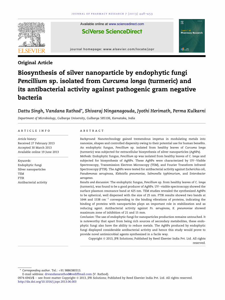
3.3.1. UVevisible spectroscopySilver nanoparticle synthesized, initially observed by color
change from pale white to brown was further conformed by
UVevisible spectroscopy. The color change occurs due to the
excitation of surface plasmon resonance in the silver metal
nanoparticle. Silver nanoparticles from endophytic fungi, Pen-
cillium sp showedmaximumabsorbance at 425 nmafter 24 h of
incubation (Fig. 3), implying that the bioreduction of AgNO3 has
taken place following incubation of the cell free culture filtrate
along with AgNO3. Surface plasmon peaks were also located at
410 nm as reported by Shivaraj et al15 using Aspergillus flavus.
Whereas, Afreen et al16 reported peak at 422 nmwith Rhizopus
stolonifer. Maliszewska et al17 reported the absorption spectrum
of spherical silver nanoparticles produced by Pencillium sp pre-
sents a maximum peak between 420 nm and 450 nm.
nge to reddish brown after treating with 1 mM AgNO3. (For
he reader is referred to the web version of this article.)
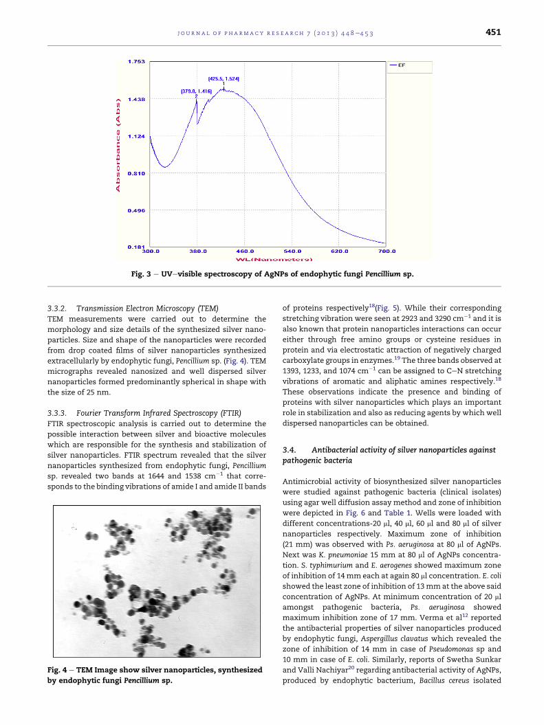
Fig. 3 e UVevisible spectroscopy of AgNPs of endophytic fungi Pencillium sp.
j o u r n a l o f p h a rm a c y r e s e a r c h 7 ( 2 0 1 3 ) 4 4 8e4 5 3 451
3.3.2. Transmission Electron Microscopy (TEM)TEM measurements were carried out to determine the
morphology and size details of the synthesized silver nano-
particles. Size and shape of the nanoparticles were recorded
from drop coated films of silver nanoparticles synthesized
extracellularly by endophytic fungi, Pencillium sp. (Fig. 4). TEM
micrographs revealed nanosized and well dispersed silver
nanoparticles formed predominantly spherical in shape with
the size of 25 nm.
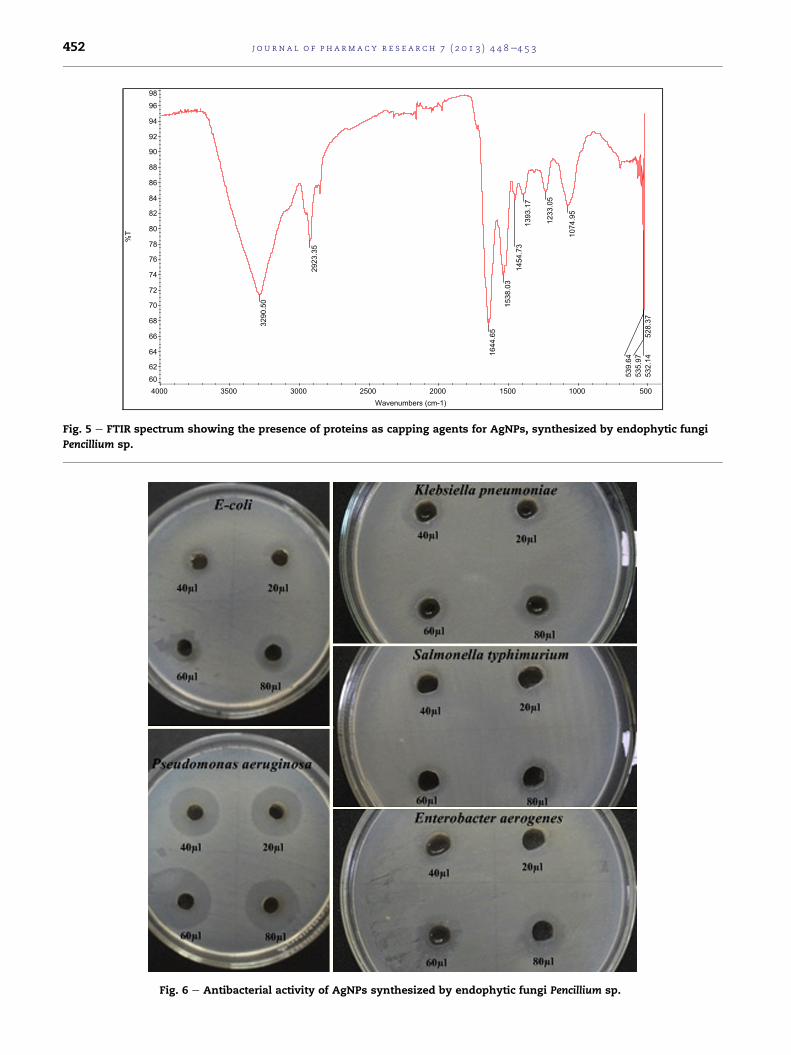
3.3.3. Fourier Transform Infrared Spectroscopy (FTIR)FTIR spectroscopic analysis is carried out to determine the
possible interaction between silver and bioactive molecules
which are responsible for the synthesis and stabilization of
silver nanoparticles. FTIR spectrum revealed that the silver
nanoparticles synthesized from endophytic fungi, Pencillium
sp. revealed two bands at 1644 and 1538 cm�1 that corre-
sponds to the binding vibrations of amide I and amide II bands
Fig. 4 e TEM Image show silver nanoparticles, synthesized
by endophytic fungi Pencillium sp.
of proteins respectively18(Fig. 5). While their corresponding
stretching vibration were seen at 2923 and 3290 cm�1 and it is
also known that protein nanoparticles interactions can occur
either through free amino groups or cysteine residues in
protein and via electrostatic attraction of negatively charged
carboxylate groups in enzymes.19 The three bands observed at
1393, 1233, and 1074 cm�1 can be assigned to CeN stretching
vibrations of aromatic and aliphatic amines respectively.18
These observations indicate the presence and binding of
proteins with silver nanoparticles which plays an important
role in stabilization and also as reducing agents by which well
dispersed nanoparticles can be obtained.
3.4. Antibacterial activity of silver nanoparticles againstpathogenic bacteria
Antimicrobial activity of biosynthesized silver nanoparticles
were studied against pathogenic bacteria (clinical isolates)
using agar well diffusion assay method and zone of inhibition
were depicted in Fig. 6 and Table 1. Wells were loaded with
different concentrations-20 ml, 40 ml, 60 ml and 80 ml of silver
nanoparticles respectively. Maximum zone of inhibition
(21 mm) was observed with Ps. aeruginosa at 80 ml of AgNPs.
Next was K. pneumoniae 15 mm at 80 ml of AgNPs concentra-
tion. S. typhimurium and E. aerogenes showed maximum zone
of inhibition of 14mm each at again 80 ml concentration. E. coli
showed the least zone of inhibition of 13mmat the above said
concentration of AgNPs. At minimum concentration of 20 ml
amongst pathogenic bacteria, Ps. aeruginosa showed
maximum inhibition zone of 17 mm. Verma et al12 reported
the antibacterial properties of silver nanoparticles produced
by endophytic fungi, Aspergillus clavatus which revealed the
zone of inhibition of 14 mm in case of Pseudomonas sp and
10 mm in case of E. coli. Similarly, reports of Swetha Sunkar
and Valli Nachiyar20 regarding antibacterial activity of AgNPs,
produced by endophytic bacterium, Bacillus cereus isolated
528.
3753
2.14
535.
9753
9.64
1074
.95
1233
.05
1393
.17
1454
.73
1538
.03
1644
.65
2923
.35
3290
.50
60 62
64
66
68
70
72
74
76
78
80
82
84
86
88
90
92
94
96 98
%T
500 1000 1500 2000 2500 3000 3500 4000Wavenumbers (cm-1)
Fig. 5 e FTIR spectrum showing the presence of proteins as capping agents for AgNPs, synthesized by endophytic fungi
Pencillium sp.
Fig. 6 e Antibacterial activity of AgNPs synthesized by endophytic fungi Pencillium sp.
j o u rn a l o f p h a rma c y r e s e a r c h 7 ( 2 0 1 3 ) 4 4 8e4 5 3452
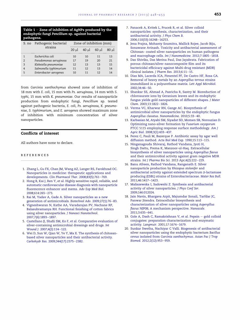
Table 1 e Zone of inhibition of AgNPs produced by theendophytic fungi Pencillium sp. against bacterialpathogens.
S. no Pathogenic bacterialstrains
Zone of inhibition (mm)
20 ml 40 ml 60 ml 80 ml
1 Escherichia coli 10 10 11 13
2 Pseudomonas aeruginosa 17 19 20 21
3 Klebsiella pneumoniae 12 13 13 15
4 Salmonella typhimurium 11 12 13 14
5 Enterobacter aerogenes 10 11 12 14
j o u r n a l o f p h a rm a c y r e s e a r c h 7 ( 2 0 1 3 ) 4 4 8e4 5 3 453
from Garcinia xanthochymus showed zone of inhibition of
18 mm with E. coli, 15 mm with Ps. aeruginosa, 14 mm with S.
typhi, 15 mm with K. pneumoniae. Our results of nanoparticle
production from endophytic fungi, Pencillium sp. tested
against pathogenic bacteria, E. coli, Ps. aeruginosa, K. pneumo-
niae, S. typhimurium, and E. aerogenes showed maximum zone
of inhibition with minimum concentration of silver
nanoparticles.
Conflicts of interest
All authors have none to declare.
r e f e r e n c e s
1. Zhang L, Gu FX, Chan JM, Wang AZ, Langer RS, Farokhzad OC.Nanoparticles in medicine: therapeutic applications anddevelopments. Clin Pharmacol Ther. 2008;83(5):761e769.
2. Hong B, Kai J, Ren Y, et al. Highly sensitive rapid, reliable, andautomatic cardiovascular disease diagnosis with nanoparticlefluorescence enhancer and mems. Adv Exp Med Biol.2008;614:265e273.
3. Rai M, Yadav A, Gade A. Silver nanoparticles as a newgeneration of antimicrobials. Biotechnol Adv. 2009;27(1):76e83.
4. Vigneshwaran N, Kathe AA, Varadarajan PV, Nachane RP,Balasubramanya RH. Functional finishing of cotton fabricsusing silver nanoparticles. J Nanosci Nanotechnol.2007;7(6):1893e1897.
5. Castellano JJ, Shafii SM, Ko F, et al. Comparative evaluation ofsilver-containing antimicrobial dressings and drugs. IntWound J. 2007;4(2):114e122.
6. Wei D, Sun W, Qian W, Ye Y, Ma X. The synthesis of chitosan-based silver nanoparticles and their antibacterial activity.Carbohydr Res. 2009;344(17):2375e2382.
7. Panacek A, Kvitek L, Prucek R, et al. Silver colloidnanoparticles: synthesis, characterization, and theirantibacterial activity. J Phys Chem B.2006;110(33):16248e16253.
8. Jena Prajna, Mohanty Soumitra, Mallick Rojee, Jacob Biju,Sonawane Avinash. Toxicity and antibacterial assessment ofChitosan- coated silver nanoparticles on human pathogensand macrophage cells. Int J Nanomedicine. 2012;7:1805e1818.
9. Das Shivika, Das Merina Paul, Das Jayabrata. Fabrication ofporous chitosan/silver nanocomposite film and itsbactericidal efficicacy against Multi-drug resistant (MDR)clinical isolates. J Pharm Res. 2013;6:11e15.
10. Dias MA, Lacerda ICA, Pimentel PF, De Castro HF, Rosa CA.Removal of heavy metals by an Aspergillus terreus strainsimmobilized in a polyurethane matrix. Lett Appl Microbiol.2002;34:46e50.
11. Shankar SS, Ahmad A, Pasricha R, Sastry M. Bioreduction ofchloroaurate ions by Geranium leaves and its endophyticfungus yields gold nanoparticles of different shapes. J MaterChem. 2003;13:1822e1826.
12. Verma VC, Kharwar RN, Gange AC. Biosynthesis ofantimicrobial silver nanoparticles by the endophytic fungusAspergillus clavatus. Nanomedicine. 2010;5:33e40.
13. Karbasian M, Atyabi SM, Siyadat SD, Momen SB, Norouzian D.Optimizing nano-silver formation by Fusarium oxysporumPTCC 5115 employing response surface methodology. Am JAgric Biol. 2008;3(1):433e437.
14. Perez C, Pauli M, Bazerque P. Antibiotic assay by agar welldiffusion method. Acta Biol Med Exp. 1990;15:113e115.
15. Ninganagouda Shivaraj, Rathod Vandana, Jyoti H,Singh Dattu, Prema K, Manzoor-ul-Haq. Extracellularbiosynthesis of silver nanoparticles using Aspergillus flavusand their antimicrobial activity against gram negative MDRstrains. Int J Pharma Bio Sci. 2013 Apr;4(2):222e229.
16. Banu Afreen, Rathod Vandana, Ranganath E. Silvernanoparticle production by Rhizopus stolonifer andantibacterial activity against extended spectrum b-lactamaseproducing (ESBL) strains of Enterobacteriaceae. Mater Res Bull.2011;46:1417e1423.
17. Maliszewska I, Sadowshi Z. Synthesis and antibacterialactivity of silver nanoparticles. J Phys Conf Ser.2009;146:012024.
18. Jain Navin, Bhargava Arpit, Majumdar Sonali, Tarfdar JC,Panwar Jitendra. Extracellular biosynthesis andcharacterization of silver nanoparticles using Aspergillusflavus NJP08; A mechanism perspective. Nanoscale.2011;3:635e641.
19. Gole A, Dash C, Ramakrishnan V, et al. Pepsin e gold colloidconjugates: preparation characterization and enzymaticactivity. Langmuir. 2001;17:1674e1679.
20. Sunkar Swetha, Nachiyar C Valli. Biogenesis of antibacterialsilver nanoparticles using the endophytic bacterium Bacilluscereus isolated from Garcinia xanthochymus. Asian Pac J TropBiomed. 2012;2(12):953e959.
Related Documents


![Review Curcumol: From Plant Roots to Cancer RootsPlant tissue culture approach has conventionally recognized as a ... Curcuma longa Common turmeric Rhizome Antifungal [37] Curcuma](https://static.cupdf.com/doc/110x72/5f7f3f3d7d5b343f5c214108/review-curcumol-from-plant-roots-to-cancer-roots-plant-tissue-culture-approach.jpg)


![Assessment Report on Curcuma Longa L. Rhizoma · Curcuma longa rhizome varies from 0.6 to 5% of the dry mass [15]. The dry turmeric rhizomes contain 3-5% curcumin, the curcumin content](https://static.cupdf.com/doc/110x72/5e0fa7a6b6de3d3894037369/assessment-report-on-curcuma-longa-l-rhizoma-curcuma-longa-rhizome-varies-from.jpg)