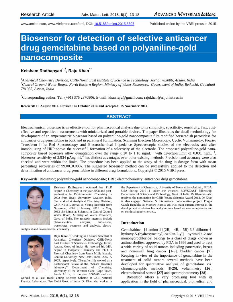
Research Article Adv. Mater. Lett. 2015, 6(1), 13-18 ADVANCED MATERIALS Letters Adv. Mater. Lett. 2015, 6(1), 13-18 Copyright © 2015 VBRI Press www.amlett.com, www.vbripress.com/aml, DOI: 10.5185/amlett.2015.5607 Published online by the VBRI press in 2015 Biosensor for detection of selective anticancer drug gemcitabine based on polyaniline-gold nanocomposite Keisham Radhapyari 1,2 , Raju Khan 1* 1 Analytical Chemistry Division, CSIR-North East Institute of Science & Technology, Jorhat 785006, Assam, India 2 Central Ground Water Board, North Eastern Region, Ministry of Water Resources, Government of India, Betkuchi, Guwahati 781035, Assam, India * Corresponding author. Tel: (+91) 376 2370806; E-mail: [email protected]; [email protected] Received: 10 August 2014, Revised: 26 October 2014 and Accepted: 15 November 2014 ABSTRACT Electrochemical biosensor is an effective tool for pharmaceutical analysis due to its simplicity, specificity, sensitivity, fast, cost- effective and repetitive measurements with miniaturized and portable devices. The paper illustrates the detail methodology for development of an amperometric biosensor based on polyaniline-gold nanocomposite film modified horseradish peroxidase for anticancer drug gemcitabine in bulk and in parenteral formulation. Scanning Electron Microscopy, Cyclic Voltammetry, Fourier Transform Infra Red Spectroscopy and Electrochemical Impedance Spectroscopic studies of the electrodes and after immobilizing of HRP shows the successful formation of a selectivity of the electrode. The proposed polyaniline-gold nano- composite based biosensor allow quantitation over the range 0.10 to 1.10 ngmL −1 with detection limit of 0.031 ngmL −1 , biosensor sensitivity of 2.934 μAng mL -1 has distinct advantages over other existing methods. Precision and accuracy were also checked and were within the limits. The procedure has been applied to the assay of the drug in dosage form with mean percentage recoveries of 99.00±0.08%. The suggested biosensor method can be successfully applied to the detection and determination of anticancer drug gemcitabine in different drug formulations. Copyright © 2015 VBRI press. Keywords: Biosensor; polyaniline-gold nanocomposite; HRP; electrochemistry; anticancer drug gemcitabine. Keisham Radhapyari obtained her Ph.D degree in Chemistry in the year 2008 and post- graduation in Environmental Chemistry in 2002 from Jiwaji University, Gwalior, India. She worked at Analytical Chemistry Division, CSIR-NEIST, Jorhat as Young Scientist from November, 2009 to January, 2013. In May, 2013 she joined as Scientist in Central Ground Water Board, Ministry of Water Resources, Govt. of India. Her research interests include pharmaceutical analysis, biosensors, wastewater treatment and analysis, electro- analytical and environmental chemistry. Raju Khan is working as a Senior Scientist at Analytical Chemistry Division, CSIR-North East Institute of Science & Technology, Jorhat, Assam, Govt. of India. He received his MSc degree in Inorganic Chemistry and PhD in Physical Chemistry from Jamia Millia Islamia, Central University, New Delhi, India, 2002 & 2005, respectively. Thereafter, He worked as a Postdoctoral Fellow at the “Sensor Research laboratory” Department of Chemistry, University of the Western Cape, Cape Town, South Africa, in the year 2005-06 and also worked as a Fast Track Young Scientist Scheme at CSIR-National Physical Laboratory, New Delhi Govt. of India. Dr Khan also worked in the Department of Chemistry; University of Texas at San-Antonio, UTSA, USA during 2010-11 under the awarded BOYSCAST fellowship, Department of Science and Technology, Govt. of India. Dr Khan has also received the nomination for CSIR Young Scientist Award 2012. Dr Khan is also engaged National & International collaborative project, Prague Czech Republic & Moscow Russia etc. His main current interest in the development of electrochemically sensors based on nano-composites and on conducting polymers etc. Introduction Gemcitabine [4-amino-1-[(2R, 4R, 5R)-3,3-difluoro-4- hydroxy-5-(hydroxymethyl)-oxolan-2-yl] pyrimidin-2-one monohydrochloride] belongs to a class of drugs known as antimetabolites, approved by FDA in 1996 and used to treat a wide variety of solid tumors including pancreatic, breast and non-small lung cancer [1-6], bladder cancer [7]. Keeping in view of the importance of gemcitabine in the treatment of solid tumors several methods have been developed for quantitative analysis of gemcitabine viz. chromatographic methods [8-25], voltammetry [26], electrochemical sensor [27] and spectrophotometry [28]. Biosensor offers a very promising tool with its application in the field of pharmaceutical, biomedical and

Welcome message from author
This document is posted to help you gain knowledge. Please leave a comment to let me know what you think about it! Share it to your friends and learn new things together.
Transcript
Research Article Adv. Mater. Lett. 2015, 6(1), 13-18 ADVANCED MATERIALS Letters
Adv. Mater. Lett. 2015, 6(1), 13-18 Copyright © 2015 VBRI Press
www.amlett.com, www.vbripress.com/aml, DOI: 10.5185/amlett.2015.5607 Published online by the VBRI press in 2015
Biosensor for detection of selective anticancer drug gemcitabine based on polyaniline-gold nanocomposite
Keisham Radhapyari1,2
, Raju Khan1*
1Analytical Chemistry Division, CSIR-North East Institute of Science & Technology, Jorhat 785006, Assam, India
2Central Ground Water Board, North Eastern Region, Ministry of Water Resources, Government of India, Betkuchi, Guwahati
781035, Assam, India *Corresponding author. Tel: (+91) 376 2370806; E-mail: [email protected]; [email protected]
Received: 10 August 2014, Revised: 26 October 2014 and Accepted: 15 November 2014
ABSTRACT
Electrochemical biosensor is an effective tool for pharmaceutical analysis due to its simplicity, specificity, sensitivity, fast, cost-effective and repetitive measurements with miniaturized and portable devices. The paper illustrates the detail methodology for development of an amperometric biosensor based on polyaniline-gold nanocomposite film modified horseradish peroxidase for anticancer drug gemcitabine in bulk and in parenteral formulation. Scanning Electron Microscopy, Cyclic Voltammetry, Fourier Transform Infra Red Spectroscopy and Electrochemical Impedance Spectroscopic studies of the electrodes and after immobilizing of HRP shows the successful formation of a selectivity of the electrode. The proposed polyaniline-gold nano-composite based biosensor allow quantitation over the range 0.10 to 1.10 ngmL
−1 with detection limit of 0.031 ngmL
−1,
biosensor sensitivity of 2.934 µAng mL-1
has distinct advantages over other existing methods. Precision and accuracy were also checked and were within the limits. The procedure has been applied to the assay of the drug in dosage form with mean percentage recoveries of 99.00±0.08%. The suggested biosensor method can be successfully applied to the detection and determination of anticancer drug gemcitabine in different drug formulations. Copyright © 2015 VBRI press.
Keywords: Biosensor; polyaniline-gold nanocomposite; HRP; electrochemistry; anticancer drug gemcitabine.
Keisham Radhapyari obtained her Ph.D degree in Chemistry in the year 2008 and post-graduation in Environmental Chemistry in 2002 from Jiwaji University, Gwalior, India. She worked at Analytical Chemistry Division, CSIR-NEIST, Jorhat as Young Scientist from November, 2009 to January, 2013. In May, 2013 she joined as Scientist in Central Ground Water Board, Ministry of Water Resources, Govt. of India. Her research interests include pharmaceutical analysis, biosensors,
wastewater treatment and analysis, electro-analytical and environmental chemistry.
Raju Khan is working as a Senior Scientist at Analytical Chemistry Division, CSIR-North East Institute of Science & Technology, Jorhat, Assam, Govt. of India. He received his MSc degree in Inorganic Chemistry and PhD in Physical Chemistry from Jamia Millia Islamia, Central University, New Delhi, India, 2002 & 2005, respectively. Thereafter, He worked as a Postdoctoral Fellow at the “Sensor Research laboratory” Department of Chemistry, University of the Western Cape, Cape Town, South Africa, in the year 2005-06 and also
worked as a Fast Track Young Scientist Scheme at CSIR-National Physical Laboratory, New Delhi Govt. of India. Dr Khan also worked in
the Department of Chemistry; University of Texas at San-Antonio, UTSA, USA during 2010-11 under the awarded BOYSCAST fellowship, Department of Science and Technology, Govt. of India. Dr Khan has also received the nomination for CSIR Young Scientist Award 2012. Dr Khan is also engaged National & International collaborative project, Prague Czech Republic & Moscow Russia etc. His main current interest in the development of electrochemically sensors based on nano-composites and on conducting polymers etc.
Introduction
Gemcitabine [4-amino-1-[(2R, 4R, 5R)-3,3-difluoro-4-hydroxy-5-(hydroxymethyl)-oxolan-2-yl] pyrimidin-2-one monohydrochloride] belongs to a class of drugs known as antimetabolites, approved by FDA in 1996 and used to treat a wide variety of solid tumors including pancreatic, breast
and non-small lung cancer [1-6], bladder cancer [7]. Keeping in view of the importance of gemcitabine in the treatment of solid tumors several methods have been developed for quantitative analysis of gemcitabine viz.
chromatographic methods [8-25], voltammetry [26],
electrochemical sensor [27] and spectrophotometry [28]. Biosensor offers a very promising tool with its
application in the field of pharmaceutical, biomedical and
Radhapyari and Khan
Adv. Mater. Lett. 2015, 6(1), 13-18 Copyright © 2015 VBRI Press 14
environmental. For the last two decades, conducting polymers have emerged as one of the most interesting materials for the fabrication of biosensors and
electrochemical sensors [29]. Polyaniline (PANI), one of the promising and unique conducting polymers has various advantages in the field of fabrication of gas sensors,
biosensors and field emitters [30-32]. Furthermore, PANI is not only environmentally stable but also dramatically changeable in its electronic structure and physical
properties in pro-activate state [33]. Above facts facilitates functionalization of PANI with metal nanoparticles that will provides a new class of advanced hybrid nanocomposite materials.
Gold nanoparticles (AuNPs) enhance polyaniline
performance in a neutral pH [34]. AuNPs display unique structural, optical, electronic, magnetic and catalytic properties making this material a very attractive material for biosensor, chemisensor, electrocatalyst, drug delivery
and sensing [35-38]. The high surface-to-volume ratio and high surface energy of AuNPs provide stable immobilization of biomolecules retaining their bioactivity. The presence of amine groups and cysteine residues in the
enzymes are known to bind strongly with gold colloids [39,
40] making AuNPs a favorable candidates for the
immobilization of enzymes [41, 42]. The application of AuNPs as a detection probe for the analysis of
pharmaceuticals [43-45] was reported. Menon et al. reported a strategy for detection and quantification of gemcitabine by colorimetric method using AuNPs as
complexing agent [24]. Gemcitabine has electroactive groups and its electrochemical behaviour has been
investigated by Keerti et al. [46] using gold electrode. In this manuscript we propose an electrochemical
preparation of inorganic-organic hybrid probe followed by immobilization of horseradish peroxidase (HRP) enzyme for determination of the gemcitabine in pharmaceutical formulations. The electrochemical investigation of gemcitabine at AuNPs/PANI nanocomposite based biosensor has been undertaken. The study will give some insight into its redox process, which is important for our understanding of its property as well as its metabolism in biological system. Furthermore, there appears to be no electrochemical biosensor method based on polyaniline-gold- horseradish peroxidase (PANI-AuNPs-HRP) for the determination of gemcitabine in pharmaceutical formulation and bulk form.
The present communication reports a validated, rapid and selective PANI-AuNPs-HRP based biosensor method for the simple and direct determination of anti cancer drug gemcitabine in bulk form and pharmaceutical formulation without any time-consuming extraction or separation steps prior to drug assay.
Experimental
Reagents and materials
Gemcitabine hydrochloride (99% purity) was procured from Enzo Life Sciences (USA). Parenteral gemcitabine (Tabicad) labeled to contain 200 mg gemcitabine per bottle was obtained from commercial sources. HRP, aniline monomer (99.5%) of analytical grade, Gold (III) chloride trihydrate, indium tin oxide (ITO) coated glass plates was
supplied by Sigma Aldrich (Germany). For experimental investigations, stock solution (1.0 mg mL
-1) of gemcitabine
was prepared by direct dissolution of 10 mg of gemcitabine in 10 mL freshly prepared 0.05M phosphate buffer solution (PBS) of pH 7.0±0.1. Simultaneously, standard working solutions were prepared by appropriate dilutions of the stock solution. Potassium ferrocyanide containing phosphate buffer solutions of 200 ml capacity with ionic strength 0.05M in the pH range 5.5-9.0 were prepared in de-ionized water (TKA Millipore water system) by adding appropriately measured amounts of mono sodium dihydrogen phosphate, disodium mono hydrogen phosphate and adjusting with 1M HCl and 0.1 M NaOH solution. All the reagents used in the present study were of analytical and molecular biology grade. Instrumentation
Electrochemical measurements were made using Autolab Potentiostat/galvanostat (Eco Chemie, Netherlands) with NOVA software and potentiostat/galvanostat/ZRA (Gamry Reference 3000, United States of America) with Gamry Echem Analyst Software in which working electrode was replaced with PANI/ITO, PANI/AuNP/ITO and PANI/AuNP/HRP/ITO. Platinum wire and Ag/AgCl (3M KCl) were used as counter and reference electrodes, respectively. Cyclic voltammetry (CV) and electrochemical impedance spectroscopy (EIS) were carried out in a 20 mL Dr Bob’s electrochemical cell stand. For chemical characterization of PANI-AuNPs nanocomposite and its HRP interaction, Fourier transform infrared (FT-IR) spectrophotometer (Spectrum 100 with software version CPU32) has been used. The surface topology of respective films was studied by using Scanning Electron Microscopy (JEOL-JSM-6390LV). Electrochemical polymerization of PANI-Au nanocomposite film The nanocomposites of PANI-AuNPs were prepared by electrochemical deposition of a mixture of HCl (1.0M), aniline (0.2M) and AuNPs (500 µL of 3 mg mL
-1
HAuCl4.3H2O in de-ionized water) and thoroughly sonicated for 15 minutes and these were introduced in a three-electrode electrochemical cell of Autolab Potentiostat/Galvonostat (EcoChemie, Netherlands, Model 101N). The cell consists of Ag/AgCl (3M KCl) as reference, Pt wire as counter electrode and ITO glass plate (0.25 cm
2) as working electrode. The electro-
polymerization was demonstrated at scan rate of 20 mVs-1
in the potential range from -2.0 to 1.1 V. The PANI/AuNPs/ITO electrode was washed with distilled water to clean the untreated AuNPs-PANI and was kept for drying. Immobilization of HRP on PANI-Au nanocomposite film
The bio-electrode for detection of gemcitabine was prepared by immobilizing HRP enzyme (1 mgmL
-1 solution
in phosphate buffer, pH 7.0±0.1) onto the PANI-Au nanocomposites by adsorption technique (overnight dipping in a special assembled cell). It is further followed by incubation at 4
0C for overnight using glutaraldehyde which
Research Article Adv. Mater. Lett. 2015, 6(1), 13-18 ADVANCED MATERIALS Letters
Adv. Mater. Lett. 2015, 6(1), 13-18 Copyright © 2015 VBRI Press
acts as a cross linker. The conditions for the immobilization
of the enzyme were selected based on our prior studies [47,
48]. To remove loosely-bound material, the biosensors were rinsed with a buffer solution and preserved at 4
0C at
pH 7.0±0.1 phosphate buffer solution for further use.
Results and discussion
Cyclic voltammetric (CV) studies
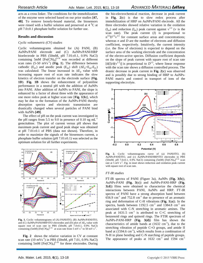
Cyclic voltammograms obtained for (A) PANI, (B) AuNPs/PANI electrode and (C) AuNPs/PANI/HRP bioelectrode in PBS (50mM, pH 7.01±0.1, 0.9% NaCl) containing 5mM [Fe(CN)6]
3-/4- was recorded at different
scan rates (5-50 mVs-1
) (Fig. 1). The difference between cathodic (Epc) and anodic peak (Epa) shift (∆Ep=Epa-Epc) was calculated. The linear increased in ∆Ep value with increasing square root of scan rate indicates the slow
kinetics of electron transfer on the electrode surface (Fig.
1D). Fig. 1B shows the enhancement of polyaniline performance in a neutral pH with the addition of AuNPs into PANI. After addition of AuNPs to PANI, the slope is enhanced by a factor of about three with the appearance of
one more redox peak at higher scan rate [Fig. 1(b)], which may be due to the formation of the AuNPs-PANI thereby absorption spectra and electronic transmission are drastically changed when several particles of PANI bind
with AuNPs [49]. The effect of pH on the peak current was investigated in
the pH ranges from 5.5 to 9.0 in presence of 0.10 ng mL-1
gemcitabine. The plot of current versus pH shows that maximum peak current and good peak shape was obtained at pH 7.01±0.1 of PBS (data not shown). Therefore, in order to maximize the signals of the biosensors current, a phosphate buffer solution (pH 7.01±0.1) was selected as the optimum solution for all further experiments.
-0.2 0.0 0.2 0.4 0.6 0.8
-2.0
-1.5
-1.0
-0.5
0.0
0.5
1.0
1.5
2.0
2.5
Cu
rre
nt
(A
)
Potential (V)
Increasing scan rate
5 - 50 mV s-1
(A)
-0.2 0.0 0.2 0.4 0.6 0.8
-12
-9
-6
-3
0
3
6
9
12
Cu
rre
nt
(A
)
Potential (V)
(B)
Increasing scan rate
5 - 50 mV s-1
-0.2 0.0 0.2 0.4 0.6 0.8
-3
-2
-1
0
1
2
3
Cu
rre
nt
(A
)
Potential (V)
(C)
Increasing scan rate
5 - 50 mV s-1
2 3 4 5 6 7
0.1
0.2
0.3
0.4
0.5
0.6
0.7
E
= E
pa-E
pc (
mV
)
Square root of scan rate, 1/2
(mVs-1)
1/2
Equation y = a + b*x
Adj. R-Square 0.99433 0.99479
PANI (a) Slope 0.03619
PANI-Au (b) Slope 0.10242
PANI-Au (b') Slope 0.10846
PANI-Au-HRP Slope 9.30084E-6
(a)
(c)
(b'')
(b)
(D)
Fig. 1. Cyclic voltammograms of (A) PANI/ITO, (B) AuNPs/PANI/ITO, and (C) AuNPs/PANI/HRP/ITO electrodes and (D) plot of ∆Ep value with square root of scan rate in PBS (50mM, pH 7.0±0.1, 0.9% NaCl) containing (5mM) [Fe(CN)6]
3-/4- at scan rate from 5 mV s-1 to 50 mV s-1.
Fig. 2 shows the relative variation in CV at constant scan rate (10 mVs
-1) in PBS (50mM, pH 7.01, 0.9% NaCl)
containing 5mM [Fe(CN)6]3-/4-
for three electrodes. During
the bio-electrochemical reaction, decrease in peak current
in Fig. 2(c) is due to slow redox process after immobilization of HRP on AuNPs/PANI electrode. All the three electrodes showed relative variation in the oxidation
(Ipa) and reduction (Ipc) peak current against 1/2 ( is the scan rate). The peak current (I) is proportional to
n3/2
D1/2
1/2 for constant surface areas and concentrations; whereas n and D are the number of electrons and diffusion coefficient, respectively. Intuitively, the current intensity (i.e. the flow of electrons) is expected to depend on the surface area of the working electrode and the concentration of the electro-active species. Diffusion coefficient depends on the slope of peak current with square root of scan rate
[d(I)/d(is proportional to D
1/2, where linear response
with the scan rate shows a diffusion controlled process. The
drastic decrease in peak current in Fig. 2(c) was observed and is possibly due to strong binding of HRP to AuNPs-PANI matrix and control in transport of ions of the supporting electrolyte.
-0.2 0.0 0.2 0.4 0.6 0.8
-2
-1
0
1
2
3
Cu
rre
nt
(A
)
Potential (V)
(a)
(b)
(c)
2 3 4 5 6 7
0
2
4
6
8
10
12
(c)
(b)
Cu
rre
nt
(A
)
Square root of scan rate, 1/2
(mVs-1)
1/2
(a)
Slope (a) =2.212A(mVs-1)
1/2
Slope (b) =1.848A(mVs-1)
1/2
Slope (c) =4.285A(mVs-1)
1/2
2 3 4 5 6 7
-12
-10
-8
-6
-4
-2
0
C
urr
en
t (
A)
Square root of scan rate, 1/2
(mVs-1)
1/2
Slope (a) = - 0.317A(mVs-1)
1/2
Slope (b) =- 2.068A(mVs-1)
1/2
Slope (c) = - 0.477A(mVs-1)
1/2
Fig. 2. Cyclic voltammograms curve of (a) PANI/ITO, (b) AuNPs/PANI/ITO, and (c) AuNPs/PANI/HRP/ITO electrodes in PBS (50mM, pH 7.0±0.1, 0.9% NaCl) containing (5mM) [Fe(CN)6]
3-/4- scan rate at 5 mV s-1. Fig. in inset shows reduction and oxidation peak current with square root of scan rate.
FT-IR studies
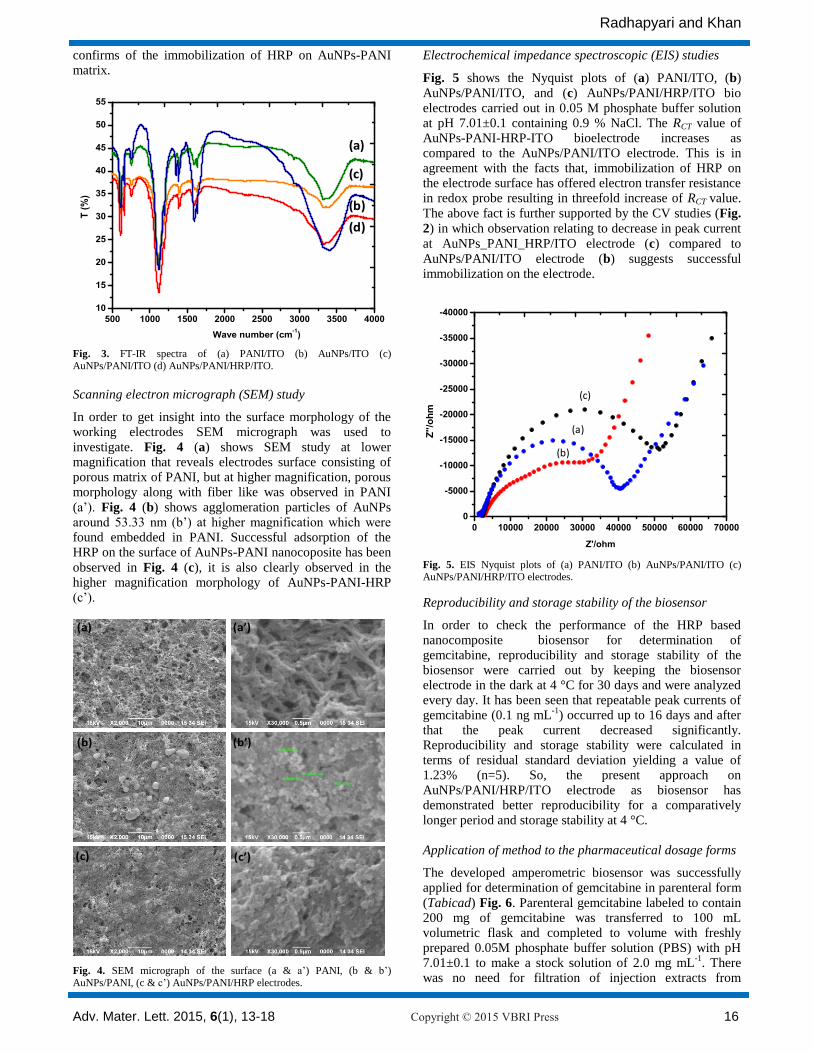
FT-IR spectra of PANI (Figure 3a), AuNPs (Fig. 3(b)),
AuNPs-PANI (Fig. 3(c)) and AuNPs-PANI-HRP (Fig.
3(d)) films were obtained to characterize the chemical interactions between PANI, AuNPs and HRP. FT-IR spectra of PANI have a strong absorption band between 656.9 cm
-1 and 752.8 cm
-1 due to presence of an aromatic
ring and deformation of C-H vibrations (Fig. 1(a)). In the spectra, bands between 1192.5 cm
-1 and 1384.8 cm
-1 are
associated with C-N stretching in aromatic amines. The peak at 1631.5 cm
-1 is attributed to C=C stretching of
benzenoid rings and quinoid rings. The FTIR spectrum of
AuNPs-PANI-HRP (Fig. 3(d)) film has shown the characteristics of amide bands at (1632 cm
-1), due to the
stretching vibration of peptide C=O groups, and amide II band at (1594.6 cm
-1), which results from a combination of
N-H in plane bending and C-N starching of peptide groups. The appearance of peaks at 1632 cm
-1 and 1594 cm
-1
Radhapyari and Khan
Adv. Mater. Lett. 2015, 6(1), 13-18 Copyright © 2015 VBRI Press 16
confirms of the immobilization of HRP on AuNPs-PANI matrix.
500 1000 1500 2000 2500 3000 3500 4000
10
15
20
25
30
35
40
45
50
55
T (
%)
Wave number (cm-1)
(a)
(c)
(b)
(d)
Fig. 3. FT-IR spectra of (a) PANI/ITO (b) AuNPs/ITO (c) AuNPs/PANI/ITO (d) AuNPs/PANI/HRP/ITO.
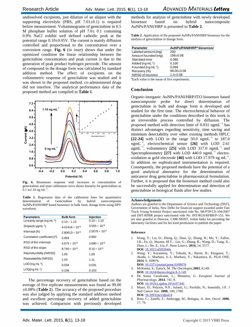
Scanning electron micrograph (SEM) study
In order to get insight into the surface morphology of the working electrodes SEM micrograph was used to
investigate. Fig. 4 (a) shows SEM study at lower magnification that reveals electrodes surface consisting of porous matrix of PANI, but at higher magnification, porous morphology along with fiber like was observed in PANI
(a’). Fig. 4 (b) shows agglomeration particles of AuNPs around 53.33 nm (b’) at higher magnification which were found embedded in PANI. Successful adsorption of the HRP on the surface of AuNPs-PANI nanocoposite has been
observed in Fig. 4 (c), it is also clearly observed in the higher magnification morphology of AuNPs-PANI-HRP (c’).
(a)
(c’)
(a’)
(b’)(b)
(c)
Fig. 4. SEM micrograph of the surface (a & a’) PANI, (b & b’) AuNPs/PANI, (c & c’) AuNPs/PANI/HRP electrodes.
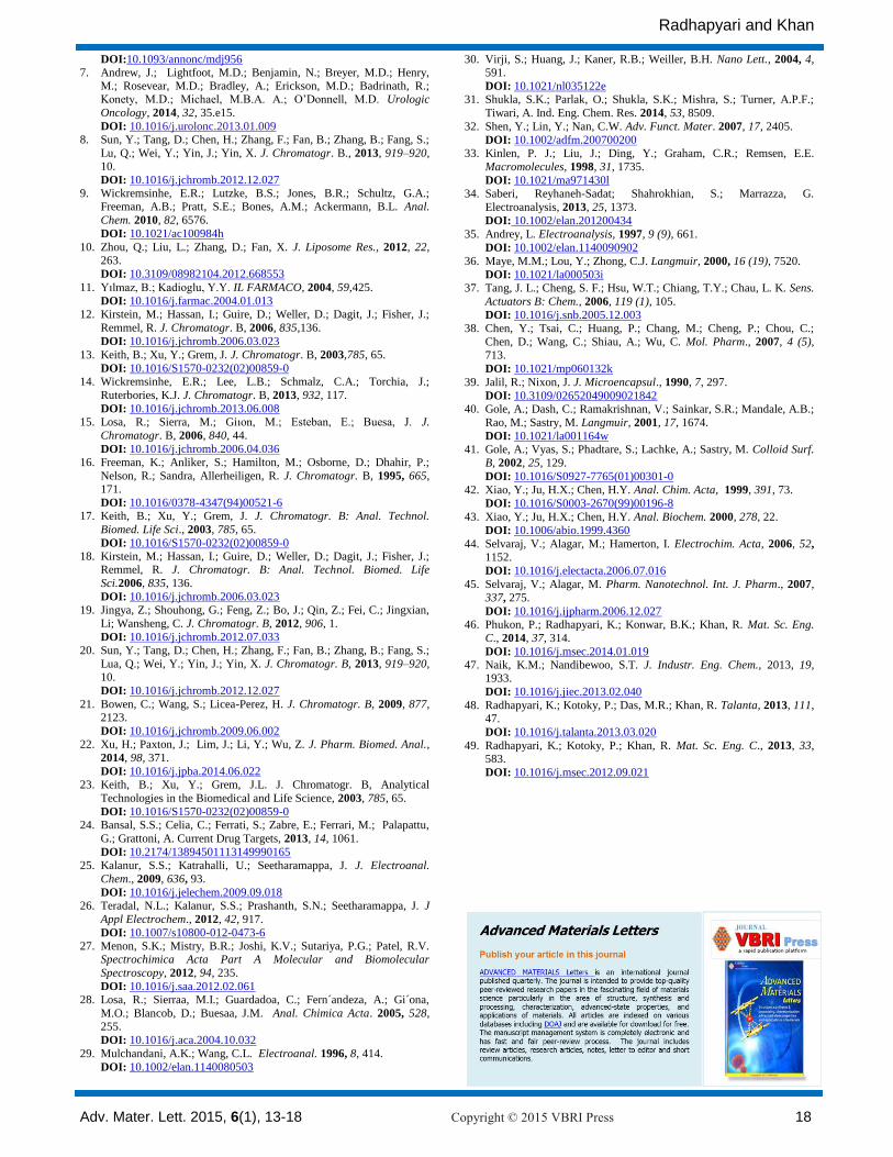
Electrochemical impedance spectroscopic (EIS) studies
Fig. 5 shows the Nyquist plots of (a) PANI/ITO, (b)
AuNPs/PANI/ITO, and (c) AuNPs/PANI/HRP/ITO bio electrodes carried out in 0.05 M phosphate buffer solution at pH 7.01±0.1 containing 0.9 % NaCl. The RCT value of AuNPs-PANI-HRP-ITO bioelectrode increases as compared to the AuNPs/PANI/ITO electrode. This is in agreement with the facts that, immobilization of HRP on the electrode surface has offered electron transfer resistance in redox probe resulting in threefold increase of RCT value. The above fact is further supported by the CV studies (Fig.
2) in which observation relating to decrease in peak current
at AuNPs_PANI_HRP/ITO electrode (c) compared to
AuNPs/PANI/ITO electrode (b) suggests successful immobilization on the electrode.
0 10000 20000 30000 40000 50000 60000 70000
0
-5000
-10000
-15000
-20000
-25000
-30000
-35000
-40000
Z''/o
hm
Z'/ohm
(a)
(c)
(b)
Fig. 5. EIS Nyquist plots of (a) PANI/ITO (b) AuNPs/PANI/ITO (c) AuNPs/PANI/HRP/ITO electrodes.
Reproducibility and storage stability of the biosensor
In order to check the performance of the HRP based nanocomposite biosensor for determination of gemcitabine, reproducibility and storage stability of the biosensor were carried out by keeping the biosensor electrode in the dark at 4 °C for 30 days and were analyzed every day. It has been seen that repeatable peak currents of gemcitabine (0.1 ng mL
-1) occurred up to 16 days and after
that the peak current decreased significantly. Reproducibility and storage stability were calculated in terms of residual standard deviation yielding a value of 1.23% (n=5). So, the present approach on AuNPs/PANI/HRP/ITO electrode as biosensor has demonstrated better reproducibility for a comparatively longer period and storage stability at 4 °C. Application of method to the pharmaceutical dosage forms
The developed amperometric biosensor was successfully applied for determination of gemcitabine in parenteral form
(Tabicad) Fig. 6. Parenteral gemcitabine labeled to contain 200 mg of gemcitabine was transferred to 100 mL volumetric flask and completed to volume with freshly prepared 0.05M phosphate buffer solution (PBS) with pH 7.01±0.1 to make a stock solution of 2.0 mg mL
-1. There
was no need for filtration of injection extracts from
Research Article Adv. Mater. Lett. 2015, 6(1), 13-18 ADVANCED MATERIALS Letters
Adv. Mater. Lett. 2015, 6(1), 13-18 Copyright © 2015 VBRI Press
undissolved excipients, just dilution of an aliquot with the supporting electrolyte (PBS, pH 7.01±0.1) is required before measurement. Voltammograms of gemcitabine in 0.5 M phosphate buffer solution of pH 7.0± 0.1 containing 0.9% NaCl exhibit well defined cathodic peak at the potential range 0.10±0.05V. The current is mainly diffusion controlled and proportional to the concentration over a
convenient range. Fig. 6 (in inset) shows that under the optimized condition the linear relationship between the gemcitabine concentration and peak current is due to the generation of peak product hydrogen peroxide. The amount of compound in the dosage form was calculated by standard addition method. The effect of excipients on the voltammetric response of gemcitabine was studied and it was shown in the proposed method, co-administered drugs did not interfere. The analytical performance data of the
proposed method are compiled in Table 1.
-0.4 -0.2 0.0 0.2 0.4 0.6 0.8 1.0
0.0
0.8
1.6
2.4
3.2
4.0
4.8
5.6
6.4
7.2
Cu
rre
nt
(A
)
Potential (V)
0 ng mL-1
1.10 ng mL-1
0.0 0.2 0.4 0.6 0.8 1.0 1.2
3
4
5
6
7
Cu
rre
nt
(A
)
Gemcitabine (ng mL-1)
Fig. 6. Biosensors response with increases in concentration of gemcitabine and inset calibration curve shows linearity for gemcitabine as 0.1 to1.10 ng mL-1.
Table 1. Regression data of the calibration lines for quantitative determination of Gemcitabine by hybrid nanocomposite AuNPs/PANI/HRP based biosensor in bulk form, dosage form using DPV waveform.
Parameters Bulk form Injection
Linearity range (ng mL-1) 0.10 – 1.10 0.10 – 1.10
Slope(A/ µgmL-1) 4.01416 × 10⁻⁶ 3.555 × 10⁻⁶
Intercept (A) 2.80613 × 10⁻⁶ 2.5576 × 10⁻⁶
Correlation coefficient (r2) 0.994 0.992
RSD of the intercept 4.675 × 10⁻⁸ 3.690 × 10⁻⁸
RSD of the slope 8.740 × 10⁻⁸ 9.10 × 10⁻⁸
Reproducibility (%RSD) 1.05 1.09
Repeatability (%RSD) 1.03 1.11
LOD (ng mL-1) 0.034 0.031
LOQ(ng mL-1) 0.106 0.103
The percentage recovery of gemcitabine based on the
average of five replicate measurements was found as 99.00
±0.08% (Table 2). The accuracy of the proposed procedure was also judged by applying the standard addition method and excellent percentage recovery of added gemcitabine was achieved. Comparison with previously developed
methods for analysis of gemcitabine with newly developed biosensor based on hybrid nanocomposite
AuNPs/PANI/HRP is presented in Table 2.
Table 2. Application of the proposed AuNPs/PANI/HRP biosensor for the analysis of gemcitabine in dosage form.
Parameter AuNPs/PANI/HRPa biosensor
Labeled amount (mg) 200
Amount founded (mg) 198±0.08
Standard error 0.080
Added (ng mL-1) 0.100
Founded (ng mL-1) 0.099
Recovery (%) 99.00±0.08
%RSD of recovery 1.4±0.08 aEach value is the mean of five experiments.
Conclusion
Organic-inorganic AuNPs/PANI/HRP/ITO biosensor based nanocomposite probe for direct determination of gemcitabine in bulk and dosage form is developed and studied for the first time. The electrochemical behavior of gemcitabine under the conditions described in this work is an irreversible process controlled by diffusion. The proposed method with detection limit of 0.031 ngmL
−1 has
distinct advantages regarding sensitivity, time saving and minimum detectability over other existing methods HPLC
[22-24] with LOD in the range 50.0 ngmL−1
to 187.0
ngmL−1
, electrochemical sensor [26] with LOD 2.61
ngmL−1
, voltammetry [25] with LOD 317.0 ngmL−1
and
Spectrophotometry [27] with LOD 440.0 ngmL−1
electro-
oxidation at gold electrode [46] with LOD 17.979 ng mL-1
. In addition no sophisticated instrumentation is required. Consequently, the proposed methods have the potential of a good analytical alternative for the determination of anticancer drug gemcitabine in pharmaceutical formulation. Further, it is proposed that the biosensor method could also be successfully applied for determination and detection of gemcitabine in biological fluids after few studies.
Acknowledgements Authors are grateful to the Department of Science and Technology (DST), Government of India, New Delhi for financial support awarded under Fast Track Young Scientist Project sanctioned vide No. SR/FTP/CS-104/2007 and DST-RFBR project sanctioned vide No. INT/RUS/RFBR/P-153. We are also grateful to Director, CSIR-NEIST, Jorhat India for providing the laboratory facilities and for his kind permission to publish the paper.
Reference
1. Wang, Y.; Liu, D.; Zheng, Q.; Zhao, Q.; Zhang, H.; Ma, Y.; Fallon,
J.K.; Fu, Q.; Haynes, M.T.; Lin, G.; Zhang, R.; Wang, D.; Yang, X.;
Zhao, L.; He, Z.; Liu, F. Nano Letters, 2014, 14, 5577.
DOI: 10.1021/nl502044x 2. Wang, Y.; Kuramitsu, Y.; Tokuda, K.; Baron, B.; Kitagawa, T.;
Akada, J.; Maehara, S.-I.; Maehara, Y.; Nakamura, K. PLoS ONE,
2014, 9, 109076.
DOI: 10.1371/journal.pone.0109076
3. McKenna, S.; Eatock, M. The Oncologist,2003, 8,149.
DOI: 10.1634/theoncologist.8-2-149 4. De Sousa Cavalcante, L.; Monteiro, G. European Journal of
Pharmacology, 2014, 741, 8.
DOI: 10.1016/j.ejphar.2014.07.041 5. Mauri, D.; Polyzos, N.P.; Salanti, G.; Pavlidis, N.; Ioannidis, J.P. J
Natl Cancer Inst. 2008, 100, 1780.
DOI: 10.1093/jnci/djn414
6. Boni, C.; Zanelli, F.; Ambroggi, M.; Bologna, A. Ann. Oncol. 2006, 17, 79.
Radhapyari and Khan
Adv. Mater. Lett. 2015, 6(1), 13-18 Copyright © 2015 VBRI Press 18
DOI:10.1093/annonc/mdj956 7. Andrew, J.; Lightfoot, M.D.; Benjamin, N.; Breyer, M.D.; Henry,
M.; Rosevear, M.D.; Bradley, A.; Erickson, M.D.; Badrinath, R.; Konety, M.D.; Michael, M.B.A. A.; O’Donnell, M.D. Urologic
Oncology, 2014, 32, 35.e15.
DOI: 10.1016/j.urolonc.2013.01.009 8. Sun, Y.; Tang, D.; Chen, H.; Zhang, F.; Fan, B.; Zhang, B.; Fang, S.;
Lu, Q.; Wei, Y.; Yin, J.; Yin, X. J. Chromatogr. B., 2013, 919–920, 10.
DOI: 10.1016/j.jchromb.2012.12.027 9. Wickremsinhe, E.R.; Lutzke, B.S.; Jones, B.R.; Schultz, G.A.;
Freeman, A.B.; Pratt, S.E.; Bones, A.M.; Ackermann, B.L. Anal.
Chem. 2010, 82, 6576.
DOI: 10.1021/ac100984h
10. Zhou, Q.; Liu, L.; Zhang, D.; Fan, X. J. Liposome Res., 2012, 22, 263.
DOI: 10.3109/08982104.2012.668553
11. Yılmaz, B.; Kadioglu, Y.Y. IL FARMACO, 2004, 59,425.
DOI: 10.1016/j.farmac.2004.01.013 12. Kirstein, M.; Hassan, I.; Guire, D.; Weller, D.; Dagit, J.; Fisher, J.;
Remmel, R. J. Chromatogr. B, 2006, 835,136.
DOI: 10.1016/j.jchromb.2006.03.023
13. Keith, B.; Xu, Y.; Grem, J. J. Chromatogr. B, 2003,785, 65.
DOI: 10.1016/S1570-0232(02)00859-0 14. Wickremsinhe, E.R.; Lee, L.B.; Schmalz, C.A.; Torchia, J.;
Ruterbories, K.J. J. Chromatogr. B, 2013, 932, 117.
DOI: 10.1016/j.jchromb.2013.06.008 15. Losa, R.; Sierra, M.; Giıon, M.; Esteban, E.; Buesa, J. J.
Chromatogr. B, 2006, 840, 44.
DOI: 10.1016/j.jchromb.2006.04.036 16. Freeman, K.; Anliker, S.; Hamilton, M.; Osborne, D.; Dhahir, P.;
Nelson, R.; Sandra, Allerheiligen, R. J. Chromatogr. B, 1995, 665, 171.
DOI: 10.1016/0378-4347(94)00521-6 17. Keith, B.; Xu, Y.; Grem, J. J. Chromatogr. B: Anal. Technol.
Biomed. Life Sci., 2003, 785, 65.
DOI: 10.1016/S1570-0232(02)00859-0 18. Kirstein, M.; Hassan, I.; Guire, D.; Weller, D.; Dagit, J.; Fisher, J.;
Remmel, R. J. Chromatogr. B: Anal. Technol. Biomed. Life
Sci.2006, 835, 136.
DOI: 10.1016/j.jchromb.2006.03.023
19. Jingya, Z.; Shouhong, G.; Feng, Z.; Bo, J.; Qin, Z.; Fei, C.; Jingxian,
Li; Wansheng, C. J. Chromatogr. B, 2012, 906, 1.
DOI: 10.1016/j.jchromb.2012.07.033 20. Sun, Y.; Tang, D.; Chen, H.; Zhang, F.; Fan, B.; Zhang, B.; Fang, S.;
Lua, Q.; Wei, Y.; Yin, J.; Yin, X. J. Chromatogr. B, 2013, 919–920, 10.
DOI: 10.1016/j.jchromb.2012.12.027
21. Bowen, C.; Wang, S.; Licea-Perez, H. J. Chromatogr. B, 2009, 877, 2123.
DOI: 10.1016/j.jchromb.2009.06.002 22. Xu, H.; Paxton, J.; Lim, J.; Li, Y.; Wu, Z. J. Pharm. Biomed. Anal.,
2014, 98, 371.
DOI: 10.1016/j.jpba.2014.06.022 23. Keith, B.; Xu, Y.; Grem, J.L. J. Chromatogr. B, Analytical
Technologies in the Biomedical and Life Science, 2003, 785, 65.
DOI: 10.1016/S1570-0232(02)00859-0 24. Bansal, S.S.; Celia, C.; Ferrati, S.; Zabre, E.; Ferrari, M.; Palapattu,
G.; Grattoni, A. Current Drug Targets, 2013, 14, 1061.
DOI: 10.2174/13894501113149990165
25. Kalanur, S.S.; Katrahalli, U.; Seetharamappa, J. J. Electroanal.
Chem., 2009, 636, 93.
DOI: 10.1016/j.jelechem.2009.09.018 26. Teradal, N.L.; Kalanur, S.S.; Prashanth, S.N.; Seetharamappa, J. J
Appl Electrochem., 2012, 42, 917.
DOI: 10.1007/s10800-012-0473-6 27. Menon, S.K.; Mistry, B.R.; Joshi, K.V.; Sutariya, P.G.; Patel, R.V.
Spectrochimica Acta Part A Molecular and Biomolecular
Spectroscopy, 2012, 94, 235.
DOI: 10.1016/j.saa.2012.02.061 28. Losa, R.; Sierraa, M.I.; Guardadoa, C.; Fern´andeza, A.; Gi´ona,
M.O.; Blancob, D.; Buesaa, J.M. Anal. Chimica Acta. 2005, 528, 255.
DOI: 10.1016/j.aca.2004.10.032
29. Mulchandani, A.K.; Wang, C.L. Electroanal. 1996, 8, 414.
DOI: 10.1002/elan.1140080503
30. Virji, S.; Huang, J.; Kaner, R.B.; Weiller, B.H. Nano Lett., 2004, 4, 591.
DOI: 10.1021/nl035122e 31. Shukla, S.K.; Parlak, O.; Shukla, S.K.; Mishra, S.; Turner, A.P.F.;
Tiwari, A. Ind. Eng. Chem. Res. 2014, 53, 8509.
32. Shen, Y.; Lin, Y.; Nan, C.W. Adv. Funct. Mater. 2007, 17, 2405.
DOI: 10.1002/adfm.200700200 33. Kinlen, P. J.; Liu, J.; Ding, Y.; Graham, C.R.; Remsen, E.E.
Macromolecules, 1998, 31, 1735.
DOI: 10.1021/ma971430l 34. Saberi, Reyhaneh-Sadat; Shahrokhian, S.; Marrazza, G.
Electroanalysis, 2013, 25, 1373.
DOI: 10.1002/elan.201200434
35. Andrey, L. Electroanalysis, 1997, 9 (9), 661.
DOI: 10.1002/elan.1140090902
36. Maye, M.M.; Lou, Y.; Zhong, C.J. Langmuir, 2000, 16 (19), 7520.
DOI: 10.1021/la000503i 37. Tang, J. L.; Cheng, S. F.; Hsu, W.T.; Chiang, T.Y.; Chau, L. K. Sens.
Actuators B: Chem., 2006, 119 (1), 105.
DOI: 10.1016/j.snb.2005.12.003 38. Chen, Y.; Tsai, C.; Huang, P.; Chang, M.; Cheng, P.; Chou, C.;
Chen, D.; Wang, C.; Shiau, A.; Wu, C. Mol. Pharm., 2007, 4 (5), 713.
DOI: 10.1021/mp060132k
39. Jalil, R.; Nixon, J. J. Microencapsul., 1990, 7, 297.
DOI: 10.3109/02652049009021842 40. Gole, A.; Dash, C.; Ramakrishnan, V.; Sainkar, S.R.; Mandale, A.B.;
Rao, M.; Sastry, M. Langmuir, 2001, 17, 1674.
DOI: 10.1021/la001164w 41. Gole, A.; Vyas, S.; Phadtare, S.; Lachke, A.; Sastry, M. Colloid Surf.
B, 2002, 25, 129.
DOI: 10.1016/S0927-7765(01)00301-0
42. Xiao, Y.; Ju, H.X.; Chen, H.Y. Anal. Chim. Acta, 1999, 391, 73.
DOI: 10.1016/S0003-2670(99)00196-8
43. Xiao, Y.; Ju, H.X.; Chen, H.Y. Anal. Biochem. 2000, 278, 22.
DOI: 10.1006/abio.1999.4360
44. Selvaraj, V.; Alagar, M.; Hamerton, I. Electrochim. Acta, 2006, 52, 1152.
DOI: 10.1016/j.electacta.2006.07.016
45. Selvaraj, V.; Alagar, M. Pharm. Nanotechnol. Int. J. Pharm., 2007,
337, 275.
DOI: 10.1016/j.ijpharm.2006.12.027 46. Phukon, P.; Radhapyari, K.; Konwar, B.K.; Khan, R. Mat. Sc. Eng.
C., 2014, 37, 314.
DOI: 10.1016/j.msec.2014.01.019 47. Naik, K.M.; Nandibewoo, S.T. J. Industr. Eng. Chem., 2013, 19,
1933.
DOI: 10.1016/j.jiec.2013.02.040
48. Radhapyari, K.; Kotoky, P.; Das, M.R.; Khan, R. Talanta, 2013, 111, 47.
DOI: 10.1016/j.talanta.2013.03.020
49. Radhapyari, K.; Kotoky, P.; Khan, R. Mat. Sc. Eng. C., 2013, 33, 583.
DOI: 10.1016/j.msec.2012.09.021
Related Documents