ISSN: 0975-8585 January – February 2015 RJPBCS 6(1) Page No. 1202 Research Journal of Pharmaceutical, Biological and Chemical Sciences Bioremediation of Textile Reactive Blue Azo Dye Residues using Nanobiotechnology Approaches. Osama M Darwesh *1 , Hassan Moawad 1 , Olfat S Barakat 2 and Wafaa M Abd El-Rahim 1 . 1 Agricultural Microbiology Department, National Research Center, Cairo, Egypt 2 Microbiology Department, Faculty of Agriculture, Cairo University, Cairo, Egypt. ABSTRACT Microbial cells contain wide assortment of specific enzymes capable to biodegrade various organic chemicals, Lignin peroxidase (LiP) plays an important role in biodegradation and mineralization of recalcitrant aromatic compounds, polychlorinated biphenyls and dyes. This study focuses on bioremediation of Reactive Blue azo dye residues generated from textile dye basins. Immobilization and stabilization of lignin peroxidase enzyme form potential bioremediation bacterial strain was among the nanobiotechnological approaches tested in this study. The lignin peroxidase enzyme isolated from Pseudomonas aeruginosa strain OS4 was partially purified. The enzyme purity was checked using SDS/PAGE. The results showed single band on gel having the molecular weight of approximately 38 kDa. Magnetic (Fe 3 O 4 ) nanoparticles were prepared by co- precipitation technique. The size and shape of Fe 3 O 4 was examined by TEM and the average particles size was 16- 20 nm. The surface charges of Fe 3 O 4 magnetic nanoparticles were modified using glutaraldehyde as cross- linker to modify the nanoparticles surface for conjugation with the enzyme. The active groups support the covalent bioconjucation properties necessary for enzyme immobilization. The modified Fe 3 O 4 nanoparticls were used for immobilization and stabilization of LiP enzyme as well as whole bacterial cells. The immobilized enzyme on magnetic nanoparticles performed much better than free enzyme in decolourization of the dye residues. The repeated use of the enzyme linked to magnetic nanoparticle is very promising for environmental application. Keywords: Magnetic (Fe 3 O 4 ) nanoparticles, Immobilized LiP enzyme, Nanotechnology, Bioremediation, TEM. *Corresponding author

Welcome message from author
This document is posted to help you gain knowledge. Please leave a comment to let me know what you think about it! Share it to your friends and learn new things together.
Transcript
ISSN: 0975-8585
January – February 2015 RJPBCS 6(1) Page No. 1202
Research Journal of Pharmaceutical, Biological and Chemical
Sciences
Bioremediation of Textile Reactive Blue Azo Dye Residues using
Nanobiotechnology Approaches.
Osama M Darwesh*1
, Hassan Moawad1, Olfat S Barakat
2 and Wafaa M Abd El-Rahim
1.
1Agricultural Microbiology Department, National Research Center, Cairo, Egypt
2Microbiology Department, Faculty of Agriculture, Cairo University, Cairo, Egypt.
ABSTRACT
Microbial cells contain wide assortment of specific enzymes capable to biodegrade various organic
chemicals, Lignin peroxidase (LiP) plays an important role in biodegradation and mineralization of recalcitrant
aromatic compounds, polychlorinated biphenyls and dyes. This study focuses on bioremediation of Reactive
Blue azo dye residues generated from textile dye basins. Immobilization and stabilization of lignin peroxidase
enzyme form potential bioremediation bacterial strain was among the nanobiotechnological approaches
tested in this study. The lignin peroxidase enzyme isolated from Pseudomonas aeruginosa strain OS4 was
partially purified. The enzyme purity was checked using SDS/PAGE. The results showed single band on gel
having the molecular weight of approximately 38 kDa. Magnetic (Fe3O4) nanoparticles were prepared by co-
precipitation technique. The size and shape of Fe3O4 was examined by TEM and the average particles size was
16- 20 nm. The surface charges of Fe3O4 magnetic nanoparticles were modified using glutaraldehyde as cross-
linker to modify the nanoparticles surface for conjugation with the enzyme. The active groups support the
covalent bioconjucation properties necessary for enzyme immobilization. The modified Fe3O4 nanoparticls
were used for immobilization and stabilization of LiP enzyme as well as whole bacterial cells. The immobilized
enzyme on magnetic nanoparticles performed much better than free enzyme in decolourization of the dye
residues. The repeated use of the enzyme linked to magnetic nanoparticle is very promising for environmental
application.
Keywords: Magnetic (Fe3O4) nanoparticles, Immobilized LiP enzyme, Nanotechnology, Bioremediation, TEM.
*Corresponding author
ISSN: 0975-8585
January – February 2015 RJPBCS 6(1) Page No. 1203
INTRODUCTION
Lignin peroxidase (LiP) catalyses several oxidations in the side chains of lignin and related compounds.
This enzyme contributes to mineralization of variety of recalcitrant aromatic compounds, polychlorinated
biphenyls and dyes [1, 2]. Franciscon et al. [3] described the transformations of six industrial azo and
phthalocyanine dyes by lignolytic peroxidases from Bjerkandera adusta. Several studies reported that LiP
enzyme from bacteria can play role in the decolourization of azo dyes [2,4,5]. Dawkar et al. [6] and Ghodake et
al. [7] performed studies concerning the purification and characterization of the lignin peroxidase from the
Bacillus sp. strain VUS and from Acenetobacter calcoaceticus NCIM 2890. They observed that purified enzyme
has a greater ability to degrade various azo dyes. The high cost of enzymes represents a hurdle in broad
industrial applications. Consequently, seeking new techniques to improve enzyme applicability and reduce
their cost is of great importance. Enzyme immobilization is one of the most powerful tools in this direction [8].
Co-precipitation of Fe2+
/Fe3+
ions under alkaline conditions has been the method of choice for the synthesis of
iron oxide nanoparticles applied in bioconjugation [9]. This synthetic method often produces particles
averaging less than 10 nm in diameter with broad size distribution [9]. Superparamagnetic materials become
magnetic only in the presence of a magnetic field. Fortunately, magnetic particles with size less than 30 nm
show superparamagnetism, meaning that they disperse easily in solution and can be recovered by use of a
simple magnet [10]. Studies with magnetic nanoparticles have been mostly dedicated for improvement of
enzyme activity and loading, rather than to enzyme stabilization. Magnetic separation has been successfully
used to concentrate biological samples for purification, isolation, and subsequent quantitative assays [11]. For
example, bio-functionalized magnetic nanoparticles were recently used to capture and detect bacteria at very
low concentration [11] and for biomolecule purification [12]. Covalent immobilization of lipase [13], and yeast
alcohol dehydrogenase on magnetic nanoparticles increased the stability of the enzymes [14].
The immobilization of enzymes on magnetic nanoparticles could potentially result in unique
properties of these bioactive magnetic particles. First, the increased surface area of nanoparticles compared
with membranes, films, or micrometric particles, would enable immobilization of larger amounts of enzyme on
the particles, which would lead to increased enzyme activity. Second, the ability to disperse the bioactive
magnetic nanoparticles in solutions would enable rapid contact between the enzyme and its substrate, and
reduce mass-transfer limitations. This would ultimately result in a lower limit of detection and faster analysis
time. Finally, the magnetic properties would permit easy separation and reuse of the bioactive magnetic
particles by cycles of magnetic separation, sample replacement and re-dispersion of the particles in the
solution [9].
In this work the lignin peroxidase enzyme was covalently attached to modified magnetic
nanoparticles. Magnetite (Fe3O4) nanoparticles averaging 16-20 nm in diameter were synthesized under
alkaline conditions. The surface charges of Fe3O4 magnetic nanoparticles were modified using glutaraldehyde
as cross-linker groups to support the covalent bioconjucation properties of enzyme immobilization. The
modified Fe3O4 was used in immobilization and possible stabilization of LiP enzyme.
MATERIALS AND METHODS
Isolation and purification of lignin peroxidase (LiP) enzyme
One litter of bioremediations products from upflow fixed-film column (UFC) bioreactor and
continuously stirred aerobic (CSA) bioreactor [5] was centrifuged at 3000 rpm for 10 min. The supernatant was
pooled and solid ammonium sulphate was added to reach 40 % saturation and was left overnight at 4 °C. Then
the mixture was centrifuged at 10000 rpm for 10 min, the supernatant was pooled and amended with solid
ammonium sulphate to 80 % saturation. The solution was kept overnight at 4 °C and centrifuged at 10000 rpm
for 10 min. The precipitate was dissolved in distilled water and dialyzed against 0.1 M phosphate buffer (pH 6)
to remove ammonium sulphate. The previous solution was subjected to gel filtration on Sephadex G-100
column (4×70 cm) previously equilibrated with 0.1 M phosphate buffer (pH 6). The column was eluted using
the same buffer at the flow rate of 1 ml/min. The eluted protein fractions were collected (3 ml/min) and
peroxidase activity was measured. The fractions exhibiting high peroxidase activity were pooled together and
passed through diethyl amino ethyl (DEAE) cellulose columns(1.5×30 cm), which were equilibrated with 0.1 M
phosphate buffer (pH 6) containing 1 mM CaCl2 and 0.5 mM NaCl. The proteins were determined by Bradford
assay [15], and bovine serum albumin was used as standard.

ISSN: 0975-8585
January – February 2015 RJPBCS 6(1) Page No. 1204
Molecular weight determination of the isolated Lip enzyme
To determine the relative molecular weight of purified LiP enzyme, sodium dodecyl sulphate
polyacrylamide gel electrophoresis (SDS-PAGE) was performed on a stacking and separating gel according to
the method of Laemmli [16] using Mini-gel electrophoresis (BioRad, USA). The molecular weight of the purified
LiP was estimated in comparison to standard molecular weight markers (standard protein markers, 21-116
kDa; Sigma, USA). The protein bands were visualized by staining with Coomassie Brilliant Blue G-250 (Sigma,
USA) after documentation.
Preparation of magnetic (Fe3O4) nanoparticles
The nanoparticles were prepared by the co-precipitation of Fe2+
and Fe3+
ions (molar ratio 2:1) at 25 ◦C
and a concentration of 0.3 M iron ions with ammonia solution (29.6 %) at pH 10, then the hydrothermal
treatment at 80 ◦C for 30 min, and finally the vacuum drying at 70
◦C after being washed several times with
water and ethanol [17].
Activation and modification of the magnetic nanoparticles
The synthesized magnetic nanoparticles (~2 g) were dispersed in ethanol and sonicated for about 10
min for getting the complete dispersion. Half ml of carbodiimide solution (25 mg/ml) and 1 ml of
glutaraldehyde solution (10 %) were added to the above particles and incubated at room temperature for
about 2 h. The particles were then washed with water to remove the excess glutaraldehyde [18].
Immobilization of lignin peroxidase on the magnetic nanoparticles
Activated MNPs were stored in 0.1 M potassium phosphate buffer, pH 6.0 at 4 °C overnight. After
separation of MNPs, 50 ml of phosphate buffer, pH 6.0 containing peroxidase enzyme (5 U/ ml) was added to
activate MNPs and the mixture was incubated under shaking condition for 24 h. The peroxidase-bound MNPs
were then decanted using permanent magnet and washed several times by deionized water.
Examination of magnetic nanoparticles and immobilized enzyme
Examination using transmission electron microscopy (TEM)
The average particle size, size distribution and morphology of the magnetic nanoparticles and
immobilized enzyme were studied using transmission electron microscope JEOL (JEM-1400 TEM). A drop of
well dispersed nanoparticle was placed onto the amorphous carbon-coated 200 mesh carbon grid, followed by
drying the sample at ambient temperature, before it was loaded into the microscope [19].
Examination using scanning electron microscopy (SEM)
The morphology of the magnetic nanoparticles and immobilized enzyme were studied using scanning
electron microscopy [20].
Examination using energy-dispersive X-ray spectroscopy
The structure and composition of magnetic nanoparticles and immobilized enzyme were studied
using energy-dispersive X-ray spectroscopy (EDX) [21].
Decolourization of wastewater containing Reactive Blue azo dye
Free lignin Peroxidase (LiP) enzyme, immobilized LiP enzyme on magnetic nanoparticles, Fe3O4
magnetic nanoparticles, free Pseudomonas aeruginosa cells and Pseudomonas aeruginosa immobilized on
magnetic nanoparticles were tested for decolourization of textile wastewater containing 300 ppm of Reactive
Blue azo dye.For testing the free bacteria, 2 ml of Pseudomonas aeruginosa 2 days old culture were
transferred to sterile 20 ml tubes. The tubes were filled to 18 ml working volume by sterile wastewater
containing Reactive Blue dye (300 ppm) amended with yeast extract to give concentration of 0.5 g/l. The tubes

ISSN: 0975-8585
January – February 2015 RJPBCS 6(1) Page No. 1205
were sealed with screw caps to achieve anoxic conditions as described by Darwesh et al. [22+2]. For testing the
immobilized bacteria, 1gm of immobilization product [2] was transferred to sterile 20 ml tubes. The tubes
were filled to 18 ml working volume by sterile wastewater containing Reactive Blue dye (300 ppm) amended
with yeast extract as above mentioned. The tubes were sealed with screw caps. For testing the free and
immobilized LiP enzyme, 1 ml containing 10 units of the enzyme was transferred to sterile 20 ml tubes. The
tubes were filled to 18 ml working volume by sterile wastewater containing Reactive Blue dye (300 ppm)
amended with yeast extract. The tubes were sealed with screw caps. For testing the magnetic nanoparticles, 1
gm of Fe3O4 magnetic nanoparticles was transferred to sterile 20 ml tubes. The tubes were filled to 18 ml
working volume by sterile wastewater containing Reactive Blue dye (300 ppm) amended with yeast extract.
The tubes were sealed with screw caps. Tubes containing 18 ml of sterile wastewater containing Reactive Blue
dye (300 ppm) and amended with yeast extract were used as a control. All tubes were incubated under static
conditions at 28 ◦C for 3 days. The decolourization of wastewater was measured at 24 hours intervals [2].
RESULTS AND DISCUSSION
Isolation and purification of lignin peroxidase (LiP)
Lignin peroxidase (LiP) enzyme is known to catalyse several oxidations in the side chains of lignin and
related compounds resulting in mineralization of variety of recalcitrant aromatic compounds, polychlorinated
biphenyls and dyes [1]. LiP enzyme was partially purified from the bioremediation products of textile
wastewater containing Reactive Blue azo dye in a bioremediation UFC/CSA prototype bioreactor. The enzyme
was isolated by precipitation using ammonium sulphate then dialyzed by dialysis bag against 0.1 M phosphate
buffer (pH 6) to remove excess ammonium sulphate followed by passing the solution through Sephadex G-100
column. The fractions containing high activity of LiP enzyme were collected and purified by diethyl amino ethyl
(DEAE) cellulose column. The molecular weight and purity of the LiP enzyme was assessed by SDS-PAGE
electrophoreses. After Coomassie blue staining of the gel, a single band was detected (Figure 1). The purified
LiP enzyme had molecular mass of approximately 38 kDa as compared with protein marker (Figure 1). This is
likely to be near to complete purification of LiP enzyme. The molecular weight of the obtained enzyme is
different from the same enzyme isolated from other microorganisms, for example, the molecular weight of LiP
from Cunninghamella elegans and Phanerochaete sordida YK-624 was 50 kDa [23]. The LiP enzyme from
Hexagona tenuis MTCC 1119 had molecular weight of 48 kDa [24], from Loweporus lividus MTCC-1178 had
MW of 40 kDa [25] and from Trametes versicolor IBL-04 had MW of 30 kDa [26].
Figure 1: Sodium dodecyl sulphate polyacrylamide gel electrophoresis (SDS-PAGE) of purified LiP enzyme from
Pseudomonas aeruginosa strain OS4.
Where; P, purified LiP enzyme and M, protein marker.
ISSN: 0975-8585
January – February 2015 RJPBCS 6(1) Page No. 1206
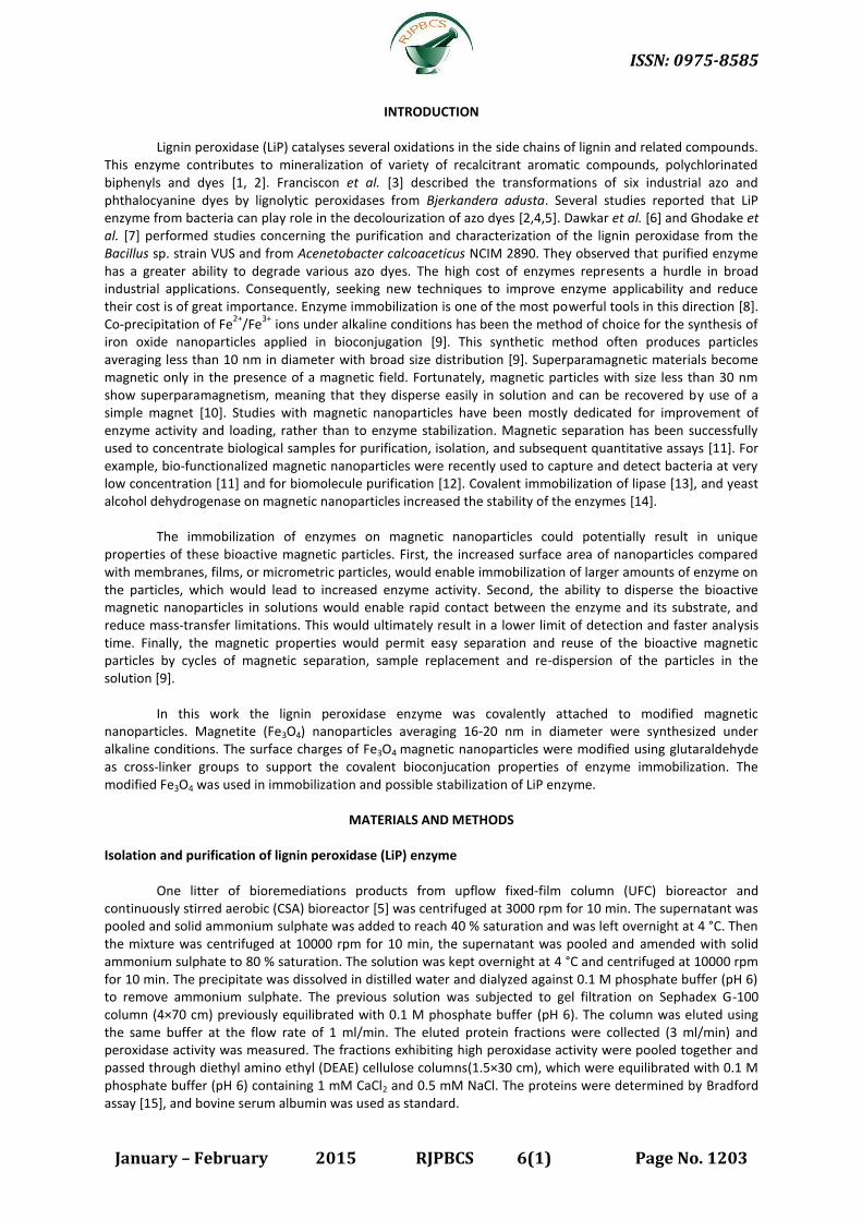
Properties of prepared magnetic nanoparticles
Magnetic (Fe3O4) nanoparticles were prepared by co-precipitation technique. The obtained
nanoparticles changed in colour, size, shape, magnetic properties and surface area charge as compared with
Fe+3
molecule. The colour of prepared magnetic nanoparticles was black (Figure 2A). The Fe3O4 nanoparticles
were separated from suspension by permanent magnet as shown in Figure (2B).
Figure 2. Fe3O4 Magnetic nanoparticles prepared by co-precipitation technique, A; dispersed nanoparticles, B;
precipitated nanoparticles.
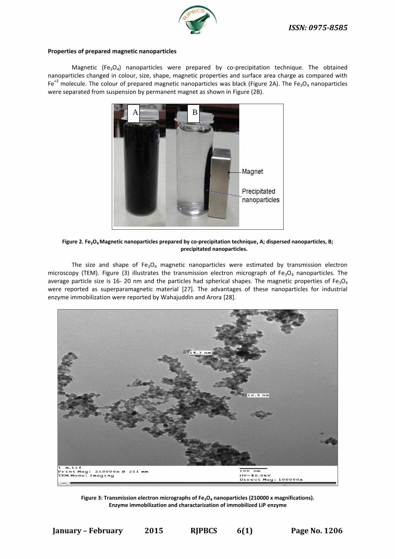
The size and shape of Fe3O4 magnetic nanoparticles were estimated by transmission electron
microscopy (TEM). Figure (3) illustrates the transmission electron micrograph of Fe3O4 nanoparticles. The
average particle size is 16- 20 nm and the particles had spherical shapes. The magnetic properties of Fe3O4
were reported as superparamagnetic material [27]. The advantages of these nanoparticles for industrial
enzyme immobilization were reported by Wahajuddin and Arora [28].
Figure 3: Transmission electron micrographs of Fe3O4 nanoparticles (210000 x magnifications).
Enzyme immobilization and charactarization of immobilized LiP enzyme
A B
ISSN: 0975-8585
January – February 2015 RJPBCS 6(1) Page No. 1207
One of the main objectives of this study is to enhance the stability and efficiency of LiP azo dye
degrading enzyme. Covalant bioconjugation experiments were performed to immobilize the LiP enzyme on
magnetic nanoparticles. Glutaraldehyde was used as a cross-linking and modifying agent for covalent coupling
of LiP enzyme to magnetic nanoparticles. The crude surface of magnetic nanoparticles modified by using
glutaraldehyde as cross-linker to synthesise aldehyde as an active group with a positive charge. These active
groups support the covalent bioconjucation of Fe3O4 nanoparticles and enzyme as well as whole bacterial cells.
The modified Fe3O4 was used in immobilization and stabilization of both LiP enzyme and whole bacterial cells.
Same approach was used by Mahdizadeh et al. [29] for preparation of Fe3O4 magnetic nanoparticles for
immobilization of Glucose Oxidase and application of immobilized enzyme for Water Deoxygenation. Bahrami
and Hejazi [30] prepared modified magnetic Fe3O4 nanoparticles and used it for immobilization of Pectinase
enzyme. This technique facilitated the economic use of several enzymes.
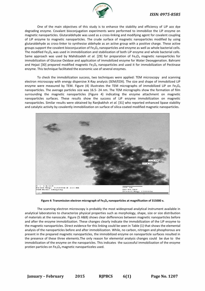
To check the immobilization success, two techniques were applied: TEM microscopy and scanning
electron microscopy with energy dispersive X-Ray analysis (SEM/EDX). The size and shape of immobilized LiP
enzyme were measured by TEM. Figure (4) illustrates the TEM micrographs of immobilized LiP on Fe3O4
nanoparticles. The average particles size was 16.5- 24 nm. The TEM micrographs show the formation of film
surrounding the magnetic nanoparticles (Figure 4) indicating the enzyme attachment on magnetic
nanoparticles surfaces. These results show the success of LiP enzyme immobilization on magnetic
nanoparticles. Similar results were obtained by Ranjbakhsh et al. [31] who reported enhanced lipase stability
and catalytic activity by covalently immobilization on surface of silica-coated modified magnetic nanoparticles.
Figure 4: Transmission electron micrograph of Fe3O4 nanoparticles at magnification of 315000 x.
The scanning electron microscopy is probably the most widespread analytical instrument available in
analytical laboratories to characterize physical properties such as morphology, shape, size or size distribution
of materials at the nanoscale. Figure (5 A&B) shows clear defferences between magnetic nanoparticles before
and after the enzyme immobilization. These changes clearly indicate the immobilization of the LiP enzyme to
the magnetic nanoparticles. Direct evidence for this linking could be seen in Table (1) that shows the elemental
analysis of the nanoparticles before and after immobilization. While, no carbon, nitrogen and phosphorous are
present in the prepared magnetic nanoparticles, the immobilized enzyme on nanoparticle surfaces resulted in
the presence of these three elements.The only reason for elemental analysis changes could be due to the
immobilization of the enzyme on the nanoparicles. This indicates the successful immobilization of the enzyme
protien particles on Fe3O4 magnetic nanoparticeles used.

ISSN: 0975-8585
January – February 2015 RJPBCS 6(1) Page No. 1208
Figure 5: Micrographs of scanning electron microscopic analysis (SEM) of Fe3O4 magnetic nanoparticles (A) and
immobilized LiP enzyme on the nanoparticles (B).
Table 1: Energy-dispersive X-ray spectrum (EDX) analysis of magnetic nanoparticles before and after immobilization with
LiP enzyme.
Element Weight of elements %
Magnetic nanoparticles Immobilized enzyme
C - 14.83
N - 10.92
O 63.12 39.86
P - 3.79
Fe 36.88 30.61
Totals 100.00 100.00
Energy dispersive X-ray diffraction (EDXD) analysis was very effective tool used for investigation of
the elemental composition of the nanoparticles [32]. Figure (6 A&B) shows the energy-dispersive X-ray
spectrum (EDX) analysis of magnetic nanoparticles (Fe3O4) before and after enzyme immobilization. The results
revealed the presence of N, C and P elements in the samples of immobilized LiP on nanoparticles. This once
more indicates the presence of enzyme protein the surface of magnetic nanoparticles. These elements were
not found on the nanoparticles without enzyme immobilization. Siemieniec et al. [33] reported that the EDX
spectrum analysis is a helpful tool to confirm the magnetic nanoparticles attached to organic molecules.
The successful LiP enzyme immobilization was further assessed by testing the enzyme activity using
pyrogallol as specific substrate. The immobilized enzyme turns pyrogallol to dark yellow colour indicating the
successful enzymatic reaction. It is likely that the conjugation between enzyme and immobilization matrix was
out of the enzyme active site [34].
A
B

ISSN: 0975-8585
January – February 2015 RJPBCS 6(1) Page No. 1209
Figure 6: Energy-dispersive X-ray spectrum (EDX) analysis of magnetic nanoparticles (A) and immobilized enzyme (B).
Efficiency of enzyme immobilization
The immobilization of enzymes onto nanoparticles usually depends on various reaction factors such
as immobilization time, quantity of nanoparticles, reaction temperature and buffer solution [18]. In this study,
the efficiency of LiP enzyme immobilization was studied with respect to immobilization time. In Figure (7),
immobilized lignin peroxidase showed maximum activity after 24 h of the start of the immobilization. The
activity of immobilized enzyme showed gradual increase as the reaction time proceeds. On contrary, the
activity of the free enzyme decreased gradually with time. The maximum activity of the immobilized LiP was
set to 100 % as a reference value. The immobilization efficiency of 100 % means that, all LiP free enzymes
added was immobilized onto the nanoparticles (Figure 7).
0
20
40
60
80
100
120
0 2 4 6 12 18 24
Time (h)
Re
lati
ve
ac
tiv
ity
(%
)
Free peroxidase Immobilized peroxidase
Figure 7: The LiP activity performance as a factor of time and the enzyme immobilization efficiency with respect to the
immobilization time.
A
B

ISSN: 0975-8585
January – February 2015 RJPBCS 6(1) Page No. 1210
Bioremediation of textile industry wastewater containing Reactive Blue azo dye
The Reactive Blue azo dye is one of the widely used textile dyes. Unfortunately the dying basins
wastewaters were found to contain large amounts of dye residues amounting up to 50 % of the used dye [22].
The save disposal of the dye wastewater requires special technology to remove the dye residues to reach the
acceptable levels by environmental authorities. The cost of physical and/or chemical techniques used for this
purpose was found to be uneconomic. In this study we test new approaches for removal of textile dye residues
using nanobiotechnological bioremediation approaches. These approaches are based on the selection of
potent microbial agents capable to remove the dye in rather short time and use these microbes either as it is
or LiP enzyme isolated from their cells and known to contribute significantly in azo dye
biodegradation/bioremoval [2]. The partially purified enzyme obtained from microbial cells is cross-linked to
magnetic nanoparticles prepared specially for this bioremediation purpose. The bacterium used in this study is
Pseudomonas aeruginosa strain OS4. This strain was proven in our previous studies [2,5] to be highly efficient
in dye removal in rather short time. The Fe3O4 magnetic nanoparticles were prepared according to the method
described in details in materials and methods section. In order to test the efficiency of the suggested
nanobiotechnological approach several controls were included for comparison. The results in Table (2) show
that in controls where the dye alone and or in the presence of nanoparticles alone no change in solution
colour was observed till the end of the experiment (72 hours). The treatment of dye containing wastewater
with LiP enzyme either in free state or immobilized on Fe3O4 magnetic nanoparticles induced colour removal at
various degrees. The free LiP enzyme started to remove dye colour after 24 hours where it could remove only
6.7% of the colour .The colour removal in this treatment increased to 15.4 after 72 hours. The immobilization
of the enzyme on magnetic nanoparticles induced early decolourization and enhanced the removal markedly
as compared with the free Lip enzyme. The application of whole bacterial cells either in free state or
immobilized on Fe3O4 nanoparticles resulted in extremely higher colour removal starting at 24 hours. The
colour removal at 48 hours sampling showed that the immobilized whole bacterial cells were superior in colour
removal as compared with the free bacterial cells being 93.4% in the first compared to 58.4% in the second.
After 72 hours, the last two treatments almost removed most of the dye colour resulting in 95.2% of colour
removal in the first and 97.5% in the second (Table 2). This clearly shows that the bioremediation by either
Pseudomonas aeruginosa whole cells or the LiP enzyme extracted from it play important role in dye
bioremoval. The immobilization of Free Pseudomonas aeruginosa strain OS4 whole cells on magnetic
nanoparticles has dramatically improved the performance of this bacterium in bioremediation of a commonly
used textile dye residues. The results are close to those obtained by using partially purified LiP enzyme.
However the advantage of the enzyme conjugated to nanoparticles is mainly linked to the repeated use of
nanoparticles –enzyme complex to effectively perform the bioremediation. The limited cycles of immobilized
bacterial cells as compared to the immobilized enzymes were previously reported [2,35,36]. In addition, the
tolerance of enzymes to the storage and the adaptation of the industries to the use of chemicals including
enzymes in different forms render the use of enzyme immobilized nanoparticles more easy and practical than
using living microbial cells.
Table 2: Decolourization % of textile wastewater containing Reactive Blue azo dye.
Treatments Incubation time (hours)
24 48 72
Control 0.0 0.0 0.0
Fe3O4 magnetic nanoparticles 0.0 0.0 0.0
Free LiP enzyme 0.0 6.7 15.4
Immobilized LiP on Fe3O4 10.2 30.6 54.3
Free Pseudomonas aeruginosa strain OS4 23.7 58.4 95.2
Immobilized Pseudomonas aeruginosa on Fe3O4 56.8 93.4 97.5

ISSN: 0975-8585
January – February 2015 RJPBCS 6(1) Page No. 1211
CONCLUSION
The enzymatic bioremediation technology using magnetic nanoparticles represents promising
nanobiotechnological approach that has wide range of application in industry in generated and bioremediation
of recalcitrant chemical residues in particular.
REFERENCES
[1] Elumalai S, Tobimatsu Y, Grabber J H, Pan X, Ralph J. Biotechnol Biofuel 2012;5(59): 1-14.
[2] Darwesh OM, Moawad H, Wafaa MA, Olfat SB, Sedik MZ. Res J Pharm Biol Chem Sci 2014;5(4):1203-
1219.
[3] Franciscon E, Grossman M J, Paschoal J R, Reyes F G, Durrant L R. Springer Plus 2012;1(37): 1-10.
[4] Dhanve R S, Kalyani D C, Phugare S S, Jadhav J P. Biodegradation 2009;20(2): 245-255.
[5] Moawad H, Darwesh O M, Wafaa M A, Olfat S B, Sedik M Z. Int J Adv Res 2013;1(7): 272-284.
[6] Dawkar V V, Jadhav U U, Jadhav S U, Govindwar S P. J Appl Microb 2008;105:14-24.
[7] Ghodake G S, Kalme S D, Jadhav J P, Govindwar S P. Appl Biochem Biotech 2008;152(1): 6-14.
[8] Guisan J M. 2006, Methods in Biotechnology: Immobilization of Enzymes and Cells. Humana Press Inc, 2nd
Ed, ISBN: 1-59745-053-7, 449 p.
[9] Rossi L M, Quach A D, Rosenzweig Z. Anal Bioanal Chem 2004;380(4): 606-613.
[10] Gupta M N, Kaloti M, Kapoor M, Solanki K. Artif Cells Bl Subst Biotechn 2011;39: 98-109.
[11] Gu H, Ho P L, Tsang K W T, Yu C W, Xu B. Chem Commun 2013;15:1966-1967.
[12] Bucak S, Jones D A, Laibinis P E, Hatton A. Biotechnol Progr 2003;19: 477- 484.
[13] Huang S H, Liao M H, Chen D H. Biotechnol Progr 2003;19: 1095- 1100.
[14] Chen D H, Liao M H. J Mol Catalysis B: Enzymatic 2002;16: 283-291.
[15] Bradford M. Anal Biochem 1976;72: 248-254.
[16] Laemmli U K. Nature 1970;227(5259): 680-685.
[17] Fan L, Zhang Y, Luoa C, Lua F, Qiua H, Sun M. Int J Biol Macromol 2012;50: 444- 450.
[18] Sohn O, Kim C, Rhee J. Biotechn Biopr Eng 2008; 13: 716-723.
[19] Wang Y, Zhu Z, Xu F, Wei X. J Nanopart Res 2012;14: 755-761.
[20] Gu T, Wang J, Xia H, Wang S, Yu X. Materials 2014; 7: 1069-1083.
[21] Chicea D, Indrea E, Cretu M. J Optoelectr Advanc Mater 2012;14(5-6): 460-466.
[22] Darwesh O M, Wafaa M A, Olfat B, Sedik O Z, Moawad H. CATRINA 2008;3(1): 71-80.
[23] Roushdy M M, Abdel-Shakour E H, El-Agamy E I. J Am Sci 2011; 7: 6-13.
[24] Yadav M, Singh S K, Yadav S K, Yadav K D S. Ind J Chem 2011;49B:489-494.
[25] Yadav M, Yadav P, Yadav K D S. Life Sci 2009;9(2): 124- 129.
[26] Asgher M, Iqbal H M, Irshad M. BMC Biotechnol 2012; 12: 46-54.
[27] Teja A S, Koh P. Materials 2009;55: 22-45.
[28] Wahajuddin, Arora S. Int J Nanomed 2012;7: 3445-3471.
[29] Mahdizadeh F, Karimi A, Ranjbarian L. Int J Sci Eng Res 2012;3(5): 1-6.
[30] Bahrami A, Hejazi P. J Mol Catalysis B: Enzymatic 2013;93: 1-7.
[31] Ranjbakhsh E, Bordbar A K, Abbasi M, Khosropour A R, Shams E. Chem Eng J 2012;179:272-276.
[32] Faraji M, Yamini Y, Rezaee M. J Iran Chem Soc 2012;7(1): 1-37.
[33] Siemieniec J, Kafarski P, Plucinski P. Molecules 2013;18(7): 8473-8484.
[34] Liu G, Zhou J, Chen C, Jing W, Jin R, Lv H. Appl Microbiol Biotechnol 2013 97(17): 7935-7942.
[35] Chen Y, Tsai M, Chi M, Wang T, Lin L. Int J Mol Sci 2013;14(3): 4613- 4628.
[36] Shah M P, Patel K A, Nair S S, Darji A M, Maharaul S. J Bioremed Biodeg 2013;4: 197-203.
Related Documents