Bioreactor Systems for Bone Tissue Engineering Juliane Rauh, Ph.D., 1,2 Falk Milan, M.Eng., 1,2 Klaus-Peter Gu ¨ nther, M.D., 1,2 and Maik Stiehler, M.D., Ph.D. 1,2 Bone graft material is often required for the treatment of osseous defects. However, due to limitations and risks associated with autologous as well as allogenic bone grafting procedures, alternative strategies are needed. In this context, ex vivo tissue engineering (TE) strategies for de novo generation of bone tissue include the combined use of autologous bone-forming cells and three-dimensional (3D) porous scaffold materials serving as structural support for the cells. Three-dimensional cultivation of osteoprogenitor cells presents several challenges, for example, insufficient nutrient and oxygen transport to and removal of waste products from the cells at the interior of the scaffold. By providing physical stimulation of tissue-engineered constructs and resolving mass transport limitations bioreactor systems denote key components for bone TE strategies. A variety of dynamic 3D bioreactor concepts mimicking the native microenvironment in bone tissue, for example, spinner flasks, rotating wall vessel constructs, perfusion bioreactors, and systems based on mechanical or electromagnetic stimulation of cell/scaffold composites, have been developed. These techniques differ considerably with respect to ease of use, cost-effectiveness, and degree of additional osteogenic stimuli, as well as monitoring and manipulation options. This review provides an overview of the concepts, advantages, challenges, and potential future applications associated with current bioreactor systems for bone TE. Introduction D ue to increasing life expectancy, bone diseases—for example, bone infection, fractures, osteoarthritis, oste- oporosis, rheumatoid arthritis, and spine disorders—requir- ing surgical interventions are becoming a major socioeconomic problem. 1 These conditions are often associ- ated with major structural defects that may need recon- struction procedures. The commonly used application of autologous bone grafting is associated with potential draw- backs, for example, the risk of donor-site morbidity (e.g., neurovascular injury, infection, persisting pain, and fracture) and the limited availability of bone graft material. 2 There- fore, alternative strategies for skeletal reconstruction are needed. Tissue engineering (TE) applies the principles of biology and engineering for the development of functional substitutes for damaged tissues. 3 The field of TE has devel- oped rapidly over the last two decades. Great research efforts have been made to reconstruct a variety of musculoskeletal (e.g., bone, 4 cartilage, 5 and muscle 6 ) and other (e.g., skin, 7 cardiac valve, 8 retina, 9 vocal fold, 10 liver, 11 and nerves 12 ) types of tissue. To engineer bone tissue successfully it may be beneficial to mimic the in vivo environment of osteopro- genitor cells by exposing them to adequate stimuli. For this purpose, different kinds of dynamic bioreactor-based culti- vation systems have been proposed. Bone tissue engineering The discipline of bone TE involves the combined use of osteoconductive matrices, bone-forming cells, and osteogenic growth factors. Notably, the tissue constructs need to be maintained in a suitable cultivation environment. Cells are the living component of such a construct, capable of pro- ducing matrix-forming neotissue. Osteoblasts have been widely used for generating mineralized cell/scaffold con- structs in vitro. 13–15 However, osteoblasts are in an almost mature stage therefore showing less proliferative potential compared to osteoblast precursor cells. In contrast, mesen- chymal stromal cells (MSCs) represent a proliferating and undifferentiated cell source. MSCs are mostly isolated from bone marrow aspirates, but can also be obtained from other tissues, for example, adipose tissue or cord blood. 16 MSCs have the potential to differentiate toward diverse mesen- chymal lineages, including osteoblasts, chondrocytes, adi- pocytes, and myocytes. 17 Drawbacks of the application of MSCs are their limited availability and their in vitro repli- cative senescence. 18 In this context, the lifespan of human MSCs can be extended by ectopic expression of human tel- omerase reverse transcriptase (hTERT). In various in vitro studies hTERT-MSCs were used for seeding scaffolds and cultivating the cell/scaffold constructs under dynamic conditions. 19–21 1 Department of Orthopedics and Centre for Translational Bone, Joint, and Soft Tissue Research, University Hospital Carl Gustav Carus, Dresden, Germany. 2 Center for Regenerative Therapies Dresden, Dresden University of Technology, Dresden, Germany. TISSUE ENGINEERING: Part B Volume 17, Number 4, 2011 ª Mary Ann Liebert, Inc. DOI: 10.1089/ten.teb.2010.0612 263

Welcome message from author
This document is posted to help you gain knowledge. Please leave a comment to let me know what you think about it! Share it to your friends and learn new things together.
Transcript

Bioreactor Systems for Bone Tissue Engineering
Juliane Rauh, Ph.D.,1,2 Falk Milan, M.Eng.,1,2 Klaus-Peter Gunther, M.D.,1,2 and Maik Stiehler, M.D., Ph.D.1,2
Bone graft material is often required for the treatment of osseous defects. However, due to limitations and risksassociated with autologous as well as allogenic bone grafting procedures, alternative strategies are needed. Inthis context, ex vivo tissue engineering (TE) strategies for de novo generation of bone tissue include the combineduse of autologous bone-forming cells and three-dimensional (3D) porous scaffold materials serving as structuralsupport for the cells. Three-dimensional cultivation of osteoprogenitor cells presents several challenges, forexample, insufficient nutrient and oxygen transport to and removal of waste products from the cells at theinterior of the scaffold. By providing physical stimulation of tissue-engineered constructs and resolving masstransport limitations bioreactor systems denote key components for bone TE strategies. A variety of dynamic 3Dbioreactor concepts mimicking the native microenvironment in bone tissue, for example, spinner flasks, rotatingwall vessel constructs, perfusion bioreactors, and systems based on mechanical or electromagnetic stimulation ofcell/scaffold composites, have been developed. These techniques differ considerably with respect to ease of use,cost-effectiveness, and degree of additional osteogenic stimuli, as well as monitoring and manipulation options.This review provides an overview of the concepts, advantages, challenges, and potential future applicationsassociated with current bioreactor systems for bone TE.
Introduction
Due to increasing life expectancy, bone diseases—forexample, bone infection, fractures, osteoarthritis, oste-
oporosis, rheumatoid arthritis, and spine disorders—requir-ing surgical interventions are becoming a majorsocioeconomic problem.1 These conditions are often associ-ated with major structural defects that may need recon-struction procedures. The commonly used application ofautologous bone grafting is associated with potential draw-backs, for example, the risk of donor-site morbidity (e.g.,neurovascular injury, infection, persisting pain, and fracture)and the limited availability of bone graft material.2 There-fore, alternative strategies for skeletal reconstruction areneeded. Tissue engineering (TE) applies the principles ofbiology and engineering for the development of functionalsubstitutes for damaged tissues.3 The field of TE has devel-oped rapidly over the last two decades. Great research effortshave been made to reconstruct a variety of musculoskeletal(e.g., bone,4 cartilage,5 and muscle6) and other (e.g., skin,7
cardiac valve,8 retina,9 vocal fold,10 liver,11 and nerves12)types of tissue. To engineer bone tissue successfully it may bebeneficial to mimic the in vivo environment of osteopro-genitor cells by exposing them to adequate stimuli. For thispurpose, different kinds of dynamic bioreactor-based culti-vation systems have been proposed.
Bone tissue engineering
The discipline of bone TE involves the combined use ofosteoconductive matrices, bone-forming cells, and osteogenicgrowth factors. Notably, the tissue constructs need to bemaintained in a suitable cultivation environment. Cells arethe living component of such a construct, capable of pro-ducing matrix-forming neotissue. Osteoblasts have beenwidely used for generating mineralized cell/scaffold con-structs in vitro.13–15 However, osteoblasts are in an almostmature stage therefore showing less proliferative potentialcompared to osteoblast precursor cells. In contrast, mesen-chymal stromal cells (MSCs) represent a proliferating andundifferentiated cell source. MSCs are mostly isolated frombone marrow aspirates, but can also be obtained from othertissues, for example, adipose tissue or cord blood.16 MSCshave the potential to differentiate toward diverse mesen-chymal lineages, including osteoblasts, chondrocytes, adi-pocytes, and myocytes.17 Drawbacks of the application ofMSCs are their limited availability and their in vitro repli-cative senescence.18 In this context, the lifespan of humanMSCs can be extended by ectopic expression of human tel-omerase reverse transcriptase (hTERT). In various in vitrostudies hTERT-MSCs were used for seeding scaffolds andcultivating the cell/scaffold constructs under dynamicconditions.19–21
1Department of Orthopedics and Centre for Translational Bone, Joint, and Soft Tissue Research, University Hospital Carl Gustav Carus,Dresden, Germany.
2Center for Regenerative Therapies Dresden, Dresden University of Technology, Dresden, Germany.
TISSUE ENGINEERING: Part BVolume 17, Number 4, 2011ª Mary Ann Liebert, Inc.DOI: 10.1089/ten.teb.2010.0612
263

The ideal scaffold should possess mechanical propertiescomparable to bone and should be easily fabricated in adesired shape. A suitable scaffold material for generatingmineralized cell/scaffold constructs should support cell at-tachment and ingrowth by the presence of an interconnectedpore network. Further, the biomaterial should be biode-gradable to facilitate natural bone formation and remodeling.Several types of synthetic or naturally occurring scaffoldmaterials, including corals, bioceramics, biopolymers, andmetals, have been suggested for generating mineralized cell/scaffold constructs.22–24 According to in vivo studies an op-timal scaffold pore size for osteoblasts ranges from 200 to400 mm.25,26 In addition, an interconnected pore network ofthe scaffold is needed to support vascularization. Using acomputer-based simulation model, Khayyeri et al. showedthat higher scaffold stiffness keeping the pore structureconstant enhanced bone healing.27 Further, scaffold internalpore architecture may influence the distribution of shearstress, the range of mechanical stimuli, and the proliferationand differentiation of osteoprogenitor cells.28–30
Besides their biophysical stimulation, osteoprogenitor cellsare biochemically stimulated in vivo by specific cytokines andgrowth factors. A number of these factors are available forosteogenic differentiation of bone-forming cells in vitro.31
Bone morphogenetic proteins (BMPs) are the most relevantfactors in bone morphogenesis.32 They belong to the trans-forming growth factor beta (TGF-b) superfamily of poly-peptides displaying extensive conservation among species.BMP-2, - 4, and - 6 are the most readily detectable BMPs inosteoblast cultures. Currently, only BMP-2 and BMP-7 areapproved for clinical application. Recombinant human BMP-2 (INFUSE� Bone Graft; Medtronic Spinal and Biologics) isendorsed for the treatment of spinal fusion, fresh tibialfractures, and for oral and maxillofacial bone grafting pro-cedures.33 BMP-7 (OP-1/BMP-7; Stryker�) is approved forthe therapy of long bone fractures, nonunions, and for spinalfusions.34 Another strategy to stimulate osteoprogenitor cellsby growth factors is the use of autologous platelet-richplasma, which has been reported to be an effective bioactivesupplement for both soft- and hard-TE applications.35–37
Platelet-rich plasma contains osteogenic and angiogenicgrowth factors such as TGF-b1, platelet-derived growthfactor insulin-like growth factor-1, and vascular endothelialgrowth factor.38
Challenges in bioreactor-based bone tissue engineering
When using bioreactor systems, technical prerequisitesand requirements related to the equipment used need to beconsidered. Bioreactors are classically used to facilitate,monitor, and control biological or biochemical processes, forexample, in the context of industrial fermentation, wastewater treatment, food processing, and production of phar-maceuticals.39 The parameters that modulate growth incomplex bioreactors include temperature, oxygen concen-tration, pH, nutrient concentration, and biochemical andmechanical stimuli. Closed bioreactor systems offer majoradvantages with respect to good manufacturing practice(GMP)-conform manufacturing of tissue-engineered prod-ucts. The devices are usually composed of biologically inertand noncorrosive material to prevent toxic reactions or cor-rosion under a humidified atmosphere. The dimensions of a
bioreactor are generally adapted to the spatial proportions ofconventional incubators to guarantee favorable culturingconditions, for example, 99% humidity, 37�C, and 5% CO2.Bioreactors are frequently assembled by components con-sisting of synthetic polymers, for example, poly-methylmethacrylate (PMMA), polyoxymethylene, orpolysulfone that can withstand sterilization techniques.Using current new-generation bioreactor systems, crucialparameters can be monitored and controlled online.
Another crucial aspect that needs to be addressed duringthe design of a bioreactor is the diffusion limit. Systems usedshould provide the cell/scaffold constructs with efficientnutrients, oxygen, and a biophysical stimulus to direct cel-lular differentiation. The supply of oxygen and soluble nu-trients becomes critical when the diffusion distance exceeds adistance of 100–200 mm.40 Studies demonstrated low ex-pression of osteogenic marker proteins and decreased pro-liferation of bone marrow stromal cells under staticcultivation conditions in large cell/scaffold constructs.41
Static cultivation methods can lead to an inhomogeneousconcentration of nutrients and oxygen and consequently toan under-supply of cells in the interior of the scaffold po-tentially inducing cell death. Thus, the size of the scaffoldthat can be sucessfully used for static cultivation is restricted.
Uniform cell distribution and enhanced cellularity withina scaffold are prerequisites for the engineering of functionalbone substitutes. Static seeding in a dropwise manner resultsin low seeding efficiencies and inhomogeneous spacial celldistribution.42,43 The use of bioreactor systems based on fluidflow has been proven beneficial for cell seeding compared tostatic seeding methods.44–47 Current dynamic seeding tech-niques include convection of medium using spinner flasks,48
centrifugation,49 perfusion,50 and oscillatory perfusion.51 Bycomparing two dynamic seeding methods, Godbey et al.showed that the centrifugation method led to more homo-geneous cellular distribution throughout the scaffold as op-posed to the spinner flask method.49 Du et al. comparedunidirectional perfusion and oscillatory flow as dynamic cellseeding methods for porous beta-tricalcium phosphate (b-TCP) ceramic scaffolds.51 Only the back-and-forth nature ofoscillatory flow resulted in a uniform proliferation of en-gineered bone in vitro compared to both unidirectional per-fusion and static seeding.
In addition to the seeding technique another importantfactor in bone TE is the number of cells used for seeding.Cell–cell recognition and adhesion are essential for successfulosteogenic differentiation of human osteoblasts.52 Van denDolder et al. observed higher calcium contents in rat bonemarrow cells when being seeded with higher cellularity ontitanium fiber mesh scaffolds.53 In contrast, Wiedmann-Al-Ahmad and co-workers showed that the lowest cell densityof 1 · 105 human primary osteoblasts/mL used in their studyshowed the best results with respect to proliferation, celldistribution, and vitality compared to higher seeding densi-ties.54 Kruyt et al. investigated in vivo bone formation afterusing different cell seeding densities on porous biphasiccalcium phosphate implanted in dutch milk goats.55 Aminimum of 8 · 104 and an optimum of 8 · 106 bone marrowstromal cells/cm3 scaffold were determined for successfulbone formation. Further, Impens et al. analyzed various pa-rameters potentially influencing cell seeding efficiency.56 Theauthors reported that besides the cell density also the volume
264 RAUH ET AL.

of seeding medium-to-free scaffold volume ratio, the seedingincubation time, and the scaffold morphology affected thecell seeding efficiency.
Further, bioreactor systems may be used to enhance os-teogenic cellular differentiation by simulation of biophysicalforces mimicking those physiologically occurring in vivo. Inthis respect, the following aspects have to be consideredduring the design phase. In vivo, bone is constantly exposedto mechanical stimulation in vivo by muscular contractionand body movements. Forces applied to bone during bodymovement result in changes of hydrostatic pressure, directcell strain, fluid flow-induced shear stress, and electricfields.57 Bone cells are more sensitive to mechanical defor-mation than most other cell types.58 Mechanical loadingstimulates bone formation and leads to an overall increase inbone mass. Shear stress generated by turbulence flow orperfusion stimulates proliferation and differentiation of hu-man osteoblasts by activation of extracellular signal-regulated kinases-dependent and other pathways.59 Andoand co-workers were the first to demonstrate that fluid-induced shear stress stimulates intracellular Ca2 + release invascular endothelial cells.60 In fact, Ca2 + acts as a secondmessenger by activating other proteins of signaling path-ways. The activation of intracellular Ca2 + correlates withapplied cell strain in single osteocytes in response to fluidflow.61 Different signaling pathways, for example, those in-cluding wingless-type MMTV integration site family (WNT),estrogen receptor (ER), insulin-like growth factor-I, andBMPs, seem to be involved in the process of mechan-otransduction.62 If shear stress exceeds a certain limit, alka-line phosphatase (ALP) activity can be downregulated.63
Weinbaum et al. predicted shear stress levels between 8 and30 dyn cm - 2 in an in vitro model for excitation of osteocytesby bone fluid shear stress induced by mechanical load.64 Theauthors describe that the magnitude of hydrodynamic shearstress is comparable with that observed in osteoblasts andother intracellular Ca2 + shear stress responses.
Providing mechanical stimulation and resolving masstransport limitations bioreactor systems are key componentsfor bone TE strategies. This article intends to give an over-view of the concepts, advantages, challenges, and potentialfuture applications associated with current bioreactor sys-tems for bone TE.
Bioreactor Systems for Bone Tissue Engineering
In this section bioreactor-based concepts for bone TE areoutlined. Systems using hydrodynamic shear stress, includ-ing spinner flasks, and rotating and perfusion bioreactors,are introduced. Moreover, bioreactors with direct mechanicalstrain, pulsed electromagnetic fields (PEMFs), and the con-cept of in vivo bioreactors are discussed. Finally, selectedcommercially available systems will be presented.
Systems using hydrodynamic shear stress
Local internal shear stress created by systems using hy-drodynamic forces and experienced by the cells on three-dimensional (3D) matrices is influenced not only by themedium flow rate but also by other parameters that have tobe considered, for example, the porosity, the dimensions, thematerial and the geometry of the scaffold, the size, the an-
isotropy, and the degree of interconnectivity of pores, as wellas the viscosity of the medium.
The fluid flow and surface shear stress in 3D tissue-engineered constructs cultivated in bioreactor systems can becharacterized by optical measuring techniques, for example,particle image velocimetry or calculated by computationalfluid dynamic (CFD) modeling, for example, Lattice–Boltz-mann method and finite element analysis.65 A detailedoverview of the rapidly growing field of CFD modeling foranalyzing and observing the impact of fluidic forces is givenby Hutmacher and Singh.66,67 Further, studies often combinepractical approaches and computer simulation to character-ize flow fields, shear stress, and cell responses. The dynamicenvironment within a spinner flask system, for example,was characterized by Sucosky et al. combining laboratory(particle-image velocimetry) and numerical experimentationwith emphasis on, for example, rotating turbulent flow andporosity of scaffolds.68 The calculated results of maximumshear stress generated by the numerical model were inagreement with the experimental results. Finite elementsimulations were used by Pollack et al. combining the sim-ulation and real-time optical techniques to describe motionsof microcarriers in a rotating bioreactor.69 Likewise, a bi-modular fluid characterization using CFD simulations andmicroparticle image velocimetry measurements on realisticconditions was performed by De Boodt and co-workers.70
Porter et al., for instance, used the Lattice–Boltzmann methodas described by Martys and Chen71 for the simulation of flowconditions in combination with microcomputed tomographyimaging to define the scaffold microarchitecture in a perfu-sion bioreactor.72 The authors observed that an averagesurface shear stress of 5 · 10 - 5 Pa corresponds to increasedcell proliferation, whereas higher shear stress levels wereassociated with the upregulation of bone marker genes. Aprediction of the micro-fluid dynamic environment imposedto three-dimensional engineered cell systems in bioreactorshas been published by Boschetti et al. addressing the influ-ence of pore size and scaffold porosity.73 The authors foundthat pore size is a variable strongly influencing the predictedshear stress level, whereas the porosity is a parameterstrongly affecting the statistical distribution of the shearstresses, but not their magnitude.
To determine shear-stress-related cell responses in vitro,Bancroft et al. observed that rat marrow stromal osteoblastscultivated on fiber mesh titanium scaffolds at continuouslow, medium, and high media flow rates of 0.3, 1, and 3 mL/min, respectively, in a perfusion system demonstrated that a3-fold increase in flow rate was associated with an oversixfold increase in calcium content, indicating enhanced ex-tracellular matrix (ECM) mineralization.13 In a study usingflow rates of 0.01, 0.1, 0.2, and 1.0 mL/min, the authorsconcluded that cultures of MC3T3-E1 osteoblast-like cells onhuman trabecular bone scaffolds at a flow rate of 1.0 mL/min resulted in substantial cell death, whereas lowering theflow rate led to an increasing proportion of viable cells,particularly at the center of the constructs.74 However, as thedegree of shear stress resulting from fluid flow sensed by 3Dcultivated osteoprogenitor cells is influenced by scaffoldmaterial characteristics, for example, pore size and porosity,the mentioned studies are not comparable. Instead, CDFmodeling for the prediction of shear stress levels adapted tothe particular scaffolds and bioreactor system used may be a
BIOREACTOR SYSTEMS FOR BONE TISSUE ENGINEERING 265

beneficial additional tool for developing optimized tissue-engineered constructs. In this context, a combined practicaland theoretical approach is an appealing strategy. Likewise,to establish predictive correlations between perfusion ratesand osteogenesis of human MSCs, Grayson et al. examinedthe effects of a wide range of medium flow rates (80–1800 mm/s) on the formation of engineered bone con-structs.75 Based on histological analyses and protein-basedassays the authors found that increasing the flow velocitysignificantly affected cell morphology, cell–cell interactions,matrix production and composition, and the expression ofosteogenic genes and that the linear velocity of mediumperfusion in the range of 400–800 mm/s resulted in thehighest matrix deposition.
Spinner flask bioreactor. The spinner flask is a simpleand inexpensive bioreactor system. Convective forces areprovided by a stirrer and medium flows around the cell/scaffold constructs that are positioned in the center of theflask (Fig. 1). Scaffolds are attached to a needle connected tothe lid of the flask. Two angled side arms equipped withfilters guarantee oxygenation of the medium. The wholesystem is placed in an incubator controlling temperature andoxygen content. The degree of shear stress depends on thestirring speed. A stirring rate of 50 rpm was used in a studywith collagen (Col) and silk scaffolds seeded with humanMSCs.76 Sikavitsas et al. used a stirring speed of 30 rpm in a120 mL flask.77 Various studies showed positive osteogeniceffects by an increased level of the early osteoblastic differ-entiation marker ALP in rat osteoblasts,23 rat77 or humanMSCs,76,78 and hTERT-MSCs.20,21 Additionally, in the ma-jority of the cases, proliferation, expression of osteogenicmarker genes, and mineralization were increased comparedto static controls (Table 1A). Spinner flasks are offered byvarious companies in different sizes (Table 2). However,minor modifications of the system have to be carried out bycustomers to ensure proper attachment of the scaffoldswithin the flask. This type of bioreactor is also used as adynamic cell seeding device.48,79
Besides the beneficial effects with respect to differentiationand proliferation, another advantage of the system is its lowcost of acquisition. A drawback of cultivating cell/scaffoldconstructs in a spinner flask systems is the possible forma-tion of a dense superficial cell layer, which may hamperoxygen and nutrient supply of the cells in the center of thescaffolds.21 In addition, shear stress is not applied homo-genously as there appears to be a gradient of convectiveforces within a spinner flask with the highest level on thebottom of the vessel in proximity to the stirrer.
Rotating bioreactor systems. Rotating wall vessel (RWV)devices were originally designed by the National Aero-nautics and Space Administration to simulate microgravi-ty.80–82 The low levels of shear stress generated by thelaminar flow of a rotating vessel along a horizontal axis areefficient to reduce diffusional limitations of nutrients andwaste products. To date, different derivative designs of ro-tating bioreactor systems have been developed for dynamic3D bone TE (Fig. 2A–C). Studies using dynamic rotation-based cultivation methods are specified in Table 1B. Qiu et al.applied an RWV system along with hydroxyapatite-coatedhollow microcarrier scaffolds exhibiting a density similar tothe medium, thereby avoiding collision with the wall of thebioreactor83 (Fig. 2A). This cultivation method induced ECMformation in rat MSCs and cells of the osteosarcoma cell lineROS 17/2.8. Botchwey and co-workers employed a com-mercially available system (Synthecon Inc.) with poly(D,L-lactic-co-glycolide) ‘‘lighter-than-water’’ microcarriers.84
Motions of microspheres were characterized by direct mea-surement using an in situ particle tracking system, originallydeveloped by Pollack et al.85 Seeding of scaffolds with hu-man sarcoma osteogenic cells was performed in the biore-actor. An increase in ALP activity and enhancedmineralization compared to static culture was observed. Incontrast, Goldstein and co-workers demonstrated decreasedALP activity and no change in OC level using the samebioreactor system.86 These unfavorable results were con-firmed by Sikavitsas et al., who reported decreased levels ofALP activity and Ca2 + as well as no change in proliferationand OC expression levels in rat MSCs using an RWV sys-tem.77 Other studies proved enhanced differentiation of os-teoprogenitor cells by RWV-based bioreactor systems.87–89
Song et al. developed an RWV bioreactor with the scaffoldsattached to the outer wall by use of stainless steel clamps.88
Outer and inner cylinders were driven by step motors (Fig.2B). Compared to the cultivation in spinner flasks or staticculture, the RWV resulted in better cell proliferation anddifferentiation. A different design of a rotating system wasdeveloped by Zellwerk GmbH in form of a rotating bedbioreactor (RBB). The schematic view of the bioreactor isshown in Figure 2C. In an RBB cell/scaffold constructs areattached directly on the axis and moved between gas andliquid phases in an alternating manner.90–92 One majorbenefit of the system besides the positive effects in terms ofproliferation and differentiation is its compliance with GMPstandards. Disadvantages of RWV systems, for example,collision of scaffolds with the bioreactor wall, which maydamage scaffolds and disrupt attached cells, can be omittedby use of the RBB concept. Regardless of the advantages,however, one major potential disadvantage of the rotatingsystem is that mineralization effects and culturing benefits
FIG. 1. Schematic view of a spinner flask bioreactor. Thecell/scaffold constructs are attached to a needle, and shearstress is applied by convection of medium.
266 RAUH ET AL.

are limited to the outside of the scaffolds. Internal nutrienttransport limitations could not be eliminated by rotation-based bioreactor systems.77
Perfusion-based bioreactor systems. The knowledge ofdiffusional limitations involved in rotation-based bioreactor
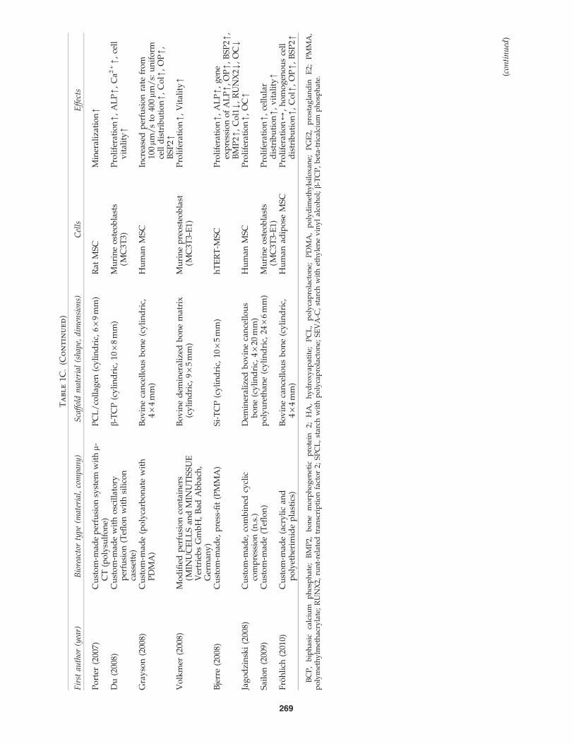
systems has implicated the development of perfusion biore-actors creating a laminar fluid flow and enabling masstransport of nutrients and oxygen through the entire scaffold.Investigations using either custom-made or commerciallyavailable perfusion bioreactor systems are specified in Table1C. A recent review by McCoy and O’Brien provides a
Table 1A. Investigations Using Spinner Flask Bioreactor Systems for Bone Tissue Engineering
First author (year) Scaffold material (shape, dimensions) Cells Effects
Goldstein (2001) PLGA foam discs(cylindric, 12.7 · 6 mm)
Rat osteoblast Proliferation4, ALP[, OC4
Sikavitsas (2002) PLGA foam discs(cylindric, 12.7 · 6 mm)
Rat MSC Proliferation[,ALP[, OC[, Ca2 + [
Meinel (2004) Collagen films andscaffolds (cylindric, 11 · 1.5 mm),silk scaffolds (cylindric, 6 · 1.5 mm)
Human MSC ALP[, Ca2 + [[
Kim (2007) Aqueous-derived fibroin silkscaffolds (cylindric, 15 · 5 mm)
Human MSC Proliferation[, ALP[,Ca2 + [, gene expressionof ALP/OP/BSP/Col1[,compressive modulus[ +compressive strength[
Mygind (2007) Coralline hydroxyapatite scaffolds(cylindric, 10 · 2 mm)
Human TERT-MSC Proliferation[, ALP[,cellular distribution[
Stiehler (2008) PLGA (cubic, 8 · 8 · 5 mm) Human TERT-MSC Proliferation[, ALP[, Ca2 + [
Arrows indicate the effects by the systems compared to static cultivation:[, positive effect;4, no effect;Y, negative effect.ALP, alkaline phosphatase; BSP, bone sialoprotein; Col1, collagen 1; MSC, mesenchymal stromal cell; OC, osteocalcin; OP, osteopontin;
PLGA, poly(lactic-co-glycolic acid; TERT, telomerase reverse transcriptase.
Table 1B. Studies Using Rotating Bioreactor Systems for Bone Tissue Engineering
First author (year)Type of bioreactor
(company)Scaffold material
(shape, dimensions) Cells Effects
Qiu (1999) Custom-made RWV Hollow glas microcarrierswith calcium phosphatesurface (spheric,diameter = 100–200 mm)
Rat MSC, ratosteo-sarcomacells (ROS 17/2.8)
ECM formation[
Botchwey (2001) RWV (Synthecon,Houston)
PLAGA microcarriers(spheric, diameter =500–860 mm)
Human SAOS-2 line ALP[, Ca2 + [,mineralization[
Goldstein (2001) RWV (Synthecon,Houston)
PLGA foam discs(cylindric, 12.7 · 6 mm)
Rat osteoblasts Cell distribution[,ALPY, OC4,Proliferation4
Sikavitsas (2002) RWV (Synthecon,Houston)
PLGA (cylindric,12.7 · 6 mm)
Rat MSC Proliferation4, ALPY,OC4, Ca2 + Y
Yu (2004) RWV (Synthecon,Houston)
PLAGA (cylindric,4 · 2.5 mm)
Rat osteoblasts Proliferation4, Ca2 +
[, ALP[, OC[,OP gene expression[
Pound (2007) RWV (Synthecon,Houston)
Alginate/chitosanmicrocapsules
(spheric, diameter = 5 mm)
Human MSC,human
chondrocytes
Proliferation4, OC[,
Song (2007) Custom-made RWV Demineralizedhuman boneallografts(cubic, 3 · 8 · 8 mm)
Rat osteoblasts Proliferation[, ALP[,Ca2 + [, LDH[,glucoseY, pH[
Diederichs (2009) RBS (Sartorius AG,Gottingen, Germany)
Sponceram� ceramiccarrier discs(thickness = 3 mm,diameter = 65 mm),Zellwerk GmbH
(Eichstadt, Germany)
Human adiposeMSC
Proliferation[,ALP[, Ca2 + [
ECM, extracellular matrix; LDH, lactate dehydrogenase; PLAGA, poly(lactide-coglycolide); RBS, rotating bed system; SAOS, sarcomaosteogenic; RWV, rotating wall vessel.
(continued)
BIOREACTOR SYSTEMS FOR BONE TISSUE ENGINEERING 267

Ta
bl
e1C
.In
ve
st
ig
at
io
ns
Usin
gP
er
fu
sio
nB
io
re
ac
to
rS
yst
em
sfo
rB
on
eT
issu
eE
ng
in
ee
rin
g
Fir
stau
thor
(yea
r)B
iore
acto
rty
pe
(mat
eria
l,co
mp
any
)S
caff
old
mat
eria
l(s
hap
e,d
imen
sion
s)C
ells
Eff
ects
Go
ldst
ein
(200
1)C
ust
om
-mad
e(p
oly
carb
on
ate)
PL
GA
(cy
lin
dri
c,12
.7·
6m
m)
Rat
ost
eob
last
sP
roli
fera
tio
n4
,A
LP
[,
OC
[B
ancr
oft
(200
2)C
ust
om
-mad
e(P
MM
A)
Tit
aniu
mfi
ber
mes
h(c
yli
nd
ric,
10·
0.8
mm
)R
atM
SC
Pro
life
rati
on
[,
AL
P[
,C
a2+
[,
OP
[
Van
der
Do
lder
(200
2)C
ust
om
-mad
e(p
oly
carb
on
ate)
Tit
aniu
mfi
ber
mes
h(c
yli
nd
ric,
10·
0.8
mm
)R
ato
steo
bla
sts
Pro
life
rati
on
[,
AL
P[
,C
a2+
[
Wan
g(2
003)
Per
fusi
on
con
tain
ers
(MIN
UC
EL
LS
and
MIN
UT
ISS
UE
Ver
trie
bs
Gm
bH
,B
adA
bb
ach
,G
erm
any
)
b-T
CP
(cu
bic
,5
·5
·5
mm
)R
ato
steo
bla
sts
AL
P[
(in
vit
ro+
inv
ivo)
,O
C[
and
inv
ivo
bo
ne
form
atio
n[
Uem
ura
(200
3)P
erfu
sio
nco
nta
iner
s(M
INU
CE
LL
San
dM
INU
TIS
SU
EV
ertr
ieb
sG
mb
H,
Bad
Ab
bac
h,
Ger
man
y)
b-T
CP
,H
A,
coll
agen
-p
ho
sph
op
ho
ryn
spo
ng
eR
ato
steo
bla
sts
AL
P[
,in
viv
ob
on
efo
rmat
ion
[
Car
tmel
l(2
003)
Cu
sto
m-m
ade
(sta
inle
ssst
eel)
Hu
man
dem
iner
aliz
edca
nce
llo
us
bo
ne
(cy
lin
dri
c,6.
4·
3m
m)
Mu
rin
eo
steo
bla
sts
(MC
3T3-
E1)
Pro
life
rati
on
[,
gen
eex
pre
ssio
no
fR
UN
X2[
,A
LP
4,
OC
4G
om
es(2
003)
SE
VA
-Can
dS
PC
L(d
isc,
8·
2m
m)
Rat
MS
CS
EV
A-C
:Pro
life
rati
on
[,
AL
P[
,C
a2+
[S
PC
L:P
roli
fera
tio
n4
,A
LP
4,
Ca2
+[
Mei
nel
(200
4)P
erfu
sio
nca
rtri
dg
es(p
oly
carb
on
ate)
Co
llag
en(fi
lman
dcy
lin
dri
c,11
·1.
5m
m),
silk
(cy
lin
dri
c,6
·1.
5m
m)
Hu
man
MS
C(n
=2
do
no
rs)
Ca2
+[
Ho
lth
of
(200
5)C
ust
om
-mad
e(P
MM
A)
Tit
aniu
mm
esh
(cy
lin
dri
c,8
·0.
8cm
)R
atM
SC
Pro
life
rati
on
[,
AL
P[
,C
a2+
[,
Ost
eop
on
tin
[V
ance
(200
5)O
scil
lati
ng
per
fusi
on
bio
reac
tor
Cal
ciu
mp
ho
sph
ate
(cy
lin
dri
c,5
·3.
5m
m)
Mu
rin
eo
steo
bla
sts
(MC
3T3)
PG
E2[
,P
roli
fera
tio
n4
Jan
ssen
(200
6)C
ust
om
-mad
e,in
clu
din
gce
ll-s
eed
ing
loo
p(p
oly
carb
on
ate)
BC
P-g
ran
ule
s(d
iam
eter
=2–
6m
m)
Go
atM
SC
Cal
ciu
mp
ho
sph
ate
no
du
les[
,in
viv
ob
on
efo
rmat
ion
4B
ern
har
dt
(200
7)P
erfu
sio
nco
nta
iner
s(M
INU
CE
LL
San
dM
INU
TIS
SU
EV
ertr
ieb
sG
mb
H,
Bad
Ab
bac
h,
Ger
man
y)
Min
eral
ized
coll
agen
mem
bra
nes
(dia
met
er=
7m
m)
Hu
man
MS
CP
roli
fera
tio
nY
,A
LP
Y
Oli
vie
r(2
007)
Cu
sto
m-m
ade,
div
erg
ent
and
con
ver
gen
tp
erfu
sio
n(c
ell
cult
ure
flas
k)
b-T
CP
(cy
lin
dri
c,33
·14
mm
,20
·4
mm
cen
tral
ho
le)
Hu
man
MG
63o
steo
bla
st-l
ike
cell
sP
roli
fera
tio
n[
,G
luco
seco
nsu
mp
tio
n[
(con
tin
ued
)
268
Ta
bl
e1C
.(C
on
tin
ue
d)
Fir
stau
thor
(yea
r)B
iore
acto
rty
pe
(mat
eria
l,co
mp
any
)S
caff
old
mat
eria
l(s
hap
e,d
imen
sion
s)C
ells
Eff
ects
Po
rter
(200
7)C
ust
om
-mad
ep
erfu
sio
nsy
stem
wit
hm-
CT
(po
lysu
lfo
ne)
PC
L/
coll
agen
(cy
lin
dri
c,6
·9
mm
)R
atM
SC
Min
eral
izat
ion
[
Du
(200
8)C
ust
om
-mad
ew
ith
osc
illa
tory
per
fusi
on
(Tefl
on
wit
hsi
lico
nca
sset
te)
b-T
CP
(cy
lin
dri
c,10
·8
mm
)M
uri
ne
ost
eob
last
s(M
C3T
3)P
roli
fera
tio
n[
,A
LP
[,
Ca2
+[
,ce
llv
ital
ity
[
Gra
yso
n(2
008)
Cu
sto
m-m
ade
(po
lyca
rbo
nat
ew
ith
PD
MA
)B
ov
ine
can
cell
ou
sb
on
e(c
yli
nd
ric,
4·
4m
m)
Hu
man
MS
CIn
crea
sed
per
fusi
on
rate
fro
m10
0mm
/s
to40
0mm
/s:
un
ifo
rmce
lld
istr
ibu
tio
n[
,C
ol[
,O
P[
,B
SP
2[V
olk
mer
(200
8)M
od
ified
per
fusi
on
con
tain
ers
(MIN
UC
EL
LS
and
MIN
UT
ISS
UE
Ver
trie
bs
Gm
bH
,B
adA
bb
ach
,G
erm
any
)
Bo
vin
ed
emin
eral
ized
bo
ne
mat
rix
(cy
lin
dri
c,9
·5
mm
)M
uri
ne
pre
ost
eob
last
(MC
3T3-
E1)
Pro
life
rati
on
[,
Vit
alit
y[
Bje
rre
(200
8)C
ust
om
-mad
e,p
ress
-fit
(PM
MA
)S
i-T
CP
(cy
lin
dri
c,10
·5
mm
)h
TE
RT
-MS
CP
roli
fera
tio
n[
,A
LP
[,
gen
eex
pre
ssio
no
fA
LP
[,
OP
[,
BS
P2[
,B
MP
2[,
Co
l1Y
,R
UN
X2Y
,O
CY
Jag
od
zin
ski
(200
8)C
ust
om
-mad
e,co
mb
ined
cycl
icco
mp
ress
ion
(n.s
.)D
emin
eral
ized
bo
vin
eca
nce
llo
us
bo
ne
(cy
lin
dri
c,4
·20
mm
)H
um
anM
SC
Pro
life
rati
on
[,
OC
[
Sai
lon
(200
9)C
ust
om
-mad
e(T
eflo
n)
po
lyu
reth
ane
(cy
lin
dri
c,24
·6
mm
)M
uri
ne
ost
eob
last
s(M
C3T
3-E
1)P
roli
fera
tio
n[
,ce
llu
lar
dis
trib
uti
on
[,
vit
alit
y[
Fro
hli
ch(2
010)
Cu
sto
m-m
ade
(acr
yli
can
dp
oly
eth
erim
ide
pla
stic
s)B
ov
ine
can
cell
ou
sb
on
e(c
yli
nd
ric,
4·
4m
m)
Hu
man
adip
ose
MS
CP
roli
fera
tio
n4
,h
om
og
eno
us
cell
dis
trib
uti
on
[,
Co
l[,
OP
[,
BS
P2[
BC
P,
bip
has
icca
lciu
mp
ho
sph
ate;
BM
P2,
bo
ne
mo
rph
og
enet
icp
rote
in2;
HA
,h
yd
roxy
apat
ite;
PC
L,
po
lyca
pro
lact
on
e;P
DM
A,
po
lyd
imet
hy
lsil
ox
ane;
PG
E2,
pro
stag
lan
din
E2;
PM
MA
,p
oly
met
hy
lmet
hac
ryla
te;
RU
NX
2,ru
nt-
rela
ted
tran
scri
pti
on
fact
or
2;S
PC
L,
star
chw
ith
po
lyca
pro
lact
on
e;S
EV
A-C
,st
arch
wit
het
hy
len
ev
iny
lal
coh
ol;b-
TC
P,
bet
a-tr
ical
ciu
mp
ho
sph
ate.
(con
tin
ued
)
269

detailed overview to the influence of fluid shear stress inperfusion bioreactor cultures for bone tissue constructs.93
Perfusion systems generally consist of containers, chambers,or cartridges harboring the cell/scaffold constructs. Cellculture medium is piped through tubes by a peristaltic rollerpump to the scaffold and can either flow in a closed loop orthe system provides a reservoir and a waste vessel (Fig. 3).
Oxygenation of the medium is ensured by either gas-permeable silicon tubes13 or by an oxygenator device.50 Themode of fluid flow can influence the effects of osteogenicstimulation. Jacobs et al. found that oscillating flow was amuch less potent stimulator of bone cells than either steadyor pulsing flow. Further, a decrease in responsiveness withincreasing frequency was observed for the dynamic flow
mode.94 Enhanced levels of prostaglandin E2 (PGE2) inMC3T3 osteoblastic cells were measured upon oscillatingflow type compared to perfusion-based and static culturesystems.95 Bakker et al. demonstrated that fluid flow-inducedshear stress induces PGE2 production and release by bonecells in vitro.96
Perfusion-based bioreactors can be divided into systemsusing indirect and direct medium perfusion (Fig. 4A, B). Inindirect perfusion systems the scaffold attached to the cas-sette is not tightly sealed, thereby enabling medium to fol-low the path of least resistance around the scaffold. Thus,flow-derived shear stress may not reach the cells in the in-terior of the construct (Fig. 4A). Using Minucells perfusioncontainers (MINUCELLS and MINUTISSUE Vertriebs
Table 1D. Studies Using Systems with Direct Mechanical Strain for Bone Tissue Engineering
First author (year) Type of strain (parameters)Scaffold material
(shape, dimensions Cells Effects
Neidlinger-Wilke (1994)
Cyclic stretching(1.0, 2.4, 5.3 8.8%surface strains)
Silicone dish,(cubic, l · w · h,10 · 3.5 · 1.75 cm)
Human osteoblasts Proliferation[,LDH4, ALP4
Akhouayri (1999) Rotation (50 or 25 rpm),contraction
Col1 gel, (cubic,3,2 · 2.7 cm)
Rat osteosarcomacells (ROS17/2.8)
Proliferation[,ALP[,osteocalcin[, Ca2 + [
Mauney (2004) Four point bending(max displacementof 0.2 mm for 250cycles every 24 h)
Partially demineralizedbovine cancellousbone, (cubic, l · w· h, 63 · 6 · 6 mm)
Human MSC ALP[, geneexpression ofALP[, OC4,OP[, Ca2 + [
Ignatius (2005) Uniaxial stretching(1 Hz, 1800 cycles)
Collagen type I gel(cubic, 3 · 3 · 0.4 cm)
Human osteoblasticcell line (hFOB1.19)
Proliferation[,gene expressionof RUNX2[,ALP[, OC[,OP[, Col1[
Wartella andWayne (2009)
Biaxial; cycliccompressionand tension
Collagen type I scaffold(cubic; 10 · 20 · 1.5 mm)
Human MSC Proteoglycan[,matrix deposition[
Table 1E. Investigations Using Custom-Made Systems with Pulsed Electromagnetic Fields
for Bone Tissue Engineering
First author (year)
PEMF parameters(frequency [Hz],pulse duration
[ms], intensity [mT])Scaffold material
(shape, dimensions) Cells Effects
Bodamyali (1998) 15 Hz, 4.5 ms 2D, tissue culturepolystyrene
Rat osteoblasts Ca2 + [, Gene expression ofBMP-2[and BMP-4[
Wiesmann (2001) 16 Hz, 63 ms 2D, tissue culturepolystyrene
Bovine osteoblasts Ca2 + [
Fassina (2006) 75 Hz, 2 mT Polyurethane porousscaffold (cylindric,15 · 2 mm)
Human osteosarcomaSAOS-2 cell line
Ca2 + [, Gene expression ofDCN[, OC[, OPN[,TGF-ß[and Col1[
Tsai (2007) 7.5 Hz, 0.3 ms, 0.13,0.24, 0.32 mT
PLGA (cylindric,6 · 3 mm)
Rat osteoblasts Proliferation[(0.13 mT),ALP[
Schwartz (2008) 15 Hz, 4.5 ms Calcium phosphate(cylindric,12.7 mm · 0.6 mm)
Human MSC ProliferationY, incombination with BMP-2: ALP[, OC[, TGF-b1[
Sun (2010) 15 Hz, 4.5 ms,1.8 mT
2D, tissue culturepolystyrene
Human MSC Proliferation[, Multi-linage differentiation4,CD phenotype4
CD, cluster of differentiation; DCN, decorin; Hz, hertz; mT, milli tesla; PEMF, pulsed electromagnetic fields; TGF-b, transforming growthfactor beta.
270 RAUH ET AL.

GmbH; Table 2), an indirect perfusion system, Wang et al.and Uemura and co-workers observed an increase in ALPactivity and osteocalcin (OC) protein expression levels97,98
by dynamic cultivation of rat osteoblasts on b-TCP scaf-folds. Further, bone formation was determined in a subcu-taneous rat model by these authors: perfusion-stimulatedcell/scaffold composites showed significantly enhancedbone formation compared to statically cultivated controls.Using a similar perfusion system human MSCs were culti-vated on membranes made of mineralized Col1.99 Lower
levels of ALP activity and a decreased proliferation ratewere observed as compared with statically cultivated cell/scaffold constructs. Volkmer et al. modified that type ofperfusion containers by adding a carrier cassette resulting ina forced perfusion system,100 demonstrating increasedamounts of viable cells in the center of the constructscompared to indirect perfusion method. The oxygen con-centrations measured in the centers of the scaffolds did notchange between the two dynamic cultivation setups. How-ever, a definitive conclusion on which cultivation method is
Table 2. Selected Commercially Available Bioreactor Systems
Company Product Type (features, options)
Bellco Biotechnology (Vineland, NJ) Bell-FlowTM Spinner flaskCorning� Lifesciences (Lowell, MA) ProCulture� glass spinner flask Spinner flask
(disposable or autoclavable)MINUCELLS and MINUTISSUE
Vertriebs GmbH (Bad Abbach, Germany)Tissue engineering container Perfusion bioreactor (indirect
perfusion)Tissue Growth Technologies (Minnetonka) OsteoGen Bioreactor Perfusion bioreactor (pulsatile
fluid flow stimulator, mCT)Zellwerk GmbH (Oberkramer, Germany) BIOSTAT� Bplus
RBS 500-System,Z� RP cell- and tissueculturing systems
Rotating bed bioreactor(GMP conform)
Synthecon, Inc. (Houston) STLV Bioreactor Rotating wall vesselB. Braun Biotech International
GmbH (Melsungen, Germany)Medistat RBS Rotating bed bioreactor
(GMP conform)Flexcell International Corporation
(Hillsborough, NC)e.g., BioPress� Compression
Plates, Flexcell� FX-5000�Compression System
Systems using tension,compression and shear stress
GMP, Good Manufacturing Practice; RBS, rotating bed system; STLV, slow turning lateral vessel; mCT, micro-computed tomography.
FIG. 2. (A–C) Schematicdrawings of rotation-basedbioreactor systems. Cultiva-tion in a free-fall manner (A).Scaffolds are attached to theouter vessel wall during cul-tivation (B). Rotating bedbioreactor (RBB) (C). Cells areseeded on discs rotating onthe horizontal axis. On theleft side, the different systemsare shown three dimension-ally. On the right side, crosssections of the respectivevessels are demonstrated.Arrows illustrate the direc-tion of rotation.
BIOREACTOR SYSTEMS FOR BONE TISSUE ENGINEERING 271

more favorable cannot be drawn from these experiments asessential parameters, for example, ALP activity, were notmeasured.
Bioreactors with direct perfusion allow the reduction ofinternal mass transfer limitations and exert biophysical for-ces by fluid flow in the interior of the so cultivated cell/scaffold constructs (Fig. 4B). Usually, the scaffolds are fixatedin containers or cassettes in a press-fit manner.101 Systemsusing direct perfusion have been shown to enhance celldensity in the scaffold center,102 cell proliferation and dif-ferentiation of osteoprogenitor cells, as well as the depositionof mineralized ECM.13,19,103,104 Various systems applyingdirect perfusion method have been described.51,105–108
Janssen et al. introduced a custom-made direct perfusionsystem equipped with a seeding loop, an oxygenator device,and an online oxygen measurement unit with sensors posi-tioned at the inlet and outlet of the medium flow.50,109
However, in vivo studies in mice using this system showedno statistically significant differences in new bone formationcomparing statically and dynamically cultured constructs.The OsteoGen bioreactor (Tissue Growth Technologies;Table 2) allows noninvasive online monitoring of minerali-zation of 3D cultivated cell/scaffold constructs by use ofmicro-computed tomography (mCT) technology.110 The rateof mineralized matrix formation in the perfused constructsincreased significantly from 0.69 mm3/week during the first3 weeks of culture to 1.03 mm3/week over the last 2 weeks.In contrast, the rate of mineral deposition in the static con-trols was 0.01 and 0.16 mm3/week, respectively. Meinel andcolleagues compared the effect of different Col- and silk-based scaffold materials and the influence of hydrodynamicenvironment (static culture, spinner flask, or perfused car-tridge) on the osteogenic differentiation of human MSCs.76
The authors observed enhanced mineralization on biode-gradable Col-based scaffolds in spinner flask cultures com-pared to perfusion-based bioreactor cultivation. The authorsargue that the advanced degradation of the used Col scaf-folds by the perfusion bioreactor may be a reason for theunfavorable results obtained with this system. The distribu-tion of mineralization was limited to the outer rim in spinnerflask-cultivated constructs, whereas mineralized matrix wasmore evenly distributed in perfused scaffolds. The authorsconclude that osteogenesis in cultured MSCs can be modu-lated by both scaffold biomaterial properties and flowenvironment. Frohlich et al. cultivated bovine cancellousbone cylinders seeded with human adipose-derived stemcells in a novel perfusion bioreactor system. In this bioreac-tor, medium flowed through a central port at the bottom ofthe bioreactor vessel from where it was evenly distributedinto six channels leading into individual culture wells loadedwith the scaffolds.111 The authors observed enhanced celldistribution, osteogenic differentiation, and bone matrixformation in perfused constructs compared to statically cul-tivated controls.
In summary, evidence exists that the use of perfusion-based bioreactor systems in bone TE results in improvedcellular proliferation, distribution, differentiation, and via-bility in the interior of scaffolds when compared to staticcultivation. Existing devices vary with respect to additionalequipment and fluid flow options. Some types of bioreactorsystems are suitable for cell seeding,50 and others can becombined with mechanical stimulation (cyclic compres-
sion)112 or online-monitoring of mineralization.110 Reportedconstant perfusion flow rates range from 199 to 600 mL/h.112
Flow rate levels exceeding a specific range have shown topromote the washing out of cells due to excessive shearstress.113 It is therefore advisable to determine the optimalmedium flow rate for each bioreactor-based setup. In gen-eral, upscaling of bioreactor-based engineered bone tissuefor clinical use still needs further optimization as the di-mensions of cell/scaffold constructs cultivated by use ofcurrent perfusion-based systems are comparatively smallranging from 0.04 to 2.7 cm3 (Fig. 5). In general, upscaling ofconstruct dimensions can be either addressed by furtherupscaling of existing perfusion bioreactor systems to obtaina vascularized construct or by combining smaller tissue-engineered constructs synthesized simultaneously.50 In aneffort to scale up existing perfusion bioreactor systems,Olivier et al. introduced a bioreactor using relatively largeporous b-TCP cylinders of 33 · 14 mm (4.8 cm3) with me-dium perfused through a dead ending hole.114 However,this method could lead to an inhomogenous flow and noresults were presented about the vitality of the cells in theinner parts of the scaffold.
Systems using direct mechanical strain
Since the German anatomist Julius Wolff in 1892 postu-lated that bone remodeling depends on mechanical load,numerous scientists have focused on biomechanical effectson the cellular level.115–119 Bone is constantly renewed bybone-forming osteoblasts and bone-resorbing osteoclasts,thereby establishing a homeostasis in healthy humans.120,121
Osteocytes are assumed to sense mechanical stimuli by dif-ferent means, for example, through the cell body, the den-dritic processes, or bending of cilia.122 The signal transfer ismediated by gap junctions and hemichannels, and the re-lease of signaling molecules into the bone fluid.123,124 How-ever, the distinct pathways involved have not beencompletely discovered to date. Mechanical unloading thatoccurs in microgravity during space flights or extended bedrest reduces the number of osteocytes.125 Loading of bonewith strains below 500mstrains was associated with boneloss, for loading with up to 1000 mstrain the original bonegeometry and mass were maintained, and strains between1000 and 4000mstrains increased new bone formation pro-gressively.126 Recent studies identified WNT signaling as animportant pathway promoting the early phase of commit-ment to the osteogenic lineage and subsequent differentia-tion of C57BL/6J osteoblasts62 and osteoblastic precursors ingeneral.127 WNT signaling enhances the expression of os-teoprotegerin, but inhibits the expression of high levels ofOC, a typical feature of mature, matrix-synthesizing bonecells.127
Various studies confirmed the principle of mechanicalconditioning by the application of direct mechanical strainusing, for example, bending, stretching, contraction, andcompression (Table 1D). These types of mechanical strainapplication will be discussed in the following. The mechan-ical stimulation by a 4-point-bending device (Fig. 6A) re-sulted in increased levels of ALP activity, mineralized matrixproduction, and gene expression of ALP and OP in MSCsloaded on demineralized bovine cancellous bone grafts.48
Interestingly, this effect was only detectable in the presence
272 RAUH ET AL.

of dexamethasone at concentrations of 10 nM, but not100 nM.
Cyclic stretching of human osteoblasts attached to silicondishes enhanced proliferation, but did not affect ALP activ-ity.15 The principle of uniaxial stretching is shown schemat-ically in Figure 6B. Uniaxial stretching of a humanosteoblastic cell line in Col1 gels applied for 21 days with amagnitude of 1% (10,000 mstrain) increased both proliferationand gene expression of ALP, OC, OP, and Col1 compared tostatic controls. Further, the cells and the newly producedtype of ECM were strictly oriented according to the directionof the applied mechanical stress. The cell-stretching systemconsists of rectangular elastic silicone dishes, which weredesigned for use of a six-station stimulation apparatus dri-ven by an eccentric motor.128,129 Another study supportingthe effects of contraction was performed by Akhouayri et al.,who observed increased proliferation, ALP activity, as wellas Ca2 + and OC protein expression levels by contracted ratosteosarcoma cells cultivated on 3D Col1-matrices.130
When analyzing effects of compression (Fig. 6C), ma-chines originally fabricated for material testing were usedfrequently. For example, a study by Lanyon and Rubin from1984 showed that intermittent dynamic as opposed to con-tinous compression loading induced bone formation in vivousing an avian ulnar defect model.117 The applied type ofstress can influence the effect of tissue response. Using low
FIG. 5. Scaffold volumedata of studies using perfu-sion bioreactor systems.
FIG. 4. (A, B) Schematic drawings of perfusion flow prin-ciples. (A) shows indirect perfusion where medium flowsaround and only partly through the scaffold. (B) In directperfusion medium flow is forced through the scaffold andshear stress is directly transferred to the cells within thescaffold. Arrows illustrate the magnitude and direction offluid flow.
FIG. 3. Flow chart of a perfusion bioreactor system. Arrowsindicate flow direction in a closed loop.
BIOREACTOR SYSTEMS FOR BONE TISSUE ENGINEERING 273

hydrostatic pressure in a bone organ culture of murine fetalmetatarsal and calvariae Burger et al. observed that inter-mittent stress enhanced mineralization more effectively thancontinuous stimulation.131 Wartella and Wayne recently in-troduced a biaxial bioreactor system for mechanical stimu-lation of tissue constructs in two perpendicular directions.131
This type of bioreactor applying both compression and ten-sion forces resulted in elevated proteoglycan production andmatrix deposition by human MSCs.
Currently existing bioreactor systems for direct mechani-cal stimulation have shown beneficial effects on proliferation,osteogenic differentiation, and matrix formation. Severalauthors used biomechanically instable Col1 gels as matricesfor mechanical strain-based cellular stimulation.129,130 Thismay be disadvantageous in situations where a certain levelof initial mechanical stability of an implantable cell/scaffoldconstruct is required for effective bone regeneration and nofurther stabilization is applied. Further, diffusional limita-tions occurring in larger constructs in mechanical load-basedbioreactor systems may be addressed additionally by otherstrategies.
EMF-based bioreactor systems
Electric and EMFs have been applied for bone regenera-tion purposes in patients with, for example, osteoporosis andnonunions as well as supportive therapy during limblengthening and revision alloarthroplasty procedures for thelast three decades.133–137 PEMF has been shown to signifi-cantly reduce the loss of bone mass and to accelerate boneformation in vivo.138,139 Endogenous EMF and PEMF arisefrom muscle movements.140 The electric potentials generatedby mechanical deformation in bone cause piezoelectricity.When bone is fractured, electrons migrate to the injured site,causing a negative potential. Vibrations of human muscles
induce mechanical strains and currents of specific frequen-cies. Frequencies in the ranges of 5–30 Hz and < 10 Hz wereobserved during postural muscle activity and walking, re-spectively.141 Interestingly, bone cells exhibit a strong fre-quency selectivity with EMF effectiveness peaking at15 Hz.142 Studies suggest that EMFs affect different subcel-lular proliferation- and differentiation-related signalingpathways, for example, those including parathyroid hor-
FIG. 6. (A–C) Schematicdrawing of systems usingdirect mechanical strain. (A)illustrates the four-pointbending method. On the left,the scaffold is shown inpassive state. On the right,mechanical loading isapplied. (B) demonstrates theprinciple of uniaxial cyclicstretching, which is appliedin elastic silicon dishes. Cellsare embedded in three-dimensional collagen type Imatrices (dark gray). (C)represents a uniaxial me-chanical loading device. Aplunger is pushed in a cyclicmanner on the scaffold.Arrows illustrate mechanicalforces applied.
FIG. 7. Schematic illustration of a bioreactor system basedon pulsed electromagnetic fields (PEMF). The Helmholtzcoils are powered by a PEMF generator. The scaffold in thebioreactor chamber is positioned between two Helmholtzcoils. The bioreactor applies PEMF stimuli to the cells withinthe scaffold at a defined frequency, amplitude, intensity, andpulse duration.
274 RAUH ET AL.

mone and adenosine A2A receptor, resulting in conformationchanges or in increase of the receptor density.143
To utilize these effects for bone TE, EMF-based bioreactors(Fig. 7) were developed. Typically, these systems consist ofHelmholtz coils powered by a PEMF generator. The cell/scaffold construct is positioned between two Helmholtz coilsand an EMF of a defined intensity is applied. In vitro studiesshowed that EMFs induce and enhance osteogenesis in hu-man MSCs144,145 and osteoblasts146–148 (Table 1E). Fassinaet al. introduced a simple EMF-based bioreactor system witha standard well plate and two parallel Helmholtz coils beingkept in a PMMA tube.149 The applied PEMF frequency usedin that study was 75 Hz with an intensity of 2 mT and themagnetic field was measured using a Hall Effect transversegaussmeter probe. PEMF-stimulated human sarcoma osteo-genic-2 cells exhibited increased mineralization and geneexpression of decorin, OC, OPN, TGF-b, and Col1. In a studyby Schwartz and co-workers, human MSCs cultivated oncalcium phosphate discs demonstrated decreased prolifera-tion but enhanced ALP activity and protein levels of OC andTGF-b in the combined presence of BMP-2 and PEMF stim-ulation.144 Bodamyali et al. showed superior mineralizationand expression of BMP-2 and BMP-4 genes upon PEMFstimulation in rat osteoblasts.146 The device utilized a sawtooth waveform consisting of 4.5-ms bursts of pulses, re-peating at a rate of 15 Hz. Increased proliferation and ALPactivity were reported by Tsai et al., who stimulated rat os-teoblasts seeded on poly(lactic-co-glycolic acid) scaffoldswith PEMFs at a frequency of 7.5 Hz.150
In summary, the use of EMF-based bioreactor systems forbone TE resulted in enhanced osteogenic differentiation ofcell/scaffold constructs compared to static cultivation. In-terestingly, as observed in some studies, PEMF also stimu-lated proliferation of osteoprogenitor cells. The high initialequipment costs required for PEMF-based bioreactor sys-tems denote a major disadvantage. On the other hand, thenoninvasiveness of PEMF-based systems is clearly advanta-geous with respect to handling and potential GMP approval.
In vivo bioreactor systems
The concept of in vivo bioreactor systems takes advantageof the physiological environment and supply of a cell-loadedscaffold biomaterial with necessary growth factors and nu-trients provided by the host organism. Several in vivo bio-reactors were developed to generate vascularized bone tissueusing different animal models, for example, in mice,151
rats,152 rabbits,153 and miniature pigs,154 resulting in site-specific de novo bone regeneration. Petite et al. used a com-bination of a coral scaffold with in vitro-expanded MSCsleading to complete recorticalization and the formation ofmature lamellar cortical bone in sheep.155 Even a man canserve as an in vivo bone bioreactor as described by Warnkeet al.156 A titanium mesh cage filled with bone mineralblocks, infiltrated with 7 mg recombinant human BMP-7 andautologous bone marrow, was implanted in a latissimusdorsi muscle pouch. After 7 weeks the construct was trans-planted to repair a mandibular defect. Successful bony re-construction resulted in improvement in the quality of life ofthis patient.157 However, although occasionally applied withsuccess, the application of in vivo bioreactor concepts iscurrently limited to individual cases.
Commercially available bioreactor systemsfor bone tissue engineering
Currently, various bioreactor systems for generatingmineralized cell/scaffold constructs are commercially avail-able (Table 2). In the following we focus on selected systemsrepresentative for the respective type of bioreactor. Besidesthe systems mentioned, other bioreactor systems are avail-able for bone TE applications.
Possibly, the most inexpensive systems are spinner flasks.The Bell-Flow� spinner flask from Bellco Biotechnology ismanufactured from autoclavable borosilicate glass and isavailable in volumes ranging from 100 mL to 3 L(www.bellcoglass.com). Corning� Lifesciences offers auto-clavable spinner flasks as well as disposable systems made ofplastic (www.corning.com/index.aspx).
MINUCELLS and MINUTISSUE GmbH offers a variety ofperfusion containers, for example, for cultivating cartilageconstructs or several types of epithelia in their organo-typicalenvironment (www.minucells.de/index.html). The reactorsare referred to the indirect perfusion method. The OsteoGenBioreactor, a device for direct perfusion, is commerciallyavailable from Tissue Growth Technologies. The design iscompact and able fit in a standard incubator, and all com-ponents of the system are autoclavable. The chambers aredesigned only for cylindrical scaffolds with 10 · 10 mm. Be-sides an optional pulsatile hydrostatic pressure stimulator,the company offers a ‘‘GrowthWorks Software and Controlplatform’’ (www.tissuegrowth.com/).
Zellwerk GmbH distributes the GMP-conform RBB tissueculturing system BIOSTAT� Bplus RBS (www.zellwerk.biz/).The complete cultivation system comprises a bioreactor, aGMP breeder, and a control unit. Other rotating bioreactorsystems are available from for example, Synthecon, Inc.These rotary cell culture microgravity bioreactors originallydesigned by National Aeronautics and Space Administrationare produced as autoclavable and disposable systems. Thecompany also offers a perfused rotating bioreactor allowingthe online monitoring of crucial parameters, for example,pH, oxygen, and glucose levels (www.synthecon.com). An-other dynamic cultivation system is available from B. BraunBiotech International GmbH offering an RBB meeting GMPstandards (www.chemietechnik.de/company).
Systems using tension, compression, and shear stressare available from, for example, Flexcell International Cor-poration. The Flexcell� FX-5000� Tension System andFlexcell FX-5000 Compression System apply cyclic or staticstrain to cells cultured on flexible-bottomed culture plates.Special devices allow to observe signaling responses uponstrain stimulation in real-time on a microscope stage (www.flexcellint.com/).
Bioreactors and GMP
GMP is a quality assurance system for medicinal products.Several regulatory requirements, for example, productionaccording to validated standard operating procedures,demonstration of quality control, and in-process controls,have to be applied. In Europe respective directives andguidelines ensure quality and safety standards for donation,procurement, testing, processing, preservation, storage, andthe distribution of human tissue and cells.158 In this con-text, compliance with the annually updated guidelines for
BIOREACTOR SYSTEMS FOR BONE TISSUE ENGINEERING 275

‘‘Current Good Manufacturing Practice’’ (cGMP) is requiredin the United States.159
Protocols were developed to facilitate the adaption ofGMP standards for the expansion of human embryonic stemcells in a stirred bioreactor system.160 In addition, a strategywas described to develop and validate a closed, automatedproduction process to expand stem and progenitor cells inthe presence of human bone marrow mononuclear cells.161
The RBBs Medistat RBS (B. Braun Biotech InternationalGmbH) and Z�RP cell- and tissue culturing systems (Zell-werk GmbH), allowing the cultivation and manufacturing of3D tissue-engineered transplants91,92 conform to require-ments of GMP standards. Successful translation from thelaboratory to clinical application can be exemplified in thefield of skin TE. The commercially available products Trans-Cyte� and Dermagraft�40 are generated in a closed perfusionbioreactor system. For this purpose, a scaffold (Biobrane�) isseeded with allogenic dermal fibroblasts and cultivated in abioreactor system allowing automated cell seeding, mediachange, in-process monitoring of growth, storage, and deliv-ery simultaneously. To the authors’ best knowledge, however,no cell-based, tissue-engineered bone substitute constructcultivated in a bioreactor system has been applied clinically todate. To pave the way for bioreactor-stimulated, tissue-engineered constructs for bone regeneration from bench tobedside, the compliance of potential products with GMPstandards will be a basic prerequisite.
Conclusion
Bone graft material is often needed for the treatment ofosseous defects. Due to limitations and risks associated withautologous as well as allogenic bone grafting procedures,alternative strategies are needed for skeletal reconstruction.The concept of TE constitutes the framework for the im-plementation of cell-based bone regeneration strategies. Tooptimize the cultivation of cell/scaffold constructs, dynamicbioreactor systems, enhancing cellular proliferation and dif-ferentiation and resolving mass transport limitations, areappealing components. Bone bioreactor systems includespinner flasks, RWV constructs, perfusion bioreactors, andsystems based on mechanical or electromagnetic stimulationof cell/scaffold composites. These systems differ consider-ably with respect to ease of use, cost-effectiveness, and de-gree of additional osteogenic stimuli provided, as well asmonitoring and manipulation options. Currently availablebone bioreactors enable adequate monitoring and controllingof specific biological, physical, and chemical parametersduring the process of in vitro bone formation. Further opti-mization of these systems may be achieved by adaptingspecific stimuli, for example, shear stress, load, or EMF, andby combining biophysical and biochemical stimuli withinone system. A major challenge, however, will be the trans-lation of bioreactor-based concepts into clinically applicable,GMP-conform systems generating newly formed mineral-ized cell/scaffold constructs.
Acknowledgments
We thank Mike Tipsword, Angela Jacobi, and CorinaVater for proofreading this article. The authors have receivedfinancial support from the German Academic Exchange
Service/German Federal Ministry of Education and Research(Grant No. D/09/04774) and from the Center for Re-generative Therapies Dresden, Germany.
Disclosure Statement
No competing financial interests exist. We do not claimcompleteness with respect to the bioreactor systems dis-cussed and do regret if certain products or concepts are notcovered in this article.
References
1. Brooks, P.M. The burden of musculoskeletal disease—aglobal perspective. Clin Rheumatol 25, 778, 2006.
2. Arrington, E.D., Smith, W.J., Chambers, H.G., Bucknell,A.L., and Davino, N.A. Complications of iliac crest bonegraft harvesting. Clin Orthop Relat Res (329), 300, 1996.
3. Langer, R., and Vacanti, J.P. Tissue engineering. Science260, 920, 1993.
4. Drosse, I., et al. Tissue engineering for bone defect healing:an update on a multi-component approach. Injury 39
Suppl 2, S9, 2008.5. Stoddart, M.J., Grad, S., Eglin, D., and Alini, M. Cells and
biomaterials in cartilage tissue engineering. Regen Med 4,
81, 2009.6. Lv, S., et al. Designed biomaterials to mimic the mechanical
properties of muscles. Nature 465, 69, 2010.7. Mansbridge, J. Skin tissue engineering. J Biomater Sci
Polym Ed 19, 955, 2008.8. Curtis, M.W., and Russell, B. Cardiac tissue engineering. J
Cardiovasc Nurs 24, 87, 2009.9. Hynes, S.R., and Lavik, E.B. A tissue-engineered approach
towards retinal repair: scaffolds for cell transplantation tothe subretinal space. Graefes Arch Clin Exp Ophthalmol248, 763, 2010.
10. Kutty, J.K., and Webb, K. Tissue engineering therapies forthe vocal fold lamina propria. Tissue Eng Part B Rev 15,
249, 2009.11. Fiegel, H.C., et al. Hepatic tissue engineering: from trans-
plantation to customized cell-based liver directed therapiesfrom the laboratory. J Cell Mol Med 12, 56, 2008.
12. Ghasemi-Mobarakeh, L., Prabhakaran, M.P., Morshed, M.,Nasr-Esfahani, M.H., and Ramakrishna, S. Electrospunpoly(epsilon-caprolactone)/gelatin nanofibrous scaffoldsfor nerve tissue engineering. Biomaterials 29, 4532, 2008.
13. Bancroft, G.N., et al. Fluid flow increases mineralized ma-trix deposition in 3D perfusion culture of marrow stromalosteoblasts in a dose-dependent manner. Proc Natl AcadSci USA 99, 12600, 2002.
14. Fassina, L., Visai, L., De Angelis, M.G., Benazzo, F., andMagenes, G. Surface modification of a porous polyurethanethrough a culture of human osteoblasts and an electro-magnetic bioreactor. Technol Health Care 15, 33, 2007.
15. Neidlinger-Wilke, C., Wilke, H.J., and Claes, L. Cyclicstretching of human osteoblasts affects proliferation andmetabolism: a new experimental method and its applica-tion. J Orthop Res 12, 70, 1994.
16. Kern, S., Eichler, H., Stoeve, J., Kluter, H., and Bieback, K.Comparative analysis of mesenchymal stem cells frombone marrow, umbilical cord blood, or adipose tissue. StemCells 24, 1294, 2006.
17. Pittenger, M.F., et al. Multilineage potential of adult humanmesenchymal stem cells. Science 284, 143, 1999.
276 RAUH ET AL.

18. Kassem, M., and Abdallah, B.M. Human bone-marrow-derived mesenchymal stem cells: biological characteristicsand potential role in therapy of degenerative diseases. CellTissue Res 331, 157, 2008.
19. Bjerre, L., Bunger, C.E., Kassem, M., and Mygind, T. Flowperfusion culture of human mesenchymal stem cells onsilicate-substituted tricalcium phosphate scaffolds. Bioma-terials 29, 2616, 2008.
20. Mygind, T., et al. Mesenchymal stem cell ingrowth anddifferentiation on coralline hydroxyapatite scaffolds. Bio-materials 28, 1036, 2007.
21. Stiehler, M., et al. Effect of dynamic 3-D culture on prolifer-ation, distribution, and osteogenic differentiation of humanmesenchymal stem cells. J Biomed Mater Res A 89, 96, 2009.
22. Hutmacher, D.W. Scaffolds in tissue engineering bone andcartilage. Biomaterials 21, 2529, 2000.
23. Wang, X., Li, Y., Hodgson, P.D., and Wen, C. Biomimeticmodification of porous TiNbZr alloy scaffold for bone tis-sue engineering. Tissue Eng Part A 16, 309, 2010.
24. Wu, Y.C., Lee, T.M., Chiu, K.H., Shaw, S.Y., and Yang, C.Y.A comparative study of the physical and mechanicalproperties of three natural corals based on the criteria forbone-tissue engineering scaffolds. J Mater Sci Mater Med20, 1273, 2009.
25. Dennis, J.E., Haynesworth, S.E., Young, R.G., and Caplan,A.I. Osteogenesis in marrow-derived mesenchymal cellporous ceramic composites transplanted subcutaneously:effect of fibronectin and laminin on cell retention and rateof osteogenic expression. Cell Transplant 1, 23, 1992.
26. Boyan, B.D., Hummert, T.W., Dean, D.D., and Schwartz, Z.Role of material surfaces in regulating bone and cartilagecell response. Biomaterials 17, 137, 1996.
27. Khayyeri, H., Checa, S., Tagil, M., O’Brien, F.J., and Pre-ndergast, P.J. Tissue differentiation in an in vivo bioreactor:in silico investigations of scaffold stiffness. J Mater Sci MaterMed 21, 2331, 2010.
28. Olivares, A.L., Marsal, E., Planell, J.A., and Lacroix, D. Fi-nite element study of scaffold architecture design and cul-ture conditions for tissue engineering. Biomaterials 30,
6142, 2009.29. VanGordon, S.B., et al. Effects of scaffold architecture on
preosteoblastic cultures under continuous fluid shear. IndEng Chem Res 50, 620, 2010.
30. Olivares, A.L., Marsal, E., Planell, J.A., and Lacroix, D. Fi-nite element study of scaffold architecture design and cul-ture conditions for tissue engineering. Biomaterials 30,
6142, 2009.31. Vater, C., Kasten, P., and Stiehler, M. Culture media for the
differentiation of mesenchymal stromal cells. Acta Bio-mater 7, 463, 2011.
32. Canalis, E., Economides, A.N., and Gazzerro, E. Bonemorphogenetic proteins, their antagonists, and the skele-ton. Endocr Rev 24, 218, 2003.
33. McKay, W.F., Peckham, S.M., and Badura, J.M. A com-prehensive clinical review of recombinant human bonemorphogenetic protein-2 (INFUSE Bone Graft). Int Orthop31, 729, 2007.
34. White, A.P., et al. Clinical applications of BMP-7/OP-1 infractures, nonunions and spinal fusion. Int Orthop 31, 735,2007.
35. Whitman, D.H., Berry, R.L., and Green, D.M. Platelet gel:an autologous alternative to fibrin glue with applications inoral and maxillofacial surgery. J Oral Maxillofac Surg 55,
1294, 1997.
36. Gardner, M.J., Demetrakopoulos, D., Klepchick, P.R., andMooar, P.A. The efficacy of autologous platelet gel in paincontrol and blood loss in total knee arthroplasty. An anal-ysis of the haemoglobin, narcotic requirement and range ofmotion. Int Orthop 31, 309, 2007.
37. Marx, R.E., et al. Platelet-rich plasma: growth factor en-hancement for bone grafts. Oral Surg Oral Med Oral PatholOral Radiol Endod 85, 638, 1998.
38. Eppley, B.L., Woodell, J.E., and Higgins, J. Platelet quanti-fication and growth factor analysis from platelet-richplasma: implications for wound healing. Plast ReconstrSurg 114, 1502, 2004.
39. Martin, I., Wendt, D., and Heberer, M. The role of biore-actors in tissue engineering. Trends Biotechnol 22, 80, 2004.
40. Ratcliffe, A., and Niklason, L.E. Bioreactors and biopro-cessing for tissue engineering. Ann NY Acad Sci 961, 210,2002.
41. Glowacki, J., Mizuno, S., and Greenberger, J.S. Perfusionenhances functions of bone marrow stromal cells in three-dimensional culture. Cell Transplant 7, 319, 1998.
42. Alvarez-Barreto, J.F., Linehan, S.M., Shambaugh, R.L., andSikavitsas, V.I. Flow perfusion improves seeding of tissueengineering scaffolds with different architectures. AnnBiomed Eng 35, 429, 2007.
43. Burg, K.J., et al. Comparative study of seeding methods forthree-dimensional polymeric scaffolds. J Biomed Mater Res51, 642, 2000.
44. Alvarez-Barreto, J.F., and Sikavitsas, V.I. Improved mes-enchymal stem cell seeding on RGD-modified poly(L-lacticacid) scaffolds using flow perfusion. Macromol Biosci 7,
579, 2007.45. Griffon, D.J., Abulencia, J.P., Ragetly, G.R., Fredericks, L.P.,
and Chaieb, S. A comparative study of seeding techniquesand three-dimensional matrices for mesenchymal cell at-tachment. J Tissue Eng Regen Med 5, 169, 2011.
46. Timmins, N.E., et al. Three-dimensional cell culture andtissue engineering in a T-CUP (tissue culture under perfu-sion). Tissue Eng 13, 2021, 2007.
47. Zhao, F., and Ma, T. Perfusion bioreactor system for humanmesenchymal stem cell tissue engineering: dynamic cellseeding and construct development. Biotechnol Bioeng 91,
482, 2005.48. Mauney, J.R., et al. Mechanical stimulation promotes oste-
ogenic differentiation of human bone marrow stromal cellson 3-D partially demineralized bone scaffolds in vitro.Calcif Tissue Int 74, 458, 2004.
49. Godbey, W.T., Hindy, S.B., Sherman, M.E., and Atala, A. Anovel use of centrifugal force for cell seeding into porousscaffolds. Biomaterials 25, 2799, 2004.
50. Janssen, F.W., Oostra, J., Oorschot, A., and van Blitterswijk,C.A. A perfusion bioreactor system capable of producingclinically relevant volumes of tissue-engineered bone:in vivo bone formation showing proof of concept. Bioma-terials 27, 315, 2006.
51. Du, D., Furukawa, K.S., and Ushida, T. 3D culture of os-teoblast-like cells by unidirectional or oscillatory flow forbone tissue engineering. Biotechnol Bioeng 102, 1670, 2009.
52. Cheng, S.L., et al. Human osteoblasts express a repertoire ofcadherins, which are critical for BMP-2-induced osteogenicdifferentiation. J Bone Miner Res 13, 633, 1998.
53. van den Dolder, J., Spauwen, P.H., and Jansen, J.A.Evaluation of various seeding techniques for culturingosteogenic cells on titanium fiber mesh. Tissue Eng 9, 315,2003.
BIOREACTOR SYSTEMS FOR BONE TISSUE ENGINEERING 277
54. Wiedmann-Al-Ahmad, M., Gutwald, R., Lauer, G., Hub-ner, U., and Schmelzeisen, R. How to optimize seeding andculturing of human osteoblast-like cells on various bioma-terials. Biomaterials 23, 3319, 2002.
55. Kruyt, M., et al. Analysis of the dynamics of bone forma-tion, effect of cell seeding density, and potential of alloge-neic cells in cell-based bone tissue engineering in goats.Tissue Eng Part A 14, 1081, 2008.
56. Impens, S., Chen, Y., Mullens, S., Luyten, F., and Schrooten,J. Controlled cell-seeding methodologies: a first step to-ward clinically relevant bone tissue engineering strategies.Tissue Eng Part C Methods 16, 1575, 2010.
57. Cowin, S.C. Bone poroelasticity. J Biomech 32, 217, 1999.58. Meyer, U., et al. Strain-related bone remodeling in distrac-
tion osteogenesis of the mandible. Plast Reconstr Surg 103,
800, 1999.59. Kapur, S., Baylink, D.J., and Lau, K.H. Fluid flow shear
stress stimulates human osteoblast proliferation and dif-ferentiation through multiple interacting and competingsignal transduction pathways. Bone 32, 241, 2003.
60. Ando, J., Komatsuda, T., and Kamiya, A. Cytoplasmiccalcium response to fluid shear stress in cultured vascularendothelial cells. In Vitro Cell Dev Biol 24, 871, 1988.
61. Rath, A.L., et al. Correlation of cell strain in single oste-ocytes with intracellular calcium, but not intracellularnitric oxide, in response to fluid flow. J Biomech 43, 1560,2010.
62. Lau, K.H., Kapur, S., Kesavan, C., and Baylink, D.J. Up-regulation of the Wnt, estrogen receptor, insulin-likegrowth factor-I, and bone morphogenetic protein pathwaysin C57BL/6J osteoblasts as opposed to C3H/HeJ osteo-blasts in part contributes to the differential anabolic re-sponse to fluid shear. J Biol Chem 281, 9576, 2006.
63. Hillsley, M.V., and Frangos, J.A. Alkaline phosphatase inosteoblasts is down-regulated by pulsatile fluid flow. CalcifTissue Int 60, 48, 1997.
64. Weinbaum, S., Cowin, S.C., and Zeng, Y. A model for theexcitation of osteocytes by mechanical loading-inducedbone fluid shear stresses. J Biomech 3, 60, 1994.
65. Weyand, B., Israelowitz, M., von Schroeder, H.P., andVogt, P.M. Fluid dynamics in bioreactor design: consider-ations for the theoretical and practical approach. Adv Bio-chem Eng Biotechnol 112, 251, 2009.
66. Singh, H., and Hutmacher, D.W. Bioreactor studies andcomputational fluid dynamics. Adv Biochem Eng Bio-technol 112, 231, 2009.
67. Hutmacher, D.W., and Singh, H. Computational fluid dy-namics for improved bioreactor design and 3D culture.Trends Biotechnol 26, 166, 2008.
68. Sucosky, P., Osorio, D.F., Brown, J.B., and Neitzel, G.P.Fluid mechanics of a spinner-flask bioreactor. BiotechnolBioeng 85, 34, 2004.
69. Pollack, S.R., Meaney, D.F., Levine, E.M., Litt, M., andJohnston, E.D. Numerical model and experimental valida-tion of microcarrier motion in a rotating bioreactor. TissueEng 6, 519, 2000.
70. De Boodt, S., et al. Bi-modular flow characterization in tis-sue engineering scaffolds using computational fluid dy-namics and particle imaging velocimetry. Tissue Eng Part CMethods 16, 1553, 2010.
71. Martys, N.S., and Chen, H. Simulation of multicomponentfluids in complex three-dimensional geometries by thelattice Boltzmann method. Phys Rev E Stat Phys PlasmasFluids Relat Interdiscip Topics 53, 743, 1996.
72. Porter, B., Zauel, R., Stockman, H., Guldberg, R., andFyhrie, D. 3-D computational modeling of media flowthrough scaffolds in a perfusion bioreactor. J Biomech 38,
543, 2005.73. Boschetti, F., Raimondi, M.T., Migliavacca, F., and Dubini,
G. Prediction of the micro-fluid dynamic environment im-posed to three-dimensional engineered cell systems inbioreactors. J Biomech 39, 418, 2006.
74. Cartmell, S.H., Porter, B.D., Garcia, A.J., and Guldberg, R.E.Effects of medium perfusion rate on cell-seeded three-dimensional bone constructs in vitro. Tissue Eng 9, 1197, 2003.
75. Grayson, W.L., et al. Optimizing the medium perfusion ratein bone tissue engineering bioreactors. Biotechnol Bioeng108, 1159, 2011.
76. Meinel, L., et al. Bone tissue engineering using humanmesenchymal stem cells: effects of scaffold material andmedium flow. Ann Biomed Eng 32, 112, 2004.
77. Sikavitsas, V.I., Bancroft, G.N., and Mikos, A.G. Formationof three-dimensional cell/polymer constructs for bone tis-sue engineering in a spinner flask and a rotating wall vesselbioreactor. J Biomed Mater Res 62, 136, 2002.
78. Kim, H.J., et al. Bone regeneration on macroporous aque-ous-derived silk 3-D scaffolds. Macromol Biosci 7, 643, 2007.
79. Vunjak-Novakovic, G., et al. Dynamic cell seeding ofpolymer scaffolds for cartilage tissue engineering. Bio-technol Prog 14, 193, 1998.
80. Freed, L.E., and Vunjak-Novakovic, G. Microgravity tissueengineering. In Vitro Cell Dev Biol Anim 33, 381, 1997.
81. Goodwin, T.J., Prewett, T.L., Wolf, D.A., and Spaulding,G.F. Reduced shear stress: a major component in the abil-ity of mammalian tissues to form three-dimensionalassemblies in simulated microgravity. J Cell Biochem 51,
301, 1993.82. Schwarz, R.P., Goodwin, T.J., and Wolf, D.A. Cell culture
for three-dimensional modeling in rotating-wall vessels: anapplication of simulated microgravity. J Tissue CultMethods 14, 51, 1992.
83. Qiu, Q.Q., Ducheyne, P., and Ayyaswamy, P.S. Fabrication,characterization and evaluation of bioceramic hollowmicrospheres used as microcarriers for 3-D bone tissueformation in rotating bioreactors. Biomaterials 20, 989,1999.
84. Botchwey, E.A., Pollack, S.R., Levine, E.M., and Laurencin,C.T. Bone tissue engineering in a rotating bioreactor using amicrocarrier matrix system. J Biomed Mater Res 55, 242,2001.
85. Pollack, S.R., Meaney, D.F., Levine, E.M., Litt, M., andJohnston, E.D. Numerical model and experimental valida-tion of microcarrier motion in a rotating bioreactor. TissueEng 6, 519, 2000.
86. Goldstein, A.S., Juarez, T.M., Helmke, C.D., Gustin, M.C.,and Mikos, A.G. Effect of convection on osteoblastic cellgrowth and function in biodegradable polymer foam scaf-folds. Biomaterials 22, 1279, 2001.
87. Pound, J.C., Green, D.W., Roach, H.I., Mann, S., and Oreffo,R.O. An ex vivo model for chondrogenesis and osteogen-esis. Biomaterials 28, 2839, 2007.
88. Song, K., Liu, T., Cui, Z., Li, X., and Ma, X. Three-dimensional fabrication of engineered bone with humanbio-derived bone scaffolds in a rotating wall vessel biore-actor. J Biomed Mater Res A 86, 323, 2008.
89. Yu, X., Botchwey, E.A., Levine, E.M., Pollack, S.R., andLaurencin, C.T. Bioreactor-based bone tissue engineering:the influence of dynamic flow on osteoblast phenotypic
278 RAUH ET AL.

expression and matrix mineralization. Proc Natl Acad SciUSA 101, 11203, 2004.
90. Anton, F., et al. Design and characterization of a rotatingbed system bioreactor for tissue engineering applications.Biotechnol Prog 24, 140, 2008.
91. Diederichs, S., et al. Dynamic cultivation of human mes-enchymal stem cells in a rotating bed bioreactor systembased on the Z RP platform. Biotechnol Prog 25, 1762, 2009.
92. Suck, K., et al. Cultivation of MC3T3-E1 cells on a newlydeveloped material (Sponceram) using a rotating bed sys-tem bioreactor. J Biomed Mater Res A 80, 268, 2007.
93. McCoy, R.J., and O’Brien, F.J. Influence of shear stress inperfusion bioreactor cultures for the development of three-dimensional bone tissue constructs: a review. Tissue EngPart B Rev 16, 587, 2010.
94. Jacobs, C.R., et al. Differential effect of steady versus os-cillating flow on bone cells. J Biomech 31, 969, 1998.
95. Vance, J., Galley, S., Liu, D.F., and Donahue, S.W. Me-chanical stimulation of MC3T3 osteoblastic cells in a bonetissue-engineering bioreactor enhances prostaglandin E2release. Tissue Eng 11, 1832, 2005.
96. Bakker, A.D., Joldersma, M., Klein-Nulend, J., and Burger,E.H. Interactive effects of PTH and mechanical stress onnitric oxide and PGE2 production by primary mouse os-teoblastic cells. Am J Physiol Endocrinol Metab 285, E608,2003.
97. Uemura, T., et al. Transplantation of cultured bone cellsusing combinations of scaffolds and culture techniques.Biomaterials 24, 2277, 2003.
98. Wang, Y., et al. Application of perfusion culture systemimproves in vitro and in vivo osteogenesis of bone marrow-derived osteoblastic cells in porous ceramic materials. Tis-sue Eng 9, 1205, 2003.
99. Bernhardt, A., Lode, A., Boxberger, S., Pompe, W., andGelinsky, M. Mineralised collagen-an artificial, extracellu-lar bone matrix-improves osteogenic differentiation of bonemarrow stromal cells. J Mater Sci Mater Med 19, 269, 2008.
100. Volkmer, E., et al. Hypoxia in static and dynamic 3D culturesystems for tissue engineering of bone. Tissue Eng Part A14, 1331, 2008.
101. Bancroft, G.N., Sikavitsas, V.I., and Mikos, A.G. Designof a flow perfusion bioreactor system for bone tissue-engineering applications. Tissue Eng 9, 549, 2003.
102. Sailon, A.M., et al. A novel flow-perfusion bioreactor sup-ports 3D dynamic cell culture. J Biomed Biotechnol 2009,
873816, 2009.103. Holtorf, H.L., Jansen, J.A., and Mikos, A.G. Flow perfusion
culture induces the osteoblastic differentiation of marrowstroma cell-scaffold constructs in the absence of dexa-methasone. J Biomed Mater Res A 72, 326, 2005.
104. Hofmann, A., et al. Bioengineered human bone tissue usingautogenous osteoblasts cultured on different biomatrices.J Biomed Mater Res A 67, 191, 2003.
105. Cartmell, S.H., Porter, B.D., Garcia, A.J., and Guldberg, R.E.Effects of medium perfusion rate on cell-seeded three-dimensional bone constructs in vitro. Tissue Eng 9, 1197, 2003.
106. Gomes, M.E., Sikavitsas, V.I., Behravesh, E., Reis, R.L., andMikos, A.G. Effect of flow perfusion on the osteogenicdifferentiation of bone marrow stromal cells cultured onstarch-based three-dimensional scaffolds. J Biomed MaterRes A 67, 87, 2003.
107. Grayson, W.L., et al. Effects of initial seeding density andfluid perfusion rate on formation of tissue-engineered bone.Tissue Eng Part A 14, 1809, 2008.
108. van den, D.J., et al. Flow perfusion culture of marrowstromal osteoblasts in titanium fiber mesh. J Biomed MaterRes A 64, 235, 2003.
109. Janssen, F.W., et al. Human tissue-engineered bone pro-duced in clinically relevant amounts using a semi-automated perfusion bioreactor system: a preliminarystudy. J Tissue Eng Regen Med 4, 12, 2010.
110. Porter, B.D., Lin, A.S., Peister, A., Hutmacher, D., andGuldberg, R.E. Noninvasive image analysis of 3D constructmineralization in a perfusion bioreactor. Biomaterials 28,
2525, 2007.111. Frohlich, M., et al. Bone grafts engineered from human
adipose-derived stem cells in perfusion bioreactor culture.Tissue Eng Part A 16, 179, 2010.
112. Jagodzinski, M., et al. Influence of perfusion and cycliccompression on proliferation and differentiation of bonemarrow stromal cells in 3-dimensional culture. J Biomech41, 1885, 2008.
113. Singh, H., Ang, E.S., Lim, T.T., and Hutmacher, D.W. Flowmodeling in a novel non-perfusion conical bioreactor. Bio-technol Bioeng 97, 1291, 2007.
114. Olivier, V., Hivart, P., Descamps, M., and Hardouin, P.In vitro culture of large bone substitutes in a new bioreac-tor: importance of the flow direction. Biomed Mater 2, 174,2007.
115. Buckley, M.J., et al. Osteoblasts increase their rate of divi-sion and align in response to cyclic, mechanical tensionin vitro. Bone Miner 4, 225, 1988.
116. Chamay, A., and Tschantz, P. Mechanical influences inbone remodeling. Experimental research on Wolff’s law.J Biomech 5, 173, 1972.
117. Lanyon, L.E., and Rubin, C.T. Static vs dynamic loadsas an influence on bone remodelling. J Biomech 17, 897,1984.
118. Liedert, A., et al. Estrogen receptor and Wnt signaling in-teract to regulate early gene expression in response tomechanical strain in osteoblastic cells. Biochem BiophysRes Commun 394, 755, 2010.
119. Meyer, U., et al. Strain-related bone remodeling in distrac-tion osteogenesis of the mandible. Plast Reconstr Surg 103,
800, 1999.120. Parfitt, A.M. Targeted and nontargeted bone remodeling:
relationship to basic multicellular unit origination andprogression. Bone 30, 5, 2002.
121. Frost, H.M. Bone dynamics in metabolic bone disease. JBone Joint Surg Am 48, 1192, 1966.
122. Bonewald, L.F. Mechanosensation and transduction in os-teocytes. Bonekey Osteovision 3, 7, 2006.
123. Jiang, J.X., Siller-Jackson, A.J., and Burra, S. Roles of gapjunctions and hemichannels in bone cell functions and insignal transmission of mechanical stress. Front Biosci 12,
1450, 2007.124. Siller-Jackson, A.J., et al. Adaptation of connexin 43-hemi-
channel prostaglandin release to mechanical loading. J BiolChem 283, 26374, 2008.
125. Sikavitsas, V.I., Temenoff, J.S., and Mikos, A.G. Biomater-ials and bone mechanotransduction. Biomaterials 22, 2581,2001.
126. Rubin, C.T., and Lanyon, L.E. Regulation of bone mass bymechanical strain magnitude. Calcif Tissue Int 37, 411,1985.
127. Galli, C., Passeri, G., and Macaluso, G.M. Osteocytes andWNT: the mechanical control of bone formation. J Dent Res89, 331, 2010.
BIOREACTOR SYSTEMS FOR BONE TISSUE ENGINEERING 279
128. Ignatius, A., et al. [Effects of mechanical strain on humanosteoblastic precursor cells in type I collagen matrices].Orthopade 33, 1386, 2004.
129. Ignatius, A., et al. Tissue engineering of bone: effects ofmechanical strain on osteoblastic cells in type I collagenmatrices. Biomaterials 26, 311, 2005.
130. Akhouayri, O., et al. Effects of static or dynamic mechanicalstresses on osteoblast phenotype expression in three-dimensional contractile collagen gels. J Cell Biochem 76,
217, 1999.131. Burger, E.H., Klein-Nulend, J., and Veldhuijzen, J.P. Me-
chanical stress and osteogenesis in vitro. J Bone Miner Res 7
Suppl 2, S397, 1992.132. Wartella, K.A., and Wayne, J.S. Bioreactor for biaxial me-
chanical stimulation to tissue engineered constructs. J Bio-mech Eng 131, 044501, 2009.
133. Aaron, R.K., Ciombor, D.M., and Simon, B.J. Treatment ofnonunions with electric and electromagnetic fields. ClinOrthop Relat Res (419), 21, 2004.
134. Bassett, C.A., Pilla, A.A., and Pawluk, R.J. A non-operativesalvage of surgically-resistant pseudarthroses and non-unions by pulsing electromagnetic fields. A preliminaryreport. Clin Orthop Relat Res (124), 128, 1977.
135. Dallari, D., et al. Effects of pulsed electromagnetic stimu-lation on patients undergoing hip revision prostheses: arandomized prospective double-blind study. Bioelec-tromagnetics 30, 423, 2009.
136. Eyres, K.S., Saleh, M., and Kanis, J.A. Effect of pulsedelectromagnetic fields on bone formation and bone lossduring limb lengthening. Bone 18, 505, 1996.
137. Gossling, H.R., Bernstein, R.A., and Abbott, J. Treatment ofununited tibial fractures: a comparison of surgery andpulsed electromagnetic fields (PEMF). Orthopedics 15, 711,1992.
138. Midura, R.J., et al. Pulsed electromagnetic field treatmentsenhance the healing of fibular osteotomies. J Orthop Res 23,
1035, 2005.139. Rubin, C.T., McLeod, K.J., and Lanyon, L.E. Prevention of
osteoporosis by pulsed electromagnetic fields. J Bone JointSurg Am 71, 411, 1989.
140. Funk, R.H., Monsees, T., and Ozkucur, N. ElectromagneticeffectsFrom cell biology to medicine. Prog Histochem Cy-tochem 43, 177, 2009.
141. Antonsson, E.K., and Mann, R.W. The frequency content ofgait. J Biomech 18, 39, 1985.
142. Rubin, C.T., Donahue, H.J., Rubin, J.E., and McLeod, K.J.Optimization of electric field parameters for the control ofbone remodeling: exploitation of an indigenous mechanismfor the prevention of osteopenia. J Bone Miner Res 8 Suppl
2, S573, 1993.143. Aaron, R.K., Ciombor, D.M., Wang, S., and Simon, B.
Clinical biophysics: the promotion of skeletal repair byphysical forces. Ann NY Acad Sci 1068, 513, 2006.
144. Schwartz, Z., et al. Pulsed electromagnetic fields en-hance BMP-2 dependent osteoblastic differentiation ofhuman mesenchymal stem cells. J Orthop Res 26, 1250,2008.
145. Sun, L.Y., Hsieh, D.K., Lin, P.C., Chiu, H.T., and Chiou,T.W. Pulsed electromagnetic fields accelerate proliferationand osteogenic gene expression in human bone marrowmesenchymal stem cells during osteogenic differentiation.Bioelectromagnetics 31, 209, 2010.
146. Bodamyali, T., et al. Pulsed electromagnetic fields si-multaneously induce osteogenesis and upregulate tran-
scription of bone morphogenetic proteins 2 and 4 in ratosteoblasts in vitro. Biochem Biophys Res Commun 250,
458, 1998.147. Icaro, C.A., et al. Stimulation of osteoblast growth by an
electromagnetic field in a model of bone-like construct. EurJ Histochem 50, 199, 2006.
148. Wiesmann, H., Hartig, M., Stratmann, U., Meyer, U., andJoos, U. Electrical stimulation influences mineral formationof osteoblast-like cells in vitro. Biochim Biophys Acta 1538,
28, 2001.149. Fassina, L., et al. Effects of electromagnetic stimulation on
calcified matrix production by SAOS-2 cells over a poly-urethane porous scaffold. Tissue Eng 12, 1985, 2006.
150. Tsai, M.T., Chang, W.H., Chang, K., Hou, R.J., and Wu,T.W. Pulsed electromagnetic fields affect osteoblast prolif-eration and differentiation in bone tissue engineering.Bioelectromagnetics 28, 519, 2007.
151. Halpern, J., et al. The application of a murine bone biore-actor as a model of tumor: bone interaction. Clin Exp Me-tastasis 23, 345, 2006.
152. Holt, G.E., Halpern, J.L., Lynch, C.C., Devin, C.J., andSchwartz, H.S. Imaging analysis of the in vivo bioreactor: apreliminary study. Clin Orthop Relat Res 466, 1890, 2008.
153. Stevens, M.M., et al. In vivo engineering of organs: the bonebioreactor. Proc Natl Acad Sci USA 102, 11450, 2005.
154. Terheyden, H., Jepsen, S., and Rueger, D.R. Mandibularreconstruction in miniature pigs with prefabricated vascu-larized bone grafts using recombinant human osteogenicprotein-1: a preliminary study. Int J Oral Maxillofac Surg28, 461, 1999.
155. Petite, H., et al. Tissue-engineered bone regeneration. NatBiotechnol 18, 959, 2000.
156. Warnke, P.H., et al. Growth and transplantation of a cus-tom vascularised bone graft in a man. Lancet 364, 766, 2004.
157. Warnke, P.H., et al. Man as living bioreactor: fate of anexogenously prepared customized tissue-engineered man-dible. Biomaterials 27, 3163, 2006.
158. Unger, C., Skottman, H., Blomberg, P., Dilber, M.S., and Ho-vatta, O. Good manufacturing practice and clinical-grade hu-man embryonic stem cell lines. Hum Mol Genet 17, R48, 2008.
159. Current good manufacturing practice regulation and in-vestigational new drugs. Direct final rule. Fed Regist 71,
2458, 2006.160. Krawetz, R., et al. Large-scale expansion of pluripotent
human embryonic stem cells in stirred suspension biore-actors. Tissue Eng Part C Methods 16, 573, 2010.
161. Gastens, M.H., et al. Good manufacturing practice-compliant expansion of marrow-derived stem and pro-genitor cells for cell therapy. Cell Transplant 16, 685, 2007.
Address correspondence to:Maik Stiehler, M.D., Ph.D.
Department of Orthopedics and Centre for Translational Bone,Joint, and Soft Tissue Research
University Hospital Carl Gustav CarusFetscherstr. 74
D-01307 DresdenGermany
E-mail: [email protected]
Received: January 25, 2011Accepted: April 15, 2011
Online Publication Date: June 3, 2011
280 RAUH ET AL.

This article has been cited by:
1. Tiffany N. Vo, F. Kurtis Kasper, Antonios G. Mikos. 2012. Strategies for Controlled Delivery of Growth Factors and Cellsfor Bone Regeneration. Advanced Drug Delivery Reviews . [CrossRef]
2. Céline Colnot . 2011. Cell Sources for Bone Tissue Engineering: Insights from Basic Science. Tissue Engineering Part B:Reviews 17:6, 449-457. [Abstract] [Full Text HTML] [Full Text PDF] [Full Text PDF with Links]
Related Documents