Landscape BIOPHOTONICS www.iipta.com IIPTA Launch a Career. Be Awesome

Welcome message from author
This document is posted to help you gain knowledge. Please leave a comment to let me know what you think about it! Share it to your friends and learn new things together.
Transcript
Landscape
BIOPHOTONICSwww.iipta.com
IIPTALaunch a Career. Be Awesome

Biophotonics Landscape
www.iipta.com1
The innovation in Biophotonics technology is today’s need as
there is a huge growing demand in portable, cost effective diagnostics and imaging application of biophotonics. A landscape report is prepared on biophotonics technology focused patents filed in year 2008 to 2013. The patent applications were analyzed during this period by forming keywords based on application of biophotonics like diagnostics, imaging, analytics etc and were searched to obtain patents from the databases - USPTO, WIPO and EPO. The non-technical or bibliographical information are represented in a table which includes publication number, inventor name, applicant name, priority year, international classification, types of patents, and status of patents. The technical information is also extracted and represented as a tabular form which includes
Abstract
types of patents, components, process involved, and applications. A detailed SWOT analysis of biophotonics is also carried out and a detailed competitive intelligence among the top global players has been conducted. The landscape report aims to cover the overall technological aspects of biophotonics technology and the limitations are covered in white gap analysis. In future there is a huge scope and market for biophotonics and more innovations are required to fill the white gap of the biophotonics technology.

Biophotonics Landscape
www.iipta.com 2
1. Introduction ..................................................................................................
1.1 History .....................................................................................................
1.2 Technology Trends .................................................................................
1.3 Current Global Trends in Biophotonics ..............................................
1.4 Applications of Biophotonics ................................................................
1.5 Recent products launched in Market (2013) ......................................
1.6 Market Research .....................................................................................
2. Patent Search Methodology ........................................................................
3. Result And Statistical Analysis ...................................................................
4. SWOT Analysis ............................................................................................
5. Competitive Intelligence .............................................................................
6. Discussions ....................................................................................................
7. References ......................................................................................................
TOPIC PAGE NO.
3
4
4 - 6
6 - 13
14 - 19
19 - 23
24 - 26
27
28 - 36
37 -38
39 -41
42 - 43
44 - 46
TAblE Of CONTENTS

Biophotonics Landscape
www.iipta.com3
Biophotonics is the study of the interaction of light with biological material, where “light” includes all forms of radiant energy whose quantum unit is the photon. It is the science of generating and harnessing light (photons) to image, detect and manipulate biological materials. Biophotonics is a new
extension of photonics, which involves a fusion of photonics and biology. Biophotonics is a technology which deals with interaction between light and biological matter.
The use of photonics for optical diagnostics, as well as for light-activated and light-guided therapy, will have a major impact on health care. Biophotonics is an emerging area of scientific research that uses light and other forms of radiant energy to understand the inner
workings of cells and tissues in living organisms. The approach allows researchers to see, measure, analyze and manipulate living tissues in a way that has not been possible before. [1] The expression itself is a combination of the Greek
syllables bios standing for life and phos standing for light. Photonics is the technical term for all methodologies and technologies utilizing light over the whole spectrum from ultraviolet through the visible and the infrared to the terahertz region, and its interaction with any matter.[2]
1. Introduction

Biophotonics Landscape
www.iipta.com 4
Biophotonics is the study of biological materials and its relations and associations with light and forms of radiant energy. It is mainly focused on applying new discoveries of lasers and light into useful sources. Understanding biophotonics will allow scientists to find different ways to create new medical tools. Two of the many applications of biophotonics are found in medical imaging and in vitriol diagnostics. [3]
Alexander Gurwitsch in 1923 suggested a type of connection between photon emission and cell division rate, which was detected and discovered by his observations and discovery of an “ultra-weak” photon emission from things such as onions and yeast. This photo emission was then referred to as “mitogenetic radiation” in his terms. Through many years, Gurswitsch’s discovery was forgotten. However, in the year of 1950, Russian scientists came to a similar discovery of the “ultra-weak
photon emission” from living organisms.
Many motivations had driven researchers from different areas of the world such as Germany, Japan, and Poland to use their greatest abilities to provide evidence and proof of ultra-weak photon emission from biological systems. They each individually contributed to support and provide evidence of such discovery by using the modern single-photon counting system. Japan and Poland group were on the side of the Imperfection Theory. In contrast, the Germany group, which included Fritz-Albert Popp, opposed the other groups and came up with an opposite theory. Their phenomenon was referred to as “Biophotons”, which is a single quanta that is transited by living systems in a continuous and repeated cycle. Around the world, the scientists and experts that stand with this idea referred the radiation biophotons and systematic fields as “BIOPHOTONICS”. [4][5]
1.2.Technology Trends
1.1 History
Biophotonics is the combination of biology as well as photonics. The technologies that give straight head to head challenge are microscopy
and spectroscopy. The trends of biophotonics can easily be discussed by means of global trends of biophotonics.

Biophotonics Landscape
www.iipta.com5
A fluorescence microscope is as much the same as a conventional light microscope with added features to enhance its capabilities. The conventional microscope uses visible light (400-700 nanometers) to illuminate and produce a magnified image of a sample. A fluorescence microscope, on the other hand, uses a much higher intensity light source which excites a fluorescent microbe species in a sample of interest. This fluorescent species in turn emits a lower energy light of a longer wavelength that produces the magnified
image instead of the original light source.
Fluorescent microscopy is often used to image specific features of small specimens such as microbes. It is also used to visually enhance 3-D features at small scales. This can be accomplished by attaching fluorescent tags to anti-bodies that in turn are attached to targeted features, or by staining in a less specific manner. When the reflected light and background fluorescence is filtered in this type of microscopy, the targeted parts of a given sample can
be imaged. This gives an investigator the ability to visualize desired organelles or unique surface features of a sample of interest. Confocal fluorescent microscopy is most often used to accentuate the 3-D nature of samples. This is achieved by using powerful light sources, such as lasers, that can be focused to a pinpoint. This focusing is done repeatedly throughout one level of a specimen after another. Most often an image reconstruction program combines the multi level image data together into a 3-D reconstruction of the targeted sample. [10]
A scanning electron microscope (SEM) is a type of electron microscope that produces images of a sample by scanning it with a focused beam of high energy electrons. The electrons interact with atoms in the sample, producing various signals that can be detected and that contain external morphology (texture), chemical composition, and crystalline structure and
orientation of materials making up the sample. The principle behind SEM technology is accelerated electrons in an SEM carry significant amounts of kinetic energy, and this energy is dissipated as a variety of signals produced by electron-sample interactions when the incident electrons are decelerated in the solid sample. These signals include secondary electrons (that
produce SEM images), backscattered electrons, diffracted backscattered electrons (that are used to determine crystal structures and orientations of minerals), photons (characteristic X-rays that are used for elemental analysis and continuum X-rays), visible light (cathodoluminescence--CL), and heat.
1.2.1Fluorescence Microscopy:
1.2.2 Scanning Electron Microscopy:
Biophotonics Landscape
www.iipta.com 6
Secondary electrons and backscattered electrons are commonly used for imaging samples: secondary electrons are most valuable for showing morphology and topography on samples and backscattered electrons are most valuable for illustrating contrasts in composition in multiphase samples (i.e. for rapid phase discrimination).
SEM is has applications on generating high-resolution images of shapes of solid objects and to show spatial variations in chemical compositions. There is arguably no other instrument with the breadth
of applications in the study of solid materials that compares with the SEM. The SEM is critical in all fields that require characterization of solid materials. While this contribution is most concerned with geological applications, it is important to note that these applications are a very small subset of the scientific and industrial applications that exist for this instrumentation. The limitations Samples must be solid and they must fit into the microscope chamber. Maximum size in horizontal dimensions is usually on the order of 10 cm; vertical dimensions are generally
much more limited and rarely exceed 40 mm. For most instruments samples must be stable in a vacuum on the order of 10-5 - 10-6 torr. Samples likely to outgas at low pressures (rocks saturated with hydrocarbons, “wet” samples such as coal, organic materials or swelling clays, and samples likely to decrepitate at low pressure) are unsuitable for examination in conventional SEM’s. However, “low vacuum” and “environmental” SEMs also exist, and many of these types of samples can be successfully examined in these specialized instruments. [23]
• From microscopy of fixed (dead) cells towards video-rate nanoscopy of processes between living cells in 3D
• From structural imaging (morphology) towards molecular and functional imaging.
• From two-dimensional towards multidimensional imaging.
• Seamless imaging from the level of the entire body down to the sub cellular and
molecular level• From biopsy and ex vivo examination
towards minimally-invasive or non-invasive in vivo diagnostics (Optical Biopsy)
• Closer alliance of diagnosis and therapy (e.g., intra operative diagnosis, feedback-controlled therapies, combined diagnostic–therapeutic laser systems).
1.3. Current Global Trends in BiophotonicsThe advancement in biophotonics due to application of photonics in molecular level has changed the traditional method of analyzing and diagnosing.

Biophotonics Landscape
www.iipta.com7
Today, a wide range of microscopic and spectroscopic methods allow the detailed examination of physiological processes in cells and cellular networks and thus have paved the way for molecular cell research. The investigation of cellular communication, or in more general the investigation of life processes on a cellular or even sub cellular level, is a prerequisite for a fundamental understanding of the origin and progression of diseases. This understanding could open up new avenues of curing diseases right at their stage of origin. For these scientific questions, fluorescence methods have become indispensable tools. Especially fluorescence microscopy has provided us with novel, manifold insights into the building blocks of the human body. However, until recently such studies were restricted to the examination of fixed cells, that is, dead cells and thus could only deliver snapshots. Moreover, with lateral resolutions of above 200 nm, many important
cellular components were not resolved sufficiently. In contrast, a true understanding of signaling processes, cellular communications or transport processes require the observation of living cells and cell aggregates, namely three-dimensional, real-time imaging with improved penetration depth and at resolutions down to a few nanometers in all spatial directions. A major breakthrough was the introduction of stimulated emission depletion (STED) microscopy, a few years ago. This technology allows the mapping of cellular structures with a lateral resolution of a few nanometers. The technique uses the nonlinear de-excitation of fluorescent dyes to narrow the size of the excitation spot. Unfortunately, this requires high laser intensities and thus might induce photo bleaching and damage living cells. Resolution on the nanoscale is also provided by single-molecule microscopic techniques such as PALM (photo activated
localization microscopy) and STORM (stochastic optical reconstruction microscopy). Here, special fluorescent dyes are used that can be switched on and off optically and reversibly, which allows their localization as single molecules. Nevertheless, improvements in order to leverage nanoscopy for widespread use in biochemical laboratories, or even in clinical environments, are still necessary. This includes the development of novel fluorescent labels. Here, novel fluorescent proteins will probably play a major role, as they allow photostable and highly specific labeling. Further innovations are also required on the hardware side for the examination of living cells, including concepts for their handling novel short-pulse lasers for non-damaging excitation, fast detection methods and innovative software and data management in order to handle the enormous amount of data associated with 3D and video-rate imaging.[24]
1.3.1. Photonic Methods for Biomedical Research

Biophotonics Landscape
www.iipta.com 8
Dynamic processes in and within cells can particularly be observed using special fluorescence methods such as FLIM (fluorescence lifetime imaging) and FRET (Förster resonance energy transfer). Both methods can also be carried out in a spatially resolved mode and thus can be used for imaging. Even more detailed insights down to the molecular level are provided by the Raman-based CARS
(coherent anti-Stokes Raman scattering) microscopy, SRS (Stimulated Raman Scattering), and fluorescence correlation spectrometry (FCS). All these methods are fairly demanding in technical respects and therefore are not yet used extensively, but are in the process of being commercialized. In contrast, two-photon microscopy is already widespread. With gentle illumination of the
sample and comparably high penetration depth, this technology is ideally suited for the spatial imaging of living cellular structures and is already being used in clinical vivo diagnostics. As an alternative to fluorescence microscopy, marker-free technologies such as DHM and phase contrast microscopy are being developed further.
For an improved early recognition or even prevention of diseases or for therapy monitoring, the further spread of low-cost, easy-to-handle test systems for the fast examination of readily obtainable samples such as body fluids would be highly beneficial. Such POC devices could facilitate routine examination of patients and risk groups directly at the doctor’s practice. Technically, this application is covered by chip-based systems that work on a molecular level: selected proteins, DNA, or small biomolecules are detected as biomarkers, that is, as a precursor or an early sign of a possible disease, or
quantified to monitor the progress of a therapy. Crucial success factors for novel POC devices are the sensitivity, selectivity, and robustness of detection, but also the cost per information, the time elapsed from sample collection to result, and the possibility of acquiring several parameters in parallel. Here optical technologies offer a unique potential. This is also reflected by their widespread use in comparable diagnostic applications performed in the laboratory. However, existing systems that work on a macroscopic scale cannot be miniaturized arbitrarily as, for example; many optical detection paradigms
such as absorbance and fluorescence suffer at smaller geometries. In many cases, new principles that are only effective on the micro scale must be implemented. A current approach is also the combination of electrochemical and photonic detection (electro-photonic biochip). Microfluidics also plays an important role, as it permits the handling of liquids (e.g., dosage, mixture, separation) in extremely small amounts. Especially sample preparation and manipulation can be automated and miniaturized in this way. This is one of the key factors for devices that can be handled easily and
1.3.2. Photonic Methods for Point-of-Care Diagnostics
Biophotonics Landscape
www.iipta.com9
possibly even by untrained persons, for example, patient to self-test. Most often, sample preparation is the most critical and difficult analysis step, especially with complex samples or samples with complex optical behavior such as blood. The combination of micro fluidic and optical technologies result in a so-called opt fluidic system.
An important area of application of molecular-based POC devices is the detection of risk factors for cardiovascular diseases. Medical research has revealed a number of relevant biomarkers, including certain protein fragments, proteins and enzymes. [11]
For the analysis of single biomarkers, for example, C-reactive protein (CRP), compact, fast immunoassays have already been introduced in the market. However, a fast test that detects several biomarkers in parallel would deliver more significant results. The implementation
of such multiplex assays ranks among the most important research topics in the field of optical POC testing. For this purpose, multichannel readers for fluorescence lifetime measurements and spectroscopic methods such as SERS (surface-enhanced Raman spectroscopy) are currently being investigated, among other methods. For fluorescence methods, stable fluorescence labels must be developed that bind to the desired biomarkers with high selectivity. Here, so-called quantum dots hold great promise.[11] In addition to POC solutions, systems for patient self-testing have considerable market potential. Generally, they must meet even higher requirements concerning cost and reliability and therefore will need more time to succeed in the market compared with systems for the doctor’s practice. Here, remarkable potential lies also in the fields of wellness and health maintenance. Portable systems might help
to adjust physical training by monitoring physiological parameters during sports activities, and to control nutrition by monitoring selected blood parameters such as triglycerides, cholesterol and -glutamyl transpeptidase ( -GT) values.
An urgent, yet largely unmet, medical need is the fast identification of pathogens in the case of sepsis. Despite all hygiene efforts, about 150 people per day die from sepsis in German hospitals alone. [10] In order to avoid mortal cases, the time for pathogen identification must be reduced from currently about 36 h to about 30 min after onset of sepsis. Here, optical cell-based diagnostics (readout of spectral signatures) promises a considerable gain of time compared to conventional culture-based methods. Not only must the pathogen be reliably identified but also resistances and host responses must be determined.

Biophotonics Landscape
www.iipta.com 10
Clinical imaging delivers images of the human body (or parts and function thereof) for clinical purposes, not only for the diagnosis of diseases, but also for prognosis, follow-up, and therapy control. In addition to widespread methods such as positron emission tomography (PET) and magnetic resonance tomography (MRT), optical methods have become increasingly important. Technically, this includes a broad range of methods that deliver two- or multi-dimensional images of tissue surfaces or from within the body. Preferably, the optical window in biological tissue is used, which lies in the near-infrared (NIR) region. The most important optical technologies for clinical imaging are endoscopy, OCT, microscopy and spectroscopy, and also combinations of them (so-called multimodal approaches). In the sense of patient-friendly diagnostics, non invasive and minimally invasive technologies are advanced. As biopsies cannot be avoided in all cases, the imaging of tissue samples remains an
important field of research.
In addition to the well-established structural (or morphological) imaging, current research also tends towards functional and molecular imaging. While functional imaging seeks to explore, for example, changes in metabolism, blood flow, and regional chemical composition, molecular imaging aims at the visualization of biological processes in living organisms on a cellular and molecular level. Especially molecular imaging is expected to revolutionize clinical diagnostics, as this method can trace pathological changes in the body already on a molecular scale – and thus possibly long before the first perceptible symptoms occur. It is known that some types of cancer can lead to the production of modified proteins as soon as 10 years before they develop into a solid, palpable tumour. This offers the opportunity to detect and influence abnormally changed cells before they turn into malignant tumour cells.
Similar latency is presumed for neurodegenerative diseases such as Alzheimer’s disease, which currently can only be diagnosed safely at an advanced stage when the brain is already severely damaged. Furthermore, the understanding and targeted treatment of widespread skin diseases such as neurodermatitis and actinic keratosis might be improved due to molecular imaging. For these purposes, multi-photon imaging is already deployed in clinical environments. Endoscopic molecular imaging also allows a sensitive and gentle intra operative diagnosis. Further improvements in diagnostic imaging are needed, especially with respect to selectivity. Possible approaches are the development of new fluorescent labels, and the further development of label-free methods. Although optical imaging methods are already well established in oncology and ophthalmology, great demand exists also in cardiology and in the area of neurodegenerative diseases.
1.3.3.Photonic Methods for Clinical Imaging

Biophotonics Landscape
www.iipta.com11
In the field of cancer diagnostics, current biophotonics research seeks to expand the existing methods in three directions:
i. Early recognition
The recognition of cancer at the earliest possible stage remains one of the most important aims in modern health care. As an example, cancer organizations estimate that the number of mortal cases due to colon cancer (the most prevalent form of cancer, e.g., in Germany) might be reduced by about half if screening was performed consistently. Today, tumours can only be visualized by radiography at a size of about 100 million cells or more. Here, the method of fluorescence endoscopy holds great promise: Current research gives rise to the hope that soon small tumours of about 1 mm diameter (1 million cells) or even smaller might be differentiated safely from healthy tissue. Fluorescence endoscopy is already used to detect tumours of the bladder and brain, and is currently being extended to permit highly specific detection of tumours in other organs (e.g., colon, lung) [16]. Typically 5-aminolevulinic acid (5-ALA) is administered to the patient ahead of the examination, which induces increased generation and accumulation of a fluorescent dye in possibly existing tumor cells. The future perspective is to intervene at a much earlier stage of tumorigenesis and to recognize the very first tumor precursors. However, this requires the development of novel, highly specific fluorescent probes that bind to known tumor markers such as TKTL-1 [17]. At present, peptide and nanoparticulate probes are being investigated for this purpose. As the development of such probes is comparably extensive, marker-free technologies such as auto fluorescence, Raman spectroscopy and CARS are also advanced further.
ii. Closer linkage of diagnosis and therapy
At present, fluorescence endoscopy enables the surgeon to recognize the exact borders of tumorous lesions in internal organs and thus to perform surgery in a highly precise and gentle mode. In the future, small tumors might even be removed easily during endoscopic examination using an endoscope that is equipped for laser-induced, thermal, or photodynamic destruction of the detected tumor [10].

Biophotonics Landscape
www.iipta.com 12
iii. Improved grading and individual therapy recommendations
Today, optical technologies offer important improvements for the histological examination of tissue samples. Although pathologists can answer the question “cancer – yes or no?” safely after microscopic inspection of tissue slices, unfortunately their prognosis of the course of disease often remains vague. Many experts are convinced that multimodal tumor diagnostics can fundamentally change this. By refining classical morphological diagnostics and combining it with molecular methods, for example, optical spectroscopy and biomolecular assays, a better grading and prognosis might become possible, which would permit individual therapy recommendations.
In the field of ophthalmology, OCT has become the gold standard for detecting morphological changes in the eye over the last 20 years. In contrast to fundus imaging, OCT allows one to visualize also deeper layers below the surface, opening the third dimension and allowing, for example, macular thickness measurements. Thereby a multitude of different eye diseases can be diagnosed, such as pigment epithelial detachment (PED) and the diagnosis of glaucoma in a non-invasive and fast way [9]. On the other hand, many age-related diseases such as cardiovascular diseases, diabetes, and neurodegenerative diseases might be diagnosed at an early stage and in a gentle manner by utilizing the eye as a “diagnostic window to the body”. Here, suitable techniques for the measurement of morphological and functional parameters, for example, auto fluorescence and fluorescence lifetime measurements are currently being advanced towards clinical systems. Again, multimodal approaches seem favorable – especially combinations of OCT with fluorescence methods.
At present, laser radiation is routinely used for the targeted treatment of tissues. For this purpose, the radiation can be conducted through optical fibers and adjusted for a highly precise local application. Different effects can be achieved by adjusting wavelength, intensity, and duration of radiation. The most common application lies in ophthalmology (e.g., refractive surgery), followed by applications in
dermatology (e.g., PDT) and surgery. More optical procedures for clinical use are currently being developed, including the use of ultra short laser pulses for precision in surgery of the eye, brain, and nerves, and therapeutic lasers for dentistry. Furthermore, closer diagnostic feedback will fundamentally change and enrich medical interventions, as indicated above. They will become even more preciseand gentle
(due to, e.g., intra operative in vivo diagnostics), and also develop into personalized therapies (due to, e.g., multimodal tumor diagnostics). Moreover, novel methods such as the optical selection of living cells from tissues or cultures and their examination and manipulation will provide new perspectives for regenerative medicine.
1.3.4.Photonic Methods for Therapeutic Applications

Biophotonics Landscape
www.iipta.com13
Optical POC devices can match urgent analytical needs in the fields of environmental analysis, food analysis, and drug safety. This includes agricultural applications such as prevention or stemming of animal and plant epidemics. In addition, wellness applications can be assumed to gain further importance, for example, the evaluation of blood values (lactose, antioxidant agents) to determine the level of fitness and to control the success of exercises and also the evaluation of stress levels. Another important
contribution of biophotonics to this field is the development of optical systems for the continuous monitoring of the quality of air, water, soil, food, pharmaceuticals, and cosmetics with respect to quality and possible contamination. Already introduced on the market are online monitoring systems for pollen and for germs in air. [8] Current research includes the fast detection of germs in potable water and an improved detection method for fine dust in air, which might even help to trace individual sources of
pollution. [19] Furthermore, security applications have become more and more important over recent years, comprising the detection of harmful biological and chemical substances (pathogens, toxins, explosives, etc.) and weapons. Last but not least, the search for new biomarkers and the screening for new pharmaceutics via optical high-throughput and content screening belong to this group of applications, which will be covered in more detail in volume III of this handbook.
1.3.5. Photonics in Pharmaceutics, Bioanalysis, and Environmental Reearch

Biophotonics Landscape
www.iipta.com 14
1.4Applications of Biophotonics
The impact and amount of information contained in visual data is almost impossible to underestimate. For this reason, imaging remains one of the most powerful tools in biomedical research. Bioimaging technology can be considered both a basic and applied science. In the scale of human biology, tools such as X-rays, computed tomography, and light microscopy are able to
image life down to the cellular level. Recent advances in genomics and proteomics, on the other hand, have revealed much about the structure of biological systems at the atomic and molecular scale. In between these two regimes, a critical gap exists in our ability to image at the level of molecular complexes and sub cellular structures. The S&T Program is supporting several research
projects that are developing new photonic solutions for advanced bioimaging. These projects include work to develop X-ray lasers to enable diffraction imaging of single biomolecules, new gene-based optical labels for fluorescence imaging, and “structured” illumination to achieve new levels of resolution with light microscopy.
1.4.1 Bioimaging
Application areas of biophotonics
Bioimaging
3D Imaging Nanophosphores Drug Molecule Biofunction
Optical Diagnostic Devices
Diagnostics
Flow CytometryBiosensorsDrug Characterization
Therapeutic
Light-Based Devices
Information technology for Data Analysis and Management
Bioinformatics Drug DiscoveryMedical Bracelet
Medical Lasers Artificial Vision TissueEngineering/Welding
Light-Guided/Activated TherapiesPhoto-Dynamic Therapy Nanomedicine/Nanoclininc Drug DeliveryBioadhesives
0102
0304
0506
07

Biophotonics Landscape
www.iipta.com15
This theme area focuses on research to improve understanding of basic biological mechanisms. Having shown that biophotonic technology is uniquely capable of
observing and analyzing dynamic, molecular systems, we are focused on two areas that have been particularly difficult to study in the past: (1) DNA-protein interactions (relevant to genetic damage
recognition and repair, cancer and aging); and (2) cell membrane physiology (particularly relevant to understanding the mechanism and development of atherosclerosis).
With recent advances in light sources and detection technologies, biophotonics is playing a critical role in the development of new medical diagnostic and therapeutic applications. In the area of optical sensors and assays, we are developing a host of new devices, including: fiber optic-based enzymatic sensors; chemical
and biological nanoprobes utilizing surface-enhanced Raman spectroscopy (SERS); protein microarrays for cancer detection; and new multi-photon systems for ultrasensitive microbial detection. We are exploring new ways to use femtosecond-pulsed lasers and advanced photodynamic therapy (PDT) to bring surgical
precision to the level of single cells. Finally in this theme area, we are developing new methods to perform real-time, spectroscopic characterization of tissues in vivo. These methods can be used to provide early warnings of organ transplant rejection; and Raman characterization of single cells in vitro.
The fluorescence lifetime - the average decay time of a fluorescence molecule’s excited state - is a quantitative signature which can be used to probe structure and dynamics at micro- and nano scales. FLIM (Fluorescence Lifetime Imaging Microscopy) is used as a routine technique
in cell biology to map the lifetime within living cells, tissues and whole organisms. The fluorescence lifetime is affected by a range of biophysical phenomena and hence the applications of FLIM are many: from ion imaging and oxygen imaging to studying cell function and
cell disease in quantitative cell biology using FRET. A key advantage of the fluorescence lifetime is that it is a basic physical parameter that does not change with variations in local fluorophore concentration and is independent of the fluorescence excitation.
1.4.4Fluorescence lifetime imaging:
1.4.3Medical Biophotonics:
1.4.2 Cellular and Molecular Biophotonics:

Biophotonics Landscape
www.iipta.com 16
Hence the lifetime is a direct quantitative measure, and its measurement - in contrast to e.g. the recorded fluorescence intensity - does not require detailed calibrations. Excited state lifetimes are also independent of the optical path of the microscope, photobleaching (at least to first order), and the local fluorescence detection efficiency.
The fluorescence lifetime does change when the molecules undergo de-excitation through other processes than fluorescence such as dynamic quenching through molecular collisions with small soluble molecules like ions or oxygen (Stern-Volmer quenching) or energy transfer to a nearby molecule through FRET. As a result the fluorophores (in the excited state) lose their energy at a higher rate, causing a distinct decrease in the fluorescence lifetime. The measured rate of fluorescence is actually
a summation of all of the rates of de-excitation. In this way the fluorescent lifetime mirrors any process in the micro-environment that quenches the fluorophores; and spatial differences in the amount of quenching reveals itself as contrast in a lifetime image.
The fluorescence lifetime - the average decay time of the excited state electron - is a quantitative signature of a fluorescent material which can be used to probe its structure and dynamics at micro- and nano scales. FLIM (Fluorescence Lifetime Imaging Microscopy) is used as a routine technique in cell biology to map the lifetime within living cells, tissues and whole organisms. The fluorescence lifetime is affected by a range of biophysical phenomena, and hence the applications of FLIM are many: from ion imaging and oxygen imaging to studying cell function and
cell disease in quantitative cell biology using FRET. [9]
Fluorescence resonance energy transfer (FRET) is a distance-dependent interaction between the electronic excited states of two dye molecules in which excitation is transferred from a donor molecule to an acceptor molecule without emission of a photon. The efficiency of FRET is dependent on the inverse sixth power of the intermolecular separation, making it useful over distances comparable to the dimensions of biological macromolecules. Thus, FRET is an important technique for investigating a variety of biological phenomena that produce changes in molecular proximity. When FRET is used as a contrast mechanism, co localization of proteins and other molecules can be imaged with spatial resolution beyond the limits of conventional optical microscopy. [10]
The oncology community is testing several novel targeted approaches for the treatment
of a variety of cancers. With regard to monitoring vasculature, it is desirable
to develop and assess noninvasive and quantitative techniques that can not only
1.4.5 Multispectral imaging:

Biophotonics Landscape
www.iipta.com17
monitor structural changes but can also assess the functional characteristics or the metabolic status of the tumor. We are testing three potential noninvasive imaging techniques to monitor patients undergoing experimental therapy: infrared thermal imaging (thermography), laser Doppler imaging (LDI), and multi-spectral imaging. We are testing the imaging techniques on subjects with Kaposi’s sarcoma (KS), a highly vascular tumor that occurs frequently among patients infected with HIV. Cutaneous KS lesions are easily accessible for
noninvasive techniques to image tumor vasculature and thus represent a tumor model in which to assess certain parameters of angiogenesis. We are pursuing KS studies in ongoing clinical trials under four different NCI protocols. We have shown that our multi-modality techniques can non-invasively monitor the functional properties of the tumor and surrounding tissues and have the potential to predict treatment outcomes. We recently obtained angiogenic markers (blood volume and oxygenation) in real time. Using novel data analysis tools based on Principal
Component Analysis, we showed that a KS lesion can be imaged and the functional state of the tumor assessed in real time. In combination with optical coherence tomography (OCT), we recently obtained quantitative measures of blood volume and oxygenation in real time. We also found that quantitative results can be obtained if the underlying skin structures are taken into account. In order to obtain those structures, we developed a spectral domain OCT system that provides 3D images of the skin.
Photodynamic therapy has been approved as a new cancer treatment modality for various early cancers and for palliative care. It is based on the systemic administration of a nontoxic drug, called as photosensitize, which preferentially localizes to the tumour cells. Red light provided by a laser is directed onto the tumour
tissue and, when absorbed by photosensitize, results in the production of short-lived toxic components that cannot spread beyond the illuminated volume. Hence, selectivity is provided not only by the pharmacokinetics of photosensitize but also by the dissymmetry of the activation light, practically eliminating the systemic toxicity. Clinical
use of photodynamic therapy is currently available for the GI tract, lung, brain, prostate, and skin, to name a few. Research has heavily centered on the development of new, more efficient drugs that can be activated by a far red or near infrared wavelength in order to increase the volumes that can be treated by surface illumination.
1.4.6Therapeutics

Biophotonics Landscape
www.iipta.com 18
While modern biotechnology is based on knowledge of molecular biology, determination or quantification of DNA, mRNA, or protein content relies on optical methods to extract the information, predominantly through the use of fluorescent-labeled antibodies or similar labels. This is evident in flow cytometry, confocal fluorescence microscopy,
or the all-too-well-known GeneChips. One of the limitations facing standard optical techniques in molecular biology and biotechnology is that only a limited number of chemical species can be monitored and quantified in a given sample. Through the use of better wavelength selectivity on the source and detection side, the number of monitored species can be increased by a factor
of 10 or more. The ultimate goal is a system that enables determination of the entire genetic content (DNA), the translated portion (mRNA), or the transcribed material (proteins) from a given sample or population. This technology could undoubtedly speed up the process of getting a product into the marketplace.
1.4.7 Biotechnology
The use of photons in therapeutic, diagnostic, and biotechnology applications will expand tremendously in the upcoming decade. An increasing number of large optical and optoelectronics companies are jumping on the biophotonics wagon, such as Motorola, who just created their own bioengineering and optics division, and Quadralogic Technologies of Vancouver, currently rated the 12th largest biotech company worldwide. Biotech companies that collaborate with photonics companies, or have in-house photonics or optical engineering
expertise, are most certainly better situated for success.
The full extent of the industrial R&D in biophotonics is difficult to assess as start-up companies usually protect their IP during the early phases of development. However, biophotonics companies are represented in all four optics industries clusters in Canada (Vancouver, South Western Ontario, Ottawa, and Quebec City). Additionally, the Institute National Optique in Quebec and Photonics research Ontario in Ontario have
active research and industry support programs for R&D in biophotonics. The Canadian Institute for Photonics Innovation, a federal centre of excellence, directs about 30% of its activities toward biophotonics research.
The breakthrough of biophotonics application in biotech and medicine is currently limited by two factors, namely the lack of awareness among start-up CEOs that biophotonics can play an important part in the development of new products and the current shortage of technicians, technologists,
1.4.8 The Future for R&D

Biophotonics Landscape
www.iipta.com19
and scientists with a multidisciplinary education--a shortage that will most likely persist in the next few years. Biophotonics demands expertise from a wide variety of disciplines, and scientists will often have a physics,
engineering, and optics background but at the same time possess knowledge about laser-tissue interactions and good general knowledge of biology, anatomy, and physiology. Biologists and physiologists with a broad
level of interdisciplinary experience and know-how will also be in demand in this field. Biophotonics research may still be in its infancy, but the potential demand for the technology and qualified researchers is huge. [8]
1.5 Recent Products launched in Market (2013)
The Spectroline Bi-O-Vision series transilluminators from Spectronics Corp. produce both 312-nm UV and white light to view fluorescent gels or visible blots for life science applications. The devices feature two adjacent workstations, each with a filter area of 20 × 20 cm. The UV side is lit by five 8-W UV-B tubes and delivers nanogram sensitivity
for detecting ethidium bromide-stained DNA or RNA. The white-light side has three 8-W fluorescent tubes for viewing Coomassie blue-stained protein gels, methylene blue-stained DNA gels and autoradiogram. The unique feature of this product is that intensity units are continually adjustable from 100 percent to 50 percent. [25]
Promega Corp. has launched the ROS-Glo H2O2, a nonhorseradish peroxidase (HRP)-dependent, plate-based bioluminescent assay designed to detect reactive oxygen species (ROS). It measures the activity of enzymes that generate or eliminate H2O2 and enables small-molecule screening, including low false hit
rate, signal stability and compatibility with liquid handling.
The homogeneous assay does not rely on a reaction catalyzed by HRP, which causes a high number of false hits. It also can be used to measure changes in the level of ROS by directly detecting H2O2 in cultured
mammalian cells after two reagent additions, with no cell sample preparation, reducing variability and the number of cells required. Large compound libraries can be screened for their capacity to alter H2O2 levels in cultured cells or in enzymatic reactions. [26]
1.5.2 ROS-Glo™ H2O2 Assay
1.5.1Bi-O-Vision Transilluminators
Biophotonics Landscape
www.iipta.com 20
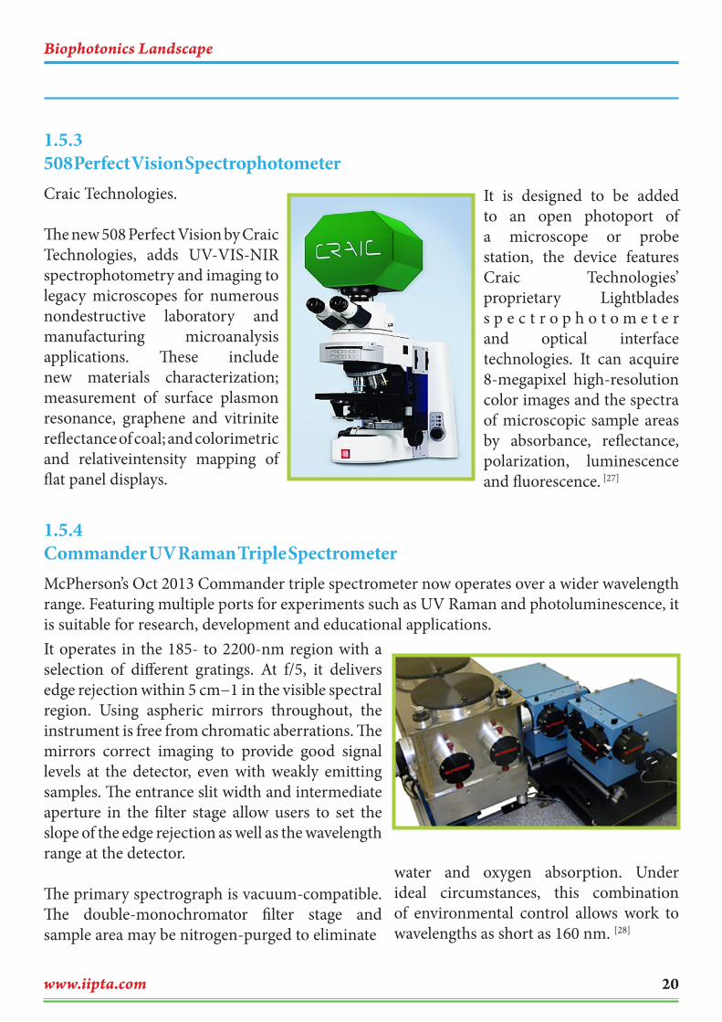
Craic Technologies.
The new 508 Perfect Vision by Craic Technologies, adds UV-VIS-NIR spectrophotometry and imaging to legacy microscopes for numerous nondestructive laboratory and manufacturing microanalysis applications. These include new materials characterization; measurement of surface plasmon resonance, graphene and vitrinite reflectance of coal; and colorimetric and relativeintensity mapping of flat panel displays.
It is designed to be added to an open photoport of a microscope or probe station, the device features Craic Technologies’ proprietary Lightblades s p e c t r o p h o t o m e t e r and optical interface technologies. It can acquire 8-megapixel high-resolution color images and the spectra of microscopic sample areas by absorbance, reflectance, polarization, luminescence and fluorescence. [27]
1.5.3508 Perfect Vision Spectrophotometer
McPherson’s Oct 2013 Commander triple spectrometer now operates over a wider wavelength range. Featuring multiple ports for experiments such as UV Raman and photoluminescence, it is suitable for research, development and educational applications.
1.5.4 Commander UV Raman Triple Spectrometer
It operates in the 185- to 2200-nm region with a selection of different gratings. At f/5, it delivers edge rejection within 5 cm−1 in the visible spectral region. Using aspheric mirrors throughout, the instrument is free from chromatic aberrations. The mirrors correct imaging to provide good signal levels at the detector, even with weakly emitting samples. The entrance slit width and intermediate aperture in the filter stage allow users to set the slope of the edge rejection as well as the wavelength range at the detector.
The primary spectrograph is vacuum-compatible. The double-monochromator filter stage and sample area may be nitrogen-purged to eliminate
water and oxygen absorption. Under ideal circumstances, this combination of environmental control allows work to wavelengths as short as 160 nm. [28]

Biophotonics Landscape
www.iipta.com21
Excelitas Technologies has added two new models to its line of silicon photomultipliers (SiPM) for low-light level detection. The C30742-11-050 and C30742-66-050 are suitable for positron
1.5.5Silicon Photomultipliers
emission tomography (PET) and high-energy physics and for analytical measurements for fluorescence and fluorescence lifetime detection. Available in 1 × 1 mm (C30742-11-050) and 6 × 6 mm (C30742-66-050) chip areas, the latest SiPMs provide additional options for OEM applications that require high photon detection efficiency. Both solutions are based on proprietary avalanche photodiode
processes that offer low timing resolution, extremely low dark count and low crosstalk for demanding high-volume applications such as molecular imaging and radiation detection.
The SiPMs were designed for photon detection in the 350- to 850-nm range and offer an alternative to photomultiplier tubes for situations where
immunity to electromagnetic fields is crucial. With a large operating voltage range (5-10 V), the 1 × 1 mm and 6 × 6 mm SiPM are available
on either ceramic mount or patented leadless laminated carrier surface mount to meet a variety of requirements. [29]
The LA series planar diffraction gratings by Shimadzu Corp. are designed for laser systems and are suitable for analytical instruments for research in the life sciences, chemistry and environmental monitoring, and for synchrotron radiation and optical communications applications.
1.5.6 LA Series Planar Diffraction Gratings
Designed for the 633- to 1064-nm waveband with 1600 lines per millimeter, they exhibit a diffraction efficiency of 98%.
This feature is important for expansion and compression of laser pulses for light sources used in laser processing. The
devices offer an efficiency of more than 90% for near-IR wavelength laser compressor applications. [30]
Biophotonics Landscape
www.iipta.com 22
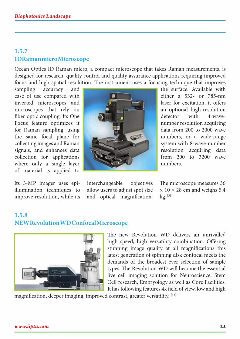
sampling accuracy and ease of use compared with inverted microscopes and microscopes that rely on fiber optic coupling. Its One Focus feature optimizes it for Raman sampling, using the same focal plane for collecting images and Raman signals, and enhances data collection for applications where only a single layer of material is applied to
the surface. Available with either a 532- or 785-nm laser for excitation, it offers an optional high-resolution detector with 4-wave-number resolution acquiring data from 200 to 2000 wave numbers, or a wide-range system with 8-wave-number resolution acquiring data from 200 to 3200 wave numbers.
1.5.7 IDRaman micro Microscope
Its 3-MP imager uses epi-illumination techniques to improve resolution, while its
interchangeable objectives allow users to adjust spot size and optical magnification.
The microscope measures 36 × 10 × 28 cm and weighs 5.4 kg. [31]
Ocean Optics ID Raman micro, a compact microscope that takes Raman measurements, is designed for research, quality control and quality assurance applications requiring improved focus and high spatial resolution. The instrument uses a focusing technique that improves
1.5.8 NEW Revolution WD Confocal Microscope
The new Revolution WD delivers an unrivalled high speed, high versatility combination. Offering stunning image quality at all magnifications this latest generation of spinning disk confocal meets the demands of the broadest ever selection of sample types. The Revolution WD will become the essential live cell imaging solution for Neuroscience, Stem Cell research, Embryology as well as Core Facilities. It has following features 4x field of view, low and high
magnification, deeper imaging, improved contrast, greater versatility. [32]

Biophotonics Landscape
www.iipta.com23
Carl Zeiss Microscopy GmbH, Optical Sensor Systems
Carl Zeiss Group’s next-generation EVO series scanning electron microscopes (SEMs) deliver work-flow automation, beam-deceleration technology and high-definition imaging for
materials and life sciences applications. Work flow is reduced from more than 400 steps to 15, enabling automated settings such as beam alignment, magnification and focus to provide imaging of areas of interest in the shortest possible time. A new midcolumn click-stop aperture changer offers reproducibility.
1.5.9 EVO Series SEMs
The beam deceleration technology and high-definition BSE (backscatter electron) detector provide fine topographical and image detail. It benefit from full environmental capabilities to observe nano
scale interactions of life science samples at different temperatures, pressures and humidities. The unique X-ray geometry of EVO gives highest resolution performance at analytical working conditions. There
are three different chamber sizes accept a wide range of specimen sizes for imaging. It uses the high definition beam source technology of EVO HD to further increase image contrast and resolution. [33]
Biophotonics Landscape
www.iipta.com 24
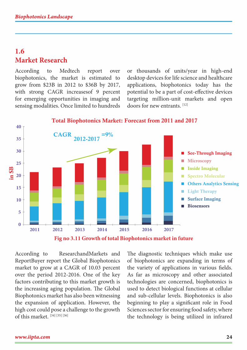
According to Medtech report over biophotonics, the market is estimated to grow from $23B in 2012 to $36B by 2017, with strong CAGR increasesof 9 percent for emerging opportunities in imaging and sensing modalities. Once limited to hundreds
or thousands of units/year in high-end desktop devices for life science and healthcare applications, biophotonics today has the potential to be a part of cost-effective devices targeting million-unit markets and open doors for new entrants. [12]
According to ResearchandMarkets and ReportBuyer report the Global Biophotonics market to grow at a CAGR of 10.03 percent over the period 2012-2016. One of the key factors contributing to this market growth is the increasing aging population. The Global Biophotonics market has also been witnessing the expansion of application. However, the high cost could pose a challenge to the growth of this market. [34] [35] [36]
The diagnostic techniques which make use of biophotonics are expanding in terms of the variety of applications in various fields. As far as microscopy and other associated technologies are concerned, biophotonics is used to detect biological functions at cellular and sub-cellular levels. Biophotonics is also beginning to play a significant role in Food Sciences sector for ensuring food safety, where the technology is being utilized in infrared
1.6 Market Research
Fig no 3.11 Growth of total Biophotonics market in future
Total Biophotonics Market: Forecast from 2011 and 2017
See-Through Imaging
Microscopy
Inside Imaging
Spectro Molecular
Others Analytics Sensing
Light Therapy
Surface Imaging
Biosensors
2011
40
35
30
25
20in S
B
15
10
5
02012 2013 2014 2015 2016 2017
CAGR =9%2012-2017

Biophotonics Landscape
www.iipta.com25
and mass spectroscopy to detect pollutants in foodstuffs. Biophotonics-based imaging technology is also finding increasing use in various fields of study, including nanobiotechnology and cell biology. Further, it is expected in the near future that biopsies and existing mechanical scanning equipment will be replaced by biomedical imaging.
According to BioOptics World report Global biophotonics market should exceed $99B by 2018 Aging population and demand for quality healthcare across the world are mentioned as major drivers, as biophotonics can enable cost-effective medical diagnostic and therapeutic tools that facilitate faster detection and optimum treatment of critical illnesses. To that end, biophotonic applications in microscopy, cytometry, and mass spectroscopy are expected to gain traction over traditional diagnostic techniques. Demographic aging as well as the growing demand for quality healthcare across the planet are expected to drive the market for biophotonics in the near term. Over the last few years, demand for semiconductor lasers for laser therapy applications witnessed an upsurge, with applications expanding from aesthetic treatments to surgeries, skin cancer therapy, and pain-relief treatments. In the near future, biomedical imaging is expected to substitute biopsies and existing mechanical scanning equipment. [37]
In recent times, photodynamic therapy (PDT) has been widely accepted as an effective medium for palliative care and for treating cancer during early stages. Several cancer-
detection technologies already make use of photonic and similar optical technologies such as the use of gold markers derived from nanoshells. Through the integration of imaging modalities, namely ultrasound, photoacoustic imaging, and optical coherence tomography (OCT), into a single 5 mm biophotonics device, researchers can now detect presence of ovarian cancer among high-risk women at early stages. In case of timely detection of debilitating conditions like heart diseases, biophotonics comes into the picture, aiding the development of advanced laser mass spectroscopy-based tests that can provide conclusive results within moments of the onset of a heart attack. Presently focused on microscopy and other associated technologies, biophotonics are used for detecting biological functions at cellular and sub-cellular levels. Additionally, new technologies are gradually emerging on the basis of electrophysiology and other probes for quantifying biological functionalities. Biophotonics is also beginning to play a vital role in the food sciences sector for ensuring food safety, where the technology is being deployed in infrared (IR) and mass spectroscopy to detect contaminants in foodstuffs. Biophotonics-based imaging technology is also finding increasing application in diverse fields of study, including nano-biotech, cell biology, cell or animal green fluorescent protein (GFP), and nonlinear microscopy. Live-cell imaging, involving the study of protein expression, protein-to-protein interactions, and localization, as well as transgenics for channel photoactivation is some of the most vibrant end-use application segments of biophotonics.

Biophotonics Landscape
www.iipta.com 26
The U.S. represents the single largest region for biophotonics technologies, capturing a sizeable chunk of the global market, as stated by the GIA report. Asia-Pacific, powered by emerging markets of India and China, is poised to grow at an impressive pace of over 23% through 2018. By end-use, the medical applications market represents a major segment for biophotonics, primarily
in the field of diagnostics and therapeutics. Developed regions of North America and Europe comprise the largest markets for biophotonics medical applications. Non-medical applications of biophotonics are currently the subject of intense research and are expected to gain significant mileage over the ensuing years. [37]
Biophotonics Landscape
www.iipta.com27
The first step of patent analysis was to retrieve the patents filed related to biophotonics from year 2008 to 2013. Patents were retrieved from freely available database- (WIPO), USPTO, EPO, etc. The patents were found from the different keywords mentioned in title, abstract, claims and descriptions.
From the literature review, we have found several keywords segregating the topic into several sections in biophotonics applications.
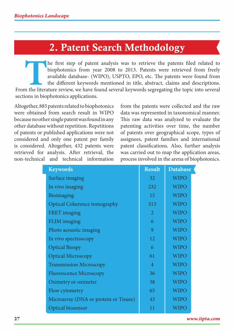
Altogether, 885 patents related to biophotonics were obtained from search result in WIPO because no other single patent was found in any other database without repetition. Repetitions of patents or published applications were not considered and only one patent per family is considered. Altogether, 432 patents were retrieved for analysis. After retrieval, the non-technical and technical information
from the patents were collected and the raw data was represented in taxonomical manner. This raw data was analyzed to evaluate the patenting activities over time, the number of patents over geographical scope, types of assignees, patent families and international patent classifications. Also, further analysis was carried out to map the application areas, process involved in the arena of biophotonics.
KeywordsSurface imagingIn vivo imagingBioimagingOptical Coherence tomography FRET imagingFLIM imagingPhoto acoustic imagingIn vivo spectroscopy Optical BiospyOptical MicroscopyTransmission MicroscopyFluorescence Microscopy Oximetry or oximeterFlow cytometryMicroarray (DNA or protein or Tisuee)Optical biosensor
Result32
23215
313269
126
614
3638654311
DatabaseWIPOWIPOWIPOWIPOWIPOWIPOWIPOWIPOWIPOWIPOWIPOWIPOWIPOWIPOWIPOWIPO
2. Patent Search Methodology
Biophotonics Landscape
www.iipta.com 28
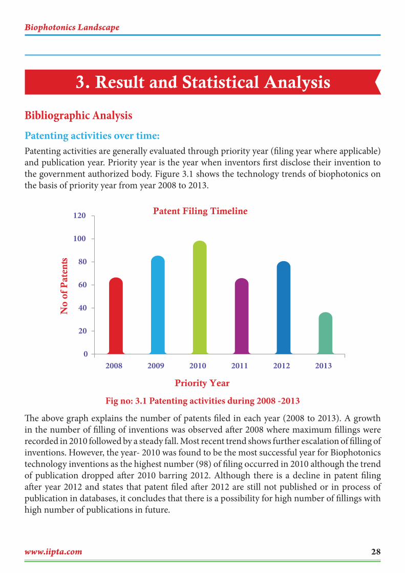
Bibliographic AnalysisPatenting activities over time: Patenting activities are generally evaluated through priority year (filing year where applicable) and publication year. Priority year is the year when inventors first disclose their invention to the government authorized body. Figure 3.1 shows the technology trends of biophotonics on the basis of priority year from year 2008 to 2013.
Fig no: 3.1 Patenting activities during 2008 -2013
The above graph explains the number of patents filed in each year (2008 to 2013). A growth in the number of filling of inventions was observed after 2008 where maximum fillings were recorded in 2010 followed by a steady fall. Most recent trend shows further escalation of filling of inventions. However, the year- 2010 was found to be the most successful year for Biophotonics technology inventions as the highest number (98) of filing occurred in 2010 although the trend of publication dropped after 2010 barring 2012. Although there is a decline in patent filing after year 2012 and states that patent filed after 2012 are still not published or in process of publication in databases, it concludes that there is a possibility for high number of fillings with high number of publications in future.
3. Result and Statistical Analysis
Priority Year
Patent Filing Timeline
0
20
40
60
80
100
120
2008 2009 2010 2011 2012 2013
No
of P
aten
ts

Biophotonics Landscape
www.iipta.com29
Number of patents by inventors: This analysis (Figure 3.2) highlights on the topmost inventors who contributed into Biophotonics innovation.
Fig no: 3.2 Top Inventors in Biophotonics Technology
Izatt Joseph is known for his extreme contribution (24) patents followed by Steward Lance and Nebosis Rainer (17), Indrevoll Bard (15), Tie Qiao (15), Jaffe Claudia (13). The nationality of maximum inventors is the United States.
No of Patents
Top 10 Inventors
05 10 15 20 25 30
Hayden Oliver
Leghton Stepan B
Bhalla Rajiv
Helou Michael Johannes
Tie Qiao
Indrevoll Bard
Nebosis Rainer
Steward Lance
Izatt Joseph A

Biophotonics Landscape
www.iipta.com 30
Top Patent applicants: The analysis of assignee or applicant who contributed to Biophtonics technology. The top applicants are mentioned below Fig no: 3.3
Applicant analysis shows that GE healthcare was the top applicant with maximum patents (98), and Olympus and Carl Zeisis shared almost similar involvements (80) with entire contributions from companies. Apart from them- University of Leland Stanford and Califronia Institute of Technology were among the top two university applicants with 13 and 11 patents respectively. Canon, Beckman Coulter, Affymetrix Inc, Allergen Inc, Omron companies made considerable contribution to the Biophotonics (Figure 3.3).
Fig no: 3.3 Top 10 Patent Applicants
Place of first filling: The place of first filling reflects the applicant’s preference to get patent protection in that specific territory considering the demand of his invention in the said territory. So, according to the place of applicant’s preference of first filling- the United States of America is the leading country (136) followed by World Intellectual Property Organization (80), China (68), European Patent Organization (50), Japan (46), South Korea (24), Canada (10) and other (18) respectively (Figure 3.4).
California Institute of technology
University of Leland Stanford
Omron
Allergen INC
Beckman Coulter
Canon
Carl Zeisis
Olympus
GE Healthcare
01 02 03 04 05 06 07 08 09 0 100
No of Patents
Top 10 Patent Applicants

Biophotonics Landscape
www.iipta.com31
Fig no 3.4: Place of first filling vs. number of patents
Patent distribution under IPCs: An invention must belong to an international patent classification (IPC). This section analyzes the number of patents in each class (Figure 3.5)
Fig no: 3.5 Patent distributions under IPCs
0
20
40
60
80
100
120
A61B G01N G01B A61K C12Q G02B C40B C12M CO7BB 82B
No
of P
aten
ts
IPC Classification
Top IPC Classification
Place of firt filing of Patents
31%
18%16%
12%
11%
6%
2%
4%
US
WIPO
China
Europe
Japan
South Korea
Canada
Others
Biophotonics Landscape
www.iipta.com 32
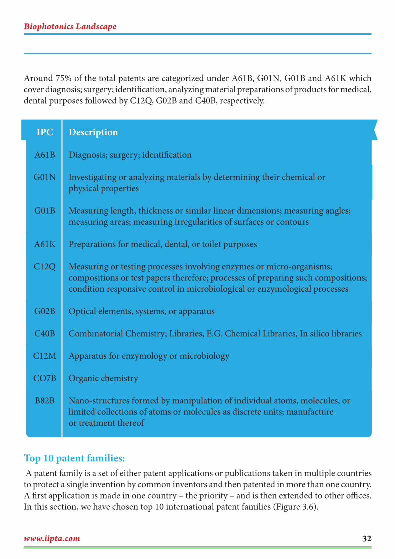
Around 75% of the total patents are categorized under A61B, G01N, G01B and A61K which cover diagnosis; surgery; identification, analyzing material preparations of products for medical, dental purposes followed by C12Q, G02B and C40B, respectively.
Top 10 patent families: A patent family is a set of either patent applications or publications taken in multiple countries to protect a single invention by common inventors and then patented in more than one country. A first application is made in one country – the priority – and is then extended to other offices. In this section, we have chosen top 10 international patent families (Figure 3.6).
IPC
A61B
G01N
G01B
A61K
C12Q
G02B
C40B
C12M
CO7B
B82B
Description
Diagnosis; surgery; identification
Investigating or analyzing materials by determining their chemical or physical properties
Measuring length, thickness or similar linear dimensions; measuring angles; measuring areas; measuring irregularities of surfaces or contours
Preparations for medical, dental, or toilet purposes
Measuring or testing processes involving enzymes or micro-organisms; compositions or test papers therefore; processes of preparing such compositions; condition responsive control in microbiological or enzymological processes
Optical elements, systems, or apparatus
Combinatorial Chemistry; Libraries, E.G. Chemical Libraries, In silico libraries
Apparatus for enzymology or microbiology
Organic chemistry
Nano-structures formed by manipulation of individual atoms, molecules, or limited collections of atoms or molecules as discrete units; manufacture or treatment thereof

Biophotonics Landscape
www.iipta.com33
Fig no: 3.6 Top patent Families
Top Patent families
US 20130258083 A1
JP 2008201786 A
US 20110066015 A1
US 20090117572 A1
US 20120200684 A1
JP 2009198511 A
US 20090005662 A1
Inventor
Iddan Gavriel J, Avni Dov, Glukhovsky, Arkady Meron Gavriel
Steward Lance E , Fernandez-salas Ester, Aoki K Roger
David Swedlow, Michael E. Fein, Marcia Fein, Paul D Mannheimer
Ester Fernandez-Salas, Lance E. Steward, Kei Roger Aoki
Arkady Glukhovsky, Gavriel Meron, Doron Adler, Ofra Zinati, Jerome Avron
Malachowski George C
Petersen Ethan Shea William Chew Bradford B
Assignee
Given Imaging Ltd
Allergen Inc
Nellcor Puritan Bennett Inc.
Allergan, Inc.
Given Imaging Ltd
Beckman Coulter Inc
Nellcor Puritan Bennett Inc
Top Patent Families
17%
18%
17%14%
13%
11%
10%
US 2013/0258083 A1
JP 2008201786 A
US 2011/0066015 A1
US 2009/0117572 A1
US 2012/0200684 A1
JP 2009198511 A
US 2009/0005662 A1
Biophotonics Landscape
www.iipta.com 34
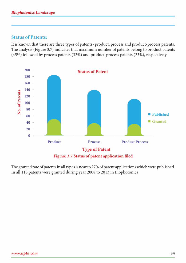
Status of Patents: It is known that there are three types of patents- product, process and product-process patents. The analysis (Figure 3.7) indicates that maximum number of patents belong to product patents (45%) followed by process patents (32%) and product-process patents (23%), respectively.
Fig no: 3.7 Status of patent application filed
The granted rate of patents in all types is near to 27% of patent applications which were published. In all 118 patents were granted during year 2008 to 2013 in Biophotonics
0
20
40
60
80
100
120
140
160
180
200
Product Process Product Process
No.
of
Pat
ents
Type of Patent
Status of Patent
Published
Granted

Biophotonics Landscape
www.iipta.com35
Technical Analysis
Fig no: 3.8 Applications area of Bio photonics
Fig 3.9 Components used in Biophotonics Patents
Biophotonics Landscape
www.iipta.com 36
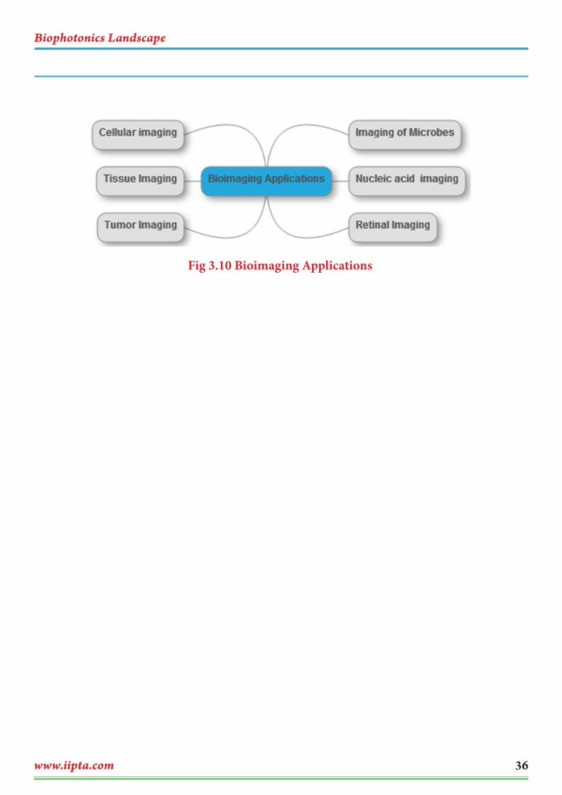
Fig 3.10 Bioimaging Applications

Biophotonics Landscape
www.iipta.com37
Strength
Optical transmission: In Biophotonics most of the techniques depend upon the optical transmission for examination. In optical transmission light passes through opaque medium containing sample.[4]
Dental: Biophotonics is mostly used in the dental sector for hardening and deep penetration of ceramic filling and other materials used for filling. The technique is also used for deep filling the cavities and drug during implants.[2]
Analytics: The technique has been used for analytical instruments as a detector unit/device. The light passes through the sample and gives a peak for a particular compound. The biophotonics technique used in IR Spectroscopy, UV Spectroscopy, Raman Spectroscopy, HPLC spectroscopy, etc.
Microscopic techniques: The biophotonics technology is used mostly for microscopic observation. This can be used as a detection source which only indicates spectrum of light for different samples.
Diagnostics: Laser-induced fluorescence is used for early detection of cancer in the gastrointestinal track and the lung. [13]
Strong and diverse biophotonics research base especially in areas such as diagnostics and applications of advanced imaging techniques
Large number of laser manufactures exporting significant system numbers for biophotonics applications
Increased precision from photonic based diagnostic and treatment methods
Strong penetration of photonics tools into life science research extensively supported by international suppliers with UK operations
Significant number of biochemistry and biophotonics research collaborations with strong support for cross-disciplinary projects from both physical and medical science funding bodies.
Weakness
Diversity of applications leading to fragmentation of development. Very less work is done over biophotonics because of limited use and of people not being aware of biophotonics.
Poor engagement with pharmaceutical industry / NHS / clinicians / medical equipment providers
Perceived higher cost of photonics based diagnostic solutions
Limited number of biophotonics industries dedicated to commercializing new advances (although those that exist are growing rapidly and visibility is not optimal)
The technique has limited use in biological as well as non-biological applications.
4. Swot Analysis
Biophotonics Landscape
www.iipta.com 38
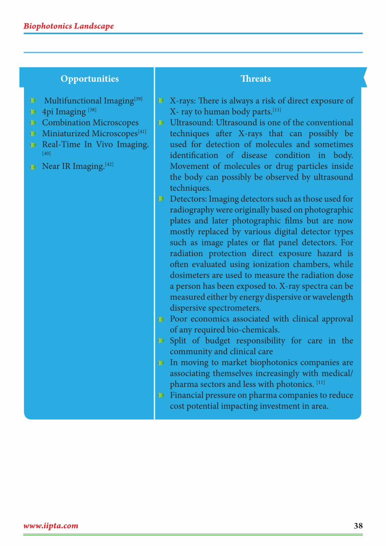
Opportunities
Multifunctional Imaging[39]
4pi Imaging [38]
Combination Microscopes Miniaturized Microscopes[41]
Real-Time In Vivo Imaging.[40]
Near IR Imaging.[42]
Threats
X-rays: There is always a risk of direct exposure of X- ray to human body parts.[11]
Ultrasound: Ultrasound is one of the conventional techniques after X-rays that can possibly be used for detection of molecules and sometimes identification of disease condition in body. Movement of molecules or drug particles inside the body can possibly be observed by ultrasound techniques.
Detectors: Imaging detectors such as those used for radiography were originally based on photographic plates and later photographic films but are now mostly replaced by various digital detector types such as image plates or flat panel detectors. For radiation protection direct exposure hazard is often evaluated using ionization chambers, while dosimeters are used to measure the radiation dose a person has been exposed to. X-ray spectra can be measured either by energy dispersive or wavelength dispersive spectrometers.
Poor economics associated with clinical approval of any required bio-chemicals.
Split of budget responsibility for care in the community and clinical care
In moving to market biophotonics companies are associating themselves increasingly with medical/ pharma sectors and less with photonics. [11]
Financial pressure on pharma companies to reduce cost potential impacting investment in area.
Biophotonics Landscape
www.iipta.com39
5. Competitive Intelligence
There are many companies which are carrying out their research programs in the field of biophotonics. But still they are fewer who filed and/or granted patents for their technologies. This is an emerging field of Life science and a limited number
of companies are carrying research in this field. That is, there is relatively less competitions among companies. They are as follows:
1. AffymzetrixInc.Industry Type: BioinformaticsAffymetrix, Inc. is an American company that manufactures DNA microarrays; it is based in Santa Clara, California, United States. AffymetrixInc was founded in year 1992 on basis of GeneChip® technology using semiconductor manufacturing techniques. Affymetrix manufactures its GeneChips using photolithography. The company also manufactures machinery for high speed analysis of biological samples. The Company works on has vast range of patent portfolio in Biophotonics. [16]
Affymetrix has wide range of solutions in following fields; Microarray Solutions, Panomics Quantitative Assays, Molecular Biology Kits and Reagents, PCR, Molecular Biology Enzymes, Biochemical, Purification, Immunology Reagents, Detergents and Lipids. Affymetrix has about 1,100 employees worldwide and maintains sales and distribution operations across Asia, Europe, Latin America, and North America
AcquisitionsPanomics, Inc. In November 2008, Affymetrix acquired Panomics Inc., a privately held Fremont, California-based company that offers a powerful suite of assay products for
a wide variety of low- to mid-plex genetic, protein, and cellular analysis applications.
True Materials Inc. Affymetrix acquired True Materials Inc., a privately held San Francisco-based company that has developed a technology capable of multiplexing up to 10,000 markers, in July 2008.
USB Corporation Affymetrix acquired USB Corporation, a privately held Cleveland, Ohio-based company that provides an extensive line of molecular biology and biochemical reagent products, in December 2007. The acquisition included Anatrace, Inc., a wholly owned subsidiary that provides membrane protein extraction and purification products.
ParAllele BioScience Inc. Affymetrix acquired ParAllele BioScience, Inc., a privately held provider of highly scalable technology for comprehensive genetic studies, in October 2005.
Neomorphic, Inc. Affymetrix acquired Neomorphic, Inc., a privately held computational genomics company, in October 2000.
Genetic MicroSystems, Inc. Affymetrix acquired Genetic MicroSystems, Inc., a privately held Massachusetts instrumentation company specializing in DNA array technology, in February 2000.

Biophotonics Landscape
www.iipta.com 40
2. Allergan Inc.Industry type: PharmaceuticalsAllergan, Inc. is a multi-specialty health care company focused on discovering, developing and commercializing innovative pharmaceuticals, biologics, medical devices and over-the-counter consumer products. Allergen was founded in year 1948. The head quarters of the company are located in Irvine, CA, United States of America. Their product ranges include ophthalmic pharmaceuticals, dermatology products, and neurological products. The financial performance of the company is $1,391.1 million total product net sales- 6.1 % compared to total product net sales in 2011; revenue $ 5.7 billion (in 201) with a growth of 9% than 2011.[17]
3. Beckman CoulterIndustry type: Diagnostics and analyticsBeckman Coulter Inc, is an American company that makes biomedical laboratory instruments. Founded by Caltech professor Arnold O. Beckman in 1935 as National Technical Laboratories to commercialize a pH meter that he had invented, the company eventually grew to employ over 10,000 people, with $2.4 billion in annual sales by 2004. Its current headquarters are in Brea, California. The company mainly operates on Poteintiometer and spectroscopy business.[18]
4. Carl Zeiss AGInusdtry Type: Optics and Optoelectronics IndustryCarl Zeiss AG is a German manufacturer of optical systems, industrial measurements and
medical devices, founded in Jena, Germany in 1846 by optician Carl Zeiss. The company is headquartered in Oberkochen, Germany. The company product comprises of various optical and vision technology field like camera lens, microscope lens, ophthalmology products binoculars and optics for military applications. Carl Zeiss AG’s revenue is generated by its Semiconductor Manufacturing Technologies division, which produces lithographic systems for the semiconductor industry as well as process control solutions (electron microscopes, mask repair tools, helium ion microscopes) [19]
Carl Zeiss is present in over 40 countries around the globe with 40 production facilities and 24,000 employees worldwide.
DivisionsSemiconductor Manufacturing Technology, Industrial Metrology, Microscopy, Medical Technology, Vision Care, Camera Lenses, Sports Optics, Planetariums
5. GE HealthcaresIndustry type: Diagnostics and analyticsGE Healthcare currently has 7 primary business units:
GE Healthcare Global Diagnostic Imaging, headquartered in Pewaukee (near Milwaukee), Wisconsin, USA. The Diagnostic Imaging business includes X-ray, digital mammography, computed tomography (CT), magnetic resonance (MR) and molecular imaging technologies. GE Healthcare Clinical Systems, headquartered in Wauwatosa (suburb of Milwaukee), Wisconsin, USA. This business provides a range of healthcare
Biophotonics Landscape
www.iipta.com41
technologies and services for clinicians and healthcare administrators. It includes ultrasound, ECG, bone densitometry, patient monitoring, incubators and infant warmers, respiratory care and anesthesia management.[20]
GE Healthcare IT, headquartered in Barrington, Illinois, USA. Healthcare IT provides clinical & financial information technology solutions such as departmental IT products, RIS/PACS (Radiology Information Systems/Picture Archiving and Communication Systems) and CVIS (Cardiovascular Information Systems), as well as revenue cycle management and practice applications.
GE Healthcare Medical Diagnostics, headquartered in Little Chalfont, Buckinghamshire, UK. Medical Diagnostics researches, manufactures and markets imaging agents used during medical scanning procedures to highlight organs, tissue and functions inside the human body.
GE Healthcare Life Sciences, headquartered in Uppsala, Sweden. This division produces technology for drug discovery, biopharmaceutical manufacturing and cellular technologies. It also makes systems and equipment for the purification of biopharmaceuticals. The Life Sciences business unit is led by Kieran Murphy.
GE Healthcare Surgery, headquartered in Salt Lake City, Utah, USA. This provides tools and technologies for cardiac, surgical and interventional care, from cardiac catheterization labs, diagnostic monitoring systems, data management systems to mobile
fluoroscopic imaging systems, navigation and 3D visualization instrumentation.
6. Olympus CorporationIndustry type: Electronics and opticsOlympus Corporation is a Japan based manufacturer of optic and reprography products. Olympus was established on 12 October 1919, initially specializing in microscope and thermometer businesses. Olympus enjoys a majority share of the world market in gastro-intestinal endoscopes. Olympus’s main business fields are medical Life sciences, industrial, camera and audio, research & development and Olympus PR movie.[21]
7. OmronIndustry type: DiagnosticsOmron Corporation is Japanese based multinational electronics company incorporated in 1948. Omron’s primary business is the manufacture and sale of automation components, equipment and systems, but it is generally known for medical equipment such as digital thermometers, blood pressure monitors and nebulizers. Omron developed the world’s first electronic ticket gate, which was named as IEEE milestone in 2007 and was one of the first manufactures of automated teller machines (ATM) with magnetic stripe card readers. The company main business fields are Industrial Automation, Electronic components, automotive electronic components, Social systems and healthcare business.[22]
Biophotonics Landscape
www.iipta.com 42
The innovation on Biophotonics technology started in the 21st century. Maximum number of fillings (98) was recorded in 2010 followed by a steady decline up to 2013 although a probability of higher number of fillings is expected in future. As
mentioned in Medtech report on biophotonics market, new technologies based on photon interaction with tissues will expand the biophotonics market from $23B in 2012 to $36B by 2017.The main application areas of biophotonics are diagnostics, therapeutics, Bio Imaging, Analytics and R&D. For obtaining patents different keywords have been framed that represent the application technique of biophotonics. From the bibliographic analysis Izatt Joseph A, Steward Lance, Nebosis Rainer, Indrevoll Bard are the inventors who applied for maximum number of patents/patent applications for their inventions. These inventors work over optical transmission, microarray, cytometry, in vivo imaging, and photon operating device, optical information, and transmission of optical signals.
Applicant analysis show that GE healthcare was the top applicant with maximum patents (98), and Olympus and Carl Zeisis shared almost
similar involvements (80) with entire contributions from their respective companies. Apart from them- University of Leland Stanford and Califronia Institute of Technology were among the top two university applicants. It has been shown that these are the global players who file maximum number of inventions over biophotonics and related applications. Timeline of patenting activities show maximum patents were filed in the year 2010, followed by decline in patenting activity. IPC (International patent classification) A61B, G01N, G01B, A61K are the IPC codes which were maximally used for filing the patent applications. This shows that biophotonics has a vast range of applications. The granted patents seem to be around 98 and published patents around 432 in the last 5 years.
From SWOT analysis the major strengths of biophotonics techniques are optical transmission, analytics, and microscopic techniques. Few are the technologies in which biophotonics technique is currently used. Major weaknesses of biophotonics are instruments; the devices are too costly which restrict its use to explore all market segments. The bulky instruments in diagnostics need to be small and portable. The threat is that biophotonics is most widely used only in dental sector, the technique can possibly be used in detection of diseases but it lacks usage in other fields due to the dearth of work over biophotonics in other fields. Major opportunities are in real time imaging, portable microscopy, 4pi imaging. Major threats are x-rays, ultrasounds and
6. Discussions
Biophotonics Landscape
www.iipta.com43
detectors. Scanning electron microscopy (SEM) and fluoresence microscopy are the major technology trends which can possibly be used in biophotonics for detection and analysis. The imaging techniques in biophotonics will see huge revolution in the upcoming years. The competitive intelligence shows that all the big global industries are mainly active in biophotonics technology. They are GE Healthcare, Canon, Afferymetrix, Omron, Carl Zeiss, and Olympus etc. These companies operate on wide range of applications of
biophotonics like imaging, diagnostic, analytics etc.
Overall development in biophotonics technology seems to be more promising in healthcare, food and medical research areas for future prospects. The innovation in biophotonics seems to be improved from previous heavy and bulky technology of earlier method of diagnostics. The involvement of electronics in these fields plays an important role in making portable size of biophotonics instruments and devices. Still biophotonics
needs lots of innovations to make applications of biophotonics technology easier and simpler so that the technology is affordable and widely available. Today, healthcare is moving from a treatment-oriented system to a diagnostic-oriented one, which proves that diagnostic applications of biophotonics are the driving force for innovation in optical technology. The end-goal being companion diagnostic which is promising but still far away.
Biophotonics Landscape
www.iipta.com 44
Prasad, Paras N. [2003] Introduction to biophotonics. John Wiley & Sons, Inc., Hoboken, New Jersey
Anil Kishen & Anand Asundi [2006] Fundamentals and Applications of Biophotonics in Dentistry
http://www.salk.edu/biophotonics/about_biophotonics.php Last Access 31st December, 2013.
http://www.photonics.com Last Access 31st December, 2013.
ht tp : / / s c ience care ers . s c iencemag .org/care er_magaz ine/pre v ious_ issues /articles/2001_09_21/nodoi.15764833591429456823 Last Access 31st December, 2013
Susan Swapp, Scanning Electron Microscopy, University of Wyoming http://serc.carleton.edu/research_education/geochemsheets/techniques/SEM.html.Last Access 31st December, 2013.
George Rice, Fluorescent Microscopy http://serc.carleton.edu/microbelife/research_methods/microscopy/fluromic.html.Last Access 31st December, 2013.
Biophotonics Market, Focus on Life Sciences & Health Applications http://www.i-micronews.com/reports/Biophotonics-Market/4/360/. Last Access 31st December, 2013.
Lother Lilge [2001] Biophotonics Opportunities in Medicine and Biotech http://www.lambertinstruments.com/technologies/flim/fluorescence-lifetime-imaging-microscopy. Last Access 31st December, 2013.
http://www.lifetechnologies.com/in/en/home/references/molecular-probes-the-handbook/technical-notes-and-product-highlights/fluorescence-resonance-energy-transfer-fret.html .Last Access 31st December, 2013.
http://photonicsuk.org/2013/06/21/uk-photonics-swot-life-sciences-and-biophotonics.Last Access 31st December, 2013.
http://www.i-micronews.com/reports/Biophotonics-Market/4/360/ Last Access 31st December, 2013
7. References
Biophotonics Landscape
www.iipta.com45
http://www.microscopyu.com/articles/fluorescence/multiphoton/multiphotonintro.html Last Access 31st December, 2013
http://www.medscape.com/viewarticle/727980_6 Last Access 31st December, 2013
http://www.ccm.pitt.edu/sites/default/files/ebm/nejm_pulse_oximetry.pdf Last Access 31st December, 2013
www.affymetrix.com/ Last Access 31st December, 2013
http://www.allergan.com/index.htm Last Access 31st December, 2013
https://www.beckmancoulter.com/wsrportal/wsrportal.portal Last Access 31st December, 2013
http://corporate.zeiss.com/about/en_in/about-carl-zeiss.html Last Access 31st December, 2013
http://www3.gehealthcare.in/ Last Access 31st December, 2013
http://www.olympus-global.com/en/ Last Access 31st December, 2013
http://www.omron.com/ Last Access 31st December, 2013
http://serc.carleton.edu/research_education/geochemsheets/techniques/SEM.html Last Access 31st December, 2013
Jürgens etal (2008) Examining and sorting – new device concepts for the microscopy of living cells. Photonics Int., 2, 24–27.
http://www.spectroline.com/content/two-one-transilluminator-provides-both-ultraviolet-and-white-light-illumination-1#bi-o-vision
http://www.promega.com/products/cell-health-and-metabolism/oxidative-stress-assays/ros_glo-h2o2-assay/
http://www.elliotscientific.com/216-0-360-442/CRAIC-Spectrophotometers/
http://mcphersoninc.com/ramanspectroscopy/commander.htm
Biophotonics Landscape
www.iipta.com 46
http://www.excelitas.com/Pages/Product/Silicon-Photomultipliers.aspx
http://www.shimadzu.com/about/pressrelease/5iqj1d000001w58i.html
http://www.oceanoptics.com/idraman/micro.asp
http://www.andor.com/pdfs/literature/Andor_Revolution-XD_Brochure.pdf
https://microscopy.zeiss.com/microscopy/en_de/products/scanning-electron-microscopes/evo-life-science.html
http://www.researchandmarkets.com/research/2t7qgg/global
http://www.reportbuyer.com/pharma_healthcare/treatments/global_biophotonics_market_2012_2016.html
http://www.bharatbook.com/healthcare-market-research-reports/global-biophotonics-market-2012-2016.html
http://www.bioopticsworld.com/articles/2012/07/global-biophotonics-market-should-exceed-99b-by-2018-says-gia.html
http://zeiss-campus.magnet.fsu.edu/referencelibrary/superresolution/fourpi.html
http://onlinelibrary.wiley.com/doi/10.1002/wnan.72/abstract
http://www.ncbi.nlm.nih.gov/pubmed/22968700
http://www.nature.com/news/2011/110911/full/news.2011.528.html
http://www.scholarpedia.org/article/Near_infrared_imaging

Related Documents