International Journal of Molecular Sciences Review Biomineralization of Collagen-Based Materials for Hard Tissue Repair Le Yu 1 and Mei Wei 1,2, * Citation: Yu, L.; Wei, M. Biomineralization of Collagen-Based Materials for Hard Tissue Repair. Int. J. Mol. Sci. 2021, 22, 944. https:// doi.org/10.3390/ijms22020944 Received: 25 December 2020 Accepted: 16 January 2021 Published: 19 January 2021 Publisher’s Note: MDPI stays neutral with regard to jurisdictional claims in published maps and institutional affil- iations. Copyright: © 2021 by the authors. Licensee MDPI, Basel, Switzerland. This article is an open access article distributed under the terms and conditions of the Creative Commons Attribution (CC BY) license (https:// creativecommons.org/licenses/by/ 4.0/). 1 Department of Chemical and Biomolecular Engineering, Ohio University, Athens, OH 45701, USA; [email protected] 2 Department of Mechanical Engineering, Ohio University, Athens, OH 45701, USA * Correspondence: [email protected]; Tel.: +1-740-593-1474 Abstract: Hydroxyapatite (HA) reinforced collagen fibrils serve as the basic building blocks of natural bone and dentin. Mineralization of collagen fibrils play an essential role in ensuring the structural and mechanical functionalities of hard tissues such as bone and dentin. Biomineraliza- tion of collagen can be divided into intrafibrillar and extrafibrillar mineralization in terms of HA distribution relative to collagen fibrils. Intrafibrillar mineralization is termed when HA minerals are incorporated within the gap zone of collagen fibrils, while extrafibrillar mineralization refers to the minerals that are formed on the surface of collagen fibrils. However, the mechanisms re- sulting in these two types of mineralization still remain debatable. In this review, the evolution of both classical and non-classical biomineralization theories is summarized. Different intrafibrillar mineralization mechanisms, including polymer induced liquid precursor (PILP), capillary action, electrostatic attraction, size exclusion, Gibbs-Donnan equilibrium, and interfacial energy guided theories, are discussed. Exemplary strategies to induce biomimetic intrafibrillar mineralization using non-collagenous proteins (NCPs), polymer analogs, small molecules, and fluidic shear stress are discussed, and recent applications of mineralized collagen fibers for bone regeneration and dentin repair are included. Finally, conclusions are drawn on these proposed mechanisms, and the future trend of collagen-based materials for bone regeneration and tooth repair is speculated. Keywords: biomineralization; intrafibrillar; extrafibrillar; collagen; tissue engineering 1. Introduction Natural bone is an organic and inorganic composite constituting of mineralized collagen fibrils, which serves as the second level of its seven hierarchical levels. The inner dentin-pulp layer of human tooth has a similar mineralization profile to native bone [1]. The main inorganic component in these tissues is nanosized hydroxyapatite (HA; Ca 10 (PO 4 ) 6 (OH) 2 ). Its crystal structure ascribes to the hexagonal crystal system, which enables it with structural and compositional flexibility and stability. It allows for an abundant variety of ion substitutions involving divalent or trivalent cations for the Ca 2+ sites while anions for the phosphate and hydroxyl sites. For examples, carbonate- substituted (carbonated) hydroxyapatite forms the basic mineral component of hard tissues in vertebrates [2]; fluoride-substituted hydroxyapatite (fluorapatite) is found in the teeth of sharks and other fishes [3]. It is a common strategy to use transition metals, such as Mn, Fe, Co and Ni, to substitute Ca(I) (surrounded by phosphate), and Ca(II) (surrounded by hydroxyl) in the HA lattice to grant materials with magnetic properties to be used in many biomedical applications, such as drug delivery, diagnostic testing, and magnetic resonance imaging [4–6]. The main organic component in bone and dentin is type I collagen, which has a com- plex structure including four levels of hierarchy. The primary level is amino acid triplets consisting mostly of proline (Pro), hydroxyproline (Hyp), and glycyl (Gly), following the pattern of Gly-Pro-X or Gly-X-Hyp, where X can be any of other amino acid species [7]. Int. J. Mol. Sci. 2021, 22, 944. https://doi.org/10.3390/ijms22020944 https://www.mdpi.com/journal/ijms

Welcome message from author
This document is posted to help you gain knowledge. Please leave a comment to let me know what you think about it! Share it to your friends and learn new things together.
Transcript
International Journal of
Molecular Sciences
Review
Biomineralization of Collagen-Based Materials for HardTissue Repair
Le Yu 1 and Mei Wei 1,2,*
�����������������
Citation: Yu, L.; Wei, M.
Biomineralization of Collagen-Based
Materials for Hard Tissue Repair. Int.
J. Mol. Sci. 2021, 22, 944. https://
doi.org/10.3390/ijms22020944
Received: 25 December 2020
Accepted: 16 January 2021
Published: 19 January 2021
Publisher’s Note: MDPI stays neutral
with regard to jurisdictional claims in
published maps and institutional affil-
iations.
Copyright: © 2021 by the authors.
Licensee MDPI, Basel, Switzerland.
This article is an open access article
distributed under the terms and
conditions of the Creative Commons
Attribution (CC BY) license (https://
creativecommons.org/licenses/by/
4.0/).
1 Department of Chemical and Biomolecular Engineering, Ohio University, Athens, OH 45701, USA;[email protected]
2 Department of Mechanical Engineering, Ohio University, Athens, OH 45701, USA* Correspondence: [email protected]; Tel.: +1-740-593-1474
Abstract: Hydroxyapatite (HA) reinforced collagen fibrils serve as the basic building blocks ofnatural bone and dentin. Mineralization of collagen fibrils play an essential role in ensuring thestructural and mechanical functionalities of hard tissues such as bone and dentin. Biomineraliza-tion of collagen can be divided into intrafibrillar and extrafibrillar mineralization in terms of HAdistribution relative to collagen fibrils. Intrafibrillar mineralization is termed when HA mineralsare incorporated within the gap zone of collagen fibrils, while extrafibrillar mineralization refersto the minerals that are formed on the surface of collagen fibrils. However, the mechanisms re-sulting in these two types of mineralization still remain debatable. In this review, the evolution ofboth classical and non-classical biomineralization theories is summarized. Different intrafibrillarmineralization mechanisms, including polymer induced liquid precursor (PILP), capillary action,electrostatic attraction, size exclusion, Gibbs-Donnan equilibrium, and interfacial energy guidedtheories, are discussed. Exemplary strategies to induce biomimetic intrafibrillar mineralization usingnon-collagenous proteins (NCPs), polymer analogs, small molecules, and fluidic shear stress arediscussed, and recent applications of mineralized collagen fibers for bone regeneration and dentinrepair are included. Finally, conclusions are drawn on these proposed mechanisms, and the futuretrend of collagen-based materials for bone regeneration and tooth repair is speculated.
Keywords: biomineralization; intrafibrillar; extrafibrillar; collagen; tissue engineering
1. Introduction
Natural bone is an organic and inorganic composite constituting of mineralizedcollagen fibrils, which serves as the second level of its seven hierarchical levels. Theinner dentin-pulp layer of human tooth has a similar mineralization profile to nativebone [1]. The main inorganic component in these tissues is nanosized hydroxyapatite(HA; Ca10(PO4)6(OH)2). Its crystal structure ascribes to the hexagonal crystal system,which enables it with structural and compositional flexibility and stability. It allows foran abundant variety of ion substitutions involving divalent or trivalent cations for theCa2+ sites while anions for the phosphate and hydroxyl sites. For examples, carbonate-substituted (carbonated) hydroxyapatite forms the basic mineral component of hard tissuesin vertebrates [2]; fluoride-substituted hydroxyapatite (fluorapatite) is found in the teeth ofsharks and other fishes [3]. It is a common strategy to use transition metals, such as Mn,Fe, Co and Ni, to substitute Ca(I) (surrounded by phosphate), and Ca(II) (surrounded byhydroxyl) in the HA lattice to grant materials with magnetic properties to be used in manybiomedical applications, such as drug delivery, diagnostic testing, and magnetic resonanceimaging [4–6].
The main organic component in bone and dentin is type I collagen, which has a com-plex structure including four levels of hierarchy. The primary level is amino acid tripletsconsisting mostly of proline (Pro), hydroxyproline (Hyp), and glycyl (Gly), following thepattern of Gly-Pro-X or Gly-X-Hyp, where X can be any of other amino acid species [7].
Int. J. Mol. Sci. 2021, 22, 944. https://doi.org/10.3390/ijms22020944 https://www.mdpi.com/journal/ijms
Int. J. Mol. Sci. 2021, 22, 944 2 of 17
The amino acid triplets stack repeatedly to form its secondary level of structure [8]. Thetertiary level is the well-known triple helix consisting of three interconnected α-chains [9].The last hierarchy is in supermolecular level that forms collagen fibrils or fibers [10]. Thesecollagen fibrils then continue to self-assemble both linearly and laterally into a hydrogelnetwork under suitable physicochemical conditions [11]. As a naturally extracted polymerfrom extracellular matrix (ECM), collagen-based materials demonstrate excellent biocom-patibility, which supports cellular adhesion, proliferation, migration and differentiation.As a result, they have been widely used in many biomedical applications, such as drugdelivery, tissue engineering, wound healing, and cosmetic surgery [8,12,13].
Bone tissue engineering aims to repair or regenerate native bones in defective areasthat are beyond their self-healing capabilities with the aid of grafting materials, typicallyscaffolds, progenitor cells and/or growth factors. Thus, selection of scaffolds becomesthe key to the success of bone tissue engineering [14]. First, the material should be bio-compatible and not induce any adverse effects or toxicity to the host. Second, it shouldbe biodegradable to avoid secondary surgery after healing. Third, it should have inter-connected pores to support cell adhesion, vascularization and transportation of nutrientsand metabolic wastes. Finally, it should possess suitable mechanical properties to againstvarious stresses and maintain structural integrity [8,15]. Collagen-hydroxyapatite-based(Col-HA-based) biomimetic scaffolds that structurally and compositionally resemble thenatural bone have drawn extensive attention from researchers because of their excellentbiocompatibility, biodegradability, low antigenicity, and compositional and structural flex-ibility. In contrast to bone, dental hard tissues, such as the outmost enamel and innerdentin-pulp complex, cannot self-heal if damaged. The cause of dental caries is a dynamicprocess of imbalance between demineralization and remineralization [16]. The caries istypically filled with artificial materials, such as amalgam, ceramics and resin composites.However, these materials could also bring about side effects, including trauma to sounddental tissue, dentin hypersensitivity, and interface microleakage between the filling mate-rials and dental tissue [17]. Thus, biomimetic Col-HA-based materials provide a promisingalternative for accelerating remineralization of dentin for tooth defect repair at an earlystage of pathological conditions [18]. Overall, biomimetic mineralization of collagen be-comes an essential strategy for developing bioinspired materials for both orthopedic anddental applications.
Existing reviews about collagen-based materials have been focused on (1) the collagensources, such as bovine tendons, rat tails, and fish, (2) the physical forms and morphologies,such as hydrogels, fibers, microspheres, and scaffolds or (3) their applications, such astissue engineering, wound healing, and cosmetic surgery. Thus, there is a need to develop acomprehensive review on these mechanisms, especially to include the most recent progressof biomineralization mechanisms. In this review, both intrafibrillar and extrafibrillarmineralization mechanisms are discussed and strategies for biomimetic mineralization ofcollagen-based materials are reviewed, and their applications in bone tissue engineeringand tooth remineralization are covered. The future trend of mineralized collagen-basedmaterial in tissue engineering and dentin repair is speculated.
2. Biomineralization of Collagen
Ordered biomineralization holds the basis of structural organization and functionalityfor multiple organisms. In comparison, pathological mineralization leads to the formationof undesired biominerals, such as kidney stones and dental calculus [19]. It has been re-cently reported that deficiency of pyrophosphate, a known endogenous biomineralizationinhibitor, could result in abnormal calcification process such as ectopic vascular calcifica-tion in the form of hydroxyapatite crystals and thus increase the risk of cardiovasculardisease [20–22]. Developing an in-depth understanding of biomineralization mechanism(s)is therefore imperative for promoting the desired mineralization while minimizing theundesired ones. It will also shed light on synthetic chemistry to develop bioinspiredmaterials via biomimetic mineralization. As shown in Figure 1, the biomineralization of
Int. J. Mol. Sci. 2021, 22, 944 3 of 17
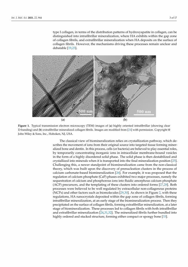
type I collagen, in terms of the distribution patterns of hydroxyapatite in collagen, can bedistinguished into intrafibrillar mineralization, where HA exhibits within the gap zoneof collagen fibrils, and extrafibrillar mineralization when HA deposits on the surface ofcollagen fibrils. However, the mechanisms driving these processes remain unclear anddebatable [19,23].
Int. J. Mol. Sci. 2021, 22, x FOR PEER REVIEW 3 of 17
ing the undesired ones. It will also shed light on synthetic chemistry to develop bioin-spired materials via biomimetic mineralization. As shown in Figure 1, the biomineraliza-tion of type I collagen, in terms of the distribution patterns of hydroxyapatite in collagen, can be distinguished into intrafibrillar mineralization, where HA exhibits within the gap zone of collagen fibrils, and extrafibrillar mineralization when HA deposits on the surface of collagen fibrils. However, the mechanisms driving these processes remain unclear and debatable [19,23].
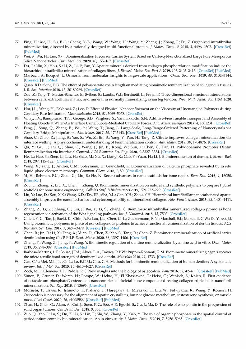
(a) (b)
Figure 1. Typical transmission electron microscopy (TEM) images of (a) highly oriented intrafibril-lar (showing clear D-banding) and (b) extrafibrillar mineralized collagen fibrils. Images are modi-fied from [24] with permission. Copyright @ John Wiley & Sons, Inc.
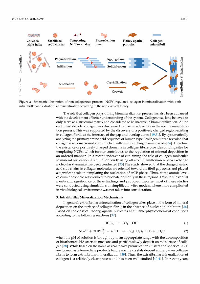
The classical view of biomineralization relies on crystallization pathway, which de-scribes the movement of ions from their original source into targeted tissue forming min-eralized bone and dentin. In this process, cells (or bacteria) are believed to play essential roles, by temporarily concentrating inorganic ions in intracellular membrane-bound ves-icles in the form of a highly disordered solid phase. The solid phase is then destabilized and crystallized into minerals when it is transported into the final mineralization position [25]. Challenging this, a newer standpoint of biomineralization came from the non-classi-cal theory, which was built upon the discovery of prenucleation clusters in the process of calcium carbonate-based biomineralization [26]. For example, it was proposed that the regulation of calcium phosphate (CaP) phases exhibited two major processes, namely the sequestration of calcium and phosphorous ions into fluidic amorphous calcium phos-phate (ACP) precursors, and the templating of these clusters into ordered forms [27,28]. Both processes were believed to be well regulated by extracellular non-collagenous pro-teins (NCPs) and other factors such as biomolecules [29,30]. As shown in Figure 2, with these regulations, HA nanocrystals deposited within the gap zone of collagen fibrils, form-ing intrafibrillar mineralization, at an early stage of the biomineralization process. Then they precipitated on the surface of collagen fibrils, forming extrafibrillar mineralization, at a later stage of biomineralization. These processes led to collagen fibrils with both in-trafibrillar and extrafibrillar mineralization [24,31,32]. The mineralized fibrils further bun-dled into highly ordered and stacked structure, forming either compact or spongy bone [33].
Figure 1. Typical transmission electron microscopy (TEM) images of (a) highly oriented intrafibrillar (showing clearD-banding) and (b) extrafibrillar mineralized collagen fibrils. Images are modified from [24] with permission. Copyright @John Wiley & Sons, Inc., Hoboken, NJ, USA.
The classical view of biomineralization relies on crystallization pathway, which de-scribes the movement of ions from their original source into targeted tissue forming miner-alized bone and dentin. In this process, cells (or bacteria) are believed to play essential roles,by temporarily concentrating inorganic ions in intracellular membrane-bound vesiclesin the form of a highly disordered solid phase. The solid phase is then destabilized andcrystallized into minerals when it is transported into the final mineralization position [25].Challenging this, a newer standpoint of biomineralization came from the non-classicaltheory, which was built upon the discovery of prenucleation clusters in the process ofcalcium carbonate-based biomineralization [26]. For example, it was proposed that theregulation of calcium phosphate (CaP) phases exhibited two major processes, namely thesequestration of calcium and phosphorous ions into fluidic amorphous calcium phosphate(ACP) precursors, and the templating of these clusters into ordered forms [27,28]. Bothprocesses were believed to be well regulated by extracellular non-collagenous proteins(NCPs) and other factors such as biomolecules [29,30]. As shown in Figure 2, with theseregulations, HA nanocrystals deposited within the gap zone of collagen fibrils, formingintrafibrillar mineralization, at an early stage of the biomineralization process. Then theyprecipitated on the surface of collagen fibrils, forming extrafibrillar mineralization, at a laterstage of biomineralization. These processes led to collagen fibrils with both intrafibrillarand extrafibrillar mineralization [24,31,32]. The mineralized fibrils further bundled intohighly ordered and stacked structure, forming either compact or spongy bone [33].
Int. J. Mol. Sci. 2021, 22, 944 4 of 17Int. J. Mol. Sci. 2021, 22, x FOR PEER REVIEW 4 of 17
Figure 2. Schematic illustration of non-collagenous proteins (NCPs)-regulated collagen biomineralization with both in-trafibrillar and extrafibrillar mineralization according to the non-classical theory.
The role that collagen plays during biomineralization process has also been advanced with the development of better understanding of the system. Collagen was long believed to only serve as a structural matrix and considered to be inactive in biomineralization. At the end of last decade, collagen was discovered to play an active role in the apatite min-eralization process. This was supported by the discovery of a positively charged region existing in collagen fibrils at the interface of the gap and overlap zones [19,31]. By system-atically analyzing the primary amino acid sequence of human type I collagen, it was re-vealed that collagen is a biomacromolecule enriched with multiple charged amino acids [34]. Therefore, the existence of positively charged domains in collagen fibrils provides binding sites for templating NCPs, which further contributes to the regulation of mineral deposition in an ordered manner. In a recent endeavor of explaining the role of collagen molecules in mineral nucleation, a simulation study using all-atom Hamiltonian replica exchange molecular dynamics has been conducted [35] The study showed that the charged amino acid side chains in collagen molecules are oriented toward the fibril gap zones and played a significant role in templating the nucleation of ACP phase. Thus, at the atomic level, calcium phosphate was verified to nucleate primarily in these regions. Despite substantial merits and significance of these findings and proposed theories, most of these studies were conducted using simulations or simplified in vitro models, where more complicated in vivo biological environment was not taken into consideration.
3. Intrafibrillar Mineralization Mechanisms In general, extrafibrillar mineralization of collagen takes place in the form of mineral
deposition on the surface of collagen fibrils in the absence of nucleation inhibitors [36]. Based on the classical theory, apatite nucleates at suitable physicochemical conditions ac-cording to the following reactions [37]: HCO → CO + OH (1) 5Ca + 3HPO + 4OH → Ca (PO ) (OH) + 3H O (2)
when the pH of solution is brought up to an appropriate range with the decomposition of bicarbonate, HA starts to nucleate, and particles slowly deposit on the surface of collagen [38]. While based on the non-classical theory, prenucleation clusters and spherical ACP are formed as intermediate products before apatite crystals deposit and grow on collagen fibrils to form extrafibrillar mineralization [39]. Thus, the extrafibrillar mineralization of collagen is a relatively clear process and has been well studied [40,41]. In recent years, extensive studies have been focused particularly on the exploration of the mechanisms of
Figure 2. Schematic illustration of non-collagenous proteins (NCPs)-regulated collagen biomineralization with bothintrafibrillar and extrafibrillar mineralization according to the non-classical theory.
The role that collagen plays during biomineralization process has also been advancedwith the development of better understanding of the system. Collagen was long believed toonly serve as a structural matrix and considered to be inactive in biomineralization. At theend of last decade, collagen was discovered to play an active role in the apatite mineraliza-tion process. This was supported by the discovery of a positively charged region existingin collagen fibrils at the interface of the gap and overlap zones [19,31]. By systematicallyanalyzing the primary amino acid sequence of human type I collagen, it was revealed thatcollagen is a biomacromolecule enriched with multiple charged amino acids [34]. Therefore,the existence of positively charged domains in collagen fibrils provides binding sites fortemplating NCPs, which further contributes to the regulation of mineral deposition inan ordered manner. In a recent endeavor of explaining the role of collagen moleculesin mineral nucleation, a simulation study using all-atom Hamiltonian replica exchangemolecular dynamics has been conducted [35] The study showed that the charged aminoacid side chains in collagen molecules are oriented toward the fibril gap zones and playeda significant role in templating the nucleation of ACP phase. Thus, at the atomic level,calcium phosphate was verified to nucleate primarily in these regions. Despite substantialmerits and significance of these findings and proposed theories, most of these studieswere conducted using simulations or simplified in vitro models, where more complicatedin vivo biological environment was not taken into consideration.
3. Intrafibrillar Mineralization Mechanisms
In general, extrafibrillar mineralization of collagen takes place in the form of mineraldeposition on the surface of collagen fibrils in the absence of nucleation inhibitors [36].Based on the classical theory, apatite nucleates at suitable physicochemical conditionsaccording to the following reactions [37]:
HCO−3 → CO2 + OH− (1)
5Ca2+ + 3HPO2−4 + 4OH− → Ca5(PO4)3(OH) + 3H2O (2)
when the pH of solution is brought up to an appropriate range with the decompositionof bicarbonate, HA starts to nucleate, and particles slowly deposit on the surface of colla-gen [38]. While based on the non-classical theory, prenucleation clusters and spherical ACPare formed as intermediate products before apatite crystals deposit and grow on collagenfibrils to form extrafibrillar mineralization [39]. Thus, the extrafibrillar mineralization ofcollagen is a relatively clear process and has been well studied [40,41]. In recent years,
Int. J. Mol. Sci. 2021, 22, 944 5 of 17
extensive studies have been focused particularly on the exploration of the mechanisms ofintrafibrillar mineralization of collagen, which were mostly built upon the classical and thenon-classical theory.
3.1. Capillary Action
Polymer induced liquid precursor (PILP) is considered to be one of the most importanttransient states before mineral infiltration into collagen fibrils and crystallization duringintrafibrillar mineralization. The term “PILP” was first introduced by Gower et al. [42,43]along with the discovery of liquid-liquid phase separation of amorphous calcium carbonate(ACC) in the presence of polyanionic polyaspartic acid (PASP) under optical microscopy.They proposed that during the PILP process, the charged polymer acted as a processing-directing agent, with the aid of which the conventional solution crystallization processwas converted into an amorphous liquid precursor-like process [44]. Using the formationof calcium carbonate and CaP based on PILP as examples, the dynamic compositionalevolution and interactions between the polymer and the amorphous precursor have beenrevealed recently with the aid of advanced analytical techniques, such as cryogenic trans-mission electron microscopy (cryo-TEM), in situ atomic force microscopy (AFM), nuclearmagnetic resonance (NMR), and modeling simulations [45–48]. A comprehensive under-standing of PILP theory along with its applications for bone regeneration has been reportedrecently [49]. The basis for the PILP theory is the liquid-liquid phase separation as liquid orliquid-like component can take flexible shape prior to solidification, which largely facilitatesthe infiltration of minerals into collagen fibrils to form intrafibrillar mineralization.
Upon the proposal of PILP, Gower et al. [50,51] opined subsequently that a liquidphase mineral precursor could be drawn into the gap spaces of collagen fibrils by capillaryaction in the PILP process before it was solidified and crystallized into minerals, formingintrafibrillar mineralized collagen fibrils that were embedded with nano crystallites. Theassumption was initially made based on their observations from capillary forces actingon the phase boundaries between the liquid precursor phase and collagen fibrils in acalcium carbonate system [50,52]. With the evolution of techniques and knowledge, theysubsequently confirmed their theory by duplicating similar observations via a combinationanalyses of transmission electron microscopy (TEM), scanning electron microscopy (SEM),confocal microscopy, and X-ray diffraction (XRD) in a CaP system that is more closelymimicking the nanostructure of natural bone [29,44,53]. Briefly, they found that PASP(processing-directing agent) incorporated with calcium ions could only penetrate intoturkey tendon for around 100 µm in comparison to PASP incorporated with CaP fluidicprecursor that had a penetration depth over 500 µm. With a smaller Stokes radius, PASP+ Ca would be expected to penetrate deeper into the dense-packed collagen scaffolds ofturkey tendon than PASP + CaP if they migrated through ion diffusion [29]. Therefore,they suggested that a capillary action might have been involved in intrafibrillar mineraliza-tion as capillary forces are expected to provide rapid and long-range transportation of afluidic precursor compared to ion diffusion. In this process, the fluidic nature of the ACPprecursors allowed them to be drawn into the nano gaps and grooves of collagen fibrils viacapillary infiltration [53,54]. However, whether capillary action is involved in intrafibrillarmineralization due to PILP is still debatable as this has not yet been directly observedthrough experiments. Exclusion of ion diffusion does not guarantee that capillary actionplays a role in the intrafibrillar mineralization process. Nevertheless, capillary action hasbeen regarded as one of the major mechanisms to date to induce intrafibrillar mineralizationof collagen, although other mechanisms may have also been involved [1,24,28,55–57].
3.2. Electrostatic Attraction
The phenomenon of electrostatic attraction in collagen mineralization has been recog-nized as early as last century [58]. Not until a decade ago that the role of collagen in boneapatite mineralization was convincingly revealed [31], had electrostatic attraction beenfully accepted as one of the major mechanisms in inducing intrafibrillar mineralization
Int. J. Mol. Sci. 2021, 22, 944 6 of 17
of collagen fibrils. With a combination of cryo-TEM, cryogenic electron tomography andmolecular modeling, Nudelman el at. [31] found that the positive net charge close to theC-terminal end of collagen molecules promotes the infiltration of ACP into collagen fibrils.Moreover, they revealed that the clusters of charged amino acids in the gap and overlapzones formed nucleation sites for apatite crystallization converted from ACP, giving a par-allel array of oriented apatite crystals to collagen fibrils. At the same time, they identifiedthe negative net charge of PASP stabilized ACP precursors. Based on these findings, theyopined that, in vivo, negatively charged NCPs might not only stabilize the amorphousphase by inhibiting apatite nucleation, but also play an active role in aiding the formationof negatively charged complexes of mineral precursors that allowed the minerals to enterthe collagen through electrostatic attraction [31,59]. The predominant role of collagen inapatite nucleation, growth, structure and orientation during bone mineralization was thor-oughly investigated in a subsequent study reported by Wang et al. [60]. They discoveredthat the collagen matrix controls the size and three-dimensional distribution of apatite,further supporting the mechanisms proposed by Nudelman et al. Thereafter, electrostaticattraction has become a popular mechanism and has been accepted in many studies [61–63].In some of our work, we also believe that electrostatic attraction is one of the mechanismsin inducing intrafibrillar mineralization while polyanionic analogs of NCPs are used tostabilize the amorphous precursors of minerals, though other mechanisms such as capillaryaction and size exclusion theories may have also played a role [24,28,64,65]. Nevertheless,the mechanism alters while polycationic analogs are involved in the reaction [66].
In recent years, there are more and more experimental observations suggesting thatthe collagen intrafibrillar mineralization process cannot be simply explained by electro-static attraction. For example, it is hard to explain why site specificity exists in differentparts of turkey leg tendons as some parts are heavily mineralized while some are nevermineralized [67], giving that positively charged zones are distributed periodically on thesurface of the entire collagen molecules. It is also incapable of explaining why electrostaticinteraction occurs only at a-band of collagen molecules while both the a-band and c-bandexhibit positively charged regions [68]. It has been found in a few studies that cationic poly-electrolyte, such as polyallyamine hydrochloride (PAH), could also stabilize amorphousprecursors of mineral crystals and induce intrafibrillar mineralization thereafter [66,69,70].These cationic polymers are unlikely to bind to the positive net charge of collagen fibrilsthrough electrostatic attraction and facilitate the infiltration of amorphous precursors. Arecent work conducted by Song et al. [71] indicated that the zeta potential of collagenbound with high-molecular weight PAA (450 kDa) was –17.17 ± 1.98 eV, while that of CaPprecursors was –0.88 ± 0.07 eV. Apparently, attachment of these two negatively chargedspecies cannot be simply explained by electrostatic attractions according to the basis ofCoulombic interpretations.
3.3. Size Exclusion
The size exclusion characteristics in collagen mineralization was observed in 2007by Price et al. [72]. It was derived from the theory that the collagen fibrils were formedfirst and then water compartment within the fibrils was replaced with minerals duringmineralization. It was proposed that collagen not only provided the position for min-eral deposition and growth, but also acted as a gatekeeper to screen different moleculesand determine whether they could diffuse into the inner spaces of collagen fibrils. A gelfiltration-like column chromatography was used in the study to investigate the size exclu-sion characteristic of type I collagen extracted from bovine Achilles tendon. The resultsdemonstrated that molecules smaller than 6 kDa could diffuse into all of the water com-partment within the collagen fibril, whereas molecules larger than 40 kDa were excludedfrom this liquid. This means protein molecules smaller than 6 kDa, such as osteocalcin(OCN; 5.8 kDa) and bone Gla protein (BGP; 5.7 kDa), can interact directly with mineralsand grow within collagen fibrils, while those larger than 40 kDa, such as fetuin (48 kDa)and albumin (66 kDa), cannot [32,73]. Importantly, Price et al. [73] suggested that although
Int. J. Mol. Sci. 2021, 22, 944 7 of 17
large molecules could not directly penetrate into collagen fibrils, some large proteins couldstill facilitate collagen intrafibrillar mineralization via other mechanisms. For example,osteopontin (OPN; 60–65 kDa) and fetuin, can direct intrafibrillar mineralization by in-hibiting apatite nucleation and growth everywhere but within collagen fibrils [55,74–76].While some other large molecules, such as bone sialoprotein (BSP; 60–80 kDa), can generateapatite crystals outside collagen fibrils, and these small crystals then diffuse into the gapzone of collagen and grow to replace water compartment inside collagen fibrils, therebyforming intrafibrillar mineralized collagen [75,77]. This theory was termed “mineralizationby inhibitor exclusion”.
In a recent work, Niu et al. [70] examined this theory in PAH-directed collagen intrafib-rillar mineralization where electrostatic attraction theory was not applicable as discussedin the last section. They found that although amine-containing PAH (15 kDa) could onlyaccess to 50.5% of the intrafibrillar water, it was still able to act as a nucleation inhibitorand produce excellent intrafibrillar mineralization. Interestingly, when another amine-containing small molecule, spermine (202 Da), which can access 98.64% of intrafibrillarwater, was added to PAH-containing mineralization solution, the rate of collagen intrafib-rillar mineralization was significantly reduced. Hence, the rate or degree of intrafibrillarmineralization induced by NCPs is more likely dependent on the nature of NCPs, suchas their ability to inhibit nucleation of CaP, rather than their size or accessibility to watercompartment within collagen fibrils. This was also evidenced in one of our recent works,where carboxyl-rich brush-like polymers were used to induce collagen intrafibrillar miner-alization [64]. Two large-molecular, brush-like polymers with enriched carboxyl branches,carboxylated polyethylene glycol terpolymer (PEG-COOH; 42 kDa) and polyethylene gly-col/polyacrylic acid copolymer (PEG-PAA; 44 kDa), were synthesized in house. Comparedto linear PAA (2 kDa), which theoretically has full access to the intrafibrillar water, bothbrush-like polymers have demonstrated better capability to induce intrafibrillar mineral-ization, which might be attributed to the high availability of carboxylic groups of thesepolymers [61,78,79]. These results partially revealed the limitation of size exclusion theoryin intrafibrillar mineralization but also endorsed the selective permeability of collagen.
3.4. Gibbs-Donnan Equilibrium
After verifying the selective permeability of collagen in PAH-directed mineralization,Niu et al. [70] compared the effect of polycationic and polyanionic electrolyte in directingintrafibrillar mineralization. Based on experimental observations and molecular dynamicsimulations, they proposed that in polyelectrolyte-directed (both polycation- and polyanion-directed) collagen mineralization systems, Gibbs-Donnan equilibrium had to be reachedbetween the outside (extrafibrillar) and inside (intrafibrillar) spaces of collagen fibrilsvia simultaneously balancing electrostatic neutrality and osmotic equilibrium. Since nodifference was observed while either polycationic or polyanionic electrolyte was used toinduce collagen mineralization, they concluded that electrostatic attraction was not the onlymechanism for inducing intrafibrillar mineralization of collagen suggesting that additionaldriving forces might have controlled the movement of prenucleation clusters (classicaltheory) or ACP (non-classical theory) toward collagen fibrils. Osmosis describes how watermoves from a low concentration to a higher concentration region across a semipermeablemembrane [80]. While collagen is evidenced to have selective permeability, the osmolalityof PAH in mineralization solution (ACP solution) and water was compared. It was foundthat the osmolality of PAH-ACP is about 20-fold as high as that of PAH in water at thesame concentration. Hence, due to concentration differences of osmotically-active speciesand salt ions (Na+, Cl–) within and outside of collagen fibrils, it is reasonable to assumethat the high oncotic pressure between the intrafibrillar and extrafibrillar spaces in thepresence of a selectively permeable membrane (collagen) could provide a long-rangedinteraction for ACP to infiltrate into fibrillar collagen. In one of their subsequent work,Ma et al. [47] further highlighted experimentally the importance of establishing Gibbs-
Int. J. Mol. Sci. 2021, 22, 944 8 of 17
Donnan equilibrium in forming intrafibrillar mineralization of collagen in a PASP and PAHdual-analog-directed mineralization system.
Overall, the establishment of Gibbs-Donnan equilibrium suggested that both short-range electrostatic forces driven by charge distribution and long-range osmosis drivenby osmotic pressure between the extrafibrillar and intrafibrillar water compartments areresponsible for collagen mineralization. The work provided significant insight of theeffect of driving forces on ACP infiltration and for the first time disclosed the crucialrole of osmotic forces in collagen mineralization [75,81,82]. It opened a new pathway inexplaining collagen mineralization mechanisms. Despite of the great breakthrough, onlypartial in vitro models and molecular simulations were employed in these studies, so thereis a need to develop direct experiments to support these theories. Moreover, none ofthese studies have ruled out the role of capillary infiltration in intrafibrillar mineralization,which is believed to provide rapid and long-range transportation of either prenucleationclusters or fluidic amorphous precursors [50,83–85]. Still, the question raised in Section 3.2regarding the site specificity of mineralization observed in turkey tendon is not answeredby the Gibbs-Donnan equilibrium theory.
3.5. Interfacial Energy Guided Mineralization
Recently, Kim el at. [23] proposed an intrafibrillar mineralization mechanism directedby nucleation energy barrier. By using in situ small-angle and wide-angle X-ray scattering(SAXS and WAXS) observations and classical nucleation theory, they found that PASP couldincrease interfacial energy between CaP nuclei and mineralization fluid. In contrast, theconfined gap region within collagen fibrils could decrease the energy barrier by reducingthe reactive surface area of CaP nuclei and corresponding surface energy penalty. Therefore,it was believed that the confined gap geometry guided the two-dimensional morphologyand structure of apatite and changes of nucleation pathway by reducing the total energybarrier for intrafibrillar mineralization. Their work for the first time calculated the freeenergy change per molecule (∆G) during apatite nucleation and the interfacial energy (α)between CaP nuclei and mineralization solution, for intrafibrillar and extrafibrillar min-eralization, respectively, at different nucleation sites. Another work regarding interfacialenergy guided collagen mineralization was reported by Shao et al. [86] in a citrate-aidedintrafibrillar mineralization system. It was found that the adsorbed citrate molecules oncollagen fibrils significantly reduced the interfacial energy between the collagen matrixand the ACP precursor via interface wetting effect, and therefore facilitating intrafibrillarmineralization of apatite at an early stage of biomineralization. Similarly, polydopamine(PDA) was utilized in their subsequent work by Qu et al. [87] to promote dentin reminer-alization with decreased heterogeneous nucleation energy barrier by reducing interfacialenergy of collagen/ACP.
Overall, these studies further advanced our understanding on the interactions be-tween collagen fibrils and minerals during intrafibrillar mineralization, and quantitativelyemphasized the importance of reducing interfacial energy of collagen/ACP to promoteheterogeneous nucleation, which could potentially enhance mineral formation withincollagen fibrils. However, the proposed mechanism still has some limitations. For instance,the molecular weight of PASP that Kim et al. used was about 5 kDa, which is supposedto be completely permeable to intrafibrillar water compartment [72]. Thus, the interfacialenergies at both the extrafibrillar and intrafibrillar spaces would be affected by PASP,but it was assumed that the addition of PASP only kinetically inhibited the formation ofextrafibrillar mineralization. Moreover, the calculation of heterogenous nucleation barrierrelied largely on the classical nucleation theory, which is still debatable from the point ofview of non-classical theory [88,89].
4. Biomimetic Intrafibrillar Mineralization Strategies for Hard Tissue Repair
The high strength of mussel shells and seashells reflects how nature applies nanotech-nology to strengthen load-bearing hard tissues, and biomineralization of collagen is an
Int. J. Mol. Sci. 2021, 22, 944 9 of 17
excellent example [27,90]. Conversely, self-assembly of collagen-based materials throughbiomimetic mineralization is a nanotechnology inspired by nature. It provides an effectiveapproach to develop advanced biomaterials in tissue engineering with abilities to not onlycontrol the functions of osteoblasts and bone marrow mesenchymal cells (BMSCs), butalso guide and promote new bone regeneration in defined shapes [91]. Compared to purecollagen and extrafibrillar mineralized collagen, collagen-based tissue engineering scaffoldscomposed of intrafibrillar mineralized fibrils demonstrated solid advantages, such as en-hanced mechanical property, increased in vitro osteogenesis and in vivo bone healing [92].Intrafibrillar mineralized collagen was also thought to promote bone regeneration viaactivation of the Wnt signaling pathway [93]. The acceleration of dentin remineralizationthrough various biomimetic strategies have also been reported recently to be an effectiveapproach for tooth defect repair [94–97]. In this section, typical strategies for biomimeticsynthesis of intrafibrillar mineralized collagen-based materials are included.
4.1. NCPs
Besides HA and collagen, there are various kinds of NCPs and extracellular moleculesexisting in natural bone. Hence, it is reasonable to believe that biomineralization is reg-ulated by these components. As discussed above, collagen has been suggested to beresponsible for the deposition of apatite minerals even at the absence of other factors,such as NCPs [23]. However, based on most published studies, NCPs are believed tomediate vertebrate mineralization associated with collagen by influencing the formationand maintenance of the mineralized matrix [98]. OCN is the most abundant NCP in boneinfluencing matrix mineralization and global metabolism that exclusively produced byosteoblasts [99]. Simon el at. [100] first evidenced the importance of this protein using highresolution TEM and Fourier analysis during collagen triple helix fibril mineralization. Itwas found that OCN was directly bonded with octacalcium phosphate and was often seenspreading between and over collagen fibrils like pearl necklace strings or as single sphericalparticles. This study revealed experimentally that OCN is attached to the collagen structureand interacts with the Ca-sites on the (100) plane dominated HA platelets. Wang et al. [75]found that in the absence of OCN, rapid and random extrafibrillar mineralization of flakeyCaP particles was observed mainly on the surfaces of collagen fibrils through TEM. Incontrast, hydrated, spherical nanoclusters of CaP were observed on the surface of collagenfibrils when OCN was added. These nanoclusters then infiltrated into the fibrils, formingintrafibrillarly mineralized collagen with HA nanocrystals aligned within the collagenfibrils. Recently, OCN has been found to be key to the alignment of apatite crystallitesduring mineralization [101]. OPN is another important NCP that mediates various bi-ological events involving the immune and the vascular systems [102]. It was used toinduce collagen intrafibrillar mineralization by serving as a process-directing agent [62].In this study, OPN was also found to promote the interaction of mouse marrow-derivedosteoclasts with re-mineralized bone, contributing to both biomineralization and boneremodeling. Such prepared intrafibrillarly mineralized materials therefore have demon-strated great potential in bone tissue engineering. Meanwhile, França et al. [55] engineeredintrafibrillarly mineralized scaffolds through an OPN-guided PILP process to reproducethe nanostructure of bone. The obtained scaffolds had a similar mineral/matrix ration tonative bone and its compressive modulus was increased up to 15-fold compared to purecollagen without mineralization. Although other natural NCPs, such as fetuin-A, BSP, detinmatrix protein 1 (DMP1) and dentin sialophosphoprotein (DSPP), were revealed to mediateintrafibrillar mineralization [88,103–105], limited studies have been conducted to directlyuse these proteins to synthesize intrafibrillarly mineralized materials for orthopedic anddental applications due to their low availability and high cost.
4.2. Polymer Analogs
To overcome the limitations of natural NCPs, synthetic polymer analogs of NCPs areusually used to replicate their functions and induce collagen intrafibrillar mineralization.
Int. J. Mol. Sci. 2021, 22, 944 10 of 17
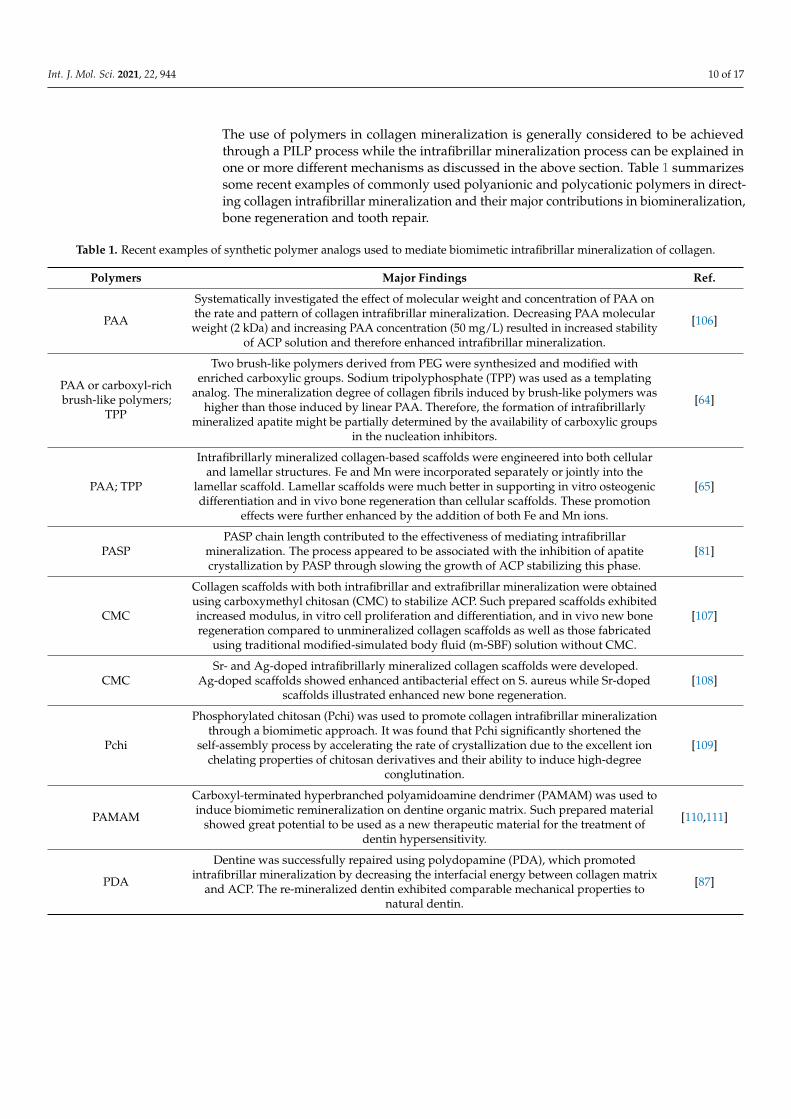
The use of polymers in collagen mineralization is generally considered to be achievedthrough a PILP process while the intrafibrillar mineralization process can be explained inone or more different mechanisms as discussed in the above section. Table 1 summarizessome recent examples of commonly used polyanionic and polycationic polymers in direct-ing collagen intrafibrillar mineralization and their major contributions in biomineralization,bone regeneration and tooth repair.
Table 1. Recent examples of synthetic polymer analogs used to mediate biomimetic intrafibrillar mineralization of collagen.
Polymers Major Findings Ref.
PAA
Systematically investigated the effect of molecular weight and concentration of PAA onthe rate and pattern of collagen intrafibrillar mineralization. Decreasing PAA molecularweight (2 kDa) and increasing PAA concentration (50 mg/L) resulted in increased stability
of ACP solution and therefore enhanced intrafibrillar mineralization.
[106]
PAA or carboxyl-richbrush-like polymers;
TPP
Two brush-like polymers derived from PEG were synthesized and modified withenriched carboxylic groups. Sodium tripolyphosphate (TPP) was used as a templating
analog. The mineralization degree of collagen fibrils induced by brush-like polymers washigher than those induced by linear PAA. Therefore, the formation of intrafibrillarly
mineralized apatite might be partially determined by the availability of carboxylic groupsin the nucleation inhibitors.
[64]
PAA; TPP
Intrafibrillarly mineralized collagen-based scaffolds were engineered into both cellularand lamellar structures. Fe and Mn were incorporated separately or jointly into the
lamellar scaffold. Lamellar scaffolds were much better in supporting in vitro osteogenicdifferentiation and in vivo bone regeneration than cellular scaffolds. These promotion
effects were further enhanced by the addition of both Fe and Mn ions.
[65]
PASPPASP chain length contributed to the effectiveness of mediating intrafibrillar
mineralization. The process appeared to be associated with the inhibition of apatitecrystallization by PASP through slowing the growth of ACP stabilizing this phase.
[81]
CMC
Collagen scaffolds with both intrafibrillar and extrafibrillar mineralization were obtainedusing carboxymethyl chitosan (CMC) to stabilize ACP. Such prepared scaffolds exhibitedincreased modulus, in vitro cell proliferation and differentiation, and in vivo new boneregeneration compared to unmineralized collagen scaffolds as well as those fabricated
using traditional modified-simulated body fluid (m-SBF) solution without CMC.
[107]
CMCSr- and Ag-doped intrafibrillarly mineralized collagen scaffolds were developed.
Ag-doped scaffolds showed enhanced antibacterial effect on S. aureus while Sr-dopedscaffolds illustrated enhanced new bone regeneration.
[108]
Pchi
Phosphorylated chitosan (Pchi) was used to promote collagen intrafibrillar mineralizationthrough a biomimetic approach. It was found that Pchi significantly shortened the
self-assembly process by accelerating the rate of crystallization due to the excellent ionchelating properties of chitosan derivatives and their ability to induce high-degree
conglutination.
[109]
PAMAM
Carboxyl-terminated hyperbranched polyamidoamine dendrimer (PAMAM) was used toinduce biomimetic remineralization on dentine organic matrix. Such prepared material
showed great potential to be used as a new therapeutic material for the treatment ofdentin hypersensitivity.
[110,111]
PDA
Dentine was successfully repaired using polydopamine (PDA), which promotedintrafibrillar mineralization by decreasing the interfacial energy between collagen matrix
and ACP. The re-mineralized dentin exhibited comparable mechanical properties tonatural dentin.
[87]
Int. J. Mol. Sci. 2021, 22, 944 11 of 17
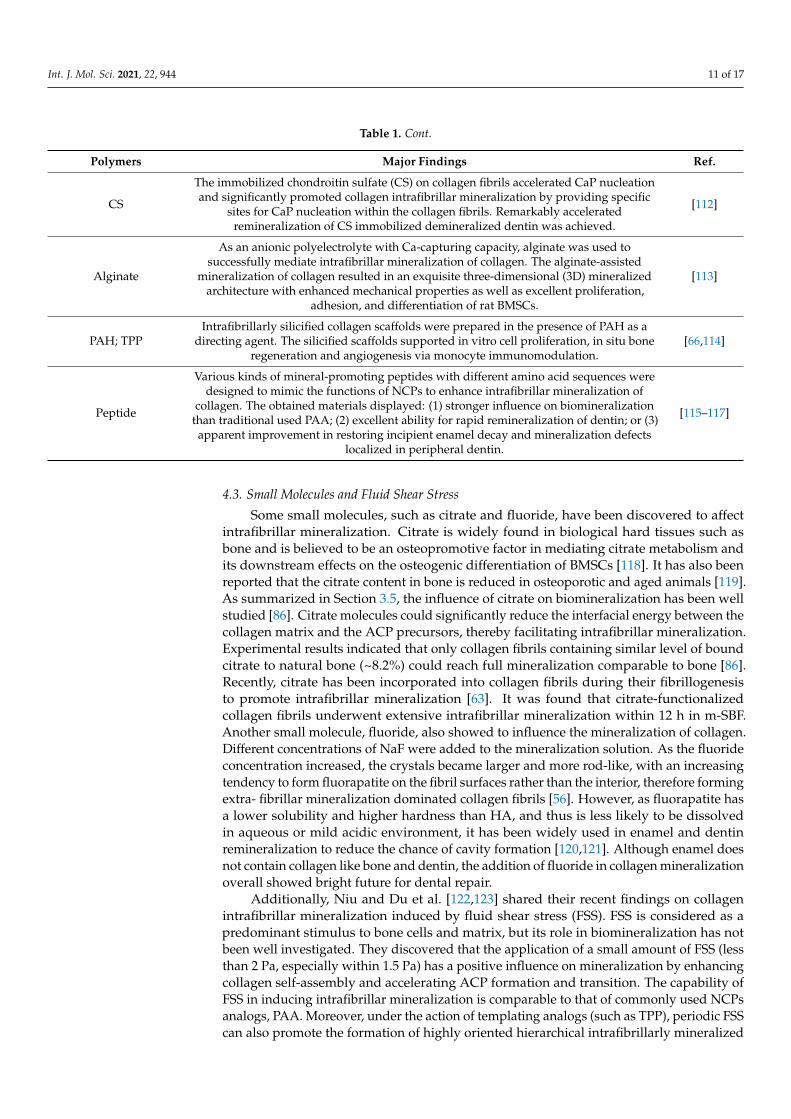
Table 1. Cont.
Polymers Major Findings Ref.
CS
The immobilized chondroitin sulfate (CS) on collagen fibrils accelerated CaP nucleationand significantly promoted collagen intrafibrillar mineralization by providing specific
sites for CaP nucleation within the collagen fibrils. Remarkably acceleratedremineralization of CS immobilized demineralized dentin was achieved.
[112]
Alginate
As an anionic polyelectrolyte with Ca-capturing capacity, alginate was used tosuccessfully mediate intrafibrillar mineralization of collagen. The alginate-assisted
mineralization of collagen resulted in an exquisite three-dimensional (3D) mineralizedarchitecture with enhanced mechanical properties as well as excellent proliferation,
adhesion, and differentiation of rat BMSCs.
[113]
PAH; TPPIntrafibrillarly silicified collagen scaffolds were prepared in the presence of PAH as a
directing agent. The silicified scaffolds supported in vitro cell proliferation, in situ boneregeneration and angiogenesis via monocyte immunomodulation.
[66,114]
Peptide
Various kinds of mineral-promoting peptides with different amino acid sequences weredesigned to mimic the functions of NCPs to enhance intrafibrillar mineralization of
collagen. The obtained materials displayed: (1) stronger influence on biomineralizationthan traditional used PAA; (2) excellent ability for rapid remineralization of dentin; or (3)apparent improvement in restoring incipient enamel decay and mineralization defects
localized in peripheral dentin.
[115–117]
4.3. Small Molecules and Fluid Shear Stress
Some small molecules, such as citrate and fluoride, have been discovered to affectintrafibrillar mineralization. Citrate is widely found in biological hard tissues such asbone and is believed to be an osteopromotive factor in mediating citrate metabolism andits downstream effects on the osteogenic differentiation of BMSCs [118]. It has also beenreported that the citrate content in bone is reduced in osteoporotic and aged animals [119].As summarized in Section 3.5, the influence of citrate on biomineralization has been wellstudied [86]. Citrate molecules could significantly reduce the interfacial energy between thecollagen matrix and the ACP precursors, thereby facilitating intrafibrillar mineralization.Experimental results indicated that only collagen fibrils containing similar level of boundcitrate to natural bone (~8.2%) could reach full mineralization comparable to bone [86].Recently, citrate has been incorporated into collagen fibrils during their fibrillogenesisto promote intrafibrillar mineralization [63]. It was found that citrate-functionalizedcollagen fibrils underwent extensive intrafibrillar mineralization within 12 h in m-SBF.Another small molecule, fluoride, also showed to influence the mineralization of collagen.Different concentrations of NaF were added to the mineralization solution. As the fluorideconcentration increased, the crystals became larger and more rod-like, with an increasingtendency to form fluorapatite on the fibril surfaces rather than the interior, therefore formingextra- fibrillar mineralization dominated collagen fibrils [56]. However, as fluorapatite hasa lower solubility and higher hardness than HA, and thus is less likely to be dissolvedin aqueous or mild acidic environment, it has been widely used in enamel and dentinremineralization to reduce the chance of cavity formation [120,121]. Although enamel doesnot contain collagen like bone and dentin, the addition of fluoride in collagen mineralizationoverall showed bright future for dental repair.
Additionally, Niu and Du et al. [122,123] shared their recent findings on collagenintrafibrillar mineralization induced by fluid shear stress (FSS). FSS is considered as apredominant stimulus to bone cells and matrix, but its role in biomineralization has notbeen well investigated. They discovered that the application of a small amount of FSS (lessthan 2 Pa, especially within 1.5 Pa) has a positive influence on mineralization by enhancingcollagen self-assembly and accelerating ACP formation and transition. The capability ofFSS in inducing intrafibrillar mineralization is comparable to that of commonly used NCPsanalogs, PAA. Moreover, under the action of templating analogs (such as TPP), periodic FSScan also promote the formation of highly oriented hierarchical intrafibrillarly mineralized
Int. J. Mol. Sci. 2021, 22, 944 12 of 17
collagen, giving D-banding patterns. Their work opened a new avenue in investigationof biomineralization mechanisms by introducing vascular flow in in vitro models. It alsoprovided a novel biomimetic strategy to induce intrafibrillar mineralization without theaddition of NCPs and biomolecules.
5. Conclusions and Perspectives
Natural bone and dentin are organic-inorganic composites mainly consisting of colla-gen and HA that are arranged in multilevel hierarchical structures from atomic- to macro-scale. Their excellent mechanical property and favorable biocompatibility are largelydetermined by the biomineralization of collagen fibrils. To have a better understanding oncollagen biomineralization and provide insight on the design of mineralized collagen-basedmaterials, this review summarized current collagen mineralization mechanisms and recentbiomimetic strategies for bone regeneration and tooth repair. Overall, these proposed mech-anisms are either derived from the classical ion-cluster-based or non-classical amorphousprecursor-based theories. A complementary proposal to both the classical and non-classicaltheories could be a future trend in establishing new mechanisms or perfecting existing ones.In addition, in vitro models involving more complicated conditions, such as fluid flow andcell loading, that better mimic the in vivo environment could provide clearer and morerealistic cognition on biomineralization. The improvement of bioinspired collagen-basedmaterials could start from the development of a simple, reliable, reproducible, and costlesscollagen extraction process. Despite extensive efforts, to date, only simplified biomimeticstructures have been achieved. Therefore, biomaterials better mimicking the complexhierarchical natural hard tissues may be achieved using newly developed techniques suchas 3D printing in combination with biomimetic strategies. Appropriate incorporation ofessential elements, drugs, or biomolecules into well-designed 3D collagen-based structureduring biomimetic mineralization could also be a direction to promote their performancefor bone regeneration and tooth repair.
Author Contributions: Conceptualization, L.Y. and M.W.; investigation, L.Y.; writing—original draftpreparation, L.Y.; writing—review and editing, M.W.; funding acquisition, M.W. Both authors haveread and agreed to the published version of the manuscript.
Funding: This research received no external funding.
Conflicts of Interest: Wei has an ownership interest in OrteoPoniX LLC, which develops andcommercializes biomaterials for orthopedic applications.
Abbreviations
HA HydroxyapatitePro ProlineHyp HydroxyprolineGly GlycylECM Extracellular matrixCol-HA Collagen-hydroxyapatiteCaP Calcium phosphateNCPs Non-collagenous proteinsACP Amorphous calcium phosphatePILP Polymer induced liquid precursorACC Amorphous calcium carbonatePASP Polyaspartic acidcyro-TEM Cryogenic transmission electron microscopyAFM Atomic force microscopyNMR Nuclear magnetic resonance
Int. J. Mol. Sci. 2021, 22, 944 13 of 17
PAA Polyacrylic acidSEM Scanning electron microscopyTEM Transmission electron microscopyXRD X-ray diffractionPAH Polyallyamine hydrochlorideOCN OsteocalcinBGP Bone Gla proteinOPN OsteopontinBSP Bone sialoproteinPEG-COOH Carboxylated polyethylene glycol terpolymerPEG-PAA Polyethylene glycol/polyacrylic acid copolymerSAXS Small-angle X-ray scatteringWAXS Wide-angle X-ray scatteringPDA PolydopamineTPP Sodium tripolyphosphateCMC Carboxymethyl chitosanm-SBF Modified-simulated body fluidPchi Phosphorylated chitosanPAMAM Polyamidoamine dendrimerCS Chondroitin sulfate3D Three-dimensionalFSS Fluid shear stress
References1. Thrivikraman, G.; Athirasala, A.; Gordon, R.; Zhang, L.; Bergan, R.; Keene, D.R.; Jones, J.M.; Xie, H.; Chen, Z.; Tao, J. Rapid
fabrication of vascularized and innervated cell-laden bone models with biomimetic intrafibrillar collagen mineralization. Nat.Commun. 2019, 10, 3520. [CrossRef] [PubMed]
2. Young, R.; Brown, W. Biological Mineralization and Demineralization; Springer: Berlin/Heidelberg, Germany, 1982.3. Chang, H.-H.; Chien, M.-J.; Kao, C.-C.; Chao, Y.-J.; Yu, P.-T.; Chang, C.-Y.; Huang, S.-J.; Lee, Y.-L.; Chan, J.C. Structural
characterization of fluoride species in shark teeth. Chem. Commun. 2017, 53, 3838–3841. [CrossRef] [PubMed]4. Zilm, M.E.; Yu, L.; Hines, W.A.; Wei, M. Magnetic properties and cytocompatibility of transition-metal-incorporated hydroxyap-
atite. Mater. Sci. Eng. C 2018, 87, 112–119. [CrossRef] [PubMed]5. Xu, Y.-J.; Dong, L.; Lu, Y.; Zhang, L.-C.; An, D.; Gao, H.-L.; Yang, D.-M.; Hu, W.; Sui, C.; Xu, W.-P. Magnetic hydroxyapatite
nanoworms for magnetic resonance diagnosis of acute hepatic injury. Nanoscale 2016, 8, 1684–1690. [CrossRef] [PubMed]6. Mondal, S.; Manivasagan, P.; Bharathiraja, S.; Moorthy, M.S.; Kim, H.H.; Seo, H.; Lee, K.D.; Oh, J. Magnetic hydroxyapatite: A
promising multifunctional platform for nanomedicine application. Int. J. Nanomed. 2017, 12, 8389. [CrossRef]7. Shoulders, M.D.; Raines, R.T. Collagen structure and stability. Annu. Rev. Biochem. 2009, 78, 929–958. [CrossRef]8. Lin, K.; Zhang, D.; Macedo, M.H.; Cui, W.; Sarmento, B.; Shen, G. Advanced Collagen-Based Biomaterials for Regenerative
Biomedicine. Adv. Funct. Mater. 2019, 29, 1804943. [CrossRef]9. Ottani, V.; Martini, D.; Franchi, M.; Ruggeri, A.; Raspanti, M. Hierarchical structures in fibrillar collagens. Micron 2002, 33,
587–596. [CrossRef]10. Ferreira, A.M.; Gentile, P.; Chiono, V.; Ciardelli, G. Collagen for bone tissue regeneration. Acta Biomater. 2012, 8, 3191–3200.
[CrossRef] [PubMed]11. O’leary, L.E.; Fallas, J.A.; Bakota, E.L.; Kang, M.K.; Hartgerink, J.D. Multi-hierarchical self-assembly of a collagen mimetic peptide
from triple helix to nanofibre and hydrogel. Nat. Chem. 2011, 3, 821. [CrossRef]12. Lutolf, M.P.; Gilbert, P.M.; Blau, H.M. Designing materials to direct stem-cell fate. Nature 2009, 462, 433–441. [CrossRef] [PubMed]13. Yoo, T.K.; Han, S.-H.; Han, J. Protective effects of biodegradable collagen implants on thinned sclera after strabismus surgery: A
paired-eye study. J. Am. Assoc. Pediatric Ophthalmol. Strabismus 2017, 21, 467–471. [CrossRef] [PubMed]14. Di Martino, A.; Liverani, L.; Rainer, A.; Salvatore, G.; Trombetta, M.; Denaro, V. Electrospun scaffolds for bone tissue engineering.
Musculoskelet. Surg. 2011, 95, 69–80. [CrossRef] [PubMed]15. Kim, W.; Kim, M.; Kim, G.H. 3D-printed biomimetic scaffold simulating microfibril muscle structure. Adv. Funct. Mater. 2018, 28,
1800405. [CrossRef]16. Featherstone, J.D. Dental caries: A dynamic disease process. Aust. Dent. J. 2008, 53, 286–291. [CrossRef] [PubMed]17. Han, M.; Li, Q.-L.; Cao, Y.; Fang, H.; Xia, R.; Zhang, Z.-H. In vivo remineralization of dentin using an agarose hydrogel biomimetic
mineralization system. Sci. Rep. 2017, 7, 41955. [CrossRef]18. Habibah, T.U.; Salisbury, H.G. StatPearls [Internet]; StatPearls Publishing: Petersburg, FL, USA, 2019.19. Cölfen, H. A crystal-clear view. Nat. Mater. 2010, 9, 960–961. [CrossRef]20. Azpiazu, D.; Gonzalo, S.; González-Parra, E.; Egido, J.; Villa-Bellosta, R. Role of pyrophosphate in vascular calcification in chronic
kidney disease. Nefrologia 2018, 38, 250–257. [CrossRef]
Int. J. Mol. Sci. 2021, 22, 944 14 of 17
21. Bhadada, S.K.; Rao, S.D. Role of phosphate in biomineralization. Calcif. Tissue Int. 2020, 108, 32–40. [CrossRef]22. Ziegler, S.G.; Ferreira, C.R.; MacFarlane, E.G.; Riddle, R.C.; Tomlinson, R.E.; Chew, E.Y.; Martin, L.; Ma, C.-T.; Sergienko, E.; Pinker-
ton, A.B. Ectopic calcification in pseudoxanthoma elasticum responds to inhibition of tissue-nonspecific alkaline phosphatase.Sci. Transl. Med. 2017, 9. [CrossRef] [PubMed]
23. Kim, D.; Lee, B.; Thomopoulos, S.; Jun, Y.-S. The role of confined collagen geometry in decreasing nucleation energy barriers tointrafibrillar mineralization. Nat. Commun. 2018, 9, 962. [CrossRef] [PubMed]
24. Hu, C.; Zilm, M.; Wei, M. Fabrication of intrafibrillar and extrafibrillar mineralized collagen/apatite scaffolds with a hierarchicalstructure. J. Biomed. Mater. Res. Part A 2016, 104, 1153–1161. [CrossRef] [PubMed]
25. Weiner, S.; Addadi, L. Crystallization pathways in biomineralization. Annu. Rev. Mater. Res. 2011, 41, 21–40. [CrossRef]26. Gebauer, D.; Völkel, A.; Cölfen, H. Stable prenucleation calcium carbonate clusters. Science 2008, 322, 1819–1822. [CrossRef]27. Liu, Y.; Li, N.; Qi, Y.p.; Dai, L.; Bryan, T.E.; Mao, J.; Pashley, D.H.; Tay, F.R. Intrafibrillar collagen mineralization produced by
biomimetic hierarchical nanoapatite assembly. Adv. Mater. 2011, 23, 975–980. [CrossRef] [PubMed]28. Hu, C.; Zhang, L.; Wei, M. Development of biomimetic scaffolds with both intrafibrillar and extrafibrillar mineralization. ACS
Biomater. Sci. Eng. 2015, 1, 669–676. [CrossRef]29. Jee, S.-S.; Thula, T.T.; Gower, L.B. Development of bone-like composites via the polymer-induced liquid-precursor (PILP) process.
Part 1: Influence of polymer molecular weight. Acta Biomater. 2010, 6, 3676–3686. [CrossRef] [PubMed]30. Kim, Y.K.; Gu, L.-S.; Bryan, T.E.; Kim, J.R.; Chen, L.; Liu, Y.; Yoon, J.C.; Breschi, L.; Pashley, D.H.; Tay, F.R. Mineralisation of
reconstituted collagen using polyvinylphosphonic acid/polyacrylic acid templating matrix protein analogues in the presence ofcalcium, phosphate and hydroxyl ions. Biomaterials 2010, 31, 6618–6627. [CrossRef] [PubMed]
31. Nudelman, F.; Pieterse, K.; George, A.; Bomans, P.H.; Friedrich, H.; Brylka, L.J.; Hilbers, P.A.; de With, G.; Sommerdijk, N.A.The role of collagen in bone apatite formation in the presence of hydroxyapatite nucleation inhibitors. Nat. Mater. 2010, 9, 1004.[CrossRef] [PubMed]
32. Chen, L.; Jacquet, R.; Lowder, E.; Landis, W.J. Refinement of collagen–mineral interaction: A possible role for osteocalcin inapatite crystal nucleation, growth and development. Bone 2015, 71, 7–16. [CrossRef] [PubMed]
33. Thula, T.T.; Rodriguez, D.E.; Lee, M.H.; Pendi, L.; Podschun, J.; Gower, L.B. In vitro mineralization of dense collagen substrates:A biomimetic approach toward the development of bone-graft materials. Acta Biomater. 2011, 7, 3158–3169. [CrossRef] [PubMed]
34. Silver, F.H.; Landis, W.J. Deposition of apatite in mineralizing vertebrate extracellular matrices: A model of possible nucleationsites on type I collagen. Connect. Tissue Res. 2011, 52, 242–254. [CrossRef] [PubMed]
35. Xu, Z.; Yang, Y.; Zhao, W.; Wang, Z.; Landis, W.J.; Cui, Q.; Sahai, N. Molecular mechanisms for intrafibrillar collagen mineralizationin skeletal tissues. Biomaterials 2015, 39, 59–66. [CrossRef] [PubMed]
36. Kim, D.; Lee, B.; Thomopoulos, S.; Jun, Y.-S. In situ evaluation of calcium phosphate nucleation kinetics and pathways duringintra-and extrafibrillar mineralization of collagen matrices. Cryst. Growth Des. 2016, 16, 5359–5366. [CrossRef]
37. Qu, H.; Xia, Z.; Knecht, D.A.; Wei, M. Synthesis of dense collagen/apatite composites using a biomimetic method. J. Am. Ceram.Soc. 2008, 91, 3211–3215. [CrossRef]
38. Hu, C.; Yu, L.; Wei, M. Sectioning studies of biomimetic collagen-hydroxyapatite coatings on Ti-6Al-4V substrates using focusedion beam. Appl. Surf. Sci. 2018, 444, 590–597. [CrossRef]
39. Gebauer, D.; Cölfen, H. Prenucleation clusters and non-classical nucleation. Nano Today 2011, 6, 564–584. [CrossRef]40. Habraken, W.J.; Tao, J.; Brylka, L.J.; Friedrich, H.; Bertinetti, L.; Schenk, A.S.; Verch, A.; Dmitrovic, V.; Bomans, P.H.; Frederik, P.M.
Ion-association complexes unite classical and non-classical theories for the biomimetic nucleation of calcium phosphate. Nat.Commun. 2013, 4, 1507. [CrossRef]
41. Deshpande, A.S.; Beniash, E. Bioinspired synthesis of mineralized collagen fibrils. Cryst. Growth Des. 2008, 8, 3084–3090.[CrossRef]
42. Gower, L.; Tirrell, D. Calcium carbonate films and helices grown in solutions of poly (aspartate). J. Cryst. Growth 1998, 191,153–160. [CrossRef]
43. Gower, L.B.; Odom, D.J. Deposition of calcium carbonate films by a polymer-induced liquid-precursor (PILP) process. J. Cryst.Growth 2000, 210, 719–734. [CrossRef]
44. Olszta, M.J.; Cheng, X.; Jee, S.S.; Kumar, R.; Kim, Y.-Y.; Kaufman, M.J.; Douglas, E.P.; Gower, L.B. Bone structure and formation: Anew perspective. Mater. Sci. Eng. R Rep. 2007, 58, 77–116. [CrossRef]
45. Xu, Y.; Tijssen, K.C.; Bomans, P.H.; Akiva, A.; Friedrich, H.; Kentgens, A.P.; Sommerdijk, N.A. Microscopic structure of thepolymer-induced liquid precursor for calcium carbonate. Nat. Commun. 2018, 9, 2582. [CrossRef]
46. Wolf, S.L.; Caballero, L.; Melo, F.; Cölfen, H. Gel-like calcium carbonate precursors observed by in situ AFM. Langmuir 2017, 33,158–163. [CrossRef] [PubMed]
47. Ma, Y.-X.; Hoff, S.E.; Huang, X.-Q.; Liu, J.; Wan, Q.-Q.; Song, Q.; Gu, J.-T.; Heinz, H.; Tay, F.R.; Niu, L.-N. Involvement ofprenucleation clusters in calcium phosphate mineralization of collagen. Acta Biomater. 2020, 120, 213–223. [CrossRef] [PubMed]
48. Garcia, N.A.; Malini, R.I.; Freeman, C.L.; Demichelis, R.; Raiteri, P.; Sommerdijk, N.A.; Harding, J.H.; Gale, J.D. Simulation ofcalcium phosphate prenucleation clusters in aqueous solution: Association beyond ion pairing. Cryst. Growth Des. 2019, 19,6422–6430. [CrossRef] [PubMed]
49. Sugawara-Narutaki, A.; Nakamura, J.; Ohtsuki, C. Bioceramics; Elsevier: Amsterdam, The Netherlands, 2020.
Int. J. Mol. Sci. 2021, 22, 944 15 of 17
50. Olszta, M.; Douglas, E.; Gower, L. Scanning electron microscopic analysis of the mineralization of type I collagen via a polymer-induced liquid-precursor (PILP) process. Calcif. Tissue Int. 2003, 72, 583–591. [CrossRef]
51. Olszta, M.J.; Douglas, E.P.; Gower, L.B. Intrafibrillar mineralization of collagen using a liquid-phase mineral precursor. MRSOnline Proc. Libr. Arch. 2003, 774. [CrossRef]
52. Olszta, M.J.; Odom, D.J.; Douglas, E.P.; Gower, L.B. A new paradigm for biomineral formation: Mineralization via an amorphousliquid-phase precursor. Connect. Tissue Res. 2003, 44, 326–334. [CrossRef]
53. Jee, S.S.; Culver, L.; Li, Y.; Douglas, E.P.; Gower, L.B. Biomimetic mineralization of collagen via an enzyme-aided PILP process.J. Cryst. Growth 2010, 312, 1249–1256. [CrossRef]
54. Jee, S.S.; Kasinath, R.K.; DiMasi, E.; Kim, Y.-Y.; Gower, L. Oriented hydroxyapatite in turkey tendon mineralized via thepolymer-induced liquid-precursor (PILP) process. CrystEngComm 2011, 13, 2077–2083. [CrossRef]
55. França, C.M.; Thrivikraman, G.; Athirasala, A.; Tahayeri, A.; Gower, L.B.; Bertassoni, L.E. The influence of osteopontin-guidedcollagen intrafibrillar mineralization on pericyte differentiation and vascularization of engineered bone scaffolds. J. Biomed. Mater.Res. Part B Appl. Biomater. 2019, 107, 1522–1532. [CrossRef] [PubMed]
56. Saxena, N.; Cremer, M.A.; Dolling, E.S.; Nurrohman, H.; Habelitz, S.; Marshall, G.W.; Gower, L.B. Influence of fluoride on themineralization of collagen via the polymer-induced liquid-precursor (PILP) process. Dent. Mater. 2018, 34, 1378–1390. [CrossRef]
57. James, B.D.; Guerin, P.; Iverson, Z.; Allen, J.B. Mineralized DNA-collagen complex-based biomaterials for bone tissue engineering.Int. J. Biol. Macromol. 2020, 161, 1127–1139. [CrossRef] [PubMed]
58. Li, S.T.; Katz, E.P. An electrostatic model for collagen fibrils. The interaction of reconstituted collagen with Ca++, Na+, and Cl−.Biopolym. Orig. Res. Biomol. 1976, 15, 1439–1460. [CrossRef] [PubMed]
59. Nudelman, F.; Lausch, A.J.; Sommerdijk, N.A.; Sone, E.D. In vitro models of collagen biomineralization. J. Struct. Biol. 2013, 183,258–269. [CrossRef]
60. Wang, Y.; Azaïs, T.; Robin, M.; Vallée, A.; Catania, C.; Legriel, P.; Pehau-Arnaudet, G.; Babonneau, F.; Giraud-Guille, M.-M.;Nassif, N. The predominant role of collagen in the nucleation, growth, structure and orientation of bone apatite. Nat. Mater. 2012,11, 724–733. [CrossRef] [PubMed]
61. Wu, S.; Gu, L.; Huang, Z.; Sun, Q.; Chen, H.; Ling, J.; Mai, S. Intrafibrillar mineralization of polyacrylic acid-bound collagen fibrilsusing a two-dimensional collagen model and Portland cement-based resins. Eur. J. Oral Sci. 2017, 125, 72–80. [CrossRef]
62. Rodriguez, D.E.; Thula-Mata, T.; Toro, E.J.; Yeh, Y.-W.; Holt, C.; Holliday, L.S.; Gower, L.B. Multifunctional role of osteopontin indirecting intrafibrillar mineralization of collagen and activation of osteoclasts. Acta Biomater. 2014, 10, 494–507. [CrossRef]
63. Jiang, W.; Griffanti, G.; Tamimi, F.; McKee, M.D.; Nazhat, S.N. Multiscale structural evolution of citrate-triggered intrafibrillarand interfibrillar mineralization in dense collagen gels. J. Struct. Biol. 2020, 212, 107592. [CrossRef]
64. Yu, L.; Martin, I.J.; Kasi, R.M.; Wei, M. Enhanced intrafibrillar mineralization of collagen fibrils induced by brushlike polymers.ACS Appl. Mater. Interfaces 2018, 10, 28440–28449. [CrossRef] [PubMed]
65. Yu, L.; Rowe, D.W.; Perera, I.P.; Zhang, J.; Suib, S.L.; Xin, X.; Wei, M. Intrafibrillar Mineralized Collagen-Hydroxyapatite-BasedScaffolds for Bone Regeneration. ACS Appl. Mater. Interfaces 2020, 12, 18235–18249. [CrossRef]
66. Hu, C.; Yu, L.; Wei, M. Biomimetic intrafibrillar silicification of collagen fibrils through a one-step collagen self-assembly/silicificationapproach. RSC Adv. 2017, 7, 34624–34632. [CrossRef]
67. Kerns, J.G.; Buckley, K.; Churchwell, J.; Parker, A.W.; Matousek, P.; Goodship, A.E. Is the collagen primed for mineralization inspecific regions of the turkey tendon? An investigation of the protein–mineral interface using Raman spectroscopy. Anal. Chem.2016, 88, 1559–1563. [CrossRef] [PubMed]
68. Jiao, K.; Niu, L.N.; Ma, C.F.; Huang, X.Q.; Pei, D.D.; Luo, T.; Huang, Q.; Chen, J.H.; Tay, F.R. Complementarity and uncertainty inintrafibrillar mineralization of collagen. Adv. Funct. Mater. 2016, 26, 6858–6875. [CrossRef]
69. Li, Y.; Rodriguez-Cabello, J.C.; Aparicio, C. Intrafibrillar mineralization of self-assembled elastin-like recombinamer fibrils. ACSAppl. Mater. Interfaces 2017, 9, 5838–5846. [CrossRef]
70. Niu, L.-N.; Jee, S.E.; Jiao, K.; Tonggu, L.; Li, M.; Wang, L.; Yang, Y.-D.; Bian, J.-H.; Breschi, L.; Jang, S.S. Collagen intrafibrillarmineralization as a result of the balance between osmotic equilibrium and electroneutrality. Nat. Mater. 2017, 16, 370–378.[CrossRef]
71. Song, Q.; Jiao, K.; Tonggu, L.; Wang, L.; Zhang, S.; Yang, Y.; Zhang, L.; Bian, J.; Hao, D.; Wang, C. Contribution of biomimeticcollagen-ligand interaction to intrafibrillar mineralization. Sci. Adv. 2019, 5, eaav9075. [CrossRef]
72. Toroian, D.; Lim, J.E.; Price, P.A. The size exclusion characteristics of type I collagen implications for the role of noncollagenousbone constituents in mineralization. J. Biol. Chem. 2007, 282, 22437–22447. [CrossRef] [PubMed]
73. Price, P.A.; Toroian, D.; Lim, J.E. Mineralization by inhibitor exclusion the calcification of collagen with fetuin. J. Biol. Chem. 2009,284, 17092–17101. [CrossRef]
74. Singh, A.; Gill, G.; Kaur, H.; Amhmed, M.; Jakhu, H. Role of osteopontin in bone remodeling and orthodontic tooth movement:A review. Prog. Orthod. 2018, 19, 18. [CrossRef] [PubMed]
75. Wang, Z.; Ustriyana, P.; Chen, K.; Zhao, W.; Xu, Z.; Sahai, N. Towards the Understanding of Small Protein-Mediated CollagenIntrafibrillar Mineralization. ACS Biomater. Sci. Eng. 2020. [CrossRef]
76. Cai, M.M.; Smith, E.R.; Holt, S.G. The role of fetuin-A in mineral trafficking and deposition. BoneKEy Rep. 2015, 4. [CrossRef][PubMed]
Int. J. Mol. Sci. 2021, 22, 944 16 of 17
77. Ping, H.; Xie, H.; Su, B.-L.; Cheng, Y.-B.; Wang, W.; Wang, H.; Wang, Y.; Zhang, J.; Zhang, F.; Fu, Z. Organized intrafibrillarmineralization, directed by a rationally designed multi-functional protein. J. Mater. Chem. B 2015, 3, 4496–4502. [CrossRef][PubMed]
78. Wei, S.; Wu, H.; Luo, X.-J. Biomineralization Precursor Carrier System Based on Carboxyl-Functionalized Large Pore MesoporousSilica Nanoparticles. Curr. Med. Sci. 2020, 40, 155–167. [CrossRef]
79. Du, T.; Niu, X.; Hou, S.; Li, Z.; Li, P.; Fan, Y. Apatite minerals derived from collagen phosphorylation modification induce thehierarchical intrafibrillar mineralization of collagen fibers. J. Biomed. Mater. Res. Part A 2019, 107, 2403–2413. [CrossRef] [PubMed]
80. Marbach, S.; Bocquet, L. Osmosis, from molecular insights to large-scale applications. Chem. Soc. Rev. 2019, 48, 3102–3144.[CrossRef] [PubMed]
81. Quan, B.D.; Sone, E.D. The effect of polyaspartate chain length on mediating biomimetic remineralization of collagenous tissues.J. R. Soc. Interface 2018, 15, 20180269. [CrossRef]
82. Zou, Z.; Tang, T.; Macías-Sánchez, E.; Sviben, S.; Landis, W.J.; Bertinetti, L.; Fratzl, P. Three-dimensional structural interrelationsbetween cells, extracellular matrix, and mineral in normally mineralizing avian leg tendon. Proc. Natl. Acad. Sci. USA 2020.[CrossRef]
83. Hor, J.L.; Wang, H.; Fakhraai, Z.; Lee, D. Effect of Physical Nanoconfinement on the Viscosity of Unentangled Polymers duringCapillary Rise Infiltration. Macromolecules 2018, 51, 5069–5078. [CrossRef]
84. Vinay, T.V.; Banuprasad, T.N.; George, S.D.; Varghese, S.; Varanakkottu, S.N. Additive-Free Tunable Transport and Assembly ofFloating Objects at Water-Air Interface Using Bubble-Mediated Capillary Forces. Adv. Mater. Interfaces 2017, 4, 1601231. [CrossRef]
85. Feng, J.; Song, Q.; Zhang, B.; Wu, Y.; Wang, T.; Jiang, L. Large-Scale, Long-Range-Ordered Patterning of Nanocrystals viaCapillary-Bridge Manipulation. Adv. Mater. 2017, 29, 1703143. [CrossRef] [PubMed]
86. Shao, C.; Zhao, R.; Jiang, S.; Yao, S.; Wu, Z.; Jin, B.; Yang, Y.; Pan, H.; Tang, R. Citrate improves collagen mineralization viainterface wetting: A physicochemical understanding of biomineralization control. Adv. Mater. 2018, 30, 1704876. [CrossRef]
87. Qu, Y.; Gu, T.; Du, Q.; Shao, C.; Wang, J.; Jin, B.; Kong, W.; Sun, J.; Chen, C.; Pan, H. Polydopamine Promotes DentinRemineralization via Interfacial Control. ACS Biomater. Sci. Eng. 2020, 6, 3327–3334. [CrossRef]
88. He, L.; Hao, Y.; Zhen, L.; Liu, H.; Shao, M.; Xu, X.; Liang, K.; Gao, Y.; Yuan, H.; Li, J. Biomineralization of dentin. J. Struct. Biol.2019, 207, 115–122. [CrossRef]
89. Wang, X.; Yang, J.; Andrei, C.M.; Soleymani, L.; Grandfield, K. Biomineralization of calcium phosphate revealed by in situliquid-phase electron microscopy. Commun. Chem. 2018, 1, 80. [CrossRef]
90. Yi, H.; Rehman, F.U.; Zhao, C.; Liu, B.; He, N. Recent advances in nano scaffolds for bone repair. Bone Res. 2016, 4, 16050.[CrossRef]
91. Zou, L.; Zhang, Y.; Liu, X.; Chen, J.; Zhang, Q. Biomimetic mineralization on natural and synthetic polymers to prepare hybridscaffolds for bone tissue engineering. Colloids Surf. B Biointerfaces 2019, 178, 222–229. [CrossRef]
92. Liu, Y.; Luo, D.; Kou, X.X.; Wang, X.D.; Tay, F.R.; Sha, Y.L.; Gan, Y.H.; Zhou, Y.H. Hierarchical intrafibrillar nanocarbonated apatiteassembly improves the nanomechanics and cytocompatibility of mineralized collagen. Adv. Funct. Mater. 2013, 23, 1404–1411.[CrossRef]
93. Zhang, Z.; Li, Z.; Zhang, C.; Liu, J.; Bai, Y.; Li, S.; Zhang, C. Biomimetic intrafibrillar mineralized collagen promotes boneregeneration via activation of the Wnt signaling pathway. Int. J. Nanomed. 2018, 13, 7503. [CrossRef]
94. Chien, Y.-C.; Tao, J.; Saeki, K.; Chin, A.F.; Lau, J.L.; Chen, C.-L.; Zuckermann, R.N.; Marshall, S.J.; Marshall, G.W.; De Yoreo, J.J.Using biomimetic polymers in place of noncollagenous proteins to achieve functional remineralization of dentin tissues. ACSBiomater. Sci. Eng. 2017, 3, 3469–3479. [CrossRef] [PubMed]
95. Chen, R.; Jin, R.; Li, X.; Fang, X.; Yuan, D.; Chen, Z.; Yao, S.; Tang, R.; Chen, Z. Biomimetic remineralization of artificial cariesdentin lesion using Ca/P-PILP. Dent. Mater. 2020, 36, 1397–1406. [CrossRef]
96. Zhang, Y.; Wang, Z.; Jiang, T.; Wang, Y. Biomimetic regulation of dentine remineralization by amino acid in vitro. Dent. Mater.2019, 35, 298–309. [CrossRef] [PubMed]
97. Barbosa-Martins, L.F.; Sousa, J.P.d.; Alves, L.A.; Davies, R.P.W.; Puppin-Rontanti, R.M. Biomimetic mineralizing agents recoverthe micro tensile bond strength of demineralized dentin. Materials 2018, 11, 1733. [CrossRef]
98. Cao, C.Y.; Mei, M.L.; Li, Q.-L.; Lo, E.C.M.; Chu, C.H. Methods for biomimetic remineralization of human dentine: A systematicreview. Int. J. Mol. Sci. 2015, 16, 4615–4627. [CrossRef]
99. Zoch, M.L.; Clemens, T.L.; Riddle, R.C. New insights into the biology of osteocalcin. Bone 2016, 82, 42–49. [CrossRef] [PubMed]100. Simon, P.; Grüner, D.; Worch, H.; Pompe, W.; Lichte, H.; El Khassawna, T.; Heiss, C.; Wenisch, S.; Kniep, R. First evidence
of octacalcium phosphate@ osteocalcin nanocomplex as skeletal bone component directing collagen triple–helix nanofibrilmineralization. Sci. Rep. 2018, 8, 13696. [CrossRef]
101. Moriishi, T.; Ozasa, R.; Ishimoto, T.; Nakano, T.; Hasegawa, T.; Miyazaki, T.; Liu, W.; Fukuyama, R.; Wang, Y.; Komori, H.Osteocalcin is necessary for the alignment of apatite crystallites, but not glucose metabolism, testosterone synthesis, or musclemass. PLoS Genet. 2020, 16, e1008586. [CrossRef] [PubMed]
102. Zhao, H.; Chen, Q.; Alam, A.; Cui, J.; Suen, K.C.; Soo, A.P.; Eguchi, S.; Gu, J.; Ma, D. The role of osteopontin in the progression ofsolid organ tumour. Cell Death Dis. 2018, 9, 356. [CrossRef]
103. Zuo, Q.; Yao, J.; Lu, S.; Du, Z.; Li, S.; Lin, F.; Shi, W.; Zhang, Y.; Xiao, Y. The role of organic phosphate in the spatial control ofperiodontium complex bio-mineralization: An in vitro study. J. Mater. Chem. B 2019, 7, 5956–5965. [CrossRef]
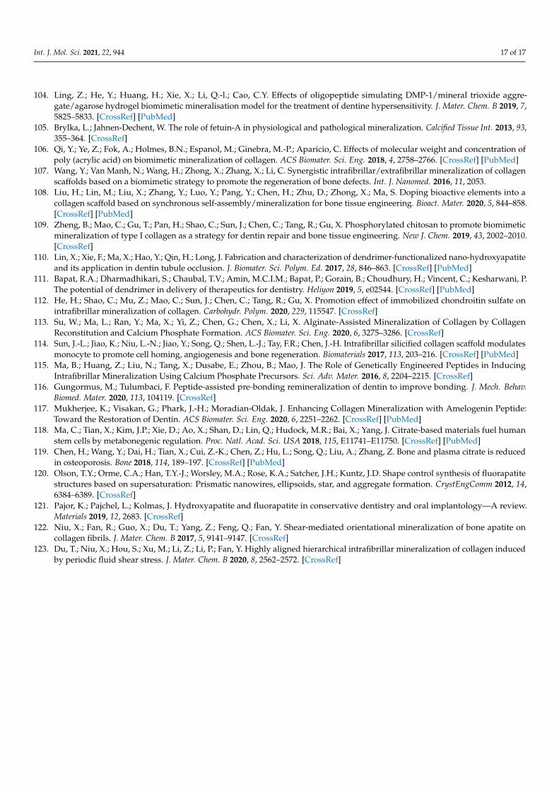
Int. J. Mol. Sci. 2021, 22, 944 17 of 17
104. Ling, Z.; He, Y.; Huang, H.; Xie, X.; Li, Q.-l.; Cao, C.Y. Effects of oligopeptide simulating DMP-1/mineral trioxide aggre-gate/agarose hydrogel biomimetic mineralisation model for the treatment of dentine hypersensitivity. J. Mater. Chem. B 2019, 7,5825–5833. [CrossRef] [PubMed]
105. Brylka, L.; Jahnen-Dechent, W. The role of fetuin-A in physiological and pathological mineralization. Calcified Tissue Int. 2013, 93,355–364. [CrossRef]
106. Qi, Y.; Ye, Z.; Fok, A.; Holmes, B.N.; Espanol, M.; Ginebra, M.-P.; Aparicio, C. Effects of molecular weight and concentration ofpoly (acrylic acid) on biomimetic mineralization of collagen. ACS Biomater. Sci. Eng. 2018, 4, 2758–2766. [CrossRef] [PubMed]
107. Wang, Y.; Van Manh, N.; Wang, H.; Zhong, X.; Zhang, X.; Li, C. Synergistic intrafibrillar/extrafibrillar mineralization of collagenscaffolds based on a biomimetic strategy to promote the regeneration of bone defects. Int. J. Nanomed. 2016, 11, 2053.
108. Liu, H.; Lin, M.; Liu, X.; Zhang, Y.; Luo, Y.; Pang, Y.; Chen, H.; Zhu, D.; Zhong, X.; Ma, S. Doping bioactive elements into acollagen scaffold based on synchronous self-assembly/mineralization for bone tissue engineering. Bioact. Mater. 2020, 5, 844–858.[CrossRef] [PubMed]
109. Zheng, B.; Mao, C.; Gu, T.; Pan, H.; Shao, C.; Sun, J.; Chen, C.; Tang, R.; Gu, X. Phosphorylated chitosan to promote biomimeticmineralization of type I collagen as a strategy for dentin repair and bone tissue engineering. New J. Chem. 2019, 43, 2002–2010.[CrossRef]
110. Lin, X.; Xie, F.; Ma, X.; Hao, Y.; Qin, H.; Long, J. Fabrication and characterization of dendrimer-functionalized nano-hydroxyapatiteand its application in dentin tubule occlusion. J. Biomater. Sci. Polym. Ed. 2017, 28, 846–863. [CrossRef] [PubMed]
111. Bapat, R.A.; Dharmadhikari, S.; Chaubal, T.V.; Amin, M.C.I.M.; Bapat, P.; Gorain, B.; Choudhury, H.; Vincent, C.; Kesharwani, P.The potential of dendrimer in delivery of therapeutics for dentistry. Heliyon 2019, 5, e02544. [CrossRef] [PubMed]
112. He, H.; Shao, C.; Mu, Z.; Mao, C.; Sun, J.; Chen, C.; Tang, R.; Gu, X. Promotion effect of immobilized chondroitin sulfate onintrafibrillar mineralization of collagen. Carbohydr. Polym. 2020, 229, 115547. [CrossRef]
113. Su, W.; Ma, L.; Ran, Y.; Ma, X.; Yi, Z.; Chen, G.; Chen, X.; Li, X. Alginate-Assisted Mineralization of Collagen by CollagenReconstitution and Calcium Phosphate Formation. ACS Biomater. Sci. Eng. 2020, 6, 3275–3286. [CrossRef]
114. Sun, J.-L.; Jiao, K.; Niu, L.-N.; Jiao, Y.; Song, Q.; Shen, L.-J.; Tay, F.R.; Chen, J.-H. Intrafibrillar silicified collagen scaffold modulatesmonocyte to promote cell homing, angiogenesis and bone regeneration. Biomaterials 2017, 113, 203–216. [CrossRef] [PubMed]
115. Ma, B.; Huang, Z.; Liu, N.; Tang, X.; Dusabe, E.; Zhou, B.; Mao, J. The Role of Genetically Engineered Peptides in InducingIntrafibrillar Mineralization Using Calcium Phosphate Precursors. Sci. Adv. Mater. 2016, 8, 2204–2215. [CrossRef]
116. Gungormus, M.; Tulumbaci, F. Peptide-assisted pre-bonding remineralization of dentin to improve bonding. J. Mech. Behav.Biomed. Mater. 2020, 113, 104119. [CrossRef]
117. Mukherjee, K.; Visakan, G.; Phark, J.-H.; Moradian-Oldak, J. Enhancing Collagen Mineralization with Amelogenin Peptide:Toward the Restoration of Dentin. ACS Biomater. Sci. Eng. 2020, 6, 2251–2262. [CrossRef] [PubMed]
118. Ma, C.; Tian, X.; Kim, J.P.; Xie, D.; Ao, X.; Shan, D.; Lin, Q.; Hudock, M.R.; Bai, X.; Yang, J. Citrate-based materials fuel humanstem cells by metabonegenic regulation. Proc. Natl. Acad. Sci. USA 2018, 115, E11741–E11750. [CrossRef] [PubMed]
119. Chen, H.; Wang, Y.; Dai, H.; Tian, X.; Cui, Z.-K.; Chen, Z.; Hu, L.; Song, Q.; Liu, A.; Zhang, Z. Bone and plasma citrate is reducedin osteoporosis. Bone 2018, 114, 189–197. [CrossRef] [PubMed]
120. Olson, T.Y.; Orme, C.A.; Han, T.Y.-J.; Worsley, M.A.; Rose, K.A.; Satcher, J.H.; Kuntz, J.D. Shape control synthesis of fluorapatitestructures based on supersaturation: Prismatic nanowires, ellipsoids, star, and aggregate formation. CrystEngComm 2012, 14,6384–6389. [CrossRef]
121. Pajor, K.; Pajchel, L.; Kolmas, J. Hydroxyapatite and fluorapatite in conservative dentistry and oral implantology—A review.Materials 2019, 12, 2683. [CrossRef]
122. Niu, X.; Fan, R.; Guo, X.; Du, T.; Yang, Z.; Feng, Q.; Fan, Y. Shear-mediated orientational mineralization of bone apatite oncollagen fibrils. J. Mater. Chem. B 2017, 5, 9141–9147. [CrossRef]
123. Du, T.; Niu, X.; Hou, S.; Xu, M.; Li, Z.; Li, P.; Fan, Y. Highly aligned hierarchical intrafibrillar mineralization of collagen inducedby periodic fluid shear stress. J. Mater. Chem. B 2020, 8, 2562–2572. [CrossRef]
Related Documents