Authors’ post print version of: Amis AA, Bull AMJ, Gupte CM, Hijazi I, Race A, Robinson JR. Biomechanics of the PCL and related structures: Posterolateral, posteromedial and meniscofemoral ligaments. Knee Surg. Sports Traumatol. Arthrosc. 11, (2003), 271-281. The original publication is available at www.springerlink.com Biomechanics of the PCL and related structures: posterolateral, posteromedial and meniscofemoral ligaments A. A. A. A. A. A. A. A. Amis Amis Amis Amis 1, 2 1, 2 1, 2 1, 2 , A. , A. , A. , A. M. M. M. M. J. J. J. J. Bull Bull Bull Bull 1, 3 1, 3 1, 3 1, 3 , C. , C. , C. , C. M. M. M. M. Gupte Gupte Gupte Gupte 1, 2, 3 1, 2, 3 1, 2, 3 1, 2, 3 , I. , I. , I. , I. Hijazi Hijazi Hijazi Hijazi 1 , A. , A. , A. , A. Race Race Race Race 1 and J. and J. and J. and J. R. R. R. R. Robinson Robinson Robinson Robinson 1, 2 1, 2 1, 2 1, 2 (1) Biomechanics Section, Mechanical Engineering Department, Imperial College, London, SW7 2AZ, UK (2) Musculoskeletal Surgery Department, Imperial College, London, SW7 2AZ, UK (3) Bioengineering Department, Imperial College, London, W6 8RF, UK A. A. Amis Email: [email protected] Phone: +44-207-5947046 Fax: +44-207-5847239 Received: Received: Received: Received: 5 September 2002 Accepted: Accepted: Accepted: Accepted: 14 November 2002 Published Published Published Published online: online: online: online: 5 September 2003 Abstract This paper reviews and updates our knowledge of the anatomy and biomechanics of the posterior cruciate ligament, and of the posterolateral, posteromedial and meniscofemoral ligaments of the knee. The posterior cruciate ligament is shown to have two functional fibre bundles that are tight at different angles of knee flexion. It is the primary restraint to tibial posterior draw at all angles of knee flexion apart from near full extension. In contrast, the posterolateral and posteromedial structures are shown to tighten as the knee extends, and to be well-aligned to resist tibial posterior draw. These structures also act as primary restraints against other tibial displacements. Tibial internal rotation is restrained by the medial and posteromedial structures, while tibial external rotation is restrained by the lateral and posterolateral structures. They are also the primary restraints against tibial abduction-adduction rotations. The meniscofemoral ligaments are shown, for the first time, to contribute significantly to resisting tibial posterior draw, and to have a strength of approximately 300 N. Taken together, this evidence shows how the posterolateral and posteromedial structures are responsible for posterior knee stability near extension, and this, along with the action of the

Welcome message from author
This document is posted to help you gain knowledge. Please leave a comment to let me know what you think about it! Share it to your friends and learn new things together.
Transcript

Authors’ post print version of:
Amis AA, Bull AMJ, Gupte CM, Hijazi I, Race A, Robinson JR. Biomechanics of the PCL and related structures: Posterolateral, posteromedial and meniscofemoral ligaments. Knee Surg. Sports Traumatol. Arthrosc. 11, (2003), 271-281.
The original publication is available at www.springerlink.com
Biomechanics of the PCL and related structures: posterolateral, posteromedial and meniscofemoral ligaments
A.A.A.A. A.A.A.A. AmisAmisAmisAmis1, 21, 21, 21, 2 , A., A., A., A. M.M.M.M. J.J.J.J. BullBullBullBull1, 31, 31, 31, 3, C., C., C., C. M.M.M.M. GupteGupteGupteGupte1, 2, 31, 2, 31, 2, 31, 2, 3, I., I., I., I. HijaziHijaziHijaziHijazi1111, A., A., A., A. RaceRaceRaceRace1111 and J.and J.and J.and J. R.R.R.R. RobinsonRobinsonRobinsonRobinson1, 21, 21, 21, 2
(1) Biomechanics Section, Mechanical Engineering Department, Imperial
College, London, SW7 2AZ, UK (2) Musculoskeletal Surgery Department, Imperial College, London, SW7 2AZ,
UK (3) Bioengineering Department, Imperial College, London, W6 8RF, UK
A. A. Amis Email: [email protected]
Phone: +44-207-5947046
Fax: +44-207-5847239
Received: Received: Received: Received: 5 September 2002 Accepted: Accepted: Accepted: Accepted: 14 November 2002 Published Published Published Published online: online: online: online: 5 September 2003
Abstract This paper reviews and updates our knowledge of the anatomy and
biomechanics of the posterior cruciate ligament, and of the posterolateral,
posteromedial and meniscofemoral ligaments of the knee. The posterior
cruciate ligament is shown to have two functional fibre bundles that are tight
at different angles of knee flexion. It is the primary restraint to tibial posterior
draw at all angles of knee flexion apart from near full extension. In contrast,
the posterolateral and posteromedial structures are shown to tighten as the
knee extends, and to be well-aligned to resist tibial posterior draw. These
structures also act as primary restraints against other tibial displacements.
Tibial internal rotation is restrained by the medial and posteromedial
structures, while tibial external rotation is restrained by the lateral and
posterolateral structures. They are also the primary restraints against tibial
abduction-adduction rotations. The meniscofemoral ligaments are shown, for
the first time, to contribute significantly to resisting tibial posterior draw, and
to have a strength of approximately 300 N. Taken together, this evidence
shows how the posterolateral and posteromedial structures are responsible for
posterior knee stability near extension, and this, along with the action of the

meniscofemoral ligaments, may explain why an isolated rupture of the
posterior cruciate ligament does not often lead to knee instability
KeywordsKeywordsKeywordsKeywords Posterior cruciate - Posterolateral - Posteromedial - Meniscofemoral - Ligament
Introduction
The overall purpose of this paper is to review and update our knowledge of the
anatomy and biomechanics of the posterior cruciate ligament, and of the
posterolateral, posteromedial and meniscofemoral ligaments of the knee.
The basic anatomy and function of the PCL are well-known. It has a relatively
compact tibial attachment located posteriorly, on the midline between the
posterior horns of the menisci and just below the joint line. The fibres are
oriented in a proximal-anterior direction, but fan out as they pass from the
tibia to the femur, thus giving an extensive area of femoral attachment. This
femoral attachment is immediately adjacent to the condylar articular cartilage,
and extends from the most anterior-distal part of the femoral intercondylar
notch (that is, the notch roof when observed in a flexed knee) to the most
posterior extent of the lateral surface of the medial femoral condyle. Several
studies have shown that the PCL is the strongest ligament crossing the knee,
and that it is the primary restraint to tibial posterior translation.
Despite this basic knowledge, the PCL remains an enigma: it is also well-
known that many patients can function well following a PCL rupture, and so
some surgeons choose never to operate on an isolated PCL rupture. There is
clearly some difficulty in reconciling the great strength of the PCL (which, if
the laws of tissue adaptation are true, implies that it transmits large forces)
versus the apparent lack of functional problems following its rupture.
When the role of ACL injury and the pivot-shift instability were first
appreciated, some thirty years ago, the initial response was to develop
peripheral extraarticular procedures. These sought to control the excessive
anterior movements of the lateral aspect of the tibial plateau. Later, as the
methods for intraarticular patellar tendon graft reconstructions were refined, so
the extraarticular reconstructions fell from favour. There has since been so
much effort to optimise intraarticular cruciate ligament reconstructions that
many surgeons have lost sight of the importance of the peripheral structures,
and of the need to reconstruct these, in addition to the cruciate ligaments, if
they have been damaged. One of the aims of this paper is to illuminate the
roles of the peripheral structures that act in synergy with the PCL, that have
received relatively little attention. Not only will this give information relating
to these structures, but it will suggest why the knee can function in the absence
of the PCL.

PCL biomechanics
Strength
The tensile strength of the PCL has been reported by a number of
investigators: Kennedy et al. [26] 1051 N, Marinozzi et al. [31] 855 N, Prietto
et al. [37] 1627 N, Trent et al. [49] 739 N. These figures may have been
underestimates, because the PCL fibres act in different directions, and so a
uniaxial test of the whole ligament may have caused a sequential tearing
failure, as different fibres became tight. This suggestion is supported by the
data of Butler et al. [6], who showed that the PCL fascicles had significantly
higher strength than the whole ligament tests had suggested.
The PCL is often considered to be made up of two functional bundles of
fibres, the anterolateral (aPC) and the posteromedial (pPC), where these refer
to their relative positions on the femur [12, 22, 35 et seqq] (Fig. 1). Race and
Amis [38] separated these bundles and found strengths of 1620 N for the aPC
and 258 N for the pPC. This difference was related to significant differences in
the cross-sectional areas (43 and 10 mm2), and to the maximum stress that the
tissues would withstand (35.9 and 24.4 MPa, respectively). Thus, this study
showed that the aPC fibre bundle is where most of the strength of the PCL is
found.
Fig. 1. Knee viewed from posteromedial aspect, after removal of the lateral
femoral condyle. The PCL has been split artificially into two functional
bundles: anterolateral, which attaches to the roof of the femoral intercondylar
notch, and posteromedial, which attaches to the medial side of the notch (from
Race and Amis [38], with permission)

These results were for specimens with a mean age of 75 years: Noyes and
Grood [34] and Woo et al. [56] found that the ACL strength was
approximately 2.5 times greater in young than in old knees. If this trend is also
true for the PCL, this suggests that the PCL strength may be 4.5 kN (5 to 6
times body weight) in young adults.
Tibial posterior translation
Many authors have noted that isolated cutting of the PCL allows a minimal
increase in posterior draw when the knee is extended, and that there is a
greater increase in laxity as the knee flexes [4, 12, 35et seqq]. Several studies
(e.g. [13, 15]) have produced graphs similar to Fig. 2 (from [2]), which shows
clearly that PCL rupture has the greatest effect at 90 degrees knee flexion.
This, therefore, is the position where the integrity of the ligament should be
tested clinically. Some investigators [11, 13, 15] have found that a posterior
draw force also induced a coupled tibial external rotation when the knee was
intact, and that this was either eliminated or reduced after the PCL was cut.
Fukubayashi et al. [11] also reported that when the coupled tibial rotation was
prevented, tibial posterior laxity was reduced by 30%.
Fig. 2. Variation of tibial posterior translation with knee flexion, for a force of
100 N, for the knee when intact and after PCL rupture (from Amis [2], with
permission)
The contribution of the PCL to resisting tibial posterior translation has been
shown by selective cutting studies. In this method, the tibia was displaced
posteriorly a known distance and the force required was recorded. When the
same displacement was repeated after the PCL had been cut, the reduction in
force represented the contribution that had been made by the PCL. For a 5 mm
posterior displacement, Butler et al. [7] found that the PCL contributed
approximately 95% at 30 and 90 degrees knee flexion. Similarly, Piziali et al.
[36] found a contribution of 83% with the knee extended. However, these
studies did not allow coupled tibial external rotation, and the PCL contribution
would be reduced in the absence of that artificial restraint, because the load
could then be shared more by the posterolateral structures.

The authors cited above who described the fibre bundles of the PCL also noted
that they had different patterns of tightening and slackening as the knee flexed
and extended. The aPC was noted to be slack in knee extension (and this is
seen on MRI, because the fibres take a curved path from femur to tibia), and to
tighten in mid flexion. The pPC fibres were tight in full knee extension and
slack in the mid range. Hughston et al. [22] showed that the pPC became well-
aligned to resist posterior tibial translation in deeper knee flexion. These
observations suggest that the two bundles act to limit tibial posterior laxity at
different angles of knee flexion.
A selective cutting study by Race and Amis [39] found that the aPC was the
primary restraint to 6 mm tibial posterior translation from 30 to 120 degrees
knee flexion (Fig. 3). The pPC shared the load with the aPC across this arc of
flexion, and became the dominant structure in deeper flexion. Although the
pPC was tight in full knee extension, it did not contribute greatly to resisting
posterior draw because its fibres were aligned in a proximal-distal direction
then, and so were not aligned to resist tibial posterior translation. It is
important to note that structures other than the PCL were dominant from full
extension to 20 degrees knee flexion. This means, in a knee with increased
posterior laxity close to full extension, that structures other than the PCL have
been damaged, and that an isolated PCL reconstruction will not restore the
laxity to normal here.
Fig. 3. Percentage contributions of the two bundles (aPC and pPC) of the
PCL, and of 'other structures' to resisting 6 mm posterior tibial translation,
across the range of knee flexion (from Race and Amis [39], with permission)
Tibial internal-external and abduction-adduction (valgus-varus) rotations
A number of studies have shown that the PCL is only a secondary restraint to
these rotations in the intact knee. This is because the PCL, being near to the
centre of the knee, has only a relatively small moment arm about the relevant
axes of rotation, and so it is at a mechanical disadvantage. These effects will
be discussed in sections below.

Posterolateral structures
Functional anatomy
The anatomy of the ligamentous structures around the posterolateral aspect of
the knee is both complex and variable. Some structures, such as the arcuate
and fabellofibular ligaments, are not always present [43, 48, 55]. In addition,
the popliteofibular ligament was not recognised for some years, despite it
being a direct ligamentous attachment from the femur to the fibula that is well-
aligned to resist tibial external rotation [32]. The action of the popliteofibular
ligament is compounded by the active tension in popliteus, which tends to
internally rotate the tibia [50]. As a general rule, the lateral collateral ligament
(LCL) and all other structures (i.e. the capsule, arcuate and fabellofibular
ligaments) that attach to the lateral femoral condyle posterior to the LCL are
tight in full knee extension, and slacken as the knee flexes. In contrast to the
other structures, the popliteofibular ligament complex is isometric, and so it
can stabilise the knee at all angles of flexion, while the LCL slackens
significantly beyond 30 degrees knee flexion [47]. The structures listed above
slant posteriorly as they pass from the femur to the tibia, and so they are well
aligned to resist tibial posterior translation and also tibial external rotation. It
follows that damage to these structures is associated with posterolateral
rotational instability (PLRI), which results from a combination of these two
components of motion (Fig. 4). In addition, the observation that these
structures tighten as the knee extends fits in with the evidence from the
posterior draw testing, that structures other than the PCL are most important at
resisting this subluxation in the extended knee.
Fig. 4. Posterolateral tibial laxity arises from a combination of posterior
translation, plus tibial external rotation (from Amis [2], with permission)

Strength of the posterolateral structures
The strengths of the lateral collateral ligament and popliteofibular ligament
have been found in two studies: Maynard et al. [32] found strengths of 750 N
and 425 N respectively, whilst Sugita and Amis [47] found strengths of 309 N
and 186 N in specimens with a mean age of approximately 70 years. Thus it
appears that the LCL is stronger than the popliteofibular ligament.
Posterior tibial translation
It was shown above that isolated PCL rupture leads to only a small increase in
posterior tibial translation when the knee is extended (Fig. 2), and that other
structures were then the primary restraint (Fig. 3). Conversely, isolated rupture
of the posterolateral structures, induced by an impact onto the anteromedial
aspect of the proximal tibia (which causes combined hyperextension and varus
angulation) has its largest effect in the extended knee. After inducing this
injury in vitro, the authors (I. Hijazi et al., unpublished) found that the intact
posterior laxity of 3 mm increased to 10 mm at full knee extension, whereas
this change was only from 5 mm to 6 mm at 90 degrees knee flexion, in
response to 100 N posterior draw force (Fig. 5). This tendency for
posterolateral damage to have the greatest effect on laxity with the knee
extended is the opposite to the effect of PCL rupture, which is most evident at
90 degrees knee flexion (Fig. 2). The studies of Gollehon et al. [13] and Grood
et al. [15] found that combined cutting of the PCL and the posterolateral
structures led to approximately 25 mm tibial posterior translation at 90 degrees
flexion. These results suggest that posterior draw tests alone are not able to
show isolated posterolateral damage clearly. An associated phenomenon, tibial
coupled rotation, provides the clue for diagnosis. A coupled rotation occurs as
an automatic secondary motion during the draw test, resulting from the greater
mobility of the lateral compartment [2]. Thus, tibial anterior draw induces
tibial internal rotation [3] and posterior draw induces tibial external rotation.
This coupled rotation increases significantly if the posterolateral structures
have been ruptured, accentuating the motion of the fibular head during a
posterior draw test (Fig. 4). This is sometimes known as a 'posterolateral draw
test' if performed at 90 degrees knee flexion.

Fig. 5. Rupture of the posterolateral structures caused posterior tibial draw
laxity at 100 N to increase greatly in the extended knee, but only a little at 90
degrees knee flexion (I. Hijazi et al., unpublished)
Tibial internal-external rotation
Because the posterolateral structures slant posteriorly as they pass distally,
tibial internal rotation slackens them, and so posterolateral damage has no
effect on internal rotation. Conversely, the posterolateral structures are
dominant in resisting tibial external rotation. Isolated PCL sectioning has no
measurable effect on tibial external rotation [13, 15]. These studies found that
posterolateral damage caused the largest increase in tibial external rotation
around 30 degrees knee flexion. At 90 degrees flexion, the change in external
rotation increased further when the PCL was also cut. Fig. 6 shows that
posterolateral damage caused the greatest increase in tibial external rotation in
the range 0 to 30 degrees knee flexion, with a smaller effect at 90 degrees
flexion (I. Hijazi et al., unpublished). The studies of Veltri et al. [51, 52]
showed that the popliteofibular ligament complex was a primary restraint to
tibial external rotation at all angles of flexion. This is because it remains well-
aligned and tight throughout the range of knee flexion [47].

Fig. 6. Rupture of the posterolateral structures caused tibial external rotation
laxity at 5 Nm torque to increase greatly in the extended knee, but only a little
at 90 degrees knee flexion (I. Hijazi et al., unpublished)
"Posterolateral rotatory instability" (PLRI) describes the hypermobility of the
lateral tibial plateau that is caused by damage to the posterolateral structures.
If the PCL remains intact, then the tibial plateau as a whole does not fall
posteriorly, and there is only an excessive tibial external rotation about a
central axis. However, if the PCL is also ruptured, the tibia is then tethered
medially, and there is a combination of posterior translation plus external
rotation that gives excessive pathological motion of the fibular head (Fig. 4).
This "coupled" rotation occurs automatically in response to a posterior
translation force on the tibia.
It is clear that a significant increase in tibial external rotation at 0 to 30
degrees knee flexion is diagnostic for posterolateral damage. This is often
demonstrated by the 'dial test', in which the feet are grasped and used to rotate
the tibia externally: the foot of the damaged limb moves further from the
'twelve o'clock' position.
Tibial adduction (varus) rotation
The lateral collateral ligament is the primary passive restraint to tibial
adduction at 5 and 25 degrees knee flexion, providing 55 and 69 percent of the
resistance to 5 degrees adduction [14]. This corresponds to it being both tight
and well-aligned here, prior to slackening in deeper knee flexion [47]. The
posterior capsular structures, such as the arcuate and fabellofibular ligaments,
contributed a further 13 percent at 5 degrees knee flexion, but slackened as the
knee flexed further [14]. The ACL is tight near knee extension, and acts as a
secondary restraint to varus joint angulation. Although the ACL is a stronger
ligament than the LCL, it has a much smaller moment arm about the adduction
pivot point, within the medial femoral condyle. Noting this, Grood et al. [14]
warned that isolated rupture of the lateral structures may not cause a large

increase in adduction laxity during clinical tests, because the tensed ACL acts
as a secondary restraint.
Posteromedial structures
Functional anatomy
The medial collateral ligament (MCL) consists of two distinct layers. The
long, parallel-fibred superficial layer (sMCL) attaches to the femur just antero-
distal to the adductor tubercle, passes distally, and attaches tangentially to the
tibial periosteum over a large area approximately 6–8 cm distal to the joint
line. The deep MCL (dMCL) attaches immediately distal to the femoral
attachment of the sMCL, and immediately below the joint line on the medial
margin of the tibial plateau, and so has much shorter fibres. En route, the deep
surface attaches to the periphery of the medial meniscus [5, 35, 44, 54].
Posterior to the parallel longitudinal fibres of the sMCL the deep and the
superficial layers merge, and there is less agreement about the description of
the structures. Hughston and Eilers [23] published diagrams of distinct fibre
bundles in the posteromedial capsule and identified a distinct ligament—the
posterior oblique ligament. Observations by the authors have confirmed those
of Warren and Marshall [54], finding a wide spread of fibres in the capsule,
rather than a distinct ligamentous band, so it is suggested that the whole of this
structure should be described simply as the 'posteromedial capsule' (PMC). In
general, these fibres are all stretched by fully extending the knee (Fig. 7), and
slacken with knee flexion (Fig. 8). Their wide femoral attachment runs
posteriorly from that of the sMCL, and passes proximal to the prominent
posterior medial femoral condyle. The condyle causes the capsule to bulge out
as the knee extends, thus tightening it. In the extended knee the PMC fibres
pass posteriorly and distally towards the joint line, and so can be seen to
tighten and resist tibial posterior translation and tibial internal rotation when
the tibia is loaded in those directions (Fig. 7). These posteromedial capsular
structures slacken and buckle as the knee flexes, and are carried deep to the
posterior margin of the sMCL in deeper flexion (Fig. 8). Distal to the joint
line, the PMC fibres attach to the posteromedial rim of the tibia and below this
are then aligned anterodistally, converging towards the sMCL, and attaching
immediately posterior to it.
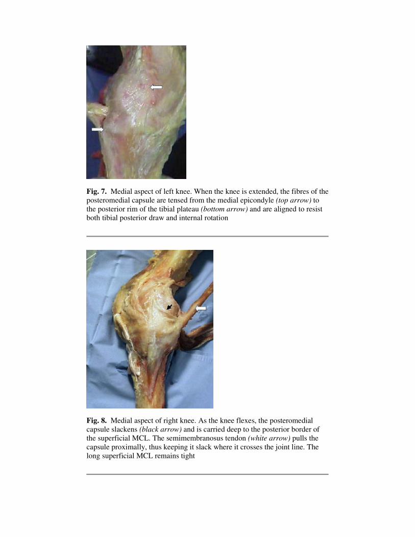
Fig. 7. Medial aspect of left knee. When the knee is extended, the fibres of the
posteromedial capsule are tensed from the medial epicondyle (top arrow) to
the posterior rim of the tibial plateau (bottom arrow) and are aligned to resist
both tibial posterior draw and internal rotation
Fig. 8. Medial aspect of right knee. As the knee flexes, the posteromedial
capsule slackens (black arrow) and is carried deep to the posterior border of
the superficial MCL. The semimembranosus tendon (white arrow) pulls the
capsule proximally, thus keeping it slack where it crosses the joint line. The
long superficial MCL remains tight

It has been suggested [46] that the PMC is tensed by the semimembranosus
tendon sheath. However, because the tendon tension acts approximately
parallel to the femur, it pulls the tendon sheath and PMC proximally when the
knee is at a low angle of flexion. The proximal part of the capsule, that crosses
the joint line, is therefore kept slack by this tendon action (Fig. 8), and so it is
then unlikely to stabilise the knee.
Ligament strength
Several authors have produced data on the strength of the MCL: Kennedy et
al. [26] 665 N, Marinozzi et al. [31] 465 N, Trent et al. [49] 516 N. However,
none of them tested the different fibre bundles separately. It has long been
known that different patterns of MCL damage can occur on the medial aspect
of the knee following an abduction (valgus) injury [45]. Ligaments fail at
approximately 20 percent tensile strain, and so a valgus opening injury will
rupture the short dMCL before the longer fibres of the sMCL reach their
ultimate strain [1]. Tensile tests of each of the structures found that the dMCL
failed at 195 N at 7 mm elongation, while the sMCL and posteromedial corner
both extended approximately 12 mm to reach loads of 784 N and 418 N,
respectively (J.R. Robinson et al., unpublished). This means that, even in
specimens with an average age of approximately 80 years, the total MCL
strength was in excess of 1 kN, which is greater than has been thought
previously, and higher than that of the ACL in this age range. Further, the data
suggests that dMCL rupture occurs at approximately 7 degrees valgus joint
opening, with complete MCL failure by 12 degrees.
Tibial posterior translation
The role of the posteromedial structures in controlling tibial posterior
translation has only been studied previously in the PCL-deficient knee at 90
degrees flexion. Ritchie et al. [42] showed that posterior translation was
reduced if the tibia was held in fixed internal rotation. Dividing the
posteromedial capsule resulted in an increase in tibial translation from 4.1 to
4.5 mm (10%). Further dividing the sMCL resulted in an increase in tibial
translation to 20.3 mm (350% increase). Thus the sMCL appeared to be the
most important secondary restraint, following PCL rupture, with the knee
flexed.
The posteromedial structures become tight and well-aligned to resist tibial
posterior translation as the knee extends (Fig. 7). Results available at the time
of writing suggest that the posteromedial structures make a significant
contribution to resisting tibial posterior translation with the tibia in internal
rotation, in both the intact and PCL-deficient knee. With sequential cutting,
the posterior laxity increased from 6.5 mm when intact, to 10.2 mm after
cutting the PCL and MFL, to 13.2 mm after cutting the PMC, at 15 degrees
flexion (C.M. Gupte et al., unpublished).
Tibial internal-external rotation
Kennedy and Fowler [25] showed that a tibial external rotation of 45 degrees
at 90 degrees of flexion tore the "capsular ligament" (dMCL) and that the
"superficial tibial collateral ligament" (sMCL) remained intact. Only with
further rotation did this part of the medial structures fail. In contrast, Warren et
al. [53] found that cutting the deep MCL in an otherwise intact knee had a
"minimum effect on restraining rotation between the femur and the tibia".
Cutting the long fibres of the sMCL first, however, resulted in a 2° increase in
external rotation at 0° knee flexion and 4° at 90° flexion.
The authors have shown that in the intact knee both the dMCL and the sMCL
restrain tibial external rotation. The dMCL provides more restraint at higher
angles of flexion, with the sMCL being important throughout the arc of
flexion. The fibres of the posteromedial capsule (PMC) are aligned such that
they are slackened by tibial external rotation, and therefore do not contribute
to restraining this movement. (J.R. Robinson et al., unpublished data).
Although the dMCL is not the strongest component of the medial ligament
complex, its position and short length mean that its tension rises quickly with
tibial external rotation, and that is why it is sometimes ruptured beneath an
intact sMCL.
The authors have found that in the intact knee both the PMC and the sMCL
are important structures in restraining tibial internal rotation. The PMC acts
when the knee is in extension and at low angles of flexion, with the sMCL
providing an increasing contribution with progressive knee flexion. In the
extended knee, cutting the sMCL and dMCL made no significant difference to
tibial internal rotation, but cutting the PMC increased internal rotation from 8
to 23 degrees (J.R. Robinson et al., unpublished data).
The PMC did not at as a significant restraint to tibial internal rotation above 30
degrees knee flexion, when the sMCL appeared to become more important.
Cutting the sMCL in isolation increased tibial internal rotation by
approximately 7 degrees at 60 to 90 degrees knee flexion (J.R. Robinson et al.,
unpublished data).
Overall, the evidence shows that the primary restraint to tibial internal rotation
is the PMC when the knee is at or close to extension, and the sMCL when the
knee is flexed.
Tibial abduction-adduction (valgus-varus) rotation
Kennedy and Fowler [25] believed that the primary lesion of valgus instability
was a tear of the capsular ligament (dMCL). Warren et al. [53] noted a
significant increase in valgus laxity after sectioning the long parallel fibres of
the sMCL. They were unable to demonstrate any significant valgus opening of
the knee by sectioning the dMCL and posterior capsule, and believed that
valgus laxity in extension was due to damage to the cruciate ligaments.
Increases in valgus laxity were measured in sequential cutting studies by
Mains et al. [29] and Nielsen et al. [33]. Mains et al. cut the sMCL, dMCL,
PMC, ACL and PCL in isolation and applied a 3.5 Nm abduction moment.
Cutting the sMCL increased valgus rotation laxity 2.5 degrees at 0 degrees
knee flexion, 5 degrees at 20–25 degrees flexion, and 4 degrees at 45 degrees
flexion. Section of the dMCL also caused 4 degrees extra valgus laxity at 45
degrees knee flexion. The sMCL was described as the prime restraint to valgus
rotation, with the dMCL as having a relatively minor role. The PMC appeared
subjectively to resist valgus rotation with the knee in hyperextension, but this
was not measured. Nielsen et al. found that maximum valgus laxity occurred
at 60 degrees of knee flexion. When applying a 2 Nm torque, cutting the entire
MCL increased valgus laxity only 2 degrees. Further transection of the capsule
increased laxity to 9 degrees. Cutting the ACL caused a further increase to 24
degrees. However the contribution of each of the structures was not evaluated
in these studies, so they did not define the primary and secondary restraints to
valgus loads.
Grood et al. [14] found that the MCL was the primary restraint to valgus laxity
when the medial aspect of the tibiofemoral joint was opened by 6 mm at 25
degrees knee flexion, resisting 78% of the load, but did not test the dMCL and
sMCL separately. They noted that the PMC slackened with flexion, so it
accounted for only 4 percent of the total restraint at 25 degrees flexion. With
the capsule tighter at 5 degrees flexion, this contribution increased to 18
percent. The ACL, PCL and posterior capsule were found to be secondary
restraints (Fig. 9) It was concluded that the capsular structures provided
important attachments for the menisci but did not have primary roles in
limiting valgus (abduction) laxity.
Fig. 9. Contributions to resisting tibial abduction (valgus) rotation, at 5 and 25
degrees knee flexion. Note that flexion slackened the posterior capsule,
decreasing its role (from Grood et al. [14], with permission)
The parallel longitudinal fibres of the superficial MCL are the primary
restraint to valgus laxity throughout the arc of knee flexion. Damage to this
structure causes increased valgus rotation of 3 degrees in extension and 5
degrees at 30 degrees flexion. This correlates well with the clinical finding
that valgus laxity due to damage of the sMCL is more demonstrable with the
knee in slight flexion, because the tight posterior capsule protects the extended
joint. There is a lesser contribution from the dMCL (approx 3 degrees from 15
to 90 degrees flexion). Although the sMCL was found to be the primary
restraint to tibial valgus rotation at all angles of knee flexion, the PMC made a
significant contribution when the knee was extended. At 0 degrees knee

flexion, the PMC resisted 29 percent of the valgus moment, which reduced to
14 percent at 30 degrees flexion and to less than 5 percent at 90 degrees
flexion (J.R. Robinson et al., unpublished).
In contrast to the neat cutting studies in vitro, clinical cases will not have
structures damaged in isolation. Even if only one major ligament bundle has
been ruptured, other related structures will have been stretched irreversibly.
This means that clinical examination will reveal larger laxities than those
reported above.
Meniscofemoral ligaments
Functional anatomy
Two meniscofemoral ligaments (MFLs) may connect the posterior horn of the
lateral meniscus to the intercondylar aspect of the medial femoral condyle,
attaching on either side of the PCL attachment. The ligament of Humphry
(aMFL) passes anterior to the PCL and attaches distally, close to the articular
cartilage (Fig. 10a). The ligament of Wrisberg (pMFL) passes posterior to the
PCL and attaches proximally, close to the roof of the intercondylar notch
(Fig. 10b). Friederich and O'Brien [10] have observed that the aMFL is tense
in flexion, whilst the pMFL is tense in extension. This reciprocal tightening
and slackening of the MFLs may be functionally similar to the two bundles of
the PCL. At least one MFL is present in 93% of specimens [16, 57], whilst
both ligaments co-exist more frequently in younger specimens [18]. The latter
suggests that they may degenerate with age.
Fig. 10. a The anterior meniscofemoral ligament of Humphry viewed in the
flexed right knee after ACL excision and tibia subluxed anteriorly. The aMFL
fibres slant across the distal surface of the PCL, which has its fibres aligned in
a sagittal plane. The MFL attaches close to the articular cartilage and
posterodistally to the lateral meniscus. The distal attachment is hidden by the
ACL in the intact knee. b Posterior view of right knee. The posterior
meniscofemoral ligament of Wrisberg slants across the posterior aspect of the
PCL. It attaches proximally in the femoral intercondylar notch, and to the
posterior horn of the lateral meniscus
Strength
The aMFL is, on average, smaller than the pMFL [21]. They have a tensile
strength (302.5 N and 300.5 N respectively) comparable to that of the pPC,
whilst their elastic modulus is similar to that of the aPC [19]. Kusayama et al.
[27] found a similar mean strength for MFLs of 297 N.
The high prevalence of the MFLs, together with their significant properties,
supports the hypothesis that they may play a functional role in the knee.
Tibial posterior translation
Radioevitch [41] proposed the MFLs as "third cruciate ligaments". Others [8,
27] have suggested that they may act as secondary restraints to posterior tibial
translation in the PCL-injured knee. Last [28] proposed that the MFLs are
involved in controlling the motion of the lateral meniscus in conjunction with
the tendon of popliteus during knee flexion, thus maintaining the congruency
of the knee joint. There have previously, however, been no objective studies to
confirm these hypotheses.
It has now been proven that the MFLs act as secondary restraints to tibial
posterior translation [17]: there was a significant increase in laxity when the
MFLs were cut, in both the intact knee and in the PCL deficient knee. This
was most clear when the knee was flexed. In the intact knee at 90 degrees
flexion, the MFLs contributed 28 percent of the resistance to posterior draw;
this contribution rose to 70 percent in the PCL deficient knee. Thus, the MFLs
act in synergy with the PCL when it is intact, and act in a similar manner to
the PCL when it has been ruptured.
Tibial internal-external rotation
The oblique orientation of the MFLs suggests that they may be involved in
limiting tibial internal rotation, but a selective cutting study in progress at the
authors' laboratory (C.M. Gupte et al., unpublished) has not found a significant
effect. This probably results from the MFLs crossing the knee close to the axis
of tibial rotation, so that their tension will have little effect on rotation.
Conclusions for the surgeon
The PCL was shown to have a divergent fibre structure that could be described
as consisting of two functional bundles of fibres. These were shown to have
different patterns of tightening and slackening, and thus to contribute to
resisting tibial posterior draw at different angles of knee flexion. This ligament

is clearly not an isometric structure, and so differs significantly from the ACL,
and an isometric reconstruction would not fit in with the natural behaviour.
This was shown by Race and Amis [40] who also, along with Harner et al.
[20] and Mannor et al. [30], showed that a double-bundled PCL reconstruction
re-established normal tibial posterior laxity significantly better than did single-
bundled reconstructions. This work in vitro, however, has not yet led to
reports of significant advantages in clinical results.
The posterolateral structures do not have a history of good results following
reconstructive surgery, and it appears that more work is needed to optimise
reconstruction methods. The treatment options were reviewed by Covey [9].
This biomechanical review has shown that the posterolateral corner is a
primary restraint to tibial adduction (varus) and to tibial external rotations, as
well as to posterior translation when the knee is near extension and thus in a
functional load-bearing posture in gait. It may be argued, therefore, that the
posterolateral structures may be more important for knee function than is the
PCL, because of their multiple roles. It is hoped that better understanding of
the biomechanics of the posterolateral structures will allow reconstruction
methods to be improved.
The medial and posteromedial structures have been relatively neglected in
recent years, and it is only now that basic data on their roles in stabilising the
knee are being produced. It is clear that, in addition to the role of the
superficial MCL as the primary restraint against tibial abduction (valgus)
rotation, these structures also act to control tibial posterior translation,
particularly when the tibia is in internal rotation. Knee extension and tibial
internal rotation both tense the posteromedial structures in an efficient
direction to resist both tibial posterior translation and internal rotation.
With the emergence of a role for the MFLs as secondary restraints to tibial
posterior translation, it may be useful to identify these structures during
arthroscopy and imaging, particularly in the PCL-deficient knee. It may also
be of benefit to attempt to preserve them during PCL reconstruction. The
authors now believe that intact MFLs help to stabilise the PCL-deficient knee,
and so contribute to the relatively good functional status following isolated
PCL rupture.
Added together, it is apparent that the combined actions of the posteromedial
and posterolateral structures of the knee are capable of controlling
tibiofemoral joint laxity in ways that have traditionally been thought to be the
role of the PCL, particularly when the knee is extended. Noting that the bulk
of the PCL is slack when the knee is extended, it appears by default that the
PCL may be so strong because of needing to resist large forces when the knee
is flexed. Further work is needed to gain a greater understanding of the
function of the PCL and its interactions with the posterolateral, posteromedial,
and meniscofemoral ligaments. This should give greater insight into the
enigma of the lack of functional deficit following isolated PCL rupture, and
guidance for the development of better reconstructive procedures.
Acknowledgements This paper includes work done over some years by the
authors, and this was supported by the Arthritis Research Campaign, the

Wishbone Trust, the Royal College of Surgeons of Edinburgh, the Smith and
Nephew Foundation, and the Ealing Hospital knee fellowship.
References
1. Amis AA (1985) The biomechanics of ligaments. In: Jenkins DHR (ed)
Ligament injuries and their treatment. Chapman and Hall, London, pp 3–28
2. Amis AA (1999) The kinematics of knee stability. In: Jakob RP, Fulford P,
Horan F (eds) EFORT European Instructional Course Lectures, vol 4. J Bone
Joint Surg Br, London, pp 96–104
3. Amis AA, Scammell BE (1993) Biomechanics of intraarticular and
extraarticular reconstructions of the anterior cruciate ligament. J Bone Joint
Surg Br 75:812–817
4. Brantigan OC, Voshell AF (1941) The mechanics of the ligaments and
menisci of the knee joint. J Bone Joint Surg Am 23:44–66
5. Brantigan OC, Voshell AF (1943) The tibial collateral ligament: its function,
its bursae, and its relation to the medial meniscus. J Bone Joint Surg Am
25:121–131
6. Butler DL, Kay MD, Stouffer DC (1986) Comparison of material properties in
fascicle-bone units from human patellar tendon and knee ligaments. J
Biomech 19:425–432
7. Butler DL, Noyes FR, Grood ES (1980) Ligamentous restraints to anterior-
posterior drawer in the human knee: a biomechanical study. J Bone Joint Surg
Am 62:259–270
8. Clancy WG J, Shelbourne KD, Zoellner GB, Keene JS, Reider B, Rosenberg
TD (1983) Treatment of knee joint instability secondary to rupture of the
posterior cruciate ligament. Report of a new procedure. J Bone Joint Surg Am
65:310–322
9. Covey DC (2001) Injuries of the posterolateral corner of the knee. J Bone
Joint Surg Am 83:106–118
10. Friederich N F, O'Brien WR (1990) Functional anatomy of the
meniscofemoral ligaments. Proc Fourth Congress of the European Society of
Sports Traumatology Knee Surgery and Arthroscopy (ESSKA)

11. Fukubayashi T, Torzilli PA, Sherman MF, Warren RF (1982) An in vitro
biomechanical evaluation of anterior-posterior motion of the knee. Tibial
displacement, rotation, and torque. J Bone Joint Surg Am 64:258–264
12. Girgis FG, Marshall JL, Al Monajem ARS (1975) The cruciate ligaments of
the knee joint. Anatomical, functional and experimental analysis. Clin Orthop
106:216–231
13. Gollehon DL, Torzilli PA, Warren RF (1987) The role of the posterolateral
and cruciate ligaments in the stability of the human knee. A biomechanical
study. J Bone Joint Surg Am 69:233–242
14. Grood ES, Noyes FR, Butler DL, Suntay WJ (1981) Ligamentous and capsular
restraints preventing straight medial and lateral laxity in intact human cadaver
knees. J Bone Joint Surg Am 63:1257–1269
15. Grood ES, Stowers SF, Noyes FR (1988) Limits of movement in the human
knee. Effects of sectioning the posterior cruciate ligament and posterolateral
structures. J Bone Joint Surg Am 70:88–97
16. Gupte CM, Bull AMJ, Thomas RD, Amis AA (2003) A Review of the
function and biomechanics of the meniscofemoral ligaments. Arthroscopy
19:161-171
17. Gupte CM, Bull AMJ, Thomas RD, Amis AA (2003) The meniscofemoral
ligaments: secondary restraints to the posterior drawer. Analysis of
anteroposterior and rotatory laxity in the intact and posterior-cruciate-deficient
knee. J Bone Joint Surg Br 85:765-773
18. Gupte CM, Smith A, McDermott ID, Bull AMJ, Thomas RD, Amis AA
(2002) Meniscofemoral ligaments revisited: incidence, age correlation and
clinical implications. J Bone Joint Surg Br 84:846–851
19. Gupte CM, Smith A, Jamieson N, Bull AMJ, Amis AA (2002)
Meniscofemoral ligaments—structural and material properties. J Biomech
35:1623-1629
20. Harner CD, Janaushek MA, Kanamori A, Yagi M, Vogrin TM, Woo SL-Y
(2000) Biomechanical analysis of a double-bundle posterior cruciate ligament
reconstruction. Am J Sports Med 28:144–151
21. Heller L, Langman J (1964) The meniscofemoral ligaments of the human
knee. J Bone Joint Surg Br 46:307–311
22. Hughston JC, Bowden JA, Andrews JR, Norwood LA (1980) Acute tears of

the posterior cruciate ligament. Results of operative treatment. J Bone Joint
Surg Am 62:438–450
23. Hughston J C, Eilers AF (1994) The role of the posteror oblique ligament in
repairs of acute medial (collateral) ligament tears of the knee. J Bone Joint
Surg Am 76:1328–1344
24. Kaneda Y, Moriya H, Takahashi K, Shimada Y, Tamaki T (1997)
Experimental study on external tibial rotation of the knee. Am J Sports Med
25:796–800
25. Kennedy JC, Fowler PJ (1971) Medial and anterior instability of the knee. J
Bone Joint Surg Am 53:1257–1270
26. Kennedy JC, Hawkins RJ, Willis RB, Danylchuck KD (1976) Tension studies
of human knee ligaments. Yield point, ultimate failure, and disruption of the
cruciate and tibial collateral ligaments. J Bone Joint Surg Am 58:350–355
27. Kusayama T, Harner CD, Carlin GJ, Xerogeanes JW, Smith BA (1994)
Anatomical and biomechanical characteristics of human meniscofemoral
ligaments. Knee Surg Sports Traumatol Arthrosc 2:234–237
28. Last RJ (1950) The popliteus muscle and the lateral meniscus. J Bone Joint
Surg Br 32:93–99
29. Mains DB, Andrews JG, Stonecipher T (1977) Medial and anterior-posterior
ligament stability of the human knee, measured with a stress apparatus. Am J
Sports Med 5:144–153
30. Mannor DA, Shearn JT, Grood ES, Noyes FR, Levy MS (2000) Two-bundle
posterior cruciate ligament reconstruction. An in vitro analysis of graft
placement and tension. Am J Sports Med 28:833–845
31. Marinozzi G, Pappalardo S, Steindler R (1983) Human knee ligaments:
mechanical tests and ultrastructural observations. Ital J Orthop Traumatol
9:231–240
32. Maynard MJ, Deng X, Wickiewicz TL, Warren RF (1996) The popliteofibular
ligament. Rediscovery of a key element in posterolateral stability. Am J Sports
Med 24:311–316
33. Nielsen S, Kromann-Andersen C, Rasmussen O, Andersen K (1984)
Instability of cadaver knees after transection of capsule and ligaments. Acta
Orthop Scand. 55:30–34
34. Noyes FR, Grood ES (1976) The strength of the anterior cruciate ligament in
humans and rhesus monkeys: age-related and species-related changes. J Bone
Joint Surg Am 58:1074–1082
35. Palmer I (1938) On the injuries to the ligaments of the knee joint. Acta Orthop
Scand Suppl 5:283
36. Piziali RL, Seering WP, Nagel DA, Schurman DJ (1980) The function of the
primary ligaments of the knee in anterior-posterior and medial-lateral motions.
J Biomech 13:777–784
37. Prietto MP, Bain JR, Stonebrook SN, Settlage RA (1988) Tensile strength of
the human posterior cruciate ligament (PCL). Trans 34th Ann ORS 13:195
38. Race A, Amis AA (1994) The mechanical properties of the two bundles of the
human posterior cruciate ligament. J Biomech 27:13–24
39. Race A, Amis AA (1996) Loading of the two bundles of the posterior cruciate
ligament: an analysis of bundle function in A-P drawer. J Biomech 29:873–
879
40. Race A, Amis AA (1998) PCL reconstruction: in vitro biomechanical
comparison of 'isometric' versus single and double-bundled 'anatomic' grafts. J
Bone Joint Surg Br 80:173–179
41. Radoïévitch S (1931) Les ligaments des ménisques interarticulaires du genou.
Ann Anat Pathol 8:400–413
42. Ritchie JR, Bergfeld JA, Kambic H, Manning T (1998) Isolated sectioning of
the medial and posteromedial capsular ligaments in the posterior cruciate
ligament-deficient knee. Influence on posterior tibial translation. Am J Sports
Med 26:389–394
43. Seebacher JR, Inglis AE, Marshall JL, Warren RF (1982) The structure of the
posteolateral aspect of the knee. J Bone Joint Surg Am 64:536–541
44. Slocum DB, Larson RL (1968) Rotatory instability of the knee. J Bone Joint
Surg Am 50:211–225
45. Smillie IS (1970) Injuries of the ligaments. In: Injuries of the knee joint, 2nd
edn. Livingstone, London, pp 189–253
46. Strobel M, Stedtfeld HW (1990) Diagnostic evaluation of the knee. Springer,
Berlin Heidelberg New York
47. Sugita T, Amis AA (2001) Anatomy and biomechanics of the lateral collateral
and popliteofibular ligaments. Am J Sports Med 29:466–472
48. Terry GC, LaPrade RF (1996) The posterolateral aspect of the knee. Anatomy
and surgical approach. Am J Sports Med 24:732–739
49. Trent PS, Walker PS, Wolf B (1976) Ligament length patterns, strength, and
rotational axes of the knee joint. Clin Orthop 117:263–270
50. Ulrich K, Krudwig WK, Witzel U (2002) Posterolateral aspect and stability of
the knee joint. I. Anatomy and function of the popliteus muscle-tendon unit:
an anatomical and biomechanical study. Knee Surg Sports Traumatol Arthrosc
10:86–90
51. Veltri DM, Deng XH, Torzilli PA, Warren RF, Maynard MJ (1995) The role
of the cruciate and posterolateral ligaments in stability of the knee. A
biomechanical study. Am J Sports Med 23:436–443
52. Veltri DM, Deng XH, Torzilli PA, Maynard MJ, Warren RF (1996) The role
of the popliteofibular ligament in stability of the human knee. A
biomechanical study. Am J Sports Med 24:19–27
53. Warren LF, Marshall JL, Girgis F (1974) The prime static stabiliser of the
medial side of the knee. J Bone Joint Surg Am 56:665–674
54. Warren LF, Marshall JL (1979) The supporting structures and layers on the
medial side of the knee. An anatomical analysis. J Bone Joint Surg Am 61:56–
62
55. Watanabe Y, Moriya H, Takahashi K, Yamagata M, Sonoda M, Shimada Y,
Tamaki T (1993) Functional anatomy of the posterolateral structures of the
knee. Arthoscopy 9:57–62
56. Woo SL-Y, Hollis JM, Adams DJ, Lyon RM, Takai S (1991) Tensile
properties of the human femur-anterior cruciate ligament-tibia complex: the
effects of specimen age and orientation. Am J Sports Med 19:217–225
57. Yamamoto M, Hirohata K (1994) Anatomical study of the meniscofemoral
ligaments of the knee. Kobe J Med Sci 37:209–226
Related Documents