Biomechanical Analysis of a ‘Heavy-Load Eccentric Calf Muscle’ Rehabilitation Exercise in persons with Achilles Tendinosis Shelley Johnson A dissertation submitted to Auckland University of Technology in partial fulfillment of the requirements for the degree of Master of Health Science (MHSc) 2008 School of Rehabilitation and Occupation Studies Primary Supervisor: Duncan Reid

Welcome message from author
This document is posted to help you gain knowledge. Please leave a comment to let me know what you think about it! Share it to your friends and learn new things together.
Transcript

Biomechanical Analysis of a ‘Heavy-Load Eccentric
Calf Muscle’ Rehabilitation Exercise in persons with Achilles Tendinosis
Shelley Johnson
A dissertation submitted to Auckland University of Technology in partial fulfillment of the
requirements for the degree of Master of Health Science (MHSc)
2008
School of Rehabilitation and Occupation Studies
Primary Supervisor: Duncan Reid

ii
Table of Contents
Attestation of Authorship………………………………………….. v
Acknowledgements………………………………………………..….. vi
Abstract………………………………………………………………………. vii
1 Introduction………………………………………………………… 1
1.1 Purpose………………………………………………………….. 2
2 Heavy Load Eccentric Calf Muscle Training for Achilles Tendinosis: A Critical Review ……... 3
2.1 Introduction……………………………………….…………….. 3
2.1.1 Purpose of Review……………………………….. 3
2.2 Selection Criteria……………………………………………… 4
2.3 Search Strategy………………………………………............ 4
2.4 Methodological Quality……………………………………… 6
2.5 Qualitative Analysis………………………………………….. 7
2.6 Results…………………………………………………………… 8
2.7 HLECM Training – The Original Study…………………. 12
2.8 Key Findings…………………………..…………………….…. 16
2.8.1 Tendon Morphology……………………………… 16
2.8.2 Compared to Concentric Training……………… 17
2.8.3 Compared to Stretching…………………………. 18
2.8.4 Compared to Night Splinting……………………. 19
2.8.5 Compared to Bracing……………………………. 20
2.8.6 Compared to Shockwave Therapy…………….. 21
2.8.7 Compared to Aprotinin………………………...... 22 2.9 Discussion………………………………………………………. 23
2.9.1 Inclusion and Exclusion Criteria………………… 24
2.9.2 Outcome Measures……………………………… 26

iii
2.9.3 Methodological Variation……………………….. 27
2.10 Muscle Activity………………………………………………… 29
2.11 Summary……………………………………………………….. 31
3 Methodology……………………………………………………….. 33
3.1 Participants…………………………………………………….. 33
3.2 Outcome Measures………………………………………….. 34 3.2.1 VISA– A…………………………………………… 34
3.2.2 Lower Limb Tasks Questionnaire……………… 35
3.2.3 Visual Analogue Scale…………………………... 35
3.3 Equipment and Procedures……………………………….. 35
3.3.1 Eccentric Exercise Performance…….…………. 35
3.3.2 Ankle Joint Motion……………………………….. 36
3.3.3 Electromyographic Activity……………………… 36
3.4 Statistics…………………………….…………………………… 38
4 Results………………………………………………………………… 40
4.1 Demographics…………………………………………………. 40
4.1.1 Outcome Measures……………………………… 40
4.2 Muscle Activity in Achilles Tendinosis………………….. 41
4.3 Muscle Activity and Knee Joint Position……………….. 42
5 Discussion…………………………………………………………. 43
5.1 Muscle Activity in Achilles Tendinosis…………………. 43
5.1.1 Pain and Muscle Activity………………………… 43
5.1.2 Biomechanical Properties and Muscle Activity.. 45
5.2 Muscle Activity and Knee Joint Position………………. 46 5.2.1 Electromyographic Studies……………………… 46
5.2.2 Rationale for Selective Muscle Activation…….. 47
5.3 Limitations of the Study…………………………………….. 50
5.4 Clinical Implications………………………………………….. 51
5.5 Future Research………………………………………………. 52

iv
5.6 Conclusion……………………………………………………… 53
References………………………………………………………………..… 54
Appendices Appendix A Operationalisation of the Criteria List........................... 61
Appendix B Patient Consent Form…………………………………… 62
Appendix C Physiotherapist Information Form…………….............. 63
Appendix D Media Recruitment Advertisement…………………….. 65
Appendix E Participant Information Sheet…………………………… 66
Appendix F Patient Demographic Form……………………………… 70
Appendix G VISA-A…………………………………………................ 71
Appendix H LLTQ………………………………………………………. 74
List of Figures Figure 2.1 Flow diagram of eccentric training search strategy….. 5
Figure 2.2 Straight knee component of HLECM training…………. 15
Figure 2.3 Bent knee component of HLECM training ……………. 15
Figure 3.1 Electrogoniometer placement………………………….. 38
Figure 3.2 Measurement of gastrocnemius MVC…………………. 39
Figure 3.3 Measurement of soleus MVC…………………………… 39
Figure 4.1 Muscle activity in experimental and control groups…... 41
Figure 4.2 Muscle activity in relation to knee joint position…….. 42
List of Tables Table 2.1 Criteria list for the methodological quality
assessment………………………………………………. 6
Table 2.2 Methodological quality scores of reviewed
studies………………..…………………………………… 7
Table 2.3 Key findings of reviewed studies………………………. 9
Table 4.1 Participant demographics…………………….………… 40

v
Attestation of Authorship “I hereby declare that this submission is my own work and that, to the
best of my knowledge and belief, it contains no material previously
published or written by another person (except where explicitly defined
in the acknowledgements), nor material which to a substantial extent has
been submitted for the award of any other degree or diploma of a
university or other institution of higher learning”.
Name: ……………………………………………………
Signed: ……………………………………………………
Date: …………………………………………………….

vi
Acknowledgments I would like to acknowledge the physiotherapists at the following clinics who
assisted in this study by distributing written information to their patients:
• Richard Ellis and Hamish Craighead – Healthzone
• Graeme White – Adidas Sports Medicine
• Chris McCullough – Forest Hill Physiotherapy
• Geoff Potts – Shore Physiotherapy
• Matt Wenham – Northcross Physiotherapy and Rehabilitation
• Jordan Salesa and Kim Rika – Active Physiotherapy Ellerslie
I would also like to thank Duncan Reid, my primary supervisor, who has continually
provided me with support and invaluable advice and also recognised the difficulty
of juggling a family and conducting practical research from afar. In addition, I wish
to thank Peter McNair for his assistance with the methodology and statistical
analysis and his positive outlook on the whole research experience.
I owe special gratitude to Kate Polson, a great friend who has been an inspiration
of mine throughout my Masters degree. By helping me remember the things that
are most important in life, Kate has helped me put things in perspective when
studying seemed all consuming. Thank you also to Lisa Hansen who has provided
me with a supportive work environment and gone out of her way to help me.
Finally, I owe a particular thank you to my family who have been so encouraging
and supportive throughout this experience. Especially to my partner Mike and
daughter Isabella who have such great faith in my ability, have put up with my
absence from home and provided me with a huge amount of love and support.
This study has been approved by the Northern Regional Ethics Committee on the
29th January 2008.
vii
Abstract
Objective The aim of this dissertation was firstly to determine the efficacy of heavy-load
eccentric calf muscle (HLECM) training for Achilles tendinosis through a review of
the current scientific literature. The second objective was to assess the
biomechanics of the HLECM training technique via an experimental study of calf
muscle activity in individuals with Achilles tendinosis.
Background Achilles tendinosis is a chronic painful condition of the Achilles tendon. HLECM
training has been developed as a popular form of conservative treatment for
Achilles tendinosis. However, there is little research investigating the biomechanics
of the HLECM training technique. A key component of the original technique is the
inclusion of a straight and bent knee condition, proposed to activate the
gastrocnemius and soleus muscles respectively. Despite widespread use of these
specific conditions in subsequent research, there is no evidence to suggest this
selective muscle activation occurs in persons with Achilles tendinosis.
Study Design A literature review was conducted to determine the effectiveness of the HLECM
training protocol for Achilles tendinosis and to also compare its efficacy against
other conservative treatment methods. The biomechanical study was a repeated
measures, cross-sectional design.
Methods A critical review of 8 studies was undertaken assessing methodological quality
through a Cochrane scoring system. A qualitative analysis to establish the level of
evidence for the efficacy of HLECM training was also undertaken.
viii
For the biomechanical study, participants (n=18) diagnosed with Achilles
tendinosis were recruited. Gastrocnemius and soleus muscle activity during the
straight and bent knee conditions of the HLECM training technique, and during a
maximum voluntary contraction (MVC), were determined through use of
electromyography (EMG). The data was expressed as a percentage of the MVC
for each muscle in each condition. Participant data sourced from a previous study,
(Potts, 2005), served as controls (n=18). A three-factor repeated measures
ANOVA was performed. The within subject factors were joint position and muscle,
while group (experimental or control) was the between subjects factor.
Results The critical review demonstrated a positive response to HLECM training but also
highlighted the presence of inconsistent inclusion and exclusion criteria, variable
outcome measures and alterations to the original HLECM protocol methodology.
These factors contributed to the difficulty in comparing the outcomes of studies and
hence the efficacy of the intervention.
Participants with Achilles tendinosis demonstrated significantly higher EMG activity
of both the gastrocnemius and soleus muscles in all conditions. There was a
significant effect of joint position on the total group (the experimental and control
group combined). The gastrocnemius muscle was significantly more active in the
straight knee condition and the soleus muscle in the bent knee condition.
Conclusion There is moderate evidence of the efficacy of HLECM training for the treatment of
Achilles tendinosis. The mechanisms of pain alleviation and return to functional
activity through this use of this regime remain unclear. There is evidence to
suggest there is a selective activation of the gastrocnemius and soleus muscles of
the calf during the straight and bent knee conditions of the HLECM training
protocol as described by Alfredson et al. (1998). Furthermore, the presence of
Achilles tendinosis pathology influences calf muscle activity levels during
performance of this training protocol.

1
1 Introduction Achilles tendinosis is a chronic, painful, degenerative condition of the Achilles
tendon. Although prevalent in non-athletic individuals, it is more common in males
between the ages of 35-45 years and those who have undertaken sports or
recreational activities involving Achilles tendon loading, such as running and
badminton (Fahlstrom, Lorentzon, & Alfredson, 2002; Kujala, Sarna, & Kaprio,
2005; Maffulli, Wong, & Almekinders, 2003). The etiology of Achilles tendinosis is
multifactorial, with excessive tendon loading being the most frequently reported
pathological stimulus (Rees, Wilson, & Wolman, 2006). Pathophysiological
changes to tendon structure and morphology have been observed in imaging
studies, however the cause of Achilles tendon pain remains unclear.
Achilles tendinosis is usually treated conservatively with interventions including
stretching, bracing, electrotherapy, orthotics and exercises (Alfredson & Cook,
2007). The use of eccentric loading has been popularised by Alfredson et al.
(1998) who developed a 12 week eccentric training regime termed “heavy-load
eccentric calf muscle” (HLECM) training for the treatment of Achilles tendinosis.
This has since been extensively utilised in subsequent research (Alfredson &
Cook, 2007). The mechanisms of efficacy of HLECM training are not known, but
are based on the concept of rendering the tendon more “load resistant” and
reversing the pathophysiological changes seen in this condition. Prospective
studies have demonstrated a reduction in tendon size and structural abnormalities,
an increase in collagen synthesis and reduced ingrowth of neovessels in the
Achilles tendon following HLECM training (Langberg, Rosendal, & Kjaer, 2001;
Ohberg, Lorentzon, & Alfredson, 2001; Shalabi, Kristoffersen-Wiberg, Svensson,
Aspelin, & Movin, 2004) A number of recent systematic reviews have been
undertaken to determine the efficacy of eccentric training for tendinopathy,
however, none have examined the HLECM training protocol specifically (Kingma,
deKnikker, Wittink, & Takken, 2006; Satyendra & Byl, 2006; Wasielewski & Kotsko,
2007; Woodley, Newsham-West, & Baxter, 2006).

2
The HLECM training protocol involves the performance of a modified heel raise
exercise where only an eccentric contraction of the calf musculature on the
symptomatic limb is permitted. The number of repetitions is high, 180 per day
were recommended in the original study. Furthermore, the exercises are often
painful for the individual to complete and this, along with a gradual increase in
weights, were considered to be essential criteria for success in the Alfredson et al.
(1998) study. The technique is divided further into a straight and bent knee
component, proposed to facilitate muscle activity in the gastrocnemius and soleus
muscles of the calf respectively. Despite the widespread use of these two
components in subsequent studies, no research exists to validate this statement.
Compliance to the HLECM training regime is not well recorded or reported in
research, but assumed to be of importance in order to achieve a graded exposure
to tendon loading. Understanding the mechanics of the exercise technique itself
may assist in the formulation of a more concise protocol for patients to follow or
one which is more effective in a shorter time frame. There is a paucity of research
investigating the mechanics of rehabilitative exercises for tendinopathy. Previous
research has demonstrated no significant difference in electromyographic (EMG)
activity in the gastrocnemius and soleus muscles during performance of each
HLECM training component in individuals without pathology (Potts, 2005). This
result does not support the assertion made in the Alfredson et al. (1998) study and
questions the necessity of inclusion of both components. However, in order to
establish whether this also occurs in a patient population, it is necessary to
investigate calf muscle activity in persons diagnosed with Achilles tendinosis.
1.1 Purpose
The purpose of this dissertation is firstly to examine the literature in order to assess
the efficacy of the HLECM training protocol as a treatment for Achilles tendinosis.
Secondly, to conduct a biomechanical study examining calf muscle activity during
performance of the training protocol in persons with this condition.

3
2 Heavy Load Eccentric Calf Muscle Training for Achilles Tendinosis: A Critical Review
A critical review of current research implementing the HLECM training protocol as
a treatment for Achilles tendinosis was undertaken. The original study by
Alfredson et al. (1998) is detailed here for comparison with subsequent research.
This is followed by an outline of the key findings of the review and a discussion
with particular emphasis on the biomechanics of the HLECM training technique.
2.1 Introduction
The HLECM regime has been studied in both randomised and clinical trials
investigating its efficacy for different populations (Fahlstrom, Jonsson, Lorentzon, &
Alfredson, 2003; Sayana & Maffulli, 2007). It has been compared to other
conventional therapies (Brown, Orchard, Kinchington, Hooper, & Nalder, 2006;
Mafi, Lorentzon, & Alfredson, 2001; Norregaard, Larsen, Bieler, & Langberg, 2007;
Petersen, Welp, & Rosenbaum, 2007; Rompe, Nafe, Furia, & Maffulli, 2007; Roos,
Engstrom, Lagerquist, & Soderberg, 2004; Tol, Vos, Weir, Visser, & deWinter,
2006) and the effect of HLECM training on tendon morphology has also been
widely examined (Alfredson & Lorentzon, 2003; Alfredson, Nordstrom, Pietila, &
Lorentzon, 1999; Knobloch et al., 2007; Langberg et al., 2007; Ohberg & Alfredson,
2004; Ohberg, Lorentzon, & Alfredson, 2004; Shalabi et al., 2004).
2.1.1 Purpose of Review
The purpose of this critical review was to determine the effectiveness of this
specific protocol on outcome measures including pain, function and tendon
morphology and to also compare its efficacy for different populations and against
other conservative treatment methods.
4
2.2 Selection Criteria
Inclusion criteria: The following criteria were used in order to select relevant
papers to be included within the review:
Type of participant: human participants diagnosed with either mid-portion or
insertional Achilles tendinosis. A clinical or imaging diagnosis was accepted.
Type of study design: randomised and quasi-randomised trials
Type of intervention: at least one of the treatment interventions was to
include the HLECM training protocol.
Exclusion criteria: Papers written in non-English languages.
2.3 Search Strategy
Electronic databases were searched and the following key words were used with
Boolean operators linked to these terms (Fig. 2.1).
The objective was to include randomised controlled trials (RCT’s) which
implemented the specific HLECM protocol as described by Alfredson et al. (1998).
The RCT is generally considered to be the paradigm of intervention research and
thus generates the strongest scientific proof of the efficacy of an intervention. Due
to there being only six RCT’s implementing the HLECM training protocol, quasi-
randomised trials (QRT’s) were also included for evaluation (vanTulder, Furlan,
Bombardier, & Bouter, 2003).
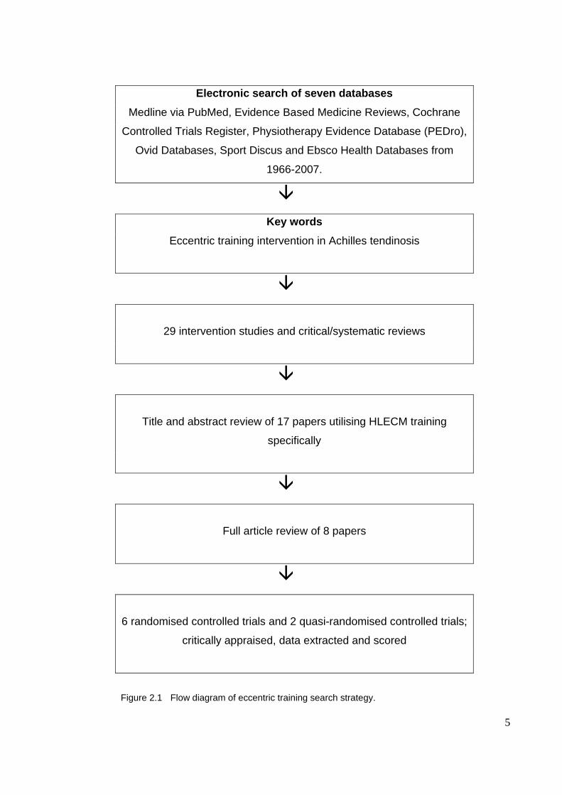
5
Electronic search of seven databases Medline via PubMed, Evidence Based Medicine Reviews, Cochrane
Controlled Trials Register, Physiotherapy Evidence Database (PEDro),
Ovid Databases, Sport Discus and Ebsco Health Databases from
1966-2007.
Key words
Eccentric training intervention in Achilles tendinosis
29 intervention studies and critical/systematic reviews
Title and abstract review of 17 papers utilising HLECM training
specifically
Full article review of 8 papers
6 randomised controlled trials and 2 quasi-randomised controlled trials;
critically appraised, data extracted and scored
Figure 2.1 Flow diagram of eccentric training search strategy.

6
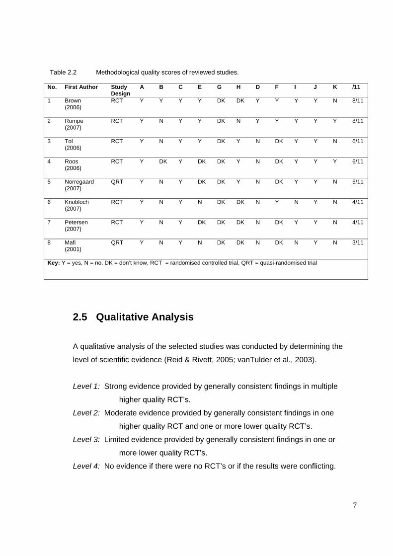
2.4 Methodological Quality
An assessment of the quality of each publication was conducted using a criteria list
recommended by the Cochrane Back Review Group (vanTulder et al., 2003)
(Table 2.1). This method was selected as its use facilitates comparison across
other Cochrane reviews of alternative interventions. The criteria list consists of 11
items which are rated with a yes (Y), no (N) or don’t know (DK) with a yes
response generating one point, thus the maximum score is 11. The criteria list
contains internal validity criteria that refer to characteristics of the study that might
be related to selection bias (A and B), performance bias (D, E, G and H), attrition
bias (I and K) and detection bias (F and J). An operationalisation of the criteria list
was used to assist with assignment of the yes/no/don’t know response (Appendix
A). The methodological quality scores are outlined in Table 2.2.
Table 2.1 Criteria list for the methodological quality assessment (van Tulder et al., 2003).
A. Was a method of randomization performed?
B. Was the treatment allocation concealed?
C. Were the groups similar at baseline regarding the most important prognostic indicators?
D. Was the patient blinded to the intervention?
E. Was the care provider blinded to the intervention?
F. Was the outcome assessor blinded to the intervention?
G. Were co-interventions avoided or similar?
H. Was the compliance acceptable in all groups?
I. Was the drop-out rate described and acceptable?
J. Was the timing of the outcome assessment in all groups similar?
K. Did the analysis include an intention-to-treat analysis?
7
Table 2.2 Methodological quality scores of reviewed studies.
No. First Author
Study Design
A B C E G H D F I J K /11
1 Brown (2006)
RCT Y Y Y Y DK DK Y Y Y Y N 8/11
2 Rompe (2007)
RCT Y N Y Y DK N Y Y Y Y Y 8/11
3 Tol (2006)
RCT Y N Y Y DK Y N DK Y Y N 6/11
4 Roos (2006)
RCT Y DK Y DK DK Y N DK Y Y Y 6/11
5 Norregaard (2007)
QRT Y N Y DK DK Y N DK Y Y N 5/11
6 Knobloch (2007)
RCT Y N Y N DK DK N Y N Y N 4/11
7 Petersen (2007)
RCT Y N Y DK DK DK N DK Y Y N 4/11
8 Mafi (2001)
QRT Y N Y N DK DK N DK N Y N 3/11
Key: Y = yes, N = no, DK = don’t know, RCT = randomised controlled trial, QRT = quasi-randomised trial
2.5 Qualitative Analysis
A qualitative analysis of the selected studies was conducted by determining the
level of scientific evidence (Reid & Rivett, 2005; vanTulder et al., 2003).
Level 1: Strong evidence provided by generally consistent findings in multiple
higher quality RCT’s.
Level 2: Moderate evidence provided by generally consistent findings in one
higher quality RCT and one or more lower quality RCT’s.
Level 3: Limited evidence provided by generally consistent findings in one or
more lower quality RCT’s.
Level 4: No evidence if there were no RCT’s or if the results were conflicting.

8
An arbitrary ranking of methodological quality was assigned to each study based
on their Cochrane score (Reid & Rivett, 2005):
Cochrane score Ranking Number of studies
8-11 High quality 2
5-8 Moderate quality 3
1-4 Low quality 3
2.6 Results
A total of eight studies met the criteria and were selected for the critical review.
These included six RCT’s and two QRT’s. The methodological quality of the
reviewed studies is displayed in Table 2.1 and the key features of the studies in
Table 2.3.
The methodological quality scores of the studies ranged from 3 to 8 out of a
possible 11 points. The majority of the studies had comparable groups at baseline,
had an acceptable drop-out rate and had similar timing of outcome assessments.
Approximately half of the studies detailed subject and treatment provider blinding
to treatment status. Few included an intention to treat analysis or described the
avoidance or detail of con-interventions.
From the qualitative analysis outlined above, there is moderate evidence to
suggest HLECM training is effective for treating Achilles tendinosis.
The following is an overview of the key findings of each study when comparing the
efficacy of HLECM training to other conservative treatment methods and the
proposed mechanisms of efficacy via its influence on tendon morphology. This is
preceded by an overview of the original Alfredson et al. (1998) research.

9
Table 2.3: Key findings of reviewed studies. Score
Study
Participants Mean Age Symptom Duration
Intervention Outcome Measures Key Findings
8/11 Brown et al. (2006)
Group 1: N = 13 46.3 yrs 8.1 mnth Group 2: N = 13 46.3 yrs 10.9 mnth
Group 1: Aprotinin injection x3 over 3 weeks and HLECM training Group 2: Placebo injection x 3 over 3 weeks and HLECM training
VISA-A Tenderness assessment Pain VAS Single leg hops Return to sport
No significant benefit of Aprotinin over Placebo in any outcome measures Significant increase in VISA-A scores following HLECM training
8/11 Rompe et al. (2006)
Group 1: N = 25 48.1 yrs 10.9 mnth Group 2: N = 25 51.2 yrs 12.5 mnth Group 3: N = 25 46.4 yrs 9.2 mnth
Group 1: HLECM training + gradual increase from 1x10 reps day 1 to 3x15 reps at day 14 Group 2: 3 sessions once a week over maximal area of tenderness Group 3: medication, stretching, training modification, ergonomic advice
VISA-A Likert scale for degree of improvement Pain scale Pain pressure threshold and tenderness US tendon diameter Success of treatment = 1 or 2 on improvement scale
No significant difference between Shockwave therapy and Eccentric groups on all outcome measures Significantly better outcomes for Eccentric and Shockwave therapy than Wait-and-see group (p<0.001) Significant improvement on all outcome measures except tendon diameter in Eccentric group and Shockwave therapy groups (p<0.001)
Key: RCT = randomised controlled trial, QRT = quasi-randomised trial, N = participant number, yr/s = year/s, US = ultrasound, reps = repetitions, FAOS = foot and ankle outcome scale, VISA-A = Victorian Institute of Sport Assessment – Achilles, KOOS = knee injury osteoarthritis outcome score, VAS = visual analogue scale.

10
Score
Study/Score
Participants Mean Age Symptom Duration
Intervention Outcome Measures Results
6/11 Tol et al. (2006)
Group 1: N=34 44.1 yrs 33.7 mnth Group 2: N=36 45.1 yrs 27.7 mnth
Group 1: HLECM training Group 2: HLECM training + dorsiflexion night splint 12 weeks
VISA-A Patient satisfaction rating of poor, fair, good, excellent Treatment success = good or excellent
No significant difference between Eccentric and Night splint groups in all outcome measures Significant increase in VISA-A score in Eccentric and Night Splint groups
6/11 Roos et al. (2004)
N = 44 46 yrs, 5.5 mnth Group 1: N=16 Group 2: N=13 Group 3: N=15
Group 1: HLECM training + gradual increase of reps/ straight knee Group 2: Dorsiflexion night splint 12 weeks Group 3: HLECM training + dorsiflexion splint
FAOS Likert scale for physical activity Likert scale for difficult during sport
No significant difference between groups in any outcome measures Significantly improvement on FAOS pain subscale in all groups
5/11 Norregaard et al. (2007)
N=45 Group 1: 41 yrs 26 mnth Group 2: 31 yrs 31 mnth
Group 1: HLECM protocol + gradual increase reps avoid pain, included concentric exercise Group 2: 5x 30 second calf stretch daily
KOOS questionnaire Tenderness US tendon thickness
No significant difference between Eccentric and Stretching groups in any outcome measures
Key: RCT = randomised controlled trial, QRT = quasi-randomised trial, N = participant number, yr/s = year/s, US = ultrasound, reps = repetitions, FAOS = foot and ankle outcome scale, VISA-A = Victorian Institute of Sport Assessment – Achilles, KOOS = knee injury osteoarthritis outcome score, VAS = visual analogue scale.

11
Score Study/Score
Participants Mean Age Symptom Duration
Intervention Outcome Measures Results
4/11 Knobloch et al. (2007)
Group1: N=15 33 yrs Group 2: N=5 32 yrs
Group 1: HLECM + reps performed once daily Group 2: cryotherapy and relative rest
Capillary blood flow Tissue oxygen saturation (SO2) Post capillary venous filling pressure (rHb) Pain VAS
No significant difference in flow, SO2 between groups Significant rHb decrease in eccentric group only (p<0.05) Significant reduction of pain in eccentric group only (p<0.05)
4/11 Petersen et al. (2007)
Group 1: N=37 42.5 yrs 7.4 mnth Group 2: N=35 42.6 yrs 7.3 mnth Group 3: N=28 43 yrs 7.0 mnth
Group 1: HLECM training Group 2: AirHeel brace worn daily for 12 weeks Group 3: HLECM training and AirHeel brace
AOFAS hindfoot scale US tendon diameter measure Pain VAS
No significant difference between groups in any outcome measures Significant decrease in AOFAS score all groups (p<0.0001)
3/11 Mafi et al. (2001)
Group 1: N=22 48.1 yrs 18 mnth Group 2: N=22 48.4 yrs 23 mnth
Group 1: HLECM training Group 2: heel raises, step-ups, skipping and side jumps
Pain VAS Patient satisfaction - method unclear
Eccentric training significantly better than concentric training (p<0.002)
Key: RCT = randomised controlled trial, QRT = quasi-randomised trial, N = participant number, yr/s = year/s, mnth = months, US = ultrasound, reps = repetitions, FAOS = foot and ankle outcome scale, VISA-A = Victorian Institute of Sport Assessment – Achilles, KOOS = knee injury and osteoarthritis outcome score.

12
2.7 Heavy Load Eccentric Calf Muscle Training – The Original Study The study by Alfredson et al. (1998) was a landmark prospective study
investigating the efficacy of the unique HLECM regime for the treatment of
Achilles tendinosis. A full description of this research is presented here to allow
comparison with the studies selected for the critical review.
Alfredson et al. (1998) examined the effect of HLECM training on pain during
activity and calf muscle strength in a group of 15 recreational athletes with an
average age of 44.3 years. A control group of 15 participants selected for
surgical treatment with an average age of 39.6 years was also studied. At
baseline the surgical group had a much longer duration of symptoms, 33.5
months versus 18.3 months in the HLECM group, although the differences were
not statistically analysed. All participants had undertaken unsuccessful
conservative treatment. Inclusion in the study was based on a clinical and
ultrasound diagnosis of Achilles tendinosis 2-6 cm above the tendon insertion on
the calcaneus (termed mid-portion tendinopathy). All participants had morning
stiffness in the Achilles tendon and pain during running. Participants were
excluded if they had bilateral symptoms or restricted ankle motion due to other
injuries or conditions.
The participants were instructed to perform the HLECM training protocol twice
daily, seven days per week for 12 weeks. Continuation of running was permitted
if it could be performed with mild discomfort or pain free. Two components were
included within the HLECM protocol involving the calf muscle being eccentrically
loaded with both the knee straight and the knee bent (Fig 2.1 & 2.2). The
authors proposed this distinction allowed for preferential activation of the soleus
muscle in the latter. Each component was performed in three sets of 15
13
repetitions three times per day. Participants were instructed to continue their
exercises with the exception of pain that became disabling.
The loading method consisted of the participant standing on their forefoot, ankle
maximally plantarflexed, on the edge of a step with all their body weight on the
injured leg. From this position, the participant lowered the heel below the
forefoot. The non-injured leg was used to return to the start position to ensure
there was no concentric calf muscle activity occurring in the injured leg. The
exercise was repeated for the set number of repetitions in a rhythmical fashion.
When the participants could perform the eccentric loading exercise without pain,
the load was increased by adding weight to a backpack worn during the exercise
or the use of a weights machine. The surgical group underwent a post-operative
training regime which involved a two week immobilisation period followed by
flexibility training and both concentric and eccentric strengthening exercises for a
period of up to one year (Alfredson, Pietila, Ohberg, & Lorentzon, 1998).
Calf muscle strength was assessed using a Biodex Isokinetic dynamometer and
quantified by measurement of peak torque (Nm), the highest torque
measurement from one repetition, and total work (joules), the average work per
repetition. Peak torque was measured in the HLECM group before (week 0) and
after (week 12) the eccentric training regime and at week 0 and week 24 after
surgery in the surgical group. The rationale for a discrepancy in chronology of
outcome measurement between the two groups is not evident. Pain during
activity was measured using a visual analogue scale (VAS) and recordings were
taken with the strength measures.
At baseline, the surgical group had significantly lower concentric plantarflexion
strength at 90 s-1 and 225 s-1 (18.7% and 23.7% respectively) and lower eccentric
plantarflexion strength (13.6%) than the non-injured side. The HLECM group also
demonstrated significantly lower concentric plantarflexion strength at 90 s-1 and
225 s-1 (12.1% and 18.0% respectively) and lower eccentric plantarflexion

14
strength (15.7%) than the non-injured side. No side-to-side difference in work
data was evident during eccentric plantarflexion contractions.
Following eccentric training there was no difference in side-to-side peak torque
values at any velocity or contraction in the HLECM training group. Average work
increased only during the concentric 90° s-1 condition after eccentric training. The
surgical group continued to demonstrate significantly less peak torque values at
all velocities and during both eccentric and concentric contractions in the
operated leg compared to the uninjured side. It is not stated whether the peak
torque values increased significantly compared to pre-training peak torque values
in the surgical group. Despite the strength deficit evident in the surgical group,
pain levels decreased significantly in both groups following HLECM training
(week 12) and surgery (week 24). All patients were satisfied with treatment and
returned to their pre-injury levels within these time frames also.
From these findings, the authors concluded that HLECM training improved calf
muscle strength and returned patients to their pre-injury level more rapidly and
effectively than surgery. However, in order to make a more accurate
comparison it may have been valuable to compare the two groups at identical
timeframes and to equalise groups in terms of symptom duration at baseline. It
is also possible that the strength and functional improvements demonstrated in
this study are augmented by a generally higher activity level of recreational
athletes compared to the general population.

15
Figure 2.2: Straight knee component of HLECM training.
Figure 2.3: Bent knee component of HLECM training.
16
2.8 Key Findings
Eight studies were reviewed and their main findings are presented here. For the
studies selected, this includes the effect of eccentric training on tendon
morphology and comparison of the efficacy of HLECM training with other
conservative treatment measures. Details of the study populations, interventions
and outcomes are outlined in Table 2.3.
2.8.1 Tendon Morphology
The ingrowth of neovessels and accompanying nerve structures in the Achilles
tendon has been proposed as a cause of pain in Achilles tendinosis and
associated with a reduction in functionality and increased chronicity of
tendinopathy (Ohberg & Alfredson, 2004; Peers, Brys, & Lysens, 2003;
Snellenberg, Wiley, & Brunet, 2006). Knobloch et al. (2007) assessed changes
in Achilles tendon microcirculation following HLECM training in participants with
insertional or mid-portion tendinopathy. An eccentric training group carried out a
modified HLECM protocol while a group undergoing cryotherapy and relative rest
served as controls. The statistically significant reduction of postcapillary venous
pressure and minimal change to capillary blood flow found in this study suggests
that HLECM training does not appear to have a thrombotic effect on neovessels
as proposed by other authors (Ohberg & Alfredson, 2004). Instead, it is thought
that the mechanism of effect may be the facilitation of venous outflow and
clearance of metabolic products in the tendon.
Although the effect of HLECM training on tendon vascularity was the chief
purpose of this study, the effectiveness of the eccentric training technique for the
studied population could also be determined. In terms of treatment efficacy, the
eccentric training group demonstrated a significant reduction in pain levels (48%)
at the 12 week mark whereas the control group did not. No change in the
amount of sporting participation was demonstrated in either group.

17
2.8.2 Compared to Concentric Training
While it is accepted that a graduated loading of the Achilles tendon is valuable in
order to treat tendinopathy, no rationale is given in the current literature for why
eccentric training of the tendon may be efficacious compared to other forms of
loading. Loading of the Achilles tendon may occur during exercise or through
patient-generated stretching and night splinting which provide a passive stretch
to the tendon and calf musculature.
Mafi et al. (2001) compared the efficacy of a concentric training programme to
the HLECM training protocol in 44 participants with mid-portion tendinopathy.
The concentric training regime involved predominantly concentric activities such
as heel raises, step-ups, skipping and side jumps which were progressed in
difficulty over the 12 week period. No previous work implementing this protocol
was evident. Pain was assessed using a VAS and patient satisfaction was also
obtained although the methodology of this evaluation was not outlined.
Following the 12 week intervention, 82% of participants in the HLECM group
were satisfied with treatment and had resumed regular activity. In comparison,
36% of participants were satisfied with treatment and had returned to regular
activity in the concentric training group.
The eccentric training achieved significantly better results than a concentric
training regime in the short term in patients with mid-portion Achilles
tendinopathy. The authors postulated this may have been due to increased
eccentric calf muscle strength or caused by a lengthening of the muscle-tendon
unit and thus an increase in its ability to bear load during activity. The inclusion
of calf strength measures such as those used by Alfredson et al. (1998) or
measures of dynamic passive muscle-tendon elastic properties in this study may
have assisted with clarification of the different biomechanical effects of each form
of training (Gadjosik, Alfred, Gabbert, & Sonsteng, 2007)
18
2.8.3 Compared to Stretching
Stretching, an alternative form of loading, is often prescribed to patients with
Achilles tendinopathy, however, previously the efficacy of stretching for this
condition has been examined only as a component of a cluster of conservative
management methods (Mayer, Hirschmuller, Muller, Schuberth, & Baur, 2006). It
has been proposed the combination of specific soft tissue mobilisation and
patient-generated stretches of the Achilles tendon and calf muscles would alter
compliance of the tendon-muscle complex and thus reduce micro-failure of the
tendon when loaded (Hunter, 2000). While there may be a physiological
rationale for the use of stretching in Achilles tendinosis, no studies investigating
the efficacy of patient self-stretching have been undertaken until recently.
Norregaard and colleagues (2007) compared the effectiveness of eccentric
training and Achilles tendon stretching in a group of 45 patients with mixed
insertional and mid-portion tendinopathy. The interventions consisted of a
modified HLECM training protocol in one group and patient-generated stretches
of the calf muscle-tendon complex in another. Outcomes were determined
through assessment of tenderness by manual examination, ultrasonography, use
of the Knee Injury and Osteoarthritis Outcome Score (KOOS) and an
assessment of global improvement.
The results of the study revealed no significant difference between the stretching
and eccentric training group on all measured parameters. There were
statistically significant improvements on the KOOS score and a significant
decrease in tendon diameter and tenderness could be seen at the one year mark
in both groups. An analysis of predictors of outcome demonstrated a poorer
prognosis for women, those with thinner tendons and insertional tendinopathy.
19
2.8.4 Compared to Night Splinting
Night splinting, with the ankle in a neutral or dorsiflexed position, functions as a
form of prolonged stretching to the both the Achilles tendon and calf musculature.
Use of a night splint has been investigated in other lower limb conditions, such as
plantar fasciitis, with beneficial effects (Barry, Barry, & Chen, 2002). The effect
of night splints on Achilles tendinopathy has only recently been examined (Roos
et al., 2004; Tol et al., 2006). The rationale for the application of a prolonged
stretch in tendinopathy has not been specifically outlined by these authors.
Roos et al. (2004) investigated the effect of a modified HLECM training protocol
and night splinting alone, or in combination, in a group of 44 participants with a
clinical diagnosis of mid-portion tendinopathy. The night splint was worn over the
anterior ankle and designed to maintain a position of 90º dorsiflexion. The
outcome measures implemented were the Foot and Ankle Outcome Score
(FAOS), physical activity levels and difficulty during sporting activities which were
recorded on Likert scales unique to the study.
Following the intervention period, all groups improved significantly, demonstrated
by the decrease in pain rating on the FAOS and improvements on the Likert
scales utilised. At one year, all three groups reported decreased pain levels of
between 35-42%. No statistically significant differences were seen in pain
scores, physical activity scores or difficulty with sport measures at any time
between the three groups. There was a trend toward the HLECM training group
demonstrating a greater reduction in pain than the night splint group at 12 weeks,
deemed to be a clinical but not statistically significant difference. From these
results, the authors concluded that HLECM training reduces pain and increases
function in patients with Achilles tendinopathy up to one year. However, no
added value of wearing a night splint could be observed from the results of this
study. It was thought by the authors that a larger sample size would have
yielded a statistically (rather than clinically) significant difference in the groups.

20
Tol et al. (2006) also investigated the additional value of a night splint to the
HLECM protocol in 58 patients clinically diagnosed with chronic mid-portion
Achilles tendinopathy. Participants were randomised into HLECM training alone
or HLECM training combined with night splinting and the outcome assessments
used included the Victorian Institute of Sports Assessment- Achilles
Questionnaire (VISA-A) and a patient satisfaction rating scale.
Patient satisfaction with the treatment was 63% for the eccentric exercise group
and 48% for the night splint group. The VISA-A scores increased significantly in
both groups although again there was no significant difference observed between
the groups. Based on these findings the authors also concluded there is no
value of wearing a night splint in addition to performing the HLECM protocol for
the treatment of Achilles tendinopathy.
2.8.5 Compared to Bracing
Petersen et al. (2007) compared the effect of an AirHeel cushioned brace,
eccentric training and a combination of these therapies in 100 patients with a
clinical and ultrasound diagnosis of mid-portion tendinopathy. The AirHeel is a
specifically designed compressive brace for treatment of Achilles tendinopathy
and was instructed to be worn by patients during the day time. The compression
imparted by the brace is thought to enhance circulation and reduce swelling
associated with tendinopathy. However, this theory has not been validated by
research. The eccentric training regime utilised in this study was identical to the
HLECM training outlined by Alfredson et al. (1998) including use of the straight
and bent knee components.
This study assessed both pain and function using the Short Form-36, the
American Orthopaedic Foot and Ankle Society (AOFAS) score and the VAS in
addition to ultrasonography investigating tendon diameter following intervention.
At completion of the intervention period the results demonstrated a significant

21
decrease in the AOFAS score in all groups at 12 weeks and in only the
combination group at one year. Pain during everyday activities, walking and
sports decreased significantly in all groups although the results were less marked
than earlier studies. Tendon diameter did not alter significantly after intervention.
Based on these outcomes, no significant difference in the efficacy of the three
treatment interventions could be observed.
2.8.6 Compared to Shockwave Therapy
Extracorporeal shockwave therapy (ESWT) is thought to produce an initial
analgesic effect in tendinopathies by altering cell membrane permeability. This
means a higher stimulus is required to provoke an action potential in the sensory
neuron conveying pain messages to higher centres (Chung & Wiley, 2002). In
the long term, ESWT may increase blood flow and induce an inflammatory
mediated response through induced damage to vascular structures. Neither of
these theories have been substantiated by research.
Rompe et al., (2007) conducted a randomised controlled trial comparing the
effect of a modified HLECM training regime, shock-wave therapy or a wait-and-
see approach for mid-portion Achilles tendinopathy in 75 patients. Shock-wave
therapy was applied by the senior author in a standardised dose for three
sessions at weekly intervals. The area of maximal tenderness was treated in a
circumferential pattern starting at the point of maximum pain level. Participants
in the wait-and-see group visited their orthopaedic specialist once during the
intervention period of 12 weeks. Alternative treatment methods including
medication, stretching, training modification and ergonomic advice were
discussed with the patients in this group although it is not outlined whether they
were undertaken. A number of functional outcome measures were implemented
including the VISA-A, a degree of improvement scale and a numeric rating scale
for pain, similar to the VAS. Pain pressure threshold of the Achilles tendon was
also assessed using an algometer, which is unique to this research.
22
Participants in the HLECM training and shock-wave therapy groups improved
significantly on the VISA-A, on the Likert scale for general improvement and in
the NRS for pain and for pain threshold compared to the wait-and-see group.
Tenderness on the NRS improved in all groups significantly. Both the shock-
wave therapy and eccentric training lead to a successful outcome in 50-60% of
patients with no significant difference in any of the outcome measures between
these two forms of therapy. A proportion of participants appeared to benefit from
crossover to shock-wave therapy or the eccentric training regime following the
initial intervention. This suggests that perhaps differing conservative treatments
may be beneficial for specific subgroups of patients with Achilles tendinosis,
however, the features defining such subgroups remain unknown.
2.8.7 Compared to Aprotinin
Aprotinin is a broad-spectrum metalloprotease (MMP) inhibitor used to treat
tendinopathy among a range of other conditions. MMPs have been shown to be
present in excessive proportions in patellar and rotator cuff tendinopathy (Rukin
& Maffulli, 2006). Aprotinin is thought to normalise the concentration of MMPs,
assisting healing. Brown et al. (2006) investigated the effectiveness of aprotinin
in combination with eccentric exercise in 26 patients with mid-portion Achilles
tendinosis. Participants were randomised to either receiving an aprotinin
injection and eccentric exercise or a placebo (saline) injection and eccentric
exercise. Three aprotinin or placebo injections were administered
peritendinously once a week for the first three weeks. The eccentric exercise
programme was based on the Alfredson et al. (1998) HLECM model but not
outlined clearly in this study. An assessment of tendon tenderness, number of
hops until pain, number of single leg raises to pain, return to sport, patient
satisfaction rating and VISA-A scale were utilised.
Absolute improvements in VISA-A score were greater in the aprotinin group
compared to the placebo group but this was not statistically significant. Most

23
other evaluation measures were not statistically significantly different between
groups at any follow-up point except for number of hops to pain and patient rating
were better in the aprotinin group at the two week follow-up. Compared to other
studies, the therapeutic effect of eccentric training at the twelve week mark was
weak with only 13% of the placebo group and 31% of the aprotinin group
returned to sport. At the one year follow-up point however, the overall results
were markedly better with 85% in the aprotinin group and 77% in the placebo
group returning to previous sporting levels. Although the results were not
statistically significantly superior to placebo, the authors recommended a larger
trial be conducted due to the beneficial effects seen in previous work on patellar
tendinopathy.
2.9 Discussion
Alfredson et al. (1998) developed a unique eccentric training protocol that has
been widely utilised in subsequent research. This research has compared the
efficacy of HLECM training with many other conservative interventions for
Achilles tendinosis. In addition, the influence of eccentric training on tendon
structure and morphology has been examined. This critical review has evaluated
both the quality and key findings of this body of research in order to determine
what conclusions may be drawn currently and where future research is required.
The original study by Alfredson et al. (1998) reported 100% of participants
returned to previous activity with reduced pain levels within a 12 week period.
While other research has demonstrated good results using the HLECM protocol,
almost none have replicated these statistics. This may be due to several factors
including the following; inconsistent inclusion and exclusion criteria, different
outcome measures implemented, participant compliance levels and variations in
HLECM protocol methodology including training principles.
24
2.9.1 Inclusion and Exclusion Criteria
The diagnosis of Achilles tendinopathy may be achieved via a clinical exam,
ultrasound (US), magnetic resonance imaging (MRI) or a combination of these
(Cook, Khan, & Purdam, 2002). The studies reviewed demonstrated variability in
the use of diagnostic measures for inclusion, with five of the eight utilising a
clinical diagnosis only. It is possible a clinical diagnosis may not exclude the
presence of other associated conditions such as paratendonitis, Haglund’s
deformity or an Achilles tendon tear. The presence of other conditions may in
turn influence the efficacy of the HLECM regime in the studied population.
The criteria for inclusion within each study also varied according to age, activity
levels, location and duration of symptoms. As Achilles tendinosis primarily
affects individuals aged between 35 and 45 years, it is ideal for study populations
to reflect this. Most studies reviewed did include participants with a mean age
between 31 and 51 years. Athletic populations, such as those in the Alfredson et
al. (1998) study, demonstrate excellent results with HLECM training in a 12 week
period. A recent prospective study has demonstrated reduced efficacy of
HLECM training for non-athletic populations (Sayana & Maffulli, 2007). The
majority of studies reviewed here contained a proportion of individuals involved in
recreational sport or did not detail the composition of their study populations.
Two studies specified participation in recreational sport for inclusion and both
demonstrated significant improvements in function and reduction in pain for the
eccentric training group (Roos et al., 2004; Tol et al., 2006).
The location of symptoms in the study population was fairly standard across the
studies reviewed with six of eight studies including only those with mid-portion
tendinopathy. Clinical trials have demonstrated that individuals with insertional
tendinopathy do not respond as positively as those with mid-portion symptoms to
eccentric training (Fahlstrom et al., 2003). Norregard et al. (2007) and Knobloch
et al. (2007) did not exclude those with insertional pain however the former study

25
did observe less of a response to HLECM training in this subgroup within their
research.
In contrast, the duration of symptoms was considerably varied across the study
populations, ranging from a mean of 5.5 to 33.7 months. With exception of
Alfredson et al. (1998), all studies demonstrated similar symptom duration
periods between compared groups, although many were not statistically
analysed at baseline for differences. Tol et al. (2006) found patient satisfaction
with outcome was influenced by symptom duration whereby participants with
symptoms present less than 5.5 months rated 89% compared to 50% satisfied
when the duration was greater than 5.5 months. In contrast, Norregaard et al.
(2007) did not find symptom duration a significant predictor of outcome.
Exclusion criteria for participants with other medical conditions or those with
bilateral symptoms were also very diverse across studies. Fundamental to the
HLECM training protocol is the concept that the symptomatic tendon is subjected
to only eccentric muscle contraction of the calf musculature during the exercises.
The presence of bilateral symptoms means that in order to accomplish this, the
return to the start position must be achieved via use of the upper limbs on an
external support. The original study by Alfredson et al (1998) did not include
patients with bilateral symptoms. A number of the studies reviewed did not
exclude patients with bilateral symptoms, yet did not outline how a concentric
component was avoided (Brown et al., 2006; Knobloch et al., 2007; Norregaard
et al., 2007; Roos et al., 2004; Tol et al., 2006). Petersen et al. (2007) instructed
participants to rise up on their toes and then place their weight on the uninjured
leg. This would mean some component of the regime was concentric rather than
a purely eccentric load as intended. It is possible that the performance of the
concentric component on the affected leg in persons with bilateral symptoms
may have influenced the efficacy of the HLECM training regime in these studies.

26
A further criteria identified in some of the reviewed studies excluded those
participants who had previously performed heavy load eccentric exercises or had
been unable to perform these exercises (Norregaard et al., 2007; Tol et al.,
2006). It is possible this criterion would create a selection bias by excluding
those participants who have not responded to HLECM training in the past. This
means the remaining study population selected might demonstrate an enhanced
treatment effect and thus not be representative of a typical patient population.
2.9.2 Outcome Measures
A comparison of results across studies is complex when research populations
vary but also when different outcome measures are utilised. Earlier studies
tended to implement only the VAS for pain and in some cases an interview
method of ascertaining satisfaction and return to sport (Mafi et al., 2001). This
latter method may be influenced by the therapist-patient relationship, particularly
if the interviewer is also conducting the research as in the Alfredson et al. (1998)
study.
More recently, studies have also used functional outcome measures including
the VISA-A, the KOOS, the FAOS and unique clinical outcome scales. These
measures differed in terms of being disease specific (VISA-A) and site specific
(KOOS, FAOS). The VISA-A was the most consistently used in recent studies
and has been shown to possess excellent test-retest reliability and construct
validity (Robinson et al., 2001).
The use of unique Likert scales in research also confounds the difficulty in
comparing the efficacy of interventions. All of the studies within the current
review developed scales to determine a range of outcomes including tenderness,
patient satisfaction, return to sport, degree of improvement and participation in
physical activity. Each scale is inimitable and thus comparison of the impact of
HLECM training on each population studied is difficult to ascertain. By utilising

27
identical outcome measures, it may be possible to determine features of either
the population or intervention itself which may be more effective.
2.9.3 Methodological Variation
Variations in methodology between studies utilising the HLECM training protocol
were reasonably minimal in terms of training principles such as sets, repetitions
and frequency of performance. Some studies gradually introduced the HLECM
regime over the first few weeks (Norregaard et al., 2007; Rompe et al., 2007;
Roos et al., 2004) or reduced the frequency from twice a day (180 repetitions) to
once a day only (90 repetitions) (Knobloch et al., 2007). Progressions of the
HLECM training protocol were possibly more varied as the original study did not
outline specifically how much weight was added to each participant’s backpack,
except to state that it should remain painful to complete the exercises.
The reproduction of Achilles tendon pain with exercise performance differed
between studies as did the concurrent return to sporting or recreational activities.
Alfredson et al. (1998) advised running may be continued if it could be performed
with mild discomfort or pain free, with the majority of subsequent research
adhering to this component of the protocol. Variations included an avoidance of
tendon loading activities for the first four weeks (Tol et al., 2006) and the
continuation of normal activity throughout (Knobloch et al., 2007). Clearly this
would result in a large variation of tendon loading for each individual both within
and across studies utilising this regime.
The reproduction of pain is suggested as an important component of the HLECM
training protocol. Some studies attempted to reduce the pain associated with
eccentric loading by altering their methodology accordingly (Norregaard et al.,
2007; Rompe et al., 2007; Roos et al., 2004). This included a gradual
introduction or decrease in number of repetitions. Furthermore, Norregaard et al.
(2007) advised participants to avoid progressing exercises if painful contrary to
28
the Alfredson et al. (1998) study where patients were advised to progress
exercises in order to achieve a painful state. The rationale for the modifications
in the studies reviewed here was primarily to increase patient compliance with
the regime.
In order for a training regime to be maximally effective, it is assumed compliance
to the training principles prescribed is essential. However, most studies
investigating the efficacy of HLECM training did not report participant compliance
levels. Measurement of compliance, when detailed, was largely through training
diaries, regular telephone calls and follow-up face-to-face visits. The definition of
good compliance also varied, although was commonly classified as performance
of at least 75% of the sets and repetitions prescribed (Roos et al., 2004; Tol et
al., 2006). The number of repetitions and frequency of performance required in
the HLECM training protocol is very high and thus it is reasonable to assume a
proportion of patients would not complete the required amount each day if
unsupervised. Tol et al. (2006) reported 27% of patients were completing less
than 50% of the regime at the 12 week mark. This was the only study to
demonstrate a relationship between a more positive outcome and increased
compliance, although the results were not statistically significant.
A final methodological variation is concerned with the exercise technique itself.
Alfredson et al. (1998) proposed that the two conditions of the HLECM protocol,
the bent-knee and straight-knee components, target the soleus and
gastrocnemius muscles respectively. It is not stated why it is necessary to target
each calf muscle selectively during the eccentric training regime. The knee
flexion angle required to achieve specific activity of each calf muscle has also not
been outlined in the original study. In order to replicate a training protocol for
research purposes, it is necessary to adhere to an identical technique. As there
are currently no clear guidelines, it is possible there may have been some
variation in technique execution between studies. Almost all subsequent studies
reviewed here emphasised the inclusion of the two technique conditions, yet no

29
research has investigated whether this selective muscle activity actually occurs
during HLECM training in a patient population.
2.10 Muscle Activity
Muscle activity is commonly measured by EMG which also facilitates the
collection of information regarding muscle activation timing, fatigue and relative
activity levels of individual muscles during selected movements (Soderberg &
Knutson, 2000). Surface electrodes are usually utilised when investigating
activity in the calf muscles (triceps surae) and are placed on the skin surface
thereby providing a more global view of muscle activity during a contraction
(DeLuca, 2006; Mademli, Arampatzis, Morey-Klapsing, & Bruggemann, 2004). In
order to evaluate the relative activity levels of selected muscles, the signal must
be normalised to a maximum voluntary contraction (MVC) of each muscle
(Soderberg & Knutson, 2000). An isometric MVC is commonly utilised as this
has been demonstrated to produce less intra-individual variability than isokinetic
or dynamic methods (Burden, Trew & Baltzopoulos, 2003). Normalisation allows
the average rectified values, termed the root mean square (RMS) signal for each
muscle to be matched to a corresponding maximum value and muscle activity
levels compared (Kennedy & Cresswell, 2001).
The gastrocnemius and soleus muscles, although sharing a similar anatomical
location, differ markedly in their architecture, fiber type composition and function
(Kawakami, Ichinose, & Fukunaga, 1998). Their relationship to joint angles of
the ankle and knee are also specific. The EMG activity of the triceps surae at
varying knee joint angles has been examined in scientific research, primarily
during a maximum isometric plantarflexion contraction (Arampatzis et al., 2006;
Kennedy & Cresswell, 2001; Miaki, Someya, & Tachino, 1999; Signorile,
Applegate, Duque, Cole, & Zink, 2002).

30
As the bi-articular gastrocnemius crosses both the ankle and knee joints, it is
generally accepted that the activity and torque output of this muscle is more
dependent on the angle of these joints than is the mono-articular soleus
(Kennedy & Cresswell, 2001). Due to the force-length relationship of skeletal
muscle fibers, pronounced knee joint flexion angles cause the gastrocnemius to
become actively insufficient. This is where the muscle reaches a critical
shortened length where torque production cannot increase, even if the muscle is
fully activated (Arampatzis et al., 2006). The research suggests this does not
occur until the knee joint angle reaches at least 80º flexion (Arampatzis et al.,
2006). At angles greater than this (i.e. where the knee is less flexed), there is an
increasing amount of gastrocnemius activity up to a maximum with the knee fully
extended, measuring 180º (Miaki et al., 1999; Signorile et al., 2002).
The angle of knee flexion required to facilitate activation of the soleus muscle
during HLECM training has not been identified in the Alfredson et al. (1998)
study. Electrical activity of the soleus muscle at differing knee joint angles has
been investigated and the results demonstrate it to be maximal at 40º to 90º
flexion (Kennedy & Cresswell, 2001; Miaki et al., 1999; Signorile et al., 2002). It
is thought this may be due to a corresponding suppression of gastrocnemius
activity in this position owing to the phenomenon of active insufficiency (Kennedy
& Cresswell, 2001). In contrast, other research has demonstrated constant EMG
activity from the soleus despite alterations in knee and ankle joint angles
(Miyamoto & Oda, 2003). During the bent-knee condition of HLECM training, the
knee flexion angle is greater than 90º. Given the research findings discussed, it
appears that the gastrocnemius muscle would remain active in this condition and
not reach a position of active insufficiency. Whether bending the knee during
HLECM training preferentially targets the soleus muscle in persons with Achilles
tendinosis, as purported by Alfredson et al (1998), is not known.
Potts (2005) investigated the EMG activity of the medial gastrocnemius and
soleus muscles during performance of both conditions of the HLECM training
31
protocol in 18 participants without pathology. The EMG activity was examined
during the eccentric phase of the contraction and expressed as a percentage of a
value derived from an MVC of each corresponding muscle. During the straight-
knee condition, the gastrocnemius activated at 61% and the soleus at 59% of
their MVC values. During the bent-knee condition, the gastrocnemius activated
at 47% and the soleus at 66% of their MVC values. While there was a trend for
increasing relative activity of the soleus and decreasing relative activity of the
gastrocnemius in the bent-knee condition, the difference between the conditions
was not statistically significant. It is therefore possible that the knee flexion angle
used during HLECM training is insufficient to inhibit gastrocnemius activity and
preferentially target the soleus in persons without Achilles tendon pain.
2.11 Summary
Heavy-load eccentric calf muscle training as a treatment for Achilles tendinosis
has been studied extensively. Although difficult to compare outcomes, a
significant reduction of pain and increase in function was observed following
HLECM training in all studies reviewed here. However, none of the studies have
replicated the results of Alfredson et al. (1998). The efficacy of HLECM training
has been demonstrated as not significantly different to shockwave therapy,
stretching, bracing or night splinting. However, it has shown to be superior to
concentric training. There was a trend in the studies reviewed for the groups
participating in HLECM training to demonstrate enhanced functional outcomes
but in all cases these were not statistically significant. Future research using
larger populations may possibly yield significant differences in the efficacy of
interventions for Achilles tendinosis.
A methodological variable not well detailed in the original or reviewed studies
includes the use of a bent-knee and straight-knee condition, proposed to
selectively activate the soleus and gastrocnemius musculature. Research has
demonstrated no significance difference in activation levels of these muscles

32
during HLECM training in participants without pathology only (Potts, 2005). This
variable has not been examined in a pathological population. The purpose of the
following biomechanical study is to examine muscle activity levels of the
gastrocnemius and soleus muscles in persons with Achilles tendinosis during
performance of the HLECM training conditions and additionally, to compare this
to activity levels demonstrated in a non-pathological population.

33
3. Methodology This chapter describes the methodology of the biomechanical study and is
divided into four sections comprising of the study participants and outcome
measures utilised, equipment and procedures implemented and statistical
methods applied.
3.1 Participants
All methods utilised in this study were approved by the Northern Regional Ethics
Committee on the 29th January 2008. All participants signed a document of
informed consent (Appendix B). The sample size was determined by selecting
18 participants from a larger sample of 46. Participants were matched as closely
as possible for age with a group of 18 subjects without pathology from Potts
(2005) study. These participants, who served as controls for the current study,
were part of a previous Masters Dissertation.
Participants were recruited by information sheets located in physiotherapy and
sports medicine practices in Auckland and via local media advertising between
February and April 2008 (Appendix C and D). Participants were provided with
an information sheet (Appendix E) from their treating medical professional.
The inclusion criteria were as follows:
• Aged over 20 years.
• Diagnosed with mid-portion Achilles tendinosis by their treating
physiotherapist, sports medicine doctor or by the researcher (a practicing
physiotherapist). A clinical diagnosis of Achilles tendinosis was used in
this study. No imaging (i.e. ultrasound or MRI) was required to confirm the
presence of the condition. The diagnostic criterion was a painful area of
34
the Achilles tendon 2-6cm proximal to the calcaneal insertion point on
palpation. This may or may not have been associated with swelling.
• Unilateral or bilateral Achilles tendinosis. The electromyographic data was
collected from the lower limb deemed most symptomatic by the participant
at the time of assessment.
The exclusion criteria were as follows:
• Diagnosed with insertional Achilles tendinosis. Patients with pain and/or
swelling in the insertional area of the tendon, indicating insertional Achilles
tendinosis, were excluded from this study. This is due to the fact that
patients with insertional Achilles tendinosis have demonstrated a poorer
response to eccentric training and are infrequently included within study
populations examining the HLECM protocol.
• Have had a previous history of Achilles tendon repair or rupture.
• Have had a previous corticosteroid injection into either Achilles tendon
• Has the presence of neural signs or symptoms affecting their lower limbs.
The following demographic information concerning age, weight, height, gender
and duration of symptoms was collected (Appendix F).
3.2 Outcome Measures
3.2.1 Victorian Institute of Sports Assessment – Achilles Questionnaire (VISA-A)
The VISA-A (Appendix G) is the only disease specific questionnaire to serve as
an index of the severity of Achilles tendinopathy. It was developed by Robinson
et al. (2001) who demonstrated that it has excellent test-retest reliability (r=0.93)
and construct validity. The questionnaire covers the domains of pain, stiffness,
function in daily living and sporting activity.
35
3.2.2 Lower Limb Task Questionnaire (LLTQ) The LLTQ (Appendix H) focuses on physical tasks related to lower-limb function
and is not disease specific. Instead the LLTQ scores the ability of an individual
to perform tasks in two separate constructs; activities of daily living and
recreational activities. In addition, the importance of each task is also rated.
The validity, reliability and responsiveness of the LLTQ have been established
(McNair et al., 2007). This outcome measure was included within this study in
order to examine the influence of Achilles tendinopathy on these two constructs
separately. It is possible that this condition may impact an individual’s ability to
participate in recreational activities more than activities of daily living.
3.2.3 Visual Analogue Scale
Pain levels were rated on a VAS from 0 = no pain to 10 = maximal pain for each
exercise condition and during each maximum voluntary contraction (Katz &
Melzack, 1999). This was noted by the researcher on a recording sheet for each
subject.
3.3 Equipment and Procedures
3.3.1 Eccentric Exercise Performance
Following preparation for EMG and range of motion testing, one of two envelopes
placed on a table were randomly selected by the participant indicating which
condition was to be tested first (i.e. straight knee or bent knee). A ten minute
warm-up was then performed on a stationary bike at a low to moderate intensity
prior to performing the HLECM exercise.

36
The eccentric exercise was performed according to the HLECM training protocol
outlined by Alfredson et al. (1998). Participants were given a demonstration and
an opportunity to practice five repetitions in order to ensure their technique was
correct for each exercise condition. The start position consisted of maximal
plantarflexion in standing on the tested limb followed by an eccentric dorsiflexion
to their maximal available dorsiflexion range over the edge of the step. The knee
was fully extended to 180º in the straight knee condition and flexed at 150º in the
bent knee condition. Three repetitions of each condition were performed in each
trial with the eccentric component of the exercise timing three seconds. Three
trials were recorded for each exercise condition.
3.3.2 Ankle Joint Motion
Ankle joint range of motion measurement was necessary in order to identify the
eccentric phase of each exercise condition. Range of movement through
plantarflexion and dorsiflexion was recorded using a dual axis electrogoniometer
(model 003, Penny and Giles, Gwent, England). A line between the middle of the
lateral malleolus and the lateral epicondyle of the fibula was marked with the
patient in standing. A second line was marked between the lateral malleolus and
head of the fifth metatarsal. The measurement arm of the electrogoniometer was
adhered to the skin surface using double-sided tape (3M Healthcare, St Paul,
MN) and further reinforced with strapping tape (3M) along the marked lines (Fig.
3.1a). The reliability of this method has been established by previous research
(Soper, Reid, & Hume, 2004). Prior to data collection the electrogoniometer was
calibrated using a 90º calibration frame. Data were sampled at 500Hz via
Superscope software (GW Instruments, Washington, USA).
3.3.3 Electromyographic Activity
Electromyography (EMG) was utilised to determine levels of muscle activity
during performance of the HLECM exercise. The participants tested limb was
37
prepared by shaving and cleaning the skin with alcohol wipes. Double-
differentiated, self adhesive surface electrodes were then placed on the soleus
and medial gastrocnemius muscles according to the SENIAM guidelines for
electrode placement (Hermens, Freriks, Disselhorst-Klug, & Rau, 2000) (Fig
3.1b). These electrodes can decrease artifacts included in the EMG signal and
reduce crosstalk from surrounding muscles (Soderberg & Knutson, 2000). A
reference electrode was placed over the tibial plateau. The longitudinal axis of
the electrodes was orientated in parallel with the approximate fibre direction of
each muscle (DeLuca, 2006). The EMG signal was sampled at 500Hz, amplified
1000x, band pass filtered (20-450Hz) and relayed to a Superscope software
package (GW Instruments, Washington, USA). The data was stored in a
McIntosh computer for subsequent analyses. The analysis of this data involved
a cursor routine that utilised the signals from the electrogoniometer in order to
identify the eccentric phase of muscle activity. RMS values were calculated
within this interval for three trials and then averaged.
Normalisation of the EMG signal from the gastrocnemius and soleus muscles
was conducted using a maximal voluntary contraction (MVC) of each muscle.
The gastrocnemius MVC was tested in prone with the knee extended (Fig. 3.2),
and the soleus in a sitting position with the knee flexed to 90º (Fig. 3.3). The
participants practiced using a ramping protocol of muscle contraction where they
were asked to generate 25%, 50% and then 75% of a MVC in each position.
Following a one minute rest, an MVC was performed with verbal encouragement
for a period of five seconds. Three MVCs were conducted in each position with a
one minute rest between trials. The EMG data was analysed using a cursor
routine targeting the most stable signals within a two second epoch. The
maximal RMS value across the three MVC trials was used in subsequent
analysis. The RMS value for each muscle in each condition was expressed as a
percentage of the RMS of the corresponding MVC.

38
3.5 Statistics
The dependent variables of interest were the RMS of EMG activity from the
gastrocnemius and soleus muscles during the eccentric phase of ankle joint
movement with the knee joint either flexed or extended. These data were
expressed as a percentage of activity recorded from the MVC. Data were
checked to identify outliers and abnormalities (kurtosis and skewness) in the
distribution. A three-factor repeated measures ANOVA was performed. The
repeated measures (within subjects) factors were the joint position and muscle,
while group was the between subjects factor. The alpha level was set at 0.05.
Levene’s test for equality of variances was performed and as a repeated
measures design was being utilised, Mauchly’s sphericity test was implemented.
Figure 3.1a Electrogoniometer placement Figure 3.1b EMG electrode placement
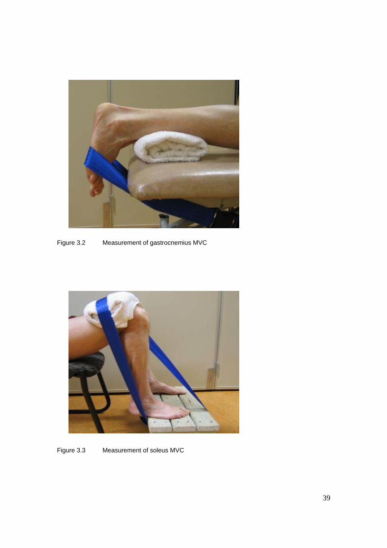
39
Figure 3.2 Measurement of gastrocnemius MVC
Figure 3.3 Measurement of soleus MVC
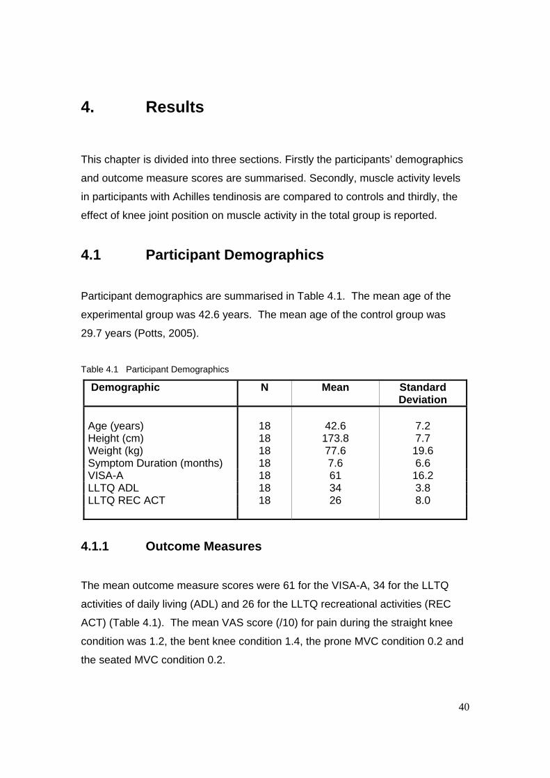
40
4. Results This chapter is divided into three sections. Firstly the participants’ demographics
and outcome measure scores are summarised. Secondly, muscle activity levels
in participants with Achilles tendinosis are compared to controls and thirdly, the
effect of knee joint position on muscle activity in the total group is reported.
4.1 Participant Demographics
Participant demographics are summarised in Table 4.1. The mean age of the
experimental group was 42.6 years. The mean age of the control group was
29.7 years (Potts, 2005).
Table 4.1 Participant Demographics
Demographic N Mean Standard Deviation
Age (years) Height (cm) Weight (kg) Symptom Duration (months)
18 18 18 18
42.6 173.8 77.6 7.6
7.2 7.7 19.6 6.6
VISA-A 18 61 16.2 LLTQ ADL 18 34 3.8 LLTQ REC ACT 18 26 8.0
4.1.1 Outcome Measures
The mean outcome measure scores were 61 for the VISA-A, 34 for the LLTQ
activities of daily living (ADL) and 26 for the LLTQ recreational activities (REC
ACT) (Table 4.1). The mean VAS score (/10) for pain during the straight knee
condition was 1.2, the bent knee condition 1.4, the prone MVC condition 0.2 and
the seated MVC condition 0.2.
41
4.2 Muscle Activity and Achilles Tendinosis
The key findings related to the electromyographic data are presented in Figure
4.1 and 4.2. Levene’s test results indicated that there was homogeneity in the
variances across all levels of the dataset. The assumption of sphericity was not
met and Greenhouse-Geisser corrections were applied to produce valid F-ratios.
These showed that there was a significant main effect for group (p<0.05).
Irrespective of knee position and muscle, EMG activity was significantly higher
(mean difference: 10%, effect size: 0.59) in those subjects with Achilles
tendinosis. Mean activity levels ranged from 55% to 72% in this group
depending upon joint position, while in the control subjects, they ranged from
47% to 62%.
0
20
40
60
80
100
GAS SK GAS BK SOL SK SOL BKExercise Condition
Mus
cle
Act
ivity
(% R
MS)
Experimental Group Control Group
Figure 4.1 Muscle activity levels in experimental and control groups during HLECM exercise
conditions. Data are mean and standard deviation.
GAS SK = gastrocnemius activity in straight knee condition
GAS BK = gastrocnemius activity in bent knee condition
SOL SK = soleus activity in straight knee condition
SOL BK = soleus activity in bent knee condition

42
0
20
40
60
80
100
GAS SK GAS BK SOL SK SOL BK
Exercise Condition
Mus
cle
Act
ivity
(% R
MS)
4.3 Muscle Activity and Knee Joint Position
There was a significant interaction effect between muscle and joint position (Fig
4.2). This indicated that differences in muscle activity depended upon whether
the knee was flexed or extended. For the gastrocnemius, EMG activity was
higher when the knee was extended, whereas when the knee was flexed, EMG
levels were decreased. In contrast, soleus activity increased slightly from the
extended to the flexed knee position. There were no other significant main
effects or interactions.
Figure 4.2 Muscle activity levels in total group during HLECM exercise conditions. Data are
mean and standard deviation. * = p<0.05.
GAS SK = gastrocnemius activity in straight knee condition
GAS BK = gastrocnemius activity in bent knee condition
SOL SK = soleus activity in straight knee condition
SOL BK = soleus activity in bent knee condition

43
5. Discussion
This chapter is divided into five sections. Firstly, muscle activity levels with
respect to the presence of pathology will be discussed. Secondly, the effect of
knee joint angle on selective calf muscle activity during performance of the
HLECM training protocol will be outlined. This will be followed by a discussion of
the limitations and clinical implications of the present study and finally areas for
future research will be identified.
5.1 Muscle Activity in Achilles Tendinosis
The main findings of the experimental study indicate that the muscle activity
levels of both the gastrocnemius and soleus muscles during each condition of the
HLECM protocol were significantly higher in persons with Achilles tendinosis
compared to controls. Muscle activity patterns provide information about neural
control strategies of movements (Lay, Hass, Nichols, & Gregor, 2007). A higher
muscle activity level corresponds to a greater proportion of motor units activated
within each muscle and/or an increase in firing rate (DeLuca, 2006). In this
study, it appears the presence of pathology influenced the neural control strategy
employed during an eccentric plantarflexor contraction, manifested by an
increase in muscle activity levels. It is possible alterations in neural control and
EMG activity in this study may be related to other factors that correspond with
pathological conditions such as tendinopathy, including pain, muscle atrophy and
reduced strength (Don et al., 2007; Valderrabano et al., 2006).
5.1.1 Pain and Muscle Activity
The mean VAS scores for pain during the HLECM protocol exercises were low in
this study (0.2/10 to 1.4/10), probably limiting the effect of this variable on muscle
activity levels. However, during performance of the full HLECM protocol in a

44
clinical and research setting, the maintenance of a painful state is considered an
essential principle (Alfredson et al. 1998). The participants in this study were
required to perform 12 repetitions of each condition, considerably less than the
180 required during the treatment protocol. It is probable that the presence of
tendon and/or muscle pain with a greater number of repetitions influences the
neural control strategies utilised by the triceps surae and associated synergists in
persons with tendinopathy.
To date there is no research investigating the effect of lower limb tendon
pathology on EMG activity of associated muscles. Maximal and sub-maximal
muscle activity has been examined in patients with rotator cuff tendinosis (Brox,
Roe, Saugen, & Vollestad, 1997). These authors demonstrated an increase in
muscle activity of the rotator cuff during a maximal voluntary contraction following
a pain relieving injection. However, muscle activity also increased during a sub-
maximal fatiguing contraction, despite pain levels increasing threefold in this
time. These authors concluded pain inhibits the central motor drive of agonist
muscles during maximal contractions only. In contrast, other research has
demonstrated muscle pain inhibits muscle activation during sub-maximal
contractions and increases the effects of fatigue (Ciubotariu, Arendt-Nielsen, &
Graven-Nielsen, 2007). Furthermore, EMG activity of synergistic muscles has
been shown to increase where pain has been induced in the corresponding
agonist, perhaps as a compensatory mechanism (Ciubotariu, Arendt-Nielsen, &
Graven-Nielsen, 2004; Schutle et al., 2004). Recent research has also
demonstrated an increase in superficial and decrease in deep cervical muscle
EMG activity in the presence of neck pain disorders (Falla, 2008). This altered
motor strategy causes a redistribution of load among synergistic muscles and
may represent reduced neuromuscular efficiency of those muscles where EMG
activity has increased. The ability to extrapolate the findings of these EMG
studies to the current one may be limited however, as they investigated only
isometric contractions and pain was induced locally into muscle, rather than
tendon tissue.

45
5.1.2 Biomechanical Properties and Muscle Activity
While there is a reasonable amount of research examining the
pathophysiological changes that occur with tendinosis, research has not yet
investigated the biomechanical effects of Achilles tendinopathy on the triceps
surae or tendon itself. However, alteration of biomechanical properties of the calf
musculature, including strength changes, have been observed for up to two
years following surgery for Achilles tendon rupture (Don et al., 2007). Of note is
the finding that EMG activity of the soleus returned to normal levels at the six
month mark despite an ongoing loss of eccentric strength of up to 30% for a
period of two years. Concentric strength however, was restored at six months
postoperatively. While this clinical situation differs to that of tendinopathy,
previous research has also demonstrated a reduction in both eccentric and
concentric strength in the calf muscles of the tendinopathic limb prior to eccentric
training (Alfredson et al. 1998, Alfredson et al., 1999). It is possible the
participants in the current study also possessed a reduction of eccentric strength,
given the chronicity of their condition. However, muscle activity levels may in fact
be independent of eccentric strength changes in this population as for those
individuals post Achilles tendon surgery.
The reason for an increase in muscle activity in the presence of pathology in this
study remains unknown. It may represent an increase in the proportionate
number of motor units activated within the plantarflexors in an attempt to perform
the task at a defined velocity with a body weight load. Many participants found it
difficult to control the eccentric descent into ankle dorsiflexion at the set three
second velocity and increased their speed. This, combined with the task being
novel for some, may have influenced their neural strategy and hence the degree
of muscle activity from the triceps surae compared to those without pathology.
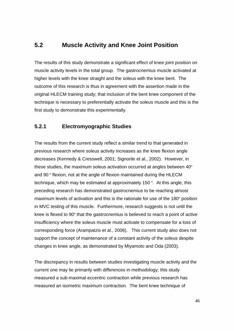
46
5.2 Muscle Activity and Knee Joint Position
The results of this study demonstrate a significant effect of knee joint position on
muscle activity levels in the total group. The gastrocnemius muscle activated at
higher levels with the knee straight and the soleus with the knee bent. The
outcome of this research is thus in agreement with the assertion made in the
original HLECM training study; that inclusion of the bent knee component of the
technique is necessary to preferentially activate the soleus muscle and this is the
first study to demonstrate this experimentally.
5.2.1 Electromyographic Studies
The results from the current study reflect a similar trend to that generated in
previous research where soleus activity increases as the knee flexion angle
decreases (Kennedy & Cresswell, 2001; Signorile et al., 2002). However, in
these studies, the maximum soleus activation occurred at angles between 40º
and 90 º flexion, not at the angle of flexion maintained during the HLECM
technique, which may be estimated at approximately 150 º. At this angle, this
preceding research has demonstrated gastrocnemius to be reaching almost
maximum levels of activation and this is the rationale for use of the 180º position
in MVC testing of this muscle. Furthermore, research suggests is not until the
knee is flexed to 90º that the gastrocnemius is believed to reach a point of active
insufficiency where the soleus muscle must activate to compensate for a loss of
corresponding force (Arampatzis et al., 2006). This current study also does not
support the concept of maintenance of a constant activity of the soleus despite
changes in knee angle, as demonstrated by Miyamoto and Oda (2003).
The discrepancy in results between studies investigating muscle activity and the
current one may lie primarily with differences in methodology; this study
measured a sub-maximal eccentric contraction while previous research has
measured an isometric maximum contraction. The bent knee technique of
47
HLECM training involves maintaining a static knee angle of approximately 150º,
while the ankle is gradually dorsiflexed to beyond neutral. Although both the
soleus and gastrocnemius muscles are lengthening, their individual length-
tension relationship will be different at the same knee and ankle joint angles due
to their anatomical position (Arampatzis et al., 2006). Perhaps the bent knee
position of the HLECM training technique, combined with an increasingly
dorsiflexed ankle places the soleus at a more optimal length to generate activity
than the gastrocnemius.
An alternative reason for the variation in results from the above EMG studies
may be the different test positions utilised, such as four-point kneeling, sitting and
standing. It has been demonstrated isometric force and muscle activity are
highest in a functional standing position but testing in prone is more reliable for
research purposes (Carlsson, Lind, Moller, Karlsson, & Svantesson, 2001).
These authors investigated only the gastrocnemius muscle, thus it is possible the
soleus behaves differently in these various positions.
5.2.2 Rationale for Selective Muscle Activation
Alfredson et al. (1998) emphasised the inclusion of the bent knee technique in
order to activate the soleus, but did not outline why it is necessary to selectively
recruit the triceps surae. The following section is a discussion of the possible
rationale for the inclusion of both components within HLECM training based on
the concept of training specificity. This is where the exercise regime of an
individual closely matches their functional activities in order to produce the
greatest performance gains (Morrissey, Harman, & Johnson, 1995). Specificity
of training incorporates variables such as technique execution, including specific
joint angles and positions, and training velocity.
The gastrocnemius and soleus muscles differ in their fiber type composition,
anatomical position and architecture (Kawakami et al., 1998). The
48
gastrocnemius muscle is comprised of a larger proportion of type II fast-twitch
fibers while soleus has an additional postural function and contains more fatigue-
resistant, type I slow-twitch fibers. During an eccentric triceps surae contraction,
the velocity of contraction has been demonstrated as a critical influence on the
relative recruitment of the gastrocnemius and soleus due to their differing fiber
type composition The more rapid the deceleration of force by the triceps surae,
the more activation of gastrocnemius and suppression of soleus is observed
(Nardone, Romano, & Schieppati, 1989). During a concentric-eccentric heel
raise protocol, it has also been demonstrated the triceps surae respond
differently to fatigue, with the soleus fatiguing less during the eccentric phase but
also displaying a reduction in muscle activation (Svantesson, Osterberg,
Thomee, & Grimby, 1998).
In the quadriceps musculature, fast-twitch fibers have exhibited greater atrophy
than slow-twitch fibers in the presence of knee joint pathology (Fink et al., 2007).
The authors proposed this may reflect pain related immobilisation of the affected
limb. Although fiber type changes have not been examined with lower limb
tendon pathology to date, it is possible similar changes may occur in the triceps
surae in the presence of Achilles tendinopathy. Due to their specific fibre type
makeup, the gastrocnemius and soleus muscles may respond differently to a
decrease in tendon load through reduced participation in daily or sporting
activities. This means they may therefore benefit from selective recruitment
during rehabilitation exercises.
The concept of velocity specificity is supported by the training literature, where
strength gains are consistently greater at the trained velocity with some carryover
to slower speeds (Morrissey et al., 1995). Within the original study no details are
provided regarding the tempo of the exercise. However, the velocity of calf
muscle contraction in HLECM training is generally slower than that of activities
such as running or walking which typically provoke tendinopathic symptoms.

49
Despite this discrepancy, many individuals return to these activities with minimal
difficulty following the three month training period.
In the current study, it was observed some participants struggled to maintain the
three second velocity of eccentric contraction required for EMG analysis possibly
due to eccentric weakness of the calf muscles. Therefore, it is possible velocity
is a particularly variable component of the technique both between individuals
and between studies, particularly when participants are unsupervised in a home
environment. Results of the outcome measures utilised in the current study
indicate a reduction in full participation in both daily (34/40) and recreational
activities (26/40) in the LLTQ, although the latter is more affected by the
presence of Achilles tendinosis. Given the differing fiber type composition and
possible variation in response to disuse, it is likely the tempo of the eccentric
phase of the HLECM technique plays a critical role in determining selective
muscle recruitment in addition to other training factors such as joint angle.
The inclusion of both components in HLECM training may also function to
reproduce joint angles similar to those generated during a gait cycle. The triceps
surae activate eccentrically during the stance phase from a straight knee position
to that of a bent knee position while the ankle is being relatively dorsiflexed
(Komi, Fukashiro, & Jarvinen, 1992). There is evidence to suggest that range of
motion specificity exists during resistance training (Morrissey et al., 1995). This
is where the greatest strength gains are made at exercised joint angles. Open
and closed kinetic chain strength training following anterior cruciate ligament
reconstruction has been demonstrated to increase strength maximally at the
angle trained with some carryover to other similar joint angles (Hooper, Hill,
Drechsler, & Morrissey, 2002).
Use of both the bent and straight knee position in HLECM training may provide a
wider range of strength gains, particularly from 180º to 150º knee flexion, than if
training with only the knee extended for example. Achilles tendinopathy

50
commonly affects individuals who participate in competitive or recreational
walking or running where the triceps surae is active at similar knee and ankle
joint angles to those adopted in the HLECM training technique (Kujala et al.,
2005). Although some EMG studies suggest the soleus may be preferentially
activated with the knee flexed to 90º, this position does not simulate those found
in symptom provoking activities. The efficacy of HLECM training may be not only
due to the eccentric loading per se but also the specificity of velocity and range of
movement resistance training and the relationship of these to functional activities.
5.3 Limitations of the Study
The diagnosis for inclusion was made by clinical exam only, without the use of
imaging, and thus the presence of mid-portion tendinopathy could not be
differentiated from other possible tendon pathologies. Differing pathologies may
render varying biomechanical effects, however, as the tissue tested in this case
is not the tendon but the adjacent musculature, it is presumed the effect on EMG
activity may not be notably different.
The performance of the exercise technique was difficult for a number of
participants in the experimental group both in terms of a lack of strength to be
able to slowly descend into a dorsiflexed position and also due to the novelty of
the task. It should be noted the bent knee position technique was much more
difficult for participants to perform correctly, even with an opportunity to practice.
All participants experienced no difficulty with the straight knee position. It may be
the bent knee position represents a more challenging movement pattern than the
straight knee position, possibly influencing the neural strategy utilised.
Measurement of the eccentric phase was taken over three cycles of movement
for three trials in each position. It may have been advantageous to teach the
technique on one day with the participant given an opportunity to practice in their
own time before testing muscle activity levels on another day. Alternatively,

51
muscle activity levels could have been tested over a greater number of trials
such as the 180 used in the Alfredson et al. (1998) study. However, this may
have caused muscle fatigue, which in turn may influence muscle activity.
Normalisation of the EMG data was carried out using an isometric maximum
voluntary contraction. There is some debate in the literature regarding the
reliability of this measure, although isometric contractions are preferential to
isotonic measures (Burden, Trew, & Baltzopoulos, 2003). Maximum voluntary
contractions are influenced by factors including familiarity with the task, verbal
encouragement and previous resistance training (Shield & Zhou, 2004). There is
good evidence to demonstrate the presence of knee joint injury reduces
voluntary muscle activation levels of the quadriceps muscles (Urbach & Awiszus,
2002). Although not examined in persons with tendon pathology, it is possible
the participants in the experimental group were not able to maximally activate
their triceps surae. Additionally, although there was opportunity to practice, the
task was unfamiliar, which also may reduce voluntary activation levels (Shield &
Zhou, 2004). The twitch interpolation technique involves a supramaximal
stimulus applied to the nerve trunk of a muscle during a voluntary contraction and
provides a more accurate assessment of the completeness of muscle activation
(Shield & Zhou, 2004). This technique may have been useful in this study to
ensure a more accurate MVC and hence RMS value was generated.
Finally, the data obtained was age matched to that from previous work by
Potts(2005) to assist in comparison of results. There was a lack of other
demographic information (i.e. gender, height and weight) available from the Potts
(2005) study to compare populations further
5.4 Clinical Implications
Almost all of the participants in the experimental group had consulted a
physiotherapist and been prescribed some form of eccentric calf muscle loading
52
exercise. This suggests the use of HLECM training is frequent in clinical
practice. Accordingly, gaining knowledge regarding the biomechanics of the
technique itself is advantageous for a number of health practitioners and
patients. As discussed, there exists limited research investigating the
biomechanics of rehabilitation exercises used for the treatment of tendinopathy.
Assessing factors such as muscle activity provides information that may be used
to explain differences in rehabilitation effects in addition to allowing design of
more effective rehabilitation programmes. Compliance to the regime has been
demonstrated to be a problem in the research utilising HLECM training and
anecdotally for the participants of this study. Assessing the biomechanics of the
HLECM training protocol may lead to a more concise programme which in turn
may improve compliance levels and possibly efficacy.
The results of this study suggest it is useful to include both components of the
HLECM training regime as they selectively activate the gastrocnemius and
soleus muscles. Given the findings related to the specificity theory of training
discussed above, it may also be useful to include varying velocities within the
regime or trial the effect of a more flexed bent knee position in order to achieve
wider range of motion training specificity.
5.5 Future Research
In order to design improved rehabilitation programmes for those with Achilles
tendinopathy it would be useful to investigate the influence of other
biomechanical variables such as eccentric strength, muscle atrophy and muscle-
tendon stiffness on the efficacy of the regime. This would also provide an
improved description of the relationship between pathology and biomechanical
changes in this particular population. By stratifying studied populations into
subgroups based on these biomechanical factors or others, such as symptom
duration and severity, an explanation of the variation in efficacy seen in the
current literature may be attained. The use of standardised inclusion and
53
exclusion criteria, functional outcome measures and clarification of the HLECM
training methodology means comparison between future studies will be
enhanced.
It may be argued that it is not specific muscle activation that is required but the
overall loading of the Achilles tendon through HLECM training that achieves an
efficacious result. How the tendon converts mechanical signals into a healing
response is currently not known (Wang, 2006). To assist with clarifying which
aspects of HLECM training may influence efficacy, modification of particular
variables such as repetition numbers, use of one component only (i.e. straight
knee position) and velocity of contraction, with use of appropriate control groups,
would also be valuable.
Given the finding that those individuals with Achilles tendinosis demonstrate
elevated levels of muscle activity of the triceps surae during the HLECM
exercises, it would also be useful to investigate whether this variable normalises
following implementation of the HLECM 12 week protocol.
5.6 Conclusion
Heavy load eccentric calf muscle training was developed by Alfredson et al.
(1998) as an intervention for Achilles tendinopathy. Results of the literature
review demonstrate there is moderate evidence for the efficacy of HLECM
training although the mechanisms of pain alleviation and return to functional
activity through the use of this regime remain unclear. This experimental study
has demonstrated the straight and bent-knee components of the HLECM training
regime selectively activate the gastrocnemius and soleus muscles respectively in
a population diagnosed with Achilles tendinosis. Additionally, individuals with
Achilles tendinosis exhibit higher muscle activation levels of the triceps surae
during the eccentric phase of this technique than controls. The reasons for this
increase in muscle activity in a pathological population are not currently known.

54
References
Alfredson, H., & Cook, J. (2007). A treatment algorithm for managing Achilles
tendinopathy: New treatment options. British Medical Journal, 41(4), 211-216.
Alfredson, H., & Lorentzon, R. (2003). Intratendinous glutamate levels and eccentric training in chronic Achilles tendinosis: A prospective study using microdialysis technique. Knee Surgery and Sports Traumatology, 11, 196-199.
Alfredson, H., Nordstrom, P., Pietila, T., & Lorentzon, R. (1999). Bone mass in the calcaneus after heavy loaded eccentric calf-muscle training in recreational athletes with chronic Achilles tendinosis. Calcified Tissue International, 64, 450-455.
Alfredson, H., Pietila, T., Ohberg, L., & Lorentzon, R. (1998). Achilles tendinosis and calf muscle strength. The effect of short-term immobilization after surgical treatment. American Journal of Sports Medicine, 26(2), 166-171.
Arampatzis, A., Karamanidis, K., Stafilidis, S., Morey-Klapsing, G., DeMonte, G., & Bruggemann, G. P. (2006). Effect of different ankle and knee-joint positions on gastrocnemius medial fascicle length and EMG activity during isometric plantar flexion. Journal of Biomechanics, 39, 1891 - 1902.
Barry, L. D., Barry, A. N., & Chen, Y. (2002). A retrospective study of standing gastrocnemius-soleus stretching versus night splinting in the treatment of plantar fasciitis. Journal of Foot and Ankle Surgery, 41(4), 221 - 227.
Brown, R., Orchard, J., Kinchington, M., Hooper, A., & Nalder, G. (2006). Aprotinin in the management of Achilles tendinopathy: A randomised controlled trial. British Journal of Sports Medicine, 40, 275-279.
Brox, J. I., Roe, C., Saugen, E., & Vollestad, N. K. (1997). Isometric abduction muscle activation in patients with rotator tendinosis of the shoulder. Archives of Physical Medicine and Rehabilitation, 78, 1260 - 1267.
Burden, A. M., Trew, M., & Baltzopoulos, V. (2003). Normalisation of gait EMGs: A re-examination. Journal of Electromyography and Kinesiology, 13, 519 - 532.
Carlsson, U., Lind, K., Moller, M., Karlsson, J., & Svantesson, U. (2001). Plantar flexor muscle function in open and closed chain. Clinical Physiology, 21, 1 - 8.
Chung, B., & Wiley, J. P. (2002). Extracorporeal shockwave therapy. A review. Sports Medicine, 32(15), 851 - 865.

55
Ciubotariu, A., Arendt-Nielsen, L., & Graven-Nielsen, T. (2004). The influence of muscle pain and fatigue on the activity of synergistic muscles of the leg. European Journal of Applied Physiology, 91, 604-614.
Ciubotariu, A., Arendt-Nielsen, L., & Graven-Nielsen, T. (2007). Localized muscle pain causes prolonged recovery after fatiguing isometric contractions. Experimental Brain Research, 181, 147-158.
Cook, J. L., Khan, K. M., & Purdam, C. (2002). Achilles tendinopathy. Manual Therapy, 7(3), 121-130.
DeLuca, C. (2006). Electromyography. In J. G. Webster (Ed.), Encyclopedia of Medical Devices and Instrumentation (pp. 98 - 109). Boston: John Wiley Publisher.
Don, R., Ranavolo, A., Cacchio, A., Serrao, M., Costabile, F., Iachelli, M., et al. (2007). Relationship between recovery of calf-muscle biomechanical properties and gait pattern following surgery for Achilles tendon rupture. Clinical Biomechanics, 22, 211-220.
Fahlstrom, M., Jonsson, J., Lorentzon, R., & Alfredson, H. (2003). Chronic Achilles tendon pain treated with eccentric calf-muscle training. Knee Surgery and Sports Traumatology, 11, 327-333.
Fahlstrom, M., Lorentzon, R., & Alfredson, H. (2002). Painful conditions in the Achilles tendon region: A common problem in middle aged competitive badminton players. Knee Surgery and Sports Traumatology, 10, 57-60.
Falla, D. (2008, June 8th - 13th). Nociceptive and sympathetic effects on cervical motor control. Paper presented at the IFOMT 2008 Connecting "Science to Quality of Life, Rotterdam.
Fink, B., Egl, M., Singer, J., Fuerst, M., Bubenheim, M., & Neuen-Jacob, E. (2007). Morphological changes in the vastus medialis muscle in patients with osteoarthritis of the knee. Arthritis and Rheumatism, 56(11), 3626-3633.
Gadjosik, R. L., Alfred, J. D., Gabbert, H. L., & Sonsteng, B. A. (2007). A stretching program increases the dynamic passive length and passive resistive properties of the calf muscle-tendon unit of unconditioned younger women. European Journal of Applied Physiology, 99, 449 - 454.
Hermens, H. J., Freriks, B., Disselhorst-Klug, C., & Rau, G. (2000). Development of recommendations for SEMG sensors and sensor placement procedures. Seniam Guidelines, 10, 361 - 374.
Hooper, D. M., Hill, H., Drechsler, W. I., & Morrissey, M. C. (2002). Range of motion specificity resulting from closed and open kinetic chain resistance
56
training after anterior cruciate ligament reconstruction. Journal of Strength and Conditioning Research, 16(3), 409 - 415.
Hunter, G. (2000). The conservative management of Achilles tendinopathy. Physical Therapy in Sport, 1, 6-14.
Katz, J., & Melzack, R. (1999). Pain measurement. Surgical Clinics of North America, 79(2), 231-252.
Kawakami, Y., Ichinose, Y., & Fukunaga, T. (1998). Architectural and functional features of human triceps surae during contraction. Journal of Applied Physiology, 85, 398 - 404.
Kennedy, P. M., & Cresswell, A. G. (2001). The effect of muscle length on motor-unit recruitment during isometric plantar flexion in humans. Experimental Brain Research, 137, 58 - 64.
Kingma, J. J., deKnikker, R., Wittink, H. W., & Takken, T. (2006). Eccentric overload training in patients with chronic Achilles tendinopathy. British Journal of Sports Medicine, 41(6), e3.
Knobloch, K., Kraemer, R., Jagodzinski, M., Zeichen, J., Meller, R., & Vogt, P. M. (2007). Eccentric training decreases paratendon capillary blood flow and preserves paratendon oxygen saturation in chronic Achilles tendinopathy. Journal of Orthopaedic and Sports Physical Therapy, 37(5), 269-276.
Komi, P., Fukashiro, S., & Jarvinen, M. (1992). Biomechanical loading of Achilles tendon during normal locomotion. Clinics in Sports Medicine, 11(3), 521 - 531.
Kujala, U. M., Sarna, S., & Kaprio, J. (2005). Cumulative incidence of Achilles tendon rupture and tendinopathy in male former elite athletes. Clinical Journal of Sports Medicine, 15(3), 133 - 135.
Langberg, H., Ellingsgaard, H., Madsen, T., Jansson, J., Magnusson, S. P., Aagard, P., et al. (2007). Eccentric rehabilitation exercise increases peritendinous Type I collagen synthesis in humans with Achilles tendinosis. Scandinavian Journal of Medicine and Science in Sports, 17, 61-66.
Langberg, H., Rosendal, L., & Kjaer, M. (2001). Training-induced changes in peritendinous Type I collagen turnover determined by microdialysis in humans. Journal of Physiology, 534(1), 297 - 302.
Lay, A. N., Hass, C. J., Nichols, R., & Gregor, R. J. (2007). The effects of sloped surfaces on locomotion: An electromyographic analysis. Journal of Biomechanics, 40, 1276 - 1285.

57
Mademli, L., Arampatzis, A., Morey-Klapsing, G., & Bruggemann, G. P. (2004). Effect of ankle joint position and electrode placement on the estimation of the antagonistic moment during maximal plantarflexion. Journal of Electromyography and Kinesiology, 14, 591 - 597.
Maffulli, N., Wong, J., & Almekinders, L. C. (2003). Types and epidemiology of tendinopathy. Clinical Sports Medicine, 22, 675 - 692.
Mafi, N., Lorentzon, R., & Alfredson, H. (2001). Superior short-term results with eccentric calf muscle training compared to concentric training in a randomized prospective multicenter study on patients with chronic Achilles tendinosis. Knee Surgery and Sports Traumatology, 9, 42-47.
Mayer, F., Hirschmuller, A., Muller, S., Schuberth, M., & Baur, H. (2006). The effect of short term treatment strategies over 4 weeks in Achilles tendinopathy. British Journal of Sports Medicine, 41(7), e6.
McNair, P., Prapavessis, H., Collier, J., Bassett, S., Bryant, A., & Larmer, P. (2007). The lower-limb tasks questionnaire: An assessment of validity, reliability, responsiveness and minimal important differences. Archives of Physical Medicine and Rehabilitation, 88, 993-1001.
Miaki, H., Someya, F., & Tachino, K. (1999). A comparison of electrical activity in the triceps surae at maximum isometric contraction with the knee and ankle at various angles. European Journal of Applied Physiology, 80, 185 - 191.
Miyamoto, N., & Oda, S. (2003). Mechanomyographic and electromyographic responses of the triceps surae during maximal voluntary contractions. Journal of Electromyography and Kinesiology, 13, 451 - 459.
Morrissey, M. C., Harman, E. A., & Johnson, M. J. (1995). Resistance training modes: specificity and effectiveness. Medicine and Science in Sports and Exercise, 27(5), 648-660.
Nardone, A., Romano, C., & Schieppati, M. (1989). Selective recruitment of high-threshold human motor units during voluntary isotonic lengthening of active muscles. Journal of Physiology, 409, 451 - 471.
Norregaard, J., Larsen, C. C., Bieler, T., & Langberg, H. (2007). Eccentric exercise in treatment of Achilles tendinopathy. Scandinavian Journal of Medicine and Science in Sports, 17, 133-138.
Ohberg, L., & Alfredson, H. (2004). Effects on neovascularisation behind the good results with eccentric training in chronic mid-portion Achilles tendinosis? Knee Surgery and Sports Traumatology, 12, 465-470.

58
Ohberg, L., Lorentzon, R., & Alfredson, H. (2001). Neovascularisation in Achilles tendons with painful tendinosis but not in normal tendons: An ultrasonographic investigation. Knee Surgery and Sports Traumatology, 9, 233-238.
Ohberg, L., Lorentzon, R., & Alfredson, H. (2004). Eccentric training in patients with chronic Achilles tendinosis: Normalised tendon structure and decreased thickness at follow up. British Journal of Sports Medicine, 38, 8-11.
Peers, K. H. E., Brys, P. P. M., & Lysens, R. J. J. (2003). Correlation between power Doppler ultrasonography and clinical severity in Achilles tendinopathy. International Orthopaedics, 27, 180-183.
Petersen, W., Welp, R., & Rosenbaum, D. (2007). Chronic Achilles tendinopathy. A prospective randomized study comparing the therapeutic effect of eccentric training, the AirHeel brace and a combination of both. American Journal of Sports Medicine, 35(10), 1659-1667.
Potts, G. (2005). Biomechanical analysis of "heavy-load eccentric calf muscle' exercise used in the rehabilitation of Achilles tendinosis. Unpublished Thesis, Auckland University of Technology, Auckland.
Rees, J. D., Wilson, A. M., & Wolman, R. L. (2006). Current concepts in the management of tendon disorders. Rheumatology, 45, 508-521.
Reid, S. A., & Rivett, D. A. (2005). Manual therapy treatment of cervicogenic dizziness: a systematic review. Manual Therapy, 10, 4-13.
Robinson, J. M., Cook, J. L., Purdam, C., Visentini, P. J., Maffulli, N., Taunto, J. E., et al. (2001). The VISA-A questionnaire: A valid and reliable index of the clinical severity of Achilles tendinopathy. British Journal of Sports Medicine, 35, 335-341.
Rompe, J. D., Nafe, B., Furia, J. P., & Maffulli, N. (2007). Eccentric loading, shock-wave therapy treatment, or a wait-and-see policy for tendinopathy of the main body of Tendo Achilles. A randomized controlled trial. American Orthopaedic Society for Sports Medicine., 35(3), 374-383.
Roos, E. M., Engstrom, M., Lagerquist, A., & Soderberg, B. (2004). Clinical improvement after 6 weeks of eccentric exercise in patients with mid-portion Achilles tendinopathy - A randomized trial with 1 year follow-up. Scandinavian Journal of Medicine and Science in Sports, 14, 286-295.
Rukin, N. J., & Maffulli, N. (2006). Systemic allergic reactions to aprotinin injection around the Achilles tendon. Journal of Science and Medicine in Sport, 10(5), 320 - 322.

59
Satyendra, L., & Byl, N. (2006). Effectiveness of physical therapy for Achilles tendinopathy: An evidence based review of eccentric exercises. Isokinetics and Exercise Science, 14, 71 - 80.
Sayana, M. K., & Maffulli, N. (2007). Eccentric calf muscle training in non-athletic patients with Achilles tendinopathy. Journal of Science and Medicine in Sport, 10, 52 - 58.
Schutle, E., Ciubotariu, A., Arendt-Nielsen, L., Disselhorst-Klug, C., Rau, G., & Graven-Nielsen, T. (2004). Experimental muscle pain increases trapezius muscle activity during sustained isometric contractions of arm muscles. Clinical Neurophysiology, 115, 1767-1778.
Shalabi, A., Kristoffersen-Wiberg, M., Svensson, L., Aspelin, P., & Movin, T. (2004). Eccentric training of the gastrocnemius-soleus complex in chronic Achilles tendinopathy results in decreased tendon volume and intratendinous signal as evaluated by MRI. American Orthopaedic Society for Sports Medicine., 32(5), 1286-1296.
Shield, A., & Zhou, S. (2004). Assessing voluntary muscle activation with the twitch interpolation technique. Sports Medicine, 34(4), 253-267.
Signorile, J. E., Applegate, B., Duque, M., Cole, N., & Zink, A. (2002). Selective recruitment of the triceps surae muscles with changes in knee angle. Journal of Strength and Conditioning, 16(3), 433 - 439.
Snellenberg, W. v., Wiley, J. P., & Brunet, G. (2006). Achilles tendon pain intensity and level of neovascularization in athletes as determined by color Doppler ultrasound. Scandinavian Journal of Medicine and Science in Sports, 17(5), 433-439.
Soderberg, G. L., & Knutson, L. M. (2000). A guide for use and interpretation of kinesiologic electromyographic data. Physical Therapy, 80(5), 485 - 498.
Soper, C., Reid, D., & Hume, P. A. (2004). Reliable passive ankle range of motion measures correlates to ankle joint motion achieved during ergonometer rowing. Physical Therapy in Sport, 5, 75 - 83.
Svantesson, U., Osterberg, U., Thomee, R., & Grimby, G. (1998). Muscle fatigue in a standing heel-rise test. Scandinavian Journal of Rehabilitation Medicine, 30, 67 - 72.
Tol, J. L., Vos, R. J. d., Weir, A., Visser, R. J. A., & deWinter, T. (2006). The additional value of a night splint to eccentric exercises in chronic midportion Achilles tendinopathy: A randomised controlled trial. British Journal of Sports Medicine, 41(7), e5.
60
Urbach, D., & Awiszus, F. (2002). Impaired ability of voluntary quadriceps activation bilaterally interferes with function testing after knee injuries: A twitch interpolation study. International Journal of Sports Medicine, 23(4), 231-236.
Valderrabano, V., vonTscharner, V., Nigg, B. M., Hintermann, B., Goepfert, B., Fung, T. S., et al. (2006). Lower leg muscle atrophy in ankle osteoarthritis. Journal of Orthopaedic Research, 24, 2159 -2169.
vanTulder, M., Furlan, A., Bombardier, C., & Bouter, L. (2003). Updated method guidelines for systematic reviews in the Cochrane Collection Back Review Group. Spine, 28(12), 1290 -1299.
Wang, J. H. C. (2006). Mechanobiology of tendon. Journal of Biomechanics, 39, 1563-1582.
Wasielewski, N. J., & Kotsko, K. M. (2007). Does eccentric exercise reduced pain and improve strength in physically active adults with symptomatic lower extremity tendinosis? A systematic review. Journal of Athletic Training, 42(3), 409 - 421.
Woodley, B. L., Newsham-West, R. J., & Baxter, G. D. (2006). Chronic tendinopathy: Effectiveness of eccentric exercise. British Journal of Sports Medicine, 41(4), 188-198.

61
Appendix A Operationalisation of the Criteria List A A random (unpredictable) assignment sequence. Examples of adequate
methods are computer generated random number tables and use of sealed opaque envelopes. Methods of allocation using date of birth, date of admission, hospital numbers or alternation should not be regarded as appropriate.
B Assignment generated by an independent person not responsible for determining
the eligibility of the patients. This person has no information about the persons included in the trial and has no influence on the assignment sequence or on the decision about eligibility of the patient.
C In order to receive a “yes”, groups have to be similar at baseline regarding
demographic factors, duration and severity of complaints, percentage of patients with neurologic symptoms and the value of the main outcome measure(s).
D The reviewer determines if enough information about the blinding is given in
order to score a “yes” E The reviewer determines if enough information about the blinding is given in
order to score a “yes” F The reviewer determines if enough information about the blinding is given in
order to score a “yes” G Cointerventions should either be avoided in the trial design or similar between
control and index groups H The reviewer determines if the compliance to the interventions is acceptable,
based on the reported intensity, duration, number and frequency of sessions for both the control and index interventions(s).
I The number of participants who were included in the study but did not complete
the observation period or were not included in the analysis must be described and reasons given. If the percentage of withdrawals and drop-outs does not exceed 20% for short-term follow-up and 30% for long-term follow-up and does not lead to a substantial bias a “yes” is scored (N.B. these percentages are arbitrary and not supported by literature).
J Timing of outcome assessment should be identical for all interventional groups
and for all important outcome assessments. K All randomised patients are reported/analysed in the group they were allocated to
by randomisation for the most important moments of effect measurement (minus missing values) irrespective of non-compliance and cointerventions.

62
Appendix B
Consent Form
Project title: Investigation of calf muscle activity during a rehabilitation exercise in patients with Achilles tendinosis
Project Supervisor: Duncan Reid Researcher: Shelley Johnson
I wish to have an interpreter (please circle) Yes No I have read and understood the information sheet dated 26th September
2007 for volunteers taking part in this study designed to investigate the activity of the calf muscles during exercise.
I have had an opportunity to discuss this study. I am satisfied with the answers I have been given.
I understand that taking part in this study is voluntary (my choice) and I may withdraw from the study at any time and this will in no way affect my future health or continuing health care.
I understand that participation in this study is confidential and that no material which could identify me will be used in any reports on this study.
I understand that the investigation will be stopped if it should appear harmful to me.
I have had time to consider whether to take part and I know who to contact if I have any side effects from the study or questions about the study.
I wish to receive a copy of the report from the research (please tick one): Yes No
I ______________________________ (full name) hereby consent to take part in this study.
Participant’s Signature: .....................................................……………………………………………………… Date: This study has received ethical approval from the Northern Regional Ethics Committee on 29 January 2008.

63
Appendix C
Physiotherapist Information Form Project Title: Biomechanical analysis of a “heavy load eccentric calf muscle” rehabilitation exercise in patients with Achilles tendinosis; a pilot study. Thank you for your assistance with this study. Attached is an information sheet, outlining what the study involves, for patients and their treating physiotherapist to read. Inclusion and Exclusion Criteria The inclusion criteria for this study are:
• Aged over 20 years. • Diagnosed with unilateral Achilles tendinosis by their treating
physiotherapist (refer note below for diagnosis criteria) The exclusion criteria for this study are:
• Diagnosed with insertional Achilles tendinosis (refer note below for diagnosis criteria)
• Have had a previous history of Achilles tendon repair or rupture. • Have had a previous corticosteroid injection into either Achilles tendon • Has Achilles tendinosis bilaterally. • Has the presence of neural signs or symptoms affecting their lower limbs.
Diagnosis of Achilles Tendinosis The diagnosis used for this study is a clinical one; no imaging (i.e. ultrasound) is required to confirm the presence of the condition. The diagnostic criterion is a painful area of the Achilles tendon 2-6cm from the tendon insertion into the calcaneus. This may or may not be associated with a thickened, swollen area. Patients with pain and/or swelling in the insertion area of the tendon, indicating insertional Achilles tendinosis are excluded from this study. This is due to the fact that patients with insertional Achilles tendinosis have demonstrated a poorer response to eccentric training programmes using the exercises described.

64
Process of Referring a Patient to this Study If you have a patient who fits the inclusion and exclusion criteria, please provide them with a patient information sheet and ask them to consider their participation in the timeframe until their next physiotherapy treatment. They are welcome to take longer if necessary. They are also able to contact the principal researcher, Shelley Johnson, if they have any questions regarding the study. Once the patient has decided to take part, they need to contact either: Shelley Johnson Principal Researcher (07) 8701033 or 021332814 [email protected] or, Duncan Reid Primary Supervisor (09) 9219999 ext 7806 [email protected] They are welcome to leave a message with contact details and they will be telephoned or emailed and a time arranged to attend the Health and Rehabilitation Research Centre, Auckland University of Technology, Akoranga Drive, Northcote, Auckland. A $20 petrol voucher will be provided to assist with travel costs. Appointment times may be outside working hours if necessary. Physiotherapist Enquiries Regarding this Study If you require clarification of any of the above information, please contact Shelley Johnson or Duncan Reid (contact details provided above).

65
Appendix D Media Recruitment Advertisement

66
Appendix E
Participant Information Sheet
26 September 2007
Project Title
Investigation of calf muscle activity during a rehabilitation exercise in patients with Achilles tendinosis: a pilot study. An Invitation
Thank you for enquiring about participation in this study which will contribute to a Master of Health Science qualification in Physiotherapy. The following is an outline of what the study involves. Your participation is entirely voluntary (your choice). You do not have to take part in this study and if you choose not to take part you will receive the usual treatment from your physiotherapist.
What is the purpose of this research?
The purpose of this research is to investigate the activity of the calf muscles while you are performing a heel raise and lowering exercise. This exercise is commonly prescribed by physiotherapists to patients with Achilles tendon pain (also known as tendonitis or tendinosis) to help reduce the pain associated with this condition and improve function. The results of this research will be presented in a dissertation document and published in national and international rehabilitation journals.
How was I chosen for this invitation? You have been selected by your physiotherapist as an ideal candidate as you have been diagnosed by them, or another medical professional, with Achilles tendinosis and also fit the following criteria:
• have no history of Achilles tendon surgery or rupture • have no history of previous corticosteroid injection into the Achilles tendon area • have Achilles tendon pain on one side only (i.e. left or right) • have no current referral of pain or neural symptoms (i.e. pins and needles) in
your legs
67
What will happen in this research? Prior to collecting the information from the exercises you will be required to fill in three forms. The first one collects general information about your age, height, weight and duration of symptoms. The second form, the VISA –A , collects information about the pain in Achilles tendon with various activities and the third one, the Lower Limb Task Questionnaire, collects information on how the condition affects you during functional and daily activities. Participating in this research involves performing a light warm-up on a stationary bike then randomly selecting a form which will allocate the order of the exercise (bent or straight leg first). A small area on the calf of your affected leg will be shaved and cleaned for the application of three self adhesive electrodes that will measure the electrical activity of the muscles. A device that measures your ankle range of movement will also be adhered to the outer side of your ankle. The exercise starts with standing on the edge of a step on your toes as a far as comfortable and you are able to touch the wall lightly in front of you for balance. One foot is removed from the step and you are then required to lower your body weight to bring your heel below the level of the step as far as is comfortable. The removed foot is then replaced on the step to raise you back up onto your toes. The sequence of lowering your body weight on one leg is repeated for two sets of 10-12 times in each set. After this you will be positioned on a table lying on your front and instructed to push as hard as possible with your calf muscle against a stationary board to measure its maximum muscle activity. What are the discomforts and risks? It is possible you may experience some mild delayed muscle soreness in the calf muscle for up to 48 hours following performing the exercises if you are not used to them. This soreness resolves naturally and is not a sign of injury. There is also a risk of Achilles tendon rupture with performing any activities that involve loading your calf muscle. However, this is extremely rare and has not happened in any other studies involving these exercises. How will these discomforts and risks be alleviated? Using the stationary bike before you perform the exercise has shown to reduce the intensity of this delayed muscle soreness. You will have full access to medical advice or treatment if the discomfort does not resolve within a couple of days or if there are any other issues relating to your lower leg symptoms. In the case of a medical emergency the researcher will refer you immediately to the appropriate medical facilities. What are the benefits? The data gained from this research will contribute towards designing better exercise programmes for people with Achilles tendinosis. This might mean that exercise programmes are effective more rapidly or that they are less arduous.

68
What compensation is available for injury or negligence?
In the unlikely event of a physical injury as a result of your participation in this study, you may be covered by ACC under the Injury Prevention, Rehabilitation and Compensation Act. ACC cover is not automatic and your case will need to be assessed by ACC according to the provisions of the 2002 Injury Prevention, Rehabilitation and Compensation Act. If your claim is accepted by ACC, you still might not get any compensation. This depends on a number of factors such as whether you are an earner or non-earner. ACC usually provides only partial reimbursement of costs and expenses and there may be no lump sum compensation payable. There is no cover for mental injury unless it is a result of physical injury. If you have ACC cover, generally this will affect your right to sue the investigators. If you have any questions about ACC, contact your nearest ACC office or the investigator. How will my privacy be protected? Privacy is of extreme importance and you will be identified only by a number. Access to the experimental data will be kept in a locked cupboard and available only to the researcher and supervisor. No material which could personally identify you will be used in any reports on this study. What are the costs of participating in this research? Participation in this research will involve approximately one hour of your time at the Auckland University of Technology Health and Rehabilitation Research Centre. A small amount will be provided to assist with travel costs.
What opportunity do I have to consider this invitation?
Once you have received this information you have until your next physiotherapy treatment to consider whether you would like to take part. This usually ranges from 24 hours to 1 week, however may be longer if your next treatment is outside this time frame.
How do I agree to participate in this research?
You will need to sign a consent form which will be provided by the researcher at the Health and Rehabilitation Research Centre.
Will I receive feedback on the results of this research?
You will be able to receive a written summary of the findings on request to the researcher.
What do I do if I have concerns about this research? Any concerns regarding the nature of this project should be notified in the first instance to the Project Supervisor (contact details below).
69
Concerns regarding the conduct of the research should be notified to the Executive Secretary, AUTEC, Madeline Banda, [email protected] , 921 9999 ext 8044.
Who do I contact for further information about this research?
Researcher Contact Details:
Shelley Johnson
(07) 838 3798 (W)
(021) 332814 (M)
Project Supervisor Contact Details:
Duncan Reid Division of Rehabilitation and Occupation Studies Auckland University of Technology (09) 921 9999 ext 7806 (W) [email protected]
This study has received ethical approval from the Northern Y Regional Ethics Committee on the 29th January 2008.
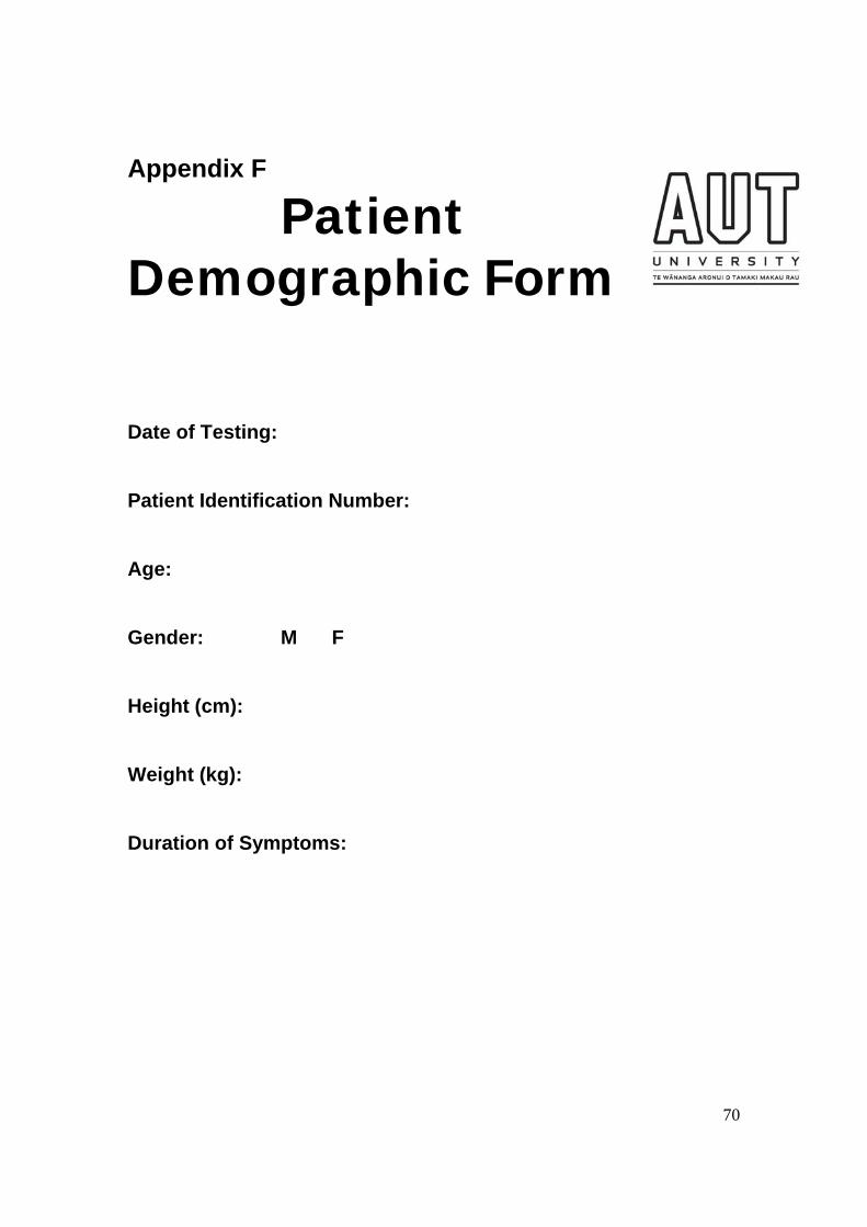
70
Appendix F
Patient Demographic Form
Date of Testing: Patient Identification Number: Age: Gender: M F Height (cm): Weight (kg): Duration of Symptoms:
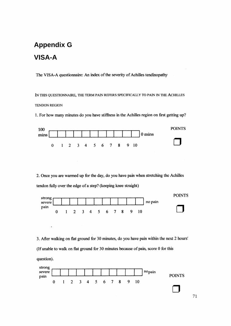
71
Appendix G
VISA-A
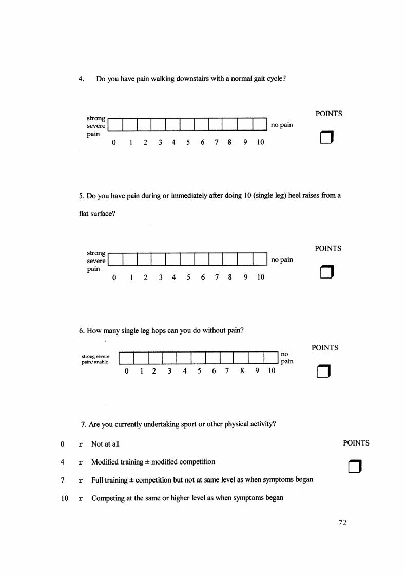
72

73

74
Appendix H
LOWER LIMB TASKS QUESTIONNAIRE ACTIVITIES OF DAILY LIVING SECTION
Patient: _______________ Date:_____________ INSTRUCTIONS Please rate your ability to do the following activities in the past 24 hours by circling the number below the appropriate response. If you did not have the opportunity to perform an activity in the past 24 hours, please make your best estimate on which response would be the most accurate. Please also rate how important each task is to you in your daily life according to the following scale: 1. = Not important 2. = Mildly important 3. = Moderately important 4. = Very important Please answer all questions. NO MILD MODERATE SEVERE IMPORTANCE DIFFICULTY DIFFICULTY DIFFICULTY DIFFICULTY UNABLE OF TASK 1. Walk for 10 minutes 4 3 2 1 0 1 2 3 4 2. Walk up or down 10 steps (1 flight) 4 3 2 1 0 1 2 3 4
3. Stand for 10 minutes 4 3 2 1 0 1 2 3 4 4. Stand for a typical work day 4 3 2 1 0 1 2 3 4 5. Get on and off a bus 4 3 2 1 0 1 2 3 4 6. Get up from a lounge chair 4 3 2 1 0 1 2 3 4 7. Push or pull a heavy trolley 4 3 2 1 0 1 2 3 4 8. Get in and out of a car 4 3 2 1 0 1 2 3 4 9. Get out of bed in the morning 4 3 2 1 0 1 2 3 4 10. Walk across a slope 4 3 2 1 0 1 2 3 4 TOTAL (/40) :_____
Enquiries concerning this questionnaire: Peter J. McNair PhD, Physical Rehabilitation Research Centre, Auckland University of Technology, Private Bag 92006, Auckland; New Zealand. email: [email protected] Phone: 921-9999 Ext 7143
Physical Rehabilitation Research Centre Auckland University of Technology
New Zealand
Time 3

75
Appendix H
LOWER LIMB TASKS QUESTIONNAIRE RECREATIONAL ACTIVITIES SECTION
Patient: _______________ Date:_____________ INSTRUCTIONS Please rate your ability to do the following activities in the past 24 hours by circling the number below the appropriate response. If you did not have the opportunity to perform an activity in the past 24 hours, please make your best estimate on which response would be the most accurate. Please also rate how important each task is to you in your daily life according to the following scale:
1. = Not important 2. = Mildly important 3. = Moderately important 4. = Very important Please answer all questions. NO MILD MODERATE SEVERE IMPORTANCE DIFFICULTY DIFFICULTY DIFFICULTY DIFFICULTY UNABLE OF TASK 1. Jog of 10 minutes 4 3 2 1 0 1 2 3 4 2. Pivot or twist quickly while walking 4 3 2 1 0 1 2 3 4 3. Jump for distance 4 3 2 1 0 1 2 3 4 4. Run fast/sprint 4 3 2 1 0 1 2 3 4 5. Stop and start moving quickly 4 3 2 1 0 1 2 3 4 6. Jump upwards and land 4 3 2 1 0 1 2 3 4 7. Kick a ball hard 4 3 2 1 0 1 2 3 4 8. Pivot or twist quickly while running 4 3 2 1 0 1 2 3 4 9. Kneel on both knees for 5 minutes 4 3 2 1 0 1 2 3 4 10. Squat to the ground/floor 4 3 2 1 0 1 2 3 4 TOTAL (/40) :_____ Enquiries concerning this questionnaire: Peter J. McNair PhD, Physical Rehabilitation Research Centre, Auckland University of Technology, Private Bag 92006, Auckland; New Zealand. email: [email protected] Phone: 921-9999 Ext 7143
Physical Rehabilitation Research Centre Auckland University of Technology
New Zealand
Related Documents