Biomarkers of oxidative stress and their application for assessment of individual radiosensitivity Siamak Haghdoost Department of Genetics, Microbiology and Toxicology Stockholm 2005 ISBN 91-7155-150-6

Welcome message from author
This document is posted to help you gain knowledge. Please leave a comment to let me know what you think about it! Share it to your friends and learn new things together.
Transcript

Biomarkers of oxidative stress and their application for assessment of
individual radiosensitivity
Siamak Haghdoost
Department of Genetics, Microbiology and Toxicology
Stockholm 2005 ISBN 91-7155-150-6

2
Abstract Radiotherapy is one of the most common therapeutic methods for
treatment of many types of cancer. Despite many decades of development and experience there is much to improve, both in efficacy of treatment and to decrease the incidences of adverse healthy tissue reactions. Around 20 % of the radiotherapy patients show a broad range in the severity of normal tissue reactions to radiotherapy, and dose limits are governed by severe reactions in the most radiosensitive patients (< 5 %). Identification of patients with low, moderate or high clinical radiosensitivity before commencing of radiotherapy would allow individual adaptation of the maximum dose with an overall increase in the cure rate. Characterization of factors that may modify the biological effects of ionizing radiation has been a subject of intense research efforts. Still, there is no assay currently available that can reliably predict the clinical radiosensitivity. The aim of this work has been to investigate the role of oxidative stress in individual radiosensitivity and evaluate novel markers of radiation response, which could be adapted for clinical use.
8-Oxo-7,8-dihydro-2´'-deoxyguanosine (8-oxo-dG), a general marker of oxidative stress, is one of the major products of interaction of ionizing radiation with DNA and the nucleotide pool of the cell. As 8-oxo-dG is highly mutagenic due to incorrect base pairing with deoxyadenosine, various repair mechanisms recognize and remove 8-oxo-dG. The repaired lesions are released from cells to the extracellular milieu (serum, urine and cell culture medium) where they can be detected as markers for free radical reactions with the nucleic acids.
Significant variations in background levels as well as in radiation induced levels of 8-oxo-dG in urine have been demonstrated in breast cancer patients (paper 1). Two major patterns were observed: high background and no therapy-related increase vs. low background and significant increase during radiotherapy for the radiosensitive and non radiosensitive patients respectively.
Studies in paper 2 indicated major contribution of the nucleotide pool to the extracellular 8-oxo-dG levels. The results also implicated induction of prolonged endogenous oxidative stress in the irradiated cells. RNA “knock-down” experiments on the nucleotide pool sanitization enzyme hMTH1 in paper 3 lend further experimental evidence to this assumption.
The applicability of 8-oxo-dG as a diagnostic marker of oxidative stress was demonstrated in paper 4. Studies on dialysis patients revealed a good correlation between inflammatory responses (known to be associated with persistent oxidative stress) and extracellular 8-oxo-dG.
In summary, our results confirm that extracellular 8-oxo-dG is a sensitive in vivo biomarker of oxidative stress, primarily formed by oxidative damage of dGTP in the nucleotide pool with a potential to become a clinical tool for prediction of individual responses to radiotherapy.

3
List of original publications
1. Haghdoost S, Svoboda P, Näslund I, Harms-Ringdahl M, Tilikides A,
Skog S. Can 8-oxo-dG be used as a predictor for individual
radiosensitivity? Int J Radiat Oncol Biol Phys. 2001 Jun 1;50(2):405-10.
2. Haghdoost S, Czene S, Näslund I, Skog S, Harms-Ringdahl M.
Extracellular 8-oxo-dG as a sensitive parameter for oxidative stress in
vivo and in vitro, Free Radic Res. 2005 Feb 39(2):153-62
3. Haghdoost S, Sjölander L Czene S, Harms-Ringdahl M.
The nucleotide pool is a significant target for oxidative stress. Submitted
4. Haghdoost S, Maruyama Y, Pecoits-Filho R, Heimburger O,
Seeberger A, Anderstam B, Suliman ME, Czene S, Lindholm B,
Stenvinkel P, Harms-Ringdahl M. Systemic inflammation in
hemodialysis patients is associated with increased oxidation of the
nucleotide pool as revealed by serum levels of 8oxo-dG. Submitted
Additional publication not included in this thesis:
5. Kämpfer P, Lindh J.M, Terenius O, Haghdoost S, Falsen E, Busse H.-J,
Faye I. Thorsellia anophelis gen. nov., sp. nov., a new genus of the
gammaproteobacteria, IJSEM Paper in Press.
Reprints were made with permission from the publisher.

4
Abbreviations
BER: base excision repair
CRP: C-reactive protein
CVD: cardio vascular disease
dNTP: deoxynucleotide triphosphate
DSB: double strand break
ELISA: enzyme-linked immunosorbent assay
ESRD: end-stage renal disease
GSH: glutathione peroxidase
Gy: gray
HD: hemodialysis
HPLC-EC: high performance liquid chromatography with electrochemical
detection
LET: linear energy transfer
MDS: multiply damaged site
NER: nucleotide excision repair
NIR: nucleotide incision repair
8-Oxo-G: 8-oxo-guanine
8-Oxo-dG: 8-oxo-7,8-dihydro-2´-deoxyguanosine
8-Oxo-Guo: 8-oxo-guanosine
ROS: reactive oxygen species
siRNA: short interfering RNA
SLE: systemic lupus erythematosus
SOD: superoxide dismutase
SSB: single strand break
Sv: sievert
UV: ultraviolet

5
TABLE OF CONTENTS
AIM.......................................................................................................................... 6 INTRODUCTION.................................................................................................. 7
Ionizing and non-ionizing radiation ............................................................................. 7 Effects of ionizing radiation on biological materials ................................................... 9 Background radiation and cancer incidence (stochastic effects) ................................. 9 Cellular targets of ionizing radiation ......................................................................... 10 Radiation and radiotherapy........................................................................................ 12 Side effects of radiotherapy (deterministic effects) and dose limiting factors............ 14 In vitro assays used for determination of radiosensitivity .......................................... 15 DNA repair and radiosensitivity ................................................................................. 16 Oxidative stress and cellular damage......................................................................... 18 8-Oxo-2´-deoxyguanosine – formation, repair and mutagenesis ............................... 20 Sources of extracellular 8-oxo-dG.............................................................................. 21 Correlation between radiosensitivity and oxidative stress ......................................... 24
RESULTS AND DISCUSSION .......................................................................... 25 Quantitative measurement of extracellular 8-hydroxy-2'-deoxyguanosine ................ 25 Individual radiosensitivity and urinary 8-oxo-dG (paper 1) ...................................... 27 Extracellular 8-oxo-dG as a marker of in vitro oxidative stress (paper 2) ................ 29 The nucleotide pool is a major source of extracellular 8-oxo-dG (paper 3).............. 30 8-oxo-dG as diagnostic marker of oxidative stress in dialysis patients (paper 4)...... 31
FUTURE PERSPECTIVES................................................................................ 32 ACKNOWLEDGEMENTS................................................................................. 35 REFERENCES..................................................................................................... 38

6
Aim The main purpose of the research studies included in this thesis was to
develop and evaluate diagnostic tools for reliable diagnosis of radiosensitive
individuals and make these tools available for the needs of individually
designed radiation therapy. Clinical availability of predictive assays for
individual radiosensitivity would allow for individually designed radiotherapy
and
• increase the probability of a better tumor control in the group of
patients with mild and moderate reactions
• decrease adverse tissue reaction in the highly radiosensitive
group of patients
7
Introduction During our whole lives we are exposed to various kinds of radiation,
ultraviolet and ionizing radiation being the dominant types with known
biological adverse effects. While the sun is the main source of UV radiation,
the major sources of ionizing radiation include medical uses, cosmic radiation,
radiation from the ground, internal radiation from 40K and radon. Human
exposures to high doses of ionizing radiation are rare. A special case of
exposure to high doses of ionizing radiation is its use for therapeutic purposes.
Ionizing and non-ionizing radiation
The various types of radiation fall into two broad categories, ionizing
and non-ionizing. When the energy of radiation is high enough it will produce
ionizations and excitations in the target. Both particle radiation (electrons,
protons, α-particles, or neutrons) and electromagnetic radiation (high energetic
photons such as X- and γ-rays) belong to the category of ionizing radiation.
Direct ionizations are produced when photons or charged particles interact
directly with the target. Indirect ionizations are produced by transfer of energy
from photons to cellular water molecules (radiolysis) forming highly reactive
intermediates e.g. hydroxyl radicals which then interact with the target.
Radiation with lower energies, on the other hand, does not produce ionizations
when interacting with matter. Examples of non-ionizing radiation are UV and
visible light, infrared radiation (IR) and micro- and radio waves.
Important definitions, terms and units in radiation biology include
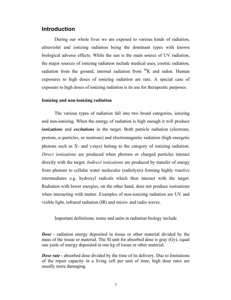
Dose - radiation energy deposited in tissue or other material divided by the mass of the tissue or material. The SI unit for absorbed dose is gray (Gy), equal one joule of energy deposited in one kg of tissue or other material. Dose rate - absorbed dose divided by the time of its delivery. Due to limitations of the repair capacity in a living cell per unit of time, high dose rates are usually more damaging.

8
Linear energy transfer (LET) - is expressed in terms of the mean energy released in keV per micrometer (µm) of the tissue traversed (keV/µm). Examples of low LET and high LET radiation are medical X-rays and alpha particles respectively. Equivalent dose - relates absorbed dose (Gy) in tissue to the biological effects of a specific radiation quality. The unit for equivalent dose is sievert (Sv). To determine equivalent dose (Sv), an absorbed dose (Gy) is multiplied by a radiation quality factor (Q). For example Q for gamma is 1, for alpha 20. In other words, one Gy equals 1 Sv for gamma radiation, but in case of alpha particles 1 Gy will equal 20 Sv.
A schematic illustration of radiation qualities with different LET is shown in
figure 1. Considering DNA as target, it is apparent that the quality of damage
induced by one Gy of high LET radiation will considerably differ from that
observed after the same dose of low LET radiation.
Low LET radiation
High LET radiation
• •
•• •
••
•• •• •• ••
• ••• • • •
•••• •••• ••
• ••
Gamma rays
Alfa particles
Target
••• • ••• •• • ••• •• • •• ••• • •• ••• •• •• ••• ••• ••• ••• • • •• ••
Figure 1. Schematic picture of ionization events (black dots) caused by high
and low LET radiation.

9
Effects of ionizing radiation on biological materials
The biological effects of radiation arise from interactions with various
components of the cell, such as proteins or nucleic acids. Radiation-induced
modifications of cellular components may destroy their proper functions and
lead to loss of cell viability, decreased enzyme activity, mutations, initiation of
cancer, and hereditary diseases. The immediate effects of high acute radiation
exposures are caused by cell membrane rupture resulting in loss of cellular
content and cell death. Acute whole body irradiation [1] can lead to organ
failure as exemplified (in rodent) by
• loss of coordination (including breathing problems) after
exposure of the central nervous system to doses above 100 Gy,
with death occurring within few minutes up to 2 days.
• nausea, vomiting, and diarrhea caused by damage to the
gastrointestinal tract from doses 9 to 100 Gy. Progressive
dehydration will result in death within 3-5 days.
• loss of appetite and hair, hemorrhaging, inflammation, and
secondary infections such as pneumonia from doses 3 to 9 Gy
due to damage to the bone marrow and other haematopoietic
tissues. This can result in death within 10-30 days. These effects
are also found in patients undergoing radiation therapy.
• loss of appetite and hair, hemorrhaging, and diarrhea observed at
doses of around 2 Gy, the consequences are rarely lethal.
Background radiation and cancer incidence (stochastic effects)
Sources of ionizing radiation include those naturally occurring and man-
made. The principal types and sources of natural background radiation are:
• Cosmic radiation from the outer space
• Terrestrial sources of naturally disintegrating radioactive
materials e.g. radium, thorium, uranium

10
• Internal sources of radioactive isotopes that are normally present
within living cells (e.g. potassium-40, carbon-14).
The background levels of radiation from natural sources show large variations
in different parts of the world (1.5 up to 100 mSv/year). Exposure levels from
radiation for diagnostic purposes (ex. nuclear medicine and diagnostic
radiology) contribute on the average with doses around 2 mSv/per year to the
Swedish population.
Increased rates of cancer and mutations are the major long-term risks of
concern associated with radiation exposure. Risk estimates for both these
effects, especially in low-dose rate chronic exposure situations, are however
difficult to perform due to confounding life-style factors (smoking, diet,
sunlight exposure) involved in the etiology of cancer. The average background
radiation dose in Sweden (including radiation from medical investigation and
radon) is 5 to 6 mSv per year which cause a few hits per cell per year. The total
cancer incidence in Sweden is around 40 000 cases per year. Genetic factors
have been estimated to cause about 20 % of the cancer incidence [2] while
contribution of life style factors may be around 70 % [3]. Based on linear
extrapolation of the relation between dose and cancer incidence, estimated from
epidemiological investigations for doses > 50 mSv, down to the low doses the
estimated number of cancers caused by background radiation in Sweden will be
1000-2000 cancers per year.
Cellular targets of ionizing radiation
Since living cells consist of ~70 % water, the majority of ionizations
(produced by radiation) take place through interactions with water molecules.
Radiolysis of cellular water will produce reactive oxygen species such as the
hydroxyl radical OH• that may interact with various cellular components. The
remaining 30 % of radiation effects in cells are mediated by direct interactions
of photons with a particular cellular component such as DNA, proteins and
membranes leading to ionizations and excitations. Of note, non-ionizing
radiation may interact with biological systems in other ways represented by e.g.

11
the photochemical reactions in the human eye, the photosynthesis of plants, and
the UV radiation-induced damage in skin.
Among the various cellular modifications induced by ionizing radiation,
persistent alterations of DNA (mutations) are of particular biological
importance, as they can be passed over to the progeny and result in hereditary
disorders. Elevated mutation frequencies and gross chromosomal changes also
play an essential role in the pathogenesis of many human disease states
including cancer, as well as in the processes of aging.
Both the direct and indirect effects of radiation can initiate damage to
DNA. Direct effects involve the formation of radicals and excited intermediates
as a result of the deposition of energy within the DNA structure. Indirect
effects involve the interaction of the DNA with radiolysis products of water
such as OH radicals, H-atoms or hydrated electrons. Low LET ionizing
radiation causes many different types of damages on DNA such as double
strand breaks (DSB, 20-40 breaks/cell per Gy), single strand breaks (SSB, 1000
breaks/cell per Gy) and base modifications (1000 modifications/cell per Gy) [4-
7]. A particular feature of radiation induced chemical alterations is the
production of unique types of damages in a small section of the DNA. These
so-called multiply damaged sites (MDS), consisting of clusters of strand breaks
and base damages within one or two turns of the DNA (10 to 20 base pairs) [8],
are suggested to be a specific signature of ionizing radiation.
Among the DNA lesions the DNA double strand breaks are of principal
interest. The genotoxic properties of many physical and chemical agents are
often closely associated with their ability to induce DSBs. Because of their high
cytotoxicity (unrepaired or misrepaired DNA double-strand breaks are often
lethal) and ability to induce chromosomal aberrations (that may ultimately lead
to carcinogenesis), cell survival and maintenance of genome integrity are
critically dependent on efficient repair of DNA DSBs. Cells have developed
different DNA repair mechanisms to deal with harmful effects of radiation on
DNA such as: homologous recombination and nonhomologous end joining
(both involved in repair of DSBs); base excision repair (BER) acting on base
12
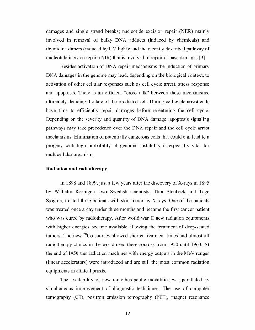
damages and single strand breaks; nucleotide excision repair (NER) mainly
involved in removal of bulky DNA adducts (induced by chemicals) and
thymidine dimers (induced by UV light); and the recently described pathway of
nucleotide incision repair (NIR) that is involved in repair of base damages [9]
Besides activation of DNA repair mechanisms the induction of primary
DNA damages in the genome may lead, depending on the biological context, to
activation of other cellular responses such as cell cycle arrest, stress response
and apoptosis. There is an efficient “cross talk” between these mechanisms,
ultimately deciding the fate of the irradiated cell. During cell cycle arrest cells
have time to efficiently repair damages before re-entering the cell cycle.
Depending on the severity and quantity of DNA damage, apoptosis signaling
pathways may take precedence over the DNA repair and the cell cycle arrest
mechanisms. Elimination of potentially dangerous cells that could e.g. lead to a
progeny with high probability of genomic instability is especially vital for
multicellular organisms.
Radiation and radiotherapy
In 1898 and 1899, just a few years after the discovery of X-rays in 1895
by Wilhelm Roentgen, two Swedish scientists, Thor Stenbeck and Tage
Sjögren, treated three patients with skin tumor by X-rays. One of the patients
was treated once a day under three months and became the first cancer patient
who was cured by radiotherapy. After world war II new radiation equipments
with higher energies became available allowing the treatment of deep-seated
tumors. The new 60Co sources allowed shorter treatment times and almost all
radiotherapy clinics in the world used these sources from 1950 until 1960. At
the end of 1950-ties radiation machines with energy outputs in the MeV ranges
(linear accelerators) were introduced and are still the most common radiation
equipments in clinical praxis.
The availability of new radiotherapeutic modalities was paralleled by
simultaneous improvement of diagnostic techniques. The use of computer
tomography (CT), positron emission tomography (PET), magnet resonance

13
imaging (MRI) and ultra sound increased the possibilities for the oncologists to
distinguish a tumor from a normal tissue and thus optimize the dose planning
and treatment regimes.
Examples of novel approaches that have led to more effective radiation
therapy of cancer include:
• Intraoperative irradiation: a large dose (8-15 Gy) of external
radiation (usually electrons) is delivered to the tumor and
surrounding tissue during surgery (e.g. in colon cancer)
• Brachytherapy, interstitial irradiation, and intracavitary
irradiation: radioactive implants are placed directly in a tumor or
body cavity (in prostate cancer, uterus cancer)
• Radiosurgery and Gamma knife: high doses of gamma radiation
(3-5 fractions of 7-15 Gy) are delivered to small solid tumors
(liver and lung metastasis, small brain metastasis)
• Radiolabeled antibodies: radiolabeled tumor-specific antibodies
are used to deliver doses of radiation directly to the cancer cells
(radioimmunotherapy).
At present, radiotherapy is used to treat about 45 % of all cancer patients in
Sweden. Radiotherapy can be used alone as a curative, adjuvant (in
combination with surgery and chemotherapy) or palliative (pain relieving,)
treatment. The aim of modern radiotherapy is to cure patients from cancer with
limited adverse effects to the normal tissue surrounding the cancer tumor. This
can be achieved by carefully planning of the dose delivery to the tumor (dose
planning system) and more detailed understanding of the tumor and healthy
tissue responses to radiation.

14
Side effects of radiotherapy (deterministic effects) and dose limiting
factors
For many years it has been recognized by clinicians that some patients
show hypersensitivity to the radiotherapy. Early and late adverse effects in
exposed healthy tissues are the major factors limiting dose escalation in
radiotherapy of cancer patients. During the radiotherapy both cancer and
normal cells are being exposed to the radiation. Radiation-induced damage in
tissues may lead to the cell death, and toxic components from dead cells may
affect the whole body and cause symptoms such as nausea.
Deterministic side effects of radiation therapy can be divided into acute
and late reactions. The dose responses for these reactions are characterized by a
threshold dose over which the effects appear. Acute reactions occur during the
ongoing treatment or within a few weeks after the treatment and are usually
reversible (ex: erythema). For example, skin reaction and pain in the irradiated
breast and chest area are the most common side effects during radiotherapy of
breast cancer patients. Acute radiosensitivity is most often manifested in tissues
with high proliferation rate such as skin, small intestine and rectum. Late
reactions occur months or years after radiation, mainly in slowly proliferating
tissues such as fatty tissues and muscles and are of permanent character
(fibrosis, telangiectasia). In addition to the radiation dose and the dose per
fraction, side effects of radiation are also dependent on the area and volume of
treatment as well as on the individual radiosensitivity. There are also
indications of the stochastic effects of radiation therapy such as increased
cancer incidence in normal tissues receiving comparatively low doses of
radiation during irradiation of the neighboring organ with the primary tumor.
Examples of stochastic effects include increased lung and esophagus cancer
incidence in breast cancer patients receiving radiotherapy [10-12].
The most commonly used system for evaluation of a patients’
radiosensitivity is the Radiation Therapy Oncology Group (RTOG) that is
based on to the intensity of the side effects (acute as well as late effects). These

15
acute and late radiation morbidity scoring criteria were developed in 1985 and
an example of such a protocol used for evaluation of the acute radiosensitivity
in breast cancer [13] is as follows:
Grade 0: no change over baseline
Grade 1: follicular or dull erythema
Grade 2: tender or bright erythema, moderate edema
Grade 3: confluent moist desquamation, skin folds, pitting edema
Grade 4: ulceration, necrosis.
About 20 % of the patients who undergo radiotherapy of breast cancer
will develop adverse reactions to the therapy [14, 15], as many as 5 % of the
patients will develop severe reactions during the course of the treatment
(RTOG 3-4). Notably, patients with adverse reactions often have better cure
rates comparing to non-reacting patients. Positive correlations between
radiation side effects and local tumor control in breast, head & neck and colon
cancer patients have been previously reported [15-17]. Local tumor recurrence
after radiation therapy is due primarily to failure to eradicate all of the cancer
cells within the irradiated fields. Theoretically, all cancers could be controlled
locally if a sufficiently high radiation dose could be delivered to a target that
encompasses all of the cancer cells. However, at the present time, doses used in
radiotherapy are adapted to what can be tolerate by the most radiosensitive
patients, keeping the risk of severe persistent normal tissue damage below 5-10
%, despite the fact that many patients could tolerate a larger dose without
severe tissue reactions. Hence, radiotherapy would greatly benefit from a
diagnostic tool providing information on individual radiosensitivity.
In vitro assays used for determination of radiosensitivity
Several attempts have been made to correlate radiation therapy side-
effects with the cellular responses to the radiation. The various endpoints of
these assays include transcriptional response employing cDNA microarrays

16
[18], assessment of DNA repair capacity by alkaline comet assay [19],
apoptosis [20] and cell survival [21, 22] as well as formation of chromosome
aberrations [23]. In a number of studies the results correlated with the clinical
observations, however these correlations only held for large groups with little
predictive value for the individual patients [24] as there was a large overlap in
the responses of radiation-sensitive and non-sensitive patients in these assays
(for review see [25]).
One of the possible explanations for the observed large variations is that
there are several underlying factors causing individual radiosensitivity. Known
risk factors include age, concurrent chemotherapy, anatomical variation (e.g.
bust size in breast cancer treatment), body mass index (BMI), tissue
oxygenation and genetic predisposition [26]. Hence, one of the potential
drawbacks of the predictive in vitro assays would be that they most often focus
on one particular cell function and thus examine only one of the many factors
involved in individual susceptibility to radiation. For instance, there are
indications that genetically defined radiosensitivity is not the predominant
clinical cause for excessive tissue reaction [27].
DNA repair and radiosensitivity
A recent review of the International Commission on Radiological
Protection (ICRP) of clinical observations and cellular as well as molecular
studies on radiosensitivity [28] has identified several genetic syndromes that
are connected with radiation hypersensitivity (table 1).
As seen from the list below, at least two groups of genes, those involved
in cell cycle control and DNA repair are strongly associated with
radiosensitivity. Seminal studies on hypersensitivity syndromes with a clear-cut
genetic component (ATM, NBS1 and Mre11 genes) identified DNA damage
recognition and repair as a central theme of radiosensitivity [14, 29]. However,
these classical cases of radiosensitivity syndromes are very rare and account
only for about one in 10 000 observed cases.

17
Table 1. Genetic syndromes associated with radiosensitivity:
Syndrome Genetic disorder
Ataxia telangiectasia DSB recognition/repair
Bloom syndrome DSB repair
Fanconi anaemia DSB repair
Li-Fraumeni syndrome Cell cycle regulation, apoptosis
Nevoid basal cell carcinoma syndrome Control of cell proliferation
Neurofibromatosis Cell cycle regulation
Nijmegen breakage syndrome DSB repair
Retinoblastoma Cell cycle regulation
As most gene products are part of multi-protein complexes and function
in complex cellular pathways, mutation of a particular gene may affect the
expression of downstream genes involved in these cellular pathways, as well as
those of genes involved in related processes trying to compensate for the
defect. Therefore, it is most likely that individuals in the relatively large group
of radiosensitive patients will carry more subtle defects in these pathways due
to presence of low-penetrance mutations, which are not limited to the two
abovementioned groups of genes.
Still, the current knowledge on genetic predisposition for individual
response to radiation damage, in particular on the involvement of low-
penetrance gene mutations, is rather sparse. It is known that cancer patients
with diabetes mellitus [30], hypertension, rheumatoid arthritis and systemic
lupus erythematosus (SLE) [31], have a higher frequency of side effects to
radiotherapy. As suggested by a recent study [18, 32], combined effects of
polymorphisms in several DNA repair genes, could also lead to manifestations
of individual radiosensitivity.

18
Oxidative stress and cellular damage
Reactive oxygen species (e.g. O2●- superoxide anion, OH● hydroxyl
radical, and H2O2 hydrogen peroxide) are continuously generated in aerobic
organisms both endogenously as by-products of oxygen metabolism as well as
by exogenous factors (figure 2)
O2-º
O2
e-From mitochondria
(1-5
% o
f tot
al O
2)
SOD H2O2
H2O
Cata
lase
Glu
tath
ione
pero
xidas
e
OHºFenton Reaction:Fe2++ H2O2=Fe3++OH
_+OHº
Gamma radiation,radiolysis of H2O
LipidsProteinsDNAdNTP
Antioxidants
X
From macrophage
Modification of:
Figure 2. Schematic picture of endogenous and exogenous production of free
radicals and cellular defense mechanisms.
The endogenous production of free radical derived DNA lesions has
been estimated to be up to 10 000 per day per cell in humans [33]. For instance,
the yield of superoxide anions has been estimated to be 1-5 % of the total
consumed oxygen, equaling to 2 kg of superoxide anions per year [33]. The
majority of the endogenously produced reactive oxygen species (ROS) is
derived from the mitochondrial electron transport chain [34]. Even though the
predominant sources of ROS in aerobic cells are the mitochondria, substantial
amounts of ROS are also generated in peroxisomes. Many chemical and

19
physical agents, among them ionizing radiation as discussed above, are known
to exert their mutagenic and carcinogenic properties through production of free
radicals [35].
The biological effects of ROS are intimately related to their tissue
concentration. At low concentrations, ROS have many important physiological
functions e.g. as secondary messengers or part of the immune defence. On the
other hand, moderate and high levels of ROS within cells may lead to
“oxidative stress”. The term oxidative stress, in essence, refers to a serious
imbalance between production of ROS and antioxidant defence in favour of the
oxidants, potentially leading to cellular damage [36]. Oxidative stress may thus
result from
1. Diminished levels of antioxidants, for example caused by, mutations
affecting the activities of antioxidant defence enzymes such as
superoxide dismutase (SOD), or glutathione (GSH) peroxidase, or toxins
that deplete antioxidant defences. For example, many xenobiotics are
metabolized by conjugation with GSH; thus high doses can deplete GSH
and cause oxidative stress even if the xenobiotic itself is not generating
reactive species. Deficiencies in dietary minerals (e.g., Zn2+, Mg2+, Fe2+,
Cu2+, and Se) and/or antioxidants can also cause oxidative stress.
and/or
2. Increased production of ROS, for example, by exposure of cells or
organisms to elevated levels of O2; physical (ionizing radiation) or
chemical agents (e.g. paraquat) that generate excess ROS; or excessive
activation of ‘natural’ systems producing such species (e.g.
inappropriate activation of phagocytic cells in chronic inflammatory
diseases).
Interaction of ROS with various cellular components such as lipids, proteins
and DNA has been shown to result in temporary as well as permanent
modifications [37], that may have deleterious consequences. Alone or in

20
combination with primary etiological factors, ROS have been implicated in
diverse diseases such as cancer, hypertension, atherosclerosis, Alzheimer’s
disease, lung injury as well as the aging process.
The detrimental effects of oxidative stress on cellular components are
counteracted by various defence mechanisms which range from low molecular
weight antioxidants through DNA repair enzymes to ultimate disposal of the
damaged cell by induction of programmed cell death (apoptosis). One of the
first and most important defences is the array of low molecular weight cellular
antioxidants, including the reduced form of GSH, vitamin E, and ascorbate
[38].
The second line of defence mechanisms involves enzymes with anti-oxidant
properties such as various SODs and catalase. For instance, superoxide anions
produced by mitochondria are first converted by SODs into the less toxic
hydrogen peroxide that in turn is disassembled into water and oxygen by
catalase [39].
Should the extent of oxidative stress overwhelm the cellular resources of
antioxidants as outlined above, the escaped ROS may damage various cellular
components, such as the DNA and nucleotide pool. While modified proteins
and lipids are removed from the cells by a normal turn over of the molecules,
specific repair pathways act to remove oxidative DNA modifications.
8-Oxo-2´-deoxyguanosine – formation, repair and mutagenesis
Oxidative modifications of nucleic acids include a range of various base
modifications such as 8-oxo-adenine, 2-hydroxy-adenine, 8-oxo-guanosine,
FaPy-guanine, thymine glycol, sugar damage, apurinic/apyrimidinic sites (AP
site), damage of the phosphodiester backbone of DNA (strand breaks) and
DNA-protein crosslinks [40]. One of the major types of base damage formed
by ionizing radiation, as well as by endogenous ROS, is 8-oxo-7,8-dihydro-2´'-
deoxyguanosine (8-oxo-dG). 8-Oxo-dG is produced mainly by hydroxylation
of the C8 position of 2´-dG in both DNA and the free nucleotide dGTP in the

21
nucleotide pool. Charge transfer events in the DNA may also contribute to
formation of 8-oxo-dG [41].
It is generally assumed that most of the oxidative DNA damage is dealt
with by base excision repair (BER) [42]. In the process of BER, DNA
glycosylases recognize and remove damaged DNA bases. After the action of
AP-endonuclease, the resulting gap is then filled by a DNA polymerase and
sealed by DNA ligase. Several glycosylases with ability to remove oxidative
base damage have been identified in both bacteria (the MutY and MutM
proteins) and higher organisms (e.g. the OGG1 and OGG2 proteins).
8-Oxo-dG is a mutagenic lesion as it can mispair with adenine [43]. If
not removed from DNA, replication of the damaged sequence will lead G→T
transversions [44]. Another mechanism for 8-oxo-dG mutagenesis is the
incorporation of free 8-oxo-dGTP from the nucleotide pool into DNA opposite
of adenine during either replication or repair. Such mis-incorporation will lead
to A→G transversions through activation of the mismatch repair pathway that
will remove A opposite of the 8-oxo-dG and replace it with C. This base pair
will be repaired in turn by base excision repair that will remove 8-oxo-dG
opposite C and replace it with G [45].
Of note, several recent studies [46, 47] indicate that the efficacy of BER
may be strongly reduced when a oxidised DNA base is a part of MDS.
Abortive repair of oxidised bases in MDS is detrimental for the cell as
indicated by decreased survival [48], increased mutation frequency [49], and
formation of DNA double strand breaks [50].
Sources of extracellular 8-oxo-dG
Despite its extensive use as a general marker for oxidative stress the
origin of extracellular 8-oxo-dG is a matter of scientific controversy. The result
of BER activity on 8-oxo-dG in DNA is 8-oxo-Gua [40] that can be detected in
urine and blood plasma. There are indications that 8-oxo-dG may be removed
from DNA also by the nucleotide excision repair (NER) [51] as well as by the
nucleotide incision repair (NIR) [52] mechanisms. As the NER pathway is

22
specific for bulky DNA adducts, its role in recognizing and removing 8-oxo-dG
from DNA is under discussion [51]. Primary fragments (containing 24-29
nucleotides) removed from DNA by NER, may undergo further reduction to 6-
7 mers or even isolated nucleosides. However, it has been demonstrated that
human urine contains only limited amounts of oligonucleotides and
mononucleotides; moreover these nucleotides do not contain oxidative
modification in form of 8-oxo-dG [53]. Thus, under normal physiological
conditions, NER seems to be of minor importance for repair of oxidative DNA
damage [54]. NIR seems to be involved in the repair of 8-oxo-dG [52], most
likely as a backup system for BER [40]. Still, the biological significance of
these findings for the production of extracellular 8-oxo-dG is yet to be shown
by further studies. Figure 3 shows the classes of repair products formed by the
NIR, NER and BER repair pathways.
C
T
C
T
G
A
G
A
NER, up to 28 bp
BER, damaged base
NIR, damaged base with sugar and phosphate
Figure 3. Schematic presentation of DNA repair products removed by BER, NIR and
NER (in bold squares)

23
Recent studies show that mitochondrial DNA (mtDNA) is yet another
target for free radical reactions. mtDNA is highly prone to oxidative damage
because of the lack of histones and also due to its proximity to one of the major
intracellular sources of ROS. The steady-state level of oxidized bases in
mtDNA is two to three fold higher than that in nuclear DNA [55]. The
importance of mitochondrial genome maintenance is demonstrated by the role
of mitochondrial gene mutations in degenerative diseases of the nervous
system, heart, muscle and kidneys [56, 57]. Still, the only documented repair
mechanism in the mitochondria up-to-date is that of BER. Thus, oxidative
mtDNA damage is not likely to contribute to the extracellular 8-oxo-dG.
Another target for radiation-induced oxidative damage is the pool of free
nucleotide triphosphates (dNTPs) available in the cytoplasm. Free radicals can
react with the dNTPs in the nucleotide pool and produce a wide spectrum of
modified dNTPs. 8-Oxo-dGTP is produced upon free radical attack on the C8
position in dGTP. 8-Oxo-dGTP can be misincorporated into DNA and cause
mutations. Bacterial cells are protected against incorporation of 8-oxo-dGTP by
8-oxo-dGTPase [45, 58] that converts 8-oxo-dGTP to 8-oxo-dGMP. The
human homologue for 8-oxo-dGTPase is named hMTH1. The importance of
nucleotide pool sanitization is demonstrated by the observation that insufficient
8-oxo-dGTPase activity may lead to cancer [44, 45].
A recurrent suggestion is that urinary 8-oxo-dG mainly originates from
the degradation and oxidation of DNA in dead cells. There is no convincing
experimental evidence however to prove this hypothesis [59]. As results from
previous studies also show that 8-oxo-dG is not absorbed through the intestinal
tract a dietary influence on extracellular 8-oxo-dG should be minimal [60-62].
It has been also discussed that oxidation of 2´-dG in serum or in urine
may significantly contribute to urinary or serum levels of 8-oxo-dG. However,
2´-dG is not oxidized in systemic circulation to 8-oxo-dG [59]. Moreover, the
presence of hydrogen peroxide or other ROS in urine did not lead to detectable
formation of 8-oxo-dG [63].
24
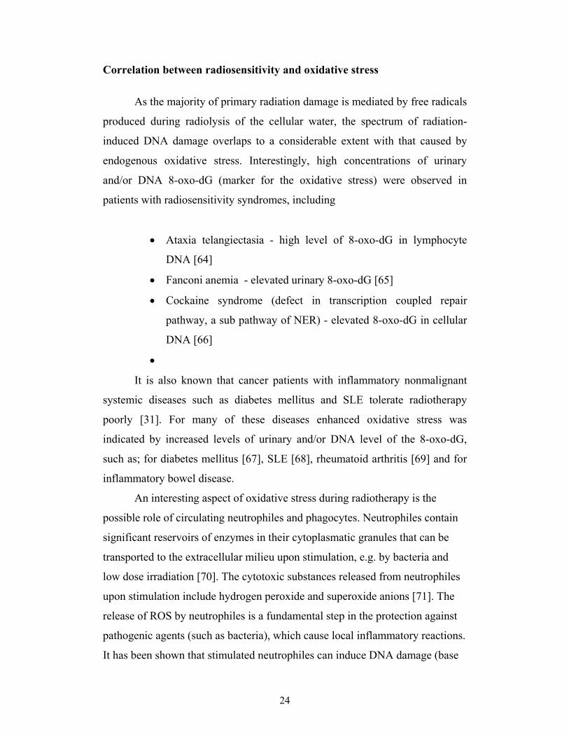
Correlation between radiosensitivity and oxidative stress
As the majority of primary radiation damage is mediated by free radicals
produced during radiolysis of the cellular water, the spectrum of radiation-
induced DNA damage overlaps to a considerable extent with that caused by
endogenous oxidative stress. Interestingly, high concentrations of urinary
and/or DNA 8-oxo-dG (marker for the oxidative stress) were observed in
patients with radiosensitivity syndromes, including
• Ataxia telangiectasia - high level of 8-oxo-dG in lymphocyte
DNA [64]
• Fanconi anemia - elevated urinary 8-oxo-dG [65]
• Cockaine syndrome (defect in transcription coupled repair
pathway, a sub pathway of NER) - elevated 8-oxo-dG in cellular
DNA [66]
•
It is also known that cancer patients with inflammatory nonmalignant
systemic diseases such as diabetes mellitus and SLE tolerate radiotherapy
poorly [31]. For many of these diseases enhanced oxidative stress was
indicated by increased levels of urinary and/or DNA level of the 8-oxo-dG,
such as; for diabetes mellitus [67], SLE [68], rheumatoid arthritis [69] and for
inflammatory bowel disease.
An interesting aspect of oxidative stress during radiotherapy is the
possible role of circulating neutrophiles and phagocytes. Neutrophiles contain
significant reservoirs of enzymes in their cytoplasmatic granules that can be
transported to the extracellular milieu upon stimulation, e.g. by bacteria and
low dose irradiation [70]. The cytotoxic substances released from neutrophiles
upon stimulation include hydrogen peroxide and superoxide anions [71]. The
release of ROS by neutrophiles is a fundamental step in the protection against
pathogenic agents (such as bacteria), which cause local inflammatory reactions.
It has been shown that stimulated neutrophiles can induce DNA damage (base

25
modification) [71] and can cause malignant transformation in other cells [72].
During radiotherapy, as neutrophiles pass through the exposed tissue, they will
receive a small radiation dose (in the cGy range). This dose could be enough to
activate the neutrophiles and give rise to inflammatory reactions, which could
theoretically contribute to various (therapeutic as well as side) effects of
radiotherapy.
Results and discussion
Quantitative measurement of extracellular 8-hydroxy-2'-deoxyguanosine
Figure 4 shows an overview of the methods used in our studies for
detection of 8-oxo-dG. In paper 1 urinary 8-oxo-dG was measured with a high
performance liquid chromatograph equipped with an electrochemical detector
(HPLC-EC). The advantage of the HPLC-EC method is that it measures only
the free form of 8-oxo-dG and not other 8-oxo-dG derivates (8-oxo-G, 8-oxo-
Guo) or oligomers containing 8-oxo-dG. Although HPLC-EC based methods
for detection of 8-oxo-dG are well established and reliable, they are less
suitable for a clinical laboratory due to low throughput (typically 1-3 hours per
sample), high cost, technical involvement and limited sensitivity for detection
of extracellular 8-oxo-dG (especially in blood serum).
We have evaluated serum levels of 8-oxo-dG as a predictive marker for
individual radiosensitivity after in vitro irradiation of whole blood samples
from patients assuming that the response may correlate with the organ response
to radiation. However the average serum level of 8-oxo-dG is relatively low
(~1.4 pmol/ml) and thus below the detection limit offered by our HPLC-EC (~1
pmol) based method. An alternative way to measure 8-oxo-dG is by enzyme
linked immunosorbent assay (ELISA). Commercial ELISA kits are sensitive,
fast and do not require sophisticated equipments. Unfortunately, the
monoclonal antibodies used in commercially available ELISA kits are not
specific for 8-oxo-dG and will even bind to some of its derivates such as 8-

26
oxo-Guo (of RNA origin) [73]. The usefulness of these kits for measurement of
8-oxo-dG in complex biological mixtures such as urine or blood serum is thus
questionable.
Samples (urine, serum, cell culture medium)
Purification by solid phase extraction (CH-bond Elute column)
Purified samples
Washing, eluting
First HPLC system based on ion pair chromatography
Urine, serum
Collection of fraction containing 8-oxo-dG
Second HPLC system,detection of 8-oxo-dG
Concentration of fraction
Medium, serum
Incubation of concentrated sample and primary antibodyin 8-oxo-dG precoated 96 well ELISA plate
Overnight incubation at 4ºC
Secondary antibody
Incubation at room temprature 2 hrs
Development and detection of colour
Figure 4. An overview of the methods for detection of 8-oxo-dG by HPLC-EC
and ELISA.
We have developed an improved ELISA for 8-oxo-dG that includes an
additional sample purification step that removes the interfering 8-oxo-Guo
before analysis. The capacity of our semi-manual ELISA method is about 100
samples per week per technician, and it has the same or better sensitivity as
commercially available kits (detection limit for 8-oxo-dG is around 0.1 pmol)
and is highly cost-effective. The reproducibility of our method for serum 8-
oxo-dG is about ± 20 % (standard deviations based on seven blood serum
samples, each analyzed 3 times, n=21). 8-Oxo-dG levels in blood serum
samples were between 0.2 up to 3 ng/ml, well within the sensitivity range of

27
our ELISA (marked as a bold line in the figure 5). To decrease the influence of
confounding factors, 8-oxo-dG is added to each sample as an internal standard.
Samples with and without internal standard were analyzed in duplicate. The
quantity of 8-oxo-dG was normalized using the internal standard.
0,1
0,2
0,3
0,1 1 10 100
8-oxo-dG (ng/ml)
OD
(450
nm
)
Figure 5. A representative standard curve for 8-oxo-dG produced by our
competitive ELISA.
Individual radiosensitivity and urinary 8-oxo-dG (paper 1)
Since the first reports on the presence of 8-oxo-dG and 8-oxo-G in urine
of human, rat and mouse [61, 74] both components have been extensively used
as markers of oxidative stress elicited by agents of both endogenous and
exogenous origin [75-79].The urinary levels of 8-oxo-G are usually 5-10 fold
higher than those of 8-oxo-dG. 8-Oxo-G, however, may also originate from
RNA degradation and its analysis is associated with technical difficulties [59,
80-82]. High levels of the 8-oxo-dG in DNA and/or urine has been detected in
cancer patients [33] and also in patients with non-cancerous diseases such as

28
Parkinson’s disease, Alzheimer’s disease, multiple sclerosis, cardiovascular
disease, chronic hepatitis, diabetes mellitus and rheumatoid arthritis [33]. It
should be noted, however, that the background levels of urinary and DNA
8-oxo-dG are also influenced by life style factors [83] and that they also are age
dependent [84, 85].
Evidence of the involvement of oxidative stress in cellular
radiosensitivity emerged from a number of in vitro and in vivo studies [86-91].
High levels of 8-oxo-dG in lymphocytes of breast cancer patients receiving
radiotherapy [79] as well as a positive correlation between urinary 8-oxo-dG
and radiation responses in lung cancer patients [92] have been reported. We
hypothesized that clinical observations of the individual radiosensitivity in
breast cancer patients undergoing radiotherapy (such as acute skin reactions)
could be related to the level of pre-existing or therapy-induced oxidative stress
response measured as urinary 8-oxo-dG. Based on clinical observations the
patients were classified as radiosensitive (major skin reactions) or non-
radiosensitive (patients with minor or no acute skin changes). The advantages
to analyze extracellular 8-oxo-dG in urine or serum are that the method is not
invasive (easy to receive patient samples), and that the 8-oxo-dG molecule is
relatively stable in extracellular milieu. The dietary effect is assumedly low.
Urine samples from a group of 9 radiosensitive and 8 non-radiosensitive
patients were collected before (overnight samples) and during the radiotherapy
session. Urinary 8-oxo-dG was analyzed by HPLC equipped with an
electrochemical detector. The urinary 8-oxo-dG levels of the radiosensitive
group were compared to the non-radiosensitive group.
Our results showed significantly higher background levels of urinary 8-
oxo-dG in the radiosensitive group as compared to the non-radiosensitive,
indicating the presence of pre-existing oxidative stress in the radiosensitive
patients before the start of the treatment. Radiotherapy of the radiosensitive
group resulted only in slight increases of the urinary 8-oxo-dG levels in
contrast to the non-radiosensitive group where low background levels and
significantly higher radiation-induced urinary 8-oxo-dG were observed. The

29
suggested mechanism for these observations was that the pre-existing oxidative
stress in radiosensitive patients led to activation of repair mechanisms that was
close to saturation. Accumulation of DNA damage due to limited availability of
the repair proteins in combination with an additional increase of ROS induced
by radiotherapy could in turn lead to clinical manifestation of radiosensitivity.
Extracellular 8-oxo-dG as a marker of in vitro oxidative stress (paper 2)
The results of paper 1 showed a significant correlation between in vivo
radiosensitivity and urinary 8-oxo-dG. Next we asked whether variations in
individual ability to cope with radiation-induced oxidative stress response
could be observed at the cellular level too. The aim of the studies in paper 2
was to establish dose response relation for radiation-induced appearance of
extracellular 8-oxo-dG in a cellular model system (whole blood samples and
isolated lymphocytes from healthy donors). For this purpose the highly
sensitive ELISA method was developed, as detailed above, for detection of
extracellular 8-oxo-dG in human serum and was used in parallel with the
HPLC method.
We found that the yields of extracellular 8-oxo-dG were dependent on
dose, individual repair capacity and secondary stress response. The observed
high yields of extracellular 8-oxo-dG suggest a two steps mechanism. The
initial one is an immediate production of 8-oxo-dG, which is primarily
mediated by radiolysis of cellular water. However, the levels of extracellular 8-
oxo-dG observed 60 min post irradiation exceeded about 30 fold the expected
yields, suggesting that radiation triggers a stress response involving production
of ROS. These in turn could lead to additional formation of 8-oxo-dG mainly
by oxidation events in the nucleotide pool. As a possible candidate for such a
stress response mechanism we suggested a radiation-induced oxidative burst in
neutrophiles present in the whole blood samples.

30
The nucleotide pool is a major source of extracellular 8-oxo-dG (paper 3)
For the past 20 years, since extracellular 8-oxo-dG has been used as a
biomarker for an oxidative stress [33, 61, 74] its origin has been discussed [51,
53, 61, 62, 93]. The amount of 8-oxo-dG excreted from cells after irradiation
(paper 2) indicated a significant contribution from other cellular target(s) then
DNA. Interestingly, a gene expression study (micro array) on blood samples
drawn from cancer patients with different radiosensitivity indicated that the
expression of the hMTH1 gene may be used to predict radiosensitivity in
cancer patients [18]. It has previously been shown that the nucleotide pool size
has an important role in the cellular radiosensitivity [94]. In this study the
authors compared human fibroblast cells derived from AT-patients and
fibroblast cells derived from normal individuals.
We wanted to investigate the role of the nucleotide pool and its
sanitization enzyme, hMTH1, in the appearance of extracellular 8-oxo-dG. The
hMTH1 enzyme in the human fibroblast cell line VH10 was down-regulated by
transfection of the cells with short interfering RNAs (siRNAs) homologous to
hMTH1 mRNA. After irradiation, the cells were incubated in serum-free cell
culture medium (DMEM). Samples of the cell culture medium were collected
after 60 and 120 min of incubation and 8-oxo-dG was analyzed by ELISA.
The “knockdown” of the hMTH1 mRNA resulted in significantly
decreased levels (60 %) of the hMTH1 protein. Irradiation resulted in increased
expression of the hMTH1 protein in control cells but not in the siRNA-
transfected cells. In the latter cells, significantly lower levels of extracellular
cellular 8-oxo-dG were observed. In another series of experiments a positive
correlation between nucleotide pool size and concentration of extracellular 8-
oxo-dG was found.

31
8-oxo-dG as diagnostic marker of oxidative stress in dialysis patients
(paper 4)
The results of papers 1-3 indicated a significant contribution to
extracellular levels of 8-oxo-dG from oxidative damage on the nucleotide pool.
As sanitization and replenishment of free nucleotides in the cytoplasm are
ubiquitous processes, the suitability to use extracellular 8-oxo-dG as a general
marker for oxidative stress was implicated. Indeed, studies reported in paper 4
seem to confirm this assumption.
Cardiovascular disease (CVD) is the main cause of mortality (50 %) in
patients with end stage renal disease (ESRD). The risk for cardiac mortality for
hemodialysis patients aged 45 years or younger has been estimated to be more
than 100-fold greater than for the general population [95]. It has also been
suggested that chronic inflammation and oxidative stress promote
atherosclerosis in ESRD patients [96]. Chronic inflammation is also associated
with an increased incidence of malignancy [97].
The plasma level of the C-reactive protein (produced by hepatocytes) is
increased in a variety of acute and chronic inflammation conditions and is used
as marker for inflammation. It is known that polymorphonuclear granulocytes
and the monocytes could be activated in hemodialysis patients by contact with
the biocompatible membrane during dialysis, leading to increased production
of ROS [98].
Under normal conditions, various defense systems (enzymes, low
molecular antioxidants, DNA repair pathways and nucleotide pool sanitization
enzymes) constantly neutralize the deleterious effects of ROS. Imbalance
between the antioxidant systems and increased ROS production may lead to
modification of lipids, proteins and also nucleic acids [99]. Persistent oxidative
stress is associated with increased oxidative damage of various cellular
components, among them DNA, and has been linked with different diseases
such as chronic inflammation, cancer and CVD.

32
In this study we have chosen a group of 14 persistently inflamed
hemodialysis patients and 19 persistently non-inflamed patients. The
classification of patients was based on their inflammatory status as revealed by
serum levels of C-reactive protein (CRP). Serum concentrations of 8-oxo-dG
were analyzed by means of ELISA. Significant differences in serum 8-oxo-dG
levels were found between the two groups of patients (p<0.05). Moreover, a
positive non-significant correlation between serum levels of 8-oxo-dG and CRP
was found. We concluded that extracellular 8-oxo-dG is a potential marker of
inflammation in dialysis patients and could, in combination with other
biomarkers of inflammation, improve early diagnosis of therapy-related side-
effects.
Future perspectives The studies included in this thesis extend the basic knowledge of the
biological significance of extracellular 8-oxo-dG and identify the nucleotide
pool as one of its major sources. Our findings implicate the suitability to use
urinary/plasma levels of 8-oxo-dG as a general biochemical marker of
oxidative stress. In particular, our studies provide experimental support for the
involvement of oxidative stress in the phenomena of individual radiosensitivity.
When further verified, our means to predict individual responses to
radiotherapy in order to provide individualized dose regimes will be enhanced.
The ELISA method we have developed during this work opens up new
opportunities for studying oxidative stress in general but also in response to
exogenous stimuli as listed below.
• Analysis of 8-oxo-dG in relation to oxidative stress (induced by
various agents) in cellular (cell culture medium and the nucleotide
pool of the cells) and animal model systems (serum and urine).
Examples of practical application could be analysis of endogenous
stress responses to drugs in animal and human trials for

33
pharmaceutical industry where there is an indication that a tested
substance induces oxidative stress.
• Antioxidant research: to test effectiveness of antioxidant drugs in
human, cellular or animal model systems.
• Prediction of radiosensitivity in cancer patients
Given the availability of specific monoclonal antibodies, our experimental
approach could also be applied to analyze urinary or serum levels of various
products of DNA repair, such as thymine glycol or UV photoproducts.
Ongoing projects
Prediction of radiosensitivity in prostate cancer patients
A pilot study has been initiated to evaluate radiation-induced
extracellular 8-oxo-dG as a predictor of individual radiosensitivity in prostate
cancer patients. This is a retrospective study on patients with documented late
effects and their matched controls. Our assay could predict 4 of 7 radiosensitive
patients and for the 5 matched controls no false positive were found. This pilot
study will be expanded in order to improve the statistical evaluation. The three
radiosensitive patients who were false negative may represent a group whose
radiosensitivity is caused by other mechanisms which deserve further attention.
Such investigations could include screening for genetic alterations which could
be responsible for the observed variations e.g. by means of protein
fingerprinting (2D-protein analysis) or SNP analysis of alleles in candidate
genes associated with radiosensitivity, particularly in the hMTH1 gene.
Low dose and low dose rate radiation effects
Risk estimates for stochastic effects (cancer, mutation) of radiation at
low doses (<50mSv) are based on the Linear-Non-Threshold (LNT) hypothesis.
The core of the LNT hypothesis is that the risk associated with radiation

34
exposure is directly proportional to dose, with no threshold. Thus regulatory
radiation exposure limits are based on extrapolation of linear dose response
relations, observed at high doses, down to the low dose range. Epidemiological
studies on effects of high doses of radiation have been performed on the
survivors of the Hiroshima and Nagasaki bombings, and also on various groups
of people given radiation for therapeutic and diagnostic purposes. There are no
epidemiological methods developed that can provide risk estimates in the low
dose range (<10 mSv). As similar uncertainties apply for most cellular and
animal models, there is a need to assess more sensitive biological markers of
the effects of low dose irradiation. The use of novel markers could help to fill
the gaps in our knowledge of the cellular responses to low doses and dose rates.
Based on the results of paper 2, we hypothesized that even low doses of
radiation could result in detectable formation of 8-oxo-dG by eliciting a
secondary stress response in the exposed cells. In pilot experiments, we have
studied the kinetics and yields of extracellular 8-oxo-dG in samples of whole
blood irradiated with doses in the mGy range. The results showed that a dose of
5 mGy increased the extracellular 8-oxo-dG yield 5 fold over the background
level. These results will be followed up in further studies.
hMTH1 expression and individual radiosensitivity
As a complementary method to 8-oxo-dG measurements, analysis of
basal and induced levels of the hMTH1 protein could also be used as a marker
for oxidative stress. Analysis in leucocytes from radiosensitive and non-
radiosensitive patients of the hMTH1 protein expression and activity is
planned.
35
Acknowledgements The financial support from the following societies and funds is gratefully
acknowledged:
Swedish Cancer and Allergy Foundation
Swedish Radiation Protection Authority
Swedish Cancer Society, Stockholm
Karolinska Institute Research Fund
Goljes Society
Nilsson Cancer Foundation
Fund for Medical Development at Karolinska Hospital
The Commission of European Union (F16R-CT-2003-508842)
Sven and Lilly Lawski Foundation for Natural Science
I would like to thank my supervisor prof. Mats Harms-Ringdahl for his supervising
and giving me all the help and encouragement that I needed. Your friendly and happy
attitude has made my life easier.
Special thanks are addressed to Doc. Ingemar Näslund, Doc. Sven Skog and prof.
Ulrik Ringborg from Karolinska Institute and Radiumhemmet; for all support and
help I got from you since the very beginning of my carrier until now, to enter the
exciting world of research.
To my co-supervisor and friend, Stefan Czene for always taking the time and helping
me to get through the complexity of the “radiobiology and genetic toxicology”, for all
scientific and non-scientific discussions, and many pleasent fishing days.
I will also thanks to my colleagues: Aris T, Gunilla I, Ulla Å, Agneta , Helena ,
Haideh, Mohsen, Arja, Christina E, Cecilia, Susanne, Maria F, Lars, Anders C,
Michaela P, Eija, Mojdeh, Gail, Karin, Peter C, Britta, Ulla Stina and all other people
at the Department of Hospital Physic and Department of Radiotherapy, Karolinska
Hospital, for all the upport, feedback that I got from you when I was close to give up
this work. You are and will always be important persons in my life.

36
Siv Ostermann-Golkar, thank you for being my teacher and good discussion partner.
Klaus Erixon, thank you for sharing your bright experience with me especially in
repair (of DNA as well as cars).
Gunilla Olsson, thank you for keeping everything clean and nice at the lab. Thank you
for our nice talks during our lunches at the “Pub”.
Lena Sjölander, Thank you being my friend. I appreciate your patience to work with
me during your radiation biology course and your exam work. Good luck with your
horses, fishes and children.
Jenny L, Carolina, Clara, Ole, Petri, Katarzyna, Mona T and Joakim, thank you for
your donation and being friends.
Peter Svoboda, thank you for sarcastic comments, HPLC helps, and good
collaboration.
Peter Stenvinkel, Björn Anderstam, Bengt Lindholm, Yukio Maruyama and other co-
authors from Huddinge Hospital, thank you for the nice collaboration.
I also want to thank the colleagues at the former Department of Radiobiology and
Department of Genetics, Microbiology and Toxicology for creating a nice and calm
working place; Gunnar Ahnström, Ainars Bajinskis, Tobias Cassel, Natalia K,
Yohannes Assefaw-Redda, Igor Belyaev, Cissi, Fredik, Ingrid Faje and her group,
Jesper Torudd, Dag Jenssen and his group, Elisabeth Haggård and her group, Ulf
Rannug and his group, Andres and Björn P.
To my ambitious students, Emma Eklöf, Lena Sjölander, Lena Brunefors and Anita
Bairamzadeh. It has been a pleasure supervising you.
To my father, mother and grand mother for raising and guiding me through my life, to
get me understand the philosophy of life.

37
Special thanks go to my family Lucia and Samuel for being with me, for giving me
your love, harmony and support. Living with you means fun, great food, great music
and stability.
This work was also influenced by: Ludka and Duro Trenkler, Young Ludka, Goli,
Omar Khayyam, Marko, Bandar Abbas, Nusrat Fatah Ali Khan, Hossein, Mareza,
Martin Trenkler and F1, Nitin Sawhney, Hafez, Tekitoi from Taha Rashid,

38
References 1 Coggle J E, Biological effetcs of radiation, 1983; Taylor & Francis inc. 2 Czene K, Lichtenstein P, Hemminki K, Environmental and heritable causes of
cancer among 9.6 million individuals in the Swedish Family-Cancer Database, Int J Cancer 2002; 99 260-266.
3 Willett W C, Balancing life-style and genomics research for disease prevention, Science 2002; 296 695-698.
4 Czene S, Harms-Ringdahl M, Detection of single-strand breaks and formamidopyrimidine-DNA glycosylase-sensitive sites in DNA of cultured human fibroblasts, Mutat Res 1995; 336 235-242.
5 Goodhead D T, Initial events in the cellular effects of ionizing radiations: clustered damage in DNA, Int J Radiat Biol 1994; 65 7-17.
6 Pouget J P, Ravanat J L, Douki T, Richard M J, Cadet J, Measurement of DNA base damage in cells exposed to low doses of gamma-radiation: comparison between the HPLC-EC and comet assays, Int J Radiat Biol 1999; 75 51-58.
7 Svoboda P, Harms-Ringdahl M, Influence of Chromatin Structure and Radical Scavengers on Yields of Radiation-Induced 8-oxo-dG and DNA Strand Breaks in Cellular Model Systems, Radiat Res 2005; 164 303-311.
8 Sutherland B M, Bennett P V, Saparbaev M, Sutherland J C, Laval J, Clustered DNA damages as dosemeters for ionising radiation exposure and biological responses, Radiat Prot Dosimetry 2001; 97 33-38.
9 Wood R D, Mitchell M, Lindahl T, Human DNA repair genes, 2005, Mutat Res 2005; 577 275-283.
10 Zablotska L B, Chak A, Das A, Neugut A I, Increased risk of squamous cell esophageal cancer after adjuvant radiation therapy for primary breast cancer, Am J Epidemiol 2005; 161 330-337.
11 Ahsan H, Neugut A I, Radiation therapy for breast cancer and increased risk for esophageal carcinoma, Ann Intern Med 1998; 128 114-117.
12 Harvey E B, Brinton L A, Second cancer following cancer of the breast in Connecticut, 1935-82, Natl Cancer Inst Monogr 1985; 68 99-112.
13 Cox J D, Stetz J, Pajak T F, Toxicity criteria of the Radiation Therapy Oncology Group (RTOG) and the European Organization for Research and Treatment of Cancer (EORTC), Int J Radiat Oncol Biol Phys 1995; 31 1341-1346.
14 Fernet M, Hall J, Genetic biomarkers of therapeutic radiation sensitivity, DNA Repair (Amst) 2004; 3 1237-1243.
15 Kuhnt T, Richter C, Enke H, Dunst J, Acute radiation reaction and local control in breast cancer patients treated with postmastectomy radiotherapy, Strahlenther Onkol 1998; 174 257-261.
16 Geara F B, Peters L J, Ang K K, et al., Comparison between normal tissue reactions and local tumor control in head and neck cancer patients treated by definitive radiotherapy, Int J Radiat Oncol Biol Phys 1996; 35 455-462.
17 Dahl O, Horn A, Mella O, Do acute side-effects during radiotherapy predict tumour response in rectal carcinoma?, Acta Oncol 1994; 33 409-413.
18 Rieger K E, Hong W J, Tusher V G, Tang J, Tibshirani R, Chu G, Toxicity from radiation therapy associated with abnormal transcriptional responses to DNA damage, Proc Natl Acad Sci U S A 2004; 101 6635-6640.
19 Popanda O, Ebbeler R, Twardella D, et al., Radiation-induced DNA damage and repair in lymphocytes from breast cancer patients and their correlation

39
with acute skin reactions to radiotherapy, Int J Radiat Oncol Biol Phys 2003; 55 1216-1225.
20 Crompton N E, Miralbell R, Rutz H P, et al., Altered apoptotic profiles in irradiated patients with increased toxicity, Int J Radiat Oncol Biol Phys 1999; 45 707-714.
21 Johansen J, Bentzen S M, Overgaard J, Overgaard M, Relationship between the in vitro radiosensitivity of skin fibroblasts and the expression of subcutaneous fibrosis, telangiectasia, and skin erythema after radiotherapy, Radiother Oncol 1996; 40 101-109.
22 Russell N S, Grummels A, Hart A A, et al., Low predictive value of intrinsic fibroblast radiosensitivity for fibrosis development following radiotherapy for breast cancer, Int J Radiat Biol 1998; 73 661-670.
23 Barber J B, Burrill W, Spreadborough A R, et al., Relationship between in vitro chromosomal radiosensitivity of peripheral blood lymphocytes and the expression of normal tissue damage following radiotherapy for breast cancer, Radiother Oncol 2000; 55 179-186.
24 Twardella D, Chang-Claude J, Studies on radiosensitivity from an epidemiological point of view - overview of methods and results, Radiother Oncol 2002; 62 249-260.
25 Russell N S, Begg A C, Editorial radiotherapy and oncology 2002: predictive assays for normal tissue damage, Radiother Oncol 2002; 64 125-129.
26 Perez C A, Patel M M, Chao K S, et al., Carcinoma of the tonsillar fossa: prognostic factors and long-term therapy outcome, Int J Radiat Oncol Biol Phys 1998; 42 1077-1084.
27 Zimmermann J S, Kumpf L, Kimmig B, Variability of individual normal tissue radiation sensitivity. An international empirical evaluation of endogenous and exogenous response modifiers, Strahlenther Onkol 1998; 174 Suppl 3 16-19.
28 ICRP, Genetic susceptibility to cancer. ICRP publication 79. Approved by the Commission in May 1997. International Commission on Radiological Protection, Ann ICRP 1998; 28 1-157.
29 Bourguignon M H, Gisone P A, Perez M R, et al., Genetic and epigenetic features in radiation sensitivity. Part II: implications for clinical practice and radiation protection, Eur J Nucl Med Mol Imaging 2005; 32 351-368.
30 Herold D M, Hanlon A L, Hanks G E, Diabetes mellitus: a predictor for late radiation morbidity, Int J Radiat Oncol Biol Phys 1999; 43 475-479.
31 Chon B H, Loeffler J S, The effect of nonmalignant systemic disease on tolerance to radiation therapy, Oncologist 2002; 7 136-143.
32 Andreassen C N, Alsner J, Overgaard J, Does variability in normal tissue reactions after radiotherapy have a genetic basis--where and how to look for it?, Radiother Oncol 2002; 64 131-140.
33 Loft S, Poulsen H E, Cancer risk and oxidative DNA damage in man, J Mol Med 1996; 74 297-312.
34 Inoue M, Sato E F, Nishikawa M, et al., Mitochondrial generation of reactive oxygen species and its role in aerobic life, Curr Med Chem 2003; 10 2495-2505.
35 Bjelland S, Seeberg E, Mutagenicity, toxicity and repair of DNA base damage induced by oxidation, Mutat Res 2003; 531 37-80.
36 Sies H, Oxidative stress: from basic research to clinical application, Am J Med 1991; 91 31S-38S.
40
37 Halliwell B, Whiteman M, Measuring reactive species and oxidative damage in vivo and in cell culture: how should you do it and what do the results mean?, Br J Pharmacol 2004; 142 231-255.
38 Vergani L, Floreani M, Russell A, et al., Antioxidant defences and homeostasis of reactive oxygen species in different human mitochondrial DNA-depleted cell lines, Eur J Biochem 2004; 271 3646-3656.
39 Slupphaug G, Kavli B, Krokan H E, The interacting pathways for prevention and repair of oxidative DNA damage, Mutat Res 2003; 531 231-251.
40 Evans M D, Dizdaroglu M, Cooke M S, Oxidative DNA damage and disease: induction, repair and significance, Mutat Res 2004; 567 1-61.
41 Giese B, Long-distance charge transport in DNA: the hopping mechanism, Acc Chem Res 2000; 33 631-636.
42 Christmann M, Tomicic M T, Roos W P, Kaina B, Mechanisms of human DNA repair: an update, Toxicology 2003; 193 3-34.
43 Michaels M L, Pham L, Cruz C, Miller J H, MutM, a protein that prevents G.C----T.A transversions, is formamidopyrimidine-DNA glycosylase, Nucleic Acids Res 1991; 19 3629-3632.
44 Nakabeppu Y, Tsuchimoto D, Ichinoe A, et al., Biological significance of the defense mechanisms against oxidative damage in nucleic acids caused by reactive oxygen species: from mitochondria to nuclei, Ann N Y Acad Sci 2004; 1011 101-111.
45 Tsuzuki T, Egashira A, Igarashi H, et al., Spontaneous tumorigenesis in mice defective in the MTH1 gene encoding 8-oxo-dGTPase, Proc Natl Acad Sci U S A 2001; 98 11456-11461.
46 Dianov G L, O'Neill P, Goodhead D T, Securing genome stability by orchestrating DNA repair: removal of radiation-induced clustered lesions in DNA, Bioessays 2001; 23 745-749.
47 David-Cordonnier M H, Laval J, O'Neill P, Recognition and kinetics for excision of a base lesion within clustered DNA damage by the Escherichia coli proteins Fpg and Nth, Biochemistry 2001; 40 5738-5746.
48 Blaisdell J O, Wallace S S, Abortive base-excision repair of radiation-induced clustered DNA lesions in Escherichia coli, Proc Natl Acad Sci U S A 2001; 98 7426-7430.
49 Pearson C G, Shikazono N, Thacker J, O'Neill P, Enhanced mutagenic potential of 8-oxo-7,8-dihydroguanine when present within a clustered DNA damage site, Nucleic Acids Res 2004; 32 263-270.
50 Gulston M, de Lara C, Jenner T, Davis E, O'Neill P, Processing of clustered DNA damage generates additional double-strand breaks in mammalian cells post-irradiation, Nucleic Acids Res 2004; 32 1602-1609.
51 Lunec J, Holloway K A, Cooke M S, Faux S, Griffiths H R, Evans M D, Urinary 8-oxo-2'-deoxyguanosine: redox regulation of DNA repair in vivo?, Free Radic Biol Med 2002; 33 875-885.
52 Ischenko A A, Saparbaev M K, Alternative nucleotide incision repair pathway for oxidative DNA damage, Nature 2002; 415 183-187.
53 Weimann A, Riis B, Poulsen H E, Oligonucleotides in human urine do not contain 8-oxo-7,8-dihydrodeoxyguanosine, Free Radical Biology and Medicine 2004; 36 1378-1382.
54 Dianov G L, Souza-Pinto N, Nyaga S G, Thybo T, Stevnsner T, Bohr V A, Base excision repair in nuclear and mitochondrial DNA, Prog Nucleic Acid Res Mol Biol 2001; 68 285-297.

41
55 Hudson E K, Hogue B A, Souza-Pinto N C, et al., Age-associated change in mitochondrial DNA damage, Free Radic Res 1998; 29 573-579.
56 Stevnsner T, Thorslund T, de Souza-Pinto N C, Bohr V A, Mitochondrial repair of 8-oxoguanine and changes with aging, Exp Gerontol 2002; 37 1189-1196.
57 Wallace D C, Brown M D, Melov S, Graham B, Lott M, Mitochondrial biology, degenerative diseases and aging, Biofactors 1998; 7 187-190.
58 Mo J Y, Maki H, Sekiguchi M, Hydrolytic elimination of a mutagenic nucleotide, 8-oxodGTP, by human 18-kilodalton protein: sanitization of nucleotide pool, Proc Natl Acad Sci U S A 1992; 89 11021-11025.
59 Cooke M S, Evans M D, Lunec J, DNA repair: insights from urinary lesion analysis, Free Radic Res 2002; 36 929-932.
60 Gackowski D, Rozalski R, Roszkowski K, Jawien A, Foksinski M, Olinski R, 8-Oxo-7,8-dihydroguanine and 8-oxo-7,8-dihydro-2'-deoxyguanosine levels in human urine do not depend on diet, Free Radic Res 2001; 35 825-832.
61 Shigenaga M K, Gimeno C J, Ames B N, Urinary 8-hydroxy-2'-deoxyguanosine as a biological marker of in vivo oxidative DNA damage, Proc Natl Acad Sci U S A 1989; 86 9697-9701.
62 Cooke M S, Evans M D, Dove R, et al., DNA repair is responsible for the presence of oxidatively damaged DNA lesions in urine, Mutat Res 2005; 574 58-66.
63 Lin H S, Jenner A M, Ong C N, Huang S H, Whiteman M, Halliwell B, A high-throughput and sensitive methodology for the quantification of urinary 8-hydroxy-2'-deoxyguanosine: measurement with gas chromatography-mass spectrometry after single solid-phase extraction, Biochem J 2004; 380 541-548.
64 Reichenbach J, Schubert R, Schindler D, Muller K, Bohles H, Zielen S, Elevated oxidative stress in patients with ataxia telangiectasia, Antioxid Redox Signal 2002; 4 465-469.
65 Pagano G, Degan P, d'Ischia M, et al., Gender- and age-related distinctions for the in vivo prooxidant state in Fanconi anaemia patients, Carcinogenesis 2004; 25 1899-1909.
66 Tuo J, Jaruga P, Rodriguez H, Bohr V A, Dizdaroglu M, Primary fibroblasts of Cockayne syndrome patients are defective in cellular repair of 8-hydroxyguanine and 8-hydroxyadenine resulting from oxidative stress, Faseb J 2003; 17 668-674.
67 Dandona P, Thusu K, Cook S, et al., Oxidative damage to DNA in diabetes mellitus, Lancet 1996; 347 444-445.
68 Lunec J, Herbert K, Blount S, Griffiths H R, Emery P, 8-Hydroxydeoxyguanosine. A marker of oxidative DNA damage in systemic lupus erythematosus, FEBS Lett 1994; 348 131-138.
69 Rall L C, Roubenoff R, Meydani S N, Han S N, Meydani M, Urinary 8-hydroxy-2'-deoxyguanosine (8-OHdG) as a marker of oxidative stress in rheumatoid arthritis and aging: effect of progressive resistance training, J Nutr Biochem 2000; 11 581-584.
70 Vicker M G, Bultmann H, Glade U, Hafker T, Ionizing radiation at low doses induces inflammatory reactions in human blood, Radiat Res 1991; 128 251-257.
42
71 Jackson J H, Gajewski E, Schraufstatter I U, et al., Damage to the bases in DNA induced by stimulated human neutrophils, J Clin Invest 1989; 84 1644-1649.
72 Fire A, Xu S, Montgomery M K, Kostas S A, Driver S E, Mello C C, Potent and specific genetic interference by double-stranded RNA in Caenorhabditis elegans, Nature 1998; 391 806-811.
73 Toyokuni S, Tanaka T, Hattori Y, et al., Quantitative immunohistochemical determination of 8-hydroxy-2'-deoxyguanosine by a monoclonal antibody N45.1: its application to ferric nitrilotriacetate-induced renal carcinogenesis model, Lab Invest 1997; 76 365-374.
74 Bergtold D S, Simic M G, Alessio H, Cutler R G, Urine biomarkers for oxidative DNA damage, Basic Life Sci 1988; 49 483-489.
75 Dizdaroglu M, Formation of an 8-hydroxyguanine moiety in deoxyribonucleic acid on gamma-irradiation in aqueous solution, Biochemistry 1985; 24 4476-4481.
76 Kasai H, Chemistry-based studies on oxidative DNA damage: formation, repair, and mutagenesis, Free Radic Biol Med 2002; 33 450-456.
77 Tagesson C, Kallberg M, Klintenberg C, Starkhammar H, Determination of urinary 8-hydroxydeoxyguanosine by automated coupled-column high performance liquid chromatography: a powerful technique for assaying in vivo oxidative DNA damage in cancer patients, Eur J Cancer 1995; 31A 934-940.
78 Haghdoost S, Svoboda P, Naslund I, Harms-Ringdahl M, Tilikides A, Skog S, Can 8-oxo-dG be used as a predictor for individual radiosensitivity?, Int J Radiat Oncol Biol Phys 2001; 50 405-410.
79 Bialkowski K, Kowara R, Windorbska W, Olinski R, 8-Oxo-2'-deoxyguanosine level in lymphocytes DNA of cancer patients undergoing radiotherapy, Cancer Lett 1996; 99 93-97.
80 Shigenaga M K, Ames B N, Assays for 8-hydroxy-2'-deoxyguanosine: a biomarker of in vivo oxidative DNA damage, Free Radic Biol Med 1991; 10 211-216.
81 Simic M G, Urinary biomarkers and the rate of DNA damage in carcinogenesis and anticarcinogenesis, Mutat Res 1992; 267 277-290.
82 Helbock H J, Beckman K B, Shigenaga M K, et al., DNA oxidation matters: the HPLC-electrochemical detection assay of 8-oxo-deoxyguanosine and 8-oxo-guanine, Proc Natl Acad Sci U S A 1998; 95 288-293.
83 Kasai H, Iwamoto-Tanaka N, Miyamoto T, et al., Life style and urinary 8-hydroxydeoxyguanosine, a marker of oxidative dna damage: effects of exercise, working conditions, meat intake, body mass index, and smoking, Jpn J Cancer Res 2001; 92 9-15.
84 Simic M G, The rate of DNA damage and aging, Exs 1992; 62 20-30. 85 Fraga C G, Shigenaga M K, Park J W, Degan P, Ames B N, Oxidative damage
to DNA during aging: 8-hydroxy-2'-deoxyguanosine in rat organ DNA and urine, Proc Natl Acad Sci U S A 1990; 87 4533-4537.
86 Rotman G, Shiloh Y, The ATM gene and protein: possible roles in genome surveillance, checkpoint controls and cellular defence against oxidative stress, Cancer Surv 1997; 29 285-304.
87 Lavin M F, Radiosensitivity and oxidative signalling in ataxia telangiectasia: an update, Radiother Oncol 1998; 47 113-123.

43
88 Dahm-Daphi J, Sass C, Alberti W, Comparison of biological effects of DNA damage induced by ionizing radiation and hydrogen peroxide in CHO cells, Int J Radiat Biol 2000; 76 67-75.
89 Epperly M W, Epstein C J, Travis E L, Greenberger J S, Decreased pulmonary radiation resistance of manganese superoxide dismutase (MnSOD)-deficient mice is corrected by human manganese superoxide dismutase-Plasmid/Liposome (SOD2-PL) intratracheal gene therapy, Radiat Res 2000; 154 365-374.
90 Park S H, Chung Y M, Lee Y S, et al., Antisense of human peroxiredoxin II enhances radiation-induced cell death, Clin Cancer Res 2000; 6 4915-4920.
91 Roy K, Kodama S, Suzuki K, Fukase K, Watanabe M, Hypoxia relieves X-ray-induced delayed effects in normal human embryo cells, Radiat Res 2000; 154 659-666.
92 Erhola M, Toyokuni S, Okada K, et al., Biomarker evidence of DNA oxidation in lung cancer patients: association of urinary 8-hydroxy-2'-deoxyguanosine excretion with radiotherapy, chemotherapy, and response to treatment, FEBS Lett 1997; 409 287-291.
93 Haghdoost S, Czene S, Naslund I, Skog S, Harms-Ringdahl M, Extracellular 8-oxo-dG as a sensitive parameter for oxidative stress in vivo and in vitro, Free Radic Res 2005; 39 153-162.
94 Booth J A, Ockey C H, Saffhill R, The sizes of cellular deoxynucleoside 5'-triphosphate pools in relation to sensitivity to electron irradiation using sensitive and resistant cell lines, Carcinogenesis 1987; 8 409-414.
95 Himmelfarb J, Stenvinkel P, Ikizler T A, Hakim R M, The elephant in uremia: oxidant stress as a unifying concept of cardiovascular disease in uremia, Kidney Int 2002; 62 1524-1538.
96 Hofseth L J, Ying L, Identifying and defusing weapons of mass inflammation in carcinogenesis, Biochim Biophys Acta 2005.
97 Dinarello C A, Lonnemann G, Bingel M, Koch K M, Shaldon S, Biological consequences of monocyte activation during hemodialysis, Contrib Nephrol 1987; 59 1-9.
98 Floccari F, Aloisi C, Crasci E, et al., Oxidative stress and uremia, Med Res Rev 2005; 25 473-486.
99 Heinzelmann M, Mercer-Jones M A, Passmore J C, Neutrophils and renal failure, Am J Kidney Dis 1999; 34 384-399.
Related Documents