University of Birmingham Biomarkers in Human Anaphylaxis: A Critical Appraisal of Current Evidence and Perspectives Beck, Sarah C.; Wilding, Thomas; Buka, Richard J.; Baretto, Richard L.; Huissoon, Aarnoud P.; Krishna, Mamidipudi T. DOI: 10.3389/fimmu.2019.00494 License: Creative Commons: Attribution (CC BY) Document Version Publisher's PDF, also known as Version of record Citation for published version (Harvard): Beck, SC, Wilding, T, Buka, RJ, Baretto, RL, Huissoon, AP & Krishna, MT 2019, 'Biomarkers in Human Anaphylaxis: A Critical Appraisal of Current Evidence and Perspectives', Frontiers in immunology, vol. 10, 494. https://doi.org/10.3389/fimmu.2019.00494 Link to publication on Research at Birmingham portal General rights Unless a licence is specified above, all rights (including copyright and moral rights) in this document are retained by the authors and/or the copyright holders. The express permission of the copyright holder must be obtained for any use of this material other than for purposes permitted by law. • Users may freely distribute the URL that is used to identify this publication. • Users may download and/or print one copy of the publication from the University of Birmingham research portal for the purpose of private study or non-commercial research. • User may use extracts from the document in line with the concept of ‘fair dealing’ under the Copyright, Designs and Patents Act 1988 (?) • Users may not further distribute the material nor use it for the purposes of commercial gain. Where a licence is displayed above, please note the terms and conditions of the licence govern your use of this document. When citing, please reference the published version. Take down policy While the University of Birmingham exercises care and attention in making items available there are rare occasions when an item has been uploaded in error or has been deemed to be commercially or otherwise sensitive. If you believe that this is the case for this document, please contact [email protected] providing details and we will remove access to the work immediately and investigate. Download date: 01. Jun. 2022

Welcome message from author
This document is posted to help you gain knowledge. Please leave a comment to let me know what you think about it! Share it to your friends and learn new things together.
Transcript
University of Birmingham
Biomarkers in Human Anaphylaxis: A CriticalAppraisal of Current Evidence and PerspectivesBeck, Sarah C.; Wilding, Thomas; Buka, Richard J.; Baretto, Richard L.; Huissoon, AarnoudP.; Krishna, Mamidipudi T.DOI:10.3389/fimmu.2019.00494
License:Creative Commons: Attribution (CC BY)
Document VersionPublisher's PDF, also known as Version of record
Citation for published version (Harvard):Beck, SC, Wilding, T, Buka, RJ, Baretto, RL, Huissoon, AP & Krishna, MT 2019, 'Biomarkers in HumanAnaphylaxis: A Critical Appraisal of Current Evidence and Perspectives', Frontiers in immunology, vol. 10, 494.https://doi.org/10.3389/fimmu.2019.00494
Link to publication on Research at Birmingham portal
General rightsUnless a licence is specified above, all rights (including copyright and moral rights) in this document are retained by the authors and/or thecopyright holders. The express permission of the copyright holder must be obtained for any use of this material other than for purposespermitted by law.
•Users may freely distribute the URL that is used to identify this publication.•Users may download and/or print one copy of the publication from the University of Birmingham research portal for the purpose of privatestudy or non-commercial research.•User may use extracts from the document in line with the concept of ‘fair dealing’ under the Copyright, Designs and Patents Act 1988 (?)•Users may not further distribute the material nor use it for the purposes of commercial gain.
Where a licence is displayed above, please note the terms and conditions of the licence govern your use of this document.
When citing, please reference the published version.
Take down policyWhile the University of Birmingham exercises care and attention in making items available there are rare occasions when an item has beenuploaded in error or has been deemed to be commercially or otherwise sensitive.
If you believe that this is the case for this document, please contact [email protected] providing details and we will remove access tothe work immediately and investigate.
Download date: 01. Jun. 2022

REVIEWpublished: 05 April 2019
doi: 10.3389/fimmu.2019.00494
Frontiers in Immunology | www.frontiersin.org 1 April 2019 | Volume 10 | Article 494
Edited by:
Jagadeesh Bayry,
Institut National de la Santé et de la
Recherche Médicale (INSERM),
France
Reviewed by:
Joana Vitte,
Aix-Marseille Université, France
Michaela Semeraro,
Necker-Enfants Malades Hospital,
France
*Correspondence:
Sarah C. Beck
Specialty section:
This article was submitted to
Molecular Innate Immunity,
a section of the journal
Frontiers in Immunology
Received: 02 November 2018
Accepted: 25 February 2019
Published: 05 April 2019
Citation:
Beck SC, Wilding T, Buka RJ,
Baretto RL, Huissoon AP and
Krishna MT (2019) Biomarkers in
Human Anaphylaxis: A Critical
Appraisal of Current Evidence and
Perspectives. Front. Immunol. 10:494.
doi: 10.3389/fimmu.2019.00494
Biomarkers in Human Anaphylaxis: ACritical Appraisal of CurrentEvidence and PerspectivesSarah C. Beck 1*, Thomas Wilding 1, Richard J. Buka 2, Richard L. Baretto 1,
Aarnoud P. Huissoon 1 and Mamidipudi T. Krishna 1,2
1Department of Allergy and Immunology, Birmingham Heartlands Hospital, University Hospitals Birmingham NHS Foundation
Trust, Birmingham, United Kingdom, 2 Institute of Immunology and Immunotherapy, University of Birmingham, Birmingham,
United Kingdom
Anaphylaxis is a type I hypersensitivity reaction that is potentially fatal if not promptly
treated. It is a clinical diagnosis, although measurement of serial serum total mast
cell tryptase (MCT) is gold standard and may help differentiate anaphylaxis from its
mimics. The performance characteristics of MCT assays in anaphylaxis has been variable
in previous studies, due to multiple factors including differences in the definition of
anaphylaxis, methods of MCT interpretation, clinical setting of anaphylaxis, causative
agents, and timing of blood sample. An international consensus equation for MCT to
interpret mast cell activation has been proposed and recently validated in the context
of peri-operative anaphylaxis during general anesthesia. There has been an interest
in the detection of newer biomarkers in anaphylaxis including platelet activation factor
(PAF), chymase, carboxypeptidase A3, dipeptidyl peptidase I (DPPI), basogranulin, and
CCL-2. The key determinants of an ideal biomarker in anaphylaxis are half-life, sample
handling and processing requirements, and cost. There may be a role for metabolomics
and systems biology in the exploration of novel biomarkers in anaphylaxis. Future
studies applying these approaches might provide greater insight into factors determining
severity, clinical risk stratification, identification of mast cell disorders and improving our
understanding of this relatively complex acute immunological condition. Post mortem
MCT evaluation is used in Forensic Medicine during autopsy for cases involving sudden
death or suspected anaphylaxis. Interpretation of post mortem MCT is challenging since
there is limited published evidence and the test is confounded by multiple variables
largely linked to putrefaction and site of sampling. Thus, there is no international
consensus on a reference range. In this state of the art review, we will focus on the
practical challenges in the laboratory diagnosis of anaphylaxis and critically appraise (a)
performance characteristics of MCT in anaphylaxis in different clinical scenarios (b) the
role for novel biomarkers and (c) post mortem MCT and its role in fatal anaphylaxis.
Keywords: anaphylaxis, mast cells, tryptase, biomarkers, diagnosis
Beck et al. Biomarkers in Human Anaphylaxis
INTRODUCTION
Anaphylaxis is a systemic hypersensitivity reaction usuallyinvolving two or more organs including skin/mucus membranes,airways, cardiovascular, and/or gastrointestinal systems. Itremains a clinical diagnosis and the World Allergy Organization(WAO) published diagnostic criteria based on clinical parameters(1, 2). The current gold standard laboratory test involvesmeasurement of serum total tryptase during an acute phasefollowed by a baseline measurement (≥24 h) (1, 2). Whilst a risein serum total mast cell tryptase (MCT) is diagnostic, this is notseen in all cases of anaphylaxis.
Serial MCT measurements are a useful adjunct duringelective specialist assessment for investigation and long-termmanagement of anaphylaxis. It is particularly useful in patientswhere there may be a paucity of historical information relating tothe index episode, to differentiate anaphylaxis from its mimics orin patients with an incomplete clinical history.
As well as reviewing the basic concepts of MCT measurementand its interpretation, this article will critically appraise thepublished literature regarding clinical utility and performance ofMCT in anaphylaxis occurring both in and outside the hospital.This article also focuses on measurement of MCT in serumobtained at post-mortem in suspected fatal anaphylaxis andexplores the role for new candidate biomarkers.
ANALYTICAL ASPECTS OF MAST CELLTRYPTASE MEASUREMENT
Tryptase, a neutral serine protease, is released from secretorygranules of mast cells (3–6). It exists in 2 isoforms, alpha and beta,encoded by separate genes and is secreted as inactive proenzymesfrom resting mast cells: alpha-protryptase and beta-protryptase(3–6). In contrast to alpha protryptase, beta protryptase is storedin its mature form in mast cell granules and released followingmast cell degranulation as a teramer bound to heparin andchondroitin sulfate (3–6). Hence, the totalMCTmeasured duringanaphylaxis is predominantly of the beta tryptase isoform.
PLATFORM FOR TRYPTASE ASSAYS
Globally there is only one manufacturer of MCT assays in theroutine diagnostic setting. External quality assurance (EQA)schemes are used by laboratories as an assessment of accuracy.The only manufacturer of in vitro measurement of MCTrepresented in the UK based EQA scheme (UKNEQAS), isThermo Fisher Scientific. The assay is available on a numberof Thermo Fisher platforms, but all use the same methodologyand reagents to measure total MCT (α and β isoforms). MCTis detected in serum using ImmunoCAP technology based onthe principle of sandwich immunoassay. Anti-tryptase antibodiesare covalently bound, to a cellulose derivative enclosed in a solidphase capsule. MCT present in serum will bind to the anti-tryptase in the solid phase. After a wash step to remove excessserum and unbound proteins, an enzyme-labeled secondary anti-tryptase antibody is added, once bound, will contain tryptase ina sandwich complex. After excess secondary antibody is washedoff, the reaction is stopped. This chemical reaction causes a
color change, fluorescence of which is measured and is directlyproportional to the concentration of MCT in the serum sample.
PERFORMANCE CHARACTERISTICS
Analytical PerformanceThe manufacturer quoted assay imprecision, determined by interand intra assay coefficient of variation (CV), across the fulldynamic range of the assay, is consistent with that expectedfor the methodology (inter-assay CV ≤7% and intra-assay CV≤4%) (7). This is supported by UK EQA data, where theoverall CV for results per distribution (of >200 participants)consistently achieves a CV of <9.5% with an average CV of7.3% over the last 6 distributions [personal communication; dataonly available to registered users of the UKNEQAS Tryptasescheme; https://www.immqas.org.uk/downloads/Participation_Handbook_2018_%202019_(V1).pdf].
TIME KINETICS OF MCT
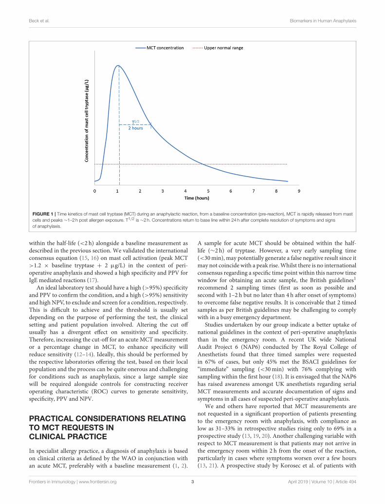
At the onset of anaphylaxis, tryptase is released from mast cellsalongside other mediators including cytokines, prostaglandinD2, and leukotriene C4 (3). Compared to histamine MCT hasa longer plasma half-life of ∼2 h (8) (Figure 1). Therefore,MCT has to date been the main target as a biomarker ofanaphylaxis. Serum MCT concentration rises steeply in the first90min from the onset of reaction, followed by a steady declinein concentration following first order kinetics (8). It can takeup to or beyond 24 h for levels to return to pre-reaction orbaseline concentrations.
Serial samples must therefore be tested to demonstrate thisnatural progression and aid in the confirmation of anaphylaxis.The UK National Institute for Health Care Excellence (NICE),the British Society for Allergy and Clinical Immunology (BSACI)and the Association of Anesthetists of Great Britain and Ireland(AAGBI) guidelines recommend serial timed MCT samples–thefirst to be taken as soon as possible after emergency treatment hascommenced, a second sample ideally within 1–2 h (but no laterthan 4 h) from the onset of symptoms and a baseline sample at≥24 h (9, 10)1.
INTERPRETATION OF RESULTS
The manufacturer of MCT states a reference range of <11.0µg/L (95 upper percentile). As with all laboratory tests, thetest performance characteristics [sensitivity, specificity, positivepredictive value (PPV), and negative predictive value (NPV)]will vary with patient populations due to differences in pre-testprobability and the method of interpretation of data. Previousstudies have reported performance characteristics based on apeak cut-off of the MCTmeasurement, a percentage change frombaseline or a combination of these (11–13). We reported nosignificant difference in these two approaches in the context ofperi-operative anaphylaxis based on area under the curve (14).Regardless, of the method of interpretation, the key determinantis to obtain one or more samples during an acute phase and
1http://guidance.nice.org.uk/CG134
Frontiers in Immunology | www.frontiersin.org 2 April 2019 | Volume 10 | Article 494
Beck et al. Biomarkers in Human Anaphylaxis
FIGURE 1 | Time kinetics of mast cell tryptase (MCT) during an anaphylactic reaction, from a baseline concentration (pre-reaction), MCT is rapidly released from mast
cells and peaks ∼1–2 h post allergen exposure. T1/2 is ∼2 h. Concentrations return to base line within 24 h after complete resolution of symptoms and signs
of anaphylaxis.
within the half-life (<2 h) alongside a baseline measurement asdescribed in the previous section. We validated the internationalconsensus equation (15, 16) on mast cell activation (peak MCT>1.2 × baseline tryptase + 2 µg/L) in the context of peri-operative anaphylaxis and showed a high specificity and PPV forIgE mediated reactions (17).
An ideal laboratory test should have a high (>95%) specificityand PPV to confirm the condition, and a high (>95%) sensitivityand high NPV, to exclude and screen for a condition, respectively.This is difficult to achieve and the threshold is usually setdepending on the purpose of performing the test, the clinicalsetting and patient population involved. Altering the cut offusually has a divergent effect on sensitivity and specificity.Therefore, increasing the cut-off for an acute MCTmeasurementor a percentage change in MCT, to enhance specificity willreduce sensitivity (12–14). Ideally, this should be performed bythe respective laboratories offering the test, based on their localpopulation and the process can be quite onerous and challengingfor conditions such as anaphylaxis, since a large sample sizewill be required alongside controls for constructing receiveroperating characteristic (ROC) curves to generate sensitivity,specificity, PPV and NPV.
PRACTICAL CONSIDERATIONS RELATINGTO MCT REQUESTS INCLINICAL PRACTICE
In specialist allergy practice, a diagnosis of anaphylaxis is basedon clinical criteria as defined by the WAO in conjunction withan acute MCT, preferably with a baseline measurement (1, 2).
A sample for acute MCT should be obtained within the half-life (∼2 h) of tryptase. However, a very early sampling time(<30min), may potentially generate a false negative result since itmay not coincide with a peak rise.Whilst there is no internationalconsensus regarding a specific time point within this narrow timewindow for obtaining an acute sample, the British guidelines1
recommend 2 sampling times (first as soon as possible andsecond with 1–2 h but no later than 4 h after onset of symptoms)to overcome false negative results. It is conceivable that 2 timedsamples as per British guidelines may be challenging to complywith in a busy emergency department.
Studies undertaken by our group indicate a better uptake ofnational guidelines in the context of peri-operative anaphylaxisthan in the emergency room. A recent UK wide NationalAudit Project 6 (NAP6) conducted by The Royal College ofAnesthetists found that three timed samples were requestedin 67% of cases, but only 45% met the BSACI guidelines for“immediate” sampling (<30min) with 76% complying withsampling within the first hour (18). It is envisaged that the NAP6has raised awareness amongst UK anesthetists regarding serialMCT measurements and accurate documentation of signs andsymptoms in all cases of suspected peri-operative anaphylaxis.
We and others have reported that MCT measurements arenot requested in a significant proportion of patients presentingto the emergency room with anaphylaxis, with compliance aslow as 31–33% in retrospective studies rising only to 69% in aprospective study (13, 19, 20). Another challenging variable withrespect to MCT measurement is that patients may not arrive inthe emergency room within 2 h from the onset of the reaction,particularly in cases where symptoms worsen over a few hours(13, 21). A prospective study by Korosec et al. of patients with
Frontiers in Immunology | www.frontiersin.org 3 April 2019 | Volume 10 | Article 494

Beck et al. Biomarkers in Human Anaphylaxis
TABLE 1 | Performance of mast cell tryptase measurement in different clinical settings.
Study Sample
number
Clinical setting Causative agent Number of samples
tested per patient
(time points, hours)
Cut-off Sensitivity Specificity PPV NPV
ACUTE/CONTROLLED SETTING
Brown et al.
(22)
64 Prospective
experimental
Challenge–venom
immunotherapy
Jack jumper ant 3 (Baseline–pre VIT,
0.25, 0.5)
9 µg/L 0.55 0.93 NS NS
12 µg/L 0.36 0.93 NS NS
12 µg/L 0.73 0.98 NS NS
Malinovsky
et al. (12)
31 Prospective Routine
clinical
General anesthesia 3 (0.5, 0.5–1, 24) 12 µg/L 0.64 1.00 1.00 0.53
25 µg/L 0.41 1.00 1.00 0.41
Ratio T0/T24>3
0.63 0.83 0.92 0.42
Krishna et al.
(14)
161 Retrospective Routine
clinical
General anesthesia 3 (0–1, 1–2, 24) 25 µg/L 0.64 0.74 0.82 0.52
33·6 µg/l 0.53 0.84 0.86 0.49
Percentage
change 506%
0.53 0.84 0.86 0.49
Baretto et al.
(17)
82 Retrospective Routine
clinical
General anesthesia 3 (0–1, 1–2, 24) peak MCT
>1.2xbaseline
tryptase +
2µg/l
0.78 0.91 0.98 0.44
Dua et al. (23) 117 Prospective
experimental Challenge
- food
Peanut 3 (pre-challenge, 0,
1–2)
30% rise from
baseline
0.53 0.85 NS NS
COMMUNITY
Buka et al.
(13)
141 retrospective
emergency
Various 12.4 µg/L 0.28 0.88 0.93 0.17
Korosec et al.
(21)
31 Prospective Emergency venoms (various) 2 (0 and 7/30 days) 11.4 µg/L 0.71 NS NS NS
NS, not stated; PPV, positive predictive value; NPV, negative predictive value.
anaphylaxis presenting to the emergency room, found a mediantime to first MCT sample of 1 h 45min, however the range was1–31 h (21).
The poor rate of MCT requests from the emergency roommay at least in part relate to the busy clinical environmentwith priority given to stabilizing the patient and lack ofawareness amongst clinicians regarding MCT measurements.Also, the sampling time depends on the time taken by thepatient to arrive in the emergency department which in turnmay be influenced by the rapidity in the onset of symptomsand their severity.
Blood samples for MCT may be taken in EDTA, heparin orplain tubes without an anticoagulant and preferably analyzedwithin 5–7 days. MCT is stable in vitro. However, if there is goingto be an anticipated delay in analysis, samples must be frozenat−20◦C.
CLINICALPERFORMANCE–CONTROLLED SETTING
Using manufacturer cut-offs, the specificity of an elevated MCTin a controlled clinical setting may show variations (Table 1). In
a study involving insect sting challenges, Brown et al reporteda relatively high specificity of 89% to 93% at a cut-off of 12µg/L but sensitivity was poor (36%). However, ROC analysisfound an optimal cut-off at 9 µg/L improved sensitivity to55% with a marginal decline in specificity (87%) (22). Thisstudy however, involved a very small sample size. At a cut-offof 12 µg/L, Malinovsky et al. reported equal specificity in aprospective study in the setting of peri-operative anaphylaxis,but superior sensitivity (64%) (12). Similarly, Mertes et al. inthe context of peri-operative anaphylaxis reported a sensitivityand specificity of 64 and 89%, respectively for a peak MCT of>25 mcg/l (11). In a similar setting, we reported a sensitivityand specificity of 64 and 74%, respectively at a peak MCTof >25 mcg/l (14).
Alternative methods of interpretations include a changein MCT concentration from baseline or “delta tryptase”(peak minus baseline) concentration may be morediscriminatory than peak MCT alone. ROC analysis maydetermine a cut-off for optimal delta tryptase to improvethe interpretation. For example, an increase in MCT of2 µg/L in the study by Brown et al found a sensitivityof 0.73 (95% CI 0.39–0.94) and specificity 0.91 (95% CI
Frontiers in Immunology | www.frontiersin.org 4 April 2019 | Volume 10 | Article 494

Beck et al. Biomarkers in Human Anaphylaxis
0.79–0.97), a superior performance to peak MCT alone(22). However, we were unable to demonstrate a significantadvantage for a percentage change from baseline vs. absolutepeak MCT measurement in the context of peri-operativeanaphylaxis (14).
The relationship between peak and baseline MCT may also beexpressed as a ratio between peak and baseline or between specifictime point concentrations. For example, Malinovsky determinedT0:T24 concentrations >3 µg/L as the discriminator. However,this was not found to be as good as increasing the cut-off ofabsolute MCT to 25 µg/L (12). As stated in the previous section,an international consensus equation has been recommended forthe interpretation of mast cell activation (15, 16).
CLINICAL PERFORMANCE OF MCT INANAPHYLAXIS PRESENTING TO THEEMERGENCY ROOM
There are limited data available regarding the performance ofMCT in an emergency room setting. Differences in performanceof MCT amongst published studies may be attributable tomultiple factors including definition of anaphylaxis employed(WAO diagnostic criteria were published in 2011), sample size,assays used for MCT measurement, sampling time, and etiology,and severity of anaphylaxis. We reported a sensitivity, specificity,PPV, and NPV of 28%, 88%, 0.93, and 0.17, respectively at apeak MCT of 12.4 µg/L (13). Practical considerations that arerelevant to this setting also include awareness of the emergencyroom physicians regarding the role for MCT in anaphylaxis.Furthermore, a significant proportion of patients are dischargedfrom the emergency room after a few hours following recovery,and may present to an allergy specialist at a different hospital ororganization, and it may be challenging to coordinate the reportsof acute MCT with a baseline measurement.
PERFORMANCE VARIATION WITHCULPRIT ALLERGEN
Regardless of the clinical setting, it is well-recognized that MCTmay not differ from baseline levels in a significant number ofcases of anaphylaxis (13, 14, 20, 24–26). This statement hasto be cautiously interpreted since some of these studies werepublished prior to the publication of theWAO diagnostic criteriafor anaphylaxis. Whilst, it is highly unlikely that this wouldhave affected cases of severe anaphylaxis as graded by Brownet al. (27), some cases of mild-moderate anaphylaxis may nothave made their way into the dataset in studies published before2011, thus potentially affecting the performance characteristics ofMCT. MCT levels have been shown to correlate with histamine.Furthermore, concurrent lack of elevation in MCT in somepatients with a raised histamine highlights basophil involvementin anaphylaxis (26). Both histamine and MCT have been shownto correlate moderately with cutaneous symptoms of erythemaand urticaria in anaphylaxis (26). There is some evidence thatseverity of anaphylaxis correlates with serumMCT and histamine(24, 25), with higher odds (13) for cases with hypotension likely
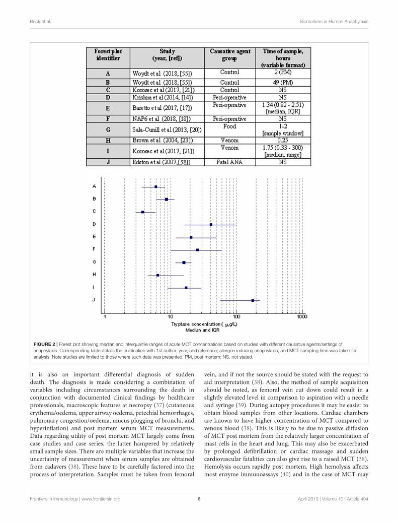
to be associated with a raised MCT. These data have to becarefully interpreted since there was no standardization withrespect to interpretation of MCT in anaphylaxis, i.e., cut offsfor an absolute measurement or a percentage change. Although,it has been suggested that food-induced anaphylaxis is lesslikely to be associated with an increased MCT, we and othersreported to the contrary (13, 19). In a recent study of double-blind-placebo controlled study involving peanut challenges, itwas shown that at least a 30% rise in MCT was seen in 62.5%of cases (n = 150), out of which 9% developed anaphylaxis,giving a sensitivity and specificity of 53 and 85%, respectively(23). It is plausible, however, that the frequency and magnitudeof elevation in MCT may be relatively lower in food-inducedanaphylaxis as opposed to severe cardiovascular anaphylaxisseen in peri-operative setting (20) (Figure 2). This needs furtherinvestigation in prospective studies where the diagnostic criteriafor anaphylaxis and methods of interpreting MCT are well-standardized (i.e., apply international consensus equation formast cell activation (15, 16).
THE CLINICAL RELEVANCE OF A RAISEDBASELINE MCT
There has been a great clinical interest in baseline MCT inthe context of patients with a previous history of anaphylaxis.It has been suggested that a constitutively elevated baselineMCT (≥11.4 µg/L) significantly enhances the risk of severeanaphylaxis, as seen in patients with hymenoptera venom allergy(HVA) (28, 29). A cut off 20 µg/L constitutes one of fourminorWHO criteria (30) for systemic mastocytosis, thus makingit an important consideration for a referral for bone marrowstudies. One large study in the context of HVA showed thatthe odds of severe anaphylaxis increases 5-fold between 5 and15 µg/L of baseline MCT (31). Hence, the BSACI criteria forinvestigation and management of HVA emphasize on a baselineMCT measurement in all cases of systemic reactions to insectstings (32). A raised baseline MCT can also be seen in the contextof chronic renal failure (33, 34) and hematological disorders (35)such as myelodysplastic syndrome, acute and chronic myeloidleukemia and chronic eosinophilic leukemia, and hence has tobe carefully interpreted with relevant investigations for renalfunction, hematinics, and a clinical examination for mastocytosisand hematological malignancy. Furthermore, it has also beenshown that 20% of the population may have an elevatedbaseline MCT due to hereditary alpha-tryptasaemia (autosomaldominant) due to increased germline copies of the alpha-tryptasegene (TPSAB1) (36). These patients have an elevated baselineMCT with or without non-specific multisystem symptoms. Thishas not yet been well-characterized.
USE OF SERUM MCT INFORENSIC MEDICINE
A post mortem differential diagnosis of anaphylaxis can be achallenging task for a histopathologist. Whilst the diagnosis canbe specifically investigated in suspected cases of anaphylaxis,
Frontiers in Immunology | www.frontiersin.org 5 April 2019 | Volume 10 | Article 494
Beck et al. Biomarkers in Human Anaphylaxis
FIGURE 2 | Forest plot showing median and interquartile ranges of acute MCT concentrations based on studies with different causative agents/settings of
anaphylaxis. Corresponding table details the publication with 1st author, year, and reference; allergen inducing anaphylaxis, and MCT sampling time was taken for
analysis. Note studies are limited to those where such data was presented. PM, post mortem; NS, not stated.
it is also an important differential diagnosis of suddendeath. The diagnosis is made considering a combination ofvariables including circumstances surrounding the death inconjunction with documented clinical findings by healthcareprofessionals, macroscopic features at necropsy (37) (cutaneouserythema/oedema, upper airway oedema, petechial hemorrhages,pulmonary congestion/oedema, mucus plugging of bronchi, andhyperinflation) and post mortem serum MCT measurements.Data regarding utility of post mortem MCT largely come fromcase studies and case series, the latter hampered by relativelysmall sample sizes. There are multiple variables that increase theuncertainty of measurement when serum samples are obtainedfrom cadavers (38). These have to be carefully factored into theprocess of interpretation. Samples must be taken from femoral
vein, and if not the source should be stated with the request toaid interpretation (38). Also, the method of sample acquisitionshould be noted, as femoral vein cut down could result in aslightly elevated level in comparison to aspiration with a needleand syringe (39). During autopsy procedures it may be easier toobtain blood samples from other locations. Cardiac chambersare known to have higher concentration of MCT compared tovenous blood (38). This is likely to be due to passive diffusionof MCT post mortem from the relatively larger concentration ofmast cells in the heart and lung. This may also be exacerbatedby prolonged defibrillation or cardiac massage and suddencardiovascular fatalities can also give rise to a raised MCT (38).Hemolysis occurs rapidly post mortem. High hemolysis affectsmost enzyme immunoassays (40) and in the case of MCT may
Frontiers in Immunology | www.frontiersin.org 6 April 2019 | Volume 10 | Article 494
Beck et al. Biomarkers in Human Anaphylaxis
cause falsely elevated results. Hemolysis may also indicate cellautolysis or tissue or blood vessel liquefaction. Cell lysis maycause increased MCT release. Hemodilution (caused from highfluid volumes ante-mortem) could also affect results causingfalse negative results, particularly if samples are taken from acentral line. Therefore, post mortem samples must be taken fromfemoral veins.
A high post mortem MCT has also been reported in theabsence of anaphylaxis. Asphyxia can contribute to a high MCTfrom femoral blood due to release from lung derived mast cells.Sudden infant death syndrome (SIDS) has also been associatedwith high MCT concentrations, although evidence regardingthis is conflicting and the biological plausibility is unclear andthere are no data on a direct cause-effect relationship (41–44).Deaths occurring from multiple trauma may have high MCTlevels post mortem as opposed to deaths from single trauma or“non-trauma” deaths (45). Acute death after heroin injectionshas also been implicated in causing increased post mortemMCT. The time delay between death and sample acquisitioncan also affect post mortem MCT concentration. In a recentpost mortem interval study by Woydt et al. (46), statisticallysignificant differences were seen between paired samples at deathand 3 h post mortem in a cohort where death was not due toanaphylaxis. One of the 20 cases breached the 44.3 µg/L cutoff at 3 h. However, variation in MCT concentrations with postmortem intervals has inconsistent evidence.
Given the confounding factors discussed above andlimitations of data, there is no standardized internationalreference range for post mortem MCT. Cut-off values proposedfor forensic identification of fatal anaphylaxis vary between10 and 54 µg/L (39, 47–49). It is worth noting that olderpublications (37) (prior to late 1990s) used β MCT as opposedto total (α and β) MCT in the last decade. Sun et al. recentlypublished a systematic review and meta-analysis to address thisquestion and concluded that at concentrations >30.4 µg/L,sensitivity and specificity for anaphylaxis were 68.5 and 83.9%,respectively (50).
At the present time, it is not a standard practice to obtain bonemarrow samples during an autopsy. Given that fatal anaphylaxisis fortunately rare, this may be considered at least in cases wheresevere cardiovascular and/or refractory anaphylaxis has occurredsince clonal mast cell disorders have been reported in suchpatients (51).
NOVEL BIOMARKERS IN ANAPHYLAXIS
The recent identification of novel biomarkers that are eitherreleased from, or expressed on the surface of activated mast cells,eosinophils, and basophils have been aided by advances in cellpurification, flow cytometry, and other laboratory techniques.There is growing evidence to support the investigationof other biomarkers, such as chymase, carboxypeptidaseA3, dipeptidyl peptidase I (DPPI), basogranulin, CCL-2,and platelet activating factor (PAF) in the diagnosis ofanaphylaxis. These are described below and summarisedin Table 2.
CHYMASE
In a similar fashion to tryptase, this serine protease is alsolargely found in the secretory granules of mast cells. One ofthe earliest reports of chymase as a potential biomarker foranaphylaxis examined 8 autopsy cases with anaphylaxis and104 control cases without (52). Chymase was detected in all 8autopsy cases with anaphylaxis, whilst it was only detected in2 of the 104 control cases. This study also found a significantpositive correlation between chymase and MCT levels in all 8cases of fatal anaphylaxis. The authors also determined chymaseto be relatively stable in serum, confirming its potential as atool in the diagnosis of anaphylaxis. The same group morerecently discovered positive chymase staining in lung mast cellsfrom 3 autopsy cases of fatal anaphylaxis, in comparison tothe control group of tissues associated with acute traumaticdeaths (53).
Another study (54) established an enzyme immunoassay(EIA) to examine serum from patients with anaphylaxisprovoked by food, drug and insect stings (n= 181), compared toa control group of healthy blood donors (n = 123) and patientswith known allergies to food (n = 76) or drugs (n = 26). Theauthors found chymase levels to be greater in serum collectedfrom patients within 8 h of anaphylaxis, compared to the controlgroup (p = 0.0069). Furthermore, the concentration of chymaseremained high at least 24 h after the onset of the reaction.
CARBOXYPEPTIDASE A3
Alongside MCT and chymase, carboxypeptidase A3 is anotherpre-formed chemical mediator of anaphylaxis released byactivated mast cells.
Brown et al (55) measured carboxypeptidase A3 levels by EIAfrom blood and saliva, in patients undergoing allergy tests forsuspected drug allergies (n = 33). The found carboxypeptidaseA3 levels to be increased in saliva, but not serum, followinga positive reaction to a drug challenge. Furthermore, baselinelevels of carboxypeptidase A3 were higher in serum and salivain patients who experienced symptoms on a drug challenge,compared to those who were asymptomatic. Baseline levelswere also higher in patients who had historically suffereda severe reaction including cardiovascular and/or respiratorycompromise, compared to those with only mild reactions.
Another study (56) also used a sandwich-based EIA tomeasure levels of carboxypeptidase A3 in cases of suspectedanaphylaxis (n = 181), systemic mastocytosis (30), and controlgroups of healthy blood donors (n = 209), or individualswith bronchial asthma (n = 15). The authors found thatcarboxypeptidase A3 levels were significantly greater in theserum or plasma collected within 8 h of an onset of allergicreaction, compared to the control cohort. They also foundcarboxypeptidase A3 levels to be elevated (although range notstated) in 83% of anaphylaxis cases with an elevated MCT.Furthermore, carboxypeptidase A3 was also elevated in 70%of the 110 cases of suspected anaphylaxis cases that wereMCT negative.
Frontiers in Immunology | www.frontiersin.org 7 April 2019 | Volume 10 | Article 494
Beck et al. Biomarkers in Human Anaphylaxis
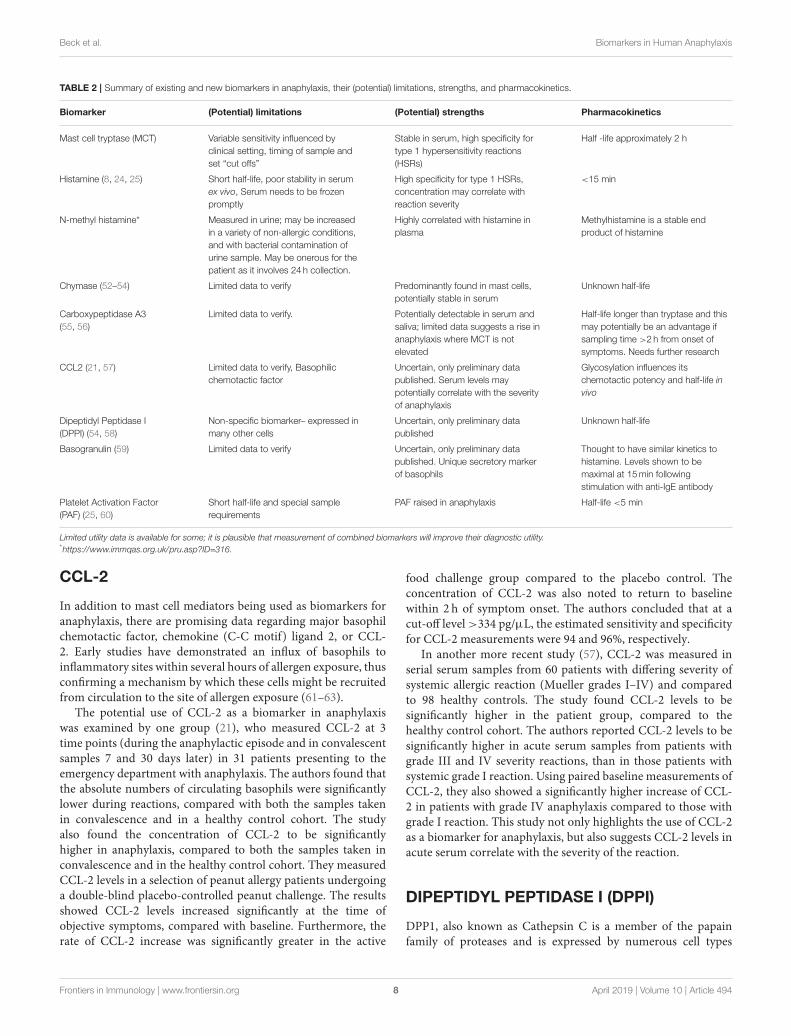
TABLE 2 | Summary of existing and new biomarkers in anaphylaxis, their (potential) limitations, strengths, and pharmacokinetics.
Biomarker (Potential) limitations (Potential) strengths Pharmacokinetics
Mast cell tryptase (MCT) Variable sensitivity influenced by
clinical setting, timing of sample and
set “cut offs”
Stable in serum, high specificity for
type 1 hypersensitivity reactions
(HSRs)
Half -life approximately 2 h
Histamine (8, 24, 25) Short half-life, poor stability in serum
ex vivo, Serum needs to be frozen
promptly
High specificity for type 1 HSRs,
concentration may correlate with
reaction severity
<15 min
N-methyl histamine* Measured in urine; may be increased
in a variety of non-allergic conditions,
and with bacterial contamination of
urine sample. May be onerous for the
patient as it involves 24 h collection.
Highly correlated with histamine in
plasma
Methylhistamine is a stable end
product of histamine
Chymase (52–54) Limited data to verify Predominantly found in mast cells,
potentially stable in serum
Unknown half-life
Carboxypeptidase A3
(55, 56)
Limited data to verify. Potentially detectable in serum and
saliva; limited data suggests a rise in
anaphylaxis where MCT is not
elevated
Half-life longer than tryptase and this
may potentially be an advantage if
sampling time >2 h from onset of
symptoms. Needs further research
CCL2 (21, 57) Limited data to verify, Basophilic
chemotactic factor
Uncertain, only preliminary data
published. Serum levels may
potentially correlate with the severity
of anaphylaxis
Glycosylation influences its
chemotactic potency and half-life in
vivo
Dipeptidyl Peptidase I
(DPPI) (54, 58)
Non-specific biomarker– expressed in
many other cells
Uncertain, only preliminary data
published
Unknown half-life
Basogranulin (59) Limited data to verify Uncertain, only preliminary data
published. Unique secretory marker
of basophils
Thought to have similar kinetics to
histamine. Levels shown to be
maximal at 15min following
stimulation with anti-IgE antibody
Platelet Activation Factor
(PAF) (25, 60)
Short half-life and special sample
requirements
PAF raised in anaphylaxis Half-life <5 min
Limited utility data is available for some; it is plausible that measurement of combined biomarkers will improve their diagnostic utility.*https://www.immqas.org.uk/pru.asp?ID=316.
CCL-2
In addition to mast cell mediators being used as biomarkers foranaphylaxis, there are promising data regarding major basophilchemotactic factor, chemokine (C-C motif) ligand 2, or CCL-2. Early studies have demonstrated an influx of basophils toinflammatory sites within several hours of allergen exposure, thusconfirming a mechanism by which these cells might be recruitedfrom circulation to the site of allergen exposure (61–63).
The potential use of CCL-2 as a biomarker in anaphylaxiswas examined by one group (21), who measured CCL-2 at 3time points (during the anaphylactic episode and in convalescentsamples 7 and 30 days later) in 31 patients presenting to theemergency department with anaphylaxis. The authors found thatthe absolute numbers of circulating basophils were significantlylower during reactions, compared with both the samples takenin convalescence and in a healthy control cohort. The studyalso found the concentration of CCL-2 to be significantlyhigher in anaphylaxis, compared to both the samples taken inconvalescence and in the healthy control cohort. They measuredCCL-2 levels in a selection of peanut allergy patients undergoinga double-blind placebo-controlled peanut challenge. The resultsshowed CCL-2 levels increased significantly at the time ofobjective symptoms, compared with baseline. Furthermore, therate of CCL-2 increase was significantly greater in the active
food challenge group compared to the placebo control. Theconcentration of CCL-2 was also noted to return to baselinewithin 2 h of symptom onset. The authors concluded that at acut-off level >334 pg/µL, the estimated sensitivity and specificityfor CCL-2 measurements were 94 and 96%, respectively.
In another more recent study (57), CCL-2 was measured inserial serum samples from 60 patients with differing severity ofsystemic allergic reaction (Mueller grades I–IV) and comparedto 98 healthy controls. The study found CCL-2 levels to besignificantly higher in the patient group, compared to thehealthy control cohort. The authors reported CCL-2 levels to besignificantly higher in acute serum samples from patients withgrade III and IV severity reactions, than in those patients withsystemic grade I reaction. Using paired baseline measurements ofCCL-2, they also showed a significantly higher increase of CCL-2 in patients with grade IV anaphylaxis compared to those withgrade I reaction. This study not only highlights the use of CCL-2as a biomarker for anaphylaxis, but also suggests CCL-2 levels inacute serum correlate with the severity of the reaction.
DIPEPTIDYL PEPTIDASE I (DPPI)
DPP1, also known as Cathepsin C is a member of the papainfamily of proteases and is expressed by numerous cell types
Frontiers in Immunology | www.frontiersin.org 8 April 2019 | Volume 10 | Article 494

Beck et al. Biomarkers in Human Anaphylaxis
including both mast cells and basophils. Studies using DPP1knockout mice have proposed a role in activation of chymase, butnot tryptase (58). This is supported by investigation of DPP1 as auseful serummarker of anaphylaxis (54), where elevated chymaseconcentrations in patients with anaphylaxis also correlated withlevels of DPP1, but not tryptase.
BASOGRANULIN
The novel basophil granule protein, termed “basogranulin” hasbeen shown to be released by basophils in parallel with histamineand is thought to be dependent on the same receptors andsignaling pathways as those for histamine release (59). Furtherwork is needed to assess the extent and role of basophil activationin anaphylaxis and whether basogranulin could be used as asuitable serum marker.
PLATELET ACTIVATION FACTOR (PAF)
There has been an interest in measuring PAF and PAFacetylhydrolase (PAF AH), an enzyme inactivating PAF. Plateletactivation factor is a proinflammatory phospholipid secretedby mast cells, monocytes, and tissue macrophages (25). PAFbinds to its receptors on platelets, monocytes, macrophages,and neutrophils. Vadas et al. (60) reported an elevated PAFconcentration in anaphylaxis which correlated with severity.There was an inverse correlation between PAF and PAF AHactivity. They also measured PAF AH activity in patientswith fatal anaphylaxis in peanut allergy and compared it withcontrols–PAF AH was significantly reduced in fatal anaphylaxissamples. In another study, Vadas et al. showed that PAFcorrelated better than MCT and histamine with severity ofanaphylaxis (25). One of the challenges withmeasurement of PAFand PAF AH in a routine clinical setting is its very short half-lifeand special sampling and transport precautions that are requiredthus making it an unattractive candidate for routine use.
FUTURE PERSPECTIVESAND CONCLUSIONS
Anaphylaxis remains a clinical diagnosis and biomarkershave no role in acute management. However, they have animportant place during specialist allergy evaluation to confirmthe diagnosis and distinguish anaphylaxis from its mimics suchas severe asthma, vocal cord dysfunction, factitious disorder orsomatoform disorders, hypotensive crisis due to non-allergiccauses and in certain circumstances where there might bea paucity of information relating to the index episode. At
the present time, the gold standard laboratory test is MCT,although other biomarkers such as chymase, carboxypeptidaseA3, and CCL-2 hold promise for the future, providing samplerequirements, assay platform, process, and costs are compatiblefor routine use in clinical diagnostic laboratories. Prospectivewell-designed multicenter studies applying the WAO diagnosticcriteria for anaphylaxis on well-characterized patients with serialtimed blood samples and standardized methods of interpretationof serumMCT are needed to firmup on the sensitivity, specificity,PPV and NPV. There may be a role for a combination ofMCT and a newer biomarker/s to enhance sensitivity andspecificity. Multiplex approach may improve efficiency in a highthroughput diagnostic laboratory using a relatively small amountof serum. Validation of biomarkers in saliva might be usefulin the interpretation of food challenges, particularly those withsubjective symptoms and minimal or no objective signs.
One of the most intriguing aspects of human anaphylaxisis that some patients with severe symptoms do not show anelevated MCT. This raises the possible involvement of otherhitherto unidentified biomarkers and possibly unidentifiedpathways. A new classification system based on phenotypes,endotypes, and biomarkers has been proposed that mightenable a more robust stratification of patients for appropriateinvestigations and selection for desensitization, in particularthose developing adverse reactions to biologics, anti-cancerdrugs and radiocontrast media (64). The systems biologyand proteomics approaches applied on serum obtainedduring an acute phase and convalescence may be worthexploring. Greater understanding of the pathogenesisunderpinning anaphylaxis might pave the way for betterrisk stratification of patients, improve diagnostics and createopportunities to develop novel immune-based treatments such asvenom immunotherapy.
AUTHOR CONTRIBUTIONS
SB, TW, and MK provided substantial contributions to theconception, design, content, and drafting of the manuscript. AH,RJB, and RLB provided critical appraisal of themanuscript as wellas substantial editorial contributions.
FUNDING
The authors’ department (SB, TW, RLB, AH, and MK) receivedfunding for PracticAllergy course in 2018 and previous yearsfrom Thermo Fisher. The authors would like to declare that saidfunding was unrelated to this study and that Thermo Fisher hadno role in the study design or manuscript preparation.
REFERENCES
1. Simons FE, Ardusso LR, Bilo MB, El-Gamal YM, Ledford DK, Ring
J, et al. World allergy organization anaphylaxis guidelines: summary. J
Allergy Clin Immunol. (2011) 127:587–93.e1-22. doi: 10.1016/j.jaci.2011.
01.038
2. Simons FE, Ardusso LR, Bilo MB, El-Gamal YM, Ledford DK, Ring
J, et al. World allergy organization guidelines for the assessment and
management of anaphylaxis. World Allergy Organ J. (2011) 4:13–37.
doi: 10.1097/WOX.0b013e318211496c
3. Schwartz LB. Tryptase, a mediator of human mast cells. J Allergy Clin
Immunol. (1990) 86:594–8. doi: 10.1016/S0091-6749(05)80222-2
Frontiers in Immunology | www.frontiersin.org 9 April 2019 | Volume 10 | Article 494
Beck et al. Biomarkers in Human Anaphylaxis
4. Schwartz LB, Atkins PC, Bradford TR, Fleekop P, Shalit M, Zweiman
B. Release of tryptase together with histamine during the immediate
cutaneous response to allergen. J Allergy Clin Immunol. (1987) 80:850–5.
doi: 10.1016/S0091-6749(87)80276-2
5. Schwartz LB, Irani AM, Roller K, Castells MC, Schechter NM. Quantitation of
histamine, tryptase, and chymase in dispersed human T and TC mast cells. J
Immunol. (1987) 138:2611–5.
6. Schwartz LB, Metcalfe DD, Miller JS, Earl H, Sullivan T. Tryptase levels as an
indicator of mast-cell activation in systemic anaphylaxis and mastocytosis. N
Engl J Med. (1987) 316:1622–6. doi: 10.1056/NEJM198706253162603
7. Lock RJ. My approach to internal quality control in a clinical immunology
laboratory. J Clin Pathol. (2006) 59:681–4. doi: 10.1136/jcp.2005.032292
8. Schwartz LB, Yunginger JW, Miller J, Bokhari R, Dull D. Time course of
appearance and disappearance of human mast cell tryptase in the circulation
after anaphylaxis. J Clin Invest. (1989) 83:1551–5. doi: 10.1172/JCI114051
9. Ewan PW, Dugue P, Mirakian R, Dixon TA, Harper JN, Nasser
SM. BSACI guidelines for the investigation of suspected anaphylaxis
during general anaesthesia. Clin Exp Allergy. (2010) 40:15–31.
doi: 10.1111/j.1365-2222.2009.03404.x
10. W.P.o.t.a.o.A.o. Great, B.a.I.a.t.B.S.f.A., Immunology C. Suspected
Anaphylactic Reactions Associatedwith Anaesthesia. London: The Association
of Anaesthetists of Great Britain and Northern Ireland and the British
Society for Allergy and Clinical Immunology, Vol. 3:1–20 (2003).
11. Mertes PM, Laxenaire MC, Alla F. Anaphylactic and anaphylactoid reactions
occurring during anesthesia in France in 1999-2000. Anesthesiology. (2003)
99:536–45. doi: 10.1097/00000542-200309000-00007
12. Malinovsky JM, Decagny S, Wessel F, Guilloux L, Mertes PM. Systematic
follow-up increases incidence of anaphylaxis during adverse reactions
in anesthetized patients. Acta Anaesthesiol Scand. (2008) 52:175–81.
doi: 10.1111/j.1399-6576.2007.01489.x
13. Buka RJ, Knibb RC, Crossman RJ, Melchior CL, Huissoon AP, Hackett S, et al.
Anaphylaxis and clinical utility of real-world measurement of acute serum
tryptase in UK emergency departments. J Allergy Clin Immunol Pract. (2017)
5:1280–7.e2. doi: 10.1016/j.jaip.2017.06.021
14. Krishna MT, York M, Chin T, Gnanakumaran G, Heslegrave J, Derbridge
C, et al. Multi-centre retrospective analysis of anaphylaxis during general
anaesthesia in the United Kingdom: aetiology diagnostic performance
of acute serum tryptase. Clin Exp Immunol. (2014) 178:399–404.
doi: 10.1111/cei.12424
15. Valent P, Akin C, Arock M, Brockow K, Butterfield JH, Carter MC,
et al. Definitions, criteria and global classification of mast cell disorders
with special reference to mast cell activation syndromes: a consensus
proposal. Int Arch Allergy Immunol. (2012) 157:215–25. doi: 10.1159/000
328760
16. Valent P, Akin C, Bonadonna P, Hartmann K, Broesby-Olsen S, Brockow
K, et al. Mast cell activation syndrome: importance of consensus criteria
and call for research. J Allergy Clin Immunol. (2018) 142:1008–10.
doi: 10.1016/j.jaci.2018.06.004
17. Baretto RL, Beck S, Heslegrave J, Melchior C, Mohamed O, Ekbote A, et al.
Validation of international consensus equation for acute serum total tryptase
in mast cell activation: a perioperative perspective. Allergy. (2017) 72:2031–4.
doi: 10.1111/all.13226
18. T.R.C.o. Anaesthetists. NAP6 Report. https://www.nationalauditprojects.org.
uk/NAP6Report?newsid=1914 (2018).
19. Srivastava S, Huissoon AP, Barrett V, Hackett S, Dorrian S, Cooke MW, et al.
Systemic reactions and anaphylaxis with an acute serum tryptase >/=14
mug/L: retrospective characterisation of aetiology, severity and adherence to
National Institute of Health and Care Excellence (NICE) guidelines for serial
tryptase measurements and specialist referral. J Clin Pathol. (2014) 67:614–9.
doi: 10.1136/jclinpath-2013-202005
20. Sala-Cunill A, Cardona V, Labrador-Horrillo M, Luengo O, Esteso O, Garriga
T, et al. Usefulness and limitations of sequential serum tryptase for the
diagnosis of anaphylaxis in 102 patients. Int Arch Allergy Immunol. (2013)
160:192–9. doi: 10.1159/000339749
21. Korosec P, Turner PJ, Silar M, Kopac P, Kosnik M, Gibbs BF, et al.
Basophils, high-affinity IgE receptors, and CCL2 in human anaphylaxis.
J Allergy Clin Immunol. (2017) 140:750–8.e15. doi: 10.1016/j.jaci.2016.
12.989
22. Brown SG, Blackman KE, Heddle RJ, Can serum mast cell tryptase
help diagnose anaphylaxis? Emerg Med Austral. (2004) 16:120–4.
doi: 10.1111/j.1742-6723.2004.00562.x
23. Dua S, Dowey J, Foley L, Islam S, King Y, Ewan P, Clark AT. Diagnostic
value of tryptase in food allergic reactions: a prospective study of 160
adult peanut challenges. J Allergy Clin Immunol Pract. (2018) 6:1692–8 e1.
doi: 10.1016/j.jaip.2018.01.006
24. Stone SF, Cotterell C, Isbister GK, Holdgate A, Brown SG, Elevated serum
cytokines during human anaphylaxis: identification of potential mediators
of acute allergic reactions. J Allergy Clin Immunol. (2009) 124:786–92 e4.
doi: 10.1016/j.jaci.2009.07.055
25. Vadas P, Perelman B, Liss G. Platelet-activating factor, histamine, and tryptase
levels in human anaphylaxis. J Allergy Clin Immunol. (2013) 131:144–9.
doi: 10.1016/j.jaci.2012.08.016
26. Lin RY, Schwartz LB, Curry A, Pesola GR, Knight RJ, Lee HS, et al.
Histamine and tryptase levels in patients with acute allergic reactions: An
emergency department-based study. J Allergy Clin Immunol. (2000) 106:65–
71. doi: 10.1067/mai.2000.107600
27. Brown SG. Clinical features and severity grading of anaphylaxis. J Allergy Clin
Immunol. (2004) 114:371–6. doi: 10.1016/j.jaci.2004.04.029
28. Haeberli G, Bronnimann M, Hunziker T, Muller U. Elevated basal serum
tryptase and hymenoptera venom allergy: relation to severity of sting reactions
and to safety and efficacy of venom immunotherapy. Clin Exp Allergy. (2003)
33:1216–20. doi: 10.1046/j.1365-2222.2003.01755.x
29. Ludolph-Hauser D, Rueff F, Fries C, Schopf P, Przybilla B. Constitutively
raised serum concentrations of mast-cell tryptase and severe
anaphylactic reactions to Hymenoptera stings. Lancet. (2001) 357:361–2.
doi: 10.1016/S0140-6736(00)03647-3
30. Valent P. Diagnostic evaluation and classification of mastocytosis. Immunol
Allergy Clin North Am. (2006) 26:515–34. doi: 10.1016/j.iac.2006.05.002
31. Rueff F, Przybilla B, Bilo MB, Muller U, Scheipl F, Aberer W, et al. Predictors
of severe systemic anaphylactic reactions in patients with Hymenoptera
venom allergy: importance of baseline serum tryptase-a study of the European
Academy of Allergology and Clinical Immunology Interest Group on
Insect Venom Hypersensitivity. J Allergy Clin Immunol. (2009) 124:1047–54.
doi: 10.1016/j.jaci.2009.08.027
32. Krishna MT, Ewan PW, Diwakar L, Durham SR, Frew AJ, Leech SC, et al.
Diagnosis and management of hymenoptera venom allergy: British Society
for Allergy and Clinical Immunology (BSACI) guidelines. Clin Exp Allergy.
(2011) 41:1201–20. doi: 10.1111/j.1365-2222.2011.03788.x
33. Dugas-Breit S, Schopf P, Dugas M, Schiffl H, Rueff F, Przybilla B. Baseline
serum levels of mast cell tryptase are raised in hemodialysis patients and
associated with severity of pruritus. J Dtsch Dermatol Ges. (2005) 3:343–7.
doi: 10.1111/j.1610-0387.2005.05706.x
34. Jesky MD, Stringer SJ, Fenton A, Ng KP, Yadav P, Ndumbo M, et al. Serum
tryptase concentration and progression to end-stage renal disease. Eur J Clin
Invest. (2016) 46:460–74. doi: 10.1111/eci.12622
35. Valent P, Sperr WR, Sotlar K, Reiter A, Akin C, Gotlib J, et al. The serum
tryptase test: an emerging robust biomarker in clinical hematology. Expert Rev
Hematol. (2014) 7:683–90. doi: 10.1586/17474086.2014.955008
36. Lyons JJ. Hereditary alpha tryptasemia: genotyping and associated
clinical features. Immunol Allergy Clin North Am. (2018) 38:483–95.
doi: 10.1016/j.iac.2018.04.003
37. Pumphrey RS, Roberts IS. Postmortem findings after fatal anaphylactic
reactions. J Clin Pathol. (2000) 53:273–6. doi: 10.1136/jcp.53.4.273
38. Sheldon J, Philips B. Laboratory investigation of anaphylaxis: not as easy as it
seems. Anaesthesia. (2015) 70:1–5. doi: 10.1111/anae.12926
39. Tse R, Garland J, Kesha K, Elstub H, Cala AD, Ahn Y, et al. Differences in
sampling techniques on total post-mortem tryptase. Int J Legal Med. (2018)
132:741–5. doi: 10.1007/s00414-017-1738-8
40. Dimeski G. Interference testing. Clin Biochem Rev. (2008) 29 (Suppl. 1):S43–8.
41. Edston E, Gidlund E, Wickman M, Ribbing H. Van Hage-
Hamsten M. Increased mast cell tryptase in sudden infant death -
anaphylaxis, hypoxia or artefact? Clin Exp Allergy. (1999) 29:1648–54.
doi: 10.1046/j.1365-2222.1999.00679.x
42. Nishio H, Suzuki K. Serum tryptase levels in sudden infant death
syndrome in forensic autopsy cases. Forensic Sci Int. (2004) 139:57–60.
doi: 10.1016/j.forsciint.2003.09.011
Frontiers in Immunology | www.frontiersin.org 10 April 2019 | Volume 10 | Article 494
Beck et al. Biomarkers in Human Anaphylaxis
43. Hagan LL, Goetz DW, Revercomb CH, Garriott J. Sudden infant death
syndrome: a search for allergen hypersensitivity. Ann Allergy Asthma
Immunol. (1998) 80:227–31. doi: 10.1016/S1081-1206(10)62962-6
44. Buckley MG, Variend S, Walls AF. Elevated serum concentrations of beta-
tryptase, but not alpha-tryptase, in Sudden Infant Death Syndrome (SIDS). An
investigation of anaphylactic mechanisms. Clin Exp Allergy. (2001) 31:1696–
704. doi: 10.1046/j.1365-2222.2001.01213.x
45. Edston E. van Hage-Hamsten M. Mast cell tryptase and
hemolysis after trauma. Forensic Sci Int. (2003) 131:8–13.
doi: 10.1016/S0379-0738(02)00383-3
46. Woydt L, Bernhard M, Kirsten H, Burkhardt R, Hammer N, Gries A, et al.
Intra-individual alterations of serum markers routinely used in forensic
pathology depending in increasing post-mortem interval. Sci Rep. (2018)
8:12811. doi: 10.1038/s41598-018-31252-5
47. Tse R, Wong CX, Kesha K, Garland J, Tran Y, Anne S, et al. Post mortem
tryptase cut-off level for anaphylactic death. Forensic Sci Int. (2018) 284:5–8.
doi: 10.1016/j.forsciint.2017.12.035
48. Edston E, van Hage-Hamsten M. beta-Tryptase measurements post-mortem
in anaphylactic deaths and in controls. Forensic Sci Int. (1998) 93:135–42.
doi: 10.1016/S0379-0738(98)00040-1
49. Edston E, Eriksson O, van Hage M. Mast cell tryptase in postmortem
serum-reference values and confounders. Int J Legal Med. (2007) 121:275–80.
doi: 10.1007/s00414-006-0101-2
50. Sun KJ, He JT, Huang HY, Xue Y, Xie XL, Wang Q. Diagnostic role of
serum tryptase in anaphylactic deaths in forensic medicine: a systematic
review and meta-analysis. Forensic Sci Med Pathol. (2018) 14:209–15.
doi: 10.1007/s12024-018-9980-z
51. Zanotti R, Lombardo C, Passalacqua G, Caimmi C, Bonifacio M, De Matteis
G, et al. Clonalmast cell disorders in patients with severeHymenoptera venom
allergy and normal serum tryptase levels. J Allergy Clin Immunol. (2015)
136:135–9. doi: 10.1016/j.jaci.2014.11.035
52. Nishio H, Takai S, Miyazaki M, Horiuchi H, Osawa M, Uemura K,
Yoshida K, et al. Usefulness of serum mast cell-specific chymase levels for
postmortem diagnosis of anaphylaxis. Int J Legal Med. (2005) 119:331–4.
doi: 10.1007/s00414-005-0524-1
53. Osawa M, Satoh F, Horiuchi H, Tian W, Kugota N, Hasegawa I.
Postmortem diagnosis of fatal anaphylaxis during intravenous administration
of therapeutic and diagnostic agents: evaluation of clinical laboratory
parameters and immunohistochemistry in three cases. Leg Med (Tokyo).
(2008) 10:143–7. doi: 10.1016/j.legalmed.2007.10.002
54. Zhou X,Whitworth HS, Khedr EM, Brown TA, Goswami R, Eren E, et al. Mast
Cell Chymase: a useful serum marker in anaphylaxis. J Allergy Clin Immunol.
(2011) 127:AB143. doi: 10.1016/j.jaci.2010.12.566
55. Brown TA, Whitworth HS, Zhou XY, Lau L, Eren E, Walls AF.
Carboxypeptidase as a Confirmatory and Predictive Marker in Allergic
Reactions to Drugs J Allergy Clin Immunol. (2011) 127(Suppl.):AB143.
doi: 10.1016/j.jaci.2010.12.567
56. Zhou X, Buckley MG, Lau LC, Sunners C, Pumphrey RSH, Walls AF, et al.
Carboxypeptidase as a new clinical marker for anaphylaxis. J Allergy Clin
Immunol. (2006) 2006:pS85. doi: 10.1016/j.jaci.2005.12.342
57. Vantur R, Koren A, Erzen R, Kosnik M, Korosec P. CCL2 and severe
anaphylaxis. Allergy. (2018) 73:315.
58. Wolters PJ, Pham CT, Muilenburg DJ, Ley TJ, Caughey GH. Dipeptidyl
peptidase I is essential for activation of mast cell chymases, but not tryptases,
in mice. J Biol Chem. (2001) 276:18551–6. doi: 10.1074/jbc.M100223200
59. Mochizuki A, McEuen AR, Buckley MG, Walls AF. The release of
basogranulin in response to IgE-dependent and IgE-independent stimuli:
validity of basogranulin measurement as an indicator of basophil activation. J
Allergy Clin Immunol. (2003) 112:102–8. doi: 10.1067/mai.2003.1511
60. Vadas P, Gold M, Perelman B, Liss GM, Lack G, Blyth T, et al. Platelet-
activating factor, PAF acetylhydrolase, and severe anaphylaxis. N Engl J Med.
(2008) 358:28–35. doi: 10.1056/NEJMoa070030
61. Nouri-Aria KT, Irani AM, Jacobson MR, O’Brien F, Varga EM, Till
SJ, et al. Basophil recruitment and IL-4 production during human
allergen-induced late asthma. J Allergy Clin Immunol. (2001) 108:205–11.
doi: 10.1067/mai.2001.117175
62. Iliopoulos O, Baroody FM, Naclerio RM, Bochner BS, Kagey-Sobotka A,
Lichtenstein LM. Histamine-containing cells obtained from the nose hours
after antigen challenge have functional and phenotypic characteristics of
basophils. J Immunol. (1992) 148:2223–8.
63. Irani AM, Huang C, Xia HZ, Kepley C, Nafie A, Fouda ED,
et al. Immunohistochemical detection of human basophils in late-
phase skin reactions. J Allergy Clin Immunol. (1998) 101:354–62.
doi: 10.1016/S0091-6749(98)70248-9
64. Jimenez-Rodriguez TW, Garcia-Neuer M, Alenazy LA, Castells
M. Anaphylaxis in the 21st century: phenotypes, endotypes, and
biomarkers. J Asthma Allergy. (2018) 11:121–42. doi: 10.2147/JAA.
S159411
Conflict of Interest Statement: The authors declare that the research was
conducted in the absence of any commercial or financial relationships that could
be construed as a potential conflict of interest.
Copyright © 2019 Beck, Wilding, Buka, Baretto, Huissoon and Krishna. This is an
open-access article distributed under the terms of the Creative Commons Attribution
License (CC BY). The use, distribution or reproduction in other forums is permitted,
provided the original author(s) and the copyright owner(s) are credited and that the
original publication in this journal is cited, in accordance with accepted academic
practice. No use, distribution or reproduction is permitted which does not comply
with these terms.
Frontiers in Immunology | www.frontiersin.org 11 April 2019 | Volume 10 | Article 494
Related Documents