Southern Illinois University Carbondale OpenSIUC Publications Department of Anatomy 8-24-2015 Biology of Muscle Atrophy and of its Recovery by FES in Aging and Mobility Impairments: Roots and By-Products. Ugo Carraro Helmut Kern Paolo Gava Christian Hofer Stefan Loefler See next page for additional authors Follow this and additional works at: hp://opensiuc.lib.siu.edu/anat_pubs Creative Commons Aribution Non-Commercial License 3.0 is Article is brought to you for free and open access by the Department of Anatomy at OpenSIUC. It has been accepted for inclusion in Publications by an authorized administrator of OpenSIUC. For more information, please contact [email protected]. Recommended Citation Carraro, Ugo, Kern, Helmut, Gava, Paolo, Hofer, Christian, Loefler, Stefan, Gargiulo, Paolo, Mosole, Simone, Zampieri, Sandra, Gobbo, Valerio, Ravara, Barbara, Piccione, Francesco, Marcante, Andrea, Baba, Alfonc, Schils, Sheila, Pond, Amber and Gava, Francesco. "Biology of Muscle Atrophy and of its Recovery by FES in Aging and Mobility Impairments: Roots and By-Products.." Eur J Transl Myol 25, No. 4 (Aug 2015): 221-230. doi:10.4081/ejtm.2015.5272.

Welcome message from author
This document is posted to help you gain knowledge. Please leave a comment to let me know what you think about it! Share it to your friends and learn new things together.
Transcript
Southern Illinois University CarbondaleOpenSIUC
Publications Department of Anatomy
8-24-2015
Biology of Muscle Atrophy and of its Recovery byFES in Aging and Mobility Impairments: Rootsand By-Products.Ugo Carraro
Helmut Kern
Paolo Gava
Christian Hofer
Stefan Loefler
See next page for additional authors
Follow this and additional works at: http://opensiuc.lib.siu.edu/anat_pubsCreative Commons Attribution Non-Commercial License 3.0
This Article is brought to you for free and open access by the Department of Anatomy at OpenSIUC. It has been accepted for inclusion in Publicationsby an authorized administrator of OpenSIUC. For more information, please contact [email protected].
Recommended CitationCarraro, Ugo, Kern, Helmut, Gava, Paolo, Hofer, Christian, Loefler, Stefan, Gargiulo, Paolo, Mosole, Simone, Zampieri, Sandra,Gobbo, Valerio, Ravara, Barbara, Piccione, Francesco, Marcante, Andrea, Baba, Alfonc, Schils, Sheila, Pond, Amber and Gava,Francesco. "Biology of Muscle Atrophy and of its Recovery by FES in Aging and Mobility Impairments: Roots and By-Products.." Eur JTransl Myol 25, No. 4 (Aug 2015): 221-230. doi:10.4081/ejtm.2015.5272.
AuthorsUgo Carraro, Helmut Kern, Paolo Gava, Christian Hofer, Stefan Loefler, Paolo Gargiulo, Simone Mosole,Sandra Zampieri, Valerio Gobbo, Barbara Ravara, Francesco Piccione, Andrea Marcante, Alfonc Baba, SheilaSchils, Amber Pond, and Francesco Gava
This article is available at OpenSIUC: http://opensiuc.lib.siu.edu/anat_pubs/13
Biology of muscle atrophy and of its recovery by FES
Eur J Transl Myol - Basic Appl Myol 2015; 25 (4): 221-230
- 221 -
Biology of muscle atrophy and of its recovery by FES in aging and
mobility impairments: roots and by-products
Ugo Carraro (1,2), Helmut Kern (3,4), Paolo Gava (2), Christian Hofer (4), Stefan Loefler (4),
Paolo Gargiulo (5,6), Simone Mosole (2,4), Sandra Zampieri (2,4), Valerio Gobbo (7), Barbara
Ravara (2,4), Francesco Piccione (1), Andrea Marcante (1), Alfonc Baba (1), Sheila Schils (8),
Amber Pond (9), Francesco Gava (2,4)
(1) IRRCS Fondazione Ospedale San Camillo, Venezia, Italy; (2) Laboratory of Translational
Myology of the Interdepartmental Research Center of Myology, Department of Biomedical
Science, University of Padova, Italy; (3) Institute of Physical Medicine and Rehabilitation,
Wilhelminenspital, Vienna, Austria; (4) Ludwig Boltzmann Institute of Electrical Stimulation
and Physical Rehabilitation, Vienna, Austria; (5) Institute for Biomedical and Neural
Engineering, Reykjavík, Iceland; (6) Landspítali, Reykjavík, Iceland; (7) C.N.R. Institute of
Neuroscience, Department of Biomedical Science, University of Padova, Italy; (8) EquiNew,
8139 900th Street, River Falls, WI, USA; (9) Anatomy Department, Southern Illinois
University, School of Medicine, Carbondale, Illinois, USA
This article is distributed under the terms of the Creative Commons Attribution Noncommercial License (by-nc 3.0) which permits any
noncommercial use, distribution, and reproduction in any medium, provided the original author(s) and source are credited.
Abstract
There is something in our genome that dictates life expectancy and there is nothing that can be
done to avoid this; indeed, there is not yet any record of a person who has cheated death. Our
physical prowess can vacillate substantially in our lifetime according to our activity levels and
nutritional status and we may fight aging, but we will inevitably lose. We have presented
strong evidence that the atrophy which accompanies aging is to some extent caused by loss of
innervation. We compared muscle biopsies of sedentary seniors to those of life long active
seniors, and show that these groups indeed have a different distribution of muscle fiber
diameter and fiber type. The senior sportsmen have many more slow fiber-type groupings than
the sedentary people which provides strong evidence of denervation-reinnervation events in
muscle fibers. It appears that activity maintains the motoneurons and the muscle fibers.
Premature or accelerated aging of muscle may occur as the result of many chronic diseases.
One extreme case is provided by irreversible damage of the Conus and Cauda Equina, a spinal
cord injury (SCI) sequela in which the human leg muscles may be completely and permanently
disconnected from the nervous system with the almost complete disappearance of muscle
fibers within 3-5 years from SCI. In cases of this extreme example of muscle degeneration, we
have used 2D Muscle Color CT to gather data supporting the idea that electrical stimulation of
denervated muscles can retain and even regain muscle. We show here that, if people are
compliant, atrophy can be reversed. A further example of activity-related muscle adaptation is
provided by the fact that mitochondrial distribution and density are significantly changed by
functional electrical stimulation in horse muscle biopsies relative to those not receiving
treatment. All together, the data indicate that FES is a good way to modify behaviors of muscle
fibers by increasing the contraction load per day. Indeed, it should be possible to defer the
muscle decline that occurs in aging people and in those who have become unable to participate
in physical activities. Thus, FES should be considered for use in rehabilitation centers, nursing
facilities and in critical care units when patients are completely inactive even for short periods
of time.
Key Words: Muscle power, master athletes, aging decay, muscle denervation/reinnervation,
type groupings, long-term denervated muscles, h-b FES-induced muscle recovery,
subsarcolemmal mitochondria, equine muscle spasm Eur J Transl Myol - Basic Appl Myol 2015; 25 (4): 221-230
Biology of muscle atrophy and of its recovery by FES
Eur J Transl Myol - Basic Appl Myol 2015; 25 (4): 221-230
- 222 -
There is something in our genome that dictates life
expectancy and nothing has yet been discovered which
can stop this decline. To date there is no record of any
immortal human being. It is a common experience that
power produced by skeletal muscle decreases as we age.
How this decline occurs and whether or not there are
abrupt increases in the rate of decay at some point are
debated questions.
Lessons from Masters: 1. World record series of
Master Athletes
Professor AV Hill stated in a famous 1925 paper that
information concerning the physiology and pathology of
mobility may be found in the results of sport
competitions.1 Indeed, the rate of muscle power
deterioration that occurs with aging can be deduced from
the decline noted in the world records of Master Athletes
in various track and field events. Studies on this subject
are numerous and our recent results, in line with those of
others,2 produce a trend-line for the power decline which
commences at the age of 30 and continues to decrease
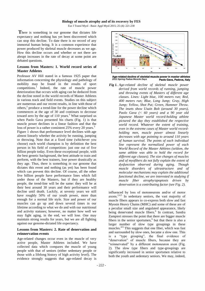
toward zero by the age of 110 years.3 What surprised us
when Paolo Gava presented his charts (Fig. 1) is that
muscle power declines in a linear fashion and that the
loss of power is a rather consistent 25% every 20 years.3
Figure 1 shows that performance level declines with age
almost linearly whether the activity be running, jumping
or throwing. Note that at a fixed time (whichever you
choose) each world champion is by definition the best
person in his field of competition: just one out of five
billion people today. Even these exceptional people, with
the best genetic background, the best attitude to train and
perform, with the best trainers, lose power drastically as
they age. Thus, there is something in our genome that
dictates this event and nothing (as yet) has been found
which can prevent this decline. Of course, all the other
five billion people have performance lines which fall
under those of the Masters, but if they are healthy
people, the trend-line will be the same: they will be at
their best around 30 years and their performance will
decline until death. Luckily, at seventy years we still
have roughly 50% of our youth power, more than
enough for a normal life style. Size and power of our
muscles can go up and down several times in our
lifetime according to what we do and with our nutritional
and activity statuses; however, no matter how well we
may fight aging, in the end, we will lose. One may
maintain strong results for years, but we are all fighting
against our genome-dictated life expectancy.
Lessons from Masters: 2. Rate of denervation and
reinnervation events
Age-related changes occur even in the muscle of very
active people, Master Athletes included. We have
collected data which compares the muscle of young
people with that of seniors (either sedentary people or
those with a lifelong history of high activity level). The
evidence strongly suggests that age-related decay is
influenced by loss of motoneurons and/or of motor
axons.2,4,5
In sedentary seniors, the vast majority of
muscle fibers appears to co-express both slow and fast
Myosin Heavy Chains (MHC) and some of these are of
a peculiar small size and angulated appearance, likely
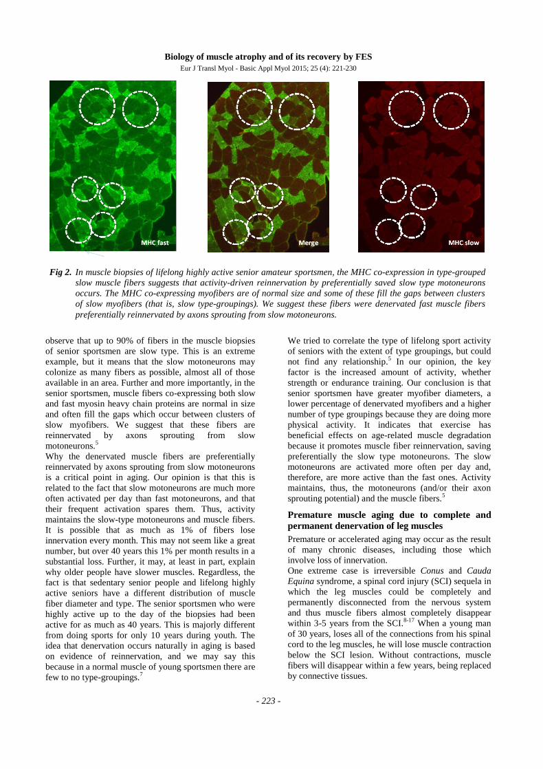
being denervated muscle fibers.6 In contrast, Sandra
Zampieri stresses the point that there are bigger muscle
fibers in the senior sportsmen,6 but that there is also a
larger number of slow type groupings in these
muscles.4-6
This suggests that one fiber, which was fast
and surrounded by slow ones, became a slow one. This
is a “type grouping”, the final evidence of
“denervation” of muscle fibers, because they are
“reinnervated” by a different motoneuron axon (Fig.
2). The slow type fibers and type-groupings are
significantly increased in senior sportsmen relative to
both the youth and sedentary seniors. We may, indeed,
Age-related decline of skeletal muscle power in master athletes
Paolo Gava, Padova, Italy2013 Spring Padua Muscle Days
P. GavaU. Bolt
90 years
Fig 1. Age-related decline of skeletal muscle power
derived from world records of running, jumping
and throwing events of Masters of different age
classes. Lines: Light blue, 100 meters run; Red,
400 meters run; Blue, Long Jump; Gray, High
Jump; Yellow, Shot Put; Green, Hammer Throw.
The insets show Usain Bolt (around 30 years),
Paolo Gava (< 60 years) and a 90 year old
Japanese Master world record-holding athlete
pictured the day they established the respective
world record. Whatever the extent of training,
even in the extreme cases of Master world record-
holding men, muscle power almost linearly
decreases with age pointing to around 110 years
of human survival. The points of each individual
line represent the normalized power of each
World Record of the Master Athletes (seldom, the
same athlete was able to hold the record for
different age classes). The size changes of muscles
and of myofibers do not fully explain the extent of
dysfunction observed during aging. Though
muscle disorders of ultrastructure and of
molecular mechanisms may explain the additional
functional decline, we are interested in studying if
muscle fiber atrophy/apoptosis driven by
denervation is a contributing factor (see Fig. 2).
Biology of muscle atrophy and of its recovery by FES
Eur J Transl Myol - Basic Appl Myol 2015; 25 (4): 221-230
- 223 -
observe that up to 90% of fibers in the muscle biopsies
of senior sportsmen are slow type. This is an extreme
example, but it means that the slow motoneurons may
colonize as many fibers as possible, almost all of those
available in an area. Further and more importantly, in the
senior sportsmen, muscle fibers co-expressing both slow
and fast myosin heavy chain proteins are normal in size
and often fill the gaps which occur between clusters of
slow myofibers. We suggest that these fibers are
reinnervated by axons sprouting from slow
motoneurons.5
Why the denervated muscle fibers are preferentially
reinnervated by axons sprouting from slow motoneurons
is a critical point in aging. Our opinion is that this is
related to the fact that slow motoneurons are much more
often activated per day than fast motoneurons, and that
their frequent activation spares them. Thus, activity
maintains the slow-type motoneurons and muscle fibers.
It is possible that as much as 1% of fibers lose
innervation every month. This may not seem like a great
number, but over 40 years this 1% per month results in a
substantial loss. Further, it may, at least in part, explain
why older people have slower muscles. Regardless, the
fact is that sedentary senior people and lifelong highly
active seniors have a different distribution of muscle
fiber diameter and type. The senior sportsmen who were
highly active up to the day of the biopsies had been
active for as much as 40 years. This is majorly different
from doing sports for only 10 years during youth. The
idea that denervation occurs naturally in aging is based
on evidence of reinnervation, and we may say this
because in a normal muscle of young sportsmen there are
few to no type-groupings.7
We tried to correlate the type of lifelong sport activity
of seniors with the extent of type groupings, but could
not find any relationship.5 In our opinion, the key
factor is the increased amount of activity, whether
strength or endurance training. Our conclusion is that
senior sportsmen have greater myofiber diameters, a
lower percentage of denervated myofibers and a higher
number of type groupings because they are doing more
physical activity. It indicates that exercise has
beneficial effects on age-related muscle degradation
because it promotes muscle fiber reinnervation, saving
preferentially the slow type motoneurons. The slow
motoneurons are activated more often per day and,
therefore, are more active than the fast ones. Activity
maintains, thus, the motoneurons (and/or their axon
sprouting potential) and the muscle fibers.5
Premature muscle aging due to complete and
permanent denervation of leg muscles
Premature or accelerated aging may occur as the result
of many chronic diseases, including those which
involve loss of innervation.
One extreme case is irreversible Conus and Cauda
Equina syndrome, a spinal cord injury (SCI) sequela in
which the leg muscles could be completely and
permanently disconnected from the nervous system
and thus muscle fibers almost completely disappear
within 3-5 years from the SCI.8-17
When a young man
of 30 years, loses all of the connections from his spinal
cord to the leg muscles, he will lose muscle contraction
below the SCI lesion. Without contractions, muscle
fibers will disappear within a few years, being replaced
by connective tissues.
Fig 2. In muscle biopsies of lifelong highly active senior amateur sportsmen, the MHC co-expression in type-grouped
slow muscle fibers suggests that activity-driven reinnervation by preferentially saved slow type motoneurons
occurs. The MHC co-expressing myofibers are of normal size and some of these fill the gaps between clusters
of slow myofibers (that is, slow type-groupings). We suggest these fibers were denervated fast muscle fibers
preferentially reinnervated by axons sprouting from slow motoneurons.
Biology of muscle atrophy and of its recovery by FES
Eur J Transl Myol - Basic Appl Myol 2015; 25 (4): 221-230
- 224 -
Is there anything that can be done in this extreme
situation to prevent these negative changes?
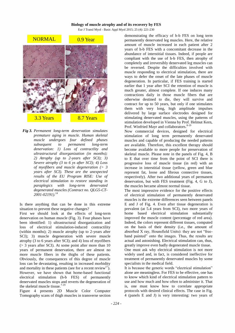
First we should look at the effects of long-term
denervation on human muscle (Fig. 3). Four phases have
been identified: 1) ultrastructural disorganization and
loss of electrical stimulation-induced contractility
(within months); 2) muscle atrophy (up to 2-years after
SCI); 3) muscle degeneration with severe muscle
atrophy (3 to 6 years after SCI); and 4) loss of myofibers
(> 3 years after SCI). At some point after more than 10
years of permanent denervation, there are almost no
more muscle fibers in the thighs of these patients.
Obviously, the consequences of this degree of muscle
loss can be devastating, resulting in increased morbidity
and mortality in these patients (see for a recent review17
).
However, we have shown that home-based functional
electrical stimulation (h-b FES) of permanently
denervated muscles stops and reverts the degeneration of
the skeletal muscle tissue.7-19
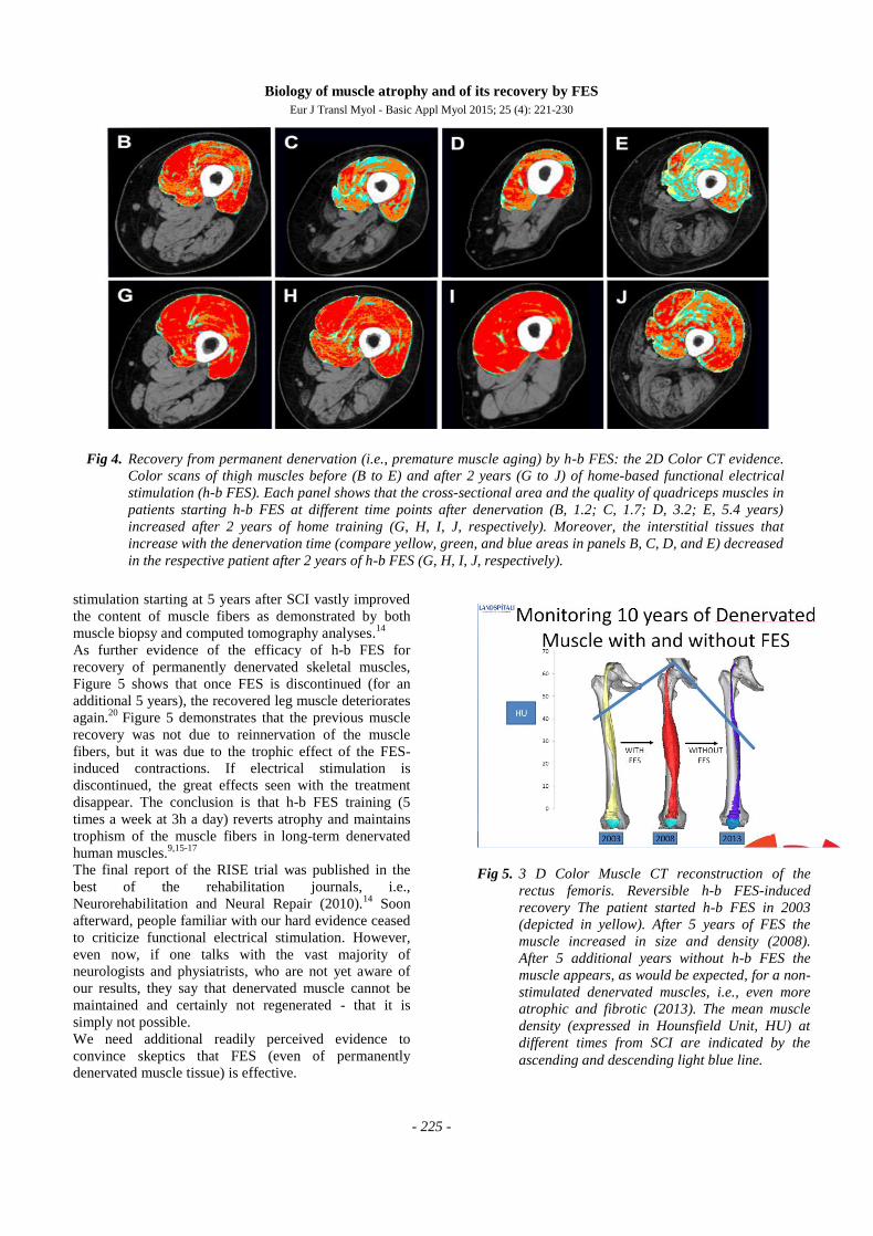
Figure 4 presents 2D Muscle Color Computer
Tomography scans of thigh muscles in transverse section
demonstrating the efficacy of h-b FES on long term
permanently denervated leg muscles. Here, the relative
amount of muscle increased in each patient after 2
years of h-b FES with a concomitant decrease in the
abundance of interstitial tissues. Indeed, if people are
compliant with the use of h-b FES, then atrophy of
completely and irreversibly denervated leg muscles can
be reversed. Despite the difficulties involved with
muscle responding to electrical stimulation, there are
ways to defer the onset of the late phases of muscle
degeneration. In particular, if FES training is started
earlier that 1 year after SCI the retention of muscle is
much greater, almost complete. If one induces many
contractions daily in those muscle fibers that are
otherwise destined to die, they will survive and
contract for up to 50 years, but only if one stimulates
them with very long, high amplitude impulses
delivered by large surface electrodes designed for
stimulating denervated muscles, using the patterns of
stimulation developed in Vienna by Prof. Helmut Kern,
Prof. Winfried Mayr and collaborators.8-19
Now commercial devices, designed for electrical
stimulation of long term permanently denervated
muscles and capable of producing the needed patterns
are available. Therefore, this excellent therapy should
become available to more people for preservation of
skeletal muscle. Please note in the panels of Fig. 4, B
to E that over time from the point of SCI there is
progressive loss of muscle tissue (in red) with an
increase in interstitial tissue (yellow, green and blue
represent fat, loose and fibrous connective tissues,
respectively). After two additional years of permanent
denervation, but with FES treatment (Fig. 4, G to J),
the muscles became almost normal tissue.
The most impressive evidence for the positive effects
of electrical stimulation of permanently denervated
muscles is the extreme differences seen between panels
E and J of Fig. 4. Even after tissue degeneration is
prevalent (at 5.4 years from SCI), two more years of
home based electrical stimulation substantially
improved the muscle content (percentage of red area).
Indeed, the colors represent different tissues, computed
on the basis of their density (i.e., the amount of
absorbed X ray, Hounsfield Units): they are not “free-
hand painted” onto the images. Thus, the results are
actual and astonishing. Electrical stimulation can, thus,
greatly improve even badly degenerated muscle tissue.
One must ask why electrical stimulation is not more
widely used and, in fact, is considered ineffective for
treatment of permanently denervated muscles by some
specialists in the medical field.
It is because the generic words “electrical stimulation”
alone are meaningless. For FES to be effective, one has
to know which kind of electrical stimulation pattern to
use and how much and how often to administer it. That
is, one must know how to correlate appropriate
protocols with desired clinical effects. The case in Fig.
4 (panels E and J) is very interesting: two years of
NORMAL
8.7 Years3.3 Years
0.9 Year
Fig 3. Permanent long-term denervation simulates
premature aging in muscle. Human skeletal
muscle undergoes four defined phases
subsequent to permanent long-term
denervation: 1) Loss of contractlity and
ultrastructural disorganization (in months);
2) Atrophy (up to 2-years after SCI); 3)
Severe atrophy (3 to 6 ys after SCI); 4) Loss
of myofibers and muscle degeneration (> 3
years after SCI). These are the unexpected
results of the EU Program RISE: Use of
electrical stimulation to restore standing in
paraplegics with long-term denervated
degenerated muscles (Contract no. QLG5-CT-
2001-02191).14
Biology of muscle atrophy and of its recovery by FES
Eur J Transl Myol - Basic Appl Myol 2015; 25 (4): 221-230
- 225 -
stimulation starting at 5 years after SCI vastly improved
the content of muscle fibers as demonstrated by both
muscle biopsy and computed tomography analyses.14
As further evidence of the efficacy of h-b FES for
recovery of permanently denervated skeletal muscles,
Figure 5 shows that once FES is discontinued (for an
additional 5 years), the recovered leg muscle deteriorates
again.20
Figure 5 demonstrates that the previous muscle
recovery was not due to reinnervation of the muscle
fibers, but it was due to the trophic effect of the FES-
induced contractions. If electrical stimulation is
discontinued, the great effects seen with the treatment
disappear. The conclusion is that h-b FES training (5
times a week at 3h a day) reverts atrophy and maintains
trophism of the muscle fibers in long-term denervated
human muscles.9,15-17
The final report of the RISE trial was published in the
best of the rehabilitation journals, i.e.,
Neurorehabilitation and Neural Repair (2010).14
Soon
afterward, people familiar with our hard evidence ceased
to criticize functional electrical stimulation. However,
even now, if one talks with the vast majority of
neurologists and physiatrists, who are not yet aware of
our results, they say that denervated muscle cannot be
maintained and certainly not regenerated - that it is
simply not possible.
We need additional readily perceived evidence to
convince skeptics that FES (even of permanently
denervated muscle tissue) is effective.
Fig 4. Recovery from permanent denervation (i.e., premature muscle aging) by h-b FES: the 2D Color CT evidence.
Color scans of thigh muscles before (B to E) and after 2 years (G to J) of home-based functional electrical
stimulation (h-b FES). Each panel shows that the cross-sectional area and the quality of quadriceps muscles in
patients starting h-b FES at different time points after denervation (B, 1.2; C, 1.7; D, 3.2; E, 5.4 years)
increased after 2 years of home training (G, H, I, J, respectively). Moreover, the interstitial tissues that
increase with the denervation time (compare yellow, green, and blue areas in panels B, C, D, and E) decreased
in the respective patient after 2 years of h-b FES (G, H, I, J, respectively).
Fig 5. 3 D Color Muscle CT reconstruction of the
rectus femoris. Reversible h-b FES-induced
recovery The patient started h-b FES in 2003
(depicted in yellow). After 5 years of FES the
muscle increased in size and density (2008).
After 5 additional years without h-b FES the
muscle appears, as would be expected, for a non-
stimulated denervated muscles, i.e., even more
atrophic and fibrotic (2013). The mean muscle
density (expressed in Hounsfield Unit, HU) at
different times from SCI are indicated by the
ascending and descending light blue line.
Biology of muscle atrophy and of its recovery by FES
Eur J Transl Myol - Basic Appl Myol 2015; 25 (4): 221-230
- 226 -
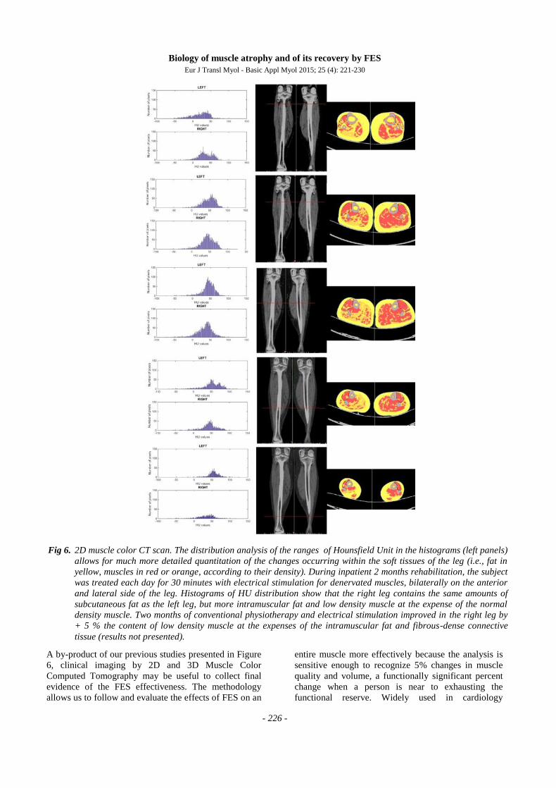
A by-product of our previous studies presented in Figure
6, clinical imaging by 2D and 3D Muscle Color
Computed Tomography may be useful to collect final
evidence of the FES effectiveness. The methodology
allows us to follow and evaluate the effects of FES on an
entire muscle more effectively because the analysis is
sensitive enough to recognize 5% changes in muscle
quality and volume, a functionally significant percent
change when a person is near to exhausting the
functional reserve. Widely used in cardiology
Fig 6. 2D muscle color CT scan. The distribution analysis of the ranges of Hounsfield Unit in the histograms (left panels)
allows for much more detailed quantitation of the changes occurring within the soft tissues of the leg (i.e., fat in
yellow, muscles in red or orange, according to their density). During inpatient 2 months rehabilitation, the subject
was treated each day for 30 minutes with electrical stimulation for denervated muscles, bilaterally on the anterior
and lateral side of the leg. Histograms of HU distribution show that the right leg contains the same amounts of
subcutaneous fat as the left leg, but more intramuscular fat and low density muscle at the expense of the normal
density muscle. Two months of conventional physiotherapy and electrical stimulation improved in the right leg by
+ 5 % the content of low density muscle at the expenses of the intramuscular fat and fibrous-dense connective
tissue (results not presented).
Biology of muscle atrophy and of its recovery by FES
Eur J Transl Myol - Basic Appl Myol 2015; 25 (4): 221-230
- 227 -
imaging,21
the false color approach is ignored in clinical
imaging of skeletal muscle tissue. We hope to have
convinced the readers that its advantages offset the low
risks of irradiation, in particular during follow-up of
supervised trials, to add unbiased quantitative evidence
to clinical assessments.
A horse model to assess FES effectiveness by
morphometry of subsarcolemmal mitochondria
Finally, we would like to discuss a recent example of the
effectiveness of FES for equine epaxial muscle spasm.
Its relevance here is related to the fact that psychological
factors are, reasonably, less important or absent in horse
treatments. In this example, we explored the different
types of mitochondria present in skeletal muscle fibers,
either subsarcolemmal or intermyofibrillar and their
differential response to needs and activation loads of
muscle fibers.22-24
We had the opportunity to analyze 12 muscle biopsies (6
pre- and 6 post-FES) from 6 FES-treated horses.
Previous preliminary histopathologic analyses suggested
that stimulated muscles were more damaged after than
before treatment.
Could this be the result of the electrical stimulation? Our
additional more careful morphometric analyses exclude
it. Indeed, only one horse presents with obvious evidence
of post-FES muscle damage (foci of severe muscle fiber
atrophy) whereas the other 5 horses, which had the same
type and amount of electrical stimulation, display only
scanty evidence of muscle atrophy (possibly resulting
from denervation) in both pre- and post-stimulation
biopsies.23
We wish to stress here that any muscle damage detected
in post-FES tissue analyses is too often immediately
attributed to FES without further evaluation. However, if
one does morphometry properly (with random sampling
and statistical evaluation) the correlation between FES
and muscle damage often disappears, as is the case in
this group of horse muscle biopsies.
Indeed, heavy electrical stimulation, (that is,
electroporation) is routinely used to deliver plasmid
DNA to the muscle tissue of experimental rodents
without reported tissue damage.25,26
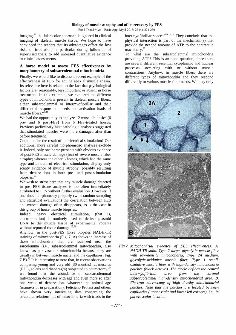
Anyhow, in the post-FES horse biopsies NADH-TR
staining of mitochondria (Fig. 7, A) shows an increase of
those mitochondria that are localized near the
sarcolemma (i.e., subsarcolemmal mitochondria, also
known as paravascular mitochondria because they are
usually in between muscle nuclei and the capillaries, Fig.
7 B).23
It is interesting to note that, in recent observations
comparing young and very old (30 months) rat muscles
(EDL, soleus and diaphragm) subjected to neurectomy,24
we found that the abundance of subsarcolemmal
mitochondria decreases with age and even more so after
one week of denervation, whatever the animal age
(manuscript in preparation). Feliciano Protasi and others
have shown very interesting data concerning the
structural relationships of mitochondria with triads in the
intermyofibrillar spaces.6,9,11,14
They conclude that the
physical interaction is part of the mechanism(s) that
provide the needed amount of ATP to the contractile
machinery.27
To what are the subsarcolemmal mitochondria
providing ATP? This is an open question, since there
are several different essential cytoplasmic and nuclear
processes occurring with or without muscle
contractions. Anyhow, in muscle fibers there are
different types of mitochondria and they respond
differently to various muscle fiber needs. We may only
Fig 7. Mitochondrial evidence of FES effectiveness. A.
NADH-TR stain. Type 2 large, glycolytic muscle fiber
with low-density mitochondria, Type 2A medium,
glycolytic-oxidative muscle fiber, Type 1 small,
oxidative muscle fiber with high-density mitochondria
patches (black arrows). The circle defines the central
intermyofibrillar area from the coronal
subsarcolemmal high-density mitochondrial area. B.
Electron microscopy of high density mitochondrial
patches. Note that the patches are located between
capillaries ( upper right and lower left corners), i.e., in
paravascular location.
Biology of muscle atrophy and of its recovery by FES
Eur J Transl Myol - Basic Appl Myol 2015; 25 (4): 221-230
- 228 -
say, for now, that mitochondrial distribution and density
are significantly changed in horses post-FES muscle
biopsies. This indicates that the clinical improvements
observed in the horse are possibly related to increased
muscle perfusion induced by FES stimulation. Likewise
as in human cases,8-20
electrical stimulation provides
clinically relevant results in the treatments of equine
epaxial muscle spasm. It seems to be a good way to
increase contraction per day, i. e., mimic volitional
exercise and, thus, modify the behaviors of the muscle
fibers.
Conclusion and perspectives
In summary, it appears that age-related decline in muscle
power is partially attributable to loss of innervation and
that this loss can be deferred by life-long high-level
activity.28
Diseases involving permanent denervation
show similar, but premature and much more severe
muscle deterioration. We have shown that, with
appropriate protocols, h-b FES can inhibit muscle
degeneration and that it can also actually reverse it.29
It should be possible to stave off age-related muscle
decline in aging people and others who have become
unable to participate in physical activities. Thus, FES
should be considered for use in rehabilitation centers,
nursing facilities and in critical care units when patients
are completely inactive even for short periods of time.
Acknowledgement
This work was supported by European Regional
Development Fund - Cross Border Cooperation
Programme Slovakia – Austria 2007–2013 (Interreg-
IVa), project Mobilität im Alter, MOBIL, N_00033
(partners: Ludwig Boltzmann Institute of Electrical
Stimulation and Physical Rehabilitation, Austria, Center
for Medical Physics and Biomedical Engineering,
Medical University of Vienna, Austria, and Faculty of
Physical Education and Sports, Comenius University in
Bratislava, Slovakia); Austrian national co-financing of
the Austrian Federal Ministry of Science and Research;
Ludwig Boltzmann Society (Vienna, Austria).
Some of the research reported in this publication was
supported by the National Institute of Arthritis and
Musculoskeletal and Skin Diseases of the National
Institutes of Health under Award Number NIH NIAMS
1R03AR053706-01A2 to ALP. The content is solely the
responsibility of the authors and does not necessarily
represent the official views of the National Institutes of
Health. This paper is an edited transcription of the talk presented by
Ugo Carraro to the Interreg IVa Final Meeting November 8
2014, Vienna, Austria.
Corresponding Author
Francesco Gava, Via Armistizio, 87, 35142, Padova,
Italy. E-mail: [email protected]
E-mail of coauthors
Ugo Carraro: [email protected]
Helmut Kern: [email protected]
Paolo Gava: [email protected]
Christian Hofer: [email protected]
Stefan Loefler: [email protected]
Paolo Gargiulo: [email protected]
Simone Mosole: [email protected]
Sandra Zampieri: [email protected]
Valerio Gobbo: [email protected]
Barbara Ravara: [email protected]
Francesco Piccione:
Andrea Marcante:
Alfonc Baba: [email protected]
Sheila Schils: [email protected]
Amber Pond: [email protected]
References
1. Hill AV. The physiological basis of athletic
records. The Scientific Monthly 1925;2:409-428.
2. Mitchell WK, Williams J, Atherton P, Larvin M,
Lund J, Narici M. Sarcopenia, dynapenia, and the
impact of advancing age on human skeletal
muscle size and strength; a quantitative review.
Front Physiol 2012 Jul 11;3:260. doi:
10.3389/fphys.2012.00260. eCollection 2012.
3. Gava P, Kern H, Carraro U. Age-associated
power decline from running, jumping, and
throwing male masters world records. Exp Aging
Res. 2015;41(2):115-35. doi:
10.1080/0361073X.2015.1001648.
4. Mosole S, Rossini K, Kern H, et al. Significant
increase of vastus lateralis reinnervation in 70-
year sportsmen with a lifelong history of high-
level exercise. Eur J Transl Myol - Basic Appl
Myol 2013;23:117-22.
5. Mosole S, Carraro U, Kern H, et al. Long-term
high-level exercise promotes muscle
reinnervation with age. J Neuropathol Exp Neurol
2014;73:284-94. doi: 10.1097/NEN.0000000000
000032.
6. Zampieri S, Pietrangelo L, Loefler S, et al.
Lifelong Physical Exercise Delays Age-
Associated Skeletal Muscle Decline. J Gerontol A
Biol Sci Med Sci 2015;70:163-73. doi:
10.1093/gerona/glu006. Epub 2014 Feb 18.
7. Kern H, Pelosi L, Coletto L, et al.
Atrophy/hypertrophy cell signaling in muscles of
young athletes trained with vibrational-
proprioceptive stimulation. Neurol Res.
2011;33:998-1009.
8. Kern H. Funktionelle Elektrostimulation
Paraplegischer Patienten. ÖZPM, Österreichi sche
Biology of muscle atrophy and of its recovery by FES
Eur J Transl Myol - Basic Appl Myol 2015; 25 (4): 221-230
- 229 -
Zeitschrift für Physikalische Medizin 1995;5:1-75.
ISSN 1021-4348.
9. Kern H, Boncompagni S, Rossini K, et al. Long-
term denervation in humans causes degeneration of
both contractile and excitation contraction coupling
apparatus, which is reversible by functional
electrical stimulation (FES). A role for myofiber
regeneration? J Neuropathol Exp Neurol
2004;63:919-31.
10. Kern H, Rossini K, Carraro U, et al. Muscle
biopsies show that FES of denervated muscles
reverses human muscle degeneration from
permanent spinal motoneuron lesion. J Rehabil Res
Dev 2005;42:43-53.
11. Boncompagni S, Kern H, Rossini K, et al.
Structural differentiation of skeletal muscle fibers
in the absence of innervation in humans. Proc Natl
Acad Sci USA 2007;104:19339-44.
12. Kern H, Hofer C, Mayr W. Protocols for clinical
work package of the European project RISE. Eur J
Transl Myol/ Basic Appl Myol 2008;18:39-44.
13. Kern H, Carraro U, Adami N, et al. One year of
home-based Functional Electrical Stimulation
(FES) in complete lower motor neuron paraplegia:
Recovery of tetanic contractility drives the
structural improvements of denervated muscle.
Neurol Res 2010;32:5-12,doi: 10.1189/ 184313209
X385644.
14. Kern H, Carraro U, Adami N, et al. Home-based
functional electrical stimulation rescues
permanently denervated muscles in paraplegic
patients with complete lower motor neuron lesion.
Neurorehabil Neural Repair 2010;24:709-21. doi:
10.1177/ 1545968310366129. Epub 2010 May 11.
15. Rossini K, Zanin ME, Carraro U. To stage and
quantify regenerative myogenesis in human long-
term permanent denervated muscle. Basic Appl
Myol 2002;12:277-87.
16. Carraro U, Rossini K, Mayr W, Kern H. Muscle
fiber regeneration in human permanent lower
motoneuron denervation: relevance to safety and
effectiveness of FES-training, which induces
muscle recovery in SCI subjects. Artif Organs
2005;29:187-91.
17. Carraro U, Boncompagni S, Gobbo V, et al.
Persistent muscle fiber regeneration in long term
denervation. Past, present, future. Eur J Transl
Myol 2015;25:77-92.
18. Gargiulo P, Helgason T, Reynisson PJ, et al.
Monitoring of muscle and bone recovery in spinal
cord injury patients treated with electrical
stimulation using three-dimensional imaging and
segmentation techniques: methodological
assessment. Artif Organs 2011:35:275-81. doi:
10.1111/j.1525-1594.2011.01214.x.
19. Gargiulo P, Reynisson PJ, Helgason B, et al.
Muscle, tendons, and bone: structural changes
during denervation and FES treatment. Neurol
Res 2011;Sep:33(7):750-8. doi: 10.1179/174313
2811Y.0000000007.
20. Carraro U, Edmunds KJ, Gargiulo P. 3D false
color computed tomography for diagnosis and
follow-up of permanent denervated human
muscles submitted to home-based Functional
Electrical Stimulation. Eur J Transl Myol - Basic
Appl Myol 2015;25:129-40.
21. Wang R, Meinel FG, Schoepf UJ, Canstein C,
Spearman JV, De Cecco CN. Performance of
Automated Software in the Assessment of
Segmental Left Ventricular Function in Cardiac
CT: Comparison with Cardiac Magnetic
Resonance. Eur Radiol. 2015 Apr 30. [Epub
ahead of print]
22. Schils SJ, Turner TA. Functional Electrical
Stimulation for equine epaxial muscle
spasms:retrospective study of 241 clinical cases.
Comparative Exercise Physiology 2014;10:89-97.
23. Ravara B, Gobbo V, Carraro U et al. Functional
electrical stimulation as a safe and effective
treatment for equine epaxial muscle spasms:
Clinical evaluations and histochemical
morphometry of mitochondria in muscle biopsies.
Eur J Transl Myol - Basic Appl Myol 2015; 25
(2): 109-120.
24. Mosole S, Zampieri S, Germinario E, et al.
Structural and functional characteristics of
denervated muscles from oldest-old rats: a
relevant animal model for FES of denervated
myofibers of the diaphragm in ALS? Eur J Transl
Myol/Basic Appl Myol 2015; 25: 151.
25. Donà M, Sandri M, Rossini K, Dell'Aica I,
Podhorska-Okolow M, Carraro U. Functional in
vivo gene transfer into the myofibers of adult
skeletal muscle. Biochem Biophys Res Commun.
2003;312:1132-8.
26. Taylor JA, Babbs CF, Alzghoul MB, et al.
Optimization of Ectopic Gene Expression in
Biology of muscle atrophy and of its recovery by FES
Eur J Transl Myol - Basic Appl Myol 2015; 25 (4): 221-230
- 230 -
Skeletal Muscle through DNA Transfer by
Electroporation. BMC Biotechnology 2004;4:11.
27. Mammucari C, Gherardi G, Zamparo I, et al. The
mitochondrial calcium uniporter controls skeletal
muscle trophism in vivo. Cell Rep. 2015 Mar
3;10(8):1269-79. doi:10.1016/j.celrep. 2015.01.
056. Epub 2015 Feb 26.
28. Kern H, Barberi L, Löfler S, et al. Electrical
stimulation counteracts muscle decline in seniors.
Front Aging Neurosci 2014;Jul 24:6:189. doi:
10.3389/fnagi.2014.00189. eCollection 2014.
29. Kern H. Electrical Stimulation on Paraplegic
Patients. Eur J Trans Myol/ Basic Appl Myol
2014;24:75-157.
Related Documents