1 The Biology of Cancer First Edition pRb and Control of the Cell Cycle Clock Nam Deuk Kim, Ph.D. Copyright © Garland Science 2007 Robert A. Weinberg

Welcome message from author
This document is posted to help you gain knowledge. Please leave a comment to let me know what you think about it! Share it to your friends and learn new things together.
Transcript

1
The Biology of Cancer First Edition
pRb and Control of the
Cell Cycle Clock
Nam Deuk Kim, Ph.D.
Copyright © Garland Science 2007
Robert A. Weinberg

2

Figure 8.1 The Biology of Cancer (© Garland Science 2007)
The central
governor of
growth and
proliferation
Summary
3
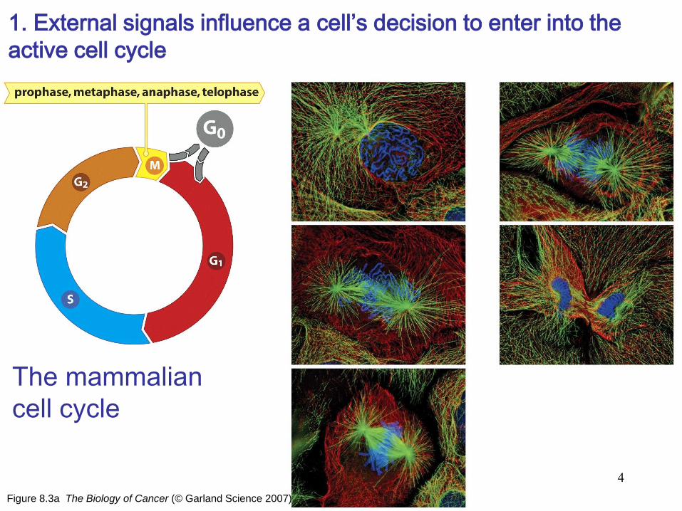
Figure 8.3a The Biology of Cancer (© Garland Science 2007)
The mammalian
cell cycle
1. External signals influence a cell’s decision to enter into the
active cell cycle
4
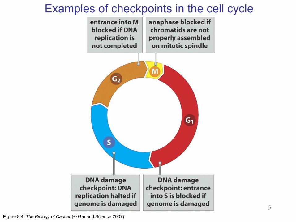
Figure 8.4 The Biology of Cancer (© Garland Science 2007)
Examples of checkpoints in the cell cycle
5

Figure 8.6 The Biology of Cancer (© Garland Science 2007)
Responsiveness to extracellular signals during the cell cycle
2. Cells make decisions about growth and quiescence during a
specific period in the G1 phase
6
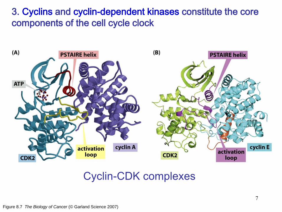
Figure 8.7 The Biology of Cancer (© Garland Science 2007)
Cyclin-CDK complexes
3. Cyclins and cyclin-dependent kinases constitute the core
components of the cell cycle clock
7
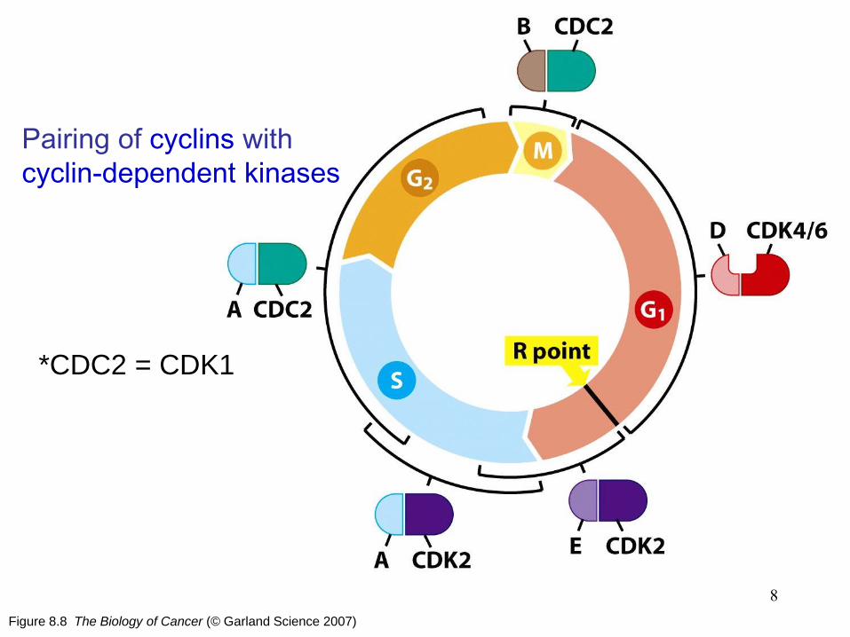
Figure 8.8 The Biology of Cancer (© Garland Science 2007)
Pairing of cyclins with
cyclin-dependent kinases
8
*CDC2 = CDK1

Figure 8.9 The Biology of Cancer (© Garland Science 2007)
Cell cycle-dependent fluctuations in cyclin B levels
9

Figure 8.10 The Biology of Cancer (© Garland Science 2007)
Fluctuation of cyclin levels during the cell cycle
10

Figure 8.11a The Biology of Cancer (© Garland Science 2007)
Control of cyclin D1 levels: Cyclin D1 was
discovered as a protein whose levels are strongly
induced by exposure of macrophages to the
mitogen CSF-1 (colony-stimulating factor-1).
11
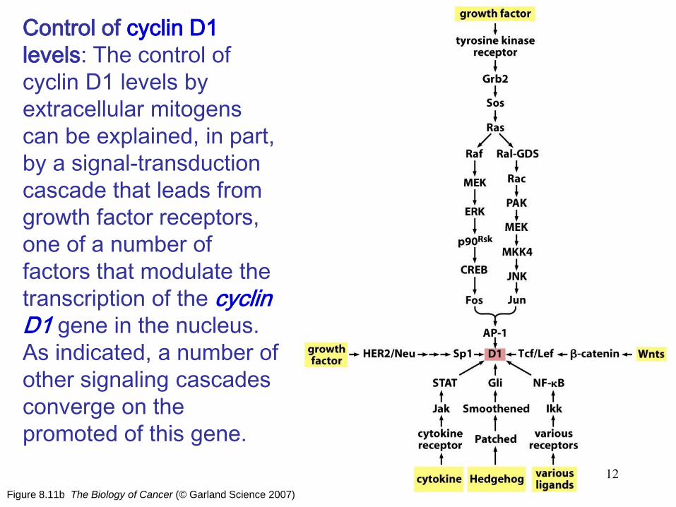
Figure 8.11b The Biology of Cancer (© Garland Science 2007)
Control of cyclin D1
levels: The control of
cyclin D1 levels by
extracellular mitogens
can be explained, in part,
by a signal-transduction
cascade that leads from
growth factor receptors,
one of a number of
factors that modulate the
transcription of the cyclin D1 gene in the nucleus.
As indicated, a number of
other signaling cascades
converge on the
promoted of this gene.
12

Table 8.1 The Biology of Cancer (© Garland Science 2007)
13

Figure 8.12 The Biology of Cancer (© Garland Science 2007)
Control of cyclin levels during the cell cycle
14

Figure 8.13a The Biology of Cancer (© Garland Science 2007)
4. Cyclin-CDK complexes are also regulated by CDK
inhibitors
Actions of CDK inhibitors 15

Figure 8.13b The Biology of Cancer (© Garland Science 2007)
The complex
between p27Kip1 and
cyclin A-CDK2,
derived by X-ray
crystallography.
16

Figure 8.13c The Biology of Cancer (© Garland Science 2007)
Inhibitors of the
INK4 class, such as
p16INK4A, bind to
CDK6. This CDK
inhibitor distort the
cyclin-binding site of
CDK6, reducing its
affinity for D-type
cyclins.
17

Figure 8.14a The Biology of Cancer (© Garland Science 2007)
TGF-β
Control of cell cycle
progression by TGF-β
18
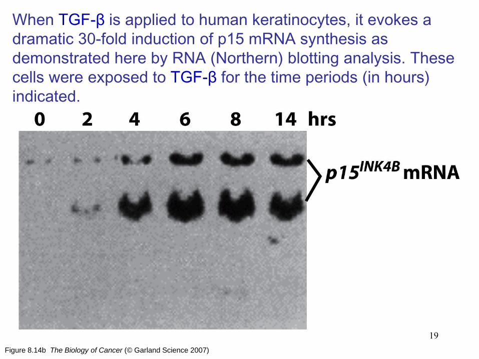
Figure 8.14b The Biology of Cancer (© Garland Science 2007)
When TGF-β is applied to human keratinocytes, it evokes a
dramatic 30-fold induction of p15 mRNA synthesis as
demonstrated here by RNA (Northern) blotting analysis. These
cells were exposed to TGF-β for the time periods (in hours)
indicated.
19
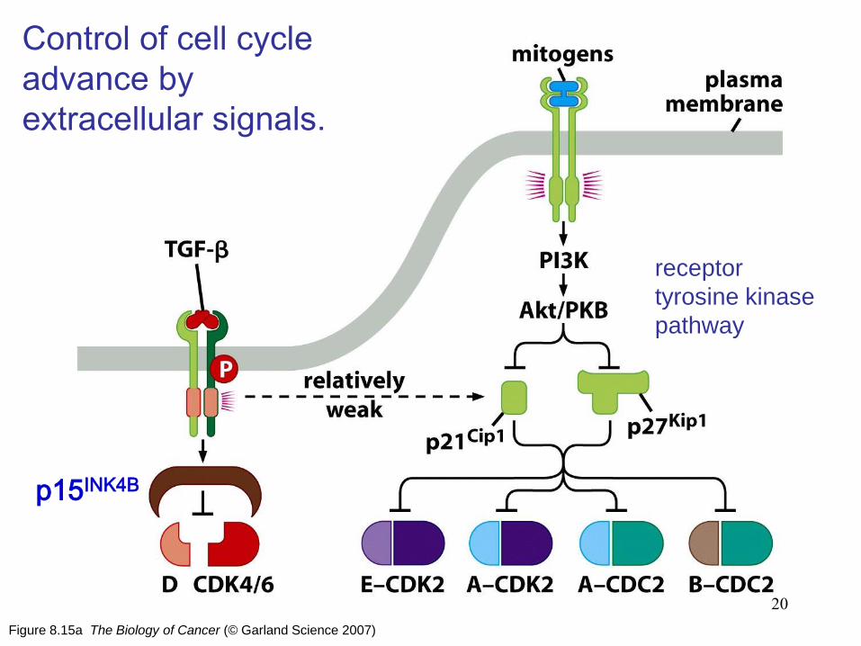
Figure 8.15a The Biology of Cancer (© Garland Science 2007)
Control of cell cycle
advance by
extracellular signals.
receptor
tyrosine kinase
pathway
20
p15INK4B
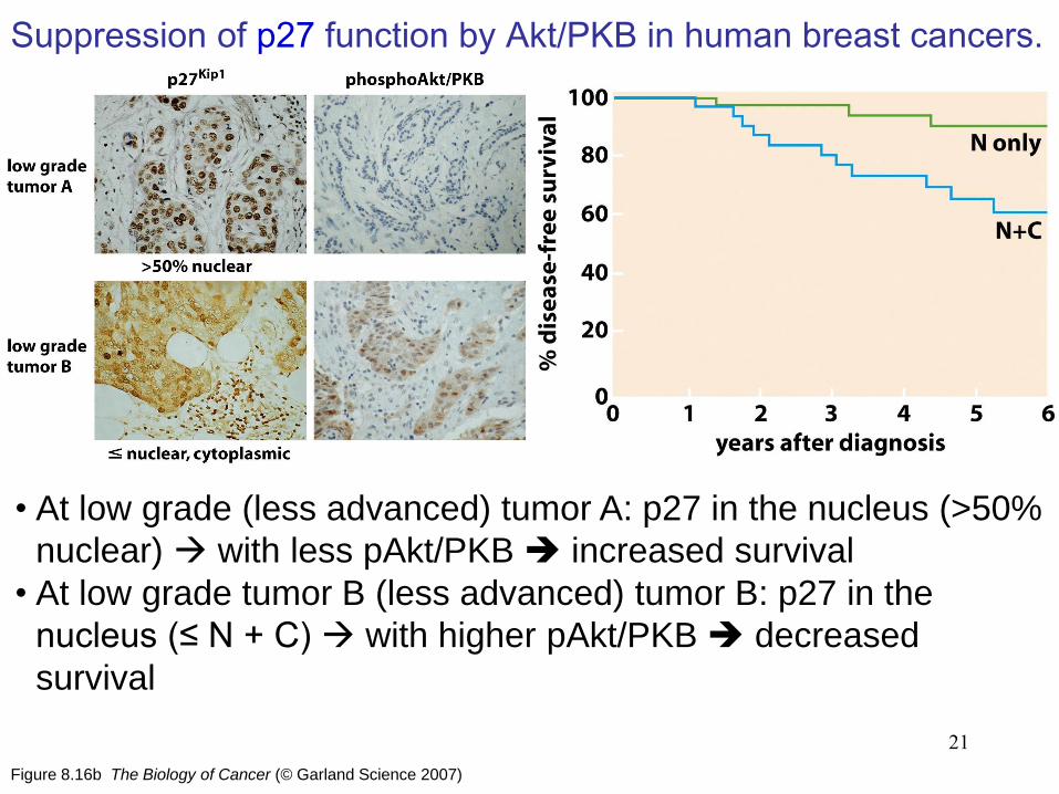
Figure 8.16b The Biology of Cancer (© Garland Science 2007)
Suppression of p27 function by Akt/PKB in human breast cancers.
21
• At low grade (less advanced) tumor A: p27 in the nucleus (>50%
nuclear) with less pAkt/PKB increased survival
• At low grade tumor B (less advanced) tumor B: p27 in the
nucleus (≤ N + C) with higher pAkt/PKB decreased
survival

Figure 8.19 The Biology of Cancer (© Garland Science 2007)
5. Viral oncoproteins reveal how pRb blocks advance
through the cell cycle
Cell cycle-
dependent
phosphorylation
of pRb
22

Figure 8.22 The Biology of Cancer (© Garland Science 2007)
Control of the restriction-point transition by mitogens.
6. pRb is deployed by the cell cycle clock to serve as
a guardian of the restriction-point gate.
23
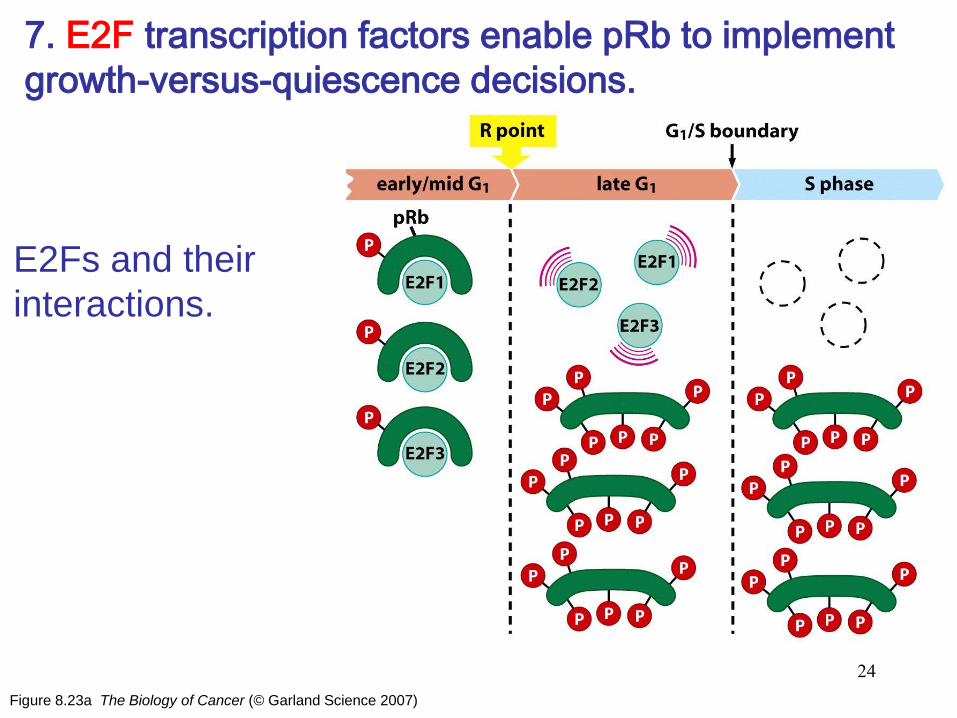
Figure 8.23a The Biology of Cancer (© Garland Science 2007)
7. E2F transcription factors enable pRb to implement
growth-versus-quiescence decisions.
E2Fs and their
interactions.
24

Figure 8.23c The Biology of Cancer (© Garland Science 2007)
The E2F transcription factors bind DNA as heterodimeric
complexes with DP partners: E2F4-DP2 complex to the DNA
double helix.
25

Figure 8.23d The Biology of Cancer (© Garland Science 2007)
The E2Fs constitute a family of at least seven distinct proteins.
• E2Fs 1, 2 and 3a: transcriptional activators
• E2F3b, E2F4, E2F5: transcriptional repressors
• E2F6, E2F7: poorly resolved
26

Figure 8.24a The Biology of Cancer (© Garland Science 2007)
Modification of chromatin by pocket proteins
27
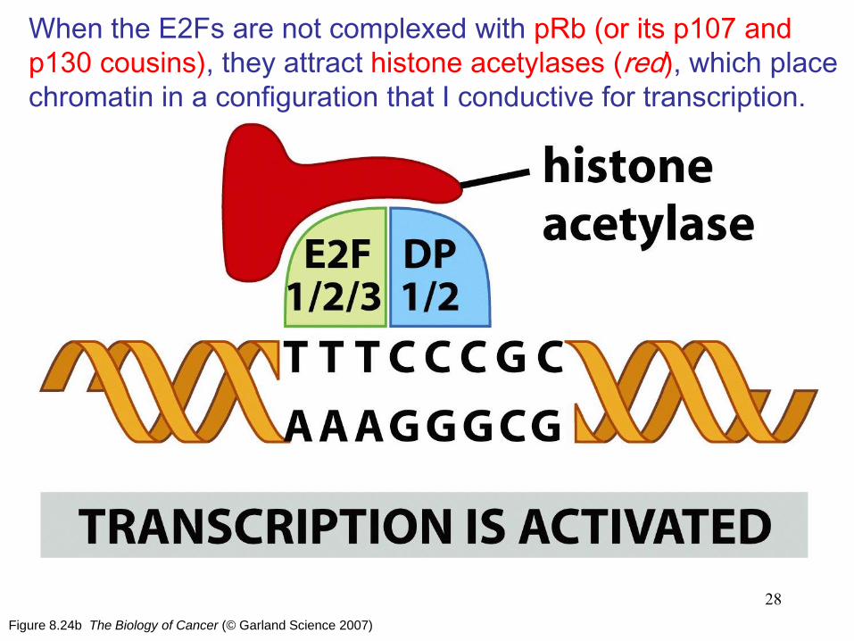
Figure 8.24b The Biology of Cancer (© Garland Science 2007)
When the E2Fs are not complexed with pRb (or its p107 and
p130 cousins), they attract histone acetylases (red), which place
chromatin in a configuration that I conductive for transcription.
28

Figure 8.25a The Biology of Cancer (© Garland Science 2007)
Positive-feedback loops and the
irreversibility of cell cycle advance.
The irreversibility of certain key steps in
cell cycle progression and the rapidity of
their execution is ensured, in part, by the
activation of certain positive-feedback
loops.
29
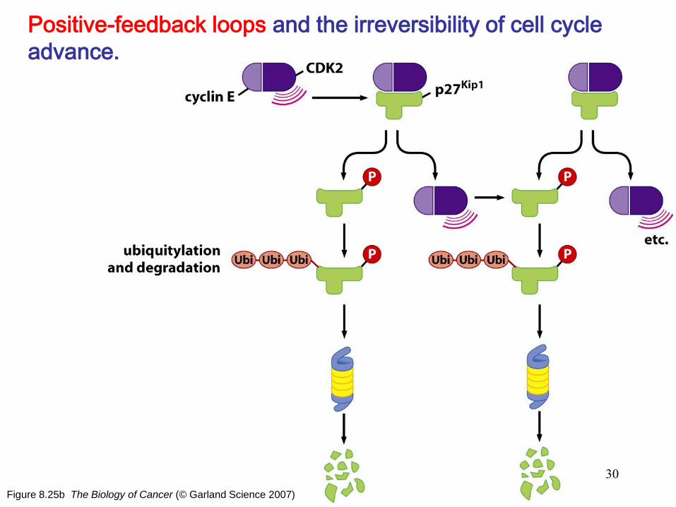
Figure 8.25b The Biology of Cancer (© Garland Science 2007)
Positive-feedback loops and the irreversibility of cell cycle
advance.
30

Figure 8.26 The Biology of Cancer (© Garland Science 2007)
8. A variety of mitogenic signaling pathways control
the phosphorylation state of pRb
• Growth factors
growth factor receptors
Ras
cyclin D1 and E
inactivation of pRb
activation of E2Fs
S-phase entrance
31
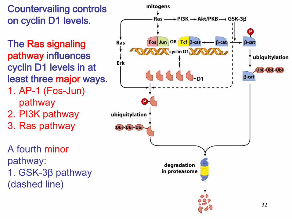
Countervailing controls
on cyclin D1 levels.
The Ras signaling
pathway influences
cyclin D1 levels in at
least three major ways.
1. AP-1 (Fos-Jun)
pathway
2. PI3K pathway
3. Ras pathway
A fourth minor
pathway:
1. GSK-3β pathway
(dashed line)
32

Figure 8.27 The Biology of Cancer (© Garland Science 2007)
9. The Myc oncoprotein perturbs the decision to phosphorylate
pRb and thereby deregulates control of cell cycle progression.
The Myc transcription factor:
• Myc belongs to a family of
bHLH (basic helix-loop-helix)
transcription factors.
• Myc-Max: promote
transcription active
proliferation
• Mad-Max: repress
transcription of most target
genes increased
differentiation
33

Figure 8.28 The Biology of Cancer (© Garland Science 2007)
Actions of Myc on the cell cycle
clock:
• Myc-Max: induce expression of the
growth-promoting proteins cyclin D2
and CDK4 promote advance
through early G1.
• By increasing the expression of
Cul1 (which is responsible for
degrading the p27 CDK inhibitor) as
well as E2Fs 1, 2, and 3, Myc
favors advance into S phase.
• In addition, Myc, acting with its Miz-
1 partner, is able to repress
expression of the p15, p21, and p27
CDK inhibitors; once again, effects
are felt both in early/mid and in
late G1.
34

Figure 8.29 The Biology of Cancer (© Garland Science 2007)
Powers of the Myc oncoprotein: The wide-ranging effects of the Myc
protein are illustrated by an experiment
in which the Myc protein has been
fused to the estrogen receptor (ER)
protein (blue). In the absence of ER
ligands, such as estrogen or tamoxifen,
the Myc-ER protein is trapped in the
cytoplasm (through association with
heat shock proteins, not shown). When
estrogen or tamoxifen ligands of the ER
(small purple ball) are added to cells,
the Myc-ER protein migrates into the
nucleus, associates with Max, and
activates Myc target genes within
minutes. Such activation, when induced
in serum-starved cells in the G0 phase,
enables them to enter the active cell
cycle and advance all the way through
G1 into the S phase.
35
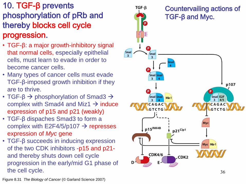
Figure 8.31 The Biology of Cancer (© Garland Science 2007)
10. TGF-β prevents
phosphorylation of pRb and
thereby blocks cell cycle
progression.
Countervailing actions of
TGF-β and Myc.
• TGF-β: a major growth-inhibitory signal
that normal cells, especially epithelial
cells, must learn to evade in order to
become cancer cells.
• Many types of cancer cells must evade
TGF-β-imposed growth inhibition if they
are to thrive.
• TGF-β phosphorylation of Smad3
complex with Smad4 and Miz1 induce
expression of p15 and p21 (weakly)
• TGF-β dispaches Smad3 to form a
complex with E2F4/5/p107 represses
expression of Myc gene
• TGF-β succeeds in inducing expression
of the two CDK inhibitors -p15 and p21-
and thereby shuts down cell cycle
progression in the early/mid G1 phase of
the cell cycle. 36
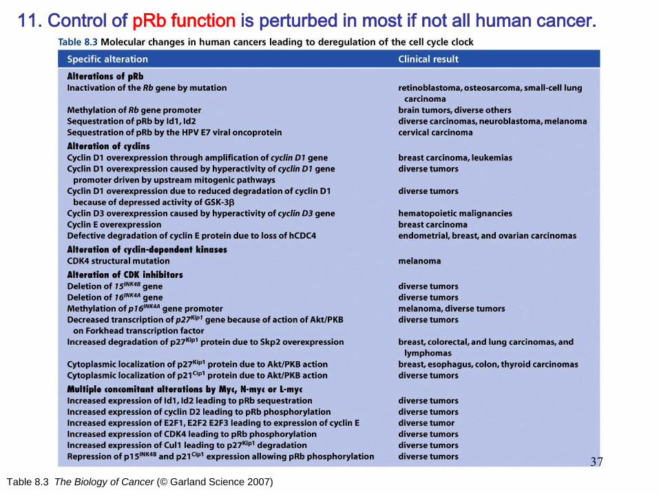
Table 8.3 The Biology of Cancer (© Garland Science 2007)
11. Control of pRb function is perturbed in most if not all human cancer.
37
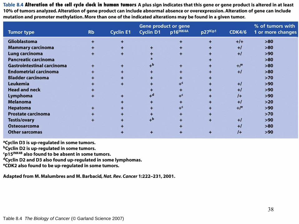
Table 8.4 The Biology of Cancer (© Garland Science 2007)
38
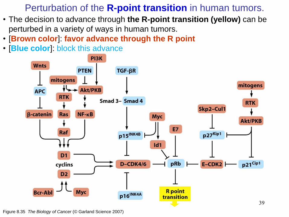
Figure 8.35 The Biology of Cancer (© Garland Science 2007)
Perturbation of the R-point transition in human tumors.
39
• The decision to advance through the R-point transition (yellow) can be
perturbed in a variety of ways in human tumors.
• [Brown color]: favor advance through the R point
• [Blue color]: block this advance

Figure 8.36 The Biology of Cancer (© Garland Science 2007)
Amplification of the cyclin D1 gene.
• Cyclin D1 gene (CCND1) in the cells
of a head-and-neck squamous cell
carcinoma (HNSCC).
• CCND1 is amplified to various
extents.
• About one-third of all of these
tumors, leading to corresponding
increases in cyclin D1 expression
and resulting loss of proper control
of pRb phosphorylation.
40
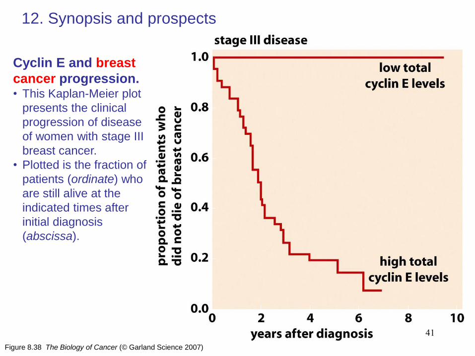
Figure 8.38 The Biology of Cancer (© Garland Science 2007)
12. Synopsis and prospects
Cyclin E and breast
cancer progression. • This Kaplan-Meier plot
presents the clinical
progression of disease
of women with stage III
breast cancer.
• Plotted is the fraction of
patients (ordinate) who
are still alive at the
indicated times after
initial diagnosis
(abscissa).
41
Related Documents