JOURNAL OF VIROLOGY, 0022-538X/00/$04.0010 Jan. 2000, p. 923–933 Vol. 74, No. 2 Copyright © 2000, American Society for Microbiology. All Rights Reserved. Biology of Attenuated Modified Vaccinia Virus Ankara Recombinant Vector in Mice: Virus Fate and Activation of B- and T-Cell Immune Responses in Comparison with the Western Reserve Strain and Advantages as a Vaccine JUAN C. RAMI ´ REZ, M. MAGDALENA GHERARDI, AND MARIANO ESTEBAN* Department of Molecular and Cellular Biology, Centro Nacional de Biotecnologı ´a, CSIC, Campus Universidad Autonoma, 28049 Madrid, Spain Received 10 August 1999/Accepted 7 October 1999 The modified vaccinia virus Ankara (MVA) strain is a candidate vector for vaccination against pathogens and tumors, due to safety concerns and the proven ability of recombinants based on this vector to trigger protection against pathogens in animals. In this study we addressed the fate of the MVA vector in BALB/c mice after intraperitoneal inoculation in comparison with that of the replication-competent Western Reserve (WR) strain by measuring levels of expression of the reporter luciferase gene, the capability to infect target tissues from the site of inoculation, and the length of time of virus persistence. We evaluated the extent of humoral and cellular immune responses induced against the virus antigens and a recombinant product (b-galactosidase). We found that MVA infects the same target tissues as the WR strain; surprisingly, within 6 h postinoculation the levels of expression of antigens were higher in tissues from MVA-infected mice than in tissues from mice infected with wild-type virus but at later times postinoculation were 2 to 4 log units higher in tissues from WR-infected mice. In spite of this, antibodies and cellular immune responses to viral vector antigens were considerably lower in MVA-inoculated mice than in WR virus-inoculated mice. In contrast, the cellular immune response to a foreign antigen expressed from MVA was similar to and even higher than that triggered by the recombinant WR virus. MVA elicited a Th1 type of immune response, and the main proinflammatory cytokines induced were interleukin-6 and tumor necrosis factor alpha. Our findings have defined the biological characteristics of MVA infection in tissues and the immune parameters activated in the course of virus infection. These results are of significance with respect to optimal use of MVA as a vaccine. The successful use of vaccinia virus (VV) during the world- wide program for eradication of smallpox (57), in conjunction with the development of strategies to generate recombinant VV (rVV) efficiently expressing foreign proteins (31, 39), has increased the potential use of poxvirus-derived vectors as de- livery systems in vaccination programs. Moreover, the advent of the AIDS pandemic has focused attention on live-virus- based vaccines, as both humoral immunity and cell-mediated immunity (CMI) are generated during replication of the viral vector. Cellular immunity seems to be relevant in the develop- ment of protection against human immunodeficiency virus type 1 (HIV-1) (45) and simian immunodeficiency virus (SIV) (25, 48), representing the immunological parameter that correlates with protection against disease. Because poxvirus-derived vec- tors activate both branches of the immune system (14), they are becoming promising vectors in efforts to develop an effi- cient anti-HIV vaccine, with economy of production and easy worldwide distribution. However, rare but grave nondesired side effects due to complications associated with vaccination with VV, increased in immunosuppressed individuals (19, 41), represent serious difficulties for wide use of VV-derived vac- cines. All of these factors have prompted the development of highly attenuated VV-derived viruses that can be used for safe vaccines if the desired immunological properties are main- tained while virulent genes are removed from the viral ge- nome. Avipoxvirus-derived vectors (55) and highly attenuated strains of VV have been considered candidate vectors (37, 42). The modified vaccinia virus Ankara (MVA), derived from the Ankara VV strain through more than 500 passages in chicken embryo fibroblasts (CEFs), was used in almost 120,000 Cau- casian individuals with no reported side effects, although many of the subjects were among the population with high risk of developing complications (33). Genetic analysis showed that during attenuation, viral genes spanning 15% of the parental genome (ca. 30 kbp) were lost (34); this loss included naturally encoded VV genes involved in host immunoregulation (8) and host range genes (1, 34). The MVA genome has been se- quenced, and the genes lost during passage have been identi- fied (2). Among the main properties that encourage the future use of MVA as a vaccine vector for humans are previous field experience and the inability of the virus to replicate produc- tively in human cell lines and primary human cell cultures (17). The defect in the viral life cycle is in the final steps of the morphogenetic program, with no alteration in early or late virus gene expression or in the levels of foreign protein expres- sion, which in cultured cells is at least as efficient as that for other, more virulent VV strains (53). Different viral and tumor animal models (11) have been used to demonstrate the efficacy of MVA recombinants in the development of protective immunity. Thus, immunity to influ- enza virus and parainfluenza virus type 3 was achieved after intramuscular and intranasal inoculation of recombinant MVA (rMVA) expressing hemagglutinin and nucleoproteins in mu- * Corresponding author. Mailing address: Department of Molecular and Cellular Biology, Centro Nacional de Biotecnologı ´a, CSIC, Cam- pus Universidad Autonoma, 28049 Madrid, Spain. Phone: 34-91 5854503. Fax: 34-91 5854506. E-mail: [email protected]. 923 on February 10, 2015 by guest http://jvi.asm.org/ Downloaded from

Welcome message from author
This document is posted to help you gain knowledge. Please leave a comment to let me know what you think about it! Share it to your friends and learn new things together.
Transcript
JOURNAL OF VIROLOGY,0022-538X/00/$04.0010
Jan. 2000, p. 923–933 Vol. 74, No. 2
Copyright © 2000, American Society for Microbiology. All Rights Reserved.
Biology of Attenuated Modified Vaccinia Virus AnkaraRecombinant Vector in Mice: Virus Fate and Activation ofB- and T-Cell Immune Responses in Comparison with the
Western Reserve Strain and Advantages as a VaccineJUAN C. RAMIREZ, M. MAGDALENA GHERARDI, AND MARIANO ESTEBAN*
Department of Molecular and Cellular Biology, Centro Nacional de Biotecnologıa, CSIC, Campus UniversidadAutonoma, 28049 Madrid, Spain
Received 10 August 1999/Accepted 7 October 1999
The modified vaccinia virus Ankara (MVA) strain is a candidate vector for vaccination against pathogensand tumors, due to safety concerns and the proven ability of recombinants based on this vector to triggerprotection against pathogens in animals. In this study we addressed the fate of the MVA vector in BALB/c miceafter intraperitoneal inoculation in comparison with that of the replication-competent Western Reserve (WR)strain by measuring levels of expression of the reporter luciferase gene, the capability to infect target tissuesfrom the site of inoculation, and the length of time of virus persistence. We evaluated the extent of humoral andcellular immune responses induced against the virus antigens and a recombinant product (b-galactosidase).We found that MVA infects the same target tissues as the WR strain; surprisingly, within 6 h postinoculationthe levels of expression of antigens were higher in tissues from MVA-infected mice than in tissues from miceinfected with wild-type virus but at later times postinoculation were 2 to 4 log units higher in tissues fromWR-infected mice. In spite of this, antibodies and cellular immune responses to viral vector antigens wereconsiderably lower in MVA-inoculated mice than in WR virus-inoculated mice. In contrast, the cellularimmune response to a foreign antigen expressed from MVA was similar to and even higher than that triggeredby the recombinant WR virus. MVA elicited a Th1 type of immune response, and the main proinflammatorycytokines induced were interleukin-6 and tumor necrosis factor alpha. Our findings have defined the biologicalcharacteristics of MVA infection in tissues and the immune parameters activated in the course of virusinfection. These results are of significance with respect to optimal use of MVA as a vaccine.
The successful use of vaccinia virus (VV) during the world-wide program for eradication of smallpox (57), in conjunctionwith the development of strategies to generate recombinantVV (rVV) efficiently expressing foreign proteins (31, 39), hasincreased the potential use of poxvirus-derived vectors as de-livery systems in vaccination programs. Moreover, the adventof the AIDS pandemic has focused attention on live-virus-based vaccines, as both humoral immunity and cell-mediatedimmunity (CMI) are generated during replication of the viralvector. Cellular immunity seems to be relevant in the develop-ment of protection against human immunodeficiency virus type1 (HIV-1) (45) and simian immunodeficiency virus (SIV) (25,48), representing the immunological parameter that correlateswith protection against disease. Because poxvirus-derived vec-tors activate both branches of the immune system (14), theyare becoming promising vectors in efforts to develop an effi-cient anti-HIV vaccine, with economy of production and easyworldwide distribution. However, rare but grave nondesiredside effects due to complications associated with vaccinationwith VV, increased in immunosuppressed individuals (19, 41),represent serious difficulties for wide use of VV-derived vac-cines. All of these factors have prompted the development ofhighly attenuated VV-derived viruses that can be used for safevaccines if the desired immunological properties are main-
tained while virulent genes are removed from the viral ge-nome.
Avipoxvirus-derived vectors (55) and highly attenuatedstrains of VV have been considered candidate vectors (37, 42).The modified vaccinia virus Ankara (MVA), derived from theAnkara VV strain through more than 500 passages in chickenembryo fibroblasts (CEFs), was used in almost 120,000 Cau-casian individuals with no reported side effects, although manyof the subjects were among the population with high risk ofdeveloping complications (33). Genetic analysis showed thatduring attenuation, viral genes spanning 15% of the parentalgenome (ca. 30 kbp) were lost (34); this loss included naturallyencoded VV genes involved in host immunoregulation (8) andhost range genes (1, 34). The MVA genome has been se-quenced, and the genes lost during passage have been identi-fied (2). Among the main properties that encourage the futureuse of MVA as a vaccine vector for humans are previous fieldexperience and the inability of the virus to replicate produc-tively in human cell lines and primary human cell cultures (17).The defect in the viral life cycle is in the final steps of themorphogenetic program, with no alteration in early or latevirus gene expression or in the levels of foreign protein expres-sion, which in cultured cells is at least as efficient as that forother, more virulent VV strains (53).
Different viral and tumor animal models (11) have beenused to demonstrate the efficacy of MVA recombinants in thedevelopment of protective immunity. Thus, immunity to influ-enza virus and parainfluenza virus type 3 was achieved afterintramuscular and intranasal inoculation of recombinant MVA(rMVA) expressing hemagglutinin and nucleoproteins in mu-
* Corresponding author. Mailing address: Department of Molecularand Cellular Biology, Centro Nacional de Biotecnologıa, CSIC, Cam-pus Universidad Autonoma, 28049 Madrid, Spain. Phone: 34-915854503. Fax: 34-91 5854506. E-mail: [email protected].
923
on February 10, 2015 by guest
http://jvi.asm.org/
Dow
nloaded from
rine models (54, 59), and protection against the latter wasobserved in rhesus monkeys (18). In the SIV model system, envgene expression by MVA in macaques conferred partial im-munity (23); more recently, mouse models have demonstratedthat MVA is an efficient vector for development of mucosalimmunity against HIV antigens (6). This vector has also provento be efficient as a booster during DNA immunization schemesdesigned to enhance the immune response against HIVepitopes (21), a property that was previously demonstrated forthe virulent WR strain in the malaria model system (49).
However, despite the use of MVA in the mouse modelsystem as a first step in vaccine research, no systematic studieshave been carried out in vivo either on the virus life cycle or onimmunological parameters triggered in the course of virus in-fection. Knowledge of these basic biological features of MVAinfection will be useful for improving immunization proceduresbased on MVA and will help to define immune parametersthat could affect the further success of vaccination strategies.
In this study, we have addressed fundamental issues relatedto the potential use of MVA as a vaccine. We have character-ized the kinetics of expression of MVA-encoded genes in dif-ferent tissues, the nature of the immune response triggered tothe MVA vector, and the effectiveness of the immune responseagainst a recombinant antigen in comparison with the labora-tory strain WR (Western Reserve). Our findings demonstratethat MVA expresses in vivo foreign antigens transiently and,compared with the WR strain, elicits a lower immune responseagainst itself while eliciting a similar or even higher immuneresponse against a foreign antigen. The cytokines activated inresponse to MVA have been characterized and found to beclearly biased toward a Th1-type pattern. Proinflammatory cy-tokines (interleukin-6 [IL-6] and tumor necrosis factor alpha[TNF-a]) were higher in MVA-infected mice than in wild-type-infected animals. These findings are relevant to MVA vacci-nation strategies in which more than one immunization dosemight be required, as the immune response against the vectoris low and the immune response to the recombinant antigenmight be increased with repeated boosters or with combinationof vectors.
MATERIALS AND METHODS
Viruses and cells. VV recombinants used in this study were derived fromeither the MVA (kindly provided by G. Sutter, Munich, Germany) or WR strain.MVAluc and WRluc, expressing the luciferase and b-galactosidase (b-Gal)genes inserted in the thymine kinase gene, were generated according to standardmethods; the latter virus has been described previously (43). MVAluc was grownin BHK-21 cells and plaque purified during six passages. WRluc was grown inHeLa cells, titrated in African green monkey kidney BSC-40 cells, and purifiedas described previously (15). Purity of stocks of both viruses was checked by PCRusing oligonucleotides complementary to the thymidine kinase sequences. CEFswere obtained from sterile pathogen-free eggs; cultures were obtained accordingto standard procedures and maintained in Dulbecco modified Eagle mediumsupplemented with 10% fetal calf serum (FCS), in the same way as the babyhamster kidney cell line BHK-21. NIH 3T3 cells were maintained in Dulbeccomodified Eagle medium supplemented with 5% calf serum.
MVA virus titration. We have analyzed different procedures for titrating MVAin an attempt to select a simple and reliable method which guarantees thatinfectious units are being counted. Purified viral stocks of MVAluc and WRlucwere titrated on BHK-21 and CEF monolayers by plaque assay and by immu-nostaining of fixed infected cultures with polyclonal serum reactive against VVproteins. For example, WRluc viral stock titers were (8.5 6 1.7) 3 109 PFU/mlor (8.7 6 1.5) 3 109 immunospots/ml; titers were (3.0 6 0.5) 3 109 PFU/ml and(2.7 6 0.2) 3 109 immunospots/ml for MVAluc virus stock. Comparable resultswere obtained with WRluc when assayed on BSC-40 cells (data not shown). Asthe visualization of MVA plaques is sometimes arduous, the results describedallowed us to establish the immunostain method with polyclonal anti-VV serumas a reliable index of the number of infectious units present in viral stocks and toassume that it corresponds to PFU.
Immunizations of mice and serum sample collection. BALB/c mice (H-2d) (6to 8 weeks old) were immunized intraperitoneally (i.p.) with different doses(indicated as PFU) of WRluc or MVAluc virus in 200 ml of sterile phosphate-
buffered saline (PBS). Fourteen days after virus inoculation, blood was obtainedfrom the retro-orbital plexus by a capillary tube, collected in an Eppendorf tube,and centrifuged; serum was obtained and stored at 220°C.
Measurement of luciferase activity in mouse tissues. Gene expression ofrecombinant viruses in different mouse tissues was monitored by a highly sensi-tive luciferase assay previously described (43). Different groups of mice receivedan i.p. inoculation of rMVAluc or rWRluc. At various times postinoculation,animals were sacrificed, and spleens, livers, and ovaries were resected, washedwith sterile PBS, and stored at 270°C. Then, tissues from individual mice werehomogenized in luciferase extraction buffer (300 ml/spleen or liver extract and100 ml/ovary extract) (Promega Corp., Madison, Wis.). Luciferase activity wasmeasured in the presence of luciferin and ATP according to the manufacturer’sinstructions, using a Lumat LB 9501 luminometer (Berthold, Nashua, N.H.), andwas expressed as luciferase reference units per milligram of protein. Proteincontent in tissue extracts was measured with a bicinchoninic protein assay re-agent kit (Pierce Co., Rockford, Ill.).
b-Gal measurement. b-Gal activity in infected cell extracts was measured incleared supernatants from scraped cell cultures, using colorimetric determina-tion with chlorophenol red-b-D-galactopyranoside (Roche Pharmaceuticals, Nut-ley, N.J.) as instructed by the manufacturer. Triplicate measurements of dilutedsamples were carried out. Mock-infected cell extracts were used for backgroundlevels. Commercial b-Gal (Sigma, St. Louis, Mo.) was used to draw a standardcurve and convert absorbance values to units of b-Gal according to the manu-facturer’s data (520 U of b-Gal/mg).
Neutralization assay. Sera from immunized mice were inactivated at 56°C for30 min, and serial dilutions in PBS supplemented with 2% FCS were incubatedwith 200 PFU of WR virus at 37°C for 1 h. Afterwards, confluent BSC-40 cellsmonolayers were infected in triplicate, and plaques were visualized 48 h posti-noculation (hpi) by crystal violet staining and counted. As a control, inactivatedserum from mock-infected mice was used. The number of plaques obtained witheach serum was normalized to this control value and used to calculate theneutralization titer.
Immunohistochemistry. Organs were aseptically removed and fixed in 10%formalin solution (Sigma), embedded in paraffin, and sectioned according tostandard procedures with a Microtome Jung RM 2155 (Leica, Cambridge,United Kingdom). For immunostaining, 5-mm-thick sections were deparaf-finized, hydrated, incubated with hyperimmune rabbit anti-VV sera at a dilutionof 1/500 and then with an ImmunoPure Elite ABC peroxidase staining kit(Pierce) as instructed by the manufacturer, and developed with 39,39-diamino-benzidine tetrahydrochloride (DAB; Sigma). After development, slices werecounterstained with hematoxylin-eosin and visualized in a Leica DMRXA mi-croscope. Images were captured with the DC100 imaging system from Leica.
Antibody measurements by ELISA. An enzyme-linked immunosorbent assay(ELISA) was used to determine the presence of antibodies against VV antigensin serum samples. The VV antigens used to coat 96-well flat-bottom plates at aconcentration of 1 mg/ml consisted of envelope proteins extracted from purifiedvirions as described previously (44). VV antigens were suspended in carbonatebuffer (pH 9.6), plated at 50 ml/well, and incubated overnight at 4°C. Afterwards,contents of the wells were removed and washed three times with PBS plus 0.05%Tween 20 (PBS-T); blocking buffer (borate-buffered saline with 1% bovine se-rum albumin [BSA], 1 mM EDTA, 0.05% and Tween 20) was added at 100ml/well, and the plates were incubated for 1 h at 37°C. The plates were washedonce with PBS-T, and samples diluted in blocking buffer were added in a volumeof 100 ml/well and incubated for 1 h at 37°C. Plates were washed three timesbefore the detection antibody was added. Peroxidase-conjugated goat anti-mouse immunoglobulin G (IgG), IgG1, or IgG2a (Southern Biotechnology As-sociates, Birmingham, Ala.) antibodies were diluted 1:1,000, 1:1,500, or 1:2,000,respectively, in blocking buffer and incubated for 1 h at 37°C. After the plateswere washed three times with PBS-T, the wells were reacted with the peroxidasesubstrate o-phenylenediamine dihydrochloride (Sigma). After 10 to 15 min ofincubation at room temperature, the reaction was stopped by adding 2 N H2SO4,and absorbance was measured at 492 nm on a Multiskan Plus plate reader(Labsystems, Chicago, Ill.).
T-cell proliferation assays. Lymphocytes were removed from spleens by pass-ing tissues through a sterile mesh to obtain cell suspensions. Cells were sus-pended in complete medium (RPMI 1640 supplemented with 10% FCS, 2 mML-glutamine, and 10 mM 2-mercaptoethanol). Erythrocytes in preparations ofspleen cells were lysed with 0.1 M ammonium chloride buffer. Splenocytes werecultured in triplicate (106 cells/well) in 96-well microtiter flat-bottom plates andstimulated with either purified VV previously inactivated by UV light at 1 mg/ml,purified b-Gal at 1 mg/ml (Sigma), or concanavalin A (1 mg/ml) (Sigma). Plateswere incubated for 3 days at 37°C in 5% CO2. After this incubation period,proliferation assays were carried out by labeling the cells with [3H]thymidine (1mCi/well) for 18 h. Following automated harvesting, [3H]thymidine incorporationwas measured by liquid scintillation counting. Cytokine (IL-10 and gamma in-terferon [IFN-g]) levels in culture supernatants were determined after 72 h ofincubation. Supernatants from triplicate cultures were pooled and stored at270°C until the assay was performed.
Evaluation of cytokine levels by ELISA. Cytokine levels in culture superna-tants and clarified spleen homogenates in PBS containing protease inhibitors(soybean trypsin inhibitor [100 mg/ml], leupeptin [10 mg/ml], and 1 mM phenyl-methylsulfonyl fluoride) were determined by ELISA using the appropriate com-
924 RAMIREZ ET AL. J. VIROL.
on February 10, 2015 by guest
http://jvi.asm.org/
Dow
nloaded from
bination of antibodies from Genzyme Diagnostics (IFN-g, IL-10, and IL-12) orPharMingen (TNF-a and IL-6). Briefly, 96-well flat-bottom plates were coatedwith 100 ml of anticytokine antibodies diluted in the buffer specified by themanufacturer and incubated overnight at 4°C. The wells were then washed withPBS-T and coated with PBS containing 1% BSA at 37°C for 2 h. Serial twofolddilutions of supernatants or sera and adequate dilutions of standard cytokineswere added in duplicate and incubated at 37°C for 1 to 2 h. The wells were thenwashed with PBS-T and incubated with the specific biotinylated anticytokineantibody diluted in PBS-T with 1% BSA for 1 to 2 h. After three to four washes,wells were incubated with horseradish peroxidase-conjugated streptavidin for 15min at 37°C and developed with tetramethylbenzidine reagent (Sigma); thereaction was stopped with 2 N H2SO4, and the absorbance was measured at 450nm.
Evaluation of CD81 T cells by the ELISPOT assay. The enzyme-linked im-munospot (ELISPOT) assay to detect antigen-specific CD81 T cells was per-formed as previously described (20). Briefly, 96-well nitrocellulose plates werecoated with 8 mg of anti-mouse IFN-g monoclonal antibody R4-6A2 (PharMin-gen) per ml in 75 ml of PBS. After overnight incubation at room temperature,wells were washed three times with RPMI 1640, 100 ml of complete mediumsupplemented with 10% FCS was added to each well, and the plates wereincubated at 37°C for 1 h. Spleen cells (depleted of erythrocytes) from differentgroups of mice were added in triplicate in twofold dilutions. P815 cells (amastocytoma cell line which expresses only major histocompatibility complexclass I molecules) were used as antigen-presenting cells. When the number ofspecific CD81 T cells against VV antigens was evaluated, P815 cells (107 cells/ml) were infected at a multiplicity of infection (MOI) of 5 PFU of WRluc virusper cell; at 4.5 h postinfection, cells were washed and treated with mitomycin C(30 mg/ml; Sigma). When the number of CD81 IFN-g-secreting cells specific forthe b-Gal protein was evaluated, splenocytes were restimulated in vitro for 5 to6 days in the presence of 1025 M synthetic peptide TPHPARIGL and 25 U ofIL-2 per ml as described previously (36). The ELISPOT assay was then per-formed by pulsing the P815 cells with 1026 M specific peptide and treating themwith mitomycin C as described above. After several washes with culture medium,105 P815 cells were added to each well. As control, P815 cells not pulsed with thepeptide or uninfected but treated under similar conditions were used. Plateswere incubated for 24 h in a 37°C incubator with a 5% CO2 atmosphere, washedextensively with PBS-T, and incubated for 2 h at 37°C with a solution of 2 mg ofbiotinylated anti-mouse IFN-g monoclonal antibody XMG1.2 (PharMingen) perml in PBS-T. Thereafter, plates were washed with PBS-T, 100 ml of peroxidase-labeled avidin (Sigma) at a 1/800 dilution in PBS-T was added to each well, andthe plates were incubated at room temperature for 1 h. Wells were washed withPBS-T and PBS, and the spots were developed by adding 1 mg of the substrateDAB (Sigma) in 50 mM Tris-HCl (pH 7.5) containing 0.015% hydrogen perox-ide. Spots were then counted with the aid of a Leica MZ122 APO stereomicro-scope and QWIN imaging system software from Leica.
RESULTS
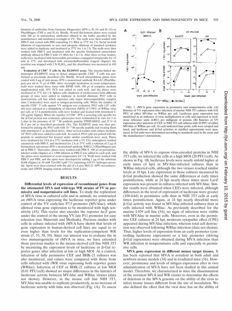
Differential levels of expression of recombinant genes fromthe attenuated MVA and wild-type WR strains of VV in per-missive and nonpermissive cell lines. To study the replicationof MVA virus in cultured cells and in animals, we constructedan rMVA virus expressing the luciferase reporter gene undercontrol of the VV early/late P7.5 promoter (MVAluc), whichallowed virus gene expression to be monitored with high sen-sitivity (43). This vector also encodes the reporter lacZ geneunder the control of the strong VV late P11 promoter for easyselection (see Materials and Methods). Previous studies withcells in culture infected with rMVA have shown that levels ofgene expression in human-derived cell lines are equal to oreven higher than levels for the replication-competent WRstrain (5, 53, 58, 59). Since our interest was to evaluate the invivo immunogenicity of rMVA in mice, we have extendedthose previous studies to the mouse-derived cell line NIH 3T3by measuring the expression levels of luciferase or b-Gal re-porter genes after infection at low or high MOI. As a control,infection of fully permissive CEF and BHK-21 cultures wasalso monitored, and values were compared with those fromcells infected with rWR virus expressing both reporter genes(WRluc). Infection of CEF or BHK-21 cultures at low MOI(0.01 PFU/cell) showed no major differences in the kinetics ofluciferase activity between MVAluc and WRluc viruses (datanot shown). However, in the mouse cell line NIH 3T3,MVAluc was unable to replicate productively, as no increase ofluciferase activity with time was observed (Fig. 1A). To assess
the ability of MVA to express virus-encoded proteins in NIH3T3 cells, we infected the cells at a high MOI (20 PFU/cell). Asshown in Fig. 1B, luciferase levels were nearly sixfold higher atearly times (4 hpi) in MVAluc-infected cultures than inWRluc-infected cells, although the two viruses reached similarlevels at 18 hpi. Late expression in those cultures measured byb-Gal production showed the same differences at early timespostinfection, while at 24 hpi nearly three times more LacZexpression was present in cultures infected with MVAluc. Sim-ilar results were obtained when CEFs were infected, althoughdifferences in the level of expression of luciferase were greater(100-fold) in permissive cells than in NIH 3T3 cells at earlytimes postinfection. Again, at 24 hpi nearly threefold moreb-Gal activity was found in MVAluc-infected cultures than incells infected with WRluc. As previously described for themurine L929 cell line (54), no signs of infection were visiblewith MVAluc in murine cells. Moreover, even in the permis-sive CEF cultures at 24 hpi, moderate cytopathic effect (CPE)appeared during MVAluc infection, whereas total cell destruc-tion was observed following WRluc infection (data not shown).Thus, higher levels of expression from an early promoter (con-trolling luciferase expression) or a late promoter (drivingb-Gal expression) were obtained during MVA infection thanWR infection in nonpermissive cells and especially in permis-sive cells.
MVA gene expression in different mouse target tissues. Ithas been reported that MVA is avirulent in both adult andnewborn mouse models (34) and in irradiated mice (56). How-ever, persistence and levels of antigen expression after in vivoadministration of MVA have not been studied in this animalmodel. Therefore, we characterized in mice the disseminationof the avirulent MVA and WR viruses to determine the effectsof deletions in the MVA genome on the ability of the virus toinfect mouse tissues different from the site of inoculation. Wealso defined the effect that the viral dose has on the ability of
FIG. 1. rMVA gene expression in permissive and nonpermissive cells. (A)Kinetics of VV expression after infection of murine NIH 3T3 cultures with 0.01PFU of either MVAluc or WRluc per cell. Luciferase gene expression wasmonitored as an indicator of virus multiplication in cells and expressed as lucif-erase reference units (LRU) per milligram of protein. (B) Kinetics of VVexpression after infection of CEF or NIH 3T3 cell cultures with 20 PFU of eitherMVAluc or WRluc per cell. At each indicated time point, cells were scraped andlysed, and luciferase and b-Gal activities in clarified supernatants were mea-sured. b-Gal units were determined according to standards used in the assay andthe manufacturer’s instructions.
VOL. 74, 2000 MVA GENE EXPRESSION AND IMMUNOGENICITY IN MICE 925
on February 10, 2015 by guest
http://jvi.asm.org/
Dow
nloaded from
MVA to reach target tissues, as well as the level of expressionof foreign MVA-encoded reporter genes.
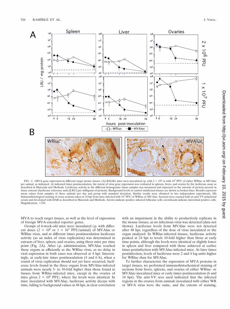
Groups of 6-week-old mice were inoculated i.p. with differ-ent doses (2 3 106 or 1 3 107 PFU/animal) of MVAluc orWRluc virus, and at different times postinoculation luciferaseactivity (as an index of virus replication) was determined inextracts of liver, spleen, and ovaries, using three mice per timepoint (Fig. 2A). After i.p. administration, MVAluc reachedthese organs as efficiently as the WRluc virus, as no delay inviral expression in both cases was observed at 4 hpi. Interest-ingly, at early-late times postinoculation (4 and 6 h), when around of virus replication should not yet have occurred, lucif-erase levels found in the three organs from MVAluc-infectedanimals were nearly 5- to 10-fold higher than those found intissues from WRluc-infected mice, except in the ovaries ofmice given 2 3 106 PFU, where the levels were identical. Inmice inoculated with MVAluc, luciferase activity decays withtime, falling to background values at 48 hpi, in clear correlation
with an impairment in the ability to productively replicate inthe mouse tissues, as no infectious virus was detected (data notshown). Luciferase levels from MVAluc were not detectedafter 48 hpi, regardless of the dose of virus inoculated or theorgan analyzed. In WRluc-infected tissues, luciferase activitypeaked at 24 hpi to levels 10-fold higher than those at earlytime points, although the levels were identical or slightly lowerin spleen and liver compared with those achieved at earliertimes postinfection with MVAluc-infected mice. At later timespostinfection, levels of luciferase were 2 and 4 log units higherfor WRluc than for MVAluc.
To further characterize the expression of MVA proteins intarget tissues, we performed immunohistochemical staining ofsections from livers, spleens, and ovaries of either WRluc- orMVAluc-inoculated mice at early times postinoculation (6 and18 hpi). The anti-VV sera used indicated that the infectedregions in the ovaries from animals inoculated with either WRor MVA virus were the same, and the extents of staining,
FIG. 2. rMVA gene expression in different target mouse tissues. (A) BALB/c mice were inoculated i.p. with 2 3 106 or with 107 PFU of either WRluc or MVAlucper animal, as indicated. At indicated times postinoculation, the extent of virus gene expression was evaluated in spleens, livers, and ovaries by the luciferase assay asdescribed in Materials and Methods. Luciferase activity in the different homogenate tissue samples was measured and expressed as the amount of protein present intissue extracts (luciferase reference units [LRU] per milligram of protein). Background levels in control uninfected tissues are shown as broken lines. Results representmean values from samples of three animals per day and group with standard deviation. Similar results were obtained in two independent experiments. (B)Immunohistological staining of ovary sections taken at 18 hpi from mice infected with 107 PFU of WRluc or MVAluc. Sections were reacted with an anti-VV polyclonalserum and developed with DAB as described in Materials and Methods. Arrows indicate positive infected follicular cells; arrowheads indicate interstitial positive cells.Magnification, 3250.
926 RAMIREZ ET AL. J. VIROL.
on February 10, 2015 by guest
http://jvi.asm.org/
Dow
nloaded from
indicative of the invasiveness of each virus, were nearly iden-tical. Areas of the ovaries positive for anti-VV sera were thefollicular cells of follicles at different stages of differentiation(Fig. 2B), cells at the corpus luteum, interstitial cells, and cellsin the germinal epithelium (not shown). Accessory fat cellswere also stained, and scattered cells of the columnar epithe-lium of the uterus appeared positive for VV antigens. Only afew positive cells per slice were present in liver and spleensections, reflecting the great differences in luciferase activitymeasured in those tissues compared to the ovaries.
The results presented in Fig. 2 revealed that MVAluc caninfect different VV target tissues following i.p. inoculation,reaching the tissues at the same time as the WR virus andexpressing viral genes at early times more efficiently than thewild-type virus. At a dose of 107 PFU, the levels of proteinexpression at early times after inoculation with MVAluc werenearly 10-fold higher in target tissues than in replication-com-petent WRluc virus-infected organs.
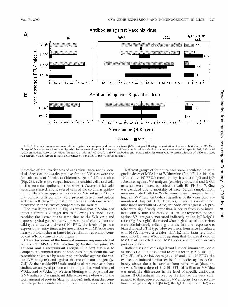
Characterization of the humoral immune response elicitedin mice after MVA or WR infection. (i) Antibodies against VVantigens and a recombinant antigen. Our next aim was tocompare the humoral immune responses induced with the tworecombinant viruses by measuring antibodies against the vec-tor (VV antigens) and against the recombinant antigen (b-Gal). As the particle/PFU ratio could be of importance in thesestudies, we analyzed protein content in purified viral stocks ofWRluc and MVAluc by Western blotting with polyclonal an-ti-VV antigens. No significant differences were observed in thetotal amount of protein (data not shown), indicating that com-parable particle numbers were present in the two virus stocks.
Different groups of four mice each were inoculated i.p. withgraded doses of MVAluc or WRluc virus (2 3 106, 1 3 107, 5 3107, and 1 3 108 PFU/mouse); 14 days later, total IgG and IgGsubclasses against VV antigens (envelope proteins) and b-Galin serum were measured. Infection with 108 PFU of WRlucwas excluded due to mortality of mice. Serum samples frommice inoculated with the WRluc virus showed comparable andhigh anti-VV IgG antibodies regardless of the virus dose ad-ministered (Fig. 3A, left). However, in serum samples frommice inoculated with MVAluc, antibody levels against VV pro-teins were significantly lower than in serum from mice inocu-lated with WRluc. The ratio of Th1 to Th2 responses inducedagainst VV antigens, measured indirectly by the IgG2a/IgG1ratio (Fig. 3A, right), decreased when high doses of either viruswere administered, indicating that the immune response wasbiased toward a Th2 type. However, sera from mice inoculatedwith MVA showed a greater Th1/Th2 ratio than sera frommice infected with WRluc, suggesting that the initial dose ofMVA had less effect since MVA does not replicate in vivopostinoculation.
Both viruses induced a significant humoral immune responseagainst b-Gal at a dose equal to or higher than 5 3 107 PFU(Fig. 3B, left). At low doses (2 3 106 and 1 3 107 PFU), thetwo vectors induced similar levels of antibodies against b-Gal,barely above those in samples from naive mice (data notshown). When a dose of 5 3 107 PFU of WRluc or MVAlucwas used, the differences in the level of specific antibodiesagainst b-Gal antigen induced by the two vectors were com-parable to those observed against VV antigens. For the recom-binant antigen analyzed (b-Gal), the IgG1 response (Th2) was
FIG. 3. Humoral immune response elicited against VV antigens and the recombinant b-Gal antigen following immunization of mice with WRluc or MVAluc.Groups of four mice were inoculated i.p. with the indicated doses of virus vectors; 14 days later, blood was obtained and sera were tested for specific IgG, IgG1, andIgG2a antibodies. Absorbance values (measured at 492 nm) of specific anti-VV antibodies and b-Gal antibodies correspond to serum dilutions of 1/400 and 1/50,respectively. Values represent mean absorbances of triplicates of pooled serum samples.
VOL. 74, 2000 MVA GENE EXPRESSION AND IMMUNOGENICITY IN MICE 927
on February 10, 2015 by guest
http://jvi.asm.org/
Dow
nloaded from
more pronounced than with VV antigens. The quantity andquality of the humoral immune response observed with a doseof 108 PFU of MVA were similar to those seen with 5 3 107
PFU of the WRluc virus.The results in Fig. 3 indicate that the extent of the humoral
immune response induced with WR and MVA viruses is de-pendent on the viral dose administered; only high doses ofMVA elicited a significant anti-VV antigen humoral response.There was also a polarization of the immune response towarda Th2 type with increased doses of both viruses, although amore pronounced Th1 type was elicited by MVA at any dose.However, higher doses of MVAluc were required to generatean antibody response against b-Gal comparable to that elicitedby WRluc.
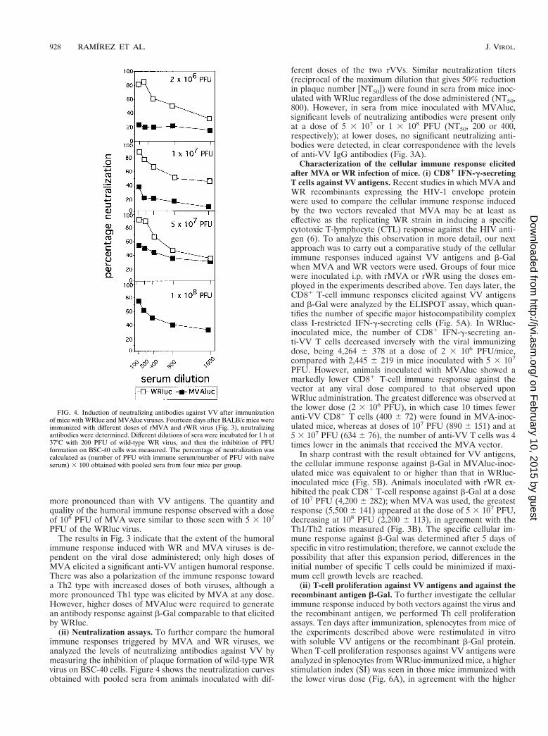
(ii) Neutralization assays. To further compare the humoralimmune responses triggered by MVA and WR viruses, weanalyzed the levels of neutralizing antibodies against VV bymeasuring the inhibition of plaque formation of wild-type WRvirus on BSC-40 cells. Figure 4 shows the neutralization curvesobtained with pooled sera from animals inoculated with dif-
ferent doses of the two rVVs. Similar neutralization titers(reciprocal of the maximum dilution that gives 50% reductionin plaque number [NT50]) were found in sera from mice inoc-ulated with WRluc regardless of the dose administered (NT50,800). However, in sera from mice inoculated with MVAluc,significant levels of neutralizing antibodies were present onlyat a dose of 5 3 107 or 1 3 108 PFU (NT50, 200 or 400,respectively); at lower doses, no significant neutralizing anti-bodies were detected, in clear correspondence with the levelsof anti-VV IgG antibodies (Fig. 3A).
Characterization of the cellular immune response elicitedafter MVA or WR infection of mice. (i) CD81 IFN-g-secretingT cells against VV antigens. Recent studies in which MVA andWR recombinants expressing the HIV-1 envelope proteinwere used to compare the cellular immune response inducedby the two vectors revealed that MVA may be at least aseffective as the replicating WR strain in inducing a specificcytotoxic T-lymphocyte (CTL) response against the HIV anti-gen (6). To analyze this observation in more detail, our nextapproach was to carry out a comparative study of the cellularimmune responses induced against VV antigens and b-Galwhen MVA and WR vectors were used. Groups of four micewere inoculated i.p. with rMVA or rWR using the doses em-ployed in the experiments described above. Ten days later, theCD81 T-cell immune responses elicited against VV antigensand b-Gal were analyzed by the ELISPOT assay, which quan-tifies the number of specific major histocompatibility complexclass I-restricted IFN-g-secreting cells (Fig. 5A). In WRluc-inoculated mice, the number of CD81 IFN-g-secreting an-ti-VV T cells decreased inversely with the viral immunizingdose, being 4,264 6 378 at a dose of 2 3 106 PFU/mice,compared with 2,445 6 219 in mice inoculated with 5 3 107
PFU. However, animals inoculated with MVAluc showed amarkedly lower CD81 T-cell immune response against thevector at any viral dose compared to that observed uponWRluc administration. The greatest difference was observed atthe lower dose (2 3 106 PFU), in which case 10 times feweranti-VV CD81 T cells (400 6 72) were found in MVA-inoc-ulated mice, whereas at doses of 107 PFU (890 6 151) and at5 3 107 PFU (634 6 76), the number of anti-VV T cells was 4times lower in the animals that received the MVA vector.
In sharp contrast with the result obtained for VV antigens,the cellular immune response against b-Gal in MVAluc-inoc-ulated mice was equivalent to or higher than that in WRluc-inoculated mice (Fig. 5B). Animals inoculated with rWR ex-hibited the peak CD81 T-cell response against b-Gal at a doseof 107 PFU (4,200 6 282); when MVA was used, the greatestresponse (5,500 6 141) appeared at the dose of 5 3 107 PFU,decreasing at 108 PFU (2,200 6 113), in agreement with theTh1/Th2 ratios measured (Fig. 3B). The specific cellular im-mune response against b-Gal was determined after 5 days ofspecific in vitro restimulation; therefore, we cannot exclude thepossibility that after this expansion period, differences in theinitial number of specific T cells could be minimized if maxi-mum cell growth levels are reached.
(ii) T-cell proliferation against VV antigens and against therecombinant antigen b-Gal. To further investigate the cellularimmune response induced by both vectors against the virus andthe recombinant antigen, we performed Th cell proliferationassays. Ten days after immunization, splenocytes from mice ofthe experiments described above were restimulated in vitrowith soluble VV antigens or the recombinant b-Gal protein.When T-cell proliferation responses against VV antigens wereanalyzed in splenocytes from WRluc-immunized mice, a higherstimulation index (SI) was seen in those mice immunized withthe lower virus dose (Fig. 6A), in agreement with the higher
FIG. 4. Induction of neutralizing antibodies against VV after immunizationof mice with WRluc and MVAluc viruses. Fourteen days after BALB/c mice wereimmunized with different doses of rMVA and rWR virus (Fig. 3), neutralizingantibodies were determined. Different dilutions of sera were incubated for 1 h at37°C with 200 PFU of wild-type WR virus, and then the inhibition of PFUformation on BSC-40 cells was measured. The percentage of neutralization wascalculated as (number of PFU with immune serum/number of PFU with naiveserum) 3 100 obtained with pooled sera from four mice per group.
928 RAMIREZ ET AL. J. VIROL.
on February 10, 2015 by guest
http://jvi.asm.org/
Dow
nloaded from
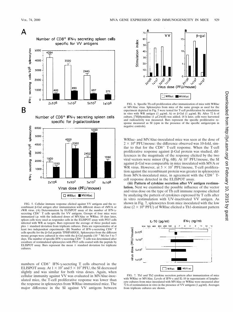
numbers of CD81 IFN-g-secreting T cells observed in theELISPOT assay. At 1 3 107 and 5 3 107 PFU, the SI decreasedslightly and was similar for both virus doses. Again, whencellular immunity against VV was evaluated in MVAluc-inoc-ulated mice, the T-cell proliferative response was lower thanthe response in splenocytes from WRluc-immunized mice. Themajor difference in the SI against VV antigens between
WRluc- and MVAluc-inoculated mice was seen at the dose of2 3 106 PFU/mouse: the difference observed was 10-fold, sim-ilar to that for the CD81 T-cell response. When the T-cellproliferative response against b-Gal protein was studied, dif-ferences in the magnitude of the response elicited by the twoviral vectors were minor (Fig. 6B). At 107 PFU/mouse, the SIagainst b-Gal was comparable in mice inoculated with MVA orWR virus. However, at 5 3 107 PFU/mouse, T-cell prolifera-tion against the recombinant protein was greater in splenocytesfrom MVA-inoculated mice, in agreement with the CD81 T-cell response detected in the ELISPOT assay.
(iii) Pattern of cytokine secretion after VV antigen restimu-lation. Next we examined the possible influence of the vectorand virus dose on the type of Th cell immune response elicitedby analyzing the pattern of cytokines expressed by T cells afterin vitro restimulation with UV-inactivated VV antigen. Asshown in Fig. 7, splenocytes from mice inoculated with the lowdose (2 3 106 PFU) of WRluc elicited a Th1-dominant pattern
FIG. 5. Cellular immune response elicited against VV antigens and the re-combinant b-Gal antigen after immunization with different doses of rMVA orrWR virus. (A) Determination by ELISPOT assay of the number of IFN-g-secreting CD81 T cells specific for VV antigens. Groups of four mice wereimmunized i.p. with the indicated doses of MVAluc or WRluc; 10 days later,spleen cells were used as responder cells in the ELISPOT assay with P815 cellsinfected with WR as targets. Bars represent the average of three pooled sam-ples 6 standard deviation from triplicate cultures. Data are representative of atleast two independent experiments. (B) Number of IFN-g-secreting CD81 Tcells specific for the b-Gal peptide TPHPARIGL. Splenocytes from the differentmouse groups were cultured in vitro with the b-Gal peptide (1025 M) for 5 to 7days. The number of specific IFN-g-secreting CD81 T cells was determined aftercoculture of restimulated splenocytes with P815 cells coated with the peptide byELISPOT assay. Bars represent the mean 6 standard deviation for triplicatecultures.
FIG. 6. Specific Th cell proliferation after immunization of mice with WRlucor MVAluc virus. Splenocytes from mice of the same groups as used for theexperiment depicted in Fig. 5 were tested for T-cell proliferation by stimulationin vitro with WR antigen (1 mg/ml; A) or b-Gal (1 mg/ml; B). After 72 h ofculture, [3H]thymidine (1 mCi/well) was added; 18 h later, cells were harvestedand radioactivity was measured. Bars represent the specific proliferative re-sponse measured as SI (cpm in the presence of the specific antigen/cpm innegative controls).
FIG. 7. Th1 and Th2 cytokine secretion pattern after immunization of micewith WRluc or MVAluc. Levels of IFN-g and IL-10 in supernatants of lympho-cyte cultures from mice inoculated with MVAluc or WRluc were measured after72 h of restimulation in vitro in the presence of VV antigens (1 mg/ml). Averagesfrom triplicate cultures are shown.
VOL. 74, 2000 MVA GENE EXPRESSION AND IMMUNOGENICITY IN MICE 929
on February 10, 2015 by guest
http://jvi.asm.org/
Dow
nloaded from
of cytokines (high levels of IFN-g and low levels of IL-10),whereas at this dose in MVAluc-inoculated mice the levelswere not significant. At 107 PFU, the levels of IFN-g secretedby splenocytes from WRluc-inoculated mice were maintained,while IL-10 levels increased. The fact that in this group of micethe immune response was biased toward the Th2 type corre-lated with the lower specific CD81 T-cell response observed inthe ELISPOT assay (Fig. 5A). At an MVA dose of 107 PFU/mouse, the pattern of cytokines secreted against VV antigenscorresponded to a Th1-dominant type. At higher doses ofMVA (5 3 107 and 1 3 108 PFU), the levels of IFN-g de-creased (Fig. 7) in correlation with the lower number of an-ti-VV CD81 IFN-g-secreting T cells (Fig. 5).
Characterization of the cellular immune response elicitedupon MVA or WR inoculation showed that the CMI inducedagainst the vector was stronger in those mice that received theWR recombinant that in those animals inoculated with theattenuated MVA vector. Nevertheless, the MVA vector wascapable of inducing a similar or even greater (at a dose of 5 3107 PFU/mouse) specific cellular immune response against theb-Gal antigen, as evaluated by CD81 T-cell ELISPOT and Thcell in vitro restimulation assays.
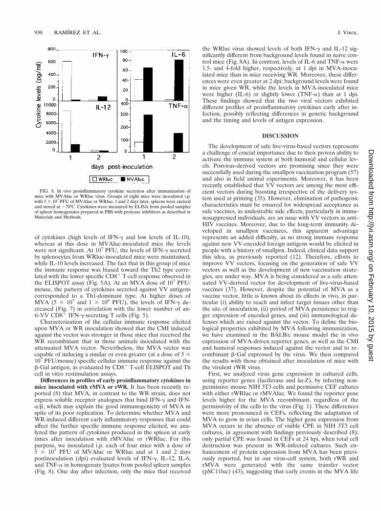
Differences in profiles of early proinflammatory cytokines inmice inoculated with rMVA or rWR. It has been recently re-ported (8) that MVA, in contrast to the WR strain, does notexpress soluble receptor analogues that bind IFN-g and IFN-a/b, which may explain the good immunogenicity of MVA inspite of its poor replication. To determine whether MVA andWR induced different early inflammatory responses that couldaffect the further specific immune response elicited, we ana-lyzed the pattern of cytokines produced in the spleen at earlytimes after inoculation with rMVAluc or rWRluc. For thispurpose, we inoculated i.p. each of four mice with a dose of5 3 107 PFU of MVAluc or WRluc and at 1 and 2 dayspostinoculation (dpi) evaluated levels of IFN-g, IL-12, IL-6,and TNF-a in homogenate lysates from pooled spleen samples(Fig. 8). One day after infection, only the mice that received
the WRluc virus showed levels of both IFN-g and IL-12 sig-nificantly different from background levels found in naive con-trol mice (Fig. 8A). In contrast, levels of IL-6 and TNF-a were1.5- and 4-fold higher, respectively, at 1 dpi in MVA-inocu-lated mice than in mice receiving WR. Moreover, these differ-ences were even greater at 2 dpi: background levels were foundin mice given WR, while the levels in MVA-inoculated micewere higher (IL-6) or slightly lower (TNF-a) than at 1 dpi.These findings showed that the two viral vectors exhibiteddifferent profiles of proinflammatory cytokines early after in-fection, possibly reflecting differences in genetic backgroundand the timing and levels of antigen expression.
DISCUSSION
The development of safe live-virus-based vectors representsa challenge of crucial importance due to their proven ability toactivate the immune system at both humoral and cellular lev-els. Poxvirus-derived vectors are promising since they weresuccessfully used during the smallpox vaccination program (57)and also in field animal experiments. Moreover, it has beenrecently established that VV vectors are among the most effi-cient vectors during boosting irrespective of the delivery sys-tem used at priming (35). However, elimination of pathogeniccharacteristics must be ensured for widespread acceptance assafe vaccines, as undesirable side effects, particularly in immu-nosuppressed individuals, are an issue with VV vectors as anti-HIV vaccines. Moreover, due to the long-term immunity de-veloped in smallpox vaccinees, this apparent advantagerepresents an added difficulty, as no strong immune responseagainst new VV-encoded foreign antigens would be elicited inpeople with a history of smallpox. Indeed, clinical data supportthis idea, as previously reported (12). Therefore, efforts toimprove VV vectors, focusing on the generation of safe VVvectors as well as the development of new vaccination strate-gies, are under way. MVA is being considered as a safe atten-uated VV-derived vector for development of live-virus-basedvaccines (37). However, despite the potential of MVA as avaccine vector, little is known about its effects in vivo, in par-ticular (i) ability to reach and infect target tissues other thanthe site of inoculation, (ii) period of MVA persistence to trig-ger expression of encoded genes, and (iii) immunological de-fenses which are raised against the vector. To define the bio-logical properties exhibited by MVA following immunization,we have examined in the BALB/c mouse model the in vivoexpression of MVA-driven reporter genes, as well as the CMIand humoral responses induced against the vector and to re-combinant b-Gal expressed by the virus. We then comparedthe results with those obtained after inoculation of mice withthe virulent rWR virus.
First, we analyzed virus gene expression in cultured cells,using reporter genes (luciferase and lacZ), by infecting non-permissive mouse NIH 3T3 cells and permissive CEF cultureswith either rWRluc or rMVAluc. We found the reporter genelevels higher for the MVA recombinant, regardless of thepermissivity of the cells to the virus (Fig. 1). These differenceswere more pronounced in CEFs, reflecting the adaptation ofMVA to grow in avian cells. The higher gene expression fromMVA occurs in the absence of visible CPE in NIH 3T3 cellcultures, in agreement with findings previously described (8);only partial CPE was found in CEFs at 24 hpi, when total celldestruction was present in WR-infected cultures. Such en-hancement of protein expression from MVA has been previ-ously reported, but in our virus-cell system, both rWR andrMVA were generated with the same transfer vector(pSC11luc) (43), suggesting that early events in the MVA life
FIG. 8. In vivo proinflammatory cytokine secretion after immunization ofmice with MVAluc or WRluc virus. Groups of eight mice were inoculated i.p.with 5 3 107 PFU of MVAluc or WRluc; 1 and 2 days later, spleens were excisedand stored at 270°C. Cytokines were measured by ELISA from pooled samplesof spleen homogenates prepared in PBS with protease inhibitors as described inMaterials and Methods.
930 RAMIREZ ET AL. J. VIROL.
on February 10, 2015 by guest
http://jvi.asm.org/
Dow
nloaded from
cycle and/or advantages in transcription or translation due tomultiple deletions in functional genes could account for themore efficient gene expression of MVA, which is cell typeindependent.
To monitor MVA infection in vivo, luciferase activity wasmeasured in target tissues (spleen, liver, and ovaries) of miceinfected with MVAluc or WRluc. We chose the i.p. route ofinoculation to achieve a systemic infection that could allow thevirus to reach all potential target tissues. We found thatMVAluc-driven protein expression peaks earlier and lasts for ashorter period of time than during WRluc infection, reachingnearly 10-times-higher luciferase levels at early times in thespleen and liver, in agreement with in vitro data (see above).With a systemic route of virus inoculation (i.p.), luciferaseactivity was found in the same target tissues as upon WRlucinoculation, indicating that MVA has retained the ability toinfect the same tissues as the WR strain. This finding wasfurther corroborated by immunohistochemical labeling of in-fected ovaries with a hyperimmune anti-VV serum: at either 6or 18 hpi, both the intensity of the signal and the infectedregions were undistinguishable between WR- and MVA-in-fected ovaries, further evidence that luciferase activity is agood indicator of viral infection in vivo. The sharp fall inluciferase activity seen in MVAluc-infected tissues at 18 hpi(spleen and liver) and 24 hpi (ovaries) (Fig. 2A) suggests alimitation in viral spread from the initially infected target cells,since replication-competent WR gene expression increases un-til 24 hpi in the spleen and liver and later in ovaries, whenluciferase activity in MVA-infected tissues was close to back-ground levels. Despite the differences in kinetic profiles andwith the exception of infection in ovaries, the maximum levelsof luciferase activity were nearly identical for the two rVVs.These findings demonstrate the main differences in in vivobehavior between the MVA vector and a wild-type vector.
Preexisting immunity to viral antigens is an important con-sideration with respect to developing vaccines based on rVV,as immunity against the heterologous gene product can beprevented when a second dose is delivered in repeated immu-nizations with rVV vectors or in vaccinee against smallpox.Although rMVA and rWR can trigger similar immune re-sponses against recombinant proteins (6, 23, 54), the antiviralimmune parameter previously analyzed in vivo in comparisonwith other VV strains was the antibody titer against the vector(23). Here we present a more exhaustive comparative study ofthe immune response generated against the MVA vector, ad-dressing both the humoral and the cellular immune responses.We found that when animals were immunized with rWR virus,levels of specific anti-VV IgGs were high and nearly identicalat any of the assayed doses, whereas rMVA induced a signif-icant humoral response at only the two higher doses used.However, in the monkey model (23), similar anti-VV antibod-ies were detected in animals immunized by different routeswith MVA or the Wyeth VV strain, indicating that results maybe specific for both the animal model and the route of inocu-lation used. The anti-VV CD81 T-cell immune response de-termined by the ELISPOT assay revealed that at any dose,MVA elicited a CMI response 4- to 10-fold lower than thatwhen WR was inoculated. The latter showed a clear dosedependence, as the numbers of IFN-g-secreting CD81 T cellsdecreased with increasing virus dose significantly (P , 0.01).
The extent of the immune response elicited for specific an-tigens depends primarily on antigen dose (30, 40), although thesustained expression of antigens appears to be even more rel-evant than antigen dose during the generation of a T-cellresponse (26). Although the levels of foreign antigen expres-sion at early times postinfection are the same or even higher in
MVA- than in WR-infected mouse tissues, the short time spanin which expression occurs leads to less sustained viral geneexpression. Moreover, as MVA does not multiply productively,levels of viral antigens are lower during MVA infection, whichcould explain the weaker humoral response elicited against VVantigens. Very large amounts of antigen often result in specificT-cell and sometimes B-cell unresponsiveness, which couldexplain why there is no enhancement in the anti-VV IgG levels,as well as the suppression of the CMI responses raised withincreased WR dose, a phenomenon not observed upon MVAinfection. Hence, viral dose and viral attenuation are factorsaffecting the level and duration of expression of antigen pro-duced, and this in turn determines the final outcome of theimmune response. In this regard, antibody-dependent immu-nological memory and memory CTL precursor frequencieshave different requirements with respect to antigen persis-tence, as described for other viral infections (3, 4, 28).
A meaningful finding was that the lower levels of anti-VVIgG circulating antibodies in MVA-inoculated mice were alsoreflected in lower levels of anti-VV neutralizing antibodies.The low antibody and CMI responses and more specifically thepoor neutralizing antibodies induced by MVA have importantimplications for repeated immunizations, as the appearance ofneutralizing antibodies in sera correlates with immunity to VV(10). Thus, low neutralizing titers against the vector might leadto an increased antibody response to a foreign antigen afterrepeated immunizations with an rVV based on the MVA vec-tor, facilitating multiple inoculations with poxvirus vectors. In-deed, previous data obtained with the SIV-macaque modelshowed an increase in the SIV-specific antibody titer afterrepeated rMVA injections (23) as well as in the specific CTLresponse (50).
The anti-b-Gal humoral immune response triggered by rVVcorrelated with the anti-VV response, as those mouse groupsthat showed higher anti-VV antibodies also had higher levelsof anti-b-Gal antibodies. Similar observations have been de-scribed for other attenuated VV strains (16, 24, 29, 51), inwhich lower anti-VV antibodies correlated with lower levels ofantibodies to specific recombinant proteins, which are directlymodulated by the extent of replication of the viral vector. Thespecific anti-b-Gal CMI was found to be lower than thatagainst VV antigens, as no measurable counts were obtained infresh ELISPOT assays (data not shown). Interestingly, whenthe ELISPOT assay was performed after 5 days of specific invitro stimulation, we found that despite the relatively weakanti-VV immune response elicited, MVA induced a CMIagainst the foreign b-Gal product equal to that induced byWR. Furthermore, it was twofold higher than in WR-infectedmice at the dose that elicited the strongest response (5 3 107
PFU) (P , 0.01). It appears that the CMI against b-Gal fol-lows a response with a peak that is achieved at higher doses forMVA than for WR inoculation and that maximum CMI levelscan be obtained during infection with the MVA vector.
Concerning the skewing of the immune responses inducedby the two viral vectors, determined by different methods (Fig.3 and 6), our findings indicate a Th1-to-Th2 polarization as theviral dose increases. It is well established that both the affinityand the amount of the antigen play a major role in activatinga particular arm of the immune system (38) and modulatingthe strength of T-cell signaling, which in turn can affect dra-matically the balance of Th1 and Th2 subsets (9). It is note-worthy that at all doses assayed, the strength of the Th1 re-sponse is higher during MVA immunization, probably due tothe low levels of circulating viral antigens, as no mature par-ticles are produced, whereas during WR infection, viral parti-cles produced increase the initial viral input.
VOL. 74, 2000 MVA GENE EXPRESSION AND IMMUNOGENICITY IN MICE 931
on February 10, 2015 by guest
http://jvi.asm.org/
Dow
nloaded from
We also studied the induction of proinflammatory cytokinesat early times postinfection during MVA inoculation in com-parison with WR inoculation. Here we show that MVA infec-tion induces higher splenic levels of IL-6 and TNF-a cytokinesat 1 and 2 dpi than during WR infection. Our observations arein concordance with other studies in which after mucosal MVAor WR immunization, higher levels of IL-6 and TNF-a werefound for monocyte cells from Peyer’s patches of MVA-inoc-ulated mice upon lipopolysaccharide stimulation (5). In con-trast, levels of IFN-g and IL-12 in the same spleen sampleswere higher than in samples from WR-inoculated mice, with apeak at 24 hpi. These results indicate that MVA and WRviruses induced different profiles of proinflammatory cyto-kines, which may be relevant in modulating the ultimate spe-cific immune response elicited. Indeed, different viral infec-tions induce distinct earlier cytokine responses; for example,mouse cytomegalovirus (MCMV) induces the production ofIL-6, a key mediator of induction of glucocorticoids that cansuppress multiple immune functions including CMI (27). Ac-cordingly, during MCMV infection, the induced glucocorticoidresponse seems to be responsible for the relatively weak T-cellresponse (46). In our study, the lower antiviral cellular immuneresponse induced by MVA inoculation might be explained inpart by the differential IL-6 cytokine induction and its possibleinfluence on glucocorticoid production, which may have adownstream immune effect on the antiviral response. As MVAdoes not express the soluble receptor for IFN-a that is encodedby WR (52), it is tempting to speculate that deletion of thisgene could play a role in the inflammatory response induced.In this regard, MVA induces high levels of IFN-a/b in primaryhuman cells (8). If it does so also in vivo, it could account forthe lower IFN-g and IL-12 levels found in MVA-infected micein comparison with those given WR, since IFN-a/b has aninhibitory action on IL-12 and IFN-g production in vivo and invitro during lymphocytic choriomeningitis virus and MCMVinfection (13).
In conclusion, in this study we have demonstrated the be-havior of MVA in mice, in particular its ability to expressantigens in target organs after i.p. inoculation and the differ-ences in the immune responses elicited in comparison with thevirulent WR strain. The most significant findings are the lowantivirus immune responses elicited at both humoral and cel-lular levels and the low neutralization titers achieved, whichmight be of relevance in future vaccination schemes. However,the CMI raised against a late foreign antigen (b-Gal) was atcertain viral doses higher than that triggered by a WR vector;also, antibodies to b-Gal were not very different for either virusat the optimal dose. Moreover, the patterns of proinflamma-tory cytokines induced early upon viral infection were differentfor MVA and WR: higher levels of IL-6 and TNF-a and nosignificant IL-12 and IFN-g were induced after MVA inocula-tion, while these two cytokines were induced during WR in-fection. Overall, the findings presented here may be relevant tothe rational design of MVA-based vaccines, as benefits derivedfrom the low immunogenicity of the vector without affectingthe immune response to foreign antigens can be applied infuture vaccination protocols using VV-derived vectors.
ACKNOWLEDGMENTS
J.C.R. and M.M.G. contributed equally to this work.We thank Antonio Alcamı and Margarita del Val for critical reading
of the manuscript. We are also indebted to Dolores Rodriguez forexpert advice about handling of MVA at early stages of this work. Theexcellent technical assistance of M. Victoria Jimenez is also acknowl-edged.
This work was supported by grants 08.6/0020/97 from the Comu-
nidad Autonoma de Madrid, SAF98-0056 from the Comision Inter-ministerial de Ciencia y Tecnologıa, Spain, and BIO4-CT98-0456 fromthe European Union. J.C.R. and M.M.G. are recipients of postdoc-toral fellowships from the Comunidad Autonoma de Madrid, Spain,and Consejo Nacional de Investigaciones Cientıficas y Tecnicas deArgentina, respectively.
REFERENCES
1. Altenburger, W., C.-P. Suter, and J. Altenburger. 1989. Partial deletion ofthe human host range in the attenuated vaccinia virus MVA. Arch. Virol.105:15–27.
2. Antoine, G., F. Scheiflinger, F. Dorner, and F. G. Falkner. 1998. The com-plete genomic sequence of the modified vaccinia Ankara strain: comparisonwith other Orthopoxviruses. Virology 244:365–396.
3. Bachmann, M. F., B. Odermatt, H. Hengartner, and R. M. Zinkernagel.1996. Induction of long-lived germinal centers associated with persistingantigen after viral infection. J. Exp. Med. 183:2259–2269.
4. Bachmann, M. F., T. M. Kundig, H. Hengartner, and R. M. Zinkernagel.1997. Protection against immunopathological consequences of a viral infec-tion by activated but not resting cytotoxic T cells: T cell memory without“memory T cells”? Proc. Natl. Acad. Sci. USA 94:640–645.
5. Belyakov, I. M., L. S. Wyatt, J. D. Ahlers, P. Earl, C. D. Pendleton, B. L.Kelsall, W. Strober, B. Moss, and J. A. Berzofsky. 1998. Induction of amucosal cytotoxic T-lymphocyte response by intrarectal immunization with areplication-deficient recombinant vaccinia virus expressing human immuno-deficiency virus 89.6 envelope protein. J. Virol. 72:8264–8272.
6. Belyakov, I. M., B. Moss, W. Strober, and J. A. Berzofsky. 1999. Mucosalvaccination overcomes the barrier to recombinant vaccinia immunizationcaused by preexisting poxvirus immunity. Proc. Natl. Acad. Sci. USA 96:4512–4517.
7. Bender, B. S., C. A. Rowe, S. F. Taylor, L. S. Wyatt, B. Moss, and P. A. Small,Jr. 1996. Oral immunization with a replication-deficient recombinant vac-cinia virus protects mice against influenza. J. Virol. 70:6418–6424.
8. Blanchard, T. J., A. Alcamı, P. Andrea, and G. L. Smith. 1998. Modifiedvaccinia virus Ankara undergoes limited replication in human cells and lacksseveral immunomodulatory proteins: implications for use as a human vac-cine. J. Gen. Virol. 79:1159–1167.
9. Bluestone, J. A. 1995. New perspectives of CD28-B7-mediated T cell co-stimulation. Immunity 2:555–559.
10. Buller, R. M., and G. J. Palumbo. 1991. Poxvirus pathogenesis. Microbiol.Rev. 55:80–122.
11. Carroll, M. W., W. Overwijk, R. S. Chamberlain, S. A. Rosenberg, B. Moss,and N. P. Restifo. 1997. Highly attenuated modified vaccinia virus Ankara(MVA) as an effective recombinant vector: a murine tumor model. Vaccine15:387–394.
12. Cooney, E. L., A. C. Collier, P. D. Greenberg, R. W. Coombs, J. Zarling, D. E.Arditti, M. C. Hoffman, S. L. Hu, and L. Corey. 1991. Safety of and immu-nological response to a recombinant vaccinia virus vaccine expressing HIVenvelope glycoprotein. Lancet 337:567–572.
13. Cousens, L. P., J. S. Orange, H. C. Sue, and C. A. Biron. 1997. Interferon-a/binhibition of interleukin 12 and interferon-g production in vitro and endo-genously during viral infection. Proc. Natl. Acad. Sci. USA 94:634–639.
14. Cox, W. I., J. Tartaglia, and E. Paoletti. 1992. Poxvirus recombinants as livevaccines, p. 123–162. In M. M. Binns and G. L. Smith (ed.), Recombinantpoxviruses. CRC Press, Boca Raton, Fla.
15. Dallo, S., and M. Esteban. 1987. Isolation and characterization of attenuatedmutants of vaccinia virus. Virology 159:408–422.
16. Dallo, S., J.-S. Maa, J. R. Rodriguez, D. Rodriguez, and M. Esteban. 1989.Humoral immune response elicited by highly attenuated variants of vacciniavirus and by an attenuated recombinant expressing HIV-1 envelope protein.Virology 173:323–329.
17. Drexler, I., K. Heller, B. Wahren, V. Erfle, and G. Sutter. 1998. Highlyattenuated modified vaccinia virus Ankara replicates in baby hamster kidneycells, a potential host for virus propagation, but not in various human trans-formed and primary cells. J. Gen. Virol. 79:347–352.
18. Durbin, A. P., L. S. Wyatt, J. Siew, B. Moss, and B. R. Murphy. 1998. Theimmunogenicity and efficacy of intranasally or parenterally administeredreplication-deficient vaccinia-parainfluenza virus type 3 recombinants in rhe-sus monkeys. Vaccine 16:1324–1330.
19. Fenner, F. 1989. Risks and benefits of vaccinia vaccine use in the worldwidesmallpox eradication campaign. Res. Virol. 140:465–466.
20. Gherardi, M. M., J. C. Ramirez, D. Rodriguez, J. R. Rodriguez, G. Sano, F.Zavala, and M. Esteban. 1999. IL-12 delivery from recombinant vacciniavirus attenuates the vector and enhances the cellular immune responseagainst HIV-1 env in a dose-dependent manner. J. Immunol. 162:6724–6733.
21. Hanke, T., T. J. Blanchard, J. Schneider, C. M. Hannan, M. Becker, S. C.Gilbert, A. V. S. Hill, G. L. Smith, and A. McMichael. 1998. Enhancement ofMHC class I-restricted peptide-specific T cell induction by a DNA prime/MVA vaccination regime. Vaccine 16:439–445.
22. Hanke, T., T. J. Blanchard, J. Schneider, G. S. Ogg, R. Tan, M. Becker, S. C.Gilbert, A. V. S. Hill, G. L. Smith, and A. McMichael. 1998. Immunogenici-
932 RAMIREZ ET AL. J. VIROL.
on February 10, 2015 by guest
http://jvi.asm.org/
Dow
nloaded from
ties of intravenous and intramuscular administrations of modified vacciniavirus Ankara-based multi-CTL epitope vaccine for human immunodeficiencyvirus type 1 in mice. J. Gen. Virol. 79:83–90.
23. Hirsch, V., T. R. Fuerst, G. Sutter, M. W. Carroll, L. C. Yang, S. Golstein,M. Piatak, W. R. Elkins, W. G. Alvord, D. Montefiori, B. Moss, and J. D.Lifson. 1996. Patterns of viral replication correlate with outcome in simianimmunodeficiency virus (SIV)-infected macaques: effect of prior immuniza-tion with a trivalent SIV vaccine in modified vaccinia virus Ankara. J. Virol.70:3741–3752.
24. Holzer, G. W., G. Remp, G. Antoine, M. Pfleiderer, O. M. Enzersberger, W.Emsenhuber, T. Hammerle, F. Gruber, C. Urban, F. G. Falkner, and F.Dorner. 1999. Highly efficient induction of protective immunity by a vacciniavirus vector defective in late gene expression. J. Virol. 73:4536–4542.
25. Jin, X., D. E. Bauer, S. E. Tuttleton, S. Lewin, A. Gettie, J. Blanchard, C. E.Irwin, J. T. Safrit, J. Mittler, L. Weinberg, L. G. Kostrikis, L. Zhang, A. S.Perelson, and D. D. Ho. 1999. Dramatic rise in plasma viremia after CD81T cell depletion in simian immunodeficiency virus-infected macaques. J. Exp.Med. 189:991–998.
26. Kundig, T. M., A. Shahinian, K. Kawai, H.-W. Mittrucker, E. Sebzda, M. F.Bachmann, T. W. Mak, and P. S. Ohashi. 1996. Duration of TCR stimulationdetermines costimulatory requirement of T cells. Immunity 5:41–52.
27. Kunicka, J. E., M. A. Talle, G. H. Denhart, M. Brown, C. A. Prince, and G.Goldstein. 1993. Immunosuppression by glucocorticoids: inhibition of pro-duction of multiple lymphokines by in vivo administration of dexamethasone.Cell. Immunol. 149:39–49.
28. Lau, L. L., B. D. Jamieson, T. Somasundaram, and R. Ahmed. 1994. Cyto-toxic T-cell memory without antigen. Nature 369:648–652.
29. Lee, M. S., J. M. Roos, L. C. McGuigan, K. A. Smith, N. Cornier, L. K.Cohen, B. E. Roberts, and L. G. Payne. 1992. Molecular attenuation ofvaccinia virus: mutant generation and animal characterization. J. Virol. 66:2617–2630.
30. Liew, F. Y., and C. R. Parish. 1974. Lack of correlation between cell-mediated immunity to the carrier and the carrier-hapten helper effect. J.Exp. Med. 139:779–784.
31. Mackett, M., G. L. Smith, and B. Moss. 1982. Vaccinia virus: a selectableeukaryotic cloning and expression vector. Proc. Natl. Acad. Sci. USA 79:7415–7419.
32. Mayr, A., V. Hoschstein-Mintzel, and H. Stickl. 1975. Abstammung, eigen-schaftenund Verwendung des attenuierten Vaccinia-Stammes MVA. Infec-tion 3:6–16.
33. Mayr, A., H. Stickl, H. K. Muller, K. Denner, and H. Singer. 1978. Thesmallpox vaccination strain MVA: marker, genetic structure, experiencegained with the parenteral vaccination and behaviour in organisms with adebilitated defence mechanism. Zentbl. Backteriol. Hyg. B 167:375–390.
34. Meyer, H., G. Sutter, and A. Mayr. 1991. Mapping of deletions in thegenome of highly attenuated vaccinia virus MVA and their influence onvirulence. J. Gen. Virol. 72:1031–1038.
35. Miyahira, Y., A. Garcıa-Sastre, D. Rodriguez, J. R. Rodriguez, K. Murata,M. Tsuji, P. Palese, M. Esteban, F. Zavala, and R. S. Nussenzweig. 1998.Recombinant viruses expressing a human malaria antigen elicit protectiveimmune CD881 T cell responses in mice. Proc. Natl. Acad. Sci. USA95:3954–3959.
36. Montoya, M., and M. del Val. 1999. Intracellular rate-limiting steps in MHCclass I antigen processing. J. Immunol. 163:1914–1922.
37. Moss, B., M. W. Carroll, L. S. Wyatt, J. R. Bennink, V. Hirsch, S. Golstein,W. R. Elkins, T. R. Fuerst, J. D. Lifson, M. Piatak, N. P. Restifo, W.Overwijk, R. Chamberlain, S. A. Rosenberg, and G. Sutter. 1996. Host rangerestricted non-replicating vaccinia virus vectors as vaccine candidates. Adv.Exp. Med. Biol. 397:7–13.
38. Murray, J. S. 1998. How the MHC selects Th1/Th2 immunity? Immunol.Today 19:157–163.
39. Panicali, D., and E. Paoletti. 1982. Construction of poxvirus as cloningvectors: insertion of the thymidine kinase gene from herpes simplex virusinto the DNA of infectious vaccinia virus. Proc. Natl. Acad. Sci. USA 79:4927–4931.
40. Parish, C. R. 1996. Immune deviation: a historical perspective. Immunol.Cell Biol. 74:449–456.
41. Redfield, R. R., D. C. Wright, W. D. James, T. S. Jones, C. Brown, and D. S.Burke. 1987. Disseminated vaccinia in a military recruit with human immu-nodeficiency virus (HIV) disease. N. Engl. J. Med. 316:673–676.
42. Rodrıguez, D., J. R. Rodrıguez, J. F. Rodrıguez, D. Trauber, and M. Esteban.1989. Highly attenuated vaccinia virus mutants for the generation of saferecombinants viruses. Proc. Natl. Acad. Sci. USA 86:1287–1291.
43. Rodrıguez, J. F., D. Rodrıguez, J. R. Rodrıguez, E. B. McGowan, and M.Esteban. 1988. Expression of the firefly luciferase gene in vaccinia virus: ahighly sensitive gene marker to follow virus dissemination in tissues ofinfected animals. Proc. Natl. Acad. Sci. USA 85:1667–1671.
44. Rodrıguez, J. R., C. Risco, J. L. Carrascosa, M. Esteban, and D. Rodrıguez.1997. Characterization of early stages in vaccinia virus membrane biogenesis:implications of the 21-kilodalton protein and a newly identified 15-kilodaltonenvelope protein. J. Virol. 71:1821–1833.
45. Rowland-Jones, S., R. Tan, and A. J. McMichael. 1997. The role of cellularimmunity in protection against HIV infection. Adv. Immunol. 65:277–346.
46. Ruzek, M. C., A. H. Millert, S. M. Opal, B. D. Pearce, and C. A. Biron. 1997.Characterization of early cytokine responses and an interleukin (IL-6)-de-pendent pathway of endogenous glucocorticoid induction during murinecytomegalovirus infection. J. Exp. Med. 185:1185–1192.
47. Scheleimer, R. P., A. Jacques, H. S. Shin, L. M. Lichstein, and M. Plaut.1984. Inhibition of T cell-mediated cytotoxicity by anti-inflammatory ste-roids. J. Immunol. 132:266–271.
48. Schmitz, J. E., M. J. Kuroda, S. Santra, V. G. Sasseville, M. A. Simon, M. A.Lifton, P. Racz, K. Tenner-Racz, M. Dalesandro, B. J. Scallon, J. Ghrayeb,M. A. Forman, D. C. Montefiori, E. P. Rieber, N. L. Letvin, and K. A.Reimann. 1999. Control of viremia in simian immunodeficiency virus infec-tion by CD81 lymphocytes. Science 283:857–860.
49. Sedegah, M., T. R. Jones, M. Kaur, R. Hedstrom, P. Hobart, J. A. Tine, andS. L. Hoffman. 1998. Boosting with recombinant vaccinia increases immu-nogenicity and protective efficacy of malaria DNA vaccine. Proc. Natl. Acad.Sci. USA 95:7648–7653.
50. Seth, A., I. Ourmanov, M. J. Kuroda, J. E. Schmitz, M. W. Carroll, L. S.Wyatt, B. Moss, M. A. Forman, V. M. Hirsch, and N. L. Letvin. 1998.Recombinant modified vaccinia virus Ankara-simian immunodeficiency virusgag pol elicits cytotoxic T lymphocytes in rhesus monkeys detected by a majorhistocompatibility complex class I/peptide tetramer. Proc. Natl. Acad. Sci.USA 95:10112–10116.
51. Shida, H., Y. Hinuma, M. Hatanaka, M. Morita, M. Kidokoro, K. Suzuki, T.Maruyama, F. Takahashi-Nishimaki, M. Sugimoto, R. Kitamura, T.Miyazawa, and M. Hayami. 1988. Effects and virulences of recombinantvaccinia viruses derived from attenuated strains that express the humanT-cell leukemia virus type I envelope gene. J. Virol. 62:4474–4480.
52. Smith, G. L., J. A. Symons, A. Khanna, A. Vanderplasschen, and A. Alcamı.1997. Vaccinia virus immune evasion. Immunol. Rev. 159:137–154.
53. Sutter, G., and B. Moss. 1992. Nonreplicating vaccinia vector efficientlyexpresses recombinant genes. Proc. Natl. Acad. Sci. USA 89:10847–10851.
54. Sutter, G., L. S. Wyatt, P. L. Foley, J. R. Bennink, and B. Moss. 1994. Arecombinant vector derived from the host-range-restricted and highly atten-uated MVA strain of vaccinia virus stimulates protective immunity in mice toinfluenza virus. Vaccine 12:1032–1040.
55. Taylor, J., R. Weinberg, J. Tartaglia, C. Richardson, G. Alkhatib, D. Breidis,M. Appel, E. Norton, and E. Paoletti. 1992. Nonreplicating viral vectors aspotential vaccines: recombinant canarypox virus expressing measles virusfusion (F) and hemagglutinin (HA) glycoproteins. Virology 187:321–328.
56. Werner, G. T., U. Jentsch, E. Metzger, and J. Simon. 1980. Studies onpoxvirus infection in irradiated animals. Arch. Virol. 64:247–256.
57. World Health Organization. 1980. The global eradication of smallpox. Finalreport of the global commission for the certification of smallpox eradication.History of international public health, no. 4. World Health Organization,Geneva, Switzerland.
58. Wyatt, L., B. Moss, and S. Rozenblatt. 1995. Replication-deficient vacciniavirus encoding bacteriophage T7 RNA polymerase for transient gene ex-pression in mammalian cells. Virology 210:202–205.
59. Wyatt, L., S. T. Shors, B. R. Murphy, and B. Moss. 1996. Development of areplication-deficient recombinant vaccinia virus vaccine effective againstparainfluenza virus 3 infection in an animal model. Vaccine 14:1451–1458.
VOL. 74, 2000 MVA GENE EXPRESSION AND IMMUNOGENICITY IN MICE 933
on February 10, 2015 by guest
http://jvi.asm.org/
Dow
nloaded from
Related Documents