Biology 211 Anatomy & Physiology I Histology of Bone

Biology 211 Anatomy & Physiology I Histology of Bone.
Dec 29, 2015
Welcome message from author
This document is posted to help you gain knowledge. Please leave a comment to let me know what you think about it! Share it to your friends and learn new things together.
Transcript
Biology 211Anatomy & Physiology I
Histology of Bone

Recall:
SYSTEMS are composed of one or more organs, all serving a common function
ORGANS are composed of one or more types of tissues, all serving a common function
TISSUES are composed of one or more types of cells and their products, all serving a common function
Organs of skeletal system = Bones (e.g. femur, ulna, vertebra, mandible)
Like all organs, these contain four types of tissue: Epithelium Connective tissue Nervous tissue Muscular tissue
However: Two types of specialized connective tissues predominate: tissue
each of which is surrounded by dense irregular connective tissue: and
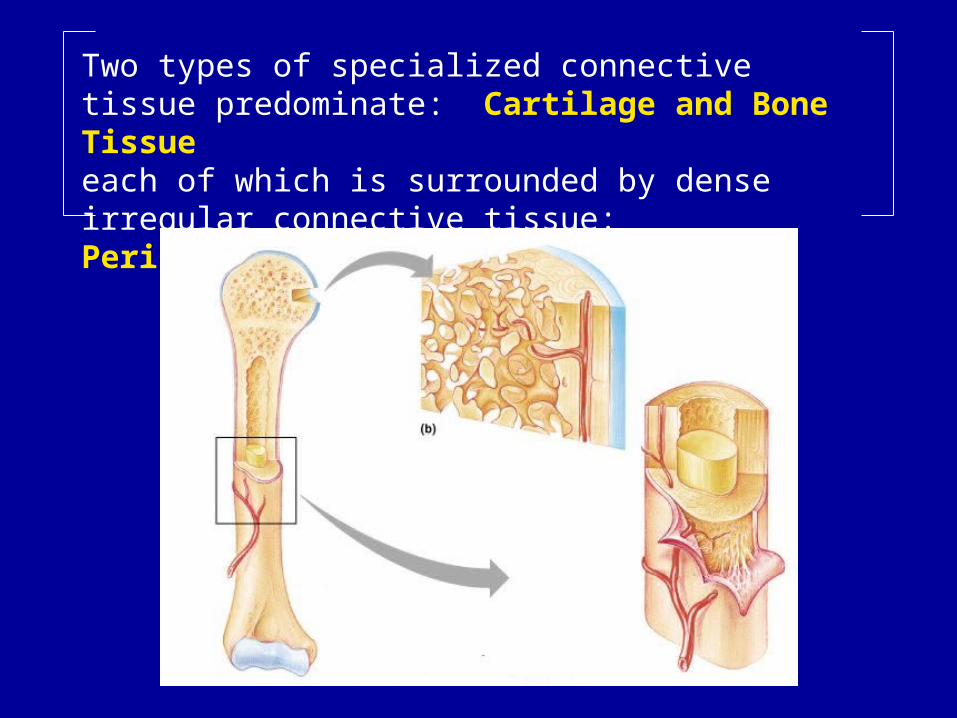
Two types of specialized connective tissue predominate: Cartilage and Bone Tissueeach of which is surrounded by dense irregular connective tissue: Perichondrium and Periosteum

Cartilage: Function = Flexible Support
Costal cartilagesIntervertebral disksExternal earExternal noseLarynxTrachea & bronchi

Cartilage: Function = Flexible Support
Costal cartilagesIntervertebral disksExternal earExternal noseLarynxTrachea & bronchi
Also: Forms embryonic "model" for many bones and: a) Remains on ends of long bones throughout lifeb) Remains at growth plates (epiphyseal plates) of immature growing bones.

Cartilage:
Young cells = Actively forming new extracellular matrix
Mature cells = Maintain and repair extracellular matrix
Extracellular Matrix Ground substance = very firm gel Fibers = collagen, elastic
Chondrocytes located in
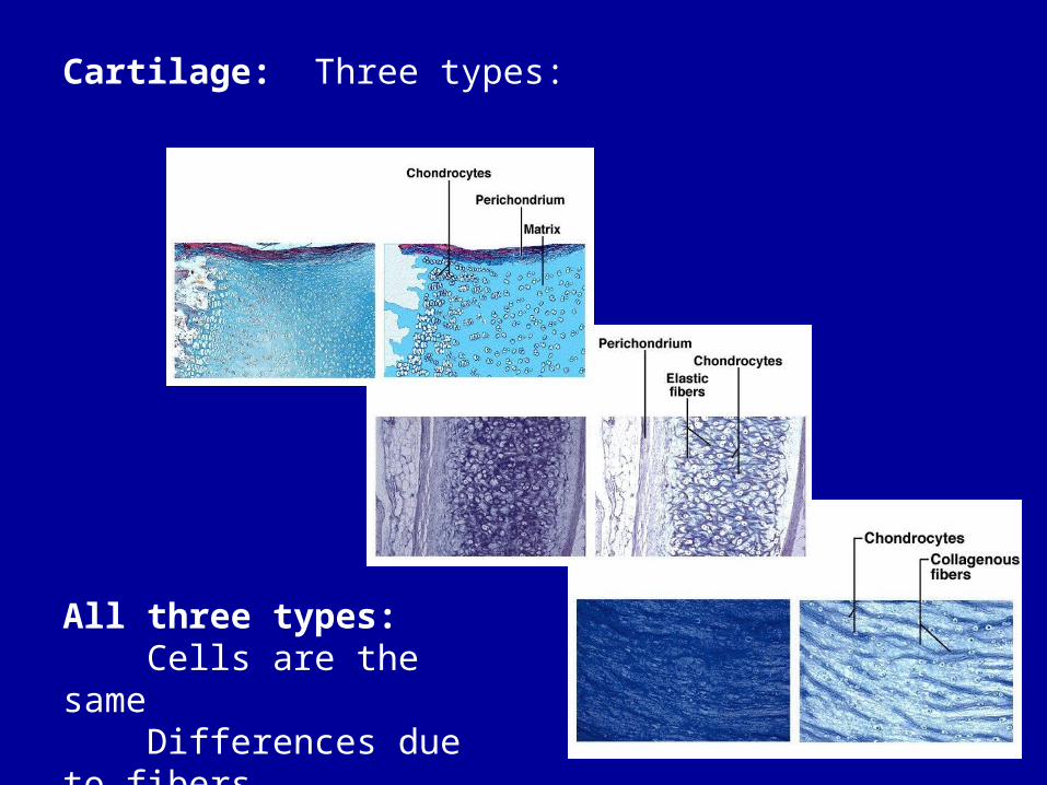
Cartilage: Three types:
All three types: Cells are the same Differences due to fibers
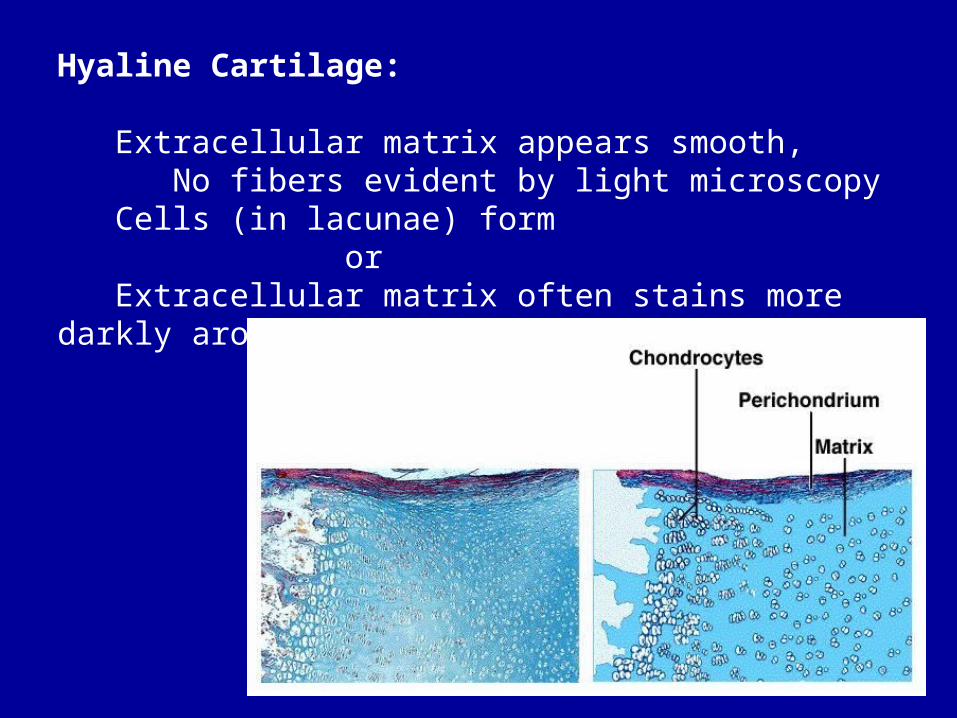
Hyaline Cartilage:
Extracellular matrix appears smooth, No fibers evident by light microscopy Cells (in lacunae) form or Extracellular matrix often stains more darkly around cells
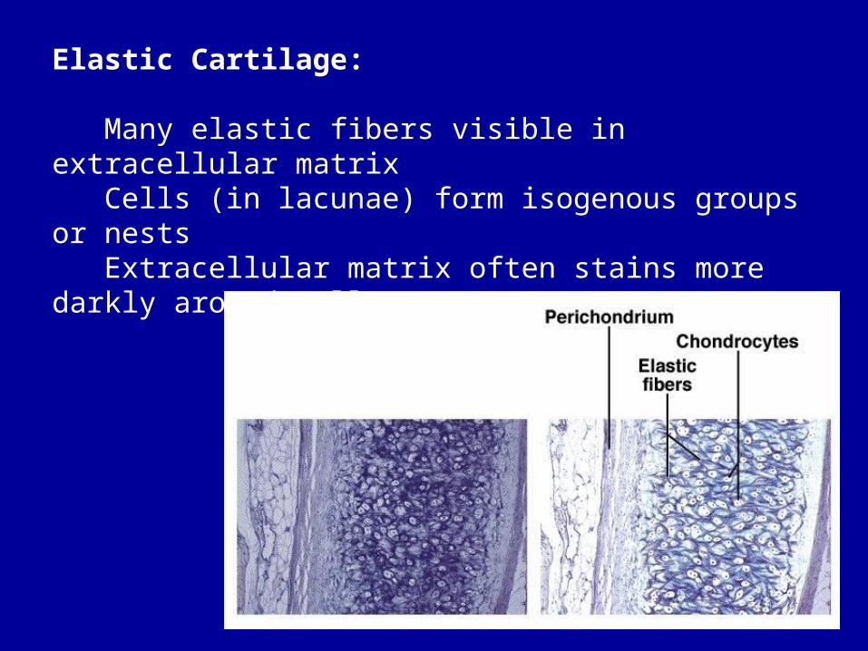
Elastic Cartilage: Many elastic fibers visible in extracellular matrix Cells (in lacunae) form isogenous groups or nests Extracellular matrix often stains more darkly around cells
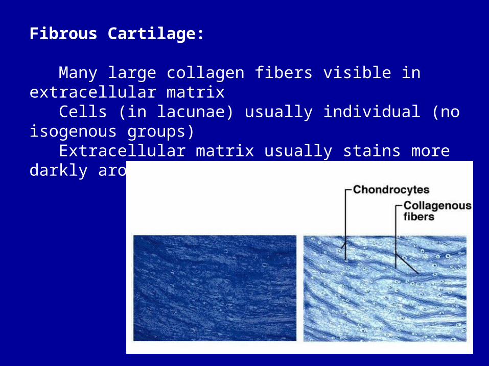
Fibrous Cartilage:
Many large collagen fibers visible in extracellular matrix Cells (in lacunae) usually individual (no isogenous groups) Extracellular matrix usually stains more darkly around cells

Bone Tissue:
Young cells = Actively forming new extracellular matrix
Mature cells = Maintain and repair extracellular matrix Located in
Third cell type = Reabsorb extracellular matrix

Bone:
Extracellular matrix: Fibers: Collagen Ground substance contains precipitated calcium phosphate called
Bone tissue constantly remodeling itself: Old extracellular matrix removed by osteclasts New extracellular matrix produced by osteocytes

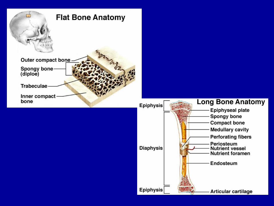
Two forms of bone tissue:
("woven", "spongy") Shelves or ridges ("trabeculae") of extracellular bone matrix with osteocytes embedded within it.
("compact", "Haversian") Concentric rings ("lamellae") of extracellular matrix and ostocytes, surrounding a containing nerves and capillaries
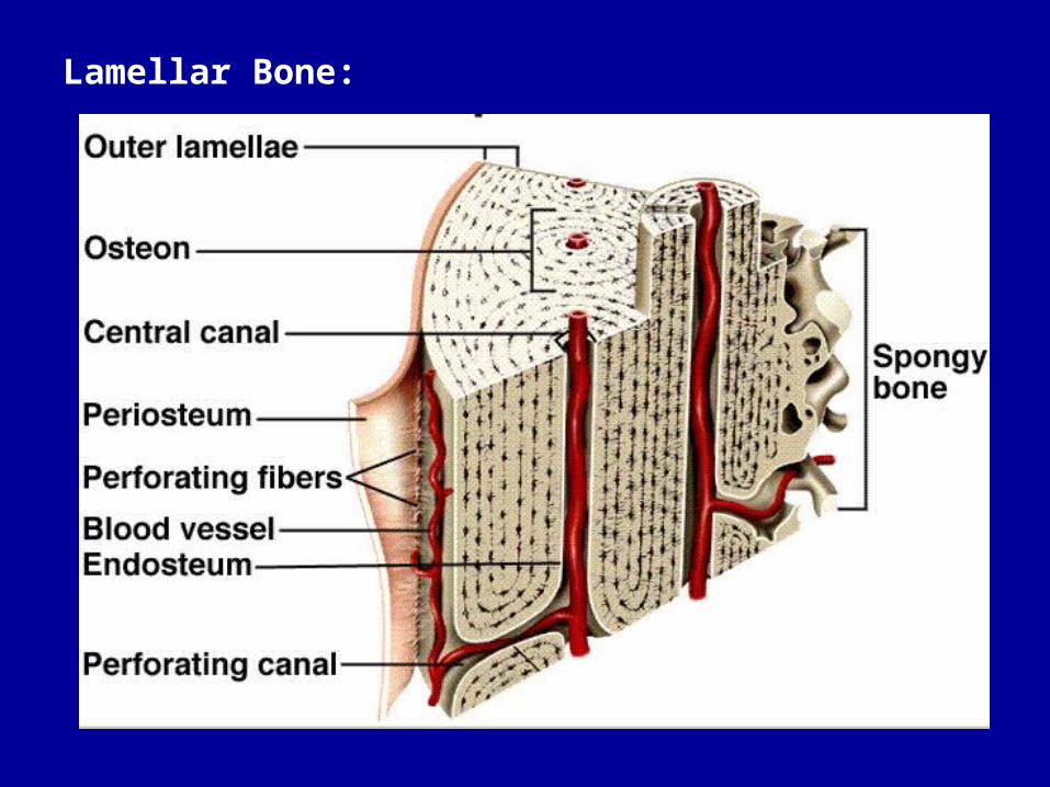
Lamellar Bone:
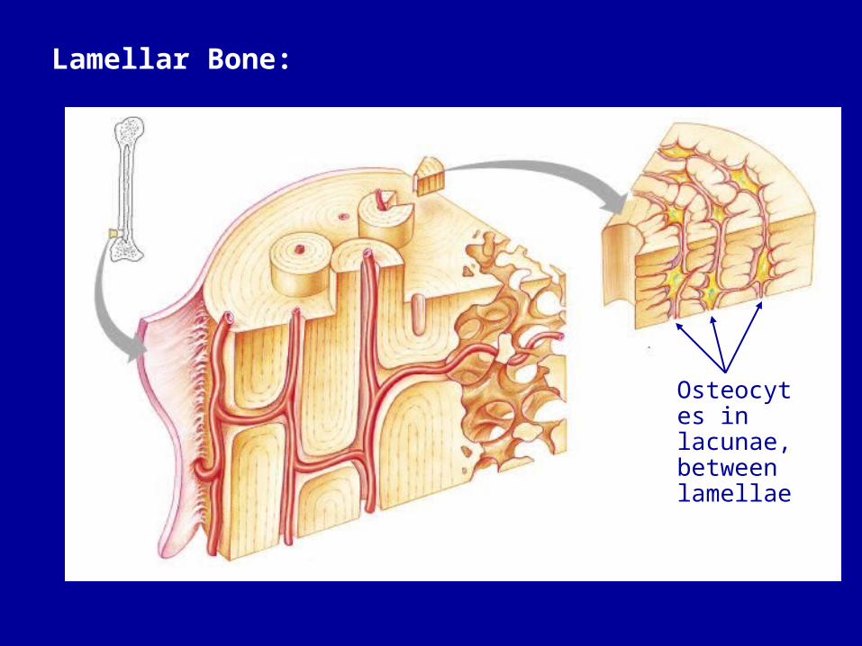
Lamellar Bone:
Osteocytes in lacunae, between lamellae
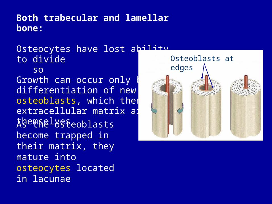
Both trabecular and lamellar bone:
Osteocytes have lost ability to divide soGrowth can occur only by differentiation of new osteoblasts, which then form extracellular matrix around themselves
As the osteoblasts become trapped in their matrix, they mature into osteocytes located in lacunae
Osteoblasts at edges
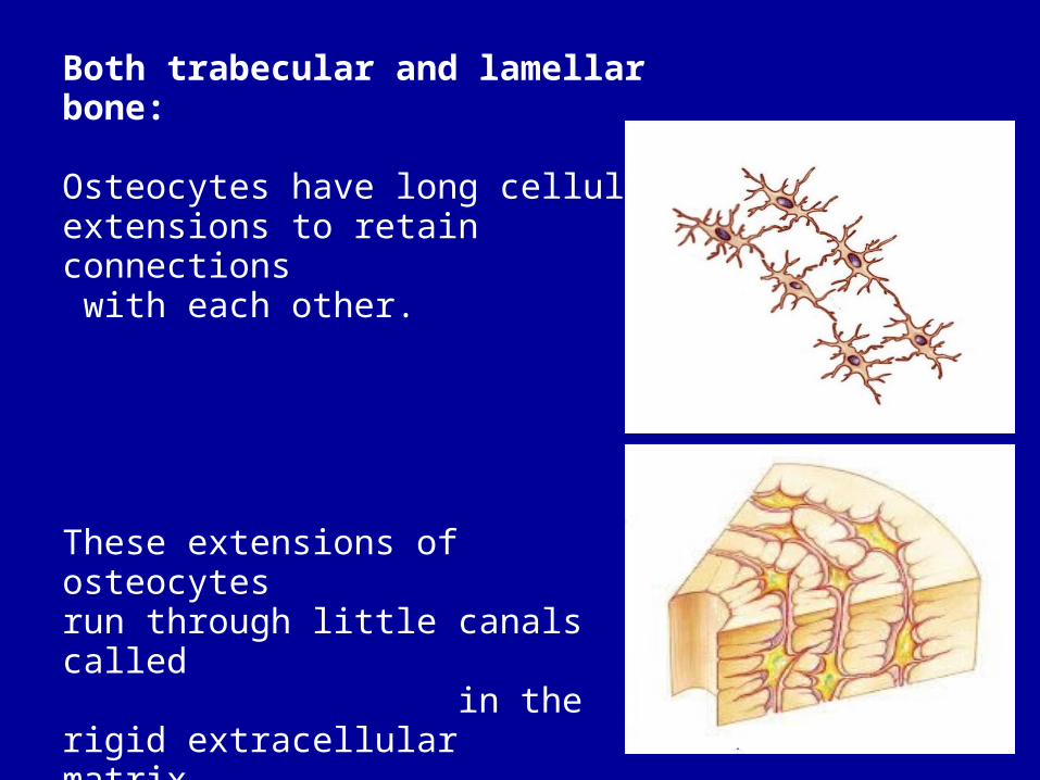
Both trabecular and lamellar bone:
Osteocytes have long cellular extensions to retain connections with each other.
These extensions of osteocytes run through little canals called in the rigid extracellularmatrix
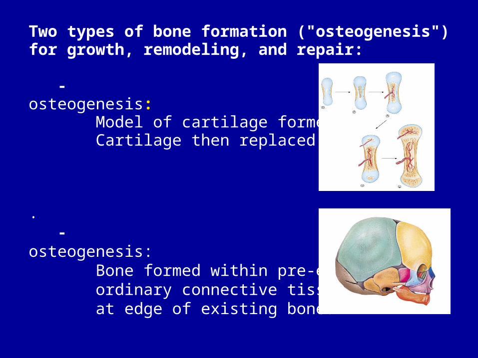
Two types of bone formation ("osteogenesis") for growth, remodeling, and repair:
- osteogenesis: Model of cartilage formed first. Cartilage then replaced by bone
. - osteogenesis: Bone formed within pre-existing ordinary connective tissue, often at edge of existing bone.
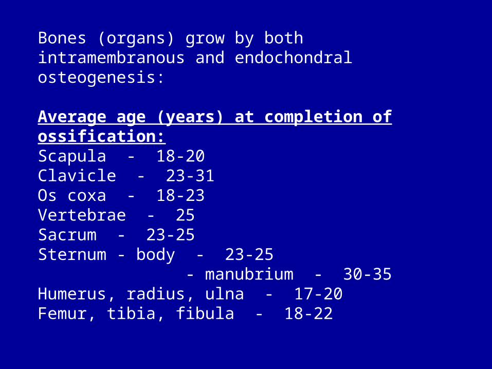
Bones (organs) grow by both intramembranous and endochondral osteogenesis:
Average age (years) at completion of ossification:Scapula - 18-20Clavicle - 23-31Os coxa - 18-23Vertebrae - 25Sacrum - 23-25Sternum - body - 23-25 - manubrium - 30-35Humerus, radius, ulna - 17-20Femur, tibia, fibula - 18-22

Bones repair fractures by both intramembranous and endochondral osteogenesis:
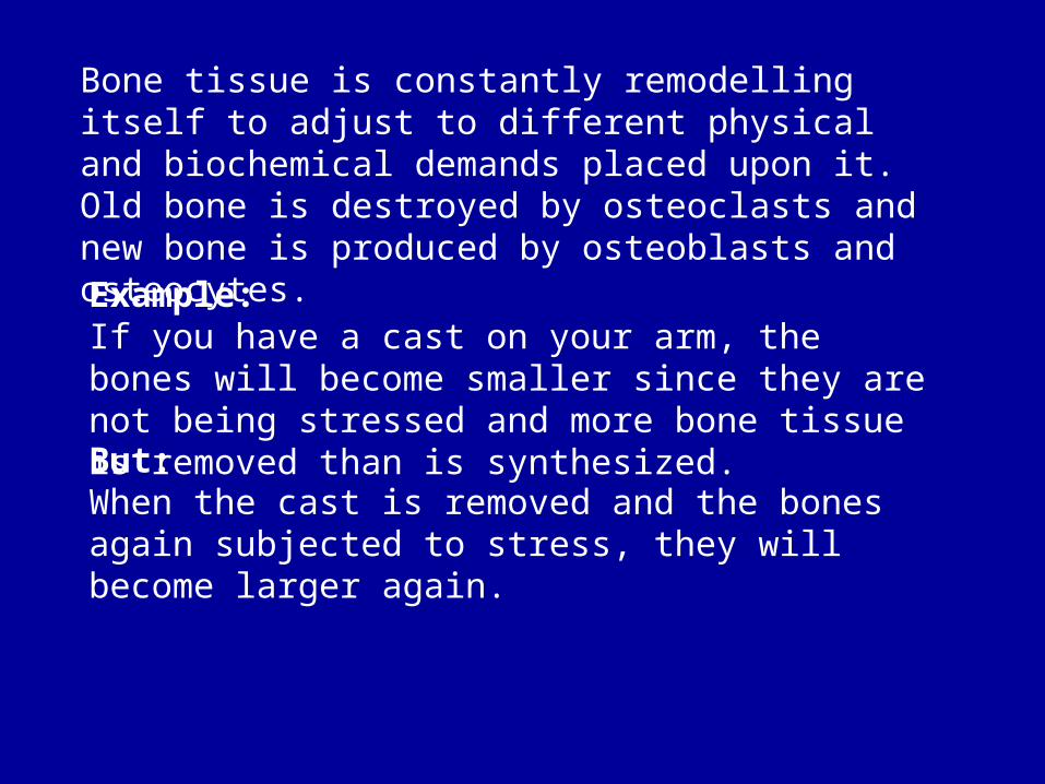
Bone tissue is constantly remodelling itself to adjust to different physical and biochemical demands placed upon it. Old bone is destroyed by osteoclasts and new bone is produced by osteoblasts and osteocytes.
Example:If you have a cast on your arm, the bones will become smaller since they are not being stressed and more bone tissue is removed than is synthesized.But:When the cast is removed and the bones again subjected to stress, they will become larger again.

Bone remodelling regulated by many hormones:
Growth hormone stimulated osteoblast activity & collagen synthesis
Thyroid hormone stimulates osteoblast activity & collagen synthesis; stimulates formation of ossification centers
Testosterone stimulates osteoblast activity & bone growth
Progesterone stimulates osteoclast activity & bone loss
Estrogen stimulates osteoblast activity & bone growth

Two hormones primarily responsible for day-to-day remodelling of bone to regulate concentration of calcium in blood:
Secreted by thyroid gland Stimulates osteoblasts to produce more matrix Inhibits osteoclasts from breaking down matrix Thus: Calcium removed from blood & stored in bone
Secreted by parathyroid glands Inhibits osteoblasts from producing more matrix Stimulates osteoclasts to break down matrix Thus: Calcium released from bone into blood
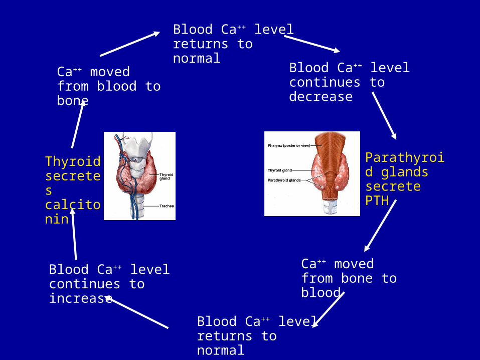
Blood Ca++ level returns to normal
Blood Ca++ level continues to increase
Blood Ca++ level continues to decrease
Blood Ca++ level returns to normal
Thyroid secretes calcitonin
Parathyroid glands secrete PTH
Ca++ moved from blood to bone
Ca++ moved from bone to blood
Related Documents