Biological sequence motif discovery using motif-x. Michael F. Chou 1 and Daniel Schwartz 2 1 Department of Genetics Harvard Medical School Boston, MA Email: mchou(at)genetics.med.harvard.edu 2 Department of Physiology and Neurobiology University of Connecticut Storrs, CT Email: daniel.schwartz(at)uconn.edu Abstract: The web-based motif-x program provides a simple interface to extract statistically significant motifs from large data sets such as MS/MS post-translational modification data and groups of proteins that share a common biological function. Users upload data files and download results using common web browsers on essentially any web-compatible computer. Once submitted, data analyses are performed rapidly on an associated high-speed computer cluster and they produce both syntactic and image- based motif results and statistics. The protocols presented demonstrate the use of motif-x in three common user scenarios. Key terms: protein motif, phosphorylation, post-translational modification (PTM), motif discovery, motif-x, mass spectrometry, proteomics.

Welcome message from author
This document is posted to help you gain knowledge. Please leave a comment to let me know what you think about it! Share it to your friends and learn new things together.
Transcript
Biological sequence motif discovery using motif-x. Michael F. Chou1 and Daniel Schwartz2 1 Department of Genetics
Harvard Medical School Boston, MA Email: mchou(at)genetics.med.harvard.edu
2 Department of Physiology and Neurobiology
University of Connecticut Storrs, CT Email: daniel.schwartz(at)uconn.edu
Abstract: The web-based motif-x program provides a simple interface to extract statistically significant motifs from large data sets such as MS/MS post-translational modification data and groups of proteins that share a common biological function. Users upload data files and download results using common web browsers on essentially any web-compatible computer. Once submitted, data analyses are performed rapidly on an associated high-speed computer cluster and they produce both syntactic and image-based motif results and statistics. The protocols presented demonstrate the use of motif-x in three common user scenarios. Key terms: protein motif, phosphorylation, post-translational modification (PTM), motif discovery, motif-x, mass spectrometry, proteomics.
2
INTRODUCTION
Using the tools of mass spectrometry and spectral identification, large-scale
proteomic experiments are now able to identify thousands of protein post-translational
modifications (PTMs) in a single experimental run, and the technology to enrich for
different modifications such as phosphorylation, acetylation, glycosylation, and others
has been steadily improving. Because of the importance of post-translational
modifications in normal and pathologic cellular physiology, the ultimate goal of
measurement is to understand the underlying biological processes that lead to these
modifications and the consequences thereof. Knowing, e.g., the preference of an
enzyme for its natural substrates can help elucidate biological pathways in which they
are involved.
Because part of the biochemical preference of an enzyme for a given substrate
may be determined by residues immediately surrounding the site of action, biochemists
have focused on identifying the critical neighboring residues that give rise to specific
enzyme-substrate interactions. This pattern of residues along the short span of a protein
or polypeptide is known as a short linear motif (or simply a motif for short). Motifs have
historically been studied using mutagenesis experiments such as alanine scanning
(Koyasu et al., 1994) and more recently, combinatorial library approaches (Hutti et al.,
2004).
The motif-x algorithm for computationally determining short linear motifs was first
described by Schwartz and Gygi in 2005 (Schwartz and Gygi, 2005), and an online
implementation was made available by Chou and Schwartz at that time
(http://motif-x.med.harvard.edu/).
3
Supplemental Data Files
Files providing supplemental data for all of the protocols presented in this unit may be
downloaded at http://www.currentprotocols.com/protocol/bi1315.
STRATEGIC PLANNING
As described in the examples that follow, motif-x was initially designed to extract
statistically significant biological motifs surrounding sites of post-translational
modification that are identified by mass spectrometry. However, as will be demonstrated
below, motif-x can also be used to perform analyses using different data sources or
biological conditions.
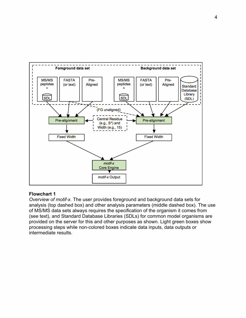
Flowchart 1 shows an overview of the variety of inputs available for use with
motif-x. There are three possible types of sequence input data that are referred to as
foreground data sets (the left side of the dashed box at the top). When a data set is
submitted on the web server, the user specifies the type of input data, and, if necessary,
motif-x translates that data into an internal format that it uses to find motifs. The three
types of data that are used as input are:
(a) Tandem Mass Spectral (MS/MS) data that is often in the form of lists of
tryptic peptides containing flags indicating which residues contain a PTM (this method is
described in the Basic Protocol below, and is the most common application);
(b) Pre-aligned data sets which are increasingly being published by web sites
containing post-translational modifications (this method is described in the Alternate
Protocol 1); and
4
Flowchart 1 Overview of motif-x. The user provides foreground and background data sets for analysis (top dashed box) and other analysis parameters (middle dashed box). The use of MS/MS data sets always requires the specification of the organism it comes from (see text), and Standard Database Libraries (SDLs) for common model organisms are provided on the server for this and other purposes as shown. Light green boxes show processing steps while non-colored boxes indicate data inputs, data outputs or intermediate results.
5
(c) FASTA-formatted data sets which may be a set of proteins with known
modifications or binding sites that the user believes to share a common motif (this
method is described in the Alternate Protocol 2).
Table 1 summarizes the various researcher objectives that can be addressed
using motif-x, as well as the format of the data used for each respective analysis.
Because residues that are significant for binding or substrate recognition can be
found both upstream and downstream of a modification site, motif-x is designed to build
motifs around a ‘central residue’. So, for example substrates of the human protein
kinase A (PKA) will often share the motif RRxS in common. That is, a serine on a
substrate that is a phospho-acceptor will often have arginines at the second and third
residues upstream from the serine which accounts for much of the specificity for that
substrate modification site. Similarly for the class of proline-directed kinases (such as
cyclin dependent kinases (CDKs) or mitogen-activated protein kinases (MAPKs)),
substrates usually have the motif SP where a proline exists one residue downstream of
the serine phospho-acceptor site. motif-x can take as input up to thousands of
substrates and determine one or more statistically overrepresented motifs in such data
sets.
By centering the analysis on a key residue (e.g., a serine known to be
phosphorylated), motif-x is able to generate a list of overrepresented motifs that may
contain residues both upstream and downstream from the key central residue.
6
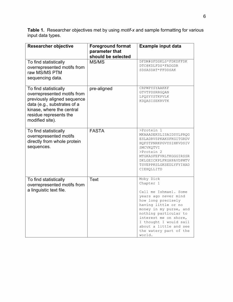
Table 1. Researcher objectives met by using motif-x and sample formatting for various input data types. Researcher objective Foreground format
parameter that should be selected
Example input data
To find statistically overrepresented motifs from raw MS/MS PTM sequencing data.
MS/MS DFDM#SFDSKLS*FDKDFFDK DTC@KDLFDS*FADGDR SDSASDAT*FFDDSAK
To find statistically overrepresented motifs from previously aligned sequence data (e.g., substrates of a kinase, where the central residue represents the modified site).
pre-aligned CRPWPYSYAAKKF GTVTPDSRRGQAN LPQSYYSTKPVLK KDQASISEKRVTK
To find statistically overrepresented motifs directly from whole protein sequences.
FASTA >Protein 1 MKAAADEKSLISAIDSYLPRQGESLADRVSPKAKSFKGITGRDVHQFSTPNRKPGVYSIHKVDSIVSMCVKQTVI >Protein 2 MTGKASFEFVRLTKGGGIRSSRDKLGEICKPLFKGHPAVDPMTVTSVEPPKSLGKSEDLYFYIHADCIEHQLLITD
To find statistically overrepresented motifs from a linguistic text file.
Text Moby Dick Chapter 1 Call me Ishmael. Some years ago never mind how long precisely having little or no money in my purse, and nothing particular to interest me on shore, I thought I would sail about a little and see the watery part of the world.
7
As described in the Basic Protocol, tandem mass spectrometry data is a natural
form of input for such analyses because other programs such as SEQUEST (Eng et al.,
1994) or Mascot (Perkins et al., 1999), can easily identify the actual modified residues in
a sample. With regard to motif extraction, a potential drawback of MS/MS data sets is
that they are fragmented (usually into tryptic peptides), completely unaligned, and each
peptide may contain one or more modified residues. However, for this form of input
data, motif-x has a special built-in preprocessing function which converts these data
sets into aligned data sets whereby each modification is converted into a fixed-width
peptide aligned on the modified residue (the native format used by the motif-x engine).
In order to perform this conversion motif-x requires the user’s specification of an
organism.
The example in Alternate Protocol 1 has a goal similar to that of the basic
protocol, and analysis proceeds the same way, except for the fact that the input data in
this case is already in the native form with fixed-width peptides surrounding a key
central residue.
Alternate Protocol 2 demonstrates the fact that data with a known modification
site is not the only data format that can be handled by motif-x. Because each of the 20
normal amino acids has a unique biochemical functionality, it is possible to use any
residue as a central residue even if a clear modification is not known. This works
because motif-x is an unbiased, statistically based algorithm which does not make a
priori assumptions regarding the function of the patterns it extracts.
An important aspect of the motif-x algorithm is the utilization of a “background
data set” for the determination of significant residues. Unlike many other motif finding
8
algorithms that either use no background frequencies or simply use the frequencies of
the 20 amino acids in the proteome as a background, motif-x (most typically) uses the
entire proteome of the organism under study as a background to empirically determine
the conditional probabilities required for significance as the algorithm proceeds.
Amongst other benefits, this allows motif-x to identify significant motifs containing
correlated residues and motifs that are directly relevant to a particular organism.
Thus, the user must specify a background data set which, like the foreground
data set, may be in a number of different formats, but is most often simply selected from
the set of built-in FASTA-formatted Standard Database Libraries from a number of
different model organisms (“SDLs” on the right side of the dashed box at the top of
Flowchart 1).
Once launched, motif-x searches for any significant motifs in the foreground data
set using the context of the background data set, and reports any motifs found to be
overrepresented in the foreground data set with respect to the background data set.
The output is in both standard syntactic form, which indicates the most significant
residues (e.g., RRxS), and in the form of a motif-x logo image. The logo indicates not
only those residues that were significant enough to exceed the user defined threshold
(such as both R residues in RRxS), but also the frequency of other residues which failed
to exceed the significance threshold, yet nevertheless may be statistically
overrepresented.
9
BASIC PROTOCOL
Extracting sequence motifs from MS/MS post-translational modification data.
Although the motif-x algorithm is general, and can be used to extract sequence
motifs from data sets of varying formats, the most common motif-x analysis involves the
extraction of motifs from large-scale post-translational modification data sets generated
by tandem mass spectrometry experiments. The following protocol will use sample
phosphorylation data obtained from the supplemental data of a study that investigated
phosphorylation changes during the early differentiation of human embryonic stem cells
(Van Hoof et al., 2009) (see Supplemental Data Files, File 1).
Necessary resources
Hardware Any computer with Internet access. Software Any web browser (e.g., Internet Explorer, Firefox, Safari, Chrome, etc.). Files Performing the basic motif-x MS/MS protocol requires a list of peptides in SEQUEST (or similar) format (see Table 1). These peptides need not be contained within a file, as it is also possible to paste peptides directly into the motif-x input text box. Users may optionally decide to upload their own background data set, rather than use the provided motif-x background options. If users choose to upload foreground and/or background files, it is important to note that they should be .txt files, as other file types (e.g., .xls, .doc, .docx, .pdf, etc.) are not supported by motif-x. In general, if a file can be viewed using Notepad (on Windows) or TextEdit (in plain text mode on a Macintosh) without odd characters, the file will be suitable for uploading. A sample MS/MS formatted foreground file corresponding to the same analysis illustrated in the protocol below is provided as a supplemental data file (see Supplemental Data Files, File 1).
10
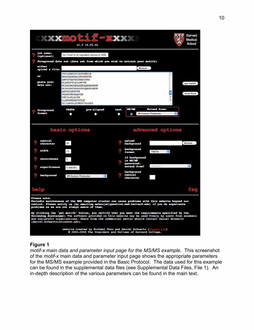
Figure 1 motif-x main data and parameter input page for the MS/MS example. This screenshot of the motif-x main data and parameter input page shows the appropriate parameters for the MS/MS example provided in the Basic Protocol. The data used for this example can be found in the supplemental data files (see Supplemental Data Files, File 1). An in-depth description of the various parameters can be found in the main text.
11
1. From your computer’s web browser navigate to the motif-x web site at http://motif-
x.med.harvard.edu and click on the motif-x logo at the top of the page to enter the
motif-x data/parameters input page (see Figure 1).
2. In the blank text box labeled “job name” enter an optional title for the job.
3. Click the “Browse” button to upload a foreground data set or paste the data set
directly into the large text box in the center of the page.
The foreground data is the data set from which you wish to extract motifs. Given the
nature of sequencing by tandem mass spectrometry, these data should be input as
peptide sequences, each separated by a carriage return. Special characters “#”, “@”,
“*”, should be used to denote sites of protein modification (see Table 1 and
Supplemental Data Files, File 1) by placing the special character immediately
following the modified amino acid. Although this peptide modification syntax is
standard for searches performed using SEQUEST, other common post-translational
modification syntaxes include the addition of lowercase letters prior to the
modification site (e.g., pS, pT, acK, (ox)M, etc.), a numerical mass change following
the modification site (e.g., S(80), K(114), etc.), or signifying modification sites as
lowercase letters themselves (e.g., s, t, y, k, etc.). Data using these alternative
syntaxes will produce incorrect results, but can typically be converted into the
SEQUEST syntax in a straightforward manner using the “find and replace” command
in Microsoft Word or Excel (e.g., replacing all “S(80)” instances with “S#” will
accomplish the task; however the final file must be plain text, not a Word or Excel
12
file). It should be noted that with the exception of the special character denoting the
modification site, motif-x removes all other non-alphanumeric characters from the
data set prior to performing a motif search. Thus, “hyphens” denoting protein termini
and “periods” denoting peptide fragment termini can be left in the data (e.g., the
peptide sequence “-MEPSSWSGSES#PAENME.R” is acceptable). Sample MS/MS
data is provided as a Supplemental Data File (see Supplemental Data Files, File 1)
for readers wishing to follow along with the present protocol.
4. In the “foreground format” field select the “MS/MS” foreground format option and
select an appropriate database in the drop-down menu from which to extend the
peptides.
Since amino acid modification sites from MS/MS data sets often lack sufficient
upstream and downstream residues necessary for the extraction of motifs, motif-x
has a built-in “peptide extension” function. This built-in function maps user-inputted
peptides back onto their appropriate proteomic database and gathers additional
sequence information surrounding the modification sites as necessary. Users should
therefore select the appropriate proteomic sequence database corresponding to the
organism from which their foreground data sample was obtained. Users following the
example presented here should select “IPI Human Proteome” as the extension
database (see Figure 1).
5. Under the “basic options” heading enter an appropriate central character into the
“central character” field.
13
Typically motif-x only takes a single character as input for this parameter; however, in
the case of MS/MS formatted data motif-x will accept two characters as input. These
two characters should reflect the appropriate syntax for modification sites in the
foreground data set (discussed in step 3). In the example provided in Supplemental
Data Files (File 1), phosphorylation sites are denoted by “#”, thus the characters “S#”
should be used in the central character input field (see Figure 1). It is very important
to note that this field is case sensitive, therefore inputting “s#” would yield no results
for this example.
6. Enter the desired motif width into the “width” field.
Protein sequence motifs, especially those involved in protein post-translational
modification tend to be between two and eight amino acids in length. Thus, choosing
a length of 13 or 15 (i.e., six or seven amino acids on each side of the central
modified residue) is expected to capture the vast majority of short-range sequence
dependencies. Motif widths between 3 and 35 (odd lengths only) can be used for this
parameter. However, choosing a motif width that is too narrow can result in the
exclusion of motifs with critical longer-range dependencies and choosing a motif
width that is too wide (without adjusting the significance threshold accordingly) can
yield spurious motif results. In the example provided, a motif width of 15 has been
chosen (see Figure 1).
7. Enter into the “occurrences” field the minimal number of times that you require your
extracted motifs to occur within the foreground data set.
14
This parameter can be used to tune the specificity of motifs since motifs with greater
specificity (i.e., more “fixed” positions) are expected to occur less often than those
motifs with lower specificity. Users that wish to extract a maximal number of motifs
should set this parameter to a low value (for example, “5”) and rely solely on the
significance parameter (see step 8) to extract motifs. On occasion it may be useful to
set this parameter as a fractional percent of the total number of modification sites in
order to compare motifs with similar specificities across data sets that vary in size
(e.g., to compare motifs from data sets of 300 and 3000 sites, one may opt to set the
occurrences parameter to 5 and 50, respectively). In the example provided, an
occurrence threshold of 5 has been chosen (see Figure 1).
8. Enter the significance threshold for the analysis into the “significance” field.
The significance parameter corresponds to the binomial probability threshold
necessary to “fix” each motif position during the motif-building phase of the algorithm.
It is critical to note that this value does not take into account a correction for multiple
hypotheses (such as the Bonferroni correction). On any given motif-x search step
there are (number of possible characters at each position) * (number of nonfixed
positions) hypotheses being tested. For example, in an S-centered analysis of width
15, there would be (20) * (14) = 280 hypotheses tested. To ensure an alpha-value of
at least 0.05 by the Bonferroni method, one would need to divide the desired alpha-
value by the total number of hypotheses tested (i.e., 0.05/280 = 0.00018). Thus, for
the previous example inputting a significance value of 0.00018 into motif-x would in
fact correspond to a p-value of 0.05. The use of a motif-x significance threshold
15
greater than 0.0005 is not suggested as it may result in the extraction of motifs that
are not statistically significant. The default value of this parameter is 0.000001, which
corresponds to an actual alpha-value of approximately 0.0003 for a protein motif
analysis of width 15 after Bonferroni correction. A significance threshold of 0.000001
was chosen for the present example.
9. In the “background” field select the appropriate background data set for the analysis.
motif-x evaluates the statistical significance of positions within motifs in the
foreground data set through a comparison to a background data set. For MS/MS
analyses, the background data set should most-often correspond to the organismal
proteomic database from which the sequencing data was obtained. Therefore, the
peptide extension database (see step 4) and the background database should
typically be the same. In the present example, since the foreground data set
originated from human embryonic stem cells, the “IPI Human Proteome” was chosen
as a background database (see Figure 1). The parameters under “advanced options”
defining the background data set are not required for a basic motif-x run. These
additional options are described in the “advanced parameters” section at the end of
the protocols.
10. Press the “get motifs” button.
Pressing “get motifs” will launch an analysis on a node of the computer cluster and
spawn a new web page as shown in Figure 2. This page will periodically reload until
the results page is loaded. On occasion it may be necessary to manually reload the
16
page using your web browser. The motif-x algorithm is relatively fast, and jobs take
under 5 minutes to perform. In an effort to minimize the burden on the computer
cluster, motif-x jobs are limited to running within 15 minutes before a “timeout”
message is generated. If users get this message they should try to simplify their
analyses by either increasing the occurrence threshold or decreasing the significance
threshold for their analysis. Please contact the authors if you believe you have an
intractably large or complex data set to analyze.
11. View the motif-x results page.
The motif-x results page for an MS/MS format analysis is organized into four major
regions: i) heading and parameters used (Figure 3), ii) data preprocessing statistics
(Figure 4), iii) motif results with sequence logos (Figure 5), and iv) raw sequence data
for each motif extracted (Figure 6).
Heading and parameters – This section provides information on the version of the
motif-x software that was used; as well as a recapitulation of the parameters selected
for the analysis.
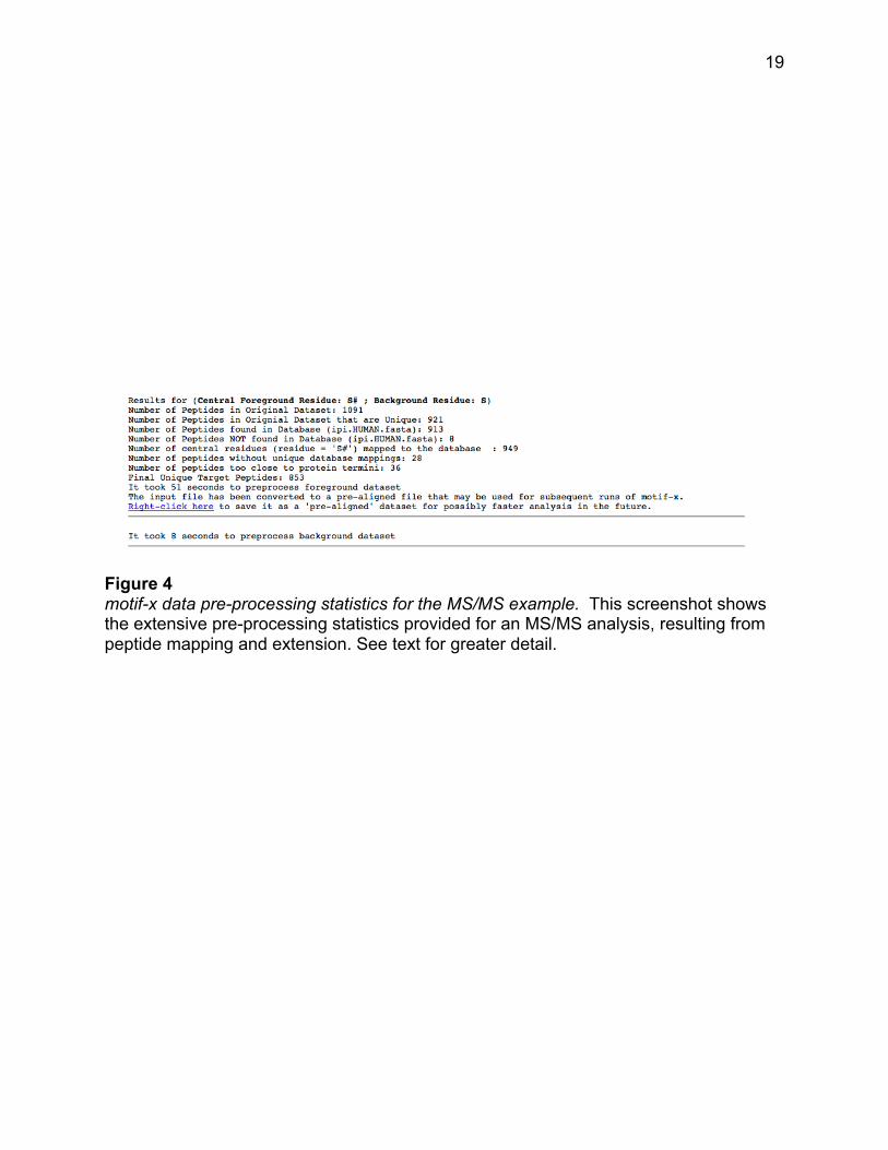
Peptide pre-processing statistics – This section provides an accounting of the steps
used to pre-process the MS/MS peptide data set into an aligned data set suitable for
the motif-x algorithm. It is noticeable from the example provided that although 1091
phosphorylated peptides were initially inputted into motif-x, following pre-processing,
only 853 serine-phosphorylated 15-mers remained.
17
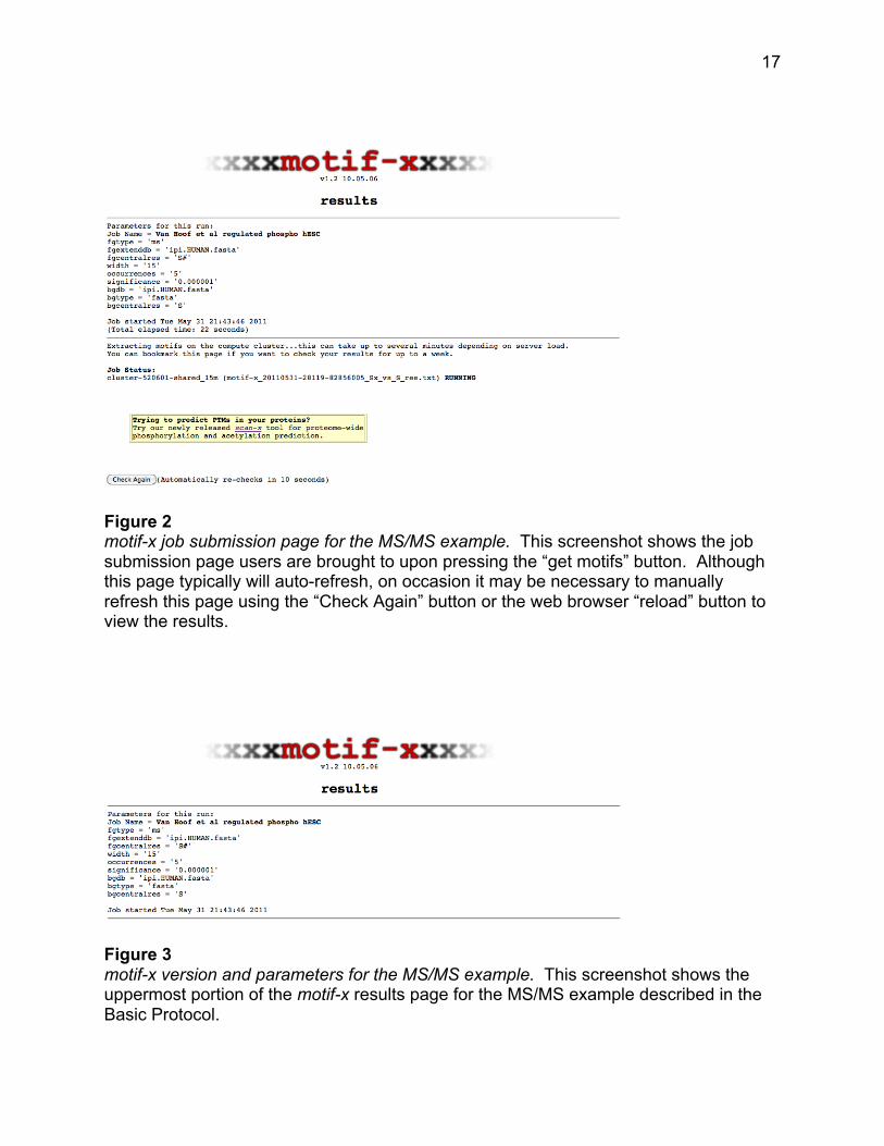
Figure 2 motif-x job submission page for the MS/MS example. This screenshot shows the job submission page users are brought to upon pressing the “get motifs” button. Although this page typically will auto-refresh, on occasion it may be necessary to manually refresh this page using the “Check Again” button or the web browser “reload” button to view the results.
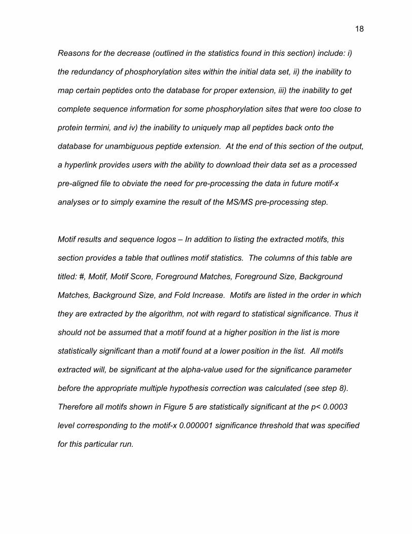
Figure 3 motif-x version and parameters for the MS/MS example. This screenshot shows the uppermost portion of the motif-x results page for the MS/MS example described in the Basic Protocol.
18
Reasons for the decrease (outlined in the statistics found in this section) include: i)
the redundancy of phosphorylation sites within the initial data set, ii) the inability to
map certain peptides onto the database for proper extension, iii) the inability to get
complete sequence information for some phosphorylation sites that were too close to
protein termini, and iv) the inability to uniquely map all peptides back onto the
database for unambiguous peptide extension. At the end of this section of the output,
a hyperlink provides users with the ability to download their data set as a processed
pre-aligned file to obviate the need for pre-processing the data in future motif-x
analyses or to simply examine the result of the MS/MS pre-processing step.
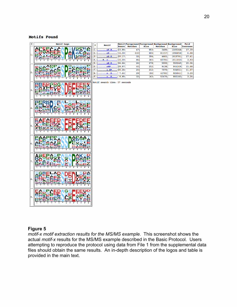
Motif results and sequence logos – In addition to listing the extracted motifs, this
section provides a table that outlines motif statistics. The columns of this table are
titled: #, Motif, Motif Score, Foreground Matches, Foreground Size, Background
Matches, Background Size, and Fold Increase. Motifs are listed in the order in which
they are extracted by the algorithm, not with regard to statistical significance. Thus it
should not be assumed that a motif found at a higher position in the list is more
statistically significant than a motif found at a lower position in the list. All motifs
extracted will, be significant at the alpha-value used for the significance parameter
before the appropriate multiple hypothesis correction was calculated (see step 8).
Therefore all motifs shown in Figure 5 are statistically significant at the p< 0.0003
level corresponding to the motif-x 0.000001 significance threshold that was specified
for this particular run.
19
Figure 4 motif-x data pre-processing statistics for the MS/MS example. This screenshot shows the extensive pre-processing statistics provided for an MS/MS analysis, resulting from peptide mapping and extension. See text for greater detail.
20
Figure 5 motif-x motif extraction results for the MS/MS example. This screenshot shows the actual motif-x results for the MS/MS example described in the Basic Protocol. Users attempting to reproduce the protocol using data from File 1 from the supplemental data files should obtain the same results. An in-depth description of the logos and table is provided in the main text.
21
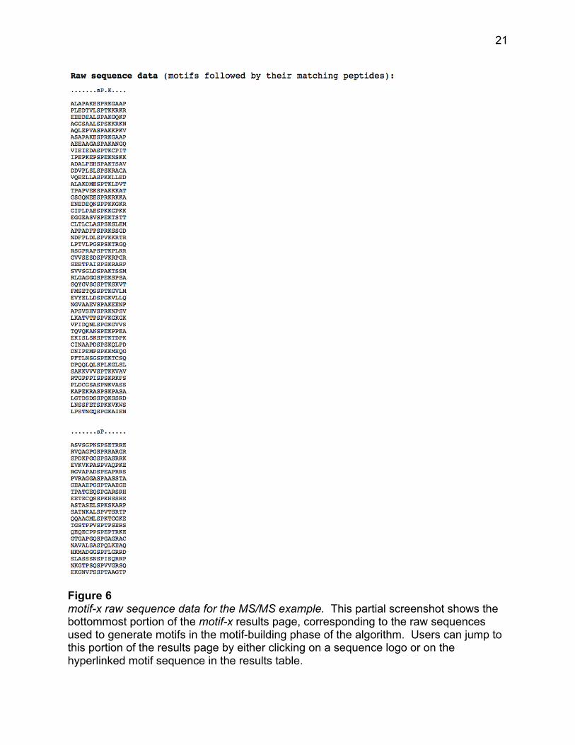
Figure 6 motif-x raw sequence data for the MS/MS example. This partial screenshot shows the bottommost portion of the motif-x results page, corresponding to the raw sequences used to generate motifs in the motif-building phase of the algorithm. Users can jump to this portion of the results page by either clicking on a sequence logo or on the hyperlinked motif sequence in the results table.
22
The determination of whether a motif is known or novel can be accomplished using a
variety of databases/tools (Edwards et al., 2008; Keshava Prasad et al., 2009) which
store motif information. Additionally, directed Google searches using “x” to represent
wildcard positions can often yield relevant primary literature sources of motif function.
For example, searching for information on the first extracted motif in Figure 5
(.......sP.K....) by performing the Google search “SPxK phosphorylation motif” quickly
reveals that the SPxK motif is the recognition sequence for cyclin dependent kinases
(CDKs).
The “motif score” is calculated by taking the sum of the negative log probabilities
used to fix each position of the motif. As such, higher motif scores typically
correspond to motifs that are more statistically significant as well as more specific
(i.e., greater number of fixed positions).
The “foreground matches” and “background matches” statistics indicate the
number of peptides containing a given motif in those respective data sets
following the removal of all peptides containing previously extracted motifs.
Because of this iterative “set reduction” strategy, the “foreground matches” and
“background matches” statistics may be less than or equal to the total number of
instances of a given motif in the whole data set.
The “foreground size” and “background size” statistics indicate the total number
of peptides contained in these data sets. The size of these data sets decreases

23
as motifs are extracted (i.e., down a column) due to the fact that peptides are
removed from both the foreground and background data sets following motif
extraction. The total number of foreground peptides not falling into any extracted
motif class can therefore be calculated as the difference between the “foreground
size” and the “foreground matches” of the final motif class (e.g., 163 – 32 = 131
unclassified peptides in Figure 5).
The “fold increase” statistic is an indicator of the enrichment level of the extracted
motifs. Specifically, it is calculated as (foreground matches/foreground
size)/(background matches/background size).
Frequency-based motif logos are provided to the left of the results table. In addition
to indicating the fixed positions of the motif at full character height, these logos
illustrate the amino acid composition of the “wildcard” positions within the motifs.
Amino acids are sorted by their frequency at each position within the motif with the
most frequent amino acids appearing closest to the top of the motif logo. Motif
positions are labeled below the x-axis and residues are colored according to their
chemical and physical properties.
Raw data – Clicking on either the links in the motif column of the results table, or on
the sequence logos themselves, brings users to the raw peptide data used to extract
the given motifs at the bottom of the motif-x results page. As such, the number of
24
peptides found for each motif in this section corresponds exactly to the number of
“foreground matches” for the motif in column 2 of the motif-x results table.
ALTERNATE PROTOCOL 1
Extracting sequence motifs from pre-aligned data.
For algorithmic reasons, aligned peptides centered on a particular amino acid
(typically a post-translational modification site) form the ideal data input format for motif-
x and are thus included among the foreground format choices on the motif-x web site
(see Table 1). This data format is also becoming increasingly adopted by protein post-
translational modification databases (Hornbeck et al., 2004; Durek et al., 2010; Dinkel et
al., 2011), as well as in the supplemental data of large-scale PTM studies (Matic et al.,
2010; Rigbolt et al., 2011). The following protocol will use pre-aligned 15-mers centered
on sumoylated lysine residues from human proteins to illustrate the use of motif-x on
this type of foreground data format (see Supplemental Data Files, File 2).
Necessary resources
Hardware Any computer with Internet access. Software Any web browser (e.g., Internet Explorer, Firefox, Safari, Chrome, etc.). Files Performing the motif-x pre-aligned protocol requires a specific list of peptides in pre-aligned format (see Table 1). These peptides need not be contained within a file, as it is also possible to paste peptides directly into the motif-x input text box. Users may optionally decide to upload their own background data set, rather than use the provided motif-x background options. If users choose to upload foreground and/or background files, it is important to note that they should be .txt files, as other file types (e.g., .xls, .doc, .docx, .pdf, etc.) are not supported by motif-x. In general, if a file can be viewed
25
using Notepad (on Windows) or TextEdit (in plain text mode on a Macintosh) without odd characters, the file will be suitable for uploading. A sample pre-aligned formatted foreground file corresponding to the same analysis illustrated in the protocol below is provided as a supplemental data file (see Supplemental Data Files, File 2).
1. From your computer’s web browser navigate to the motif-x web site at http://motif-
x.med.harvard.edu and click on the motif-x logo at the top of the page to enter the
motif-x data/parameters input page (see Figure 7).
2. In the blank text box labeled “job name” enter an optional title for the job.
3. Click the “Browse” button to upload a foreground data set or paste the data set
directly into the large text box in the center of the page.
The foreground data is the data set from which you wish to extract motifs. Data
should be uploaded or pasted in the same format as shown in the pre-aligned
example in Table 1 or the raw sequence data provided in as a supplemental data file
(see Supplemental Data Files, File 2). Peptides of equal width and centered on an
amino acid of interest should be separated by a carriage return. In contrast to the
previously described MS/MS format, which allows for special characters (“#”, “@”, “*”)
to designate modification sites, the pre-aligned data format does not allow for the
inclusion of special characters.
4. In the “foreground format” field select the “pre-aligned” option.
26
This option indicates that the inputted data is already in the ideal pre-aligned format
for motif-x, and that motif-x does not need to preprocess the data.
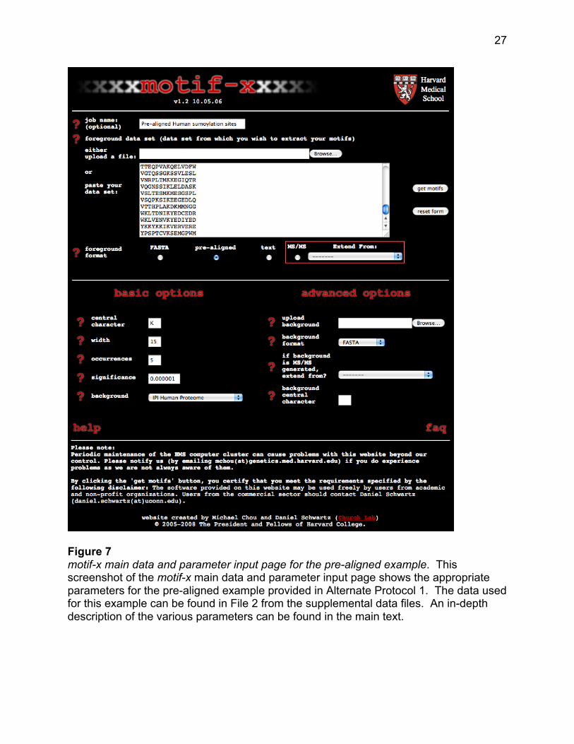
5. Under the “basic options” heading enter an appropriate central character into the
“central character” field.
A single (case sensitive) character should be entered. As mentioned in Step 3
above, combinations of residues and special characters may not be used in pre-
aligned analyses. In the sumoylation example presented here, since the central
character of the pre-aligned data corresponds to the site of lysine modification, “K” is
entered as the central character (see Figure 7). It should be noted however that all
the peptides in the input data need not contain the same central character. In the
case of pre-aligned input data containing multiple different central characters, motif-x
will carry out the motif extraction procedure on only that subset of peptides bearing
the central character specified by this field.
6. Enter the desired motif width into the “width” field.
In a pre-aligned analysis, the width parameter should correspond exactly to the width
of the pre-aligned peptides. A width of 15 was entered for the present example.
7. Enter into the “occurrences” field the minimal number of times that you require your
extracted motifs to occur within the foreground data set.
An explanation of this parameter is provided in the Basic Protocol, Step 7. An
occurrences parameter of 5 was entered for the present example.
27
Figure 7 motif-x main data and parameter input page for the pre-aligned example. This screenshot of the motif-x main data and parameter input page shows the appropriate parameters for the pre-aligned example provided in Alternate Protocol 1. The data used for this example can be found in File 2 from the supplemental data files. An in-depth description of the various parameters can be found in the main text.
28
8. Enter the significance threshold for the analysis into the “significance” field.
An explanation of this parameter is provided in Step 8 of the previously described
Basic Protocol. A significance of 0.000001 was entered for the present example.
9. In the “background” field select the appropriate background data set for the analysis.
motif-x evaluates the statistical significance of positions within motifs through a
comparison to a background data set. It is highly preferable to select the background
proteomic database corresponding to the organism from which the foreground data
was derived. In the present human sumoylation site example the “IPI Human
Proteome” is selected as a background database (see Figure 7) because the pre-
aligned foreground data originated from human proteins.
10. Press the “get motifs” button.
An explanation of the result of pressing “get motifs” is provided in Step 10 of the
previously described Basic Protocol.
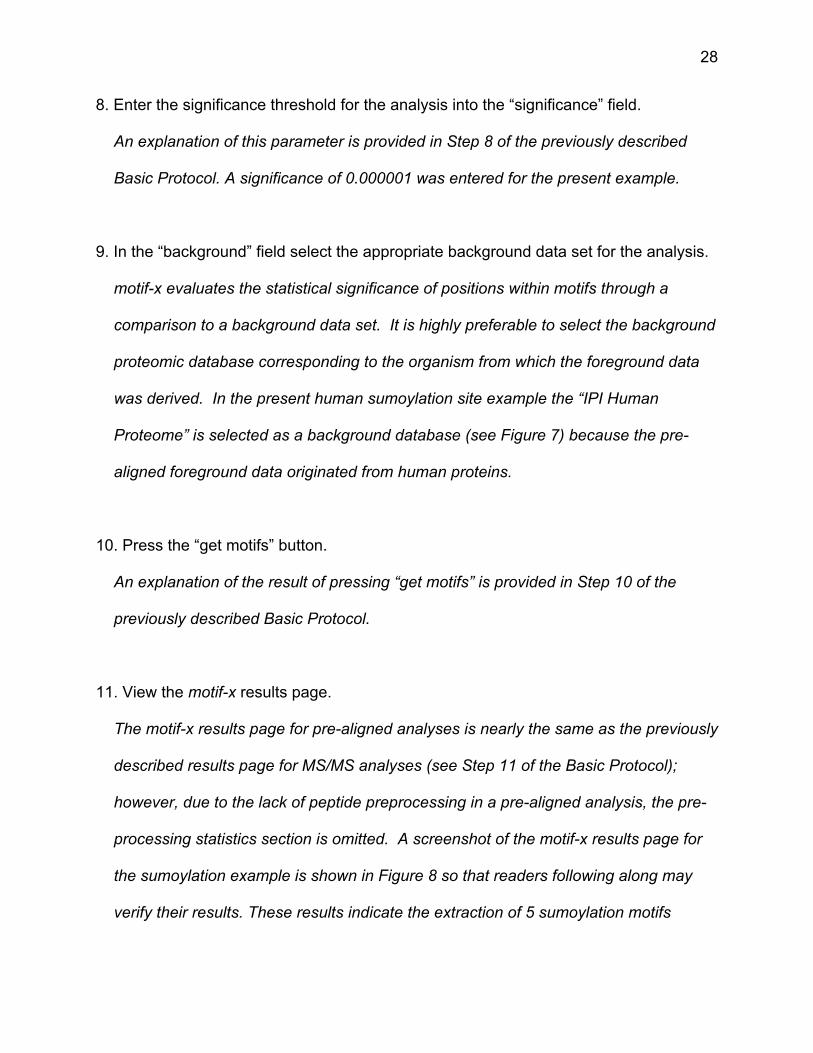
11. View the motif-x results page.
The motif-x results page for pre-aligned analyses is nearly the same as the previously
described results page for MS/MS analyses (see Step 11 of the Basic Protocol);
however, due to the lack of peptide preprocessing in a pre-aligned analysis, the pre-
processing statistics section is omitted. A screenshot of the motif-x results page for
the sumoylation example is shown in Figure 8 so that readers following along may
verify their results. These results indicate the extraction of 5 sumoylation motifs
29
Figure 8 motif-x results page for the pre-aligned example. This screenshot shows the actual motif-x results for the pre-aligned example described in Alternate Protocol 1. Users attempting to reproduce the protocol using data from File 2 from the supplemental data files should obtain the same results. An in-depth description of the logos and table is provided in the main text.

30
(IKxEP, VKxE, IKxE, LKxE, and KxE), consistent with the experimentally verified
sumoylation sequence specificity (!KxE, where ! represents a hydrophobic residue).
ALTERNATE PROTOCOL 2
Extracting sequence motifs from whole protein sequence data in FASTA format.
Following experiments, researchers are often left with lists of proteins that share
a common attribute (e.g., binding to the same partner, localizing to the same cellular
compartment, etc.). Although the motif-x algorithm was initially designed with the intent
of extracting protein post-translational modification motifs from aligned data, the
algorithm is capable of extracting overrepresented sequence patterns from any
sequence-based data set including whole protein data in FASTA format (see Table 1).
It should be noted that patterns extracted from whole protein data need not be involved
in protein post-translational modifications as motif-x extracts patterns statistically,
without regard to cellular function. The following protocol will use a group of ten
proteins selected from the ELM (Eukaryotic Linear Motif) database (Gould et al., 2010)
known to be bound by Grb2-like Src Homology 2 (SH2) domains. Src Homology 2
domains are involved in a wide variety of protein signaling pathways, and are unified in
their ability to bind phosphorylated tyrosine residues. The Grb2-like SH2 domain used in
this analysis is known to specifically bind the YxN motif (Kessels et al., 2002). The data
set used for this example is provided in Supplemental Data Files (see File 3).
31
Necessary resources
Hardware Any computer with Internet access. Software Any web browser (e.g., Internet Explorer, Firefox, Safari, Chrome, etc.). Files Performing the motif-x FASTA protocol requires a specific list of proteins in FASTA format (see Table 1). These proteins need not be contained within a file, as it is also possible to paste proteins directly into the motif-x input text box. Users may optionally decide to upload their own background data set, rather than use the provided motif-x background options. If users choose to upload foreground and/or background files, it is important to note that they should be .txt files, as other file types (e.g., .xls, .doc, .docx, .pdf, etc.) are not supported by motif-x. In general, if a file can be viewed using Notepad (on Windows) or TextEdit (in plain text mode on a Macintosh) without odd characters, the file will be suitable for uploading. A sample FASTA formatted foreground file corresponding to the same analysis illustrated in the protocol below is provided as a supplemental data file (see Supplemental Data Files, File 3).
1. From your computer’s web browser navigate to the motif-x web site at http://motif-
x.med.harvard.edu and click on the motif-x logo at the top of the page to enter the
motif-x data/parameters input page (see Figure 9).
2. In the blank text box labeled “job name” enter an optional title for the job.
3. Click the “Browse” button to upload a foreground data set or paste the data set
directly into the large text box in the center of the page.
The foreground data is the data set from which you wish to extract motifs.
32
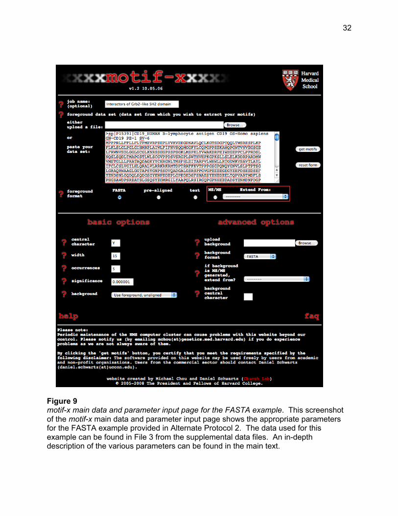
Figure 9 motif-x main data and parameter input page for the FASTA example. This screenshot of the motif-x main data and parameter input page shows the appropriate parameters for the FASTA example provided in Alternate Protocol 2. The data used for this example can be found in File 3 from the supplemental data files. An in-depth description of the various parameters can be found in the main text.
33
Data should be uploaded or pasted in the same format as shown in the FASTA
example in Table 1 or the raw sequence data provided as a supplemental data file
(see Supplemental Data Files, File 3). The FASTA data format allows one or more
protein sequences in a text file each of which can be preceded by a description line
that begins with a “greater than” (“>”) character, followed by biological sequence
starting on the line below. In contrast to the previously described MS/MS format,
which allows for special characters to designate modification sites (“#”, “@”, “*”), the
FASTA data format does not allow for the inclusion of special characters except in
the description lines, which are ignored by motif-x. It is important not to simply merge
all protein sequences together without description lines because sequences at the
junctions would form non-meaningful peptide sequences when extracted by motif-x.
4. In the “foreground format” field select the “FASTA” option.
This option indicates that the inputted data is in the FASTA data format and instructs
the motif-x web tool on the proper preprocessing of the data.
5. Under the “basic options” heading enter an appropriate central character into the
“central character” field.
A single (case sensitive) character should be entered. As mentioned in Step 3
above, combinations of residues and special characters may not be used in FASTA
analyses. In the present example, since it is known a priori that the SH2 domain
interacts with proteins bearing YxN motifs, “Y” is entered as the central character

34
(see Figure 9). However, performing a complete and unbiased motif search in a set
of proteins requires 20 independent motif-x analyses centered at each of the 20
amino acids.
6. Enter the desired motif width into the “width” field.
Any odd value between 3 and 35 can be chosen as a motif width; however, most
short linear protein motifs are under 15 amino acids long. In the present SH2 domain
example, a width of 15 has been used (see Figure 9).
7. Enter into the “occurrences” field the minimal number of times that you require your
extracted motifs to occur within the foreground data set.
This parameter can be used to tune the specificity of motifs since motifs with greater
specificity (i.e., more “fixed” positions) are expected to occur less often than those
motifs with lower specificity. Users that wish to extract a maximal number of motifs
should set this parameter to a low value (e.g., 5) and rely solely on the significance
parameter (see step 8) to extract motifs. It should be noted that raising the
occurrences threshold to a high value (e.g., 50) on a relatively small FASTA data set
(e.g., 5 proteins) is likely to result in no extracted motifs since it is unlikely that any
sequence pattern would occur such a large number of times in a small data set. An
occurrences threshold of 5 was used for the present example.
8. Enter the significance threshold for the analysis into the “significance” field.
35
An explanation of this parameter is provided in Step 8 of the previously described
Basic Protocol. A significance threshold of 0.000001 was used for the present
example.
9. In the “background” field select the appropriate background data set for the analysis.
motif-x evaluates the statistical significance of positions within motifs through a
comparison to a background data set. Although it is highly preferable to select the
background proteomic database corresponding to the organism from which the
foreground data was derived, FASTA-formatted data sets provide a unique
opportunity to use statistical characteristics from the foreground data set to derive a
relevant background data set. On the motif-x web site this option is labeled “Use
foreground, unaligned” in the drop-down menu. Because our SH2 domain example
lacks an obvious proteomic background (due to the fact that the set of 10 proteins are
derived from a variety of species), we have selected the “Use foreground, unaligned”
option in this example to illustrate its usefulness (see Figure 9). Alternatively, users
could select the background organism database that is most closely related to the
foreground data set (in the present example, this would correspond to the IPI Human
database).
36
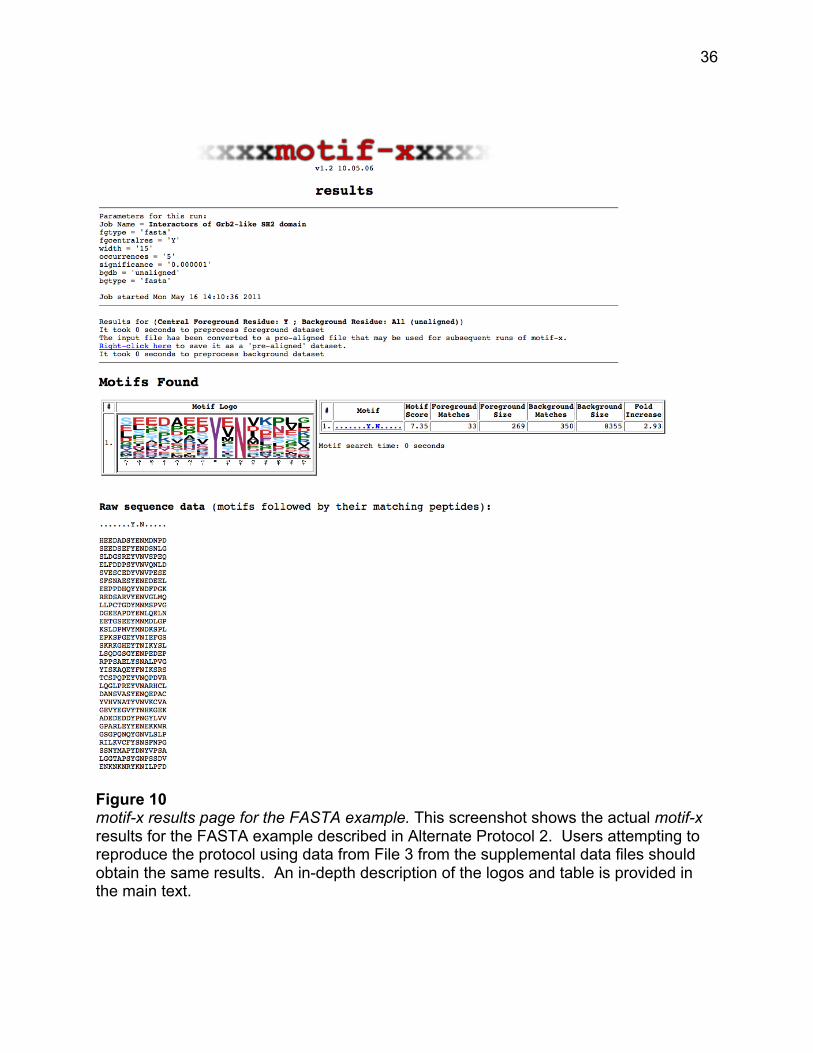
Figure 10 motif-x results page for the FASTA example. This screenshot shows the actual motif-x results for the FASTA example described in Alternate Protocol 2. Users attempting to reproduce the protocol using data from File 3 from the supplemental data files should obtain the same results. An in-depth description of the logos and table is provided in the main text.
37
10. Press the “get motifs” button.
An explanation of the result of pressing “get motifs” is provided in the Basic
Protocol, Step 10.
11. View the motif-x results page.
The motif-x results page for FASTA format analyses is nearly the same as the
previously described results page for MS/MS analyses (see Step 11 of the Basic
Protocol); however, due to the lack of significant peptide preprocessing in a FASTA
analysis, the preprocessing statistics are minimal. A screenshot of the motif-x results
page for the worked out example is shown in Figure 10. In the example, a single
motif, YxN, was extracted from the data set corresponding to the known specificity of
the Grb2 SH2 domain.
GUIDELINES FOR UNDERSTANDING RESULTS
The normal output of motif-x is a list of motifs for the given input data. Whereas
previous studies sometimes indicate the existence of some flexibility at motif residues,
motif-x deconvolutes input data into distinct and different motifs thereby avoiding over-
generalizing its findings. For instance, a hypothetical motif shown in the literature as
Tx[K/R][K/R] may be shown by motif-x as separate motifs for TxRR, TxKK and TxKR,
but not TxRK, which means that the data set supplied did not achieve significance for
that motif. It is possible that with a larger foreground data set, such a motif would
38
emerge as an additional motif in the series; however, given today’s large data sets, it is
more likely that such a motif is actually disfavored in comparison with the others.
The interpretation of the results will depend on the nature of the input data. If the
user supplies substrate data that results from just one enzyme, then one or more motifs
may emerge as preferred for that enzyme. However, shotgun proteomic data is likely to
yield a collection of motifs that are the result of multiple enzymes. Such data is useful if
one is predicting motifs that direct modification as opposed to understanding single
enzyme-substrate specificity.
Note that the results page is a unique URL, and may be bookmarked and
revisited for up to a week before it is archived. If the URL is no longer available, the user
will get an error indicating that it is expired.
COMMENTARY
Background Information
Understanding cellular functions in normal and abnormal cells ultimately depends
on understanding the molecular interactions within the cell, which includes enzymatic
and binding reactions between proteins. Computational analyses of protein sequences,
such as the determination of secondary structure, local hydrophobicity, signaling
peptide sequences and domain architecture, have all increased our understanding of
protein functions and mechanisms.
Within the enzyme-substrate subset of protein-protein interactions, it is often the
chemical reactivity of a particular amino acid side chain within the substrate or enzyme
that is a prerequisite for catalysis. Such residues thereby firmly fix at least one amino
39
acid in an enzyme or substrate that participates in the reaction. However, a repertoire
of 20 amino acids still provides little specificity, and nature has evolved secondary sites
within enzymes and substrates to further direct specificity.
Such short sequences within substrates or interaction partners have been shown
in many cases to be necessary and/or sufficient to direct highly specific interactions
including proteolysis and enzyme-catalyzed post-translational modifications, such as
phosphorylation, sumoylation, glycosylation and others (Howard et al., 1991; Songyang
and Cantley, 1995; Shakin-Eshleman et al., 1996; Sternsdorf et al., 1999).
Understanding the crucial residues and positions that are the primary
determinants of substrate specificity or protein-protein interactions can be used to
predict biological activities and mechanisms. This can be performed, for instance using
a companion tool known as scan-x (http://scan-x.med.harvard.edu) described in an
upcoming supplement of Current Protocols in Bioinformatics.
Partly because of the importance of phosphorylation and proteolysis in biology
and partly because of ease of measurement, the discovery of motifs for kinases and
proteases using biochemical means has historically led the field.
Recent advances in mass spectrometry are helping to provide thousands of new
measurements with each high-throughput paper that is published, and now data sets for
many protein post-translational modifications are quite large. However, understanding of
the enzymes responsible for these modifications is lagging behind. Knowing the identity
of preferred residues proximal to an enzymatically modified residue on a substrate
improves understanding of interactions and helps to generate hypotheses about other
40
possible interactions. Using motif-x to discover motifs can provide important information
about known enzymes and enzymes yet to be discovered in an unsupervised manner.
Often laborious bench work is required to understand biological mechanisms.
The implementation of motif-x to deconvolute sequence data into potential interaction
motifs can be used to dramatically improve the generation and testing of biological
hypotheses.
Critical Parameters
Central Character (default: blank)
Once input data sets are processed, it is this residue that all foreground
sequences will be centered upon. This is typically a site with a known or putative post-
translational modification (PTM). For example, in the case of eukaryotic
phosphorylation, one might use serine, threonine or tyrosine as a central residue. For
mass spectrometry input data, the motif-x user will usually want to distinguish between
a modified or unmodified residue in input data because, e.g., not every serine will be
modified.
Therefore, in the foreground data set, the residues may be specified with or
without a trailing symbol within MS/MS foreground data. Thus, in a list of hypothetical
input sequences, one may have the tryptic peptide, MACRTISPWESR, where only the
first S is phosphorylated. In that case, the input sequence should be modified to be
MACRTIS*PWESR. If the input has more than one modified residue per sequence (e.g.,
MACRTIS*PWES*R) then motif-x will attempt to create a fixed length sequence
surrounding each one and will be properly handled as two separate phosphorylation
41
events by motif-x. This format is the natural output format of SEQUEST, and the output
from other spectral identification pipelines will have to be processed into this format if it
is not already compliant. Once a decision has been made by the user to focus on a
particular modified residue, that same symbol can be used as the central residue. So, in
this instance, the user would specify ‘S*’ as the foreground central residue.
Note that any modified residue to be centered upon is limited to the repertoire of
symbols: “*”, “#” or “@” which must always be placed after the modified residue. While it
is acceptable for foreground data sets to have multiple different symbols (indicating
different modification types for a given peptide e.g., M@ACRTIS*PWES*R or
MACRT*IS*PWES*R), if a modified residue is to be used as a central residue, the user
must use the same residue and modifier symbol (e.g., ‘S*’) in the “central character”
field (all other symbols will be ignored for that particular analysis). Note that it is also
acceptable to use a single letter as the central residue, which will match every
occurrence of that residue regardless of modification state.
Because each analysis is specific for a residue, to find motifs centered on each
of the eukaryotic phosphorylated residues in a single data set, one can simply change
the central residue from ‘S*’ to ‘T*’ to ‘Y*’ for each of three analyses.
Width (default: 15)
The default value for the width parameter is 15 residues (i.e., 7 residues on either
side of the central residue). The user may increase this value to as high as 35, but we
have rarely found residues that are significantly correlated if they are very distant from
the central residue.
42
Occurrences (default: 20)
Normally, motif-x stops and outputs the current motif when it cannot find
additional residues that are significantly different than the background. However, an
additional stopping criterion occurs when the size of the foreground data set is reduced
to below this occurrence threshold. Typically, this is set at some low number based on
how few peptides are expected in each motif class. In general it is recommended that
the significance threshold be set in such a way that minimum foreground occurrences
are not invoked by the algorithm.
Significance (default: 0.000001)
Each step of the algorithm tests the significance of all residue/position pairs with
respect to the background frequency of those same residue/positions. The significance
threshold used by motif-x for this test is a user configurable parameter. Typically a
statistical significance (!) would be set at 0.05 or 0.01, but each step of the motif-x
algorithm is testing multiple hypotheses. Therefore this value must be reduced
accordingly. Version 1.2 of motif-x does not automatically perform Bonferroni correction,
so it must be done by the user to protect against the generation of false positives
(Bonferroni, 1935).
We recommend at least dividing your desired alpha value by: (width – 1) x 20.
So, for instance, if you want to use an alpha of 0.05, and a width of 15 for protein
analysis (i.e., 20 amino acids), then 0.05/(14x20) = 0.05/280 = 0.00018. Thus, using a
number higher than this will likely yield motifs that are not statistically significant. The
43
default is currently set at 0.000001, which corresponds to an alpha value of 0.0003 for a
width of 15. This threshold is very conservative and generally works quite well.
Background (default: IPI Human Proteome)
As mentioned previously, the background data set is the source of statistics for
each residue/position, and using the right organism will provide the most relevant
results. Multiple proteomic Standard Database Libraries are provided on the motif-x web
site, and they continue to be added over time by request to the authors.
A background data set size is typically more than an order of magnitude larger
than the foreground data set size. Users may also provide background data sets in a
number of different formats (pre-aligned, MS/MS and FASTA/text), but Standard
Database Libraries are in FASTA format by default, and using a Standard Database
Library is the most typical way to run motif-x.
A special background data set known as “Foreground Unaligned” can be
generated from the foreground data set if the foreground is in FASTA format. Using this
option, instead of processing a background data set to be centered on just one
particular residue, it is aligned on every single residue and then used as the
background. An example of the utility of this approach is shown in Alternative Protocol
2. Essentially, this allows for the discovery of motifs for a particular central residue
evaluated in comparison to all other residues. “Foreground unaligned” can also be used
when other suitable backgrounds cannot be determined.
Background central character (default: blank)
44
In general, one should set this to the same thing as the foreground central
residue without a modifier, if any. So, for instance, if the foreground central residue is
“S*”, then use “S”’ as the background central residue, if it is “T*” then use “T” as the
background central residue, and so forth. If this field is left blank, it will default to the
foreground central residue without any modifier.
Troubleshooting
Two extreme cases occur with some regularity:
1) No Motifs Found. When no motifs are found with the given input parameters, the
message that is reported in the output is “Motifs Found: None”. While it is certainly
possible that no motifs can be found at a significant level some suggested actions
are:
a) Double check that you have inputted a non-empty foreground data set and that it
contains a sufficient amount of data. It is nearly impossible to find significant motifs
with a single protein, or only a dozen MS/MS peptides. There must be enough data
to obtain at least one significant motif.
b) If performing an MS/MS analysis, download the pre-aligned data set from the
hyperlink at the end of the first section (shown as “Right-click here” in Figure 4) to
see if the pre-processing seems reasonable. There may be an obvious syntactic
45
error in the input data or specification of the central residue.
c) Double check the significance and occurrences thresholds to make sure they are
reasonable. Setting an occurrence threshold at a low number, while maintaining the
significance parameter, will still only yield statistically significant motifs even in a
small set.
d) Increase the amount of foreground input data that you have. Note that identical
peptides are not contained in motif-x’s analysis, so merely duplicating existing data
is not an effective approach. Always use real data sets to get relevant results.
e) Closely examine the foreground data to make sure the central residue is
represented correctly (i.e., “S” versus “s” or “T” versus “T*”, etc.).
f) Make certain that the foreground data is significantly different from the
background. Asking motif-x to find motifs in a data set that closely approximates the
background will not yield significant results.
g) Some data sets simply do not have any significant motifs. For example, current
ubiquitination data sets suggest that there is little evidence that short linear motifs in
the immediate vicinity of the ubiquitinated lysine residue play a primary role in
substrate specificity.
46
Note that significantly correlated residues may exist at a distance outside the scope
of motif-x that nonetheless contribute to specificity. These other contributions to
specificity may be particularly important in cases where motif-x is not otherwise able
to find a strong motif in a data set.
2) Timeouts. Occasionally, an error message will be generated indicating that a
“Timeout” has occurred. While motif-x usually runs very quickly, there are some data
sets that cause the algorithm to take longer than 15 minutes of CPU time to run. This
is usually due to extremely large data sets, and/or a significance parameter that is
too large. However, it can also be due to an overloaded computer cluster. There are
a number of things that can be done to try to generate motifs after a timeout. Any or
all of them may alleviate the timeout:
a) Run the job again at a different time of day.
b) Decrease the value of the significance parameter to a very small number (< 1e-6
or smaller). Because decreasing the value of the significance parameter makes it
harder to find significant motifs, reducing the significance threshold can actually
reduce the output and the run time. If the job completes, the number of CPU
seconds will be shown in the output, and if it is significantly less than 900 seconds
(i.e., 15 minutes), then gradually increase the value of significance towards a desired
value.
47
c) Reduce the size of the foreground data set.
d) Unfortunately, the computer cluster does occasionally become overloaded.
However, the job may still have been run. Before re-running jobs that look like they
failed for no explainable reason, it is a good idea to try to wait a few minutes and use
the “reload” button in the web browser while on the results page. In some cases,
when it refreshes, output may be visible.
A more complete list of error messages and suggested remedies can be found in the
Supplemental Data Files (see File 4). If you continue to experience problems despite
following the troubleshooting guidelines, please contact the authors using the email
address on the web site.
Advanced Parameters (optional)
Advanced options in the current version of motif-x (version 1.2) are limited.
Ability to upload a user-selected background data set
Users may upload a background data set by selecting “uploaded” under the background
parameter, and browsing for the proper file by clicking on the “Browse” button next to
the “upload background” dialog box in the “advanced options” section (see Figures 1, 7,
and 9). With regard to formatting, background data sets may exist in the same four
formats used for foreground data sets (i.e., FASTA, pre-aligned, text, and MS/MS – see
48
Table 1 for examples of correct formatting). As in the case of foreground data sets,
backgrounds in MS/MS format (i.e., proteolytically digested sequence fragments)
require users to indicate the proper database from which to extend using the drop down
menu in the “advanced options” column. Also, as discussed in the preceeding protocol,
special characters (“#”, “@”, “*”) may only be used in conjunction with MS/MS-formatted
backgrounds to denote modified residues. If a residue is not inputted into the
background central character parameter, motif-x will automatically use the same central
character that was inputted for the foreground data set, but without the modifier [so it is
important to be explicit if using an MS/MS background (e.g., S*)].
Use of “text” format
The use of ‘text’ as a foreground or background format is not useful for proteomic
analysis, and may be eliminated in future versions of the software.
Additional organism databases
Users should feel free to contact the authors to have additional commonly used
background databases added to the motif-x web site.
49
Literature Cited
Bonferroni, C.E. 1935. Il calcolo delle assicurazioni su gruppi di teste. In Studi in Onore
del Professore Salvatore Ortu Carboni.13-60.
Dinkel, H., Chica, C., Via, A., Gould, C.M., Jensen, L.J., Gibson, T.J., and Diella, F.
2011. Phospho.ELM: a database of phosphorylation sites--update 2011. Nucleic
Acids Res 39:D261-267.
Durek, P., Schmidt, R., Heazlewood, J.L., Jones, A., MacLean, D., Nagel, A., Kersten,
B., and Schulze, W.X. 2010. PhosPhAt: the Arabidopsis thaliana phosphorylation
site database. An update. Nucleic Acids Res 38:D828-834.
Edwards, R.J., Davey, N.E., and Shields, D.C. 2008. CompariMotif: quick and easy
comparisons of sequence motifs. Bioinformatics 24:1307-1309.
Eng, J.K., Mccormack, A.L., and Yates, J.R. 1994. An Approach to Correlate Tandem
Mass-Spectral Data of Peptides with Amino-Acid-Sequences in a Protein
Database. Journal of the American Society for Mass Spectrometry 5:976-989.
Gould, C.M., Diella, F., Via, A., Puntervoll, P., Gemund, C., Chabanis-Davidson, S.,
Michael, S., Sayadi, A., Bryne, J.C., Chica, C., Seiler, M., Davey, N.E., Haslam,
N., Weatheritt, R.J., Budd, A., Hughes, T., Pas, J., Rychlewski, L., Trave, G.,
Aasland, R., Helmer-Citterich, M., Linding, R., and Gibson, T.J. 2010. ELM: the
status of the 2010 eukaryotic linear motif resource. Nucleic Acids Res 38:D167-
180.
50
Hornbeck, P.V., Chabra, I., Kornhauser, J.M., Skrzypek, E., and Zhang, B. 2004.
PhosphoSite: A bioinformatics resource dedicated to physiological protein
phosphorylation. Proteomics 4:1551-1561.
Howard, A.D., Kostura, M.J., Thornberry, N., Ding, G.J., Limjuco, G., Weidner, J.,
Salley, J.P., Hogquist, K.A., Chaplin, D.D., Mumford, R.A., and et al. 1991. IL-1-
converting enzyme requires aspartic acid residues for processing of the IL-1 beta
precursor at two distinct sites and does not cleave 31-kDa IL-1 alpha. J Immunol
147:2964-2969.
Hutti, J.E., Jarrell, E.T., Chang, J.D., Abbott, D.W., Storz, P., Toker, A., Cantley, L.C.,
and Turk, B.E. 2004. A rapid method for determining protein kinase
phosphorylation specificity. Nat Methods 1:27-29.
Keshava Prasad, T.S., Goel, R., Kandasamy, K., Keerthikumar, S., Kumar, S.,
Mathivanan, S., Telikicherla, D., Raju, R., Shafreen, B., Venugopal, A.,
Balakrishnan, L., Marimuthu, A., Banerjee, S., Somanathan, D.S., Sebastian, A.,
Rani, S., Ray, S., Harrys Kishore, C.J., Kanth, S., Ahmed, M., Kashyap, M.K.,
Mohmood, R., Ramachandra, Y.L., Krishna, V., Rahiman, B.A., Mohan, S.,
Ranganathan, P., Ramabadran, S., Chaerkady, R., and Pandey, A. 2009. Human
Protein Reference Database--2009 update. Nucleic Acids Res 37:D767-772.
Kessels, H.W., Ward, A.C., and Schumacher, T.N. 2002. Specificity and affinity motifs
for Grb2 SH2-ligand interactions. Proc Natl Acad Sci U S A 99:8524-8529.
Koyasu, S., Tse, A.G., Moingeon, P., Hussey, R.E., Mildonian, A., Hannisian, J.,
Clayton, L.K., and Reinherz, E.L. 1994. Delineation of a T-cell activation motif
51
required for binding of protein tyrosine kinases containing tandem SH2 domains.
Proc Natl Acad Sci U S A 91:6693-6697.
Matic, I., Schimmel, J., Hendriks, I.A., van Santen, M.A., van de Rijke, F., van Dam, H.,
Gnad, F., Mann, M., and Vertegaal, A.C. 2010. Site-specific identification of
SUMO-2 targets in cells reveals an inverted SUMOylation motif and a
hydrophobic cluster SUMOylation motif. Mol Cell 39:641-652.
Perkins, D.N., Pappin, D.J., Creasy, D.M., and Cottrell, J.S. 1999. Probability-based
protein identification by searching sequence databases using mass spectrometry
data. Electrophoresis 20:3551-3567.
Rigbolt, K.T., Prokhorova, T.A., Akimov, V., Henningsen, J., Johansen, P.T.,
Kratchmarova, I., Kassem, M., Mann, M., Olsen, J.V., and Blagoev, B. 2011.
System-wide temporal characterization of the proteome and phosphoproteome of
human embryonic stem cell differentiation. Sci Signal 4:rs3.
Schwartz, D. and Gygi, S.P. 2005. An iterative statistical approach to the identification
of protein phosphorylation motifs from large-scale data sets. Nat Biotechnol
23:1391-1398.
Shakin-Eshleman, S.H., Spitalnik, S.L., and Kasturi, L. 1996. The amino acid at the X
position of an Asn-X-Ser sequon is an important determinant of N-linked core-
glycosylation efficiency. J Biol Chem 271:6363-6366.
Songyang, Z. and Cantley, L.C. 1995. Recognition and specificity in protein tyrosine
kinase-mediated signalling. Trends Biochem Sci 20:470-475.
52
Sternsdorf, T., Jensen, K., Reich, B., and Will, H. 1999. The nuclear dot protein sp100,
characterization of domains necessary for dimerization, subcellular localization,
and modification by small ubiquitin-like modifiers. J Biol Chem 274:12555-12566.
Van Hoof, D., Munoz, J., Braam, S.R., Pinkse, M.W., Linding, R., Heck, A.J., Mummery,
C.L., and Krijgsveld, J. 2009. Phosphorylation dynamics during early
differentiation of human embryonic stem cells. Cell Stem Cell 5:214-226.
Key Reference
Schwartz and Gygi, 2005. See above.
Original description of the motif-x algorithm.
Internet Resources
http://motif-x.med.harvard.edu
Home page for the motif-x web tool (note: the protocols in this manuscript pertain to
motif-x version 1.2).
Related Documents


