Review Biological activity of ruthenium nitrosyl complexes Elia Tfouni b , Daniela Ramos Truzzi a , Aline Tavares a , Anderson Jesus Gomes c , Leonardo Elias Figueiredo b , Douglas Wagner Franco a,⇑ a Departamento de Química e Física Molecular, Instituto de Química de São Carlos, Universidade de São Paulo, São Carlos, SP, Brazil b Departamento de Química, Faculdade de Filosofia, Ciências e Letras de Ribeirão Preto, Universidade de São Paulo, Ribeirão Preto, SP, Brazil c Faculdade de Ceilândia, Universidade de Brasília, Brasília, DF, Brazil article info Article history: Received 2 September 2011 Revised 29 November 2011 Available online 7 December 2011 Keywords: Ruthenium nitrosyls Nitric oxide carriers Reactivity and biologic activity abstract Nitric oxide plays an important role in various biological processes, such as neurotransmission, blood pressure control, immunological responses, and antioxidant action. The control of its local concentration, which is crucial for obtaining the desired effect, can be achieved with exogenous NO-carriers. Coordina- tion compounds, in particular ruthenium(III) and (II) amines, are good NO-captors and -deliverers. The chemical and photochemical properties of several ruthenium amine complexes as NO-carriers in vitro and in vivo have been reviewed. These nitrosyl complexes can stimulate mice hippocampus slices, pro- mote the lowering of blood pressure in several in vitro and in vivo models, and control Trypanosoma cruzi and Leishmania major infections, and they are also effective against tumor cells in different models of can- cer. These complexes can be activated chemically or photochemically, and the observed biological effects can be attributed to the presence of NO in the compound. Their efficiencies are explained on the basis of the [Ru II NO + ] 3+ /[Ru II NO 0 ] 2+ reduction potential, the specific rate constant for NO liberation from the [RuNO] 2+ moiety, and the quantum yield of NO release. Ó 2011 Published by Elsevier Inc. Contents Introduction............................................................................................................ 38 Nitric oxide activation on ruthenium(II) nitrosyl complexes ..................................................................... 41 Biological molecules and nitric oxide activation on ruthenium(II) nitrosyl complexes ............................................ 41 Photochemical activation ............................................................................................. 41 Biological tests.......................................................................................................... 42 Vasodilation and vasoconstriction ...................................................................................... 42 Ruthenium nitrosyls as anti-infective agents ............................................................................. 44 Cancer ............................................................................................................ 46 Immobilization of ruthenium nitrosyl complexes .......................................................................... 48 Conclusions and perspectives .............................................................................................. 48 References ........................................................................................................... 49 Introduction Coordination compounds have long been used as metallophar- maceuticals [1–3]. There is a wide range of metal complexes that have biological activities and applications for diseases, such as can- cer [4–6], cardiovascular problems [7], arthritis [8], and parasitosis [9], and some of these complexes are commercially available. The best known and most studied metal drugs are the anticancer com- pounds of platinum, which have been used since the antitumor activity of cisplatin (cis-diamminedichloridoplatinum(II), [cis- Pt II (NH 3 ) 2 Cl 2 ]) was discovered in the 1960s [3]. The continued development of metal complexes has sought to overcome the drug resistance and side effects of [cis-Pt II (NH 3 ) 2 Cl 2 ]. Iron complexes have been extensively used in the treatment of anemia, cancer, and cardiovascular problems. Iron(II) citrate is a classical anemia treatment [10]; ferrocenyl derivatives are antican- cer drugs for breast cancer [11], while sodium nitroprusside (SNP) 1089-8603/$ - see front matter Ó 2011 Published by Elsevier Inc. doi:10.1016/j.niox.2011.11.005 ⇑ Corresponding author. Fax: +55 16 3373 9976. E-mail address: [email protected] (D.W. Franco). Nitric Oxide 26 (2012) 38–53 Contents lists available at SciVerse ScienceDirect Nitric Oxide journal homepage: www.elsevier.com/locate/yniox

Welcome message from author
This document is posted to help you gain knowledge. Please leave a comment to let me know what you think about it! Share it to your friends and learn new things together.
Transcript
Nitric Oxide 26 (2012) 38–53
Contents lists available at SciVerse ScienceDirect
Nitric Oxide
journal homepage: www.elsevier .com/locate /yniox
Review
Biological activity of ruthenium nitrosyl complexes
Elia Tfouni b, Daniela Ramos Truzzi a, Aline Tavares a, Anderson Jesus Gomes c, Leonardo Elias Figueiredo b,Douglas Wagner Franco a,⇑a Departamento de Química e Física Molecular, Instituto de Química de São Carlos, Universidade de São Paulo, São Carlos, SP, Brazilb Departamento de Química, Faculdade de Filosofia, Ciências e Letras de Ribeirão Preto, Universidade de São Paulo, Ribeirão Preto, SP, Brazilc Faculdade de Ceilândia, Universidade de Brasília, Brasília, DF, Brazil
a r t i c l e i n f o
Article history:Received 2 September 2011Revised 29 November 2011Available online 7 December 2011
Keywords:Ruthenium nitrosylsNitric oxide carriersReactivity and biologic activity
1089-8603/$ - see front matter � 2011 Published bydoi:10.1016/j.niox.2011.11.005
⇑ Corresponding author. Fax: +55 16 3373 9976.E-mail address: [email protected] (D.W. Franco)
a b s t r a c t
Nitric oxide plays an important role in various biological processes, such as neurotransmission, bloodpressure control, immunological responses, and antioxidant action. The control of its local concentration,which is crucial for obtaining the desired effect, can be achieved with exogenous NO-carriers. Coordina-tion compounds, in particular ruthenium(III) and (II) amines, are good NO-captors and -deliverers. Thechemical and photochemical properties of several ruthenium amine complexes as NO-carriers in vitroand in vivo have been reviewed. These nitrosyl complexes can stimulate mice hippocampus slices, pro-mote the lowering of blood pressure in several in vitro and in vivo models, and control Trypanosoma cruziand Leishmania major infections, and they are also effective against tumor cells in different models of can-cer. These complexes can be activated chemically or photochemically, and the observed biological effectscan be attributed to the presence of NO in the compound. Their efficiencies are explained on the basis ofthe [RuIINO+]3+/[RuIINO0]2+ reduction potential, the specific rate constant for NO liberation from the[RuNO]2+ moiety, and the quantum yield of NO release.
� 2011 Published by Elsevier Inc.
Contents
Introduction. . . . . . . . . . . . . . . . . . . . . . . . . . . . . . . . . . . . . . . . . . . . . . . . . . . . . . . . . . . . . . . . . . . . . . . . . . . . . . . . . . . . . . . . . . . . . . . . . . . . . . . . . . . . 38Nitric oxide activation on ruthenium(II) nitrosyl complexes . . . . . . . . . . . . . . . . . . . . . . . . . . . . . . . . . . . . . . . . . . . . . . . . . . . . . . . . . . . . . . . . . . . . . 41
Biological molecules and nitric oxide activation on ruthenium(II) nitrosyl complexes . . . . . . . . . . . . . . . . . . . . . . . . . . . . . . . . . . . . . . . . . . . . 41Photochemical activation . . . . . . . . . . . . . . . . . . . . . . . . . . . . . . . . . . . . . . . . . . . . . . . . . . . . . . . . . . . . . . . . . . . . . . . . . . . . . . . . . . . . . . . . . . . . . 41
Biological tests. . . . . . . . . . . . . . . . . . . . . . . . . . . . . . . . . . . . . . . . . . . . . . . . . . . . . . . . . . . . . . . . . . . . . . . . . . . . . . . . . . . . . . . . . . . . . . . . . . . . . . . . . . 42
Vasodilation and vasoconstriction . . . . . . . . . . . . . . . . . . . . . . . . . . . . . . . . . . . . . . . . . . . . . . . . . . . . . . . . . . . . . . . . . . . . . . . . . . . . . . . . . . . . . . 42Ruthenium nitrosyls as anti-infective agents . . . . . . . . . . . . . . . . . . . . . . . . . . . . . . . . . . . . . . . . . . . . . . . . . . . . . . . . . . . . . . . . . . . . . . . . . . . . . 44Cancer . . . . . . . . . . . . . . . . . . . . . . . . . . . . . . . . . . . . . . . . . . . . . . . . . . . . . . . . . . . . . . . . . . . . . . . . . . . . . . . . . . . . . . . . . . . . . . . . . . . . . . . . . . . . 46Immobilization of ruthenium nitrosyl complexes. . . . . . . . . . . . . . . . . . . . . . . . . . . . . . . . . . . . . . . . . . . . . . . . . . . . . . . . . . . . . . . . . . . . . . . . . . 48Conclusions and perspectives . . . . . . . . . . . . . . . . . . . . . . . . . . . . . . . . . . . . . . . . . . . . . . . . . . . . . . . . . . . . . . . . . . . . . . . . . . . . . . . . . . . . . . . . . . . . . . 48References . . . . . . . . . . . . . . . . . . . . . . . . . . . . . . . . . . . . . . . . . . . . . . . . . . . . . . . . . . . . . . . . . . . . . . . . . . . . . . . . . . . . . . . . . . . . . . . . . . . . . . . . . . . 49
Introduction
Coordination compounds have long been used as metallophar-maceuticals [1–3]. There is a wide range of metal complexes thathave biological activities and applications for diseases, such as can-cer [4–6], cardiovascular problems [7], arthritis [8], and parasitosis[9], and some of these complexes are commercially available. The
Elsevier Inc.
.
best known and most studied metal drugs are the anticancer com-pounds of platinum, which have been used since the antitumoractivity of cisplatin (cis-diamminedichloridoplatinum(II), [cis-PtII(NH3)2Cl2]) was discovered in the 1960s [3]. The continueddevelopment of metal complexes has sought to overcome the drugresistance and side effects of [cis-PtII(NH3)2Cl2].
Iron complexes have been extensively used in the treatment ofanemia, cancer, and cardiovascular problems. Iron(II) citrate is aclassical anemia treatment [10]; ferrocenyl derivatives are antican-cer drugs for breast cancer [11], while sodium nitroprusside (SNP)

E. Tfouni et al. / Nitric Oxide 26 (2012) 38–53 39
(Na2[FeII(NO+)(CN)5]) is a vasodilator drug that is able to deliver ni-tric oxide (NO) by reduction in vivo [1,12,13]. Although SNP isadministered in the clinic [14], there are problems associated withits use, such as a susceptibility to photolysis and its oxidativebreakdown through the action of the immune system, ultimatelyreleasing cyanide [7].
Osmium compounds have not been well-studied as metal-based drugs, likely due to the higher cost of the metal, the in-creased difficulty associated with compound synthesis [15], andits high toxicity [16]. In addition, the ligand exchange rates in os-mium compounds are not favorable on the timescale of cellularprocesses and are approximately 105 times lower than those ofthe corresponding ruthenium complexes [17]. However, the kineticlability of osmium complexes can be tuned so that their anticanceractivity approaches the range of cellular processes [18].
Metallopharmaceuticals can also be activated by irradiationwith light [19–35]. Possibly, the best known examples of applica-tion are complexes designed for photodynamic therapy (PDT)[20], such as in the treatment of neoplastic tissues. The key compo-nents of PDT are a photosensitizer, light, and tissue oxygen. Theabsorption of light by a photosensitizer molecule can lead to en-ergy transfer to activate another molecule, such as the conversionof O2 to the excited singlet state (1O2) and other free radicals, suchas �OH, HO�2, O��2 , which induce damage to membranes, DNA, andother cell structures [20,36].
Of the metallo drugs, ruthenium-based complexes have re-ceived considerable attention for medical applications [19,37,38].Their kinetic behavior is similar to platinum with respect to cellu-lar division processes [19,39,40], but their toxicity is lower thanthat of cisplatin [41], probably due to their ability to mimic ironand therefore bind to many biomolecules, such as human serumalbumin and the iron transport protein transferrin [37,38].
According to its coordination sphere composition, rutheniumhas several oxidation states: Ru(II), Ru(III), and Ru(IV). Most ofthese oxidation states are accessible under physiological condi-tions [37]. Ru(III) complexes serve as precursors to Ru(II) by reduc-tion in vivo by biological reductants such as glutathione andascorbic acid [19,37,42]. Ru(II) centers are more reactive thanRu(III) in substitution reactions and are able to coordinate rapidlyand bind strongly to nitrogen and sulfur atoms available inbiomolecules.
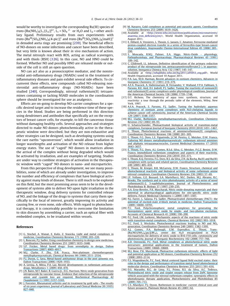
The biological activities of ruthenium complexes were first re-ported in the 1950s (Fig. 1a) [43–46] and have been reviewed else-where [19,47]. As an example, fac-[RuIII(NH3)3Cl3] exhibitsanticancer activity but is not soluble enough in aqueous solutions[48] for use in the clinic. Since then, a series of ruthenium com-plexes have been studied [49,50]. NAMI-A (trans-[RuIII(dmso-S)(Im)Cl4]ImH, dmso = dimethyl sulfoxide, Im = imidazole)
Fig. 1. (a) Dwyer compound, (b) NAMI-A, and (c) KP-1019 structures.
(Fig. 1b) inhibits the formation and growth of metastases [51],and KP-1019 (trans-[RuIII(Ind)2Cl4]IndH, Ind = indazole) (Fig. 1c) in-duces apoptosis in colorectal carcinoma cells [52]. Both are cur-rently the most promising Ru(III) complexes in cancer treatment;NAMI-A is the first anticancer ruthenium compound to success-fully complete Phase I Clinical Trials and will enter Phase II Trials,whereas KP-1019 is in Phase I Clinical Trials [53].
Ruthenium(III) and (II) tetraamines (d5 and d6 low spin) consti-tute a very interesting class of compounds for medicinal chemistrystudies because of their water solubility, stability in aqueous med-ium, and low cytotoxicity [22,25,54–58]. The amines, which areoriented along the x- and y-axes of the pseudo-octahedral com-plexes, are inert ligands that enable substitution reactions to takeplace only in the z direction. Depending on the nature of the li-gands and the ruthenium oxidation state, the photochemical reac-tivity can differ from the chemical one [21–25,59,60]. Thus, giventhe chemical and photochemical properties of this class of com-pounds [22–25,59–61], they are attractive candidates for develop-ing a metallopharmaceutical platform.
In this respect, ruthenium compounds could be useful as deliv-ery or scavenging agents for molecules that have a crucial role inphysiology. Nitric oxide (NO), for instance, is an endogenous mol-ecule that is produced by nitric oxide synthase (NOS) through theconversion of L-arginine to L-citrulline and NO [62]. There are threeNOS isozymes: the endothelial and neuronal (eNOS and nNOS) iso-forms, which are dependent on intracellular Ca2+ concentrationand expressed constitutively in several tissues, and the inducibleNOS (iNOS), which are Ca2+-independent and activated by pro-inflammatory stimuli [63,64]. An important difference betweenthe NOS isoforms is that the constitutive type produces low NOconcentrations in the nmol L�1 range, while the inducible formproduces NO in the lmol L�1 range [63]. Thus, nitric oxide playsan important role in a variety of physiological functions, such asblood pressure control [65], neurotransmission [66–68], immuno-logical responses [55,69,70], and antioxidant action [71–73]. Theseeffects are quite dependent on the local NO bioavailability and con-centration [74]. Therefore, intense efforts have been devoted todeveloping compounds that deliver NO efficiently and in a con-trolled manner in physiological medium, including NONOates[75–77], S-nitrosothiols [78–80], and metal complexes [27,30,81–83], such as ruthenium(II) nitrosyl compounds [22–25,34,59,60,84–92], and also to developing scavengers of NO, suchas ruthenium(III) edta and cyclam compounds [24,93–96].
Ruthenium tetraammine and tetraazamacrocyclic nitrosyl com-pounds [22,24,25,97–99] are of great interest as NO-donors due totheir outstanding properties, as the low cytotoxicity against hostcells, water solubility, stability to air oxidation and tailoring reac-tivity possibilities of the coordinated NO, by the choice of the axialligand (L2) in trans-[RuII(NO+)(L1)x(L2)]n+ [25,97–100]. In addition,there is also the possibility to immobilize these complexes intomatrices such as silica, organic inorganic hybrid materials, poly-urethane, poly(vinyl alcohol), poly(methylmethacrylate), and den-drimers [23]. Many of these NO-donors have presented biologicalactivities against several diseases. The challenges of providing anew platform based on nitric oxide are to overcome the resistancethat some actual drugs and treatments have encountered, and tobe site specific in order to minimize side effects.
trans-[RuII(NO+)(L1)x(L2)]n+ complexes have been widely studied[21–23,25,61,101,102]. In these complexes, the L2 ligand is veryimportant because of its ability to modulate, based on its ownproperties (r-donation, p-acceptation, orbital energy and symme-try), the reduction potential of the NO+ ligand (ENOþ=NO0 )1, the NO+
1 Considering the canonical form [RuII-NO+]; other forms include [RuIII-NO0] and[RuIV-NO�] for the [RuNO]6 moiety [59,103].
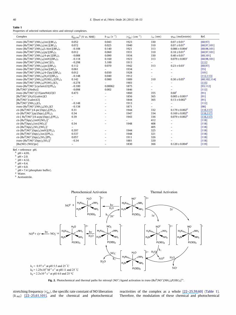
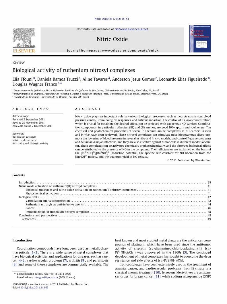
Table 1Properties of selected ruthenium nitro and nitrosyl complexes.
Complex EþNO/NO0 (V vs. NHE) k-NO (s�1) ðmþNOÞ (cm�1) kirr (nm) uNO (mol/estein) Ref.
trans-[RuII(NO+)(NH3)4(isn)](BF4)3 0.052 0.043 1923 330 0.07 ± 0.01a [60,97]trans-[RuII(NO+)(NH3)4(nic)](BF4)3 0.072 0.025 1940 310 0.07 ± 0.01b [60,97,101]trans-[RuII(NO+)(NH3)4(L-hist)](BF4)3 �0.108 0.140 1921 313 0.086 ± 0.004c [60,98,101]trans-[RuII(NO+)(NH3)4(py)](BF4)3 0.012 0.060 1931 310 0.18 ± 0.01d [60,97,101]trans-[RuII(NO+)(NH3)4(4-pic)](BF4)3 �0.008 0.090 1934 330 0.40 ± 0.01e [60,101]trans-[RuII(NO+)(NH3)4(imN)](BF4)3 �0.118 0.160 1923 313 0.079 ± 0.003c [60,98,101]trans-[RuII(NO+)(NH3)4(imC)]Cl3 �0.298 5.100 1913 – – [111]trans-[RuII(NO+)(NH3)4(pz)](BF4)3 0.112 0.070 1942 313 0.23 ± 0.03c [60,97]trans-[RuII(NO+)(NH3)4(ina)](BF4)3 0.061 – 1934 – – [55]trans-[RuII(NO+)(NH3)4(4-Clpy)](BF4)3 0.012 0.030 1928 – – [101]trans-[RuII(NO+)(NH3)4(H2O)](BF4)3 �0.148 0.040 1912 – – [112,113]trans-[RuII(NO+)(NH3)4(P(OEt)3)](PF6)3 0.142 0.980 1909 310 0.30 ± 0.05b [60,102,114]trans-[RuII(NO+)(NH3)4(P(OH)3)]Cl3 �0.278 – 1903 – – [115]trans-[RuII(NO+)(cyclam)Cl](PF6)2 �0.100 0.00062 1875 – – [93,112][RuII(NO+)(Hedta)] �0.098 0.002 1846 – – [112]trans-[RuII(NO+)([15]aneN4)Cl]Cl 0.473 – 1860 355 0.60f [91][RuII(NO+)(H2O)(salen)]Cl – – 1856 365 0.005 ± 0.001g [91][RuII(NO+)(salen)Cl] – – 1844 365 0.13 ± 0.002h [91][RuII(NO+)(NH3)5]Cl3 �0.148 – 1913 – – [112]trans-[RuII(NO+)(NH3)4(SO3)]Cl �0.138 – 1871 – – [98]cis-[RuII(NO+)(4-pic)(bpy)2](PF6)3 0.31 – 1944 332 0.179 ± 0.002b [116,117]cis-[RuII(NO+)(py)(bpy)2](PF6)3 0.34 – 1947 334 0.169 ± 0.002b [116,117]cis-[ RuII(NO+)(4-acpy)(bpy)2](PF6)3 0.39 – 1943 336 0.079 ± 0.002b [116,117]cis-[RuII(bpy)2(imN)(NO2)]+ – – – 412 – [118]cis-[RuII(bpy)2(isn)(NO2)]+ 0.34 – 1948 408 – [118]cis-[RuII(bpy)2(SO3)(NO2)]– – 405 – [118]cis-[RuII(NO+)(bpy)2(imN)](PF6)3 0.397 – 1944 325 – [118]cis-[RuII(NO+)(bpy)2(isn)](PF6)3 0.537 – 1948 321 – [118]cis-[RuII(NO+)(bpy)2(SO3)]PF6 0.057 – 1911 326 – [118]trans-[RuII(NO+)(bpy)2(SO3)]+ �0.34 – 1881 320 – [118][Ru(NO)-(NO2)pc] – – 1830 366 0.120 ± 0.004g [119]
Ref. = reference �pH.A pH = 4.95.b pH = 2.0.c pH = 4.32.d pH = 6.4.e pH = 6.0.f pH = 7.4 (phosphate buffer).g Water.h Acetonitrile.
Fig. 2. Photochemical and thermal paths for nitrosyl (NO+) ligand activation in trans-[RuII(NO+)(NH3)4P(OEt)3]3+.
40 E. Tfouni et al. / Nitric Oxide 26 (2012) 38–53
stretching frequency ðmþNOÞ, the specific rate constant of NO liberation(k-NO) [22–25,61,101], and the chemical and photochemical
reactivities of the complex as a whole [22–25,59,60] (Table 1).Therefore, the modulation of these chemical and photochemical
E. Tfouni et al. / Nitric Oxide 26 (2012) 38–53 41
properties, promoted by L2, is relevant for tailoring ruthenium com-pounds to selectively deliver or capture NO according to the charac-teristics of each target.
Nitric oxide activation on ruthenium(II) nitrosyl complexes
The trans-[RuII(NO+)(L1)x(L2)]n+ complexes can be activated torelease nitric oxide thermally and/or photochemically. Fig. 2 showsreactions involving these two activation paths for the trans-[RuII
(NO+)(L1)x(L2)]n+, exemplified by trans-[RuII(NO+)(NH3)4P(OEt)3]3+.In the thermal pathway, the NO+ ligand can be reduced by a one
or two electron reductors [104], yielding the [RuII(NO0)]2+or[RuII(NO�)]+ moieties, respectively (Fig. 2). After reduction, neithernitric oxide [25,61,101] nor the nitroxyl anion (NO�) [105] have anaffinity for the metal center, and are released with rate constantsdependent on the trans effect and trans influence promoted bythe trans ligand (L2).
Conceivably, the trans-[RuII(H2O)(L1)x(L2)](n�1)+ generated afterNO or NO�dissociation can react with nitrite (NO�2 ) present inthe plasma (114 ± 11 lmol L�1) [106], giving rise to trans-[RuII(NO2)(L1)x(L2)](n�2)+, which can be quantitatively convertedin acid medium to trans-[RuII(NO+)(L1)x(L2)]n+, regenerating the ini-tial nitrosyl compound [107–109]. This catalytic cycle should befurther explored as an NO buffer because the Ru(II) complexeswould convert NO�2 to NO+ [108,109].
The ruthenium nitrosyl complexes can also be activated by lightirradiation, yielding NO and either trans-[RuIII(H2O)(L1)x(L2)]n+ (atpH 6 2) or trans-[RuIII(OH)(L1)x(L2)](n�1)+ (at pH P 6) as the onlyphotoproducts [25,59,60] (Fig. 2). trans-[RuII(NO2)(NH3)4P(OEt)3]+
is also a potential model of a photochemical NO-donor because,after irradiation, it produces trans-[RuII(H2O)(NH3)4P(OEt)3]2+ andfree NO�2 , which, after a secondary photolysis, yields NO and O.�
[110].Table 1 shows several ruthenium complexes and the properties
that enable their use as NO-donors by thermal or photochemicalactivation.
Biological molecules and nitric oxide activation on ruthenium(II)nitrosyl complexes
The ability of ruthenium(II) nitrosyl compounds to be thermallyactivated by biological redactors to release NO was first investi-gated using mice hippocampi [120] and mitochondria [114,121].
Central nervous neurons can be excited by different factors, forinstance, electrical stimulus, which can be used at high or lowfrequencies, resulting in long-term potentiation (LTP) [122] orlong-term depression (LTD) [123] of the synaptic response. In thehippocampus, nNOS and eNOS are abundant [124], and NO pro-duced in this area can act as a mediator in LTP [120]. Therefore,the trans-[RuII(NO+)(NH3)4P(OEt)3](PF6)3 compound was investi-gated as an NO-donor in mice hippocampus slices in vitro, facilitat-ing the population spike [120]. Other ruthenium(II) compounds,such as [RuII(NO+)(NH3)5]Cl3, [RuII(NO+)Cl5]K2, trans-[RuII(NO+)(bpy)2Cl]Cl2, cis-[RuII(NO+)(bpy)2Cl](PF6)2, trans-[RuII(NO+)(14ane)Cl]Cl2�4H2O, trans-[RuII(NO+)(cyclam)Cl]Cl2�4H2O, which releaseNO at lower rate compared to the phosphite complex (see Table 1),and trans-[RuIII(cyclam)Cl2]Cl2�2H2O did not show activity. Thesefindings were explained by the ability of the trans-[RuII(NO+)(NH3)4P(OEt)3](PF6)3 compound to liberate NO rapidly (k-NO =0.98 s�1, 25 �C) and by the NO+/NO reduction potential, which isaccessible under physiological conditions [102].
The mitochondria was used as a probe because it contains sev-eral components able to reduce nitrosyl complexes, such as NADH,complex I and II, FADH2 and FMNH2 [125]. Moreover, the electrontransfer of these components in mitochondria increases in low
oxygen conditions, being an important reduction route for thetreatment of hypoxic cells. The trans-[RuII(NO+)(NH3)4L]3+
(L = P(OEt)3 or py) complexes were exposed to a mitochondrial sus-pension in which malate or succinate was used as the substrateand rotenone was used as an inhibitor of the electron transportchain to ensure that NADH was the only available reductant. The[RuIINO+]3+/[RuIINO0]2+ couple was followed by differential pulsepolarography, which indicated that the nitrosyl compounds wereconsumed completely [114,121]. Under the same conditions, mito-chondrial NADH oxidation was monitored by fluorescence[114,121]. The second-order specific rate constants for the NO+/NO reduction in trans-[RuII(NO+)(NH3)4P(OEt)3]3+ by intact orhomogenized mitochondria were of the same order of magnitudeand were calculated as 2 � 101 L mol�1 s�1 at T = 25 ± 1 �C. How-ever, the reaction between pure NADH and trans-[RuII(NO+)(NH3)4-
P(OEt)3]3+ was much slower [114], suggesting the possibleexistence of an electron-transfer mediator that accelerates theelectron flow from NADH to ruthenium(II) nitrosyl [114].
The impact of NO release from trans-[RuII(NO+)(NH3)4(py)]3+ onmitochondria was also evaluated. A decrease in membrane poten-tial and ATP levels, the generation of reactive oxygen species (ROS)and the elicitation of mitochondrial permeability transition (MPT)were observed, indicating the potential involvement of NO in cellapoptosis, which could be a new anti-cancer strategy [121].
Another possible thermal activation route for ruthenium(II)nitrosyl compounds is reaction with ROS. Recent studies [126]have shown that superoxide (O�2 Þ, which is present in the respira-tory chain [127], ischemia, and inflammatory situations [104], isable to react with trans-[RuII(NO+)(NH3)4L]3+(L = P(OEt)3, imN, nic,isn, 4-pic, or py) and [RuII(NO+)(Hedta)]3+ (reaction 1) with appar-ent bimolecular rate constants (kapp) ranging from 103 to105 L mol�1 s�1.
trans�½RuIIðNOþÞðNH3Þ4L�3þ þ O�2 !�Kapp
trans�½RuIIðNO0ÞðNH3Þ4L�2þ þ O2 ð1Þ
trans�½RuIIðNO0ÞðNH3Þ4L�2þ ! trans�½RuIIðH2OÞðNH3Þ4L�2þ þ NO0
ð2Þ
The products (NO and the corresponding aquo complexes) weredetected with an NO-selective electrode and differential pulsepolarography [126]. These findings show that reduction by super-oxide or possibly other ROS can also activate NO release in a bio-logical medium.
Photochemical activation
With the advent of PDT as a treatment for certain cancers, com-bined with the biological importance of NO, light-activated NO do-nors have also gained attention. Light irradiation provides furthercontrol for NO release, such as targeting and dosage. Several differ-ent approaches and metal complexes, including ruthenium nitros-yls, have been proposed in this regard [20–25,27,29–32,34–36,59,60,81–83,87–92,109,110,119]. Compounds reported to be NOdonors include nitrosyl complexes of ruthenium porphyrins ofthe type [Ru(P)(NO)(X)] (P = TPP, OEP; X = Cl-, ONO-). Exposure ofboth [Ru(TPP)(NO)(Cl)] and [Ru(OEP)(NO)(Cl)] to 366 nm lightleads to photolysis of the Ru–N(O) bond [128]. In recent years, var-ious exogenous NO donors have been synthesized to modulate NOconcentrations in cellular environments to kill malignant cells[129]. Alternatives for improving the cancer treatment have beenreported that use combined systems that produce NO and 1O2
simultaneously upon irradiation with light, such as cis-[Ru(H-dcbpy�)2(Cl)(NO)] (H2-dcbpy = 4,40-dicarboxy-2,20-bipyridine) andNa4[Tb(TsPc)(acac)] (TsPc = tetrasulfonated phthalocyanines;
42 E. Tfouni et al. / Nitric Oxide 26 (2012) 38–53
acac = acetylacetone), zinc phthalocyanine associated with[Ru(NH�NHq)(tpy)NO]3+, and 5,10,15,20-tetrakis(4-sulphonatoph-enyl)-21H,23H-porphyrin (TPPS) and a nitroaniline derivative[36,130,131]. However, several complexes, such as some ruthe-nium nitrosyls, have no absorption bands in the visible region[21–25], and it is necessary to use some artifice to promote thephotoinduced release of NO, especially by irradiation with 600–1100 nm light, which corresponds to the phototherapeutic window[132–134] for greater penetration in the skin and could thereforebe useful as a non-invasive clinical therapy. Nevertheless, in someinstances, such as in dermatology, compounds activated by near-UV light are needed [135–137]. Several strategies to develop sys-tems able to deliver NO by irradiation in the therapeutic windowhave been reported. One is the use of two-photon antenna systems,for which the focus is on designing compounds with antenna chro-mophores that have high absorption cross-sections at longer wave-lengths and that sensitize NO photochemical labilization from thelinked metal center [138]. An alternative means of broadening theabsorption spectrum to the visible region, with the photochemicalrelease of NO, is to modify the coordination sphere of the {M–NO}6
complexes using conjugated heterocyclic ligands that can act aslight antennae or to employ a second metal center that acts as aphotosensitizer [82,83,118,119,139–142]. Examples of the modifi-cation of the coordination sphere of ruthenium complexes are thephthalocyanines, which absorb light at long wavelengths. Thephthalocyanineruthenium(II) complex [Ru(NO)-(NO2)pc](pc = phthalocyanine) possesses an intense band(e = 2.75 � 104 L mol�1 cm�1) at 690 nm in the electronic spectra,which, under irradiation with 660 nm light, is capable of releasingNO [119]. A nice strategy is the use of quantum dots (QD) that al-low the attachment of the desired molecules at the surface of thenanocrystal and/or impart water solubility or biological specificity[143]. Quantum confinement effects permit shifts in the QD opticalabsorbance and photoluminescence by varying their size, suggest-ing that QDs should be tunable antennae for photoreaction sensiti-zation [143].
Ruthenium nitrosyls are substitution inert at room tempera-ture, but some release NO when exposed to light and/or by reduc-tion [19–25,59,60,87–89,91,92,144]. Typically, upon exposure tolight, one coordinated ligand (L) is replaced by a solvent molecule(Fig. 2) [22–25,59,60]. The number of complexes that release NOexclusively when triggered by light and are stable under
Table 2Vasorelaxant effect induced by thermally activated ruthenium(II) nitrosyl complexesin aorta ring without endothelium.
Compound Vasorelaxant effect (%) Ref.
trans-[RuII(NO+)(NH3)4(P(OEt)3)](PF6)3a 100 [104]
trans-[RuII (NO+)(NH3)4(4-pic)](BF4)3a 99.4 ± 8.0 [104]
trans-[RuII (NO+)(NH3)4(py)](BF4)3a 100 [104]
trans-[RuII (NO+)(NH3)4(pz)](BF4)3a 49.2 ± 8.0 [104]
trans-[RuII(NO+)(NH3)4(imN)](BF4)3a 82.2 ± 5.8 [104]
trans-[RuII (NO+)(NH3)4(nic)](BF4)3a 43.4 ± 5.2 [104]
trans-[RuII (NO+)(NH3)4(SO3)]Cla 36.4 ± 4.7 [104][RuII(NO+)(NH3)5]Cl3
a 37.0 ± 4.2 [104]trans-[RuII(NO+)([15]aneN4)Cl](PF6)2
b 100 [148]cis-[Ru(bpy)2(py)(NO2)]PF6
c 100 [149][RuII(NO+)(Hedta)]d 10.8 ± 6.3 [150]Na2[FeII(NO+)(CN)5]e 100 [149]
Ref. = reference;Pre-contractor/complex concentration/time passed until reaches the relaxation(%):
a 1 lmol L�1 noradrenaline/3 lmol L�1/7200 s.b 0.1 lmol L�1 norepinephrine/100 lmol L�1/595 s.c 0.1 lmol L�1phenylephrine/3 lmol L�1/240 s.d 5 lmol L�1 noradrenaline/1 lmol L�1/10 min.e 0.1 lmol L�1 norepinephrine/0.1 lmol L�1/210 s.
physiological conditions is limited [34]. The limitation imposedby decomposition of a compound under physiological conditionscan be overcome by encapsulating the complex in systems like mi-cro and nanoparticles, for example, preventing degradation andyet allowing activation through light irradiation. Notably, the irra-diation of ruthenium ammine and macrocycle nitrosyls generallydoes not lead to a mixture of several products but, rather, resultsin the release of NO and only one other product [22–25,59,60,89]. This is extremely interesting when applications areconcerned. As an example, aqueous solutions of trans-[RuII
(NO+)(NH3)4L](BF4)3 (L = L-hist, imN, 4-pic, py, pz, or nic) com-plexes are photoactive toward near-UV (310–370 nm) excitationto give trans-[RuIII(NH3)4(L)(H2O)]3+or trans-[RuIII(OH)(NH3)4(L)]2+,depending on the pH and pKa of the aquo product, and NO as prod-ucts (Fig. 2) [60]. The same behavior is observed for trans-[RuII
(NO+)(cyclam)Cl](PF6)2, trans-[RuII(NO+)([15]aneN4)Cl]Cl, 1-(3-pro-pylammonium)-1,4,8,11-tetraazacyclotetradecane, and [RuII
(NO+)(salen)Cl] [59,87–89,91,92].
Biological tests
Vasodilation and vasoconstriction
Endothelial cells play an important role in the vascular systemthrough the production of vasoactive mediators, as nitric oxide,which is also known as the endothelium-derived relaxing factor(EDRF) [64,145]. In endothelial cells, eNOS is responsible for pro-ducing nitric oxide [146,147], and dysfunctions in its expressioncan result in a range of vascular diseases. Thus, metal complexescapable of delivering NO are desired not only for understandingtheir chemical reactions mechanisms but also for future medicalapplications, including an eventual alternative to sodium nitro-prusside (SNP).
Table 2 lists the vasorelaxant effects of ruthenium(II) nitrosylcomplexes and sodium nitroprusside in endothelium-denuded aor-ta ring of male Wistar. These experiments were performed in an or-gan bath (T = 37 �C, 95% O2 and 5% CO2) with Krebs solution and atransducer connected to aorta ring was used to detect tension[104,148–150]. Among the ruthenium tetraammines, trans-[RuII
(NO+)(NH3)4L]n, only complexes in which L = P(OEt)3, 4-pic, or pyinduced complete relaxation. Indeed, all of the rutheniumtetraammines in Table 2 induced relaxation effect, but it is importantto note that the intensity and time course were different for each one.Because the autoxidation and diffusion of NO in muscle cells was thesame for all compounds, the relaxant effect is closely related to thespecific rate constant for NO liberation (k-NO), which determinesthe time course of the vasorelaxant effect [104]. For instance,trans-[RuII(NO+)(NH3)4(P(OEt)3)]3+ has the higher k-NO (0.98 s�1) va-lue among the ruthenium tetraammines and exhibited the fastervasorelaxant effect, while [RuII(NO+)(Hedta)]� (k-NO = 0.002 s�1)exhibited a slower time course. The plot of k-NO against time for50% relaxation effect displays a linear correlation, showing that thek-NO value is directly related to the relaxant effect [104].
When administered to normotensive or hypertensive rats, thecomplex trans-[RuII(NO+)(cyclam)Cl](PF6)2 (cyclam-NO) (cy-clam = 1,4,8,11-tetraazocyclotetradecane) induced a reduction inblood pressure, especially in hypertensive rats, that was 20 timeslonger than that resulting from SNP administration in either nor-motensive or hypertensive rats. The relaxation effect induced bycyclam-NO was completely inhibited by the unspecific GMPcinhibitor methylene blue and by c-PTIO and trans-[RuII(tfms)(cyclam)Cl]+ [93], which reacts with NO to give trans-[RuII(NO+)(cyclam)Cl]2+, indicating that the effect was due to NO release.Similarly, trans-[RuII(NO+)([15]aneN4)Cl]+ can be activated to deli-ver NO by chemical reduction [148] or by UV light irradiation
E. Tfouni et al. / Nitric Oxide 26 (2012) 38–53 43
(k = 355 nm) [151]. However, the time course of the relaxation ef-fect is different for each activation pathway. For the thermal acti-vation, the maximum effect was observed at 595 s, while it wasobserved at 50 s when light irradiation was used.
The trans-[RuII(NO+)(cyclam)Cl]2+ and trans-[RuII(NO+)([15]a-neN4)Cl]+ complexes can release NO photochemically; however,the pharmacological mechanism of vasorelaxation by these nitro-syl macrocyclic ruthenium complexes was impaired in rat aortasunder light irradiation [89]. The trans-[RuII(NO+)([15]aneN4)Cl]+
complex gave the maximum relaxation (22.66 ± 1.60%, n = 9) afterthe 355 nm light irradiation, while trans-[RuII(NO+)(cyclam)Cl]2+
failed to induce relaxation [89]. Diffusion of NO to the cell is oneof the pharmacological mechanisms of vasorelaxation; using thenitrosyl macrocyclic ruthenium complex, the amount of NO gener-ated by light irradiation of trans-[RuII(NO+)(cyclam)Cl]2+ is not en-ough to activate this biological pathway [89]. Because bothcomplexes release NO photochemically, the relaxation observedwith trans-[RuII(NO+)([15]aneN4)Cl]+ is consistent with a largerquantum yield (/NO 0.60 mol einstein�1, Table 1) of NO releaseby this complex, while the cyclam complex has a much lowerquantum yield (/NO 0.16 mol einstein�1, Table 1) and is unable toprovide a sufficient concentration of NO for the relaxation [89].As a result, relaxation was not observed when the cyclam nitrosylruthenium complex was used as an NO delivery agent with 355 nmlight irradiation [89].
The nitrite ruthenium complex cis-[RuII(bpy)2(py)NO2]+ is re-ported to exhibit [149] a fast relaxant effect (100% vasodilation in240 s), with the same efficiency as SNP but a lower potency. Accord-ing to the authors, the maximum relaxation requires 3 lmol L�1 cis-[RuII(bpy)2(py)NO2]+ and just 0.1 lmol L�1 SNP [149]. Similar exper-iments were carried out with NaNO2, and a concentration of2 mmol L�1 was needed to equal the performance of cis-[RuII(bpy)2
(py)NO2]+ at 3 lmol L�1 [149]. It was also reported that this complexis not activated to release NO photochemically or thermally alone;NO is released only in the presence of aortic tissue [149]. Experi-ments with a soluble guanylylcyclase (sGC) inhibitor revealed thatthe relaxation effect was partially blocked (Table 3), showing thatthe conversion of nitrite into nitric oxide in the complex cis-[RuII(b-py)2(py)NO2]+ could be catalyzed by the sGC enzyme [149].
Arterial blood pressure variations in adult male Wistar andSwiss rats were followed after intravenous administration of[FeII(NO+)(CN)5]2�, trans-[RuII(NO+)(NH3)4P(OEt)3]3+, and NaNO2,and combinations of these complexes with NaNO2 in normotensiveand hypertensive rats [54,152]. In normotensive rats, the ruthe-nium compounds resulted in dose-dependent decreases in bloodpressure, with hypotensive responses of 3–26% for doses between2.5 and 80 nmol kg�1, while for SNP it was 11–32% under the sameconditions. When acute hypertension was induced with0.3 mg kg�1 L-NAME, both complexes resulted in similar bloodpressure decreases. Moreover, NaNO2 administered alone did nothave a hypotensive effect, but when administrated in combinationwith trans-[RuII(NO+)(NH3)4P(OEt)3]3+ it increased the blood
Table 3Effects on the relaxation behavior induced by ruthenium(II) nitrosyl compounds in the pr
Compound sGC inhibitor Relaxation Effect NO scav
trans-[RuII(NO+)(cyclam)Cl](PF6)2 MB Fully blocked Carboxytrans-RuII(NO+)(NH3)4(P(OEt)3)](PF6)3 MB Fully blocked Carboxycis-[RuII(NO+)(bpy)2(Cl)]PF6 ODQ Partly blocked –[RuII(NO+)(terpy)(bdq)](PF6)3 ODQ Partly blocked OHCbltrans-[RuII(NO+)([15]aneN4)Cl]Cl – – HbO2
cis-[RuII(bpy)2(py)NO2]PF6 ODQ Partly blocked –Na2[FeII(NO+)(CN)5] MB Partly blocked Carboxy
Ref. = reference; soluble Guanylyl Cyclase (sGC); Methylene Blue (MB); 1H-[1,2,4] oxadnium (TEA); Hydroxocobalamin (OHCbl).
pressure lowering effect of this complex. However, this effectwas not observed when NaNO2 was co-administered with SNP[152]. This last observation strengthens the possibility that thereaction between trans-[RuII(H2O)(NH3)4P(OEt)3]2+ and nitrite ionsto yield trans-[RuII(NO2)(NH3)4P(OEt)3]+ (Fig. 2) could occur in vivoand thus renew the NO source. The activation of SNP results in sev-eral products [7], and therefore it is not regenerated by NaNO2 un-der these conditions.
The pathway by which ruthenium(II) nitrosyl compounds in-duce smooth muscle relaxation is also an important subject forstudy. The vasodilatation process promoted by NO seems to bemediated mainly by the soluble guanylylcyclase (sGC) stimulus,which catalyzes the production of the second messenger guano-sine-30,50-monophosphate (cGMP) from guanosine triphosphate(GTP) [146,147,153]. However, there are reports [154–157] sug-gesting that the induced NO relaxation is also related to the directactivation of potassium channels and is thus independent of thecGMP stimulus.
The relaxation mechanism of the best known inorganic vasodi-lator, SNP, is not fully understood [160,161]. NO release from SNPis accompanied by cyanide liberation, which is undesirable due tocell toxicity and other effects, such as sGC activation [162,163]. Inaddition to the activation of sGC by NO release, SNP can react withproteins and thiols [164,165]. Indeed, experiments have shown[93,159] that the relaxation effects of SNP are only partiallyblocked in the presence of both Carboxy-PTIO (NO scavenger)and MB (sGC inhibitor) and thus are only partially related to theNO-cGMP pathway. However, there is evidence that K+ channelactivation may also be related to the observed SNP effects (Table 3)[93,159]. This last behavior was also observed for [RuII(NO+)(ter-py)(bdq)]3+ under similar conditions [159].
Changes in the cytosolic calcium concentration ([Ca2+]c) are acritical process in the relaxation of vascular smooth muscle cells[166,167]. The mechanism of NO-dependent relaxation of vascularsmooth muscle cells involves a decrease in [Ca2+]c by inhibition ofCa2+ entry [168], and it was described that NO and cGMP may relaxvascular smooth muscle cells by cGMP-dependent K+ channel acti-vation [169]. Another mechanism that operates by a direct effect ofNO on Ca2+-dependent K+ channels [170] and the L-type calciumcurrent [171] and depends on cGMP has been reported. Studieson other types of smooth muscle preparations indicate that NO/cGMP may decrease [Ca2+]c and reduce the sensitivity of contrac-tile proteins to Ca2+, thereby resulting in the relaxation of smoothmuscle. Confocal microscopy assays have shown that NO donorslike trans-[RuII(NO+)([15]aneN4)Cl]+, [RuII(NO+)(NH�NHq)(terpy)]3+,and cis-[RuII(NO+)(bpy)2Cl](PF6)2 decrease [Ca2+]c in vascularsmooth muscle cells [172,173]. A decrease in [Ca2+]c, measuredas the fluorescence intensity of the Ca2+-sensitive dye Fluo-3AM,was observed after the addition of these NO donors [172,173].
In contrast to SNP, the relaxation effect of NO-donors such astrans-[RuII(NO+)(cyclam)Cl]3+ and trans-[RuII(NO+)(NH3)4P(OEt)3]3+
was completely blocked by an NO scavenger and a sGC inhibitor
esence of a sGC inhibitor, NO scavenger and K+ channel blocker.
enger Relaxation Effect K+ channel blocker Relaxation Effect Ref.
-PTIO Fully blocked – – [93]-PTIO Fully blocked – – [152]
– TEA Partly blocked [158]Partly blocked TEA Partly blocked [159]Fully blocked TEA Partly blocked [148,151]– – – [149]
-PTIO Partly blocked TEA Partly blocked [93,159]
iozolo[4,3-a]quinoxaline-1-one (ODQ); Oxyhaemoglobin (HbO2); Tetraethylammo-

44 E. Tfouni et al. / Nitric Oxide 26 (2012) 38–53
(Table 3), strongly suggesting that their mechanism of action is re-lated to the NO-cGMP pathway [93,152]. The vasorelaxant effectsof trans-[RuII(NO+)([15]aneN4)Cl]+ were also fully blocked in thepresence of the NO scavenger HbO2, but the potassium channelalso seemed to be involved [148,151].
In addition to the problems caused by low NO concentrations, itis important to highlight that also high levels of NO produced fromthe activation of the inducible nitric oxide synthase (iNOS)[174,175] have been implicated in a variety of diseases, such asseptic shock, tissue injury, inflammatory bowel disease, and rheu-matoid arthritis [176]. After stimulation by, for instance, bacteriallipopolysaccharide or inflammatory cytokines, iNOS remains activefor hours and thus increases the endogenous NO concentration[174,175]. To treat these conditions, inhibitors of iNOS have beenwidely studied [177,178]. However, adverse effects and nonselec-tive action have been observed [179].
Metal compounds (d5, slow spin) that react rapidly with NO arean attractive alternative for use as nitric oxide scavengers. Thereaction between the metal scavenger and NO is dependent onthe concentrations of both; therefore, NO capture would be moreeffective in regions of high NO concentration than in low ones[95]. This action would follow a different pathway than iNOS inhib-itors, the performance of which is NO concentration-independent.
½RuIIIðH2OÞðHedtaÞ�� þ NO0 !KþNO½RuIIIðNOþÞðHedtaÞ�� þH2O ð3Þ
[RuIII(H2O)(Hedta)]� [180] and trans-[RuIII(OH)(cyclam)(Cl)]+
[93] are NO scavengers, [RuIII(H2O)(Hedta)]� (k+NO =2 � 107 mol L�1 s�1) is 8 orders of magnitude better than trans-[RuIII(OH)(cyclam)(Cl)]+ (k+NO = 0.20 mol L�1 s�1) at pH 7 [181], act-ing according to reaction 3. It is interesting to recall that the[RuII(NO+)(Hedta)]�, the product of reaction 3, releases nitric oxidevery slowly after reduction (Table 1). The complexes [RuIII(L1)(Hed-ta)]n (L1 = H2O or Cl�) were already tested and were shown to be use-ful as alternatives to NOS inhibitors during NO overproduction[94–96].
Ruthenium nitrosyls as anti-infective agents
Neglected diseases and tuberculosis represent 12% of global dis-eases [182], affecting billions of poor and marginalized people indeveloping countries [182–186]. Among these diseases, two arecaused by flagellates of the Trypanosomatidae family: Americantrypanosomiasis, commonly known as Chagas’ disease, and leish-maniasis [187]. In both cases, the treatment is painful and basedon drugs that have serious toxicity, side effects and poor efficacy,besides high cost and increase of the parasite resistance [187].
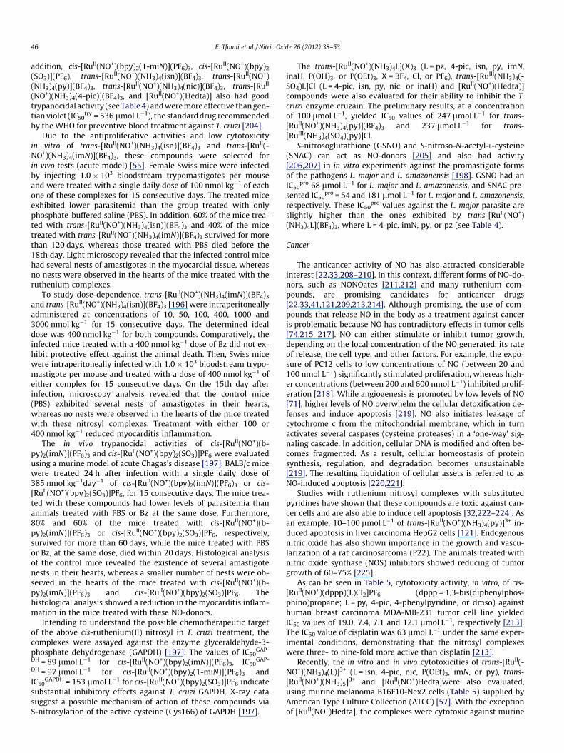
Fig. 3. Structures of (a) pentostam (antimonial) (b) pe
Because of this, are necessary additional incentives for the devel-opment of new treatments and chemotherapeutic agents that aremore effective and less toxic against these pathogenic agents.
Chagas’ disease is a parasitic infection caused by the flagellatedprotozoan Trypanosoma cruzi (T. cruzi), which is an organism ende-mic in 21 countries of Latin America. According to the WorldHealth Organization (WHO) [185,188], there are 10 million peopleinfected worldwide. It is estimated that Chagas’ disease causedmore than 10,000 deaths in 2008, making it the parasitic diseaseresponsible for the largest number of deaths in Latin America.
The life-cycle of T. cruzi includes a passage through both verte-brate and invertebrate hosts and three forms of the parasite: thetrypomastigote form (extracellular phase, that circulates in theblood), the amastigote form (intracellular phase) and the epimasti-gote form (found in the digestive tract of the vector) [187]. TheChagas’ disease presents two phases: the acute phase, where thetripomastigote form is prominent and a high number of parasitescirculate in the blood. When the disease is in this phase there aretwo available drugs for treatment: benznidazole (Bz) and nifurti-mox (Nif). In the chronic phase, the amastigote form is prominentand the parasites are mainly in the heart and digestive muscle. Inthis phase the treatment using these drugs is not effective[189,190].
Leishmaniasis is caused by about 20 species of the flagellatedprotozoan of the genus Leishmania, and affects about 12 millionpeople in 88 countries around the world. In Latin America, 90% ofcases occur in Brazil [185]. The Leishmania parasites present twoforms, the amastigotic (intracellular phase without a flagellum)and promastigotic (extracellular phase with a flagellum). The dis-ease may be cutaneous, mucocutaneous or visceral, and the thera-pies mainly include pentavalent antimonials (SbV) (Pentostam andGlucantime) [190–192], amphotericin B or pentamidine. Othereffective anti-infective drugs are miltefosine and paromomycin[192]. Some examples are shown in Fig. 3.
Nitric oxide has an important role in controlling infectionscaused by pathogenic agents such as Trypanosoma and Leishmania[193,194]. In this context, ruthenium(II) NO-donors, which couldrelease this molecule in a controlled manner, would be useful fordeveloping a platform of potential therapies for these diseases[55,56,195–198]. It is worthwhile to recall that the reduction ofthe NO+ ligand occurs at potential values accessible to the reducingenvironment present in biological media, such as NADH [101], sulf-hydryl groups (R-SH) [100] and ROS [126].
As can be seen in Table 4, the trans-[RuII(NO+)(NH3)4L]n+ com-plexes exhibited in vitro cytotoxicity against host normal cells(IC50
V79, where IC50 is the concentration in which 50% of activityis inhibited) in the range of 120 lmol L�1 (L = pz) to 2260 lmol L�1
ntamidine B (c) amphotericin and (d) miltefosine.
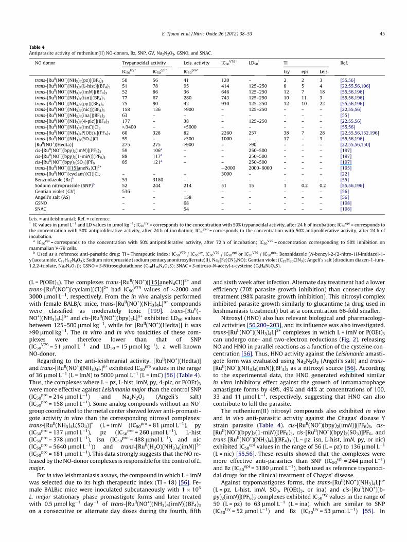
Table 4Antiparasite activity of ruthenium(II) NO-donors, Bz, SNP, GV, Na2N2O3, GSNO, and SNAC.
NO donor Trypanocidal activity Leis. activity IC50V79* LD50
* TI Ref.
IC50try* IC50
epi* IC50pro* try epi Leis.
trans-[RuII(NO+)(NH3)4(pz)](BF4)3 50 56 41 120 – 2 2 3 [55,56]trans-[RuII(NO+)(NH3)4(L-hist)](BF4)3 51 78 95 414 125–250 8 5 4 [22,55,56,196]trans-[RuII(NO+)(NH3)4(imN)](BF4)3 52 86 36 646 125–250 12 7 18 [55,56,196]trans-[RuII(NO+)(NH3)4(isn)](BF4)3 77 67 280 743 125–250 10 11 3 [55,56,196]trans-[RuII(NO+)(NH3)4(py)](BF4)3 75 90 42 930 125–250 12 10 22 [55,56,196]trans-[RuII(NO+)(NH3)4(nic)](BF4)3 158 136 >900 – 125–250 – – – [22,55,56]trans-[RuII(NO+)(NH3)4(ina)](BF4)3 63 – – – – – – – [55]trans-[RuII(NO+)(NH3)4(4-pic)](BF4)3 177 – 38 – 125–250 – – – [22,55,56]trans-[RuII(NO+)(NH3)4(imC)]Cl3 �3400 – >5000 – – – – – [55,56]trans-[RuII(NO+)(NH3)4P(OEt)3](PF6)3 60 328 82 2260 257 38 7 28 [22,55,56,152,196]trans-[RuII(NO+)(NH3)4(SO3)]Cl 59 – >300 1000 – 17 – 3 [55,56,196][RuII(NO+)(Hedta)] 275 275 >900 – >90 – – – [22,55,56,150]cis-[RuII(NO+)(bpy)2(imN)](PF6)3 59 106a – – 250–500 – – – [197]cis-[RuII(NO+)(bpy)2(1-miN)](PF6)3 88 117a – – 250–500 – – – [197]cis-[RuII(NO+)(bpy)2(SO3)]PF6 85 121a – – 250–500 – – – [197]trans-[RuII(NO+)([15]aneN4)Cl]2+ �2000 2000–6000 – – – [195]trans-[RuII(NO+)(cyclam)(Cl)]Cl2 – – – 3000 – – – – [22]Benznidazole (Bz)b 53 3180 – – – – – – [55]Sodium nitroprusside (SNP)b 52 244 214 51 15 1 0.2 0.2 [55,56,196]Gentian violet (GV) 536 – – – – – – – [56]Angeli’s salt (AS) – – 158 – – – – – [56]GSNO – – 68 – – – – – [198]SNAC – – 54 – – – – – [198]
Leis. = antileishmanial; Ref. = reference.* IC values in lmol L�1 and LD values in lmol kg�1; IC50
try = corresponds to the concentration with 50% trypanocidal activity, after 24 h of incubation; IC50epi = corresponds to
the concentration with 50% antiproliferative activity, after 24 h of incubation; IC50pro = corresponds to the concentration with 50% antiproliferative activity, after 24 h of
incubation.a IC50
epi = corresponds to the concentration with 50% antiproliferative activity, after 72 h of incubation; IC50V79 = concentration corresponding to 50% inhibition on
mammalian V-79 cells.b Used as a reference anti-parasitic drug; TI = Therapeutic Index: IC50
V79 / IC50try, IC50
V79 / IC50epi or IC50
V79 / IC50pro; Benznidazole (N-benzyl-2-(2-nitro-1H-imidazol-1-
yl)acetamide, C12H12N4O3); Sodium nitroprusside (sodium pentacyanonitrosylferrate(II), Na2[Fe(CN)5NO); Gentian violet (C25H30ClN3); Angeli’s salt (disodium diazen-1-ium-1,2,2-triolate, Na2N2O3)); GSNO = S-Nitrosoglutathione (C10H16N4O7S); SNAC = S-nitroso-N-acetyl-L-cysteine (C5H8N2O4S).
E. Tfouni et al. / Nitric Oxide 26 (2012) 38–53 45
(L = P(OEt)3). The complexes trans-[RuII(NO+)([15]aneN4Cl)]2+ andtrans-[RuII(NO+)(cyclam)(Cl)]2+ had IC50
V79 values of �2000 and3000 lmol L�1, respectively. From the in vivo analysis performedwith female BALB/c mice, trans-[RuII(NO+)(NH3)4L]n+ compoundswere classified as moderately toxic [199]. trans-[RuII(-NO+)(NH3)4L]n+ and cis-[RuII(NO+)(bpy)2L]n+ exhibited LD50 valuesbetween 125–500 lmol kg�1, while for [RuII(NO+)(Hedta)] it was>90 lmol kg�1. The in vitro and in vivo toxicities of these com-plexes were therefore lower than that of SNP(IC50
V79 = 51 lmol L�1 and LD50 = 15 lmol kg�1), a well-knownNO-donor.
Regarding to the anti-leishmanial activity, [RuII(NO+)(Hedta)]and trans-[RuII(NO+)(NH3)4L]n+ exhibited IC50
pro values in the rangeof 36 lmol L�1 (L = ImN) to 5000 lmol L�1 (L = imC) [56] (Table 4).Thus, the complexes where L = pz, L-hist, imN, py, 4-pic, or P(OEt)3
were more effective against Leishmania major than the control SNP(IC50
pro = 214 lmol L�1) and Na2N2O3 (Angeli’s salt)(IC50
pro = 158 lmol L�1). Some analog compounds without an NO+
group coordinated to the metal center showed lower anti-promasti-gote activity in vitro than the corresponding nitrosyl complexes:trans-[RuII(NH3)4L(SO4)]+ (L = imN (IC50
pro = 81 lmol L�1), py(IC50
pro = 137 lmol L�1), pz (IC50pro = 260 lmol L�1), L-hist
(IC50pro = 378 lmol L�1), isn (IC50
pro = 488 lmol L�1), and nic(IC50
pro = 5640 lmol L�1)) and trans-[RuII(H2O)(NH3)4(imN)]3+
(IC50pro = 181 lmol L�1). This data strongly suggests that the NO re-
leased by the NO-donor complexes is responsible for the control of L.major.
For in vivo leishmaniasis assays, the compound in which L = imNwas selected due to its high therapeutic index (TI = 18) [56]. Fe-male BALB/c mice were inoculated subcutaneously with 1 � 105
L. major stationary phase promastigote forms and later treatedwith 0.5 lmol kg�1 day�1 of trans-[RuII(NO+)(NH3)4(imN)](BF4)3
on a consecutive or alternate day doses during the fourth, fifth
and sixth week after infection. Alternate day treatment had a lowerefficiency (70% parasite growth inhibition) than consecutive daytreatment (98% parasite growth inhibition). This nitrosyl complexinhibited parasite growth similarly to glucantime (a drug used inleishmaniasis treatment) but at a concentration 66-fold smaller.
Nitroxyl (HNO) also has relevant biological and pharmacologi-cal activities [56,200–203], and its influence was also investigated.trans-[RuII(NO+)(NH3)4L]3+ complexes in which L = imN or P(OEt)3
can undergo one- and two-electron reductions (Fig. 2), releasingNO and HNO in parallel reactions as a function of the cysteine con-centration [56]. Thus, HNO activity against the Leishmania amasti-gote form was evaluated using Na2N2O3 (Angeli’s salt) and trans-[RuII(NO+)(NH3)4(imN)](BF4)3 as a nitroxyl source [56]. Accordingto the experimental data, the HNO generated exhibited similarin vitro inhibitory effect against the growth of intramacrophageamastigote forms by 49%, 49% and 44% at concentrations of 100,33 and 11 lmol L�1, respectively, suggesting that HNO can alsocontribute to kill the parasite.
The ruthenium(II) nitrosyl compounds also exhibited in vitroand in vivo anti-parasitic activity against the Chagas’ disease Ystrain parasite (Table 4). cis-[RuII(NO+)(bpy)2(imN)](PF6)3, cis-[RuII(NO+)(bpy)2(1-miN)](PF6)3, cis-[RuII(NO+)(bpy)2(SO3)]PF6, andtrans-[RuII(NO+)(NH3)4L](BF4)3 (L = pz, isn, L-hist, imN, py, or nic)exhibited IC50
epi values in the range of 56 (L = pz) to 136 lmol L�1
(L = nic) [55,56]. These results showed that the complexes weremore effective anti-parasitics than SNP (IC50
epi = 244 lmol L�1)and Bz (IC50
epi = 3180 lmol L�1), both used as reference trypanoci-dal drugs for the clinical treatment of Chagas’ disease.
Against trypomastigotes forms, the trans-[RuII(NO+)(NH3)4L]n+
(L = pz, L-hist, imN, SO3, P(OEt)3, or ina) and cis-[RuII(NO+)(b-py)2(imN)](PF6)3 complexes exhibited IC50
try values in the range of50 (L = pz) to 63 lmol L�1 (L = ina), which are similar to SNP(IC50
try = 52 lmol L�1) and Bz (IC50try = 53 lmol L�1) [55]. In
46 E. Tfouni et al. / Nitric Oxide 26 (2012) 38–53
addition, cis-[RuII(NO+)(bpy)2(1-miN)](PF6)3, cis-[RuII(NO+)(bpy)2
(SO3)](PF6), trans-[RuII(NO+)(NH3)4(isn)](BF4)3, trans-[RuII(NO+)(NH3)4(py)](BF4)3, trans-[RuII(NO+)(NH3)4(nic)](BF4)3, trans-[RuII
(NO+)(NH3)4(4-pic)](BF4)3, and [RuII(NO+)(Hedta)] also had goodtrypanocidal activity (see Table 4) and were more effective than gen-tian violet (IC50
try = 536 lmol L�1), the standard drug recommendedby the WHO for preventive blood treatment against T. cruzi [204].
Due to the antiproliferative activities and low cytotoxicityin vitro of trans-[RuII(NO+)(NH3)4(isn)](BF4)3 and trans-[RuII(-NO+)(NH3)4(imN)](BF4)3, these compounds were selected forin vivo tests (acute model) [55]. Female Swiss mice were infectedby injecting 1.0 � 103 bloodstream trypomastigotes per mouseand were treated with a single daily dose of 100 nmol kg�1 of eachone of these complexes for 15 consecutive days. The treated miceexhibited lower parasitemia than the group treated with onlyphosphate-buffered saline (PBS). In addition, 60% of the mice trea-ted with trans-[RuII(NO+)(NH3)4(isn)](BF4)3 and 40% of the micetreated with trans-[RuII(NO+)(NH3)4(imN)](BF4)3 survived for morethan 120 days, whereas those treated with PBS died before the18th day. Light microscopy revealed that the infected control micehad several nests of amastigotes in the myocardial tissue, whereasno nests were observed in the hearts of the mice treated with theruthenium complexes.
To study dose-dependence, trans-[RuII(NO+)(NH3)4(imN)](BF4)3
and trans-[RuII(NO+)(NH3)4(isn)](BF4)3 [196] were intraperitoneallyadministered at concentrations of 10, 50, 100, 400, 1000 and3000 nmol kg�1 for 15 consecutive days. The determined idealdose was 400 nmol kg�1 for both compounds. Comparatively, theinfected mice treated with a 400 nmol kg�1 dose of Bz did not ex-hibit protective effect against the animal death. Then, Swiss micewere intraperitoneally infected with 1.0 � 103 bloodstream trypo-mastigote per mouse and treated with a dose of 400 nmol kg�1 ofeither complex for 15 consecutive days. On the 15th day afterinfection, microscopy analysis revealed that the control mice(PBS) exhibited several nests of amastigotes in their hearts,whereas no nests were observed in the hearts of the mice treatedwith these nitrosyl complexes. Treatment with either 100 or400 nmol kg�1 reduced myocarditis inflammation.
The in vivo trypanocidal activities of cis-[RuII(NO+)(b-py)2(imN)](PF6)3 and cis-[RuII(NO+)(bpy)2(SO3)]PF6 were evaluatedusing a murine model of acute Chagas’s disease [197]. BALB/c micewere treated 24 h after infection with a single daily dose of385 nmol kg�1day�1 of cis-[RuII(NO+)(bpy)2(imN)](PF6)3 or cis-[RuII(NO+)(bpy)2(SO3)]PF6, for 15 consecutive days. The mice trea-ted with these compounds had lower levels of parasitemia thananimals treated with PBS or Bz at the same dose. Furthermore,80% and 60% of the mice treated with cis-[RuII(NO+)(b-py)2(imN)](PF6)3 or cis-[RuII(NO+)(bpy)2(SO3)]PF6, respectively,survived for more than 60 days, while the mice treated with PBSor Bz, at the same dose, died within 20 days. Histological analysisof the control mice revealed the existence of several amastigotenests in their hearts, whereas a smaller number of nests were ob-served in the hearts of the mice treated with cis-[RuII(NO+)(b-py)2(imN)](PF6)3 and cis-[RuII(NO+)(bpy)2(SO3)]PF6. Thehistological analysis showed a reduction in the myocarditis inflam-mation in the mice treated with these NO-donors.
Intending to understand the possible chemotherapeutic targetof the above cis-ruthenium(II) nitrosyl in T. cruzi treatment, thecomplexes were assayed against the enzyme glyceraldehyde-3-phosphate dehydrogenase (GAPDH) [197]. The values of IC50
GAP-
DH = 89 lmol L�1 for cis-[RuII(NO+)(bpy)2(imN)](PF6)3, IC50GAP-
DH = 97 lmol L�1 for cis-[RuII(NO+)(bpy)2(1-miN)](PF6)3 andIC50
GAPDH = 153 lmol L�1 for cis-[RuII(NO+)(bpy)2(SO3)]PF6 indicatesubstantial inhibitory effects against T. cruzi GAPDH. X-ray datasuggest a possible mechanism of action of these compounds viaS-nitrosylation of the active cysteine (Cys166) of GAPDH [197].
The trans-[RuII(NO+)(NH3)4L](X)3 (L = pz, 4-pic, isn, py, imN,inaH, P(OH)3, or P(OEt)3, X = BF4, Cl, or PF6), trans-[RuIII(NH3)4(-SO4)L]Cl (L = 4-pic, isn, py, nic, or inaH) and [RuII(NO+)(Hedta)]compounds were also evaluated for their ability to inhibit the T.cruzi enzyme cruzain. The preliminary results, at a concentrationof 100 lmol L�1, yielded IC50 values of 247 lmol L�1 for trans-[RuII(NO+)(NH3)4(py)](BF4)3 and 237 lmol L�1 for trans-[RuIII(NH3)4(SO4)(py)]Cl.
S-nitrosoglutathione (GSNO) and S-nitroso-N-acetyl-L-cysteine(SNAC) can act as NO-donors [205] and also had activity[206,207] in in vitro experiments against the promastigote formsof the pathogens L. major and L. amazonensis [198]. GSNO had anIC50
pro 68 lmol L�1 for L. major and L. amazonensis, and SNAC pre-sented IC50
pro = 54 and 181 lmol L�1 for L. major and L. amazonensis,respectively. These IC50
pro values against the L. major parasite areslightly higher than the ones exhibited by trans-[RuII(NO+)(NH3)4L](BF4)3, where L = 4-pic, imN, py, or pz (see Table 4).
Cancer
The anticancer activity of NO has also attracted considerableinterest [22,33,208–210]. In this context, different forms of NO-do-nors, such as NONOates [211,212] and many ruthenium com-pounds, are promising candidates for anticancer drugs[22,33,41,121,209,213,214]. Although promising, the use of com-pounds that release NO in the body as a treatment against canceris problematic because NO has contradictory effects in tumor cells[74,215–217]. NO can either stimulate or inhibit tumor growth,depending on the local concentration of the NO generated, its rateof release, the cell type, and other factors. For example, the expo-sure of PC12 cells to low concentrations of NO (between 20 and100 nmol L�1) significantly stimulated proliferation, whereas high-er concentrations (between 200 and 600 nmol L�1) inhibited prolif-eration [218]. While angiogenesis is promoted by low levels of NO[71], higher levels of NO overwhelm the cellular detoxification de-fenses and induce apoptosis [219]. NO also initiates leakage ofcytochrome c from the mitochondrial membrane, which in turnactivates several caspases (cysteine proteases) in a ‘one-way’ sig-naling cascade. In addition, cellular DNA is modified and often be-comes fragmented. As a result, cellular homeostasis of proteinsynthesis, regulation, and degradation becomes unsustainable[219]. The resulting liquidation of cellular assets is referred to asNO-induced apoptosis [220,221].
Studies with ruthenium nitrosyl complexes with substitutedpyridines have shown that these compounds are toxic against can-cer cells and are also able to induce cell apoptosis [32,222–224]. Asan example, 10–100 lmol L�1 of trans-[RuII(NO+)(NH3)4(py)]3+ in-duced apoptosis in liver carcinoma HepG2 cells [121]. Endogenousnitric oxide has also shown importance in the growth and vascu-larization of a rat carcinosarcoma (P22). The animals treated withnitric oxide synthase (NOS) inhibitors showed reducing of tumorgrowth of 60–75% [225].
As can be seen in Table 5, cytotoxicity activity, in vitro, of cis-[RuII(NO+)(dppp)(L)Cl2]PF6 (dppp = 1,3-bis(diphenylphos-phino)propane; L = py, 4-pic, 4-phenylpyridine, or dmso) againsthuman breast carcinoma MDA-MB-231 tumor cell line yieldedIC50 values of 19.0, 7.4, 7.1 and 12.1 lmol L�1, respectively [213].The IC50 value of cisplatin was 63 lmol L�1 under the same exper-imental conditions, demonstrating that the nitrosyl complexeswere three- to nine-fold more active than cisplatin [213].
Recently, the in vitro and in vivo cytotoxicities of trans-[RuII(-NO+)(NH3)4(L)]3+ (L = isn, 4-pic, nic, P(OEt)3, imN, or py), trans-[RuII(NO+)(NH3)5]3+ and [RuII(NO+)Hedta]were also evaluated,using murine melanoma B16F10-Nex2 cells (Table 5) supplied byAmerican Type Culture Collection (ATCC) [57]. With the exceptionof [RuII(NO+)Hedta], the complexes were cytotoxic against murine

Table 5NO-donor ruthenium complexes activity against cancer cells.
NO donor Cells IC50 Ref.
cis-[RuII(NO+)(dppp)(py)Cl2]PF6 MDA-MB-231 19.0 lmol L�1 [213]cis-[RuII(NO+)(dppp)(4-pic)Cl2]PF6 MDA-MB-231 7.4 lmol L�1 [213]cis-[RuII(NO+)(dppp)(4-phen)Cl2]PF6 MDA-MB-231 7.1 lmol L�1 [213]cis-[RuII(NO+)(dppp)(dmso)Cl2]PF6 MDA-MB-231 12.1 lmol L�1 [213]trans-[RuII(NO+)(NH3)4(isn)](BF4)3 B16F10-Nex2 1.0 lmol L�1 [57]trans-[RuII(NO+)(NH3)4(4-pic)](BF4)3 B16F10-Nex2 6.0 lmol L�1 [57]trans-[RuII(NO+)(NH3)4(nic)](BF4)3 B16F10-Nex2 20.0 lmol L�1 [57]trans-[RuII(NO+)(NH3)4(P(OEt)3)](PF6)3 B16F10-Nex2 33.0 lmol L�1 [57]trans-[RuII(NO+)(NH3)4(imN)](BF4)3 B16F10-Nex2 74.0 lmol L�1 [57]trans-[RuII(NO+)(NH3)4(py)](BF4)3 B16F10-Nex2 86.0 lmol L�1 [57]trans-[RuII(NO+)(NH3)5](BF4)3 B16F10-Nex2 44.0 lmol L�1 [57][RuII(NO+)Hedta] B16F10-Nex2 >40 � 102lmol L�1 [57]trans-[RuII(NO+)(NH3)4(inaH)](BF4)3 B16F10 12.6 lmol L�1 [222]trans-[RuII(NO+)(NH3)4(4-mapy)](BF4)3 B16F10 347 lmol L�1 [222]trans-[RuII(NO+)(NH3)4(ina-Tat48–60)](BF4)3 B16F10 49.0 lmol L�1 [222]trans-[RuII(NO+)(NH3)4(py)](BF4)3 B16F10 1.0 mmol L�1 [32]trans-[RuII(NO+)(cyclam)Cl](PF6)2 B16F10 6.8 mmol L�1 [224]trans-[RuII(NO+)(NH3)4(py)](BF4)3 Melan-a 0.9 mmol L�1 [223]trans-[RuII(NO+)(cyclam)Cl](PF6)2 Melan-a 9.8 mmol L�1 [223]trans-[RuII(NO+)(NH3)4(imN)](PF6)3 A2058 335 lmol L�1 [57]trans-[RuII(NO+)(NH3)4(imN)](PF6)3 HCT 287 lmol L�1 [57]trans-[RuII(NO+)(NH3)4(imN)](PF6)3 Siha 333 lmol L�1 [57]
MDA-MB-231 = human breast carcinoma; B16F10 = murine melanoma; B16F10-Nex2 = murine melanoma subclone Nex2; melan-a = normal murine melanocyte;A2058 = human metastatic melanoma; HCT = human colorectal tumor; Siha = human cervical cancer.
E. Tfouni et al. / Nitric Oxide 26 (2012) 38–53 47
melanoma B16F10-Nex2 in in vitro tests (IC50 values in the range of1.0 (L = isn) to 86.0 lmol L�1 (L = py)). Against V79 cells, the IC50
values were higher than 646 lmol L�1, showing that these cellswere less sensitive to these complexes than the murine melanomaB16F10-Nex2 cells [57].
In vitro assays with B16F10 cell line treated with trans-[RuII(-NO+)(NH3)4(L)]3+ (L = isonicotinic acid (inaH) or 4-(amino-methyl)pyridine (4-mapy)) were also performed [222]. Thepreliminary IC50 values obtained by the MTT cell viability assaywere 12.6 lmol�1 L�1 for L = inaH and 347 lmol�1 L�1 for L = 4-mapy (Table 5). Compared to cisplatin, which has IC value of33.3 lmol�1 L�1 against this cell line [226], the first complex isvery toxic, while the other is a little less. The difference in toxicity,however, can not be explained on the basis of the rate of release ofNO from these nitrosyls. The estimated rate constants value, k-NO,of release of NO for these complexes are similar and in the orderof 10�2 s�1. The induction of apoptosis, measured by flow cytome-try with annexin marking, using BD Pharmingen™ ‘‘PE Annexin VApoptosis Detection Kit I’’, followed the same tendency. The com-plex with inaH induced apoptosis to 51% of the cancer cells, whilefor the 4-mapy complex this percentage was 13% [222]. These re-sults illustrate the complexity of this subject and that other factorsmay be involved.
trans-[RuII(NO+)(NH3)4(imN)](PF6)3 was evaluated in vitroagainst melanoma (A2058), colorectal (HCT), and cervical human(Siha) tumor cells [57] and showed inhibitory antitumor effectswith IC50 values in the range of 287–335 lmol L�1 (Table 5). Also,an inhibitory effect on angiogenesis was observed for trans-[RuII(-NO+)(NH3)4(ImN)](PF6)3, as indicated by the in vitro cytotoxicity onhuman umbilical vein endothelial cells (HUVEC). At doses of 100and 250 lmol L�1, a 50 to 70% of reduction in the number of pro-angiogenic structures formed by HUVEC was observed, respec-tively. It is interesting to point out that even at concentrations ashigh as 0.5 mmol L�1, 60% of the endothelial cells were still viable.
trans-[RuII(NO+)(NH3)4(imN)](PF6)3 and trans-[RuII(NO+)(N-H3)4(isn)](PF6)3, which exhibited higher IC50 values in vitro, wereassayed in vivo in the murine melanoma B16F10-Nex2 model usingadult male C57Bl/6 mice [57]. In this assay, 5 � 104 B16F10-Nex2cells were subcutaneously injected in the mice. For trans-[RuII(-NO+)(NH3)4(ImN)](PF6)3, two different treatments were applied
24 h after injection. In the first treatment, doses of 210 and1050 ng kg�1 were inoculated intraperitoneally every other dayfor a 2-week period. Both doses inhibited tumor development in40% of animals even 60 days after tumor cell inoculation. In thesecond treatment protocol, 210 ng kg�1 of the complex was in-jected intraperitoneally every day for one week and every otherday on the second week. As a result, only 10% of the animals wereprotected against melanoma development. trans-[RuII(NO+)(N-H3)4(isn)](PF6)3 was injected intraperitoneally at 23 and232 ng kg�1 doses every other day for 2 weeks. Sixty days afterthe injection of tumor cells, the lowest dose inhibited tumor devel-opment in 40% of the animals, while the highest dose protectedonly 10% of the animals. The above data suggest that lower dosesand less aggressive protocols are more effective in controlling tu-mor development.
The cytotoxicity of trans-[RuII(NO+)(NH3)4(py)]3+, trans-[RuII(-NO+)(cyclam)Cl]2+, and [RuII(NO+)(Hedta)] were evaluated in mur-ine melanocyte tumorigenic and non-tumorigenic cell lines(B16F10 and melan-a, respectively) [32,223,224,227] (Table 5)using MTT assay. Cytotoxicity was observed for both trans-[RuII(-NO+)(NH3)4(py)]3+ (IC50
B16�F10 = 1.0 mmol L�1 [32,224], IC50melan-
a = 0.9 mmol L�1 [223]), and trans-[RuII(NO+)(cyclam)Cl]2+
(IC50B16�F10 = 6.8 mmol L�1 [224]; IC50
melan-a = 9.8 mmol L�1 [223])at concentrations higher than 1 � 10�3mol L�1, causing cell death.The complex [RuII(NO+)(Hedta)] was cytotoxic in the presence oflight, whereas in its absence, it had no cytotoxic effect on the cellline in the study. The behavior in the dark is consistent with thehigher NO scavenging rate constant of the aquo complex(kf = 2.24 � 107 L mol�1 s�1), which is formed after the release ofNO, than the release of NO from the nitrosyl complex (k-
NO = 7.3 � 10�3 s�1) at pH 7.4 [150,228], 53.9% for trans-[RuII(-NO+)(cyclam)Cl]2+ and 22.3% for [RuII(NO+)(Hedta)][32,223,224,227]. In addition, the phototoxicity of these complexeswas also evaluated against melan-a cells, with cell death values of57.6% for trans-[RuII(NO+)(NH3)4(py)]3+, 47.0% for trans-[RuII(-NO+)(cyclam)Cl]2+ and 17.7% for [RuII(NO+)(Hedta)][32,223,224,227].
A mixture of cis-[RuII(NO+)(H-dcbpy�)2Cl] (H2-dcbpy = 4,40-dicarboxy-2,20-bipyridine) and Na4[Tb(TsPc)(acac)] (TsPc = tetra-sulfonated phthalocyanines; acac = acetylacetone) was reported

48 E. Tfouni et al. / Nitric Oxide 26 (2012) 38–53
to simultaneously produce NO and 1O2 under irradiation with lightof k P 550 nm, strongly inhibiting B16-F10 cell viability [131].
To increase selectivity and avoid side effects, it is necessary totailor more specific ruthenium complexes to act at the desired tar-get. A ruthenium nitrosyl NO-donor linked to a penetrating peptidecalled Tat48–60 [229–231] (Tat48–60 = GRKKRRQRRRPPQ, G = gly-cine, R = arginine, K = lysine, Q = glutamine, P = proline) was syn-thesized for this purpose. This 13 aminoacid sequencecorresponds to the positions 48 to 60 of the cell penetrating Tatprotein of the HIV virus [230–234], a protein that is importantfor virus replication [229,234]. The synthesized complex wastrans-[RuII(NO+)(NH3)4(L)]3+, L = ina-Tat48–60 (ina-Tat48–60 = Tat48–60 plus inaH, leading to the sequence ina-GRKKRRQRRRPPQ). This complex was tested in vitro against the tu-mor cell line B16F10, with preliminary results similar to those ofthe complex with inaH; an IC50 value of 49 lmol L�1, and 38% of in-duced apoptosis (Table 5) [222], which is close to that of cisplatin.
Immobilization of ruthenium nitrosyl complexes
The use of nanostructured materials as drug delivery systemshas begun to impact medicine due to beneficial size-dependentphysical and chemical properties [235] and targeted selectivity.As far as ruthenium nitrosyl complexes are concerned, this ap-proach may also improve the in vivo residence time of rutheniumnitrosyl complexes, increase the number of species that could becarried by a single matrix, and avoid undesired side reactions, suchas nucleophilic attack on the coordinated nitrosyl [23]. A widerange of materials can be used as matrices. The choice of theimmobilization procedure depends on the structure of the com-plex, the material characteristics, and the desired properties. A re-view on the immobilization of ruthenium complexes has recentlybeen published [23].
The use of NO-releasing silica nanoparticles as a therapeutictool to improve NO delivery to human ovarian cancer cells hasbeen reported [235]. It was shown that the NO-releasing nanopar-ticles exhibited enhanced growth inhibition of tumor cells whencompared to both empty nanoparticles (control) and a previouslyreported small molecule NO donor, sodium 1-(pyrrolidium-1-yl)diazen-1-ium-1,2-diolate (PYRRO/NO) [235].
Incorporation of a ruthenium nitrosyl ([Ru(NO+)(Me2bpb)(4-vpy)](BF4)) in polyHEMA hydrogel via a radical-induced copoly-merization process has been described [236]. The transparent pHE-MA nitrosyl-hydrogel conjugate readily releases NO upon exposureto low-intensity UV light (5–10 mW). Because 1-pHEMA can trans-fer NO to proteins like myoglobin quite readily, this material couldbe employed in the cellular environment for light-triggered NOdelivery [236].
Photoinduced nitric oxide and singlet oxygen release from lipo-somal zinc phthalocyanine associated with the nitrosyl rutheniumcomplex [RuII(NO+)(NH�NHq)(tpy)]3+ in neoplastic cells has beenproposed in a PDT application using the B16F10 cell line [36].The dark toxicity for three ZnPC preparations (ranging from 1.0to 5.0 lmol L�1) was evaluated at a fixed [RuII(NO+)(NH�NHq)(t-py)]3+ complex concentration (0.05 mmol L�1). As observed for allof the samples studied, no dark toxicity was observed above thecell viability basal level (90%). These results led to light activationstudies (675 nm) to analyze the best combination of both drugsthat could induce cell damage. The observed ideal complex formu-lation for inducing 90% cellular death was 5.0 lmol L�1 ZnPC and0.05 mmol L�1 [RuII(NO+)(NH�NHq)(tpy)]3+ [36].
The NO donors trans-[RuII(NO+)(NH3)4(py)](BF4)3, trans-[RuII(-NO+)(cyclam)Cl](PF6)2, and [RuII(NO+)(Hedta)] were recently incor-porated into polymeric micro and nanoparticles (MP) ofpoly(lactic-co-glycolic acid) (PLGA) (py-MP, cyclam-MP and Hed-ta-MP). This system was effective for complex transport and NO
release in biological targets [32,223,224,227,237]. B16F10 mela-noma cells were chosen as the biological model to evaluate thein vitro cytotoxicity of the ruthenium nitrosyl- and MP-loadedcomplex. The MTT test showed that the nitrosyl complex wasnon-toxic in low concentrations in solution or entrapped in micro-particles, promoting a proliferative effect when solutions of thiscomplex with concentrations lower than 1 � 10�4mol L�1 were ap-plied. Toxicity was observed only for the complexes in solution athigher concentrations (>1 � 10�4 mol L�1), causing death (trans-[RuII(NO+)(NH3)4(py)](BF4)3 IC50 = 1.0 � 10�3 mol L�1; trans-[RuII(-NO+)(cyclam)Cl](PF6)2 IC50 = 6.8 � 10�3 mol L�1) [32]. The absenceof cell death when using nitrosyl complexes entrapped in PLGAmicroparticles, which are able to release NO after reduction, indi-cated that the entrapped complex was not available to the mediumand its reducing agents. The behavior of the NO donors was en-hanced under light irradiation. Phototoxicity assays of the nitrosylcomplexes were performed using the same cell lines and the MTTassay. After treating the B16F10 cells (10 min irradiation with366 nm light) with the NO donor in solution (1 � 10�4 mol L�1)in the presence of light, more cell damage was observed, leadingto 63 ± 3% of cell death in the presence of trans-[RuII(NO+)(NH3)4(-py)](BF4)3, 53.8 ± 6.2% of cell death in the presence of trans-[RuII(-NO+)(cyclam)Cl](PF6)2 and 22.3 ± 8.2% of cell death in the presenceof [RuII(NO+)(Hedta)]. When encapsulated, py-MP produced12 ± 6% of cell death, cyclam-MP 13.6 ± 6.4% of cell death and Hed-ta-MP 6.3 ± 7.6% of cell death. As the py-MP system is non-toxicwithout light irradiation, these results suggest that cell deathwas due to NO photorelease [22,32].
Nitrosyl ruthenium complex-loaded lipid carriers for topicaladministration, such as for skin cancer treatment, were also re-ported [129]. Lipid carrier systems were developed to circumventproblems related to the topical absorption of drugs, particularlypositively-charged drugs, as is the case for the [RuII(NO+)(ter-py)(bdqi)](PF6)3 complex [129].
The PAMAM dendrimers (Gx) functionalized with [RuIII(-H2O)(edta)]�(Gx/RuH2O) and [RuII(NO+)(edta)]� (Gx/RuNO) werestudied in vascular and trypanocidal models [150,238]. The initialexperiments were performed using denuded normotensive rat aor-tic rings pre-contracted with noradrenaline. Similar to the observa-tions for [RuII(NO+)(edta)]�, treatment with Gx/RuNO (x = 0 and3.3 lmol L�1) induced a slow delayed relaxation that began after15 min (14.11 ± 6.25%, n = 5), inducing a maximal relaxation effectin the second hour (36.8 ± 6.5%, n = 5). The trypanocidal activity ofG0/RuNO in vitro was evaluated using a drug-resistant strain of T.cruzi. Active bloodstream trypomastigotes (T. cruzi) were not de-tected in infected mice after 24 h of incubation with the G0/RuNOdendrimer at 1.0 � 10�3 mol L�1. The G0/RuNO dendrimer wasslightly more effective against T. cruzi (100%) than[RuII(NO+)(edta)]� (89%) [238].
Conclusions and perspectives
Nitrosyl complexes proved to be good NO-carriers. In particular,ruthenium(II) amines are very useful compounds because they arewater soluble, stable in solution in the presence of oxygen, and caneasily deliver NO after activation by chemical or photochemicalpathways. The redox potential for [RuIINO+]3+/[RuIINO0]2+ is inthe range of biological reductants, and the rate constant for releaseof NO from the [RuIINO0]2+ moiety can be modulated through coor-dination sphere tailoring. Photoactive nitrosyl complexes with re-gard to NO release can be obtained by judicious choice of theligands, resulting in complexes with absorption bands in the regionof the spectrum suitable for PDT.
Coordination compounds have been comparatively more ex-plored as NO-donors than as NO-scavengers. In this respect, it
E. Tfouni et al. / Nitric Oxide 26 (2012) 38–53 49
would be worthy to investigate the corresponding Ru(III) species oftrans-[Ru(NH3)4(L1)(L2)]n+, L1 = SO4
2� or H2O and L2 = other ancil-lary ligand. Preliminary results from ours experiments withtrans-[RuIII(SO4)(NH3)4(4-pic)]+ and trans-[RuIII(SO4)(NH3)4(inaH)]+
in denuded aorta rings are promising [239]. The beneficial effectsof NO-donors on some infections and cancer have been described,but very little is known about their in vivo mechanism of action.The metal nitrosyls react with ROS, acting as radical scavengers,and with thiols (RSH) [126]. In this case, NO and HNO could beformed. Whether NO and possibly HNO are released inside or out-side of the cell is still an open question.
NO can act also as a pronociceptive mediator, and the nonste-roidal anti-inflammatory drugs (NSAIDs) used in the treatment ofinflammatory diseases and pain exhibit several side effects. To cir-cumvent these effects, new compounds called NO-releasing non-steroidal anti-inflammatory drugs (NO-NSAIDs) have beenstudied [240]. Correspondingly, nitrosyl ruthenium(II) tetraam-mines containing or lacking NSAID molecules in the coordinationsphere should be investigated for use as analgesics.
Efforts are on-going to develop NO-carrier complexes for a spe-cific desired target and to increase the residence time of these spe-cies in the blood. Studies are being performed in this directionusing dendrimers and antibodies that specifically act on the recep-tors of breast cancer cells, for example, to kill the cancerous tissuewithout damaging healthy cells. Several approaches and strategiesdirected to make NO donors photochemically active in the thera-peutic window were described, but they are non-exhaustive andother strategies can be designed, such as developing systems usingthe rare earths ‘‘upconversion’’, which would allow irradiation inlonger wavelengths and activation of the NO release from higherenergy states. The use of ‘‘caged’’ NO donors in matrices allowsthe arrival of the complex without being degraded which wouldbe activated by irradiation, and are examples of targeting. Studiesare under way to combine strategies of activation in the therapeu-tic window with ‘‘caged’’ NO donors in nano- and microparticles.
From this perspective of drug development, we see many possi-bilities, some of which are already under investigation, to improvethe number and efficiency of complexes that have biological activ-ity against many kinds of disease. There is still much to be exploredon this field, but the most promising areas seem to be in the devel-opment of systems able to deliver NO upon light irradiation in thetherapeutic window, drug delivery systems for controlled releaseof NO, and the linkage of the drugs to systems that carry them spe-cifically to the local of interest, greatly improving its activity andcausing few, or even none, side effects. With regard to photochem-ical therapy, it is conceivably possible to overcome the limitationto skin diseases by assembling a carrier, such an optical fiber withembedded complex, to be irradiated within vessels.
References
[1] G. Stochel, A. Wanat, E. Kulis, Z. Stasicka, Light and metal complexes inmedicine, Coordination Chemistry Reviews 171 (1998) 203–220.
[2] L. Ronconi, P.J. Sadler, Using coordination chemistry to design new medicines,Coordination Chemistry Reviews 251 (2007) 1633–1648.
[3] S.P. Fricker, Metal based drugs: from serendipity to design, DaltonTransactions (2007) 4903–4917.
[4] M.J. Clarke, F. Zhu, D.R. Frasca, Non-platinum chemotherapeuticmetallopharmaceuticals, Chemical Reviews 99 (1999) 2511–2534.
[5] P.J. Dyson, G. Sava, Metal-based antitumour drugs in the post genomic era,Dalton Transactions (2006) 1929–1933.
[6] A. Levina, A. Mitra, P.A. Lay, Recent developments in ruthenium anticancerdrugs, Metallomics 1 (2009) 458–470.
[7] J.N. Bates, M.T. Baker, R. Guerra Jr., D.G. Harrison, Nitric oxide generation fromnitroprusside by vascular tissue. Evidence that reduction of the nitroprussideanion and cyanide loss are required, Biochemical Pharmacology 42(Supplement 1) (1991) S157–S165.
[8] J. Forestier, Rheumatoid arthritis and its treatment by gold salts - The resultsof six years experience, Journal of Laboratory and Clinical Medicine 20 (1935)827–840.
[9] M. Navarro, Gold complexes as potential anti-parasitic agents, CoordinationChemistry Reviews 253 (2009) 1619–1626.
[10] Available at <http://www.who.int/nutrition/publications/micronutrients/anaemia_iron_deficiency/en/>, World Health Organization, accessed 10August 2011.
[11] E. Hillard, A. Vessieres, L. Thouin, G. Jaouen, C. Amatore, Ferrocene-mediatedproton-coupled electron transfer in a series of ferrocifen-type breast-cancerdrug candidates, Angewandte Chemie-International Edition 45 (2006) 285–290.
[12] S. Moncada, R.M.J. Palmer, E.A. Higgs, Nitric-Oxide - physiology,Pathophysiology, and Pharmacology. Pharmacological Reviews 43 (1991)109–142.
[13] C. Glidewell, I.L. Johnson, Definitive identification of the primary reductionproduct of the nitroprusside ion, pentacyanonitrosylferrate(2-), in aqueous-solution, Inorganica Chimica Acta 132 (1987) 145–147.
[14] Available at <http://whqlibdoc.who.int/hq/2011/a95053_eng.pdf>, WorldHealth Organization, accessed 10 August 2011.
[15] P.A. Lay, W.D. Harman, Recent advances in osmium chemistry, Advances inInorganic Chemistry 37 (1991) 219–379.
[16] A.F.A. Peacock, A. Habtemariam, R. Fernandez, V. Walland, F.P.A. Fabbiani, S.Parsons, R.E. Aird, D.I. Jodrell, P.J. Sadler, Tuning the reactivity of osmium(II)and ruthenium(II) arene complexes under physiological conditions, Journal ofthe American Chemical Society 128 (2006) 1739–1748.
[17] D.T. Richens, The chemistry of aqua ions: synthesis, structure, andreactivity: a tour through the periodic table of the elements, Wiley, NewYork, 1997.
[18] A.F.A. Peacock, S. Parsons, P.J. Sadler, Tuning the hydrolytic aqueouschemistry of osmium arene complexes with N,O-chelating ligands toachieve cancer cell cytotoxicity, Journal of the American Chemical Society129 (2007) 3348–3357.
[19] M.J. Clarke, Ruthenium metallopharmaceuticals, Coordination ChemistryReviews 232 (2002) 69–93.
[20] U. Schatzschneider, Photoactivated biological activity of transition-metalcomplexes, European Journal of Inorganic Chemistry 2010 (2010) 1451–1467.
[21] E. Tfouni, Photochemical reactions of ammineruthenium(II) complexes,Coordination Chemistry Reviews 196 (2000) 281–305.
[22] E. Tfouni, F.G. Doro, L.E. Figueiredo, J.C.M. Pereira, G. Metzker, D.W. Franco,Tailoring NO donors metallopharmaceuticals: ruthenium nitrosyl amminesand aliphatic tetraazamacrocycles, Current Medicinal Chemistry 17 (2010)3643–3657.
[23] E. Tfouni, F.G. Doro, A.J. Gomes, R.S.d. Silva, G. Metzker, P.G.Z. Benini, D.W.Franco, Immobilized ruthenium complexes and aspects of their reactivity,Coordination Chemistry Reviews 254 (2010) 355–371.
[24] E. Tfouni, K.Q. Ferreira, F.G. Doro, R.S. da Silva, Z.N. da Rocha, Ru(II) and Ru(III)complexes with cyclam and related species, Coordination Chemistry Reviews249 (2005) 405–418.
[25] E. Tfouni, M. Krieger, B.R. McGarvey, D.W. Franco, Structure, chemical andphotochemical reactivity and biological activity of some ruthenium aminenitrosyl complexes, Coordination Chemistry Reviews 236 (2003) 57–69.
[26] J.F. Vollano, G.E. Bossard, S.A. Martellucci, M.C. Darkes, M.J. Abrams, R.C.Brooks, The synthesis and in vitro photodynamic activity of a series of novelruthenium(II)-2, 3-naphthalocyanines, Journal of Photochemistry andPhotobiology B: Biology 37 (1997) 230–235.
[27] A.A. Eroy-Reveles, P.K. Mascharak, Nitric oxide-donating materials and theirpotential in pharmacological applications for site-specific nitric oxidedelivery, Future Medicinal Chemistry 1 (2009) 1497–1507.
[28] N.J. Farrer, L. Salassa, P.J. Sadler, Photoactivated chemotherapy (PACT): thepotential of excited-state d-block metals in medicine, Dalton Transactions(2009) 10690–10701.
[29] P.C. Ford, Polychromophoric metal complexes for generating thebioregulatory agent nitric oxide by single- and two-photon excitation,Accounts of Chemical Research 41 (2008) 190–200.
[30] P.C. Ford, I.M. Lorkovic, Mechanistic aspects of the reactions of nitric oxidewith transition-metal complexes, Chemical Reviews 102 (2002) 993–1018.
[31] P.C. Ford, S. Wecksler, Photochemical reactions leading to NO and NOx
generation, Coordination Chemistry Reviews 249 (2005) 1382–1395.[32] A.J. Gomes, P.A. Barbougli, E.M. Espreafico, E. Tfouni, Trans-
[Ru(NO)(NH3)4(py)](BF4)3 center dot H2O encapsulated in PLGAmicroparticles for delivery of nitric oxide to B16–F10 cells: cytotoxicity andphototoxicity, Journal of Inorganic Biochemistry 102 (2008) 757–766.
[33] A.D. Ostrowski, P.C. Ford, Metal complexes as photochemical nitric oxideprecursors: potential applications in the treatment of tumors, DaltonTransactions (2009) 10660–10669.
[34] M.J. Rose, P.K. Mascharak, Photoactive ruthenium nitrosyls: effects of lightand potential application as NO donors, Coordination Chemistry Reviews 252(2008) 2093–2114.
[35] P.S. Wagenknecht, P.C. Ford, Metal centered ligand field excited states: theirroles in the design and performance of transition metal based photochemicalmolecular devices, Coordination Chemistry Reviews 255 (2011) 591–616.
[36] D.S. Maranho, R.G. de Lima, F.L. Primo, R.S. da Silva, A.C. Tedesco,Photoinduced nitric oxide and singlet oxygen release from ZnPC liposomevehicle associated with the nitrosyl ruthenium complex: synergistic effects inphotodynamic therapy application, Photochemistry and Photobiology 85(2009) 705–713.
[37] C.S. Allardyce, P.J. Dyson, Ruthenium in medicine: current clinical uses andfuture prospects, Platinum Metals Review 45 (2001) 62.

50 E. Tfouni et al. / Nitric Oxide 26 (2012) 38–53
[38] W.H. Ang, P.J. Dyson, Classical and non-classical ruthenium-based anticancerdrugs: towards targeted chemotherapy, European Journal of InorganicChemistry (2006) 4003–4018.
[39] J. Reedijk, Metal-ligand exchange kinetics in platinum and rutheniumcomplexes, Platinum Metals Review 52 (2008).
[40] D.V. Deubel, J.K.C. Lau, In silico evolution of substrate selectivity: comparisonof organometallic ruthenium complexes with the anticancer drug cisplatin,Chemical Communications (2006) 2451–2453.
[41] G. Sava, I. Capozzi, K. Clerici, G. Gagliardi, E. Alessio, G. Mestroni,Pharmacological control of lung metastases of solid tumours by a novelruthenium complex, Clinical & Experimental Metastasis 16 (1998) 371–379.
[42] L. Rover, N.F. Hoehr, A.P. Vellasco, L.T. Kubota, Antioxidant system involvingthe glutathione metabolic cycle associated to electroanalytical methods inthe oxidative stress evaluation, Química Nova 24 (2001) 112–119.
[43] F.P. Dwyer, E.C. Gyarfas, W.P. Rogers, J.H. Koch, Biological activity of complexions, Nature 170 (1952) 190–191.
[44] F.P. Dwyer, E. Mayhew, E.M. Roe, A. Shulman, Inhibition of landschuetzascites tumour growth by metal chelates derived from 3, 4, 7, 8-tetramethyl-1, 10-phenanthroline, British Journal of Cancer 19 (1965) 195–199.
[45] E. Meggers, Exploring biologically relevant chemical space with metalcomplexes, Current Opinion in Chemical Biology 11 (2007) 287–292.
[46] A. Bergamo, G. Sava, Ruthenium complexes can target determinants oftumour malignancy, Dalton Transactions (2007) 1267–1272.
[47] M.J. Clarke, Oncological implications of the chemistry of ruthenium, NewYork, Marcel Dekker, 1980.
[48] M.J. Clarke, Ruthenium chemistry pertaining to the design of anticanceragents, in ruthenium and other non-platinum metal complexes in cancerchemotherapy, Progress in Clinical Biochemistry and Medicine 10 (1989)25–39.
[49] M. Galanski, V.B. Arion, M.A. Jakupec, B.K. Keppler, Recent developments inthe field of tumor-inhibiting metal complexes, Current PharmaceuticalDesign 9 (2003) 2078–2089.
[50] C.G. Hartinger, S. Zorbas-Seifried, M.A. Jakupec, B. Kynast, H. Zorbas, B.K.Keppler, From bench to bedside - preclinical and early clinical development ofthe anticancer agent indazolium trans-[tetrachlorobis(1H-indazole)ruthenate(III)] (KP1019 or FFC14A), Journal of InorganicBiochemistry 100 (2006) 891–904.
[51] G. Sava, A. Bergamo, S. Zorzet, B. Gava, C. Casarsa, M. Cocchietto, A. Furlani, V.Scarcia, B. Serli, E. Iengo, E. Alessio, G. Mestroni, Influence of chemicalstability on the activity of the antimetastasis ruthenium compound NAMI-A,European Journal of Cancer 38 (2002) 427–435.
[52] S. Kapitza, M. Pongratz, M.A. Jakupec, P. Heffeter, W. Berger, L. Lackinger, B.K.Keppler, B. Marian, Heterocyclic complexes of ruthenium(III) induceapoptosis in colorectal carcinoma cells, Journal of Cancer Research andClinical Oncology 131 (2005) 101–110.
[53] C.G. Hartinger, M.A. Jakupec, S. Zorbas-Seifried, M. Groessl, A. Egger, W.Berger, H. Zorbas, P.J. Dyson, B.K. Keppler, KP1019, a new redox-activeanticancer agent - preclinical development and results of a clinical phase Istudy in tumor patients, Chemistry & Biodiversity 5 (2008) 2140–2155.
[54] B.F. de Barros, J.C. Toledo, D.W. Franco, E. Tfouni, M.H. Krieger, A newinorganic vasodilator, trans-[Ru(NO)(NH3)4(P(OEt)3](PF6)3: hypotensive effectof endothelium-dependent and -independent vasodilators in differenthypertensive animals models, Nitric Oxide-Biology and Chemistry 7 (2002)50–56.
[55] J.J. Silva, A.L. Osakabe, W.R. Pavanelli, J.S. Silva, D.W. Franco, In vitro andin vivo antiproliferative and trypanocidal activities of ruthenium NO donors,British Journal of Pharmacology 152 (2007) 112–121.
[56] J.C.M. Pereira, V. Carregaro, D.L. Costa, J.S. da Silva, F.Q. Cunha, D.W. Franco,Antileishmanial activity of ruthenium(II)tetraammine nitrosyl complexes,European Journal of Medicinal Chemistry 45 (2010) 4180–4187.
[57] R.Z. Osti, F.A. Serrano, T. Paschoalin, M. Massaoka, L.R. Travassos, D.R. Truzzi,E.G. Rodrigues, and D.W. Franco, The in vitro and in vivo antitumor activitiesof nitrosyl ruthenium am(m)ine complexes against murine and human tumorcell lines. British Journal of Pharmacology submitted for publication.
[58] D.W. Franco, Ruthenium ammine complexes of group Vb donor ligands,Coordination Chemistry Reviews 119 (1992) 199–225.
[59] J. Bordini, D.O. Novaes, I.E. Borissevitch, B.T. Owens, P.C. Ford, E. Tfouni,Acidity and photolability of ruthenium salen nitrosyl and aquo complexes inaqueous solutions, Inorganica Chimica Acta 361 (2008) 2252–2258.
[60] R.M. Carlos, A.A. Ferro, H.A.S. Silva, M.G. Gomes, S.S.S. Borges, P.C. Ford, E.Tfouni, D.W. Franco, Photochemical reactions of trans-[Ru(NH3)4L(NO)]3+
complexes, Inorganica Chimica Acta 357 (2004) 1381–1388.[61] J.C. Toledo, B.D.S.L. Neto, D.W. Franco, Mutual effects in the chemical
properties of the ruthenium metal center and ancillary ligands uponcoordination, Coordination Chemistry Reviews 249 (2005) 419–431.
[62] R.M. Palmer, D.S. Ashton, S. Moncada, Vascular endothelial cells synthesizenitric oxide from L-arginine, Nature 333 (1988) 664–666.
[63] P. Pagliaro, Differential biological effects of products of nitric oxide (NO)synthase: it is not enough to say NO, Life Sciences 73 (2003) 2137–2149.
[64] P. Pacher, J.S. Beckman, L. Liaudet, Nitric oxide and peroxynitrite in health anddisease, Physiological Reviews 87 (2007) 315–424.
[65] L.J. Ignarro, Endothelium-derived nitric-oxide - actions and properties, FasebJournal 3 (1989) 31–36.
[66] D.S. Bredt, P.M. Hwang, S.H. Snyder, Localization of nitric oxide synthaseindicating a neural role for nitric oxide, Nature 347 (1990) 768–770.
[67] J. Garthwaite, Glutamate, nitric oxide and cell-cell signalling in the nervoussystem, Trends in Neurosciences 14 (1991) 60–67.
[68] J. Garthwaite, Neural nitric oxide signalling, Trends in Neurosciences 18(1995) 51–52.
[69] J.B. Hibbs Jr., R.R. Taintor, Z. Vavrin, E.M. Rachlin, Nitric oxide: a cytotoxicactivated macrophage effector molecule, Biochemical and BiophysicalResearch Communications 157 (1988) 87–94.
[70] M.A. Marletta, P.S. Yoon, R. Iyengar, C.D. Leaf, J.S. Wishnok, Macrophageoxidation of L-arginine to nitrite and nitrate: nitric oxide is an intermediate,Biochemistry 27 (1988) 8706–8711.
[71] L.J. Ignarro, Nitric Oxide Biology and Pathobiology, Academic Press, 2000.[72] J.A. McCleverty, Chemistry of nitric oxide relevant to biology, Chemical
Reviews 104 (2004) 403–418.[73] D.A. Wink, J.B. Mitchell, Nitric oxide and cancer: an introduction, Free Radical
Biology & Medicine 34 (2003) 951–954.[74] D.A. Wink, J.B. Mitchell, Chemical biology of nitric oxide: insights into
regulatory, cytotoxic, and cytoprotective mechanisms of nitric oxide, FreeRadical Biology & Medicine 25 (1998) 434–456.
[75] L.K. Keefer, Nitric oxide (NO)- and nitroxyl (HNO)-generatingdiazeniumdiolates (NONOates): emerging commercial opportunities,Current Topics in Medicinal Chemistry 5 (2005) 625–636.
[76] L.K. Keefer, R.W. Nims, K.M. Davies, D.A. Wink, NONOates, (1-substituteddiazen-1-ium-1, 2-diolates) as nitric oxide donors: convenient nitric oxidedosage forms, Methods in Enzymology 268 (1996) 281–293.
[77] H.A. Hassanin, L. Hannibal, D.W. Jacobsen, M.F. El-Shahat, M.S. Hamza, N.E.Brasch, Mechanistic studies on the reaction between R2N-NONOates andaquacobalamin: evidence for direct transfer of a nitroxyl group from R2N-NONOates to cobalt(III) centers, Angewandte Chemie International Edition 48(2009) 8909–8913.
[78] A.B. Seabra, G.F. de Souza, L.L. da Rocha, M.N. Eberlin, M.G. de Oliveira, S-nitrosoglutathione incorporated in poly(ethylene glycol) matrix: potentialuse for topical nitric oxide delivery, Nitric Oxide-Biology and Chemistry 11(2004) 263–272.
[79] S.M. Shishido, A.B. Seabra, W. Loh, Ganzarolli M. de Oliveira, Thermal andphotochemical nitric oxide release from S-nitrosothiols incorporated inPluronic F127 gel: potential uses for local and controlled nitric oxiderelease, Biomaterials 24 (2003) 3543–3553.
[80] A.B. Seabra, A. Fitzpatrick, J. Paul, M.G. de Oliveira, R. Weller, Topically appliedS-nitrosothiol-containing hydrogels as experimental and pharmacologicalnitric oxide donors in human skin, British Journal of Dermatology 151 (2004)977–983.
[81] P.G. Wang, M. Xian, X. Tang, X. Wu, Z. Wen, T. Cai, A.J. Janczuk, Nitric oxidedonors: chemical activities and biological applications, Chemical Reviews 102(2002) 1091–1134.
[82] F. DeRosa, X. Bu, K. Pohaku, P.C. Ford, Synthesis and luminescence propertiesof Cr(III) complexes with cyclam-type ligands having pendant chromophores,trans-[Cr(L)Cl2]Cl, Inorganic Chemistry 44 (2005) 4166–4174.
[83] A.D. Ostrowski, R.O. Absalonson, M.A.D. Leo, G. Wu, J.G. Pavlovich, J. Adamson,B. Azhar, A.V. Iretskii, I.L. Megson, P.C. Ford, Photochemistry of trans-Cr(cyclam)(ONO)2+. A nitric oxide precursor, Inorganic Chemistry 50 (2011)4453–4462.
[84] N.L. Fry, M.J. Rose, D.L. Rogow, C. Nyitray, M. Kaur, P.K. Mascharak, Rutheniumnitrosyls derived from tetradentate ligands containing carboxamido-N andphenolato-o donors: syntheses, structures, photolability, and time dependentdensity functional theory studies, Inorganic Chemistry 49 (2010) 1487–1495.
[85] N.L. Fry, B.J. Heilman, P.K. Mascharak, Dye-tethered ruthenium nitrosylscontaining planar dicarboxamide tetradentate N4 ligands: effects of in-planeligand twist on NO photolability, Inorganic Chemistry 50 (2011) 317–324.
[86] F.O. Silva, M.C. Candido, A.K. Holanda, I.C. Diogenes, E.H. Sousa, L.G. Lopes,Mechanism and biological implications of the NO release of cis-[Ru(bpy)2L(NO)](n+) complexes: a key role of physiological thiols, Journal ofInorganic Biochemistry 105 (2011) 624–629.
[87] J. Bordini, D.L. Hughes, J.D. Motta Neto, C.J. da Cunha, Nitric oxidephotorelease from ruthenium salen complexes in aqueous and organicsolutions, Inorganic Chemistry 41 (2002) 5410–5416.
[88] K.Q. Ferreira, E. Tfouni, Chemical and photochemical properties of aruthenium nitrosyl complex with the N-monosubstituted cyclam 1-(3-Propylammonium)-1, 4, 8, 11-tetraazacyclotetradecane, Journal of theBrazilian Chemical Society 21 (2010) 1349–1358.
[89] F.S. Oliveira, K.Q. Ferreira, D. Bonaventura, L.M. Bendhack, A.C. Tedesco, S.P.Machado, E. Tfouni, R.S. da Silva, The macrocyclic effect and vasodilationresponse based on the photoinduced nitric oxide release from trans-[RuCl(tetraazamacrocycle)NO]2+, Journal of Inorganic Biochemistry 101(2007) 313–320.
[90] M.J. Rose, N.L. Fry, R. Marlow, L. Hinck, P.K. Mascharak, Sensitization ofruthenium nitrosyls to visible light via direct coordination of the dyeresorufin: trackable NO donors for light-triggered NO delivery to cellulartargets, Journal of the American Chemical Society 130 (2008) 8834–8846.
[91] C.F. Works, P.C. Ford, Photoreactivity of the ruthenium nitrosyl complex,Ru(salen)(Cl)(NO). Solvent effects on the back reaction of NO with the lewisacid RuIII(salen)(Cl), Journal of the American Chemical Society 122 (2000)7592–7593.
[92] C.F. Works, C.J. Jocher, G.D. Bart, X. Bu, P.C. Ford, Photochemical nitric oxideprecursors: synthesis, photochemistry, and ligand substitution kinetics ofruthenium salen nitrosyl and ruthenium salophen nitrosyl complexes,Inorganic Chemistry 41 (2002) 3728–3739.
E. Tfouni et al. / Nitric Oxide 26 (2012) 38–53 51
[93] F.G. Marcondes, A.A. Ferro, A. Souza-Torsoni, M. Sumitani, M.J. Clarke, D.W.Franco, E. Tfouni, M.H. Krieger, In vivo effects of the controlled NO donor/scavenger ruthenium cyclam complexes on blood pressure, Life Sciences 70(2002) 2735–2752.
[94] S.R. Hutchings, D. Song, S.P. Fricker, C.C. Pang, The ruthenium-based nitricoxide scavenger, AMD6221, augments cardiovascular responsiveness tonoradrenaline in rats with streptozotocin-induced diabetes, EuropeanJournal of Pharmacology 528 (2005) 132–136.
[95] R. Mosi, B. Seguin, B. Cameron, L. Amankwa, M.C. Darkes, S.P. Fricker,Mechanistic studies on AMD6221: a ruthenium-based nitric oxide scavenger,Biochemical Biophysical Research Communications 292 (2002) 519–529.
[96] S.P. Fricker, E. Slade, N.A. Powell, O.J. Vaughan, G.R. Henderson, B.A. Murrer,I.L. Megson, S.K. Bisland, F.W. Flitney, Ruthenium complexes as nitric oxidescavengers: a potential therapeutic approach to nitric oxide-mediateddiseases, British Journal of Pharmacology 122 (1997) 1441–1449.
[97] S.S. Borges, C.U. Davanzo, E.E. Castellano, Z. Schpector, J.S.C. Silva, D.W.Franco, Ruthenium nitrosyl complexes with N-heterocyclic ligands, InorganicChemistry 37 (1998) 2670–2677.
[98] M.G. Gomes, C.U. Davanzo, S.C. Silva, L.G.F. Lopes, P.S. Santos, D.W. Franco,Cis- and trans-nitrosyltetraammineruthenium(II). Spectral andelectrochemical properties and reactivity, Dalton Transactions (1998) 601–607.
[99] L.G.F. Lopes, M.G. Gomes, S.S.S. Borges, D.W. Franco, Correlation between thelever parameter and electronic properties of nitrosyl ruthenium(II)complexes, Australian Journal of Chemistry 51 (1998) 865–866.
[100] F. Roncaroli, M.E. Ruggiero, D.W. Franco, G.L. Estiu, J.A. Olabe, Kineticmechanistic, and DFT study of the electrophilic reactions of nitrosylcomplexes with hydroxide, Inorganic Chemistry 41 (2002) 5760–5769.
[101] J.C. Toledo, H.A.S. Silva, M. Scarpellini, V. Mori, A.J. Camargo, M. Bertotti, D.W.Franco, Ruthenium tetraammines as a model of nitric oxide donorcompounds, European Journal of Inorganic Chemistry (2004) 1879–1885.
[102] L.G.F. Lopes, E.E. Castellano, A.G. Ferreira, C.U. Davanzo, M.J. Clarke, D.W.Franco, Reactivity of trans-[Ru(NH3)4P(OEt)3NO]X3 (X = PF6
-, CF3COO-):modulation of the release of NO by the trans-effect, Inorganica ChimicaActa 358 (2005) 2883–2890.
[103] J.H. Enemark, R.D. Feltham, Principles of structure, bonding, and reactivity formetal nitrosyl complexes, Coordination Chemistry Reviews 13 (1974) 339–406.
[104] P.G. Zanichelli, H.F.G. Estrela, R.C. Spadari-Bratfisch, D.M. Grassi-Kassisse,D.W. Franco, The effects of ruthenium tetraammine compounds on vascularsmooth muscle, Nitric Oxide-Biology and Chemistry 16 (2007) 189–196.
[105] E.V. Stefaneli, G. Metzker, J.C.M. Pereira, F.C.A. Lima, and D.W. Franco,Selective and controlled HNO and NO formation in the reduction of trans-tetraamminenitrosyltriethylphosphiteruthenium(II) ion. Inorganica ChemicaActa (2011), submitted for publication.
[106] P.H. MacArthur, S. Shiva, M.T. Gladwin, Measurement of circulating nitriteand S-nitrosothiols by reductive chemiluminescence, Journal ofChromatography B Analytical Technologies in the Biomedical and LifeSciences 851 (2007) 93–105.
[107] P.G. Zanichelli, R.L. Sernaglia, D.W. Franco, Immobilization of the[RuII(edta)NO+] ion on the surface of functionalized silica gel, Langmuir 22(2006) 203–208.
[108] R.Z. de Osti, D.W. Franco, Aspects of nitrite association with trans-[Ru(NH3)4P(OEt)3H2O]2+, Polyhedron 26 (2007) 4746–4750.
[109] F.G. Doro, U.P. Rodrigues, E. Tfouni, A regenerable ruthenium tetraamminenitrosyl complex immobilized on a modified silica gel surface. Preparationand studies of nitric oxide release and nitrite-to-NO conversion, Journal ofColloid and Interface Science 307 (2007) 405–417.
[110] R.M. Carlos, D.R. Cardoso, E.E. Castellano, R.Z. Osti, A.J. Camargo, L.G. Macedo,D.W. Franco, Reactivity of radicals generated on irradiation of trans-[Ru(NH3)4(NO2)P(OEt)3](PF6), Journal of the American Chemical Society 126(2004) 2546–2555.
[111] L.G.F. Lopes, A. Wieraszko, Y. El-Sherif, M.J. Clarke, The trans-labilization ofnitric oxide in Ru(II) complexes by C-bound imidazoles, Inorganica ChimicaActa 312 (2001) 15–22.
[112] D.R. Lang, J.A. Davis, L.G. Lopes, A.A. Ferro, L.C. Vasconcellos, D.W. Franco, E.Tfouni, A. Wieraszko, M.J. Clarke, A controlled NO-releasing compound:synthesis, molecular structure, spectroscopy, electrochemistry, and chemicalreactivity of R, R, S, S-trans-[RuCl(NO)(cyclam)]2+ (1, 4, 8, 11-tetraazacyclotetradecane), Inorganic Chemistry 39 (2000) 2294–2300.
[113] C.W.B. Bezerra, S.C. da Silva, M.T.P. Gambardella, R.H.A. Santos, L.M.A. Plicas,E. Tfouni, D.W. Franco, Water pi-donation in trans-tetraammineruthenium(II): effect on coordinated-water properties inducedby a trans NO ligand, Inorganic Chemistry 38 (1999) 5660–5667.
[114] J.C. Toledo, L.G.D. Lopes, A.A. Alves, L.P. da Silva, D.W. Franco, Release of NOby a nitrosyl complex upon activation by the mitochondrial reducing power,Journal of Inorganic Biochemistry 89 (2002) 267–271.
[115] D.R. Truzzi, A.G. Ferreira, S.C. Silva, E.E. Castellano, F.C.A. Lima, D.W. Franco,Nitrosyl induces phosphorous-acid dissociation in ruthenium(II), DaltonTransactions 40 (2011) 12917–12925.
[116] M.G. Sauaia, R.S. da Silva, The reactivity of nitrosyl ruthenium complexescontaining polypyridyl ligands, Transition Metal Chemistry 28 (2003) 254–259.
[117] M.G. Sauaia, F.D. Oliveira, A.C. Tedesco, R.S. da Silva, Control of NO release bylight irradiation from nitrosyl-ruthenium complexes containing polypyridylligands, Inorganica Chimica Acta 355 (2003) 191–196.
[118] F.O.N. Silva, S.X.B. Araújo, A.K.M. Holanda, E. Meyer, F.A.M. Sales, I.C.N.Diógenes, I.M.M. Carvalho, Í.S. Moreira, L.G.F. Lopes, Synthesis,characterization, and NO release study of the cis- and trans-[Ru(bpy)2(SO3)(NO)]+ complexes, European Journal of Inorganic Chemistry2006 (2006) 2020–2026.
[119] Z.N. aa Rocha, R.G. de Lima, F.G. Doro, E. Tfouni, R.S. da Silva, Photochemicalproduction of nitric oxide from a nitrosyl phthalocyanine ruthenium complexby irradiation with light in the phototherapeutic window, InorganicChemistry Communications 11 (2008) 737–740.
[120] A. Wieraszko, M.J. Clarke, D.R. Lang, L.G. Lopes, D.W. Franco, The influence ofNO-containing ruthenium complexes on mouse hippocampal evokedpotentials in vitro, Life Sciences 68 (2001) 1535–1544.
[121] C.R. Pestana, D.P. Phelippin, A.C. Polizello, D.J. Dorta, S.A. Uyemura, A.C.Santos, F.G. Doro, F.P. Rodrigues, E. Tfouni, C. Curti, Effects on mitochondria ofmitochondria-induced nitric oxide release from a ruthenium nitrosylcomplex, Nitric Oxide-Biology and Chemistry 20 (2009) 24–30.
[122] T.V. Bliss, G.L. Collingridge, A synaptic model of memory: long-termpotentiation in the hippocampus, Nature 361 (1993) 31–39.
[123] A. Artola, W. Singer, Long-term depression of excitatory synaptictransmission and its relationship to long-term potentiation, Trends inNeurosciences 16 (1993) 480–487.
[124] H.E. Harooni, N. Naghdi, H. Sepehri, A.H. Rohani, The role of hippocampalnitric oxide (NO) on learning and immediate, short- and long-term memoryretrieval in inhibitory avoidance task in male adult rats, Behavioural BrainResearch 201 (2009) 166–172.
[125] D.G. Nicholls, Bioenergetic: an introduction to the chemiosmotic theory,Academic Press, London, 1982.
[126] G. Metzker, D.R. Cardoso, and D.W. Franco, Ruthenium nitrosylstetraammines as radical scavenger, In: 39 International Conference onCoordination Chemistry, Adelaide - Australia, (2010).
[127] B. Halliwell, J.M. Gutteridge, Oxygen toxicity, oxygen radicals, transitionmetals and disease, Biochemical Journal 219 (1984) 1–14.
[128] K.M. Miranda, X. Bu, I. Lorkovic, P.C. Ford, Synthesis and structuralcharacterization of several ruthenium porphyrin nitrosyl complexes,Inorganic Chemistry 36 (1997) 4838–4848.
[129] F. Marquele-Oliveira, D.C.D. Santana, S.F. Taveira, D.M. Vermeulen, A.R.M. deOliveira, R.S. da Silva, R.F.V. Lopez, Development of nitrosyl rutheniumcomplex-loaded lipid carriers for topical administration: improvement inskin stability and in nitric oxide release by visible light irradiation, Journal ofPharmaceutical and Biomedical Analysis 53 (2010) 843–851.
[130] E.B. Caruso, E. Cicciarella, S. Sortino, A multifunctional nanoassembly ofmesogen-bearing amphiphiles and porphyrins for the simultaneousphotodelivery of nitric oxide and singlet oxygen, Chemical Communications(2007) 5028–5030.
[131] S.A. Cicillini, A.C.L. Prazias, A.C. Tedesco, O.A. Serra, R.S. da Silva, Nitric oxideand singlet oxygen photo-generation by light irradiation in thephototherapeutic window of a nitrosyl ruthenium conjugated with aphthalocyanine rare earth complex, Polyhedron 28 (2009) 2766–2770.
[132] Z.J. Diwu, J.W. Lown, Phototherapeutic potential of alternativephotosensitizers to porphyrins, Pharmacology & Therapeutics 63 (1994) 1–35.
[133] A.J. Gomes, L.O. Lunardi, J.M. Marchetti, C.N. Lunardi, A.C. Tedesco,Photobiological and ultrastructural studies of nanoparticles of poly(lactic-co-glycolic acid)-containing bacteriochlorophyll-a as a photosensitizer usefulfor PDT treatment, Drug Delivery 12 (2005) 159–164.
[134] B.R. Masters, P.T.C. So, E. Gratton, Multiphoton excitation fluorescencemicroscopy and spectroscopy of in vivo human skin, Biophysical Journal 72(1997) 2405–2412.
[135] D. Iskander, R. Weller, Nitric oxide suppression of apoptosis in human skinfollowing UV irradiation, British Journal of Dermatology 152 (2005) 847.
[136] M. Mowbray, S. McLintock, R. Weerakoon, N. Lomatschinsky, S. Jones, A.G.Rossi, R.B. Weller, Enzyme-independent NO stores in human skin:quantification and influence of UV radiation, Journal of InvestigativeDermatology 129 (2009) 834–842.
[137] R. Weller, Nitric oxide: a key mediator in cutaneous physiology, Clinical andExperimental Dermatology 28 (2003) 511–514.
[138] S.R. Wecksler, A. Mikhailovsky, D. Korystov, P.C. Ford, A two-photon antennafor photochemical delivery of nitric oxide from a water-soluble, dye-derivatized iron nitrosyl complex using NIR light, Journal of the AmericanChemical Society 128 (2006) 3831–3837.
[139] A.G. de Candia, J.P. Marcolongo, R. Etchenique, L.D. Slep, Widely differingphotochemical behavior in related octahedral {Ru-NO}6 compounds:intramolecular redox isomerism of the excited state controlling thephotodelivery of NO, Inorganic Chemistry 49 (2010) 6925–6930.
[140] G.M. Halpenny, P.K. Mascharak, Accelerated photorelease of NO from {Ru-NO}6 nitrosyls containing carboxamido-N and carboxylato-O donors:syntheses, structures, and photochemistry, Inorganic Chemistry 48 (2009)1490–1497.
[141] M.G. Sauaia, R.G. de Lima, A.C. Tedesco, R.S. de Silva, Photoinduced NO releaseby visible light irradiation from pyrazi-bridged nitrosyl rutheniumcomplexes, Journal of the American Chemical Society 125 (2003) 14718–14719.
[142] M.G. Sauaia, R.G. de Lima, A.C. Tedesco, R.S. de Silva, Nitric oxide productionby visible light irradiation of aqueous solution of nitrosyl rutheniumcomplexes, Inorganic Chemistry 44 (2005) 9946–9951.
[143] D. Neuman, A.D. Ostrowski, A.A. Mikhailovsky, R.O. Absalonson, G.F. Strouse,P.C. Ford, Quantum dot fluorescence quenching pathways with Cr(III)

52 E. Tfouni et al. / Nitric Oxide 26 (2012) 38–53
complexes. Photosensitized NO production from trans-[Cr(cyclam)(ONO)]2+,Journal of the American Chemical Society 130 (2008) 168–175.
[144] R.S. da Silva, M.S.P. Marchesi, C. Khin, C.N. Lunardi, L.M. Bendhack, P.C. Ford,Photoinduced electron transfer between the cationic complexes[Ru(NH3)5pz]2+, trans-[RuCl([15]aneN4)NO]2+ mediated by phosphate ion:visible light generation of nitric oxide for biological targets, Journal ofPhysical Chemistry B 111 (2007) 6962–6968.
[145] R.F. Furchgott, J.V. Zawadzki, The obligatory role of endothelial cells in therelaxation of arterial smooth muscle by acetylcholine, Nature 288 (1980)373–376.
[146] C.M. Hu, J.J. Kang, C.C. Lee, C.H. Li, J.W. Liao, Y.W. Cheng, Induction ofvasorelaxation through activation of nitric oxide synthase in endothelial cellsby brazilin, European Journal of Pharmacology 468 (2003) 37–45.
[147] G. Yetik-Anacak, J.D. Catravas, Nitric oxide and the endothelium: history andimpact on cardiovascular disease, Vascular Pharmacology 45 (2006) 268–276.
[148] D. Bonaventura, F.S. Oliveira, V. Togniolo, A.C. Tedesco, R.S. da Silva, L.M.Bendhack, A macrocyclic nitrosyl ruthenium complex is a NO donor thatinduces rat aorta relaxation, Nitric Oxide-Biology and Chemistry 10 (2004)83–91.
[149] A.C. Pereira, P.C. Ford, R.S. da Silva, L.M. Bendhack, Ruthenium-nitritecomplex as pro-drug releases NO in a tissue and enzyme-dependent way,Nitric Oxide-Biology and Chemistry 24 (2011) 192–198.
[150] P.G. Zanichelli, A.M. Miotto, H.F.G. Estrela, F.R. Soares, D.M. Grassi-Kassisse,R.C. Spadari-Bratfisch, E.E. Castellano, F. Roncaroli, A.R. Parise, J.A. Olabe,A.R.M.S. de Brito, D.W. Franco, The [Ru(Hedta)NO]0, 1- system: structure,chemical reactivity and biological assays, Journal of Inorganic Biochemistry98 (2004) 1921–1932.
[151] C.Z. Ferezin, F.S. Oliveira, R.S. da Silva, A.R. Simioni, A.C. Tedesco, L.M.Bendhack, The complex trans-[RuCl([15]aneN4NO]2+ induces rat aortarelaxation by ultraviolet light irradiation, Nitric Oxide-Biology andChemistry 13 (2005) 170–175.
[152] A.S. Torsoni, B.F. de Barros, J.C. Toledo, M. Haun, M.H. Krieger, E. Tfouni, D.W.Franco, Hypotensive properties and acute toxicity of trans-[Ru(NH3)4P(OEt)3(NO)](PF6)3, a new nitric oxide donor, Nitric Oxide-Biologyand Chemistry 6 (2002) 247–254.
[153] M.J. Lohse, U. Forstermann, H.H. Schmidt, Pharmacology of NO: cGMP signaltransduction, Naunyn-Schmiedeberg’s Archives of Pharmacology 358 (1998)111–112.
[154] D.K. Mistry, C.J. Garland, Nitric oxide (NO)-induced activation of largeconductance Ca2+-dependent K+ channels (BK(Ca)) in smooth muscle cellsisolated from the rat mesenteric artery, British Journal of Pharmacology 124(1998) 1131–1140.
[155] K.L. Homer, J.C. Wanstall, Cyclic GMP-independent relaxation of ratpulmonary artery by spermine NONOate, a diazeniumdiolate nitric oxidedonor, British Journal of Pharmacology 131 (2000) 673–682.
[156] G.B. Wu, E.X. Zhou, D.X. Qing, Tanshinone II(A) elicited vasodilation in ratcoronary arteriole: roles of nitric oxide and potassium channels, EuropeanJournal of Pharmacology 617 (2009) 102–107.
[157] A.C. Celotto, C.B. Restini, V.K. Capellini, L.M. Bendhack, P.R. Evora, Acidosisinduces relaxation mediated by nitric oxide and potassium channels in ratthoracic aorta, European Journal of Pharmacology 656 (2011) 88–93.
[158] C.N. Lunardi, J.A. Vercesi, R.S. da Silva, L.M. Bendhack, Vasorelaxation inducedby the new nitric oxide donor cis-[Ru(Cl)(bpy)2(NO)](PF6) is due to activationof K(Ca) by a cGMP-dependent pathway, Vascular Pharmacology 47 (2007)139–144.
[159] D. Bonaventura, R.G. de Lima, J.A. Vercesi, R.S. da Silva, L.M. Bendhack,Comparison of the mechanisms underlying the relaxation induced by twonitric oxide donors: sodium nitroprusside and a new ruthenium complex,Vascular Pharmacology 46 (2007) 215–222.
[160] D. Bonaventura, C.N. Lunardi, G.J. Rodrigues, M.A. Neto, L.M. Bendhack, Anovel mechanism of vascular relaxation induced by sodium nitroprusside inthe isolated rat aorta, Nitric Oxide-Biology and Chemistry 18 (2008) 287–295.
[161] L. Li, K. Vaali, I. Paakkari, H. Vapaatalo, Bradykinin, lemakalim and sodiumnitroprusside relax the mouse trachea in vitro by different mechanisms, LifeSciences 61 (1997) PL67–PL73.
[162] P.R. Shafer, D.E. Wilcox, H. Kruszyna, R. Kruszyna, R.P. Smith, Decompositionand specific exchange of the trans-cyanide ligand on nitroprusside isfacilitated by hemoglobin, Toxicology and Applied Pharmacology 99 (1989)1–10.
[163] S. Takabuchi, K. Hirota, K. Nishi, S. Oda, T. Oda, K. Shingu, A. Takabayashi, T.Adachi, G.L. Semenza, K. Fukuda, The inhibitory effect of sodiumnitroprusside on HIF-1 activation is not dependent on nitric oxide-solubleguanylyl cyclase pathway, Biochemical and Biophysical ResearchCommunications 324 (2004) 417–423.
[164] K. Stuart-Smith, D.O. Warner, K.A. Jones, The role of cGMP in the relaxation tonitric oxide donors in airway smooth muscle, European Journal ofPharmacology 341 (1998) 225–233.
[165] D. Koesling, E. Bohme, G. Schultz, Guanylyl cyclases, a growing family ofsignal-transducing enzymes, Faseb Journal 5 (1991) 2785–2791.
[166] P.L. Li, H.C. Lee, M.T. Nelson, G.A. Meininger, C. Van Breemen, Novel Ca2+
signalling mechanisms in vascular myocytes: symposium overview, ActaPhysiologica Scandinavica 179 (2003) 339–352.
[167] A. Soloviev, V. Lehen’kyi, S. Zelensky, P. Hellstrand, Nitric oxide relaxes rat tailartery smooth muscle by cyclic GMP-independent decrease in calciumsensitivity of myofilaments, Cell Calcium 36 (2004) 165–173.
[168] B.A. Summers, J.L. Overholt, N.R. Prabhakar, Nitric oxide inhibits L-type Ca2+
current in glomus cells of the rabbit carotid body via a cGMP-independentmechanism, Journal of Neurophysiology 81 (1999) 1449–1457.
[169] S.L. Archer, J.M.C. Huang, V. Hampl, D.P. Nelson, P.J. Shultz, E.K. Weir, Nitric-oxide and cGMP cause vasorelaxation by activation of a charybdotoxin-sensitive K-channel by cGMP-dependent protein-kinase, Proceedings of theNational Academy of Sciences of the United States of America 91 (1994)7583–7587.
[170] V.M. Bolotina, S. Najibi, J.J. Palacino, P.J. Pagano, R.A. Cohen, Nitric-oxidedirectly activates calcium-dependent potassium channels in vascularsmooth-muscle, Nature 368 (1994) 850–853.
[171] D. Bonaventura, F.S. Oliveira, C.N. Lunardi, J.A. Vercesi, R.S. da Silva, L.M.Bendhack, Characterization of the mechanisms of action and nitric oxidespecies involved in the relaxation induced by the ruthenium complex, NitricOxide-Biology and Chemistry 15 (2006) 387–394.
[172] C.N. Lunardi, R.S. da Silva, L.M. Bendhack, New nitric oxide donors based onruthenium complexes, Brazilian Journal of Medical and Biological Research42 (2009) 87–93.
[173] G.J. Rodrigues, C.N. Lunardi, R.G. Lima, C.X. Santos, F.R.M. Laurindo, R.S. daSilva, L.M. Bendhack, Vitamin C improves the effect of a new nitric oxidedonor on the vascular smooth muscle from renal hypertensive rats, NitricOxide-Biology and Chemistry 18 (2008) 176–183.
[174] J.R. Parratt, Nitric oxide. A key mediator in sepsis and endotoxaemia?, Journalof Physiology and Pharmacology 48 (1997) 493–506
[175] S. Moncada, E.A. Higgs, The discovery of nitric oxide and its role in vascularbiology, British Journal of Pharmacology 147 (2006) S193–S201.
[176] E. Karpuzoglu, S.A. Ahmed, Estrogen regulation of nitric oxide and induciblenitric oxide synthase (iNOS) in immune cells: implications for immunity,autoimmune diseases, and apoptosis, Nitric Oxide-Biology and Chemistry 15(2006) 177–186.
[177] D.R. Cheshire, A. Aberg, G.M. Andersson, G. Andrews, H.G. Beaton, T.N.Birkinshaw, N. Boughton-Smith, S. Connolly, T.R. Cook, A. Cooper, S.L. Cooper,D. Cox, J. Dixon, N. Gensmantel, P.J. Hamley, R. Harrison, P. Hartopp, H. Kack,P.D. Leeson, T. Luker, A. Mete, I. Millichip, D.J. Nicholls, A.D. Pimm, S.A. St-Gallay, A.V. Wallace, The discovery of novel, potent and highly selectiveinhibitors of inducible nitric oxide synthase (iNOS), Bioorganic & MedicinalChemistry Letters 21 (2011) 2468–2471.
[178] U. Gradler, T. Fuchss, W.R. Ulrich, R. Boer, A. Strub, C. Hesslinger, C. Anezo, K.Diederichs, A. Zaliani, Novel nanomolar imidazo[4, 5-b]pyridines as selectivenitric oxide synthase (iNOS) inhibitors: SAR and structural insights,Bioorganic & Medicinal Chemistry Letters 21 (2011) 4228–4232.
[179] P.G. Wang, T.B. Cai, N. Taniguchi, Nitric oxide donors: for pharmaceutical andbiological applications, Wiley-VCH, Weinheim, 2005.
[180] N.A. Davies, M.T. Wilson, E. Slade, S.P. Fricker, B.A. Murrer, N.A. Powell, G.R.Henderson, Kinetics of nitric oxide scavenging by ruthenium(III)polyaminocarboxylates: novel therapeutic agents for septic shock, ChemicalCommunications (1997) 47–48.
[181] K.Q. Ferreira, Síntese, caracterização e propriedades químicas e fotoquímicasde nitrosilo complexos de rutênio com ligantes tetraazamacrocíclicos,Departamento de Química - Faculdade de Filosofia, Ciências e Letras deRibeirão Preto (DQ - FFCLRP), Universidade de São Paulo (USP), Ribeirão Preto(SP) - Brazil, (2004).
[182] W. de Souza, Doenças negligenciadas, Academia Brasileira de Ciências, Rio deJaneiro, (2010).
[183] Available at <http://www.who.int/topics/tropical_diseases/en/>, WorldHealth Organization, accessed 1 August 2011.
[184] First WHO Report on Neglected Tropical Diseases, available at <http://whqlibdoc.who.int/publications/2010/9789241564090_eng.pdf>, WorldHealth Organization, accessed 1 August 2011.
[185] CRS, Neglected Tropical Diseases: Background, Responses, and Issues forCongress, available at: <http://www.fas.org/sgp/crs/misc/R41607.pdf>,accessed August 2011.
[186] P. Trouiller, P. Olliaro, E. Torreele, J. Orbinski, R. Laing, N. Ford, Drugdevelopment for neglected diseases: a deficient market and a public-healthpolicy failure, Lancet 359 (2002) 2188–2194.
[187] Available at <http://www.who.int/neglected_diseases/diseases/chagas/en>and <http://www.who.int/leishmaniasis/en/>, World Health Organization,accessed 1 August 2011.
[188] Second report of the WHO Expert Committee, available at <http://whqlibdoc.who.int/trs/WHO_TRS_905.pdf>, World Health Organization,accessed 1 August 2011.
[189] J.R. Coura, S.L. Castro, A critical review on Chagas disease chemotherapy,Memórias do Instituto Oswaldo Cruz 97 (2002) 3–24.
[190] M.C. Chung, E.I. Ferreira, J.L. Santos, J. Giarolla, D.G. Rando, A.E. Almeida, P.L.Bosquesi, R.F. Menegon, L. Blau, Prodrugs for the treatment of neglecteddiseases, Molecules 13 (2008) 616–677.
[191] R. Reithinger, J.C. Dujardin, H. Louzir, C. Pirmez, B. Alexander, S. Brooker,Cutaneous leishmaniasis, Lancet Infectious Diseases 7 (2007) 581–596.
[192] Available at: <http://whqlibdoc.who.int/trs/WHO_TRS_949_eng.pdf>, WorldHealth Organization, accessed 1 August 2011.
[193] P. Ascenzi, A. Bocedi, L. Gradoni, The anti-parasitic effects of nitric oxide,IUBMB Life 55 (2003) 573–578.
[194] F.R.S. Gutierrez, T.W.P. Mineo, W.R. Pavanelli, P.M.M. Guedes, J.S. Silva,The effects of nitric oxide on the immune system during Trypanosomacruzi infection, Memórias Do Instituto Oswaldo Cruz 104 (2009) 236–245.

E. Tfouni et al. / Nitric Oxide 26 (2012) 38–53 53
[195] P.M.M. Guedes, F.S. Oliveira, F.R.S. Gutierrez, G.K. da Silva, G.J. Rodrigues, L.M.Bendhack, D.W. Franco, M.A.D. Matta, D.S. Zamboni, R.S. da Silva, J.S. Silva,Nitric oxide donor trans-[RuCl([15]aneN4)NO]2+ as a possible therapeuticapproach for Chagas’ disease, British Journal of Pharmacology 160 (2010)270–282.
[196] J.J. Silva, W.R. Pavanelli, J.C. Pereira, J.S. Silva, D.W. Franco, Experimentalchemotherapy against Trypanosoma cruzi infection using ruthenium nitricoxide donors, Antimicrobial Agents and Chemotherapy 53 (2009) 4414–4421.
[197] J.J. Silva, P.M. Guedes, A. Zottis, T.L. Balliano, F.O.N. Silva, L.G.F. Lopes, J. Ellena,G. Oliva, A.D. Andricopulo, D.W. Franco, J.S. Silva, Novel ruthenium complexesas potential drugs for Chagas’s disease: enzyme inhibition and in vitro/in vivotrypanocidal activity, British Journal of Pharmacology 160 (2010) 260–269.
[198] G.F.P. de Souza, J.K.U. Yokoyama-Yasunaka, A.B. Seabra, D.C. Miguel, M.G. deOliveira, S.R.B. Uliana, Leishmanicidal activity of primary S-nitrosothiolsagainst Leishmania major and Leishmania amazonensis: implications for thetreatment of cutaneous leishmaniasis, Nitric Oxide-Biology and Chemistry 15(2006) 209–216.
[199] E. Schlede, E. Genschow, H. Spielmann, G. Stropp, D. Kayser, Oral acute toxicclass method: a successful alternative to the oral LD50 test, RegulatoryToxicology and Pharmacology 42 (2005) 15–23.
[200] N. Paolocci, M.I. Jackson, B.E. Lopez, K. Miranda, C.G. Tocchetti, D.A. Wink, A.J.Hobbs, J.M. Fukuto, The pharmacology of nitroxyl (HNO) and its therapeuticpotential: not just the janus face of NO, Pharmacology & Therapeutics 113(2007) 442–458.
[201] C.H. Switzer, W. Flores-Santana, D. Mancardi, S. Donzelli, D. Basudhar, L.A.Ridnour, K.M. Miranda, J.M. Fukuto, N. Paolocci, D.A. Wink, The emergence ofnitroxyl (HNO) as a pharmacological agent, Biochimica et Biophysica Acta-Bioenergetics 1787 (2009) 835–840.
[202] J.M. Fukuto, K. Chiang, R. Hszieh, P. Wong, G. Chaudhuri, The pharmacologicalactivity of nitroxyl - a potent vasodilator with activity similar to nitric-oxideand or endothelium-derived relaxing factor, Journal of Pharmacology andExperimental Therapeutics 263 (1992) 546–551.
[203] J.M. Fukuto, C.H. Switzer, K.M. Miranda, D.A. Wink, Nitroxyl (HNO):chemistry, biochemistry, and pharmacology, Annual Review ofPharmacology and Toxicology 45 (2005) 335–355.
[204] R.S.F. Silva, E.M. Costa, U.L.T. Trindade, D.V. Teixeira, M.D.F.R. Pinto, G.L.Santos, V.R.S. Malta, C.A. de Simone, A.V. Pinto, S.L. de Castro, Synthesis ofnaphthofuranquinones with activity against Trypanosoma cruzi, EuropeanJournal of Medicinal Chemistry 41 (2006) 526–530.
[205] N. Hogg, Biological chemistry and clinical potential of S-nitrosothiols, FreeRadical Biology and Medicine 28 (2000) 1478–1486.
[206] A.P. Gobert, S. Semballa, S. Daulouede, S. Lesthelle, M. Taxile, B. Veyret, P.Vincendeau, Murine macrophages use oxygen- and nitric oxide-dependentmechanisms to synthesize S-nitroso-albumin and to kill extracellulartrypanosomes, Infection and Immunity 66 (1998) 4068–4072.
[207] P. Lopez-Jaramillo, C. Ruano, J. Rivera, E. Teran, R. Salazar-Irigoyen, J.V.Esplugues, S. Moncada, Treatment of cutaneous leishmaniasis with nitric-oxide donor, Lancet 351 (1998) 1176–1177.
[208] J.A. Coulter, H.O. McCarthy, J. Xiang, W. Roedl, E. Wagner, T. Robson, D.G.Hirst, Nitric oxide - A novel therapeutic for cancer, Nitric Oxide-Biology andChemistry 19 (2008) 192–198.
[209] Y. Ling, X.L. Ye, Z.Z. Zhang, Y.H. Zhang, Y.S. Lai, H. Ji, S.X. Peng, J.D. Tian, Novelnitric oxide-releasing derivatives of farnesylthiosalicylic acid: synthesis andevaluation of antihepatocellular carcinoma activity, Journal of MedicinalChemistry 54 (2011) 3251–3259.
[210] L.J. Hofseth, S.P. Hussain, G.N. Wogan, C.C. Harris, Nitric oxide in cancer andchemoprevention, Free Radical Biology and Medicine 34 (2003) 955–968.
[211] A. Weyerbrock, B. Baumer, A. Papazoglou, Growth inhibition andchemosensitization of exogenous nitric oxide released from NONOates inglioma cells in vitro, Journal of Neurosurgery 110 (2009) 128–136.
[212] C. Chen, Y. Shi, S. Li, Q. Qi, L. Gu, J. Song, P.G. Wang, A glycosylated nitric oxidedonor, beta-Gal-NONOate, and its site-specific antitumor activity, Archiv derPharmazie (Weinheim) 339 (2006) 366–371.
[213] C.C. Golfeto, G. Von Poelhsitz, H.S. Selistre-de-Araujo, M.P. de Araujo, J. Ellena,E.E. Castellano, L.G.L. Lopes, I.S. Moreira, and A.A. Batista, Synthesis,characterization and cytotoxic activities of the [RuCl2(NO)(dppp)(L)]PF6
complexes, Journal of Inorganic Biochemistry 104 (2010) 489–495.[214] B. Serli, E. Zangrando, T. Gianfeffara, L. Yellowlees, E. Alessio, Coordination
and release of NO by ruthenium-dimethylsulfoxide complexes - Implicationsfor antimetastases activity, Coordination Chemistry Reviews 245 (2003) 73–83.
[215] D. Fukumura, S. Kashiwagi, R.K. Jain, The role of nitric oxide in tumourprogression, Nature Reviews Cancer 6 (2006) 521–534.
[216] D.A. Wink, Y. Vodovotz, J. Laval, F. Laval, M.W. Dewhirst, J.B. Mitchell, Themultifaceted roles of nitric oxide in cancer, Carcinogenesis 19 (1998) 711–721.
[217] K.D. Kroncke, K. Fehsel, V. Kolb-Bachofen, Nitric oxide: cytotoxicity versuscytoprotection–how, why, when, and where? Nitric oxide 1 (1997) 107–120.
[218] A. Bal-Price, J. Gartlon, G.C. Brown, Nitric oxide stimulates PC12 cellproliferation via cGMP and inhibits at higher concentrations mainlyvia energy depletion, Nitric Oxide-Biology and Chemistry 14 (2006) 238–246.
[219] G.C. Brown, Regulation of mitochondrial respiration by nitric oxide inhibitionof cytochrome c oxidase, Biochimica Et Biophysica Acta-Bioenergetics 1504(2001) 46–57.
[220] B. Brune, Nitric oxide: NO apoptosis or turning it ON? Cell Death andDifferentiation 10 (2003) 864–869.
[221] D. Hirst, T. Robson, Targeting nitric oxide for cancer therapy, Journal ofPharmacy and Pharmacology 59 (2007) 3–13.
[222] L.E. Figueiredo, E.M. Cilli, E.M. Espreafico, and E. Tfouni, Synthesis,characterization, reactivity, and biological activity of ruthenium nitrosyl NOdonors with substituted pyridines and a cell penetrating peptide 8thInternational Congress of Pharmaceutical Sciences - CIFARP, Ribeirão Preto(SP) – Brazil, (2011).
[223] A.J. Gomes, E.M. Espreafico, and E. Tfouni, trans-[Ru(NO)Cl(cyclam)](PF6)2�H2O encapsulated in microparticles for deliveryof nitric oxide toneoplasic cells. 6th International Congress of PharmaceuticalSciences - CIFARP, Ribeirão Preto (SP) – Brazil, (2007).
[224] A.J. Gomes, E.M. Espreafico, and E. Tfouni, Action of nanoparticles loaded withtrans-[Ru(NO)(NH3)4(py)](BF3)4 and trans-[RuCl(NO)(cyclam)](PF6)2 on B16-F10 tumoral cell, In:XIV Brazilian Meeting on Inorganic Chemistry. I LatinAmerican Meeting on Biological Inorganic Chemistry, Foz do Iguaçu (PR)–Brazil, (2008).
[225] F.W. Flitney, R.J. Pritchard, G.D. Kennovin, S.K. Bisland, D.G. Hirst, S.P. Fricker,Antitumor actions of ruthenium(III)-based nitric oxide scavengers and nitricoxide synthase inhibitors, Molecular Cancer Therapeutics 10 (2011) 1571–1580.
[226] E. Gianasi, M. Wasil, E.G. Evagorou, A. Keddle, G. Wilson, R. Duncan, HPMAcopolymer platinates as novel antitumour agents: in vitro properties,pharmacokinetics and antitumour activity in vivo, European Journal ofCancer 35 (1999) 994–1002.
[227] A.J. Gomes, E.M. Espreafico, and E. Tfouni, Encapsulamento do complexo[Ru(NO)(Hedta)] em micropartículas biodegradáveis e avaliação sobre célulastumorais da linhagem B16-F10, 30ª Reunião Anual da Sociedade Brasileira deQuímica, Águas de Lindóia (MG) – Brazil, (2007).
[228] Y. Chen, R.E. Shepherd, Different isomers of [RuII(NO+)(hedta)(H2O)] preparedfrom Ru(NO)Cl3 via chelation by hedta3- than by NO2
- addition to[Ru(H2O)(hedta)]-, Inorganica Chimica Acta 343 (2003) 281–287.
[229] A.D. Frankel, C.O. Pabo, Cellular uptake of the TAT protein from humanimmunofeficiency virus, Cell 55 (1988) 1189–1193.
[230] S. Futaki, T. Suzuki, W. Ohashi, T. Yagami, S. Tanaka, K. Ueda, Y. Sugiura,Arginine-rich peptides - An abundant source of membrane-permeablepeptides having potential as carriers for intracellular protein delivery,Journal of Biological Chemistry 276 (2001) 5836–5840.
[231] J. Temsamani, P. Vidal, The use of cell-penetrating peptides for drug delivery,Drug Discovery Today 9 (2004) 1012–1019.
[232] G. Drin, S. Cottin, E. Blanc, A.R. Rees, J. Temsamani, Studies on theinternalization mechanism of cationic cell-penetrating peptides, Journal ofBiological Chemistry 278 (2003) 31192–31201.
[233] J.P. Richard, K. Melikov, E. Vives, C. Ramos, B. Verbeure, M.J. Gait, L.V.Chernomordik, B. Lebleu, Cell-penetrating peptides - A reevaluation of themechanism of cellular uptake, Journal of Biological Chemistry 278 (2003)585–590.
[234] E. Vives, P. Brodin, B. Lebleu, A truncated HIV-1 Tat protein basicdomain rapidly translocates through the plasma membrane andaccumulates in the cell nucleus, Journal of Biological Chemistry 272 (1997)16010–16017.
[235] E.V. Stevens, A.W. Carpenter, J.H. Shin, J.S. Liu, C.J. Der, M.H. Schoenfisch,Nitric oxide-releasing silica nanoparticle inhibition of ovarian cancer cellgrowth, Molecular Pharmaceutics 7 (2010) 775–785.
[236] G.M. Halpenny, M.M. Olmstead, P.K. Mascharak, Incorporation of a designedruthenium nitrosyl in PolyHEMA hydrogel and light-activated delivery of NOto myoglobin, Inorganic Chemistry 46 (2007) 6601–6606.
[237] A.J. Gomes, P.A. Barbougli, E.M. Espreafico, E. Tfouni, trans-[Ru(NO)(NH3)4(py)](BF4)3 encapsulated in PLGA nano and microparticlesfor delivery of nitric oxide to neoplasic cells. Experimental Biology (EB 2008),San Diego, (CA) - USA, 2008.
[238] P.G.Z. Benini, B.R. McGarvey, D.W. Franco, Functionalization of PAMAMdendrimers with [RuIII(edta)(H2O)]-, Nitric Oxide-Biology and Chemistry 19(2008) 245–251.
[239] A.L. Moura, R.M. Conrado, W.A. Januário, D.W. Franco, E. Tfouni, and R.C.Spadari-Bratfisch, Nitric oxide scavenge property of ruthenium compoundsin rat aortic rings. XXVI Reunião da Federação das Sociedades de BiologiaExperimental (FESBE), Rio de Janeiro - Brazil, (2011).
[240] Y. Cury, G. Picolo, V.P. Gutierrez, S.H. Ferreira, Pain and analgesia: the dualeffect of nitric oxide in the nociceptive system, Nitric oxide 25 (2011) 243–254.
Related Documents