1 Biological activities and peptidomic profile of in vitro-digested cow, camel, goat and sheep milks Davide Tagliazucchi 1* , Serena Martini 1 , Sherif Shamsia 2 , Ahmed Helal 2 , Angela Conte 1 1 Department of Life Sciences, University of Modena and Reggio Emilia, Via Amendola, 2 - Pad. Besta, 42100 Reggio Emilia, Italy 2 Department of Food and Dairy Sciences and Technology, Damanhour University, 22516 Damanhour, Egypt

Welcome message from author
This document is posted to help you gain knowledge. Please leave a comment to let me know what you think about it! Share it to your friends and learn new things together.
Transcript
1
Biological activities and peptidomic profile of in
vitro-digested cow, camel, goat and sheep milks
Davide Tagliazucchi1*, Serena Martini1, Sherif Shamsia2, Ahmed Helal2, Angela Conte1
1Department of Life Sciences, University of Modena and Reggio Emilia, Via Amendola, 2 - Pad.
Besta, 42100 Reggio Emilia, Italy
2Department of Food and Dairy Sciences and Technology, Damanhour University, 22516
Damanhour, Egypt
2
Abstract 1
The present study was designed to compare in vitro digestibility, selected biological activities 2
(antioxidant, angiotensin-converting enzyme (ACE)-inhibitory and dipeptidyl-peptidase-IV (DPP-3
IV)-inhibitory activities) and digested products of proteins from skimmed cow, camel, goat and 4
sheep milks. The experimental approach combined the recently developed harmonized in vitro 5
INFOGEST digestion model and mass spectrometry to identify peptides. Goat milk had the highest 6
digestibility, while sheep milk showed the highest ACE-inhibitory activity after digestion. Cow 7
milk was found to have the highest DPP-IV-inhibitory activity. A total of 522 peptides were 8
identified after in vitro digestion of milks. Goat and sheep milk showed the highest similarity in 9
peptide sequence with 151 common peptides. Thirteen, forty-three and twenty peptides with 10
previously demonstrated antioxidant, ACE-inhibitory and DPP-IV-inhibitory activities were found 11
in digested milks. Nineteen bioactive peptides in common were released from the different milks. 12
Despite the limitations related to the analysis of one sample of milk for each species, possible 13
differences in physiological functions after the ingestion of milk from different species are 14
suggested by our results, however this requires confirmation by in vivo testing. 15
3
1. Introduction 16
Bioactive peptides have been defined as specific protein fragments that have a positive impact on 17
body functions or conditions and may ultimately influence health (Rizzello et al., 2016). These 18
peptides are inactive within the sequence of the parent protein and can be released under proteolytic 19
conditions such as those in the gastro-intestinal tract or during food processing. These bioactive 20
peptides potentially carry out their activity in the human body after the digestion process and once 21
they are released from their original structure, and may act as regulatory compounds with hormone-22
like activity (Nongonierma & FitzGerald, 2015). The beneficial health effects of bioactive peptides 23
include antimicrobial, antioxidative, dipeptidyl peptidase-IV (DPP-IV) and angiotensin-converting 24
enzyme (ACE) inhibition, antihypertensive and immunomodulatory activities (Nongonierma & 25
FitzGerald, 2015; Rizzello et al., 2016). Today, milk proteins are considered an important source of 26
bioactive peptides and an increasing number of them have been identified in milk protein 27
hydrolysates and fermented dairy products (Hernández-Ledesma, García-Nebot, Fernández-Tomé, 28
Amigo, & Recio, 2014; FitzGerald, Murray, & Walsh, 2004; Nongonierma & FitzGerald, 2015; 29
Egger, & Ménard, 2017). 30
Besides the well-known and most commonly consumed cow milk, a high consumption of milk of 31
different origins (e.g. camel, goat and sheep milk) can be observed in other areas such as Asia, 32
Africa and many eastern European countries. These alternative milks show high biological values, 33
similar to those of cow milk, and are also used in the production of infant formulas or as a milk 34
allergy-alternatives for those who suffer allergic reactions to cow milk (El-Agamy, Nawar, 35
Shamsia, Awad, & Haenlein, 2009; Yadav, Singh, & Yadav, 2016). 36
Casein concentration is different between the different types of milk, whereas sheep milk has the 37
highest concentration among cow, camel and goat milk (Park, Juárez, Ramos, & Haenlein, 2007). 38
Moreover, the incidence of the four major caseins (αS1-, αS2-, β-, and κ-caseins) is also different and 39
related to the milk type (Tagliazucchi, Shamsia, Helal, & Conte, 2017). Divergence in the primary 40
4
structure of milk proteins across species may have an impact on the potential bioactivities of the 41
released peptides. 42
The main bioactive peptides studied are those with antioxidant, ACE-inhibitory and DPP-IV 43
inhibitory activities (Nongonierma & FitzGerald, 2015; Hernández-Ledesma, García-Nebot, 44
Fernández-Tomé, Amigo, & Recio, 2014). In most cases, the active peptides were released by 45
hydrolysis with individual proteases, such as pepsin, trypsin, papain, thermolysin or combination, or 46
through the action of microbial enzymes during milk fermentation (Rizzello et al., 2016; Abd El-47
Salam, & El-Shibiny, 2017). Some recent studies addressed the release of bioactive peptides after in 48
vitro digestion (Rutella, Solieri, Martini, Tagliazucchi, 2016; Tagliazucchi, Shamsia, & Conte, 49
2016a; Egger, & Ménard, 2017; Tagliazucchi et al., 2017); however, there is a lack of information 50
about the comparison between the bioactivities and he release of bioactive peptides from milks of 51
different species after in vitro digestion. In addition, studies found in literature were focused on the 52
release of ACE-inhibitory peptides and on determination of ACE-inhibitory activity of digested 53
milks. For example, two recent studies applied the harmonized in vitro digestive system to study the 54
release and fate of some ACE-inhibitory peptides, such as VPP, IPP, VY, HLPLPL during cow 55
milk digestion (Kopf-Bolanz et al., 2014; Rutella et al., 2016). In two additional studies, 17 and 20 56
bioactive peptides with ACE-inhibitory activity were found in camel and goat milk, respectively, 57
subjected to the harmonized in vitro digestion (Tagliazucchi et al., 2016a and 2017). Moreover, a 58
comparative analysis of the peptidomic profile of peptides released during in vitro digestion of 59
different milk has never been reported until now. 60
Therefore, the present study was designed to compare in vitro digestibility, biological activities 61
(antioxidant, ACE-inhibitory and DPP-IV-inhibitory activities) and digested products of proteins 62
from skimmed cow, camel, goat and sheep milk employing a harmonized basic static in vitro 63
digestive model, simulating human digestion and developed within the COST Action INFOGEST.64
5
2. Materials and methods 65
2.1. Materials 66
All MS/MS reagents were from Bio-Rad (Hercules, CA, U.S.A.). Chemicals and enzymes for the 67
digestion procedure, ACE and DPP-IV assays, antioxidant activity measurements and degree of 68
hydrolysis determination were purchased from Sigma-Aldrich (Milan, Italy). Amicon Ultra-4 69
regenerated cellulose filters with a molecular weight cut-off of 3 kDa were supplied by Millipore 70
(Milan, Italy). The whole milk from camel, goat and sheep were obtained from farms at El-Alamin 71
and Sidi-Barani areas around Alexandria (Egypt). Cow whole milk was obtained from a local 72
producer (Reggio Emilia, Italy). All the other reagents were from Carlo Erba (Milan, Italy). 73
74
2.2. Chemical analysis of skimmed cow, camel, goat and sheep milks 75
Skimmed cow, camel, goat and sheep milks were prepared as reported in Tagliazucchi et al. (2017) 76
and analysed for pH, fat, and lactose by phenol-sulphuric acid method, and total nitrogen, non-77
casein nitrogen by micro-Kjeldahl (Tagliazucchi et al. 2016a). Three analytical replicate for each 78
milk sample were run for each assay. 79
80
2.3. In vitro gastro-intestinal digestion of skimmed cow, camel, goat and sheep milks using the 81
harmonized protocol 82
For the in vitro digestion, the protocol previously developed within the COST Action INFOGEST 83
was followed (Minekus et al., 2014) with minor modifications for adaptation to milk (Tagliazucchi, 84
Helal, Verzelloni, Bellesia, & Conte, 2016b). The protocol consisted of three consecutive steps: 85
oral, gastric and intestinal phases. The three steps were carried out in absence of light. Simulated 86
salivary, gastric, and intestinal fluids (SSF, SGF and SIF) (Kopf-Bolanz et al., 2012) were 87
employed for each step. First, oral digestion was performed by adding 12 mL of the stock SSF 88
solution and 150 U mL-1 of porcine α-amylase to 9 mL of skimmed milk. The sample was shaken 89
for 5 min at 37°C. Second, the gastric digestion step was carried out by adding to the bolus 24 mL 90
6
of SGF. The pH was adjusted to 2.0 with 6 mol L-1 of HCl and supplemented with porcine pepsin 91
(1115 U mL-1 of simulated gastric fluid). After 2 h of incubation at 37°C, the final intestinal step 92
was carried out by adding 36 mL of SIF (prepared by mixing 24 mL of pancreatic fluid and 12 mL 93
of bile salts). Then, the pH was adjusted to 7.0, supplemented with pancreatin and the samples were 94
incubated at 37°C for 2 h. All samples were immediately cooled on ice and frozen at –80°C for 95
further analysis. The digestions were performed in triplicate. In addition, a control digestion, which 96
included only the gastro-intestinal juices and enzymes, and water in place of milk, was carried out 97
to consider the possible impact of the digestive enzymes in the subsequent analysis. For each 98
digestion, aliquots were taken after 0 and 5 minutes of salivary digestion, after 30, 60 ,90 and 120 99
minutes of gastric digestion and after 30, 60 ,90 and 120 minutes of intestinal digestion. 100
101
2.4. Assessment of protein hydrolysis during the digestion and preparation of the peptidic fractions 102
from digested cow, camel, goat and sheep milks 103
Protein hydrolysis during the in vitro digestion was followed by measuring the amounts of released 104
amino groups using the 2,4,6-trinitrobenzenesulfonic acid (TNBS) assay and leucine as standard 105
(Adler-Nissen, 1979). The obtained raw data were corrected by the contribution of the control 106
digestion and normalised with respect to the initial content in proteins of the respective milk. 107
Data are expressed as mmol leucine equivalent g-1 milk proteins and reported as a mean value and 108
standard deviation from the three analytical replicates. Low molecular weight peptides were 109
extracted by ultrafiltration (cut-off 3 kDa) from the post-pancreatic digested samples as described 110
by Tagliazucchi et al. (2017). The peptide content in the peptidic fraction was determined by using 111
the TNBS method as described above and expressing the results as mg of leucine equivalent mL-1. 112
113
2.5. Biological activities analysis 114
115
2.5.1. Antioxidant activities analysis 116
7
The antioxidant activity of the sample collected during the in vitro digestion procedure was 117
determined using the 2,2′-azino-bis(3-ethylbenzothiazoline-6-sulphonic acid (ABTS) method as 118
described in Re et al. (1999). The antioxidant properties of the peptidic fractions were evaluated 119
using three different assays. 120
The ABTS assay was carried out as described above. The capacity to scavenge hydroxyl radicals 121
was evaluated according to Tagliazucchi, Helal, Verzelloni, & Conte (2016c). In the assay, 50 μL of 122
appropriately diluted samples or standard (vitamin C) were mixed with 50 μL of TPTZ (2,4,6-tri(2-123
pyridyl)-S-triazine) at a concentration of 3 mmol L−1, 50 μL of 3 mmol L−1 FeSO4, and 50 μL of 124
0.01% (v/v) hydrogen peroxide, in a clear bottom 96-well plate. The mixture was incubated for 1 h 125
at 37°C, and the absorbance was measured at 540 nm using a microplate reader. 126
The ability to inhibit lipid peroxidation was carried out using a linoleic acid emulsion system 127
(Tagliazucchi et al., 2016c). For that purpose, 200 μL of sample (at a peptide concentration of 1g 128
L−1) were added to 200 μL of ethanol and 2.6 μL of linoleic acid, and the total volume was adjusted 129
to 500 μL with sodium phosphate buffer, 50 mmol L−1, and pH 7.0. The mixture was incubated at 130
40°C in the dark for a week. The amount of generated lipid hydroperoxide was measured by the 131
FOX assay as reported by Tagliazucchi et al. (2010). 132
The obtained raw data were corrected by the contribution of the control digestion and normalised 133
with respect to the initial content in proteins of the respective milk or to the peptide content in the 134
peptidic fractions. ABTS scavenging capacity was expressed as μmol of vitamin C g-1 milk proteins 135
or μmol vitamin C g-1 of peptides. Hydroxyl radical scavenging capacities was expressed as μmol 136
vitamin C g-1 of peptides. The lipid peroxidation inhibitory activity of the samples was expressed as 137
percentage of inhibition with respect to a control reaction carried out in presence of the peptidic 138
fraction of the control digestion. 139
Three analytical replicate were run for each sample in all the assays. 140
141
8
2.5.2. Measurements of angiotensin-converting enzyme (ACE)-inhibitory activity 142
ACE-inhibitory activity was measured by the spectrophotometric assay of Ronca-Testoni (1983) 143
using the tripeptide, N-[3-(2-furyl)acryloyl]-L-phenylalanyl-glycyl-glycine (FAPGG) as substrate. 144
For the calculation of the IC50 value, the ACE assay was carried out in presence of different 145
amounts of the milk peptidic fractions and the data were corrected for the contribution of the control 146
digestion. IC50 was defined as the concentration of peptides required to inhibit 50% of the 147
enzymatic activity and expressed as μg of peptides mL-1. The IC50 values were determined using 148
nonlinear regression analysis and fitting the data with the log (inhibitor) vs. response model 149
generated by GraphPad Prism 6.0 (GraphPad Software, San Diego, CA, USA). For the enzymatic 150
assay three analytical replicate were carried out. 151
152
2.5.3. Measurements of dipeptidyl peptidase IV (DPP-IV)-inhibitory activity 153
The enzyme DPP-IV was extracted from rat intestinal acetone powder. Namely, 100 mg of 154
intestinal acetone powder was added to 3 mL of 0.1 mol L-1 Tris-HCl pH 8.0 buffer and sonicated in 155
a sonic bath (for 30 sec 4 times). After centrifugation at 10000g for 30 min, the resulting 156
supernatant was directly analysed. For the calculation of the DPP-IV activity of the rat intestinal 157
acetone extract, variable amounts of the extract (from 5 to 40 μL) were added to 5 μL of the 158
substrate glycine-proline-p-nitroanilide (Gly-Pro-pNA 6.4 mmol L-1) and the 0.1 mol L-1 Tris-HCl 159
pH 8.0 buffer was added to reach 300 μL (final volume of the assay). After 10 min of incubation at 160
37°C, the amount of release p-nitroanilide (pNA) was measured at 405 nm using a microplate 161
reader. One unit of DPP-IV is defined as the quantity of enzyme that releases 1.0 μmol of pNA 162
from Gly-Pro-pNA per minute at pH 8.0 at 37°C. 163
For the inhibition assay, in a 96-well plate 50 μL of diluted peptidic fractions, 235 μL of 0.1 mol L-1 164
Tris-HCl pH 8.0 buffer and 10 μL of enzyme solution (0.1 U mL-1) were added. The reaction was 165
initiated by the addition of 5 μL of substrate solution (Gly-Pro-pNA 6.4 mmol L-1). After 20 min of 166
9
incubation at 37°C, the amount of release p-nitroanilide (pNA) was measured at 405 nm using a 167
microplate reader. 168
The concentration of peptides required to cause 50% inhibition of the DPP-IV activity (IC50) was 169
determined by plotting the percentage of DPP-IV inhibition as a function of sample final 170
concentration (natural logarithm). IC50 values were expressed as mg of peptides mL− 1. Data were 171
corrected for the contribution of the control digestion. For the enzymatic assay three analytical 172
replicate were carried out. 173
174
2.6. Analysis of the peptidomic profile of peptidic fractions of cow, camel, goat and sheep milks by 175
nanoflow liquid chromatography accurate mass quadrupole time-of-flight mass spectrometry with 176
electrospray ionization (LC-ESI-QTOF MS) 177
The peptidic fractions from digested cow, camel, goat and sheep milks were subjected to QTOF 178
MS/MS analysis for peptide identification. Nano LC/MS and tandem MS experiments were 179
performed on a 1200 Series Liquid Chromatographic two-dimensional system coupled to a 6520 180
Accurate-Mass Q-TOF LC/MS via a Chip Cube Interface (Agilent Technologies, Santa Clara, CA, 181
USA). Chromatographic separation was performed on a ProtID-Chip-43(II) including a 4 mm 40 nL 182
enrichment column and a 43 mm × 75μm analytical column, both packed with a Zorbax 300SB 5 183
μm C18 phase (Agilent Technologies). 184
For peptide identification, a non-targeted approach already optimized for the analysis of digested 185
milk was applied as reported by Tagliazucchi et al. (2016b). The mass spectrometer was tuned, 186
calibrated and set with the same parameters as reported by Dei Più et al. (2014). This approach 187
suffers of several limitations especially related to the detection and identification of small peptides 188
and any peptide containing free cysteine (Fricker, 2015). Small peptides (<500 Da) are often 189
inefficiently ionized giving a low intensity m/z signal which hampered the selection of precursor for 190
successive MS/MS fragmentation. To overcome this problem, each digested milk was run twice by 191
changing the range of precursor selection. In the first run MS/MS level experiments were acquired 192
10
using a 4 amu precursor selection width and m/z 500–1700 scan range. To detect also small 193
peptides, in the second run MS/MS level experiments were acquired using a 4 amu precursor 194
selection width and m/z 50–500 scan range. The database search approach also has its limitations. 195
First, if the correct fragment is not derived from one of the proteins in the database, the search 196
cannot provide the correct peptide identification. Secondly, the software commonly used for 197
proteomic study and adapted for peptide identification, such as Mascot, have normally a 198
minimum peptide length for identification of five residues and are not able to identify short peptides 199
(Koskinen, Emery, Creasy, & Cottrell, 2011). Therefore, for the identification of peptides, we used 200
a de novo sequencing software, which is able to identify also shorter peptide such as di- or tri-201
peptides. 202
For peptide identification and sequencing, MS/MS spectra were converted to .mgf and de novo 203
peptide sequencing was performed using Pepnovo software 204
(http://proteomics.ucsd.edu/ProteoSAFe/). The following parameters were considered: enzyme, 205
none; peptide mass tolerance, ± 40 ppm; fragment mass tolerance, ± 0.12 Da; variable 206
modifications, oxidation (M) and phosphorylation (ST); maximal number of PTMs permitted in a 207
single peptide, 3. A search for the biological activity of peptides identified was carried out through 208
the BIOPEP and MBPDB databases (Minkiewicz, Dziuba, Iwaniak, Dziuba, & Darewicz, 2008; 209
Nielsen, Beverly, Qu, & Dallas, 2017). 210
211
2.7. Statistical analysis 212
All data are presented as mean ± standard deviation (SD) for three replicates for each prepared 213
digestion. Univariate analysis of variance (ANOVA) with Tukey post-hoc test was applied using 214
GraphPad Prism 6.0 (GraphPad Software, San Diego, CA, USA). The differences were considered 215
significant with P<0.05. 216
11
3. Results and discussion 217
218
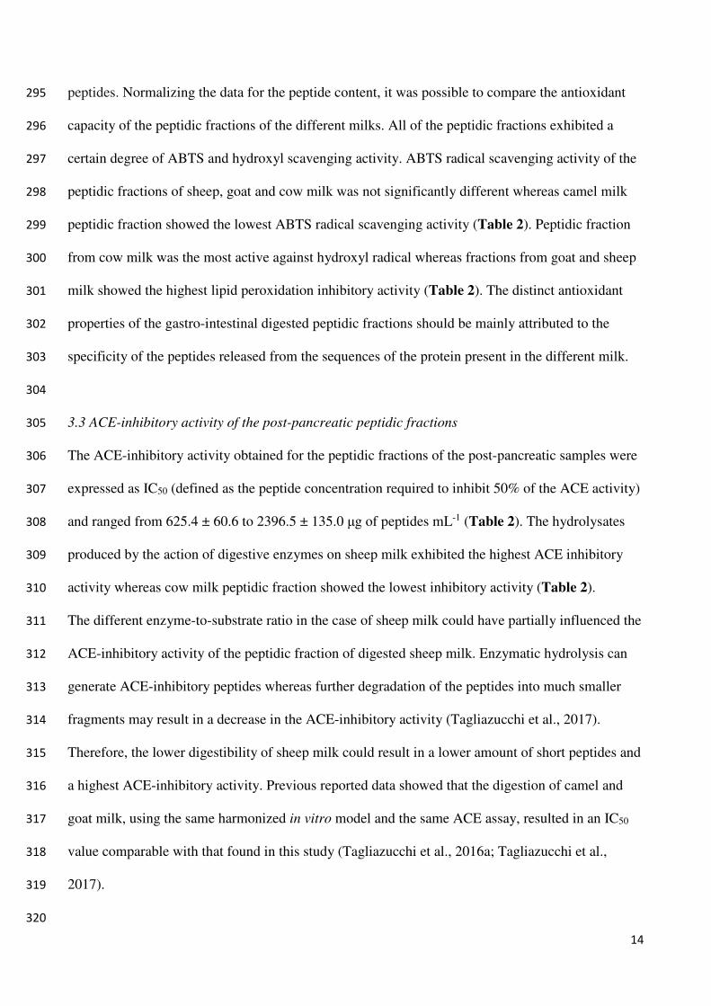
3.1 Comparison between the digestibility of cow, camel, goat and sheep milk proteins 219
The chemical composition of skimmed cow, camel, goat and sheep milks is reported in Table 1. 220
Sheep milk contained significant higher (P<0.05) amount of total proteins and caseins respect to the 221
other milks. The content in total proteins and caseins was not significant different between cow, 222
camel and goat milks. Indeed, no significant statistical differences were observed between the total 223
whey proteins and lactose content as well as the pH value of the different milk. 224
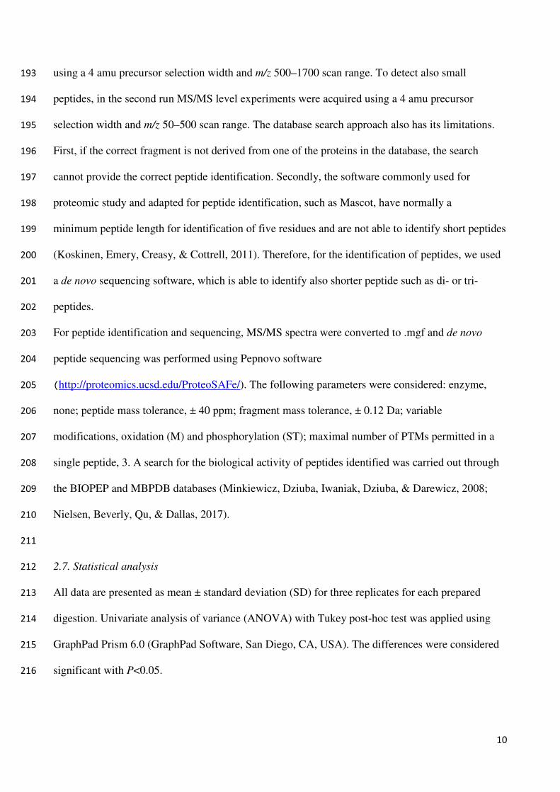
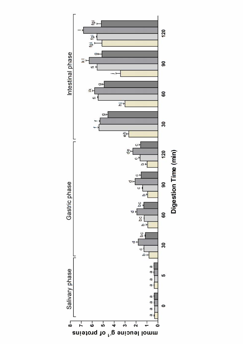
The degradation of milk proteins by gastro-intestinal proteolytic enzymes was compared by 225
measuring the amount of released free amino groups using TNBS assay (Figure 1). As expected, 226
the amount of free amino groups before the digestion (corresponding to the time 0 of the salivary 227
phase of digestion) was not significantly different between the different milk and remained constant 228
during the 5 minutes of salivary incubation. An increase in the hydrolysis was observed for milk of 229
different species during gastric digestion. After 30 minutes of gastric digestion the amount of free 230
amino groups released from goat milk was significantly higher (P<0.001) than that released from 231
cow, camel and sheep milk. No significant statistical differences were observed between the milk 232
from sheep and camel, whereas cow milk showed significantly less amino groups that the other 233
milks (P>0.05). The amount of released amino groups increased slightly but not significantly 234
during the subsequent 90 minutes of peptic digestion in all the milks. The transition from gastric to 235
pancreatic environment produced a significant increase in the amount of free amino groups in all 236
the digested milks. Subsequently, the quantity of released amino groups showed a tendency to 237
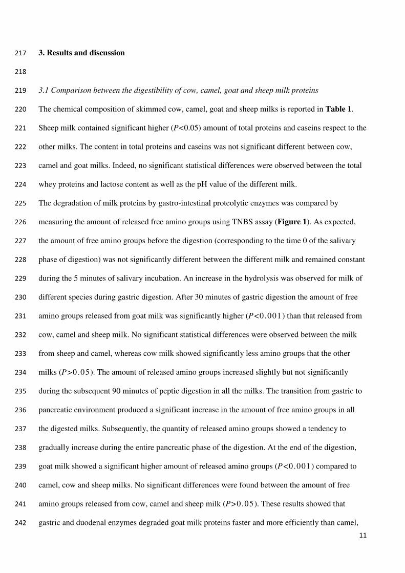
gradually increase during the entire pancreatic phase of the digestion. At the end of the digestion, 238
goat milk showed a significant higher amount of released amino groups (P<0.001) compared to 239
camel, cow and sheep milks. No significant differences were found between the amount of free 240
amino groups released from cow, camel and sheep milk (P>0.05). These results showed that 241
gastric and duodenal enzymes degraded goat milk proteins faster and more efficiently than camel, 242
12
cow and sheep milk. These conclusions are supported by comparison with previously published 243
data. For example, Almaas et al. (2006) found that goat milk proteins were degraded faster than 244
cow milk using human gastro-intestinal proteolytic enzymes. On the other hand, Salami et al. 245
(2008) found that the extent of hydrolysis of camel caseins with pancreatic enzymes was greater 246
than that of cow caseins. Digestion of camel, cow and goat milk with the same protocol used in this 247
study resulted in a higher digestibility of goat milk respect to camel and cow milk (Rutella et al., 248
2016; Tagliazucchi, et al., 2016a; Tagliazucchi et al., 2017). 249
The different enzyme-to-substrate ratio during the digestion, especially in the case of sheep milk, 250
which showed the highest initial protein content, may have had an impact on the hydrolysis of milk 251
proteins. Espejo-Carpio, Pérez-Gálvez, Guadix and Guadix (2013) reported an increase in the 252
digestibility of goat milk proteins as a function of the enzyme-to-substrate ratio. The lower 253
digestibility of sheep milk proteins can be partially attributed to the lower enzyme-to-substrate ratio 254
respect to the other digested milks. 255
256
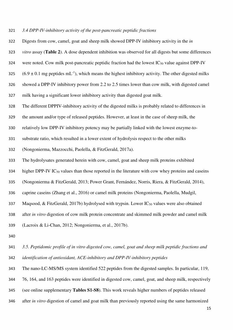
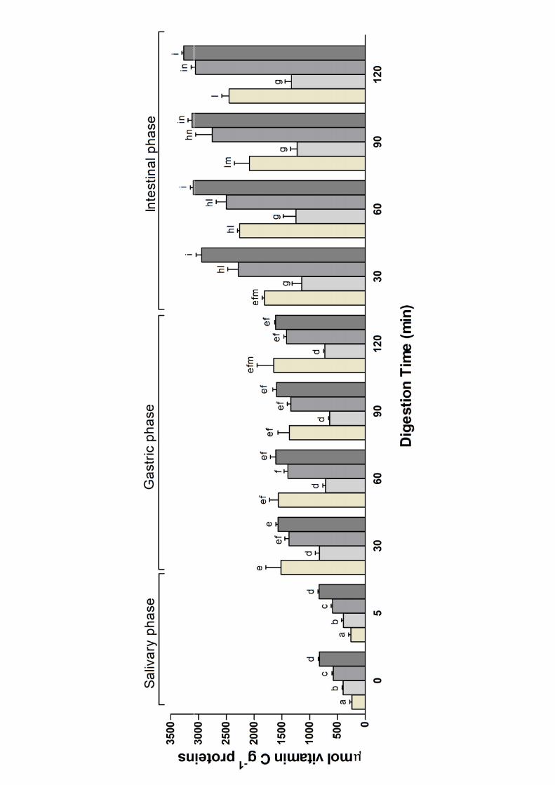
3.2 Evolution of antioxidant activity during in vitro digestion and antioxidant properties of the post-257
pancreatic peptidic fractions 258
The variation in antioxidant activity during the digestion of the different milk was followed by the 259
ABTS assay and reported in Figure 2. 260
All the studied milk showed ABTS radical scavenging activity before the digestion (corresponding 261
to the time 0 of the salivary phase of digestion), but with some differences (Figure 2). Sheep milk 262
had a significant higher ABTS radical scavenging activity with respect to the other milks (P<0.05), 263
whereas cow milk showed the lowest ABTS radical scavenging activity. Clausen, Skibsted, & 264
Stagsted (2009) found that caseins are quantitatively the highest radical scavengers in milk whereas 265
the lower contribution of the low molecular weight compounds is due to ascorbate and especially 266
urate. Caseins have a high content of antioxidative amino acids such as tyrosine, tryptophan and 267
phosphoserine, and quenching of free radicals by oxidation of these amino acids was proposed as 268
13
the explanation (Clausen et al. 2009). As expected, the ABTS radical scavenging activity of the 269
different milk remained constant during the 5 minutes of salivary incubation, whereas the ABTS 270
radical scavenging activity rose as the digestion proceeded reaching the highest value at the end of 271
the pancreatic phase of the digestion in all the analysed milks (Figure 2). This can be explained by 272
an increased number of peptides and amino acids at higher hydrolysis available for interaction with 273
the ABTS radical as already reported (De Gobba, Espejo-Carpio, Skibsted, & Otte, 2014; Kumar, 274
Chatli, Singh, Mehta, & Kumar, 2016). On the other hand, previous studies reported an increase in 275
radical scavenging activity of cow, goat and human milk after in vitro digestion (Tsopmo, et al., 276
2009; Nehir et al., 2015; Power Grant et al., 2016; Tagliazucchi et al., 2016c). Comparison of the 277
data at the end of the digestion showed that sheep and goat milk displayed the highest ABTS radical 278
scavenging activity (P>0.05) followed by cow (P<0.001) and camel (P<0.001) milk. 279
Hernández-Ledesma, Amigo, Recio and Bartolomé (2007) found that an equimolar free amino acids 280
mixture had low antioxidant activity compared to those of the corresponding peptides. Accordingly, 281
extensive hydrolysis, resulting in an increased amount of free amino acids, should bring about to a 282
lower antioxidant activity. However, digested sheep and goat milks showed the highest ABTS 283
radical scavenging activity but goat milk showed the highest digestibility whereas sheep milk the 284
lowest. Therefore, the ABTS radical scavenging activity of digested milk seems more related to the 285
specificity and amount of formed peptides than to the extent of hydrolysis. 286
To fully characterize the antioxidant properties of the digested milk and to evaluate the impact of 287
the released peptides, peptidic fractions were further extracted from the post-pancreatic digested 288
samples through ultrafiltration with a cut-off of 3 kDa and evaluated for their ABTS radical 289
scavenging activity and for their ability to scavenge hydroxyl radical and to inhibit lipid 290
peroxidation. The data regarding the antioxidant properties of the peptidic fractions of the post-291
pancreatic samples are reported in Table 2, together with the peptide content. The amount of 292
released peptides after pancreatic digestion was not significantly different between cow, camel and 293
goat milk whereas sheep milk digestion resulted in a release of significantly greater amount of 294
14
peptides. Normalizing the data for the peptide content, it was possible to compare the antioxidant 295
capacity of the peptidic fractions of the different milks. All of the peptidic fractions exhibited a 296
certain degree of ABTS and hydroxyl scavenging activity. ABTS radical scavenging activity of the 297
peptidic fractions of sheep, goat and cow milk was not significantly different whereas camel milk 298
peptidic fraction showed the lowest ABTS radical scavenging activity (Table 2). Peptidic fraction 299
from cow milk was the most active against hydroxyl radical whereas fractions from goat and sheep 300
milk showed the highest lipid peroxidation inhibitory activity (Table 2). The distinct antioxidant 301
properties of the gastro-intestinal digested peptidic fractions should be mainly attributed to the 302
specificity of the peptides released from the sequences of the protein present in the different milk. 303
304
3.3 ACE-inhibitory activity of the post-pancreatic peptidic fractions 305
The ACE-inhibitory activity obtained for the peptidic fractions of the post-pancreatic samples were 306
expressed as IC50 (defined as the peptide concentration required to inhibit 50% of the ACE activity) 307
and ranged from 625.4 ± 60.6 to 2396.5 ± 135.0 μg of peptides mL-1 (Table 2). The hydrolysates 308
produced by the action of digestive enzymes on sheep milk exhibited the highest ACE inhibitory 309
activity whereas cow milk peptidic fraction showed the lowest inhibitory activity (Table 2). 310
The different enzyme-to-substrate ratio in the case of sheep milk could have partially influenced the 311
ACE-inhibitory activity of the peptidic fraction of digested sheep milk. Enzymatic hydrolysis can 312
generate ACE-inhibitory peptides whereas further degradation of the peptides into much smaller 313
fragments may result in a decrease in the ACE-inhibitory activity (Tagliazucchi et al., 2017). 314
Therefore, the lower digestibility of sheep milk could result in a lower amount of short peptides and 315
a highest ACE-inhibitory activity. Previous reported data showed that the digestion of camel and 316
goat milk, using the same harmonized in vitro model and the same ACE assay, resulted in an IC50 317
value comparable with that found in this study (Tagliazucchi et al., 2016a; Tagliazucchi et al., 318
2017). 319
320
15
3.4 DPP-IV-inhibitory activity of the post-pancreatic peptidic fractions 321
Digests from cow, camel, goat and sheep milk showed DPP-IV inhibitory activity in the in 322
vitro assay (Table 2). A dose dependent inhibition was observed for all digests but some differences 323
were noted. Cow milk post-pancreatic peptidic fraction had the lowest IC50 value against DPP-IV 324
(6.9 ± 0.1 mg peptides mL-1), which means the highest inhibitory activity. The other digested milks 325
showed a DPP-IV inhibitory power from 2.2 to 2.5 times lower than cow milk, with digested camel 326
milk having a significant lower inhibitory activity than digested goat milk. 327
The different DPPIV-inhibitory activity of the digested milks is probably related to differences in 328
the amount and/or type of released peptides. However, at least in the case of sheep milk, the 329
relatively low DPP-IV inhibitory potency may be partially linked with the lowest enzyme-to-330
substrate ratio, which resulted in a lower extent of hydrolysis respect to the other milks 331
(Nongonierma, Mazzocchi, Paolella, & FitzGerald, 2017a). 332
The hydrolysates generated herein with cow, camel, goat and sheep milk proteins exhibited 333
higher DPP-IV IC50 values than those reported in the literature with cow whey proteins and caseins 334
(Nongonierma & FitzGerald, 2013; Power Grant, Fernández, Norris, Riera, & FitzGerald, 2014), 335
caprine caseins (Zhang et al., 2016) or camel milk proteins (Nongonierma, Paolella, Mudgil, 336
Maqsood, & FitzGerald, 2017b) hydrolysed with trypsin. Lower IC50 values were also obtained 337
after in vitro digestion of cow milk protein concentrate and skimmed milk powder and camel milk 338
(Lacroix & Li-Chan, 2012; Nongonierma, et al., 2017b). 339
340
3.5. Peptidomic profile of in vitro digested cow, camel, goat and sheep milk peptidic fractions and 341
identification of antioxidant, ACE-inhibitory and DPP-IV-inhibitory peptides 342
The nano‐LC‐MS/MS system identified 522 peptides from the digested samples. In particular, 119, 343
76, 164, and 163 peptides were identified in digested cow, camel, goat, and sheep milk, respectively 344
(see online supplementary Tables S1-S8). This work reveals higher numbers of peptides released 345
after in vitro digestion of camel and goat milk than previously reported using the same harmonized 346
16
protocol. Our previous research identified 65 and 50 peptides in digested camel and goat milk, 347
respectively (Tagliazucchi et al., 2016a; Tagliazucchi et al., 2017). Concerning cow milk, several 348
studies have already found higher amount of peptides (more than 119) than this study (Egger et al., 349
2016; Picariello et al., 2010). To the best of our knowledge, this is the first paper reporting a 350
comprehensive peptidomic profile of digested sheep milk. 351
The majority of the peptides were from caseins (71.4, 73.7, 72.0 and 71.2% of the total identified 352
peptides in digested cow, camel, goat and sheep milk, respectively) with β-casein which was the 353
best source of peptides in all the digested milk (43.7, 51.3, 42.7 and 40.5% of the total identified 354
peptides in digested cow, camel, goat and sheep milk, respectively). Whey proteins gave a lower 355
amount of peptides respect to caseins, especially in camel milk that does not contain β-lactoglobulin 356
(see online supplementary Tables S1-S8). In addition, 9 amino acids were also identified, 7 of them 357
being essential amino acids (W, L, I, V, K, R and F). 358
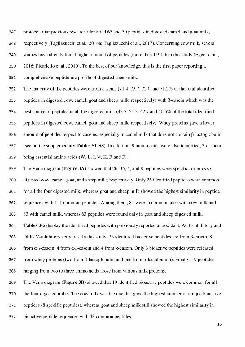
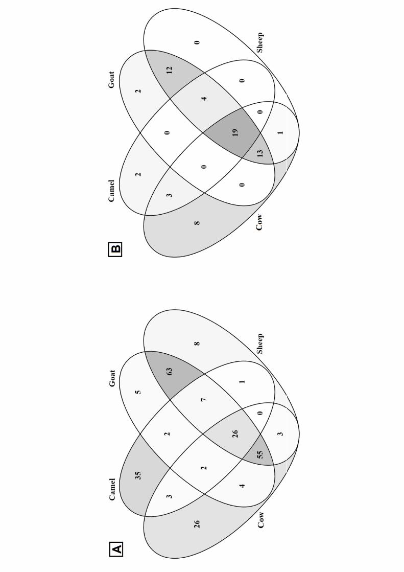
The Venn diagram (Figure 3A) showed that 26, 35, 5, and 8 peptides were specific for in vitro 359
digested cow, camel, goat, and sheep milk, respectively. Only 26 identified peptides were common 360
for all the four digested milk, whereas goat and sheep milk showed the highest similarity in peptide 361
sequences with 151 common peptides. Among them, 81 were in common also with cow milk and 362
33 with camel milk, whereas 63 peptides were found only in goat and sheep digested milk. 363
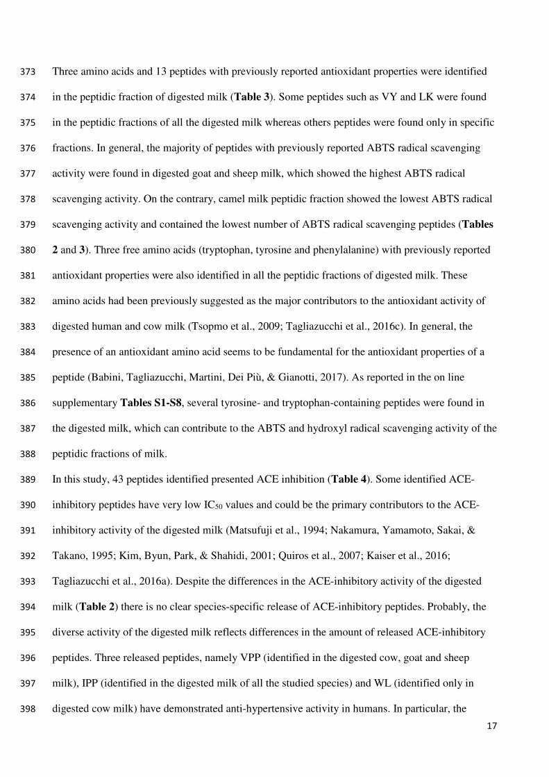
Tables 3-5 display the identified peptides with previously reported antioxidant, ACE-inhibitory and 364
DPP-IV-inhibitory activities. In this study, 26 identified bioactive peptides are from β-casein, 8 365
from αS1-casein, 4 from αS2-casein and 4 from κ-casein. Only 3 bioactive peptides were released 366
from whey proteins (two from β-lactoglobulin and one from α-lactalbumin). Finally, 19 peptides 367
ranging from two to three amino acids arose from various milk proteins. 368
The Venn diagram (Figure 3B) showed that 19 identified bioactive peptides were common for all 369
the four digested milks. The cow milk was the one that gave the highest number of unique bioactive 370
peptides (8 specific peptides), whereas goat and sheep milk still showed the highest similarity in 371
bioactive peptide sequences with 48 common peptides. 372
17
Three amino acids and 13 peptides with previously reported antioxidant properties were identified 373
in the peptidic fraction of digested milk (Table 3). Some peptides such as VY and LK were found 374
in the peptidic fractions of all the digested milk whereas others peptides were found only in specific 375
fractions. In general, the majority of peptides with previously reported ABTS radical scavenging 376
activity were found in digested goat and sheep milk, which showed the highest ABTS radical 377
scavenging activity. On the contrary, camel milk peptidic fraction showed the lowest ABTS radical 378
scavenging activity and contained the lowest number of ABTS radical scavenging peptides (Tables 379
2 and 3). Three free amino acids (tryptophan, tyrosine and phenylalanine) with previously reported 380
antioxidant properties were also identified in all the peptidic fractions of digested milk. These 381
amino acids had been previously suggested as the major contributors to the antioxidant activity of 382
digested human and cow milk (Tsopmo et al., 2009; Tagliazucchi et al., 2016c). In general, the 383
presence of an antioxidant amino acid seems to be fundamental for the antioxidant properties of a 384
peptide (Babini, Tagliazucchi, Martini, Dei Più, & Gianotti, 2017). As reported in the on line 385
supplementary Tables S1-S8, several tyrosine- and tryptophan-containing peptides were found in 386
the digested milk, which can contribute to the ABTS and hydroxyl radical scavenging activity of the 387
peptidic fractions of milk. 388
In this study, 43 peptides identified presented ACE inhibition (Table 4). Some identified ACE-389
inhibitory peptides have very low IC50 values and could be the primary contributors to the ACE-390
inhibitory activity of the digested milk (Matsufuji et al., 1994; Nakamura, Yamamoto, Sakai, & 391
Takano, 1995; Kim, Byun, Park, & Shahidi, 2001; Quiros et al., 2007; Kaiser et al., 2016; 392
Tagliazucchi et al., 2016a). Despite the differences in the ACE-inhibitory activity of the digested 393
milk (Table 2) there is no clear species-specific release of ACE-inhibitory peptides. Probably, the 394
diverse activity of the digested milk reflects differences in the amount of released ACE-inhibitory 395
peptides. Three released peptides, namely VPP (identified in the digested cow, goat and sheep 396
milk), IPP (identified in the digested milk of all the studied species) and WL (identified only in 397
digested cow milk) have demonstrated anti-hypertensive activity in humans. In particular, the 398
18
lactotripeptides VPP and IPP have been shown (at dosages between 5 and 100 mg day−1) to 399
decrease the systolic (4.0 mmHg) and diastolic (1.9 mmHg) blood pressure in hypertensive patients 400
and to positively modulate pulse wave velocity in mildly hypertensive subjects (Cicero, Fogacci, & 401
Colletti, 2017). Two recent studies showed that VPP and IPP could be released from cow and goat 402
milk during in vitro digestion at doses, which can elicit physiological effects (Rutella et al., 2016; 403
Tagliazucchi et al., 2017). The α-lactalbumin derived dipeptide WL was found to be bioavailable in 404
human subjects, reducing in vivo ACE activity (Kaiser et al., 2016). One additional peptide 405
(LHLPLP) was found to be able to decrease systolic and diastolic blood pressure in spontaneously 406
hypertensive rats (Quiros et al., 2007). Some other peptides with very low IC50 values (IY, VF and 407
LPP) have been found in plasma of human volunteers after consumption of dairy products (van 408
Platerink, Janssen, Horsten, & Haverkamp, 2006; Foltz et al., 2007). The peptide VY seems to be 409
particularly interesting, behaving as a multifunctional bioactive peptide with high ACE-inhibitory 410
and antioxidant activities (Cheng, Che, Xiong, 2010; Tagliazucchi et al., 2016a). The release of VY 411
was common in milk from the different species studied. VY has been also found in human plasma 412
after consumption of a milk beverage, indicating that this peptide is also released in vivo from cow 413
milk caseins and is bioavailable in humans (Foltz et al., 2007). 414
Finally, 20 peptides with previously demonstrated DPP-IV-inhibitory activity were identified in the 415
peptidic fractions of digested milk (Table 5). Cow milk was the best source of DPP-IV-inhibitory 416
peptides (17 out of 20) and the sample with the highest DPP-IV-inhibitory activity (Table 2). Two 417
well-known DPP-IV inhibitors, namely IPI (also known as Diprotin A) and VLP (also known as 418
Diprotin B), were released from cow milk after digestion and could be the primary contributor to the 419
DPP-IV-inhibitory activity of the digested cow milk (Table 5). Diprotin A but not Diprotin B was 420
also found in the peptidic fractions of digested goat and sheep milk. 421
422
4. Conclusion 423
19
The present study integrated peptides identified by LC-MS/MS with in vitro bioactivities of milk 424
from four different species (cow, camel, goat and sheep) after the application of the harmonized 425
INFOGEST in vitro gastro-intestinal digestion protocol. Whereas goat milk showed the highest 426
apparent digestibility, sheep milk appeared to be the best source of ACE-inhibitory peptides. 427
Moreover, cow milk was found to be the best source of DPP-IV-inhibitory peptides and antioxidant 428
peptides and amino acids. Peptidomic analysis showed that goat and sheep milk displayed the 429
highest similarity in peptide sequences identified after in vitro digestion. Most of the released 430
bioactive peptides were in common between two or more species and the peptides with the highest 431
ACE-inhibitory activity had previously demonstrated to be bioavailable in humans. 432
Although this study lays the basis to distinguish milk from different species in the light of their 433
bioactivities and bioactive peptides released during in vitro digestion, limitations have to be 434
considered. The most important is that this research was conducted analysing one sample of milk 435
from each species. Therefore, to expand the results, studies involving more milk samples are 436
required. Finally, further investigations and in vivo trials are needed to establish which of the 437
observed bioactive peptides have physiological significance.438
20
References
Abd El-Salam, M. H., & El-Shibiny, S. (2017). Preparation, properties, and uses of enzymatic milk
protein hydrolysates. Critical Reviews in Food Science and Nutrition, 57, 1119-1132.
Adler-Nissen, J. (1979). Determination of the degree of hydrolysis of food protein hydrolysates by
trinitrobenzensulfonic acid. Journal of Agricultural and Food Chemistry, 27, 1256-1262.
Almaas, H., Cases, A. L., Devold, T. G., Holm, H., Langsurd, T., Aabakken, L., Aadnoey, T., &
Vegarud, G. E. (2006). In vitro digestion of bovine and caprine milk by human gastric and
duodenal enzymes. International Dairy Journal, 16, 961-968.
Babini, E., Tagliazucchi, D., Martini, S., Dei Più, L., & Gianotti, A. (2017). LC-ESI-QTOF-MS
identification of novel antioxidant peptides obtained by enzymatic and microbial hydrolysis of
vegetable proteins. Food Chemistry, 228, 186-196.
Cheng, Y., Che, J., & Xiong, Y. L. (2010). Chromatographic separation and tandem MS
identification of active peptides in potato protein hydrolysate that inhibit autoxidation of soybean
oil-in-water emulsions. Journal of Agricultural and Food Chemistry, 58, 8825-8832.
Cicero, A. F. G., Fogacci, F., & Colletti, A. (2017). Potential role of bioactive peptides in
prevention and treatment of chronic diseases: a narrative review. British Journal of
Pharmacology, 174, 1378-1394.
Clausen, M. R., Skibsted, L. H., & Stagsted, J. (2009) Characterization of major radical scavenger
species in bovine milk through size exclusion chromatography and functional assays. Journal of
Agricultural and Food Chemistry, 57, 2912–2919.
De Gobba, C., Espejo-Carpio, F. J., Skibsted, L. H., & Otte, J. (2014). Antioxidant peptides from
goat milk protein fractions hydrolysed by two commercial proteases. International Dairy
Journal, 39, 28–40.
Dei Più, L., Tassoni, A., Serrazanetti, D. I., Ferri, M., Babini, E., Tagliazucchi, D., & Gianotti, A.
(2014). Exploitation of starch industry liquid by-product to produce bioactive peptides from rice
hydrolyzed proteins. Food Chemistry, 155, 199–206.
21
Egger, L., Ménard, O., Delgado-Andrade, C., Alvito, P., Asunção, R., Balance, S., et al. (2016). The
harmonized INFOGEST in vitro digestion method: From knowledge to action. Food Research
International, 88, 217-225.
Egger, L., & Ménard, O. (2017). Update on bioactive peptides after milk and cheese digestion.
Current Opinion in Food Science, 14, 116-121.
El-Agamy, E. I., Nawar, M., Shamsia, S. M., Awad, S., & Haenlein, G. F. W. (2009). Are camel
milk proteins convenient to the nutrition of cow milk allergic children? Small Ruminant
Research, 82, 1-6.
Espejo-Carpio, F. J., Pérez-Gálvez, R., Guadix, E. M., & Guadix, A. (2013). Optimization of the
hydrolysis of goat milk protein for the production of ACE-inhibitory peptides. Journal of Dairy
Research, 80, 214-222.
FitzGerald, R. J., Murray, B. A., & Walsh, D. J. (2004). Hypotensive peptides from milk proteins.
Journal of Nutrition, 134, 980S-988S.
Foltz, M., Meynen, E. E., Bianco, V., van Platerink, C., Koning, T. M. M. G., & Kloek, J. (2007).
Angiotensin converting enzyme inhibitory peptides from a lactotripeptide-enriched milk
beverage are absorbed intact into the circulation. Journal of Nutrition, 137, 953-958.
Fricker, L. D. (2015). Limitations of mass spectrometry-based peptidomic approaches. Journal of
the American Society for Mass Spectrometry, 26, 1981-1991.
Hernández-Ledesma, B., Amigo, L., Recio, L., & Bartolomè, B. (2007). ACE-inhibitory and
radical-scavenging activity of peptides derived from β-lactoglobulin f(19-25). Interactions with
ascorbic acid. Journal of Agricultural and Food Chemistry, 55, 3392-3397.
Hernández-Ledesma, B., García-Nebot, M. J., Fernández-Tomé, S., Amigo, L., & Recio, I. (2014).
Dairy protein hydrolysates: Peptides for health benefits. International Dairy Journal, 38, 82-100.
Kaiser, S., Martin, M., Lunow, D., Rudolph, S., Mertten, S., Möckel, U., Deußen, A., & Henle, T.
(2016). Tryptophan-containing dipeptides are bioavailable and inhibit plasma human
angiotensin-converting enzyme in vivo. International Dairy Journal, 52, 107-114.
22
Kim, S. K., Byun, H. G., Park, P. J., & Shahidi, F. (2001). Angiotensin I converting enzyme
inhibitory peptides purified from bovine skin gelatin hydrolysate. Journal of Agricultural and
Food Chemistry, 49, 2992-2997.
Kopf-Bolanz, K. A., Schwander, F., Gijs, M., Vergères, G., Portmann, R., & Egger, L. (2012).
Validation of an in vitro digestive system for studying macronutrient decomposition in humans.
Journal of Nutrition, 142, 245–250.
Kopf-Bolanz, K. A., Schwander, F., Gijs, M., Vergères, G., Portmann, R., & Egger, L. (2014).
Impact of milk processing on the generation of peptides during digestion. International Dairy
Journal, 35, 130-138.
Koskinen, V. R., Emery, P. A., Creasy, D. M., & Cottrell, J. S. (2011). Hierarchical clustering of
shotgun proteomics data. Molecular and Cellular Proteomics, 10, M110.003822.
Kumar, D., Chatli, M. K., Singh, R., Mehta, N., & Kumar, P. (2016). Enzymatic hydrolysis of
camel milk casein and its antioxidant properties. Dairy Science & Technology, 96, 391-404.
Lacroix, I., M., E., & Li-Chan, E. C. Y. (2012). Dipeptidyl peptidase-IV inhibitory activity of dairy
protein hydrolysates. International Dairy Journal, 25, 97-102.
Matsufuji, H., Matsui, T., Seki, E., Osajima, K., Nakashima, M., & Osajima, Y. (1994).
Angiotensin I-converting enzyme inhibitory peptides in an alkaline protease hydrolyzate derived
from sardine muscle. Bioscience, Biotechnology and Biochemistry, 58, 2244-2251.
Minkiewicz, P., Dziuba, J., Iwaniak, A., Dziuba, M., Darewicz M. (2008). BIOPEP database and
other programs for processing bioactive peptide sequences. Journal of AOAC International, 91,
965-980.
Minekus, M., Alminger, M., Alvito, P., Ballance, S., Bohn, T., Bourlieu, C., et al. (2014). A
standardised static in vitro digestion method suitable for food – an international consensus. Food
& Function, 5, 1113 −1124.
23
Nakamura, Y., Yamamoto, N., Sakai, K., & Takano, T. (1995). Antihypertensive effect of sour milk
and peptides isolated from it that are inhibitors to angiotensin I-converting enzyme. Journal of
Dairy Science, 78, 1253 −1257.
Nehir, S., Karakaya, S., Simsek, S., Dupont, D., Menfaatli, E., Eker, A. T. (2015). In vitro
digestibility of goat milk and kefir with a new standardised static digestion method (INFOGEST
cost action) and bioactivities of the resultant peptides. Food & Function, 6, 2322-2330.
Nielsen, S. D., Beverly, R. L., Qu, Y., & Dallas, D. C. (2017). Milk bioactive peptide database: A
comprehensive database of milk protein-derived bioactive peptides and novel visualization. Food
Chemistry, 232, 673–82.
Nongonierma, A. B., & FitzGerald, R. J. (2013). Dipeptidyl peptidase IV inhibitory and
antioxidative properties of milk-derived dipeptides and hydrolysates. Peptides, 39, 157–163.
Nongonierma, A. B., & FitzGerald, R. J. (2015). The scientific evidence for the role of milk
protein-derived bioactive peptides in humans: A Review. Journal of Functional Foods, 17, 640 -
656.
Nongonierma, A. B., Mazzocchi, C., Paolella, S., & FitzGerald, R. J. (2017a). Release of dipeptidyl
peptidase IV (DPP-IV) inhibitory peptides from milk protein isolate (MPI) during enzymatic
hydrolysis. Food Research International, 94, 79 -89.
Nongonierma, A. B., Paolella, S., Mudgil, P., Maqsood, S., & FitzGerald, R. J. (2017b). Dipeptidyl
peptidase IV (DPP-IV) inhibitory properties of camel milk protein hydrolysates generated with
trypsin. Journal of Functional Foods, 34, 49 -58.
Park, Y. W., Juárez, M., Ramos, M., & Haenlein, G. F. W. (2007). Physico-chemical characteristics
of goat and sheep milk. Small Ruminant Research, 68, 88-113.
Picariello, G., Ferranti, P., Fierro, O., Mamone, G., Caira, S., Di Luccia, A., Monica, S., Addeo, F.
(2010). Peptides surviving the simulated gastro-intestinal digestion of milk proteins: biological
and toxicological implication. Journal of Chromatography B, 878, 295-308.
24
Power Grant, O., Fernández, A., Norris, R., Riera, F. A., & FitzGerald, R. J. (2014). Selective
enrichment of bioactive properties during ultrafiltration of a tryptic digest of β-lactoglobulin.
Journal of Functional Foods, 9, 38–47.
Power Grant, O., McCormack, W. G., Ramia De Cap, M., Amigo-Benavent, M., FitzGerald, R. J.,
& Jakeman, P. (2016). Evaluation of the antioxidant capacity of a milk protein matrix in vitro
and in vivo in women aged 50-70 years. International Journal of Food Sciences and Nutrition,
67, 325-334.
Quiros, A., Ramos, M., Muguerza, B., Delgado, M. A., Miguel, M., Aleixandre, A., & Recio, I.
(2007). Identification of novel antihypertensive peptides in milk fermented with Enterococcus
faecalis. International Dairy Journal, 17, 33-41.
Re, R., Pellegrini, N., Proteggente, A., Pannala, A., Yang, M., & Rice-Evans, C. (1999) Antioxidant
activity applying an improved ABTS radical cation decolorization assay. Free Radical Biology
and Medicine, 26, 1231-1237.
Rizzello, C. G., Tagliazucchi, D., Babini, E., Rutella, G. S., Taneyo Saa, D. L., & Gianotti, A.
(2016). Bioactive peptides from vegetable food matrices: Research trends and novel
biotechnologies for synthesis and recovery. Journal of Functional Foods, 27, 549-569.
Ronca-Testoni, S. (1983). Direct spectrophotometric assay for angiotensin-converting enzyme in
serum. Clinical Chemistry, 29, 1093–1096.
Rutella, G. S., Solieri, L., Martini, S., Tagliazucchi, D. (2016). Release of the antihypertensive
tripeptides valine-proline-proline and isoleucine-proline-proline from bovine milk caseins during
in vitro gastrointestinal digestion. Journal of Agricultural and Food Chemistry, 64, 8509-8515.
Salami, M., Yousefi, R., Eshani, M. R., Dalgalarrondo, M. Chobert, J. M., Haertlé, T., Razavi, S.
H., Saboury, A. A., Niasari-Niaslaji, A., & Moosavi-Movahedi, A. A. (2008). Kinetic
characterization of hydrolysis of camel and bovine milk proteins by pancreatic enzymes.
International Dairy Journal, 18, 1097-1102.
25
Tagliazucchi, D., Verzelloni, E., & Conte, A. (2010). Effect of dietary melanoidins on lipid
peroxidation during simulated gastric digestion: their possible role in the prevention of oxidative
damage. Journal of Agricultural and Food Chemistry, 58, 2513-2519.
Tagliazucchi, D., Shamsia, S., & Conte, A. (2016a). Release of angiotensin converting-enzyme
inhibitory peptides during in vitro gastro-intestinal digestion of camel milk. International Dairy
Journal, 56, 119-128.
Tagliazucchi, D., Helal, A., Verzelloni, E., Bellesia, A., & Conte, A. (2016b). Composition and
properties of peptides that survive standardised in vitro gastro-pancreatic digestion of bovine
milk. International Dairy Journal, 61, 196-204.
Tagliazucchi, D., Helal, A., Verzelloni, E., & Conte, A. (2016c). Bovine milk antioxidant
properties: effect of in vitro digestion and identification of antioxidant compounds. Dairy
Science & Technology, 96, 657-676.
Tagliazucchi, D., Shamsia, S., Helal, A., & Conte, A. (2017). Angiotensin-converting enzyme
inhibitory peptides from goats' milk released by in vitro gastro-intestinal digestion. International
Dairy Journal, 71, 6-16.
Tsopmo, A., Dielh-Jones, B. W., Aluko, R. E., Kitts, D. D., Elisia, I., & Friel, J. K. (2009).
Tryptophan released from mother's milk has antioxidant properties. Pediatric Research, 66, 618-
618.
Van Platerink, C. J., Janssen, H. G. M., Horsten, R., & Haverkamp, J. (2006). Quantification of
ACE inhibiting peptides in human plasma using high performance liquid chromatography–mass
spectrometry. Journal of Chromatography B, 830, 151-157.
Yadav, A. K., Singh, J., & Yadav, S. K. (2016). Composition, nutritional and therapeutic values of
goat milk: A review. Asian Journal of Dairy Food and Research, 35, 96-102.
Zhang, Y., Chen, R., Zuo, F., Ma, H., Zhang, Y., & Chen, S. (2016). Comparison of dipeptidyl
peptidase IV-inhibitory activity of peptides from bovine and caprine milk casein by in silico and
in vitro analyses. International Dairy Journal, 53, 37–44.
26
Figure captions
Figure 1. Comparison between the in vitro digestibility of skimmed cow, camel, goat and sheep
milk. Release of free amino groups during in vitro gastric and pancreatic digestion of skimmed cow
( ), camel ( ), goat ( ) and sheep ( ) milk. Data were corrected by the contribution of the
control digestion and normalised with respect to the initial protein content of the different milks
studied and expressed as mmol of leucine equivalent per g of protein. Values are means of three
independent digestions ± standard deviation (SD). Different letters indicate significantly different
values (P<0.05).
Figure 2. Evolution of ABTS radical scavenging activity during the in vitro digestion of
skimmed cow ( ), camel ( ), goat ( ) and sheep ( ) milk. ABTS radical scavenging
activity was expressed as μmol of vitamin C g-1 of milk protein. Data were corrected by the
contribution of the control digestion and normalised with respect to the initial protein content of the
different milks studied. Values are means of three independent digestions ± standard deviation
(SD). Different letters indicate significantly different values (P<0.05).
Figure 3. Venn diagrams of peptides obtained from skimmed cow, camel, goat and sheep
milk. (A) Venn diagram created with all the identified peptides released after in vitro gastro-
27
intestinal digestion (see on line supplementary material Tables S1-S8 for the peptide sequences).
(B) Venn diagram created with only the bioactive peptides released and identified after in vitro
gastro-intestinal digestion (see Tables 3-5 for the peptide sequences and bioactivity).
Table 1. Chemical composition of skimmed cow, camel, goat and sheep milks. Data are expressed
as g 100 g-1 of milk.
Values represent means ± standard deviation of triplicate determination; different superscript letters within the same
row indicate that the values are significantly different (P<0.05).
Cow milk Camel milk Goat milk Sheep milk
Total proteins 3.60 ± 0.11a 3.48 ± 0.14a 3.78 ± 0.12a 5.68 ± 0.21b
Caseins 2.88 ± 0.07a 2.68 ± 0.10a 2.92 ± 0.09a 4.76 ± 0.18b
Whey proteins 0.72 ± 0.04a 0.80 ± 0.08a 0.86 ± 0.10a 0.92 ± 0.08a
Lactose 4.80 ± 0.18a 4.87 ± 0.15a 4.55 ± 0.14a 4.74 ± 0.12a
Fat < 0.05 < 0.05 < 0.05 < 0.05
pH 6.65 ± 0.03a 6.61 ± 0.05a 6.64 ± 0.05a 6.67 ± 0.04a
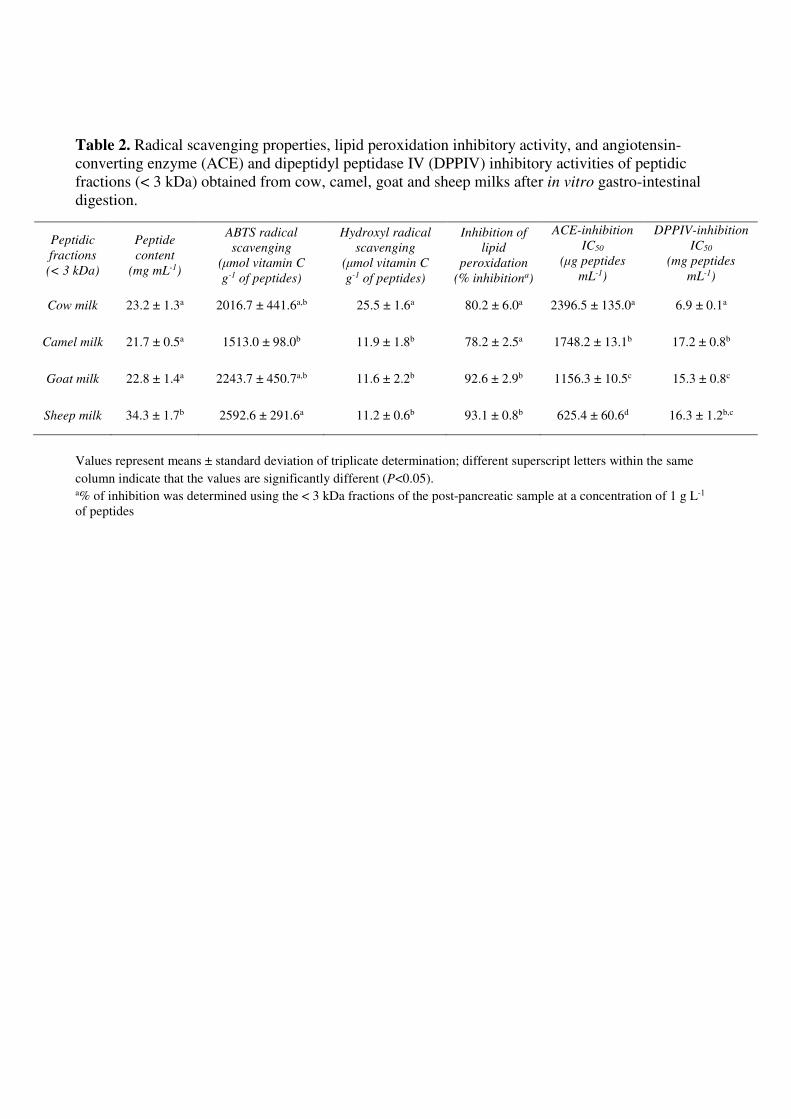
Table 2. Radical scavenging properties, lipid peroxidation inhibitory activity, and angiotensin-
converting enzyme (ACE) and dipeptidyl peptidase IV (DPPIV) inhibitory activities of peptidic
fractions (< 3 kDa) obtained from cow, camel, goat and sheep milks after in vitro gastro-intestinal
digestion.
Values represent means ± standard deviation of triplicate determination; different superscript letters within the same
column indicate that the values are significantly different (P<0.05). a% of inhibition was determined using the < 3 kDa fractions of the post-pancreatic sample at a concentration of 1 g L-1
of peptides
Peptidic
fractions
(< 3 kDa)
Peptide
content
(mg mL-1)
ABTS radical
scavenging
(μmol vitamin C
g-1 of peptides)
Hydroxyl radical
scavenging
(μmol vitamin C
g-1 of peptides)
Inhibition of
lipid
peroxidation
(% inhibitiona)
ACE-inhibition
IC50
(μg peptides
mL-1)
DPPIV-inhibition
IC50
(mg peptides
mL-1)
Cow milk 23.2 ± 1.3a 2016.7 ± 441.6a,b 25.5 ± 1.6a 80.2 ± 6.0a 2396.5 ± 135.0a 6.9 ± 0.1a
Camel milk 21.7 ± 0.5a 1513.0 ± 98.0b 11.9 ± 1.8b 78.2 ± 2.5a 1748.2 ± 13.1b 17.2 ± 0.8b
Goat milk 22.8 ± 1.4a 2243.7 ± 450.7a,b 11.6 ± 2.2b 92.6 ± 2.9b 1156.3 ± 10.5c 15.3 ± 0.8c
Sheep milk 34.3 ± 1.7b 2592.6 ± 291.6a 11.2 ± 0.6b 93.1 ± 0.8b 625.4 ± 60.6d 16.3 ± 1.2b,c
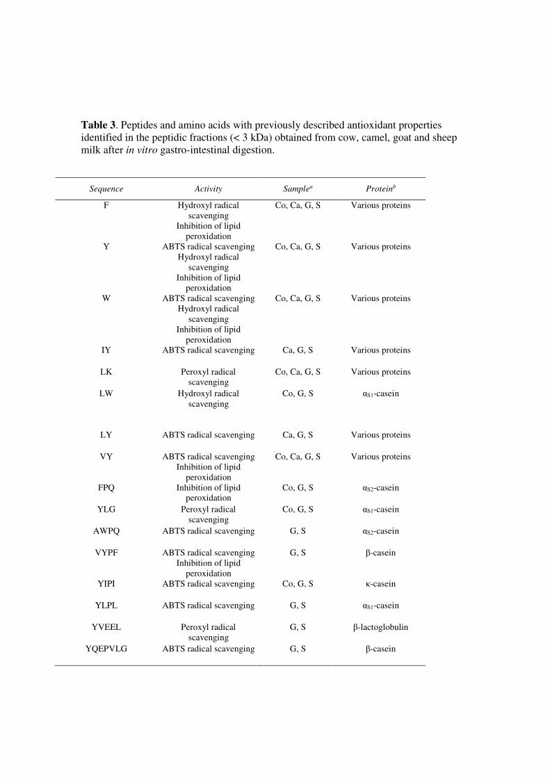
Table 3. Peptides and amino acids with previously described antioxidant properties
identified in the peptidic fractions (< 3 kDa) obtained from cow, camel, goat and sheep
milk after in vitro gastro-intestinal digestion.
Sequence Activity Samplea Proteinb
F Hydroxyl radical
scavenging
Inhibition of lipid
peroxidation
Co, Ca, G, S Various proteins
Y ABTS radical scavenging
Hydroxyl radical
scavenging
Inhibition of lipid
peroxidation
Co, Ca, G, S Various proteins
W ABTS radical scavenging
Hydroxyl radical
scavenging
Inhibition of lipid
peroxidation
Co, Ca, G, S Various proteins
IY ABTS radical scavenging Ca, G, S Various proteins
LK Peroxyl radical
scavenging
Co, Ca, G, S Various proteins
LW Hydroxyl radical
scavenging
Co, G, S αS1-casein
LY ABTS radical scavenging Ca, G, S Various proteins
VY ABTS radical scavenging
Inhibition of lipid
peroxidation
Co, Ca, G, S Various proteins
FPQ Inhibition of lipid
peroxidation
Co, G, S αS2-casein
YLG Peroxyl radical
scavenging
Co, G, S αS1-casein
AWPQ ABTS radical scavenging G, S αS2-casein
VYPF ABTS radical scavenging
Inhibition of lipid
peroxidation
G, S β-casein
YIPI ABTS radical scavenging Co, G, S κ-casein
YLPL ABTS radical scavenging G, S αS1-casein
YVEEL Peroxyl radical
scavenging
G, S β-lactoglobulin
YQEPVLG ABTS radical scavenging G, S β-casein

aSample in which the peptide was identified (Co: digested cow milk; Ca: digested camel milk; G: digested
goat milk; S: digested sheep milk) bPrecursor protein
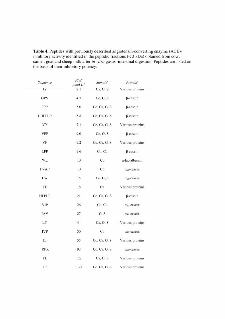
Table 4. Peptides with previously described angiotensin-converting enzyme (ACE)-
inhibitory activity identified in the peptidic fractions (< 3 kDa) obtained from cow,
camel, goat and sheep milk after in vitro gastro-intestinal digestion. Peptides are listed on
the basis of their inhibitory potency.
Sequence IC50
a
μmol L-1 Sampleb Proteinc
IY 2.1
Ca, G, S Various proteins
GPV 4.7
Co, G, S β-casein
IPP 5.0 Co, Ca, G, S β-casein
LHLPLP 5.8 Co, Ca, G, S β-casein
VY 7.1
Co, Ca, G, S Various proteins
VPP 9.0
Co, G, S β-casein
VF 9.2 Co, Ca, G, S Various proteins
LPP 9.6 Co, Ca β-casein
WL 10
Co α-lactalbumin
FVAP 10 Co αS1-casein
LW 15 Co, G, S αS1-casein
TF 18
Ca Various proteins
HLPLP 21 Co, Ca, G, S β-casein
VIP 26 Co, Ca αS2-casein
IAV 27 G, S αS1-casein
LY 44
Ca, G, S Various proteins
IVP 50 Co αS1-casein
IL 55 Co, Ca, G, S Various proteins
RPK 92
Co, Ca, G, S αS1-casein
YL 122
Ca, G, S Various proteins
IP 130
Co, Ca, G, S Various proteins
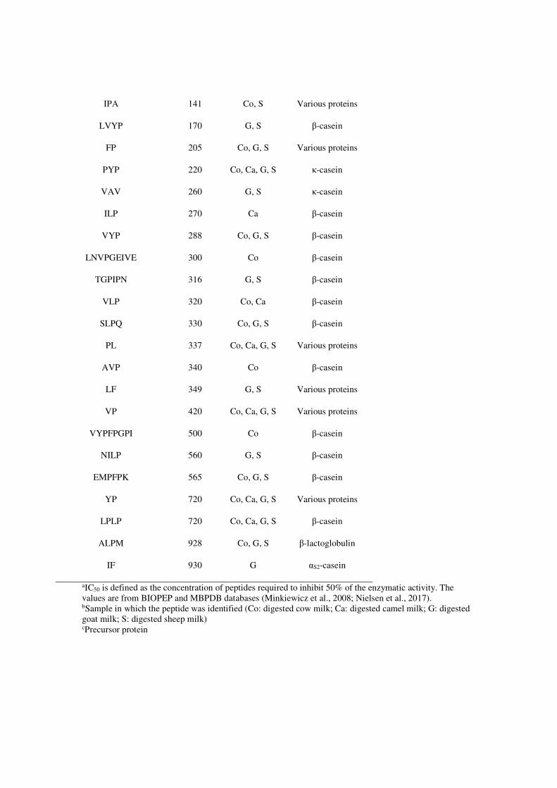
IPA 141
Co, S Various proteins
LVYP 170 G, S β-casein
FP 205
Co, G, S Various proteins
PYP 220 Co, Ca, G, S κ-casein
VAV 260 G, S κ-casein
ILP 270 Ca β-casein
VYP 288 Co, G, S β-casein
LNVPGEIVE 300
Co β-casein
TGPIPN 316 G, S β-casein
VLP 320 Co, Ca β-casein
SLPQ 330 Co, G, S β-casein
PL 337 Co, Ca, G, S Various proteins
AVP 340 Co β-casein
LF 349
G, S Various proteins
VP 420 Co, Ca, G, S Various proteins
VYPFPGPI 500 Co β-casein
NILP 560 G, S β-casein
EMPFPK 565 Co, G, S β-casein
YP 720
Co, Ca, G, S Various proteins
LPLP 720 Co, Ca, G, S β-casein
ALPM 928 Co, G, S β-lactoglobulin
IF 930 G αS2-casein
aIC50 is defined as the concentration of peptides required to inhibit 50% of the enzymatic activity. The
values are from BIOPEP and MBPDB databases (Minkiewicz et al., 2008; Nielsen et al., 2017).
bSample in which the peptide was identified (Co: digested cow milk; Ca: digested camel milk; G: digested
goat milk; S: digested sheep milk) cPrecursor protein
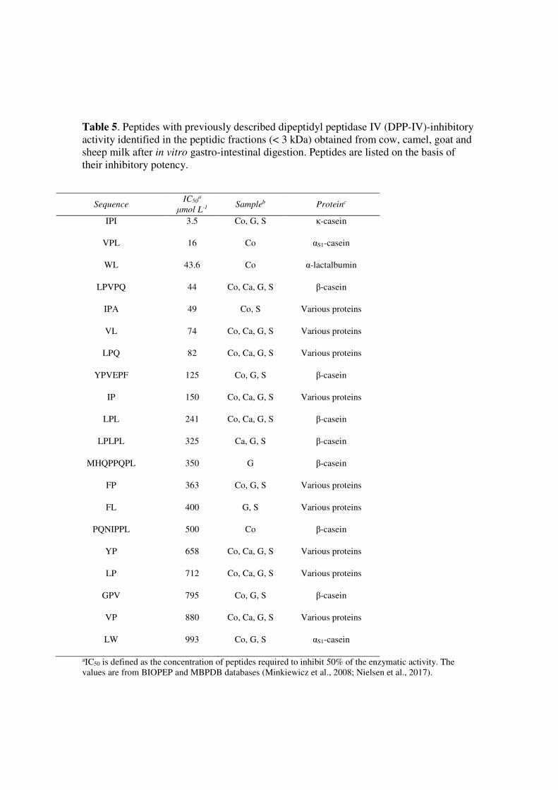
Table 5. Peptides with previously described dipeptidyl peptidase IV (DPP-IV)-inhibitory
activity identified in the peptidic fractions (< 3 kDa) obtained from cow, camel, goat and
sheep milk after in vitro gastro-intestinal digestion. Peptides are listed on the basis of
their inhibitory potency.
Sequence IC50
a
μmol L-1 Sampleb Proteinc
IPI 3.5 Co, G, S κ-casein
VPL 16 Co αS1-casein
WL 43.6 Co α-lactalbumin
LPVPQ 44 Co, Ca, G, S β-casein
IPA 49 Co, S Various proteins
VL
74
Co, Ca, G, S Various proteins
LPQ 82 Co, Ca, G, S Various proteins
YPVEPF 125
Co, G, S β-casein
IP 150 Co, Ca, G, S Various proteins
LPL 241 Co, Ca, G, S β-casein
LPLPL 325 Ca, G, S β-casein
MHQPPQPL 350
G β-casein
FP 363 Co, G, S Various proteins
FL
400
G, S Various proteins
PQNIPPL 500 Co β-casein
YP 658 Co, Ca, G, S Various proteins
LP 712 Co, Ca, G, S Various proteins
GPV 795 Co, G, S β-casein
VP 880 Co, Ca, G, S Various proteins
LW 993 Co, G, S αS1-casein
aIC50 is defined as the concentration of peptides required to inhibit 50% of the enzymatic activity. The
values are from BIOPEP and MBPDB databases (Minkiewicz et al., 2008; Nielsen et al., 2017).

bSample in which the peptide was identified (Co: digested cow milk; Ca: digested camel milk; G: digested
goat milk; S: digested sheep milk) cPrecursor protein
Related Documents