Symposium Manuscript Biologic Joint Repair Strategies: The Mizzou BioJoint Story Keiichi Kuroki 1 , Aaron M. Stoker 1 , James P. Stannard 1 , Chantelle C. Bozynski 1 , Cristi R. Cook 1 , Ferris M. Pfeiffer 1 , and James L. Cook 1 Abstract Because articular cartilage has very limited healing potential, most symptomatic cartilage injuries eventually result in end-stage osteoarthritis and are treated with artificial joint replacement. Our interdisciplinary, comparative orthopedic research performed by a team of DVMs, MDs, engineers, and basic scientists has yielded marked progress toward effective biologic joint restoration strategies by bringing bench-side ideas to fruition in bedside applications in both canine and human patients. This mini-review summarizes the progress of biologic joint restoration strategies at our center. Keywords osteoarthritis, cartilage, osteochondral allograft Articular cartilage has very limited intrinsic healing potential (Huey, Hu, and Athanasiou 2012), and its damage often leads to the development of osteoarthritis, the most common form of arthritis and a leading cause of disability throughout the world (Neogi 2013). Currently, there is no cure for osteoarthritis, and medical treatments are mainly focused on decreasing inflam- mation and pain. Surgical intervention is often used in an attempt to treat irrevocably damaged articular cartilage by removal (debridement), repair, and/or replacement. Total joint replacement (TJR) using synthetic prosthetic implants is often considered the definitive treatment option for patients with extensive articular cartilage damage. Although TJR surgery is appropriately considered to be one of the greatest surgical advances in recent times based on consistent improvements in patients’ pain, function, and quality of life, complications, mor- bidity, and revisions rates are still significant such that TJR is ideally reserved for patients older than 65 years and not involved in high-impact activities (Lachiewicz and Soileau 2009; Ritter and Meneghini 2010; Swanson, Schmalzried, and Dorey 2009). Microfracture and subchondral drilling are common proce- dures aimed at encouraging cartilage repair by marrow stimu- lation (McNickle, Provencher, and Cole 2008). Marrow stimulation allows bone marrow cells to reach to the avascular cartilage lesion to mount a healing response; however, unlike hyaline cartilage, the resultant repaired cartilage is type I collagen-rich fibrocartilage and deteriorates over time in response to the mechanical loading of the joint (Kaul et al. 2012; Murawski, Foo, and Kennedy 2010). Autologous chon- drocyte implantation (ACI) techniques use the patient’s own cells with or without scaffolds to enhance repair in cartilage defects and can yield more robust repair tissue than marrow stimulation and good clinical outcomes (Biant et al. 2014; Brittberg et al. 1994; Peterson et al. 2002). However, ACI requires 2 surgeries: harvest of the patient’s chondrocytes and implantation of culture-expanded chondrocytes. The resultant repair tissue is predominantly fibrocartilage (Gikas et al. 2009; Roberts et al. 2009), the procedure is costly, and graft failure is not an uncommon complication (Gikas et al. 2009; Niemeyer et al. 2008). Regeneration of articular cartilage using tissue engineering strategies has demonstrated promising outcomes in animals. Large tissue-engineered constructs with hyaline cartilage archi- tecture and native biomechanical properties have been success- fully elaborated in vitro (Bian et al. 2010; Roach et al. 2015). Our collaborative studies with Professor Hung at Columbia University have demonstrated that agarose constructs seeded with canine chondrocytes and subjected to dynamic deforma- tion produce tissue-engineered cartilage with material proper- ties and biochemical composition matching native canine 1 Thompson Laboratory for Regenerative Orthopaedics, The Mizzou BioJoint SM Center, Missouri Orthopaedic Institute, University of Missouri, Columbia, MO, USA Corresponding Author: Keiichi Kuroki, Thompson Laboratory for Regenerative Orthopaedics, The Mizzou BioJoint SM Center, Missouri Orthopaedic Institute, University of Missouri, 1100 Virginia Avenue, Columbia, MO 65212, USA. Email: [email protected] Toxicologic Pathology 1-8 ª The Author(s) 2017 Reprints and permission: sagepub.com/journalsPermissions.nav DOI: 10.1177/0192623317735786 journals.sagepub.com/home/tpx

Welcome message from author
This document is posted to help you gain knowledge. Please leave a comment to let me know what you think about it! Share it to your friends and learn new things together.
Transcript
Symposium Manuscript
Biologic Joint Repair Strategies: The MizzouBioJoint Story
Keiichi Kuroki1, Aaron M. Stoker1, James P. Stannard1,Chantelle C. Bozynski1, Cristi R. Cook1, Ferris M. Pfeiffer1,and James L. Cook1
AbstractBecause articular cartilage has very limited healing potential, most symptomatic cartilage injuries eventually result in end-stageosteoarthritis and are treated with artificial joint replacement. Our interdisciplinary, comparative orthopedic research performedby a team of DVMs, MDs, engineers, and basic scientists has yielded marked progress toward effective biologic joint restorationstrategies by bringing bench-side ideas to fruition in bedside applications in both canine and human patients. This mini-reviewsummarizes the progress of biologic joint restoration strategies at our center.
Keywordsosteoarthritis, cartilage, osteochondral allograft
Articular cartilage has very limited intrinsic healing potential
(Huey, Hu, and Athanasiou 2012), and its damage often leads
to the development of osteoarthritis, the most common form of
arthritis and a leading cause of disability throughout the world
(Neogi 2013). Currently, there is no cure for osteoarthritis, and
medical treatments are mainly focused on decreasing inflam-
mation and pain. Surgical intervention is often used in an
attempt to treat irrevocably damaged articular cartilage by
removal (debridement), repair, and/or replacement. Total joint
replacement (TJR) using synthetic prosthetic implants is often
considered the definitive treatment option for patients with
extensive articular cartilage damage. Although TJR surgery
is appropriately considered to be one of the greatest surgical
advances in recent times based on consistent improvements in
patients’ pain, function, and quality of life, complications, mor-
bidity, and revisions rates are still significant such that TJR is
ideally reserved for patients older than 65 years and not
involved in high-impact activities (Lachiewicz and Soileau
2009; Ritter and Meneghini 2010; Swanson, Schmalzried, and
Dorey 2009).
Microfracture and subchondral drilling are common proce-
dures aimed at encouraging cartilage repair by marrow stimu-
lation (McNickle, Provencher, and Cole 2008). Marrow
stimulation allows bone marrow cells to reach to the avascular
cartilage lesion to mount a healing response; however, unlike
hyaline cartilage, the resultant repaired cartilage is type I
collagen-rich fibrocartilage and deteriorates over time in
response to the mechanical loading of the joint (Kaul et al.
2012; Murawski, Foo, and Kennedy 2010). Autologous chon-
drocyte implantation (ACI) techniques use the patient’s own
cells with or without scaffolds to enhance repair in cartilage
defects and can yield more robust repair tissue than marrow
stimulation and good clinical outcomes (Biant et al. 2014;
Brittberg et al. 1994; Peterson et al. 2002). However, ACI
requires 2 surgeries: harvest of the patient’s chondrocytes and
implantation of culture-expanded chondrocytes. The resultant
repair tissue is predominantly fibrocartilage (Gikas et al. 2009;
Roberts et al. 2009), the procedure is costly, and graft failure is
not an uncommon complication (Gikas et al. 2009; Niemeyer
et al. 2008).
Regeneration of articular cartilage using tissue engineering
strategies has demonstrated promising outcomes in animals.
Large tissue-engineered constructs with hyaline cartilage archi-
tecture and native biomechanical properties have been success-
fully elaborated in vitro (Bian et al. 2010; Roach et al. 2015).
Our collaborative studies with Professor Hung at Columbia
University have demonstrated that agarose constructs seeded
with canine chondrocytes and subjected to dynamic deforma-
tion produce tissue-engineered cartilage with material proper-
ties and biochemical composition matching native canine
1 Thompson Laboratory for Regenerative Orthopaedics, The Mizzou BioJointSM
Center, Missouri Orthopaedic Institute, University of Missouri, Columbia,
MO, USA
Corresponding Author:
Keiichi Kuroki, Thompson Laboratory for Regenerative Orthopaedics, The
Mizzou BioJointSM Center, Missouri Orthopaedic Institute, University of
Missouri, 1100 Virginia Avenue, Columbia, MO 65212, USA.
Email: [email protected]
Toxicologic Pathology1-8ª The Author(s) 2017Reprints and permission:sagepub.com/journalsPermissions.navDOI: 10.1177/0192623317735786journals.sagepub.com/home/tpx
hyaline cartilage (Bian et al. 2010). After safety and efficacy
testing in preclinical animal models (Ng et al. 2010), 6 client-
owned canine patients with symptomatic stifle (knee) joint
articular cartilage defects were treated with agarose-based
tissue-engineered osteochondral constructs with informed cli-
ent consent and have shown good to excellent long-term (4–8
years) clinical outcomes (unpublished data). Cell-free scaffolds
aimed at homing endogenous host cells for self-repair is
another attractive articular cartilage tissue engineering
approach with evidence for applicability. In collaboration with
Professor Ma at Columbia University, our research demon-
strated that the entire articular surface of rabbit humeral heads
can be restored by homing host cells to a patient-specific scaf-
fold using a transforming growth factor (TGF) beta3-infused
polycaprolactone with hydroxyapatite bioscaffold (Lee et al.
2010). Although these and other articular cartilage tissue engi-
neering strategies hold great potential for improving cartilage
regeneration and repair treatments, these technologies are not
yet approved for use in human patients, and the regulatory and
financial hurdles to their clinical application are daunting.
Osteochondral graft transfer and transplantation are the only
currently available methods for restoring articular defects with
hyaline cartilage. Osteochondral autograft is a viable option for
treating smaller (<2 cm2) articular defects, primarily in knees,
without risk of disease transmission or immune response.
Reported clinical success rates range between 72% and 92%in long-term follow-up studies (Hangody et al. 2008; Pareek
et al. 2016). However, osteochondral autograft is associated
with donor site morbidity and is limited by defect size and
location (Camp, Stuart, and Krych 2014). Osteochondral allo-
graft (OCA) transplantation is another approved option for
restoring articular defects with hyaline articular cartilage that
is far less limited by lesion size and location. The first clinical
OCA transplantation was reported more than 100 years ago
(Lexer 1908), and it has been used clinically for more than
40 years since its modern descriptions by Gross et al. (1975)
and Meyers, Akeson, and Convery 1989. OCA transplantation
has been associated with 88% return to sport (Krych, Robert-
son, and Williams 2012) and greater than 80% 10-year graft
survivorship for treatment of large femoral condyle lesions
(Aubin et al. 2001; Gross, Shasha, and Aubin 2005). However,
the use of OCAs in clinical practice is limited by availability
(quantity) of acceptable donor tissues for eligible patients. One
of the major limitations to availability is the capability of tissue
banks to preserve OCAs with essential chondrocyte viability
(quality) for sufficient time after procurement to complete
mandatory disease screening protocols and identify, match, and
place the tissue at a center for transplantation into an eligible
patient (Capeci et al. 2013; Demange and Gomoll 2012). Stud-
ies have shown that human OCAs stored at 4�C using the
current standard of care (SOC) method at tissue banks for more
than 14 days undergo significant decrease in chondrocyte via-
bility such that the majority of grafts fall below the minimum
essential chondrocyte viability level (70% of day 0 viable
chondrocyte density [VCD]) by day 28 after procurement
(Allen et al. 2005; Ball et al. 2004; S. K. Williams et al.
2003). Data from our laboratory revealed that mean chondro-
cyte viability in SOC OCAs (n¼ 24, storage days ranging from
16 to 21 days) obtained from 2 tissue banks and designated for
transplantation into patients was only 62% (unpublished data).
Clinical data from 75 patients who underwent OCA transplan-
tation at our center for treatment of large (>2 cm2) articular
defects of the femoral condyle showed that graft storage of
>28 days at 4�C prior to implantation was associated with a
significantly and 2.6 times lower likelihood of a successful
outcome (Nuelle et al. 2017), which matches data from others
(LaPrade et al. 2009). The other major factors to alter clinical
outcomes after OCA transplantation include patients’ preo-
perative activity levels, body mass index (Nuelle et al. 2017),
and surgical techniques for graft creation and implantation.
Therefore, our team of orthopedic clinicians and scientists
designed and implemented a comparative translational
research approach to address quantity, quality, and technique
limitations for successful OCA transplantation in canine and
human patients.
Materials and Methods
All procedures were performed under institutional Animal
Care and Use Committee approvals (8235, 8236, and 8285) for
canine studies and institutional review board (IRB) approvals
(2003053, 2002628, and 2005936) for human studies. As a
critical step toward improving maintenance of essential chon-
drocyte viability of OCAs during preservation, a series of
experiments were performed using medial and lateral femoral
condyles aseptically harvested from stifle (knee) joints of adult
canine cadavers within 4 hr of euthanasia performed for unre-
lated reasons (Garrity et al. 2012; A. Stoker et al. 2012; A. M.
Stoker et al. 2011). The femoral condyles were either used as
time 0 (at harvest) controls or randomly assigned to one of
more than 40 different combinations of media, temperature,
and container characteristics to evaluate the effects of preser-
vation methods for extending the duration of OCA
preservation.
After optimizing temperature, media, and container charac-
teristics for OCA quality during extended preservation (Garrity
et al. 2012; A. Stoker et al. 2012; A. M. Stoker et al. 2011),
functional outcomes of OCAs were evaluated using a preclini-
cal canine model (Cook et al. 2014, 2016). Then, the effective-
ness of our novel protocol to maintain sufficient chondrocyte
viability, extracellular matrix composition, and material prop-
erties was evaluated using human femoral condyle OCAs (A.
M. Stoker, Stannard, et al. 2017).
For all experiments, chondrocyte viability was determined
using Calcein AM (Invitrogen, Carlsbad, CA) for the live cell
stain and either ethidium homodimer-1 (Invitrogen, Carlsbad,
CA) or SYTOX Blue (Invitrogen, Carlsbad, CA) for visualiz-
ing dead cells as described elsewhere (Cook et al. 2014, 2016;
Garrity et al. 2012; A. M. Stoker, Stannard, et al. 2017; A.
Stoker et al. 2012). In addition, histologic integrity of OCAs
was evaluated using the Osteoarthritis Research Society Inter-
national (OARSI) system (Cook, Kuroki, et al. 2010). For
2 Toxicologic Pathology XX(X)
histological processing, tissues were fixed in 10% buffered
formalin fixative for 48 hr and decalcified in 10% EDTA solu-
tion. Furthermore, biochemical evaluation of extracellular
matrix composition of OCAs was assessed using dimethyl-
methylene blue assay (DMMB) for glycosaminoglycan (Farn-
dale, Buttle, and Barrett 1986) and hydroxyproline assay for
collagen (Reddy and Enwemeka 1996), and biomechanical
properties including dynamic modules and instantaneous tissue
modules of OCAs were assessed as described previously (Cook
et al. 2016; Garrity et al. 2012). In the clinical study, outcomes
were assessed by using multiple patient-reported measures
including Visual Analog Scale for pain (VAS pain), Interna-
tional Knee Documentation Committee (IKDC; Irrgang et al.
2001), Single Assessment Numerical Evaluation (SANE; Win-
terstein et al. 2013), Tegner (Tegner and Lysholm 1985), and
Patient-Reported Outcomes Measurement Information System
(PROMIS) Mobility (Kratz et al. 2013).
Results
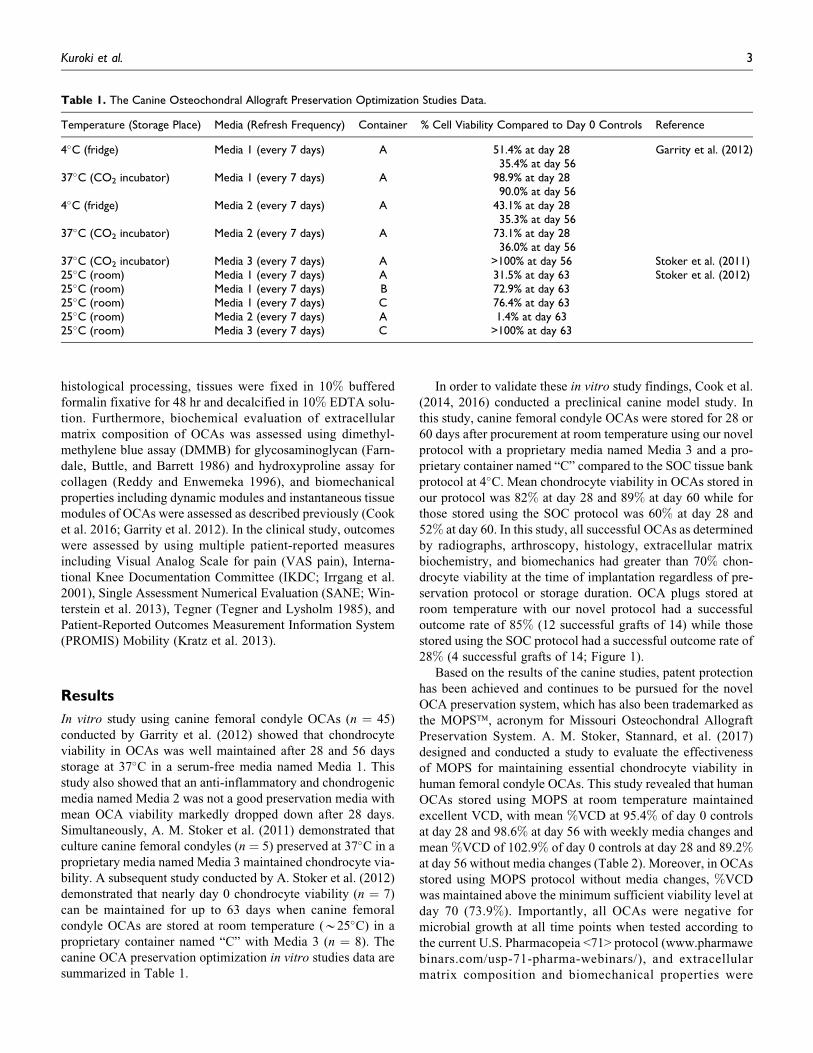
In vitro study using canine femoral condyle OCAs (n ¼ 45)
conducted by Garrity et al. (2012) showed that chondrocyte
viability in OCAs was well maintained after 28 and 56 days
storage at 37�C in a serum-free media named Media 1. This
study also showed that an anti-inflammatory and chondrogenic
media named Media 2 was not a good preservation media with
mean OCA viability markedly dropped down after 28 days.
Simultaneously, A. M. Stoker et al. (2011) demonstrated that
culture canine femoral condyles (n ¼ 5) preserved at 37�C in a
proprietary media named Media 3 maintained chondrocyte via-
bility. A subsequent study conducted by A. Stoker et al. (2012)
demonstrated that nearly day 0 chondrocyte viability (n ¼ 7)
can be maintained for up to 63 days when canine femoral
condyle OCAs are stored at room temperature (*25�C) in a
proprietary container named “C” with Media 3 (n ¼ 8). The
canine OCA preservation optimization in vitro studies data are
summarized in Table 1.
In order to validate these in vitro study findings, Cook et al.
(2014, 2016) conducted a preclinical canine model study. In
this study, canine femoral condyle OCAs were stored for 28 or
60 days after procurement at room temperature using our novel
protocol with a proprietary media named Media 3 and a pro-
prietary container named “C” compared to the SOC tissue bank
protocol at 4�C. Mean chondrocyte viability in OCAs stored in
our protocol was 82% at day 28 and 89% at day 60 while for
those stored using the SOC protocol was 60% at day 28 and
52% at day 60. In this study, all successful OCAs as determined
by radiographs, arthroscopy, histology, extracellular matrix
biochemistry, and biomechanics had greater than 70% chon-
drocyte viability at the time of implantation regardless of pre-
servation protocol or storage duration. OCA plugs stored at
room temperature with our novel protocol had a successful
outcome rate of 85% (12 successful grafts of 14) while those
stored using the SOC protocol had a successful outcome rate of
28% (4 successful grafts of 14; Figure 1).
Based on the results of the canine studies, patent protection
has been achieved and continues to be pursued for the novel
OCA preservation system, which has also been trademarked as
the MOPS™, acronym for Missouri Osteochondral Allograft
Preservation System. A. M. Stoker, Stannard, et al. (2017)
designed and conducted a study to evaluate the effectiveness
of MOPS for maintaining essential chondrocyte viability in
human femoral condyle OCAs. This study revealed that human
OCAs stored using MOPS at room temperature maintained
excellent VCD, with mean %VCD at 95.4% of day 0 controls
at day 28 and 98.6% at day 56 with weekly media changes and
mean %VCD of 102.9% of day 0 controls at day 28 and 89.2%at day 56 without media changes (Table 2). Moreover, in OCAs
stored using MOPS protocol without media changes, %VCD
was maintained above the minimum sufficient viability level at
day 70 (73.9%). Importantly, all OCAs were negative for
microbial growth at all time points when tested according to
the current U.S. Pharmacopeia <71> protocol (www.pharmawe
binars.com/usp-71-pharma-webinars/), and extracellular
matrix composition and biomechanical properties were
Table 1. The Canine Osteochondral Allograft Preservation Optimization Studies Data.
Temperature (Storage Place) Media (Refresh Frequency) Container % Cell Viability Compared to Day 0 Controls Reference
4�C (fridge) Media 1 (every 7 days) A 51.4% at day 2835.4% at day 56
Garrity et al. (2012)
37�C (CO2 incubator) Media 1 (every 7 days) A 98.9% at day 2890.0% at day 56
4�C (fridge) Media 2 (every 7 days) A 43.1% at day 2835.3% at day 56
37�C (CO2 incubator) Media 2 (every 7 days) A 73.1% at day 2836.0% at day 56
37�C (CO2 incubator) Media 3 (every 7 days) A >100% at day 56 Stoker et al. (2011)25�C (room) Media 1 (every 7 days) A 31.5% at day 63 Stoker et al. (2012)25�C (room) Media 1 (every 7 days) B 72.9% at day 6325�C (room) Media 1 (every 7 days) C 76.4% at day 6325�C (room) Media 2 (every 7 days) A 1.4% at day 6325�C (room) Media 3 (every 7 days) C >100% at day 63
Kuroki et al. 3
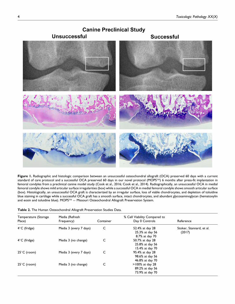
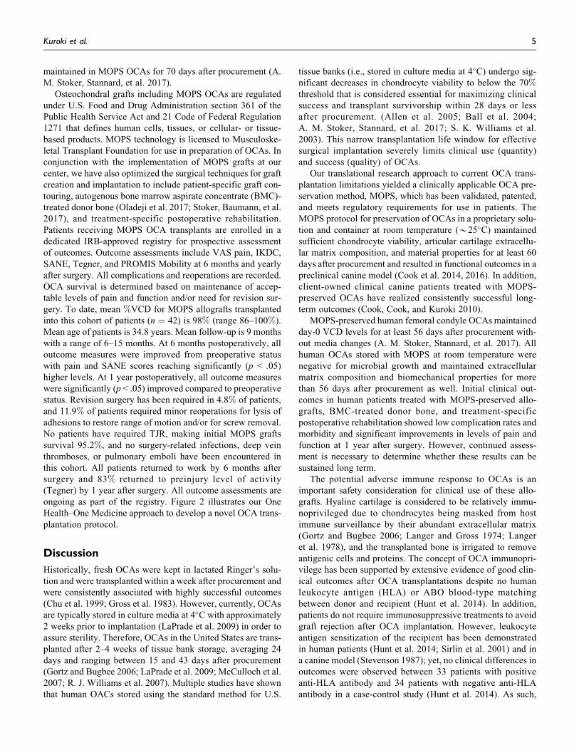
Figure 1. Radiographic and histologic comparison between an unsuccessful osteochondral allograft (OCA) preserved 60 days with a currentstandard of care protocol and a successful OCA preserved 60 days in our novel protocol (MOPS™) 6 months after press-fit implantation infemoral condyles from a preclinical canine model study (Cook et al., 2016; Cook et al., 2014). Radiographically, an unsuccessful OCA in medialfemoral condyle shows mild articular surface irregularities (box) while a successful OCA in medial femoral condyle shows smooth articular surface(box). Histologically, an unsuccessful OCA graft is characterized by an irregular surface, loss of viable chondrocytes, and depletion of toluidineblue staining in cartilage while a successful OCA graft has a smooth surface, intact chondrocytes, and abundant glycosaminoglycan (hematoxylinand eosin and toluidine blue). MOPS™ ¼ Missouri Osteochondral Allograft Preservation System.
Table 2. The Human Osteochondral Allograft Preservation Studies Data.
Temperature (StoragePlace)
Media (RefreshFrequency) Container
% Cell Viability Compared toDay 0 Controls Reference
4�C (fridge) Media 3 (every 7 days) C 52.4% at day 2825.3% at day 568.7% at day 70
Stoker, Stannard, et al.(2017)
4�C (fridge) Media 3 (no change) C 50.7% at day 2825.8% at day 5615.4% at day 70
25�C (room) Media 3 (every 7 days) C 95.4% at day 2898.6% at day 5646.8% at day 70
25�C (room) Media 3 (no change) C >100% at day 2889.2% at day 5673.9% at day 70
4 Toxicologic Pathology XX(X)
maintained in MOPS OCAs for 70 days after procurement (A.
M. Stoker, Stannard, et al. 2017).
Osteochondral grafts including MOPS OCAs are regulated
under U.S. Food and Drug Administration section 361 of the
Public Health Service Act and 21 Code of Federal Regulation
1271 that defines human cells, tissues, or cellular- or tissue-
based products. MOPS technology is licensed to Musculoske-
letal Transplant Foundation for use in preparation of OCAs. In
conjunction with the implementation of MOPS grafts at our
center, we have also optimized the surgical techniques for graft
creation and implantation to include patient-specific graft con-
touring, autogenous bone marrow aspirate concentrate (BMC)-
treated donor bone (Oladeji et al. 2017; Stoker, Baumann, et al.
2017), and treatment-specific postoperative rehabilitation.
Patients receiving MOPS OCA transplants are enrolled in a
dedicated IRB-approved registry for prospective assessment
of outcomes. Outcome assessments include VAS pain, IKDC,
SANE, Tegner, and PROMIS Mobility at 6 months and yearly
after surgery. All complications and reoperations are recorded.
OCA survival is determined based on maintenance of accep-
table levels of pain and function and/or need for revision sur-
gery. To date, mean %VCD for MOPS allografts transplanted
into this cohort of patients (n ¼ 42) is 98% (range 86–100%).
Mean age of patients is 34.8 years. Mean follow-up is 9 months
with a range of 6–15 months. At 6 months postoperatively, all
outcome measures were improved from preoperative status
with pain and SANE scores reaching significantly (p < .05)
higher levels. At 1 year postoperatively, all outcome measures
were significantly (p < .05) improved compared to preoperative
status. Revision surgery has been required in 4.8% of patients,
and 11.9% of patients required minor reoperations for lysis of
adhesions to restore range of motion and/or for screw removal.
No patients have required TJR, making initial MOPS grafts
survival 95.2%, and no surgery-related infections, deep vein
thromboses, or pulmonary emboli have been encountered in
this cohort. All patients returned to work by 6 months after
surgery and 83% returned to preinjury level of activity
(Tegner) by 1 year after surgery. All outcome assessments are
ongoing as part of the registry. Figure 2 illustrates our One
Health–One Medicine approach to develop a novel OCA trans-
plantation protocol.
Discussion
Historically, fresh OCAs were kept in lactated Ringer’s solu-
tion and were transplanted within a week after procurement and
were consistently associated with highly successful outcomes
(Chu et al. 1999; Gross et al. 1983). However, currently, OCAs
are typically stored in culture media at 4�C with approximately
2 weeks prior to implantation (LaPrade et al. 2009) in order to
assure sterility. Therefore, OCAs in the United States are trans-
planted after 2–4 weeks of tissue bank storage, averaging 24
days and ranging between 15 and 43 days after procurement
(Gortz and Bugbee 2006; LaPrade et al. 2009; McCulloch et al.
2007; R. J. Williams et al. 2007). Multiple studies have shown
that human OACs stored using the standard method for U.S.
tissue banks (i.e., stored in culture media at 4�C) undergo sig-
nificant decreases in chondrocyte viability to below the 70%threshold that is considered essential for maximizing clinical
success and transplant survivorship within 28 days or less
after procurement. (Allen et al. 2005; Ball et al. 2004;
A. M. Stoker, Stannard, et al. 2017; S. K. Williams et al.
2003). This narrow transplantation life window for effective
surgical implantation severely limits clinical use (quantity)
and success (quality) of OCAs.
Our translational research approach to current OCA trans-
plantation limitations yielded a clinically applicable OCA pre-
servation method, MOPS, which has been validated, patented,
and meets regulatory requirements for use in patients. The
MOPS protocol for preservation of OCAs in a proprietary solu-
tion and container at room temperature (*25�C) maintained
sufficient chondrocyte viability, articular cartilage extracellu-
lar matrix composition, and material properties for at least 60
days after procurement and resulted in functional outcomes in a
preclinical canine model (Cook et al. 2014, 2016). In addition,
client-owned clinical canine patients treated with MOPS-
preserved OCAs have realized consistently successful long-
term outcomes (Cook, Cook, and Kuroki 2010).
MOPS-preserved human femoral condyle OCAs maintained
day-0 VCD levels for at least 56 days after procurement with-
out media changes (A. M. Stoker, Stannard, et al. 2017). All
human OCAs stored with MOPS at room temperature were
negative for microbial growth and maintained extracellular
matrix composition and biomechanical properties for more
than 56 days after procurement as well. Initial clinical out-
comes in human patients treated with MOPS-preserved allo-
grafts, BMC-treated donor bone, and treatment-specific
postoperative rehabilitation showed low complication rates and
morbidity and significant improvements in levels of pain and
function at 1 year after surgery. However, continued assess-
ment is necessary to determine whether these results can be
sustained long term.
The potential adverse immune response to OCAs is an
important safety consideration for clinical use of these allo-
grafts. Hyaline cartilage is considered to be relatively immu-
noprivileged due to chondrocytes being masked from host
immune surveillance by their abundant extracellular matrix
(Gortz and Bugbee 2006; Langer and Gross 1974; Langer
et al. 1978), and the transplanted bone is irrigated to remove
antigenic cells and proteins. The concept of OCA immunopri-
vilege has been supported by extensive evidence of good clin-
ical outcomes after OCA transplantations despite no human
leukocyte antigen (HLA) or ABO blood-type matching
between donor and recipient (Hunt et al. 2014). In addition,
patients do not require immunosuppressive treatments to avoid
graft rejection after OCA implantation. However, leukocyte
antigen sensitization of the recipient has been demonstrated
in human patients (Hunt et al. 2014; Sirlin et al. 2001) and in
a canine model (Stevenson 1987); yet, no clinical differences in
outcomes were observed between 33 patients with positive
anti-HLA antibody and 34 patients with negative anti-HLA
antibody in a case-control study (Hunt et al. 2014). As such,
Kuroki et al. 5
the roles and clinical significance of subrejection immunologic
responses to OCAs are not fully understood and warrant further
investigation. The addition of autogenous BMC to the osseous
portion of OCAs for all patients at our center is designed in part
to mitigate any untoward immune responses, further optimiz-
ing outcomes after OCA transplantation (Oladeji et al. 2017).
This body of research has validated MOPS for significantly
increasing effective storage duration of OCAs and significantly
improving chondrocyte viability in stored OCAs at room tem-
perature without changing a proprietary media in a proprietary
container, which combined can profoundly increase the num-
ber of grafts that can be safely transplanted into eligible
patients. These improvements will allow surgeons to have
increased confidence in the quality of grafts they are transplant-
ing, provide tissue banks and graft coordinators with a much
longer period of time for matching and delivering OCAs, and
offer patients higher chances for highly functional long-term
outcomes and graft survivorship, resulting in decreased donor
tissue waste and related financial costs. Although approxi-
mately 30,000 donors provide tissue for transplant in the
United States in each year (www.aatb.org), the demand for
osteochondral tissue will continue to far exceed supply, even
with the use of MOPS. The number of total knee replacement
surgeries alone in the United States is rapidly increasing from
approximately 263,000 cases in 1999 to 616,000 cases in 2008
(Bernstein and Derman 2014). The demand for knee replace-
ments is estimated to exponentially increase to 3.48 million
cases by the year 2030 (Kurtz et al. 2007). So, although this
body of research has provided important progress in addressing
quantity, quality, and technique limitations for successful OCA
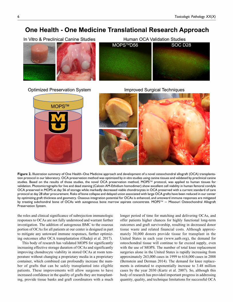
Figure 2. Illustration summary of One Health–One Medicine approach and development of a novel osteochondral allograft (OCA) transplanta-tion protocol in our laboratory. OCA preservation method was optimized by in vitro studies using canine tissues and validated by preclinical caninestudies. Based on the results of those studies, the novel OCA preservation method, MOPS™ protocol, was applied to human tissues forvalidation. Photomicrographs for live and dead staining (Calcein AM-Ethidium homodimer) show excellent cell viability in human femoral condyleOCA preserved in MOPS at day 56 of storage while markedly decreased viable chondrocytes in OCA preserved with a current standard of careprotocol at day 28 after procurement. Risks of bone collapse and delayed union associated with large OCA grafts have been reduced in our centerby optimizing graft thickness and geometry. Osseous integration potential for OCAs is enhanced, and untoward immune responses are mitigatedby treating subchondral bone of OCAs with autogenous bone marrow aspirate concentrate. MOPS™ ¼ Missouri Osteochondral AllograftPreservation System.
6 Toxicologic Pathology XX(X)
transplantation, development and validation of effective pre-
ventative, preservation, and tissue engineering strategies for
articular cartilage disorders in clinical canine and human
patients are of critical importance. The authors suggest that a
One Health–One Medicine comparative and translational
approach to addressing this critical unmet need can prove
successful.
Authors’ Contribution
All authors (KK, AS, JS, CB, CC, FP, JC) contributed to conception or
design; data acquisition, analysis, or interpretation; drafted the manu-
script; and critically revised the manuscript. All authors gave final
approval and agreed to be accountable for all aspects of work in
ensuring that questions relating to the accuracy or integrity of any
part of the work are appropriately investigated and resolved.
Declaration of Conflicting Interests
The author(s) declared the following potential conflicts of interest
with respect to the research, authorship, and/or publication of this
article: One or more of the authors (AMS and JCL) are patent holders
for the Missouri Osteochondral Allograft Preservation System
(MOPS) and receive associated royalties. One of the authors (JLC)
is a board member of the Musculoskeletal Transplant Foundation.
Funding
The author(s) disclosed receipt of the following financial support for
the research, authorship, and/or publication of this article: Portions of
this work were funded by the Musculoskeletal Transplant Foundation,
Synthes, the Thompson Laboratory for Regenerative Orthopaedics,
and the University of Missouri Department of Orthopaedic Surgery.
References
Allen, R. T., Robertson, C. M., Pennock, A. T., Bugbee, W. D., Harwood, F. L.,
Wong, V. W., Chen, A. C., Sah, R. L., and Amiel, D. (2005). Analysis of
stored osteochondral allografts at the time of surgical implantation. Am J
Sports Med 33, 1479–84.
Aubin, P. P., Cheah, H. K., Davis, A. M., and Gross, A. E. (2001). Long-term
followup of fresh femoral osteochondral allografts for posttraumatic knee
defects. Clin Orthop Relat Res 391, S318–27.
Ball, S. T., Amiel, D., Williams, S. K., Tontz, W., Chen, A. C., Sah, R. L., and
Bugbee, W. D. (2004). The effects of storage on fresh human osteochondral
allografts. Clin Orthop Relat Res 418, 246–52.
Bernstein, J., and Derman, P. (2014). Dramatic increase in total knee replace-
ment utilization rates cannot be fully explained by a disproportionate
increase among younger patients. Orthopedics 37, e656–59.
Bian, L., Fong, J. V., Lima, E. G., Stoker, A. M., Ateshian, G. A., Cook, J. L.,
and Hung, C. T. (2010). Dynamic mechanical loading enhances functional
properties of tissue-engineered cartilage using mature canine chondrocytes.
Tissue Eng Part A 16, 1781–90.
Biant, L. C., Bentley, G., Vijayan, S., Skinner, J. A., and Carrington, R. W.
(2014). Long-term results of autologous chondrocyte implantation in the
knee for chronic chondral and osteochondral defects. Am J Sports Med 42,
2178–83.
Brittberg, M., Lindahl, A., Nilsson, A., Ohlsson, C., Isaksson, O., and Peterson,
L. (1994). Treatment of deep cartilage defects in the knee with autologous
chondrocyte transplantation. N Engl J Med 331, 889–95.
Camp, C. L., Stuart, M. J., and Krych, A. J. (2014). Current concepts of
articular cartilage restoration techniques in the knee. Sports Health 6,
265–73.
Capeci, C. M., Turchiano, M., Strauss, E. J., and Youm, T. (2013). Osteochon-
dral allografts: Applications in treating articular cartilage defects in the
knee. Bull Hosp Jt Dis (2013) 71, 60–67.
Chu, C. R., Convery, F. R., Akeson, W. H., Meyers, M., and Amiel, D. (1999).
Articular cartilage transplantation. Clinical results in the knee. Clin Orthop
Relat Res 360, 159–68.
Cook, J. L., Cook, C. R., and Kuroki, K. (2010, February 20–27). Osteochon-
dral allografting in dogs: A small series of cases. Veterinary Orthopedic
Society Conference, Breckenridge, CO.
Cook, J. L., Kuroki, K., Visco, D., Pelletier, J. P., Schulz, L., and Lafeber, F. P.
(2010). The OARSI histopathology initiative recommendations for histo-
logical assessments of osteoarthritis in the dog. Osteoarthritis Cartilage 18,
S66–79.
Cook, J. L., Stannard, J. P., Stoker, A. M., Bozynski, C. C., Kuroki, K., Cook,
C. R., and Pfeiffer, F. M. (2016). Importance of donor chondrocyte viability
for osteochondral allografts. Am J Sports Med 44, 1260–68.
Cook, J. L., Stoker, A. M., Stannard, J. P., Kuroki, K., Cook, C. R., Pfeiffer, F.
M., Bozynski, C., and Hung, C. T. (2014). A novel system improves pre-
servation of osteochondral allografts. Clin Orthop Relat Res 472, 3404–14.
Demange, M., and Gomoll, A. H. (2012). The use of osteochondral allografts in
the management of cartilage defects. Curr Rev Musculoskelet Med 5,
229–35.
Farndale, R. W., Buttle, D. J., and Barrett, A. J. (1986). Improved quantitation
and discrimination of sulphated glycosaminoglycans by use of dimethyl-
methylene blue. Biochim Biophys Acta 883, 173–77.
Garrity, J. T., Stoker, A. M., Sims, H. J., and Cook, J. L. (2012). Improved
osteochondral allograft preservation using serum-free media at body tem-
perature. Am J Sports Med 40, 2542–48.
Gikas, P. D., Bayliss, L., Bentley, G., and Briggs, T. W. (2009). An overview of
autologous chondrocyte implantation. J Bone Joint Surg Br 91, 997–1006.
Gortz, S., and Bugbee, W. D. (2006). Fresh osteochondral allografts: Graft
processing and clinical applications. J Knee Surg 19, 231–40.
Gross, A. E., McKee, N. H., Pritzker, K. P., and Langer, F. (1983). Reconstruc-
tion of skeletal deficits at the knee. A comprehensive osteochondral trans-
plant program. Clin Orthop Relat Res 174, 96–106.
Gross, A. E., Shasha, N., and Aubin, P. (2005). Long-term followup of the use
of fresh osteochondral allografts for posttraumatic knee defects. Clin
Orthop Relat Res 435, 79–87.
Gross, A. E., Silverstein, E. A., Falk, J., Falk, R., and Langer, F. (1975). The
allotransplantation of partial joints in the treatment of osteoarthritis of the
knee. Clin Orthop Relat Res 108, 7–14.
Hangody, L., Vasarhelyi, G., Hangody, L. R., Sukosd, Z., Tibay, G., Bartha, L.,
and Bodo, G. (2008). Autologous osteochondral grafting—Technique and
long-term results. Injury 39, S32–39.
Huey, D. J., Hu, J. C., and Athanasiou, K. A. (2012). Unlike bone, cartilage
regeneration remains elusive. Science 338, 917–21.
Hunt, H. E., Sadr, K., Deyoung, A. J., Gortz, S., and Bugbee, W. D. (2014). The
role of immunologic response in fresh osteochondral allografting of the
knee. Am J Sports Med 42, 886–91.
Irrgang, J. J., Anderson, A. F., Boland, A. L., Harner, C. D., Kurosaka, M.,
Neyret, P., Richmond, J. C., and Shelborne, K. D. (2001). Development and
validation of the international knee documentation committee subjective
knee form. Am J Sports Med 29, 600–13.
Kaul, G., Cucchiarini, M., Remberger, K., Kohn, D., and Madry, H. (2012).
Failed cartilage repair for early osteoarthritis defects: A biochemical, his-
tological and immunohistochemical analysis of the repair tissue after treat-
ment with marrow-stimulation techniques. Knee Surg Sports Traumatol
Arthrosc 20, 2315–24.
Kratz, A. L., Slavin, M. D., Mulcahey, M. J., Jette, A. M., Tulsky, D. S., and Haley,
S. M. (2013). An examination of the PROMIS((R)) pediatric instruments to
assess mobility in children with cerebral palsy. Qual Life Res 22, 2865–76.
Krych, A. J., Robertson, C. M., and Williams, R. J., 3rd. (2012). Return to
athletic activity after osteochondral allograft transplantation in the knee.
Am J Sports Med 40, 1053–59.
Kurtz, S., Ong, K., Lau, E., Mowat, F., and Halpern, M. (2007). Projections of
primary and revision hip and knee arthroplasty in the United States from
2005 to 2030. J Bone Joint Surg Am 89, 780–85.
Lachiewicz, P. F., and Soileau, E. S. (2009). Fifteen-year survival and osteo-
lysis associated with a modular posterior stabilized knee replacement. A
concise follow-up of a previous report. J Bone Joint Surg Am 91, 1419–23.
Kuroki et al. 7
Langer, F., and Gross, A. E. (1974). Immunogenicity of allograft articular
cartilage. J Bone Joint Surg Am 56, 297–304.
Langer, F., Gross, A. E., West, M., and Urovitz, E. P. (1978). The immuno-
genicity of allograft knee joint transplants. Clin Orthop Relat Res 132,
155–62.
LaPrade, R. F., Botker, J., Herzog, M., and Agel, J. (2009). Refrigerated
osteoarticular allografts to treat articular cartilage defects of the femoral
condyles. A prospective outcomes study. J Bone Joint Surg Am 91, 805–11.
Lee, C. H., Cook, J. L., Mendelson, A., Moioli, E. K., Yao, H., and Mao, J. J.
(2010). Regeneration of the articular surface of the rabbit synovial joint by
cell homing: A proof of concept study. Lancet 376, 440–48.
Lexer, E. (1908). Substitution of whole or half joints from freshly amputated
extremities by fee plastic operations. Surg Gynecol Obstet 6, 301–07.
McCulloch, P. C., Kang, R. W., Sobhy, M. H., Hayden, J. K., and Cole, B. J.
(2007). Prospective evaluation of prolonged fresh osteochondral allograft
transplantation of the femoral condyle: Minimum 2-year follow-up. Am J
Sports Med 35, 411–20.
McNickle, A. G., Provencher, M. T., and Cole, B. J. (2008). Overview of
existing cartilage repair technology. Sports Med Arthrosc 16, 196–201.
Meyers, M. H., Akeson, W., and Convery, F. R. (1989). Resurfacing of the
knee with fresh osteochondral allograft. J Bone Joint Surg Am 71, 704–13.
Murawski, C. D., Foo, L. F., and Kennedy, J. G. (2010). A review of arthro-
scopic bone marrow stimulation techniques of the talus: The good, the bad,
and the causes for concern. Cartilage 1, 137–44.
Neogi, T. (2013). The epidemiology and impact of pain in osteoarthritis.
Osteoarthritis Cartilage 21, 1145–53.
Ng, K. W., Lima, E. G., Bian, L., O’Conor, C. J., Jayabalan, P. S., Stoker, A.
M., Kuroki, K., et al. (2010). Passaged adult chondrocytes can form engi-
neered cartilage with functional mechanical properties: A canine model.
Tissue Eng Part A 16, 1041–51.
Niemeyer, P., Pestka, J. M., Kreuz, P. C., Erggelet, C., Schmal, H., Suedkamp,
N. P., and Steinwachs, M. (2008). Characteristic complications after auto-
logous chondrocyte implantation for cartilage defects of the knee joint. Am
J Sports Med 36, 2091–99.
Nuelle, C. W., Nuelle, J. A., Cook, J. L., and Stannard, J. P. (2017). Patient
factors, donor age, and graft storage duration affect osteochondral allo-
graft outcomes in knees with or without comorbidities. J Knee Surg 30,
179–84.
Oladeji, L. O., Stannard, J. P., Cook, C. R., Kfuri, M., Crist, B. D., Smith, M. J.,
and Cook, J. L. (2017). Effects of autogenous bone marrow aspirate con-
centration on radiographic integration of femoral condylar osteochondral
allografts. Am J Sports Med. Published electronically July 1, 2017. doi:
10.1177/0363546517715725.
Pareek, A., Reardon, P. J., Maak, T. G., Levy, B. A., Stuart, M. J., and Krych, A. J.
(2016). Long-term outcomes after osteochondral autograft transfer: A sys-
tematic review at mean follow-up of 10.2 years. Arthroscopy 32, 1174–84.
Peterson, L., Brittberg, M., Kiviranta, I., Akerlund, E. L., and Lindahl, A.
(2002). Autologous chondrocyte transplantation. Biomechanics and long-
term durability. Am J Sports Med 30, 2–12.
Reddy, G. K., and Enwemeka, C. S. (1996). A simplified method for the
analysis of hydroxyproline in biological tissues. Clin Biochem 29, 225–29.
Ritter, M. A., and Meneghini, R. M. (2010). Twenty-year survivorship of
cementless anatomic graduated component total knee arthroplasty. J
Arthroplasty 25, 507–13.
Roach, B. L., Hung, C. T., Cook, J. L., Ateshian, G. A., and Tan, A. R. (2015).
Fabrication of tissue engineered osteochondral grafts for restoring the
articular surface of diarthrodial joints. Methods 84, 103–08.
Roberts, S., Menage, J., Sandell, L. J., Evans, E. H., and Richardson, J. B.
(2009). Immunohistochemical study of collagen types I and II and procol-
lagen IIA in human cartilage repair tissue following autologous chondro-
cyte implantation. Knee 16, 398–404.
Sirlin, C. B., Brossmann, J., Boutin, R. D., Pathria, M. N., Convery, F. R.,
Bugbee, W., Deutsch, R., Lebeck, L. K., and Resnick, D. (2001). Shell
osteochondral allografts of the knee: Comparison of mr imaging findings
and immunologic responses. Radiology 219, 35–43.
Stevenson, S. (1987). The immune response to osteochondral allografts in
dogs. J Bone Joint Surg Am 69, 573–82.
Stoker, A. M., Baumann, C. A., Stannard, J. P., and Cook, J. L. (2017, June 24).
Bone marrow aspirate concentrate versus platelet rich plasma to enhance
osseous integration potential for osteochondral allografts. J Knee Surg. doi:
10.1055/s-0037-1603800. [Epub ahead of print]
Stoker, A. M., Garrity, J. T., Hung, C. T., Stannard, J. P., and Cook, J. (2012).
Improved preservation of fresh osteochondral allografts for clinical use. J
Knee Surg 25, 117–25.
Stoker, A. M., Garrity, J. T., Sims, H. J., and Cook, J. L. (2011, January 13–15).
Optimization of osteochondral allograft preservation to extend the usable
life span of harvested tissue. Paper No. 1449. Orthopaedic Research Soci-
ety Annual Meeting, Long Beach, CA.
Stoker, A. M., Stannard, J. P., Kuroki, K., Bozynski, C. C., Pfeiffer, F. M., and
Cook, J. L. (2017). Validation of the Missouri osteochondral preservation
system for maintenance of osteochondral allograft quality during prolonged
storage. Am J Sports Med. Published electronically September 1, 2017. doi:
10.1177/0363546517727516.
Swanson, E. A., Schmalzried, T. P., and Dorey, F. J. (2009). Activity recom-
mendations after total hip and knee arthroplasty: A survey of the American
Association for Hip and Knee Surgeons. J Arthroplasty 24, 120–26.
Tegner, Y., and Lysholm, J. (1985). Rating systems in the evaluation of knee
ligament injuries. Clin Orthop Relat Res 198, 43–49.
Williams, S. K., Amiel, D., Ball, S. T., Allen, R. T., Wong, V. W., Chen, A. C.,
Sah, R. L., and Bugbee, W. D. (2003). Prolonged storage effects on the
articular cartilage of fresh human osteochondral allografts. J Bone Joint
Surg Am 85-A, 2111–20.
Williams, R. J. 3rd, Ranawat, A. S., Potter, H. G, Carter, T., and Warren, R. F.
(2007). Fresh stored allografts for the treatment of osteochondral defects of
the knee. J Bone Joint Surg Am 89, 718–26.
Winterstein, A. P., McGuine, T. A., Carr, K. E., and Hetzel, S. J. (2013).
Comparison of IKDC and SANE outcome measures following knee injury
in active female patients. Sports Health 5, 523–29.
8 Toxicologic Pathology XX(X)
Related Documents