Biointeractions of Nanomaterials

Welcome message from author
This document is posted to help you gain knowledge. Please leave a comment to let me know what you think about it! Share it to your friends and learn new things together.
Transcript

Biointeractions ofNanomaterials

Boca Raton London New York
CRC Press is an imprint of theTaylor & Francis Group, an informa business
Edited by
Vijaykumar B. SutariyaUniversity of South FloridaCollege of PharmacyTampa, Florida, USA Yashwant PathakUniversity of South FloridaCollege of PharmacyTampa, Florida, USA
Biointeractions ofNanomaterials

CRC PressTaylor & Francis Group6000 Broken Sound Parkway NW, Suite 300Boca Raton, FL 33487-2742
© 2015 by Taylor & Francis Group, LLCCRC Press is an imprint of Taylor & Francis Group, an Informa business
No claim to original U.S. Government worksVersion Date: 20140425
International Standard Book Number-13: 978-1-4665-8239-2 (eBook - PDF)
This book contains information obtained from authentic and highly regarded sources. While all reasonable efforts have been made to publish reliable data and information, neither the author[s] nor the publisher can accept any legal responsibility or liability for any errors or omissions that may be made. The publishers wish to make clear that any views or opinions expressed in this book by individual editors, authors or contributors are personal to them and do not necessarily reflect the views/opinions of the publishers. The information or guidance contained in this book is intended for use by medical, scientific or health-care professionals and is provided strictly as a supplement to the medical or other professional’s own judgement, their knowledge of the patient’s medical history, relevant manufacturer’s instructions and the appropriate best practice guidelines. Because of the rapid advances in medical science, any information or advice on dosages, procedures or diagnoses should be independently veri-fied. The reader is strongly urge to consult the relevant national drug formulary and the drug companies’ printed instructions, and their websites, before administering any of the drugs recommended in this book. This book does not indicate whether a particular treatment is appropriate or suitable for a particular individual. Ultimately it is the sole responsibility of the medical professional to make his or her own professional judgements, so as to advise and treat patients appropriately. The authors and publishers have also attempted to trace the copyright holders of all material reproduced in this publication and apologize to copyright holders if permission to publish in this form has not been obtained. If any copyright material has not been acknowl-edged please write and let us know so we may rectify in any future reprint.
Except as permitted under U.S. Copyright Law, no part of this book may be reprinted, reproduced, transmitted, or utilized in any form by any electronic, mechanical, or other means, now known or hereafter invented, including photocopying, microfilming, and recording, or in any information storage or retrieval system, without written permission from the publishers.
For permission to photocopy or use material electronically from this work, please access www.copyright.com (http://www.copy-right.com/) or contact the Copyright Clearance Center, Inc. (CCC), 222 Rosewood Drive, Danvers, MA 01923, 978-750-8400. CCC is a not-for-profit organization that provides licenses and registration for a variety of users. For organizations that have been granted a photocopy license by the CCC, a separate system of payment has been arranged.
Trademark Notice: Product or corporate names may be trademarks or registered trademarks, and are used only for identifica-tion and explanation without intent to infringe.
Visit the Taylor & Francis Web site athttp://www.taylorandfrancis.com
and the CRC Press Web site athttp://www.crcpress.com

Dedicated to the loving memory of my father, Bhadabhai Chakubhai Sutariya, who passed away on April 22, 2013. He was my
role model and mentor throughout my life and whatever I have achieved in life is because of his blessings. I would also like to dedicate this book to the memory of Swami Vivekananda; the
world celebrated the 150th birthday of Swamijee in 2013.
Vijaykumar B. Sutariya
To the loving memories of my parents and Dr. Keshav Baliram Hedgewar, who showed the right direction; my wife Seema, who gave my life positive meaning; and my son Sarvadaman who gave a golden lining to my life.
Yashwant Pathak


vii
ContentsForeword ...........................................................................................................................................ixPreface...............................................................................................................................................xiEditors ............................................................................................................................................ xiiiContributors .....................................................................................................................................xv
Chapter 1 Introduction—Biointeractions of Nanomaterials: Challenges and Solutions ..............1
Vijaykumar B. Sutariya, Vrinda Pathak, Ana Groshev, Mahavir B. Chougule, Sachin Naik, Deepa Patel, and Yashwant Pathak
Chapter 2 Nanoparticle Exposures in Occupational Environments ........................................... 49
Li-Hao Young, Ying-Fang Wang, Ching-Hwa Chen, Chun-Wan Chen, and Perng-Jy Tsai
Chapter 3 Physicochemical Characterization–Dependent Toxicity of Nanoparticles ................ 73
Jigar N. Shah, Ankur P. Shah, Hiral J. Shah, and Vijaykumar B. Sutariya
Chapter 4 Cytotoxicity of Stimuli-Responsive Nanomaterials: Predicting Clinical Viability through Robust Biocompatibility Pro�les ................................................. 103
Daniel Wehrung and Moses O. Oyewumi
Chapter 5 Biosensing Devices for Toxicity Assessment of Nanomaterials .............................. 117
Evangelia Hondroulis, Pratik Shah, Xuena Zhu, and Chen-Zhong Li
Chapter 6 Carbon Nanotubes and Pulmonary Toxicity ............................................................ 131
Malay K. Das and Charles Preuss
Chapter 7 Nanotoxicity of Polymeric and Solid Lipid Nanoparticles ...................................... 141
Dev Prasad and Harsh Chauhan
Chapter 8 Analytical Characterization of Nanomaterials in Biological Matrices for Hazard Assessment ............................................................................................. 159
Mingsheng Xu, Daisuke Fujita, Huanxing Su, Hongzheng Chen, and Nobutaka Hanagata
Chapter 9 Nanoparticles and Human Health: A Review of Epidemiological Studies .............. 175
Vijaykumar B. Sutariya, Ana Groshev, Vivek Dave, Hardeep Saluja, Deepak Bhatia, Prabodh Sadana, and Yashwant Pathak

viii Contents
Chapter 10 Toxicogenomic Approaches to Understanding the Toxicity of Nanoparticles .........209
Qiwen Shi, Mahavir B. Chougule, Vijaykumar B. Sutariya, and Deepak Bhatia
Chapter 11 Nanomaterial-Based Gene and Drug Delivery: Pulmonary Toxicity Considerations ..........................................................................................................225
Mahavir B. Chougule, Rakesh K. Tekade, Peter R. Hoffmann, Deepak Bhatia, Vijaykumar B. Sutariya, and Yashwant Pathak
Chapter 12 Cardiovascular Toxicity of Nanomaterials ...............................................................249
Saijie Zhu and Minghuang Hong
Chapter 13 Toxicity of Nanomaterials on the Gastrointestinal Tract ......................................... 259
Jayvadan Patel and Vibha Champavat
Chapter 14 Toxicity of Nanomaterials on the Liver, Kidney, and Spleen...................................285
Jayvadan Patel and Anita Patel
Chapter 15 Regulatory Implications of Nanotechnology ........................................................... 315
Lynn L. Bergeson and Michael F. Cole
Chapter 16 Ocular Toxicity of Nanoparticles ............................................................................. 347
Aditya Grover, Anjali Hirani, Yong Woo Lee, Vijaykumar B. Sutariya, and Yashwant Pathak
Chapter 17 Genotoxicity of Nanoparticles.................................................................................. 353
Amaya Azqueta, Leire Arbillaga, and Adela López de Cerain
Chapter 18 Interactions of Polysaccharide-Coated Nanoparticles with Proteins ....................... 365
Christine Vauthier
Chapter 19 Models for Risk Assessments of Nanoparticles ....................................................... 383
Sanjay Dey, Bhaskar Mazumder, and Yaswant Pathak
Chapter 20 Immunotoxicity of Carbon Nanoparticles ................................................................ 425
Paulami Pal, Bhaskar Mazumder, and Yaswant Pathak

ix
ForewordNanomaterials are those in the nanometer range (10−9 m). These incredibly small particles can be organic or inorganic, with examples ranging from poly(lactic-co-glycolic acid) or gold nanoparticles to carbon nanotubes and quantum dots. These particles may be used to encapsulate drugs, recognize biological markers, or visualize body tissues among many other possibilities, all enabling their widespread application in biology, medicine, and pharmaceutics. Indeed, these nanomaterials may have bene�cial effects that have not even been imagined.
The small size of these particles provides an enormous surface area, which is ideal for interactions with cells on a molecular level, but also raises the question of their biosafety. The chemical composi-tion of the diverse nanomaterials available for biological interactions
may have unforeseen consequences in living systems. Whether the good that these interactions accomplish outweighs the risk of harm will have to be addressed before nanomaterials are used on a wide scale, especially in biological systems.
This book is a collaborative effort of the editors Drs. Vijaykumar B. Sutariya and Yashwant Pathak and the numerous contributors who are leading scientists in this �eld. The subject mat-ter is of prime importance in the area of nanotechnology and its applications. These contributors, knowledgeable and experienced in their �eld, attempt to elucidate the potential biointeractions of nanomaterials with their respective applications in efforts to answer the questions posed above. This book presents the possible biointeractions of various nanomaterials with a number of different body tissues in a multitude of applications. I would like to congratulate Drs. Vijaykumar B. Sutariya and Yashwant Pathak at the University of South Florida for editing this important and timely book.
It is my great pleasure to write a foreword and present to you Biointeractions of Nanomaterials. I sincerely hope you will gain as much insight as I did from these chapters.
Shyam S. Mohapatra, PhD, MBA, FAAAAI, FNAIDistinguished USF Health Professor and Director
Division of Translational Medicine-USF Nanomedicine Research CenterVice Chair of Research
Department of Internal MedicinePresident, USF Chapter of the National Academy of Inventors


xi
PrefaceThe purpose of this book is to focus on the biointeractions of nanomaterials, an area that has not been previously addressed in detail. It also covers various techniques and tests that have been devel-oped to evaluate the toxicity of materials at the nanolevel. The interactions of nanomaterials and nanosystems within biosystems are a concern for the scienti�c community.
This book is targeted toward academic researchers as well as industry members who are involved in the development of nanosystems. Many graduate schools have initiated courses in nanotechnol-ogy and applications, and this book will be a great resource for students as well as professors. Additionally, this will be a useful tool for industrial scientists investigating technology to update their nanotoxicology and nanosafety understanding.
The objective of the book is to address issues related to the toxicity and safety of nanomaterials and nanosystems. It also covers the interactions of these in biological systems, and various tools and methods used to evaluate toxicity and safety issues.
The volume comprises 20 chapters written by leading scientists in the �eld of nanotechnol-ogy. Chapter 1 covers the challenges and solutions of biointeractions of nanomaterials. This is fol-lowed by three chapters that address the assessment and characterization of nanosystems in the bioenvironment.
The next group of chapters covers toxicity and includes biosensing devices for toxicity assess-ment, carbon nanotubes, and pulmonary toxicity, as well as nanotoxicity of solid lipid nanoparticles. The �nal group of chapters from 8 to 20 covers nanosafety concerns and solutions. Each of these chapters delves into the effects of nanoparticles on different organs and sheds light on regulatory implications of nanomaterials.
We sincerely hope this book gets an overwhelming response from the scienti�c community in the �eld of nanotechnology.
We thank and acknowledge our families, the publishers, and our contributing authors. We would also like to acknowledge Aditya Grover, Anastasia Groshev, and Anjali Hirani for their assistance in editing and obtaining copyright clearance as well as the staff of Taylor & Francis who assisted in shaping this wonderful book in the �eld of nanotechnology.
Vijaykumar B. SutariyaYashwant Pathak


xiii
EditorsDr. Vijaykumar B. Sutariya earned his bachelor of pharmacy and master of pharmacy from L. M. College of Pharmacy, Gujarat University, Ahmedabad, India and his PhD in pharmacy from The M.S. University of Baroda, Vadodara, India. He did his postdoctoral training in the �eld of pharmaceutics and drug delivery at Butler University, Indianapolis, Indiana.
Dr. Sutariya is an assistant professor in the Department of Pharmaceutical Sciences at the University of South Florida (USF) College of Pharmacy. He has a joint appointment with the Department of Internal Medicine, Division of Translational Medicine at USF.
Dr. Sutariya has published more than 30 research papers in peer-reviewed journals and has pre-sented at various national and international meetings. He is a reviewer of many international journals and an editorial board member of more than six journals related to drug delivery and pharmaceutical sciences. Dr. Sutariya’s research is focused on the development of novel drug delivery systems such as nanoparticles, liposome, and thermoreversible gel. His main research focus is on brain-targeted drug delivery and ocular drug delivery. Dr. Sutariya is currently serving as a coinvestigator on two NIH grants (R01 and R15). In addition to research, Dr. Sutariya teaches various courses related to pharmaceutics in the Doctor of Pharmacy curriculum.
Dr. Yashwant Pathak completed his MS and PhD in pharmaceutical technology at Nagpur University, India and his EMBA and MS in con�ict management from Sullivan University, Kentucky. He is an associate dean for faculty affairs at the College of Pharmacy, University of South Florida, Tampa, Florida. With extensive experience in academia as well as industry, he has to his credit more than 100 publications, 5 books on nanotechnology, 4 books on nutraceuticals, and several books on cultural studies, including 2 on aging studies from an Indian perspective. His areas of research include drug delivery systems and their characterization in animal models.


xv
Contributors
Leire ArbillagaDepartment of Pharmacology and ToxicologyUniversity of NavarraPamplona, Spain
Amaya AzquetaDepartment of Pharmacology and ToxicologyUniversity of NavarraPamplona, Spain
Lynn L. BergesonBergeson & Campbell, P.C.Washington, D.C.
Deepak BhatiaDepartment of Pharmaceutical SciencesNortheast Ohio Medical UniversityRootstown, Ohio
Vibha ChampavatNootan Pharmacy CollegeNorth Gujarat, India
Harsh ChauhanDepartment of Pharmacy SciencesCreighton UniversityOmaha, Nebraska
Ching-Hwa ChenDepartment of Environmental and
Occupational Health, Medical CollegeNational Cheng Kung UniversityTainan, Taiwan
Chun-Wan ChenInstitute of Occupational Safety and HealthMinistry of LaborTaipei, Taiwan
Hongzheng ChenDepartment of Polymer Science and
EngineeringZhejiang UniversityZhejiang, China
Mahavir B. ChouguleDepartment of Pharmaceutical SciencesUniversity of HawaiiHilo, Hawaii
Michael F. ColeBergeson & Campbell, P.C.Washington, D.C.
Malay K. DasCollege of PharmacyUniversity of South FloridaTampa, Florida
Vivek DaveWegmans School of PharmacySt. John Fisher CollegeRochester, New York
Adela López de CerainDepartment of Pharmacology and ToxicologyUniversity of NavarraPamplona, Spain
Sanjay DeyDepartment of Pharmaceutical SciencesDibrugarh UniversityDibrugarh, India
Daisuke FujitaAdvanced Key Technologies DivisionNational Institute for Materials ScienceIbaraki, Japan
Ana GroshevCollege of PharmacyUniversity of South FloridaTampa, Florida
Aditya GroverCollege of PharmacyUniversity of South FloridaTampa, Florida

xvi Contributors
Nobutaka HanagataInterdisciplinary Laboratory for Nanoscale
Science and TechnologyNational Institute for Materials ScienceIbaraki, Japan
Anjali HiraniSchool of Biomedical Engineering and
SciencesVirginia TechBlacksburg, VirginiaandCollege of PharmacyUniversity of South FloridaTampa, Florida
Peter R. HoffmannDepartment of Cell and Molecular BiologyJohn A. Burns School of MedicineHonolulu, Hawaii
Evangelia HondroulisCollege of Engineering and ComputingFlorida International UniversityMiami, Florida
Minghuang HongPharmaceutical Crystal Engineering Research
GroupShanghai Institute of Pharmaceutical IndustryShanghai, China
Yong Woo LeeSchool of Biomedical Engineering and SciencesVirginia TechBlacksburg, Virginia
Chen-Zhong LiCollege of Engineering and ComputingFlorida International UniversityMiami, Florida
Bhaskar MazumderDepartment of Pharmaceutical SciencesDibrugarh UniversityDibrugarh, India
Sachin NaikFormulation DepartmentSunPharma Advanced Research Co. Ltd.Gujarat, India
Moses O. OyewumiDepartment of Pharmaceutical SciencesNortheast Ohio Medical UniversityRootstown, Ohio
Paulami PalDepartment of Pharmaceutical
SciencesDibrugarh UniversityDibrugarh, India
Anita PatelNootan Pharmacy CollegeNorth Gujarat, India
Deepa PatelParul Institute of Pharmacy and ResearchGujarat, India
Jayvadan PatelNootan Pharmacy CollegeNorth Gujarat, India
Vrinda PathakCollege of PharmacyUniversity of South FloridaTampa, Florida
Yashwant PathakCollege of PharmacyUniversity of South FloridaTampa, Florida
Dev PrasadSchool of PharmacyMassachusetts College of Pharmacy and
Health SciencesBoston, Massachusetts
Charles PreussDepartment of Molecular Pharmacology and
PhysiologyMorsani College of MedicineUniversity of South FloridaTampa, Florida
Prabodh SadanaDepartment of Pharmaceutical SciencesNortheast Ohio Medical UniversityRootstown, Ohio
xviiContributors
Hardeep SalujaCollege of PharmacySouthwestern Oklahoma State UniversityWeatherford, Oklahoma
Ankur P. ShahPharmaceutical Technology CenterZydus Cadila Healthcare Ltd.Gujarat, India
Hiral J. ShahDepartment of PharmaceuticsArihant School of Pharmacy and BRIGujarat, India
Jigar N. ShahDepartment of PharmaceuticsNirma UniversityAhmedabad, India
Pratik ShahCollege of Engineering and ComputingFlorida International UniversityMiami, Florida
Qiwen ShiDepartment of Pharmaceutical SciencesCollege of PharmacyNortheast Ohio Medical UniversityRootstown, Ohio
Huanxing SuState Key Laboratory of Quality Research in
Chinese Medicine and Institute of Chinese Medical SciencesUniversity of MacauMacau SAR, ChinaandInterdisciplinary Laboratory for Nanoscale
Science and Technology National Institute for Materials ScienceIbaraki, Japan
Vijaykumar B. SutariyaCollege of PharmacyUniversity of South FloridaTampa, Florida
Rakesh K. TekadeDepartment of Pharmaceutical SciencesUniversity of Hawaii at HiloHilo, Hawaii
Perng-Jy TsaiDepartment of Environmental and
Occupational HealthNational Cheng Kung UniversityTainan, Taiwan
Christine VauthierInstitut Galien Paris-SudUniversité de Paris Sud Faculté de PharmacieChatenay-Malabry, France
Ying-Fang Wang Department of Environmental and
Occupational HealthMedical CollegeNational Cheng Kung UniversityTainan, Taiwan
Daniel WehrungDepartment of Pharmaceutical SciencesNortheast Ohio Medical UniversityRootstown, Ohio
Mingsheng XuDepartment of Polymer Science and
EngineeringZhejiang UniversityZhejiang, China
Li-Hao YoungDepartment of Occupational Safety
and HealthSchool Public HealthChina Medical UniversityTaichung, Taiwan
Saijie ZhuCollege of PharmacyThe University of Texas at AustinAustin, Texas
Xuena ZhuCollege of Engineering and
ComputingFlorida International UniversityMiami, Florida


1
Introduction—Biointeractions of NanomaterialsChallenges and Solutions
Vijaykumar B. Sutariya, Vrinda Pathak, Ana Groshev, Mahavir B. Chougule, Sachin Naik, Deepa Patel, and Yashwant Pathak
1
CONTENTS
1.1 Introduction ..............................................................................................................................21.1.1 What Is Nanotechnology? .............................................................................................21.1.2 Genesis of the Field ......................................................................................................4
1.2 Nanomaterials ...........................................................................................................................51.3 Classi�cation of Nanomaterials ................................................................................................61.4 Application of Nanomaterials ...................................................................................................8
1.4.1 Applications in Medicine and Pharmacy......................................................................91.4.1.1 Tissue Engineering ........................................................................................91.4.1.2 Drug Delivery Systems ................................................................................ 101.4.1.3 Nasal Vaccination ........................................................................................ 101.4.1.4 Cancer Diagnosis and Treatment ................................................................. 101.4.1.5 Local Anesthetic Toxicity ............................................................................ 111.4.1.6 Gene Therapy and Transfection ................................................................... 111.4.1.7 Molecular Diagnostics and Imaging ............................................................ 111.4.1.8 Biosensor and Biolabels ............................................................................... 121.4.1.9 Antimicrobial Nanopowders and Coatings .................................................. 121.4.1.10 Extraction and Separation Techniques ........................................................ 131.4.1.11 Nucleic Acid Sequence and Protein Detection ............................................ 13
1.4.2 Applications in Computer Technology ....................................................................... 131.4.3 Environmental Applications ....................................................................................... 14
1.4.3.1 Catalysis and Elimination of Pollutants ....................................................... 141.4.3.2 Water Remediation ....................................................................................... 151.4.3.3 Sensors ......................................................................................................... 151.4.3.4 Fuel Cells ..................................................................................................... 15
1.4.4 Applications in Commonly Used Products ................................................................ 161.4.4.1 Cosmetics ..................................................................................................... 161.4.4.2 Coatings ....................................................................................................... 161.4.4.3 Self-Cleaning Windows ............................................................................... 161.4.4.4 Scratch-Resistant Materials ......................................................................... 161.4.4.5 Textiles ......................................................................................................... 161.4.4.6 Insulation Materials ..................................................................................... 161.4.4.7 Nanocomposites ........................................................................................... 161.4.4.8 Paint ............................................................................................................. 17

2 Biointeractions of Nanomaterials
1.1 INTRODUCTION
1.1.1 WHAT IS NANOTECHNOLOGY?
Nanotechnology is the science that deals with the interactions that arise at a nanosized, molecular scale. There are several paradigms from nature, such as viruses, DNA, water molecules, and red blood cells, with sizes in the nanometer range. This chapter presents a general overview of the nanoparticles (NPs) and their biointeractions. Figure 1.1 illustrates several cases from nature and pharmaceuticals of components with nanometer dimensions. For many decades, nanotechnology has been used most frequently in the areas of engineering, electronics, and physics, and has shown remarkable developments in these �elds. However, pharmaceutical and biomedical areas of applica-tion still need to be explored.
The unique �eld of nanotechnology represents not just one speci�c �eld but a wide range of areas from basic material sciences to personal care applications. The exciting aspect of nanotechnology is the capability to fabricate formulations by manipulating molecules and supramolecular struc-tures for the development of devices with programmed functions. This is very promising when it is applied to the �eld of active pharmacological ingredient (API) delivery. The conventional form of
1.4.4.9 Cutting Tools ................................................................................................ 171.4.4.10 Lubricants .................................................................................................... 17
1.5 Nanotoxicity ............................................................................................................................ 171.6 Biointeractions of Nanomaterials ........................................................................................... 19
1.6.1 Interactions with the Environment ............................................................................. 191.6.2 Nanotoxicity in the Body ............................................................................................23
1.6.2.1 Molecular Mechanisms of Nanomaterial Toxicity ......................................231.6.2.2 Pharmacokinetics .........................................................................................25
1.6.3 Effects of Nanomaterials on Organ Systems ..............................................................261.6.3.1 Pulmonary System .......................................................................................261.6.3.2 Gastrointestinal Tract ...................................................................................261.6.3.3 Reticuloendothelial Systems ........................................................................271.6.3.4 Cardiovascular System .................................................................................271.6.3.5 Central Nervous System ..............................................................................271.6.3.6 Integumentary System .................................................................................27
1.7 Nanotoxicity: Challenges, Solutions, and the Future .............................................................271.7.1 Physicochemical Characterization..............................................................................281.7.2 In Vitro Assessment ....................................................................................................30
1.7.2.1 DNA Synthesis and Damage .......................................................................301.7.2.2 Immunogenicity ........................................................................................... 311.7.2.3 Oxidative Stress ........................................................................................... 311.7.2.4 Cell Proliferation .......................................................................................... 311.7.2.5 Exocytosis .................................................................................................... 321.7.2.6 Cell Viability and Metabolic Activity.......................................................... 321.7.2.7 Hemolysis ..................................................................................................... 32
1.7.3 In Vivo Assessment ..................................................................................................... 331.7.3.1 Absorption, Distribution, Metabolism, Excretion, and
Pharmacokinetic Studies .............................................................................341.7.3.2 Genotoxicity and Carcinogenic Studies .......................................................34
1.7.4 Considerations for Preventing Nanotoxicity ............................................................... 351.8 Future Considerations .............................................................................................................361.9 Summary ................................................................................................................................36References ........................................................................................................................................ 37

3Introduction—Biointeractions of Nanomaterials
novel carriers, such as liposomes, polymeric micelles, and NPs, are now known as nanovehicles. However, this is only in the terms of size. These conventional drug delivery systems would have developed to their current state regardless of the current development of nanotechnology. To fully understand the scope of nanotechnology in the drug delivery �eld, it may be favorable to categorize drug delivery systems by providing examples from before and after the rise of nanotechnology.
The properties of materials at the nanometer scale can be incredibly altered from those at a larger scale. As the size decreases from bulk compounds, only very small changes in the properties occur until the size of the particulates falls below 100 nm, while remarkable changes in properties can further take place. Nanostructured materials are of great interests for the development of novel properties and functions (Bhushan 2010).
Nanoparticulate drug delivery systems offer many bene�ts over conventional dosage forms. The advantages of nanoparticulate drug delivery systems include improved therapeutic ef�cacies, reduc-tions in toxicity, improved biodistributions, and improved patient compliance. Pharmaceutical NPs contain entrapped API substances and are composed of tens or hundreds of atoms or molecules, ranging from 5 to 300 nm in size and with different morphologies, such as amorphous, crystalline, spherical, and needles, among others (Saraf 2006).
Nanosized formulations and structures can be produced by using either “bottom-up” or “top-down” fabrication methods. In “bottom-up” methods, nanoparticulate structures are developed by building up atoms or molecules in a controlled manner through the regulation of thermodynamic properties such as self-assembly, precipitation, and crystallization. On the other hand, advances in nanotechnologies can be used to fabricate nanoscale structures through size-reduction approaches. These techniques, referred to as “top-down” nanofabrication technologies, include photolithography, nanomolding, dip-pen, lithography, and nano�uidics (Figure 1.2 compares the bottom-up and top-down techniques in various manufacturing processes) (Peppas 2004, Sahoo and Labhasetwar 2003).
The reduction of size, having a crucial role in pharmacy, is essential for proper unit operations. It helps in improving the performance of dosages and by providing better formulation opportunities for drugs. Drugs with sizes in the nanometer range improve performances in various dosage forms. Nano-sized formulations provide enhancements in surface area, solubility, rate of dissolution, and oral bioavailability. It may also provide a rapid onset of therapeutic action, a reduction in doses (and frequencies), and decreased fed/fasted and patient-to-patient variabilities.
Particulate dispersions, or solid particles with a size in the nanometer range (10–1000 nm), are known as NPs, in which a drug is dissolved, entrapped, encapsulated, or attached to a NP
Nature
Nanometers
Pharmaceuticalnanotechnology
10–1 101 104 106 108 1010
Watermolecule
DNA Virus Erythrocyte Apple
DendrimersNanotubes
Quantum dotsNiosome
PolymerNanoparticles
MicellesLiposome
Microparticles Tabletcapsule
FIGURE 1.1 (See color insert.) Dimensions scale of nanotechnology.

4 Biointeractions of Nanomaterials
matrix. NPs, nanospheres, or nanocapsules can be obtained in various forms, such as particles or vesicles, based on the preparation method. Major ambitions in fabricating NPs as delivery car-riers are to control the particle size, surface properties, and the release of therapeutics in order to accomplish the site-speci�c targeting of the drug at a therapeutically optimal rate and dose regimen. NPs can be made up of several materials, including polymers, metals, and ceramics. According to their methods of manufacturing and the materials used, NPs can adopt differ-ent shapes and sizes with speci�c properties. Many other types of NPs are in several stages of development as drug delivery carriers, including lipid-based carriers such as liposomes, lipid emulsions, lipid–drug complexes, polymer–drug conjugates, polymer microspheres, micelles, and various ligand-targeted carriers such as immunoconjugates (Allen 2002, LaVan et al. 2003, Liu et al. 2000, Moghimi et al. 2001).
1.1.2 GENESIS OF THE FIELD
Although nanotechnology as a �eld has developed recently, the concept has been present since much earlier. The synthesis and use of gold NPs predates the age of peer-reviewed literature. For example, artists have been utilizing colloidal gold, otherwise referred to as a gold NP solution, to create colors for pottery from the Ming dynasty and stained glass windows in medieval churches (Daniel and Astruc 2004).
The �rst published report on colloidal gold dates back to a celebrated, 1857 work by Faraday, although earlier unpublished experiments are likely (Faraday 1857). In 1959, Richard Feynman, an American physicist and Nobel Prize winner, envisioned the idea of manipulating particles or materi-als at a molecular and atomic scale in his presentation titled “There’s Plenty of Room at the Bottom” to the American Physical Society’s annual meeting. During his talk, he presented facts for generat-ing nanoscale machines to manipulate, control, and image materials at the atomic level. However, the term “nanotechnology” was coined in 1974, more than a decade later, by Norio Taniguchi, a
Chemical synthesis Particle molecules Cosmetics, fueladditives
Self-assembly Crystals, films, andtubes
Displays
Positional assembly Experimental atomicor molecular devices
Lithography
Cutting, etching, andgrinding
Electronic deviceschip masks
Precision-engineeredsurfaces
Quantum well lasersComputer chips
MEMS
Top-
dow
nBo
ttom
-up
FIGURE 1.2 Bottom-up and top-down techniques in manufacturing nanoparticles.

5Introduction—Biointeractions of Nanomaterials
scientist at the University of Tokyo, in his work titled On the Basic Concept of Nanotechnology. He described extra-high precision and ultra�ne dimensional structures, and also expected improve-ments in integrated circuits and devices of mechanical, optoelectronic, and computer memory appli-cations (Taniguchi 1974). This is called the “top-down” approach (of carving small structures from larger ones) (Thassu et al. 2007).
The creation of the scanning tunneling microscope by Gerd Binnig and Heinrich Rohrer in 1981, from IBM Zurich Laboratories, rendered a breakthrough by allowing visualizations on a nanosized scale. Further, the invention of the atomic force microscope (AFM) in 1986 made possible the imaging of structures on an atomic scale. In 1986, another scientist, K. Eric Drexler, in his book titled Engines of Creation, argued about the future of nanotechnology, speci�cally the design of larger structures from their atomic and molecular components, known as the “bottom-up approach” (Drexler 1986). He also offered thoughts for “molecular nanotechnology,” which is the self-assem-bly of particles into an ordered and functional structure.
Another major advance in the �eld of nanotechnology was established in 1985, when Harry Kroto, Robert Curl, and Richard Smalley developed a new form of carbon known as “fullerenes” (or “bucky-balls”), a single molecule containing 60 carbon atoms arranged in the shape of a soccer ball. This invention led to a Nobel Prize in Chemistry in 1996. In 2000, this new area of research received recog-nition from the government when former President Bill Clinton launched the National Nanotechnology Initiative (NNI) to promote research and development in nanotechnology. NNI de�nes research and development in nanotechnology as that on the 1–100 nm range scale to create systems with novel properties that have the capacity to function on the atomic scale (Thomas and Sayre 2005). Thus, nanotechnology aims to design the formulation of structures, devices, and systems by controlling the shape and size at a nanometer range (Varshney 2012). Today, nanotechnology has progressed into an extensive �eld of science, with multibillion dollar investments from the public and private sectors. Along with this comes the potential to generate multitrillion dollar industries in the coming decades with an enormous potential to bene�t many more applications and areas of research.
1.2 NANOMATERIALS
The history of nanomaterials (NMs) is perhaps as old as that of the universe, as nanostructures were formed in its near beginning. From the dawn of mankind, NPs were produced from �res used by early humans (Alagarasi 2011). The scienti�c community caught on to NMs much later.
A nanometer is one millionth of a millimeter, about 100,000 times smaller than the diameter of human hair. NMs are important because, at this scale, exclusive optical, magnetic, and electrical properties emerge, among others. These characteristics have great application potentials in elec-tronics, medicine, and other �elds. Owing to coatings or surface modi�cations, NMs demonstrate biocompatibility through interacting with living cells.
NMs are the foundation stones of nanoscience and nanotechnology, a large and interdisciplin-ary area of research and development that has been explosively developing globally in the past few years. It has the potential to revolutionize the approach in which NMs are developed, and the range and nature of functionalities that can be accessed. It has a signi�cant commercial impact, which will continue growing in the future.
Modi�ed NMs are resources fabricated at the nanometer scale to bene�t from small sizes and novel characteristics, normally not found in their conventional, bulk counterparts. These charac-teristics are enhanced relative surface areas and new quantum effects. NMs encompass a higher surface area to volume ratio than their conventional forms, which can lead to superior chemical reactivities and also have an effect on their strength. Moreover, at the nanorange scale, quantum effects become much more important in determining the material’s properties, leading to new opti-cal, electrical, and magnetic characters. The range of NM commercial products available today is very broad, including sunscreens, wrinkle-free textiles, stain-resistant goods, cosmetics, electron-ics, paints, and varnishes (Alagarasi 2011).

6 Biointeractions of Nanomaterials
1.3 CLASSIFICATION OF NANOMATERIALS
The classi�cation of NMs is not simple. It may be appropriate to organize the types of NMs accord-ing to their chemical and physical properties. However, different types of structures synthesized using various manufacturing processes, along with different surface coatings, can obscure classi�-cations. Other approaches to categorization may be based on NM points of origin or whether they are natural or modi�ed (Nowack and Bucheli 2007). Given the range of NM characteristics, it may be practical to assign categories based on speci�c properties, such as the potential for health risks (Tervonen et al. 2009). Therefore, in order to provide a general overview of NMs, multiple catego-rization systems should be considered.
For instance, taking the point of origin into consideration, NMs can be generated via either natu-ral or anthropogenic processes (Figure 1.3 demonstrates classi�cation organization of NMs based on their origin). Naturally produced NMs can be classi�ed into biogenic, geogenic, atmospheric, and pyrogenic categories, based on the methods and mechanisms of production by living organ-isms, the soil, the air, and heat, respectively. Anthropogenic NMs, or those produced as a result of human activity, can be classi�ed into two categories—unintentionally produced and intentionally engineered NMs (Nowack and Bucheli 2007). Most often, unintentional NMs are created as a by-product of combustion processes (Nowack and Bucheli 2007). Intentionally developed NMs can be further classi�ed into �ve different categories: carbon-based materials, metal-based materials, dendrimers, polymeric particles, and composites (Tuominen and Schultz 2010).
As NMs have an enormously small size, 100 nm or less in at least one dimension, descriptions of their size and shape have been attempted, such that if NMs have a nanometer size in one dimension, they are referred to as surface �lms; in two dimensions, �bers or strands; and in three dimensions, particles. They can be present in single, fused, aggregated, or agglomerated forms, with different shapes, such as spherical, tubular, and irregular (Figure 1.4 shows examples of NM classi�cation based on their shape).
NMs are resources that are differentiated by an ultra�ne grain size (<50 nm in size) or by a dimensionality restricted to 50 nm. NMs can be produced with different modulation dimension-alities as described by Richard W. Siegel: atomic clusters, �laments and cluster assemblies (zero), multilayers (one), ultra�ne-grained over layers or buried layers (two), and nanophase materials con-sisting of equiaxed nanometer-sized grains (three) as shown in Figure 1.4 (Siegel and Fougere 1995).
Therefore, NMs can also be classi�ed based on structure, morphology, and physicochemical properties. General classi�cations, based on the types of the NMs, take into account dendrimers, nanotubes, fullerenes, and quantum dots (QDs) (Nowack and Bucheli 2007).
Nanomaterials
Anthropogenic Naturalprocesses
Unintentional Engineered
Carbon-based materials,metal-based materials,dendrimers, polymeric
particles, and composites
Biogenic, geogenic,atmospheric, and
pyrogenic
FIGURE 1.3 Classi�cation of nanomaterials based on their origin. Natural process such as proliferation of living organisms, movement of soil, air, and the heat energy may result in the formation of nanomaterials. Anthropogenic nanomaterials are produced as a result of human activity regardless whether speci�c intent to do so was present or not. Depending on the process, unintentional processes or engineering design may result in nanomaterials of different shapes, sizes, and different properties.

7Introduction—Biointeractions of Nanomaterials
NMs can be classi�ed based on their phase composition properties, such as single-phase solids (crystalline, amorphous particles, layers, etc.), multiphase solids (matrix composites, coated parti-cles, etc.), and multiphase systems (colloids, aerogels, ferro�uids, etc.). Based on their methods of manufacturing, NMs can be classi�ed into three different categories: gas-phase reactions (�ame synthesis, condensation, etc.), liquid-phase reactions (sol–gel, precipitation, hydrothermal process-ing, etc.), and mechanical procedures (ball milling, plastic deformation, etc.) (Wolfgang 2004). Based on their structural properties, NMs can be classi�ed into two parts: (1) nanocrystalline materials, such as crystals, generally consisting of crystallite with at least one dimension in a nanometer size, and (2) nanostructured materials, such as dislocation fragments, clusters, quasicrystals, micropores, subgrains, and segregations. Nanofragmented materials, composed of dislocation fragments or sub-grains whose size is less than 100 nm (Figure 1.5a), normally consist of metals and alloys subjected to megaplastic deformations. Nonporous materials mainly exhibit a high volume density of nanopores less than 100 nm situated on the conventional grain body or along their boundaries (Figure 1.5b). Nanodendrites are materials mainly consisting dendrite solidi�cation products in the form of degen-erate dendrite nanodendrites, such as dendrite cells, and become visible upon the rapid solidi�cation
FIGURE 1.4 Classi�cation of nanomaterials. (a) Zero-dimensional spheres and clusters. (b) One-dimensional nano�bers, wires, and rods. (c) Two-dimensional �lms, plates, and networks. (d) Three-dimensional nanoma-terials. (Reprinted with permission from Alagarasi, A. 2011. Introduction to Nanomaterial. National Centre for Catalysis Research.)
FIGURE 1.5 Nanostructured materials. (a) Structure of nanofragmented material. Melts quenched FeSi alloy, TEM. (b) Structure of nonporous materials. Nanopores are located at grain boundaries in a polycrystal-line FeAl alloy produced by melt quenching, TEM. (c) Structure of nanodendrite materials. Dendrite nanocell in side grain in a FeSi produced by melt quenching are visible, SEM. (d) Structure of nanodislocation materi-als. Melt quench FeLi ally has high density of prismatic vacancy-type dislocation loop, TEM. (Reprinted with permission from Glezer, A. M. 2011. Russian Metallurgy (Metally) 4:263–269.)

8 Biointeractions of Nanomaterials
of melted compounds (Figure 1.5c). Nanodislocation materials are distinguished by a high-volume fraction of nanoscale dislocation collections or a con�guration of de�nite types (Figure 1.5d).
Nanophase materials generally contain phase transformation nanoproducts. Nanosegregations are materials that consist of grain boundaries or other element segregations with at least one dimen-sion in the nanosized scale. Based on modern theory, nano-cluster or amorphous materials are mainly multicomponent amorphous metallic glasses with a nano-cluster structure. The clusteriza-tion of amorphous alloy is highly prominent after local plastic �ow. As a result, the amorphous state of alloys manufactured by melt quenching should be considered as possessing a nanostructured shape (Glezer 2011).
NMs can be classi�ed on the basis of their source and origin, structure, morphology, or other physicochemical properties. Figure 1.6 summarizes these various methods of classi�cation (Buzea et al. 2007, Nowack and Bucheli 2007).
1.4 APPLICATION OF NANOMATERIALS
With applications in many areas, such as pharmacuticals, electronics, fuel cells, batteries, agri-culture, the food industry, and cosmetics, NMs have certainly already established themselves in the market. A variety of NM-containing products currently exist, such as sunscreens, electron-ics, paints, varnishes, stain-resistant and wrinkle-free textiles, windows, bicycles, automobiles, and sports equipment such as longer-lasting tennis balls using butyl rubber and nano-clay compos-ites. Owing to their ability to absorb ultraviolet (UV) light, numerous products exist to provide UV-blocking coatings on glass bottles. For example, nanosized titanium dioxide is widely used in sunblock creams and self-cleaning windows, and nanoscale silica is being available as a �ller in a series of products, including cosmetics and dental �llings (Alagarasi 2011).
Chemical synthesis
Self-assembly
Positional assembly
Particle molecules
Crystals, films,and tubes
Experimental atomicor molecular devices
Cosmetics, fueladditives
Displays
Lithography Electronic deviceschip masks
Quantum well lasers-Computer chips
MEMs
Cutting, etching, andgrinding
Precision-engineeredsurfacesTo
p-do
wn
Botto
m-u
p
FIGURE 1.6 Summary of classi�cation categories of the nanomaterials. NM can be classi�ed based on their structure and state, for example, agglomeration state. More commonly, NMs are categorized based on their dimensions, morphology, and composition. (Adapted with permission from Nowack, B. and T. D. Bucheli. 2007. Environmental Pollution 150(1):5–22.)

9Introduction—Biointeractions of Nanomaterials
It is obvious that NMs trump their conventional counterparts due to their exceptional formability and better chemical, physical, and mechanical properties. Modifying material properties allows for applications as diverse as advanced ceramics, semiconductor electronics, sensors, special polymers, magnetics, and membranes. Table 1.1 summarizes the applications of NMs used in different �elds.
1.4.1 APPLICATIONS IN MEDICINE AND PHARMACY
1.4.1.1 Tissue EngineeringNanotechnology has provided several elegant materials that are widely used for tissue repair and replacements and implantable devices, such as sensory aids, retina implants, structural implant materials, implant coatings, bone repairs, surgical aids, operating tools, smart instruments, tissue regeneration scaffolds, and bioresorbable materials (Table 1.2 summarizes various applications in
TABLE 1.1Applications of Nanomaterials
Field of Application Uses
Medicine and pharmacy QDs biological imaging for medical diagnostics; early diagnosis of atherosclerosis,Alzheimer’s disease, and cancer; drug delivery systems
Computer technology Nano transistors, computer and camera display and computer memory systems
Environment Solar cells, solar panels, fuel additives, alternate sources fuels (cellulose intoethanol for fuel), rechargeable batteries; longer, stronger, and lighter-weight wind mills to generate more electricity; advanced �lters for cleaner and more puri�ed water
Transportation Construction of better highways, bridges rails, tunnels, parking garages, pavements in terms of performance, cost effectiveness and longevity; advanced vehicular operation to avoid accidents; aviation
Commonly used products Baseball bats, tennis rackets,motorcycle helmets, automobile bumpers, luggage, spectacles, fabrics, food containers, sensors to alert food spoilage, cosmetic products,ceramics, tires, cleaning agents, antimicrobial/antibacterial coatings, paints, and household tools
Source: Adapted from NNI. National Nanotechnology Initiative, http://www.nano.gov/you/nanotechnology-bene�ts.
TABLE 1.2Applications of Nanomaterials in Medicine (Tissue Regeneration, Growth, and Repair)
Nanosystem Application
Tissue Regeneration, Growth, and RepairNanoengineered prosthetics Retinal, auditory, spinal, and cranial implants
Cellular manipulation Persuasion of lost nerve tissue to grow: growth of body part
Cancer TherapyCarbon nanotubes DNA mutation detection, disease protein biomarker detection
Dendrimer Controlled release drug delivery, image contrast agents
Nanocrystals Improved formulation for poorly soluble drugs
Nanoparticles MRI and ultrasound image contrast agents, targeted drug delivery, permeation enhancers, reporters of apoptosis, angiogenesis
Nanoshells Tumor-speci�c imaging, deep tissue thermal ablation
Nanowires Disease protein biomarker detection, DNA mutation detection, gene expression detection
Quantum dots Optical detection of genes and proteins in animal models and cell assays, tumor and lymph node visualization

10 Biointeractions of Nanomaterials
the area of tissue regeneration, growth, and repair). For example, nano�ber scaffolds, generally used to redevelop central nervous system cells and other organs, have been shown to facilitate the regen-eration of axonal tissue in hamsters with severed optic tracts (Ellis-Behnke et al. 2006).
1.4.1.2 Drug Delivery SystemsCommercially available and conventional dosage forms for drugs generally suffer from many draw-backs, such as the need for target speci�city, a high rate of drug metabolism, cytotoxicity, high dose and dosing frequency requirements, and poor patient compliance, among others. Nanotechnology has facili-tated drug delivery systems by improving the physical, chemical, and biological properties that can pro-vide ef�cient delivery means for currently available active pharmacological ingredients (APIs). Several nanocarriers, such as polymeric NPs, polymeric micelles, liposome, niosomes, dendrimer, polymer–drug conjugates, and antibody–drug conjugates, can generally be divided into the following categories:
1. Sustained and controlled delivery systems 2. Stimuli-sensitive/environment-sensitive delivery systems 3. Functionalized systems for the delivery of bioactives 4. Multifunctional systems for the combined delivery of therapeutics, biosensing, and
diagnostic 5. Site-speci�c, targeted drug delivery systems, including intracellular, cellular, and tissue
targeting (Vasir et al. 2005)
The direct, intravenous administration of APIs may induce toxicity due to �rst-order drug release kinetics when compared to intravenous administration. In cases where a sustained release in required, the �eld of NMs offers implantable delivery systems by virtue of their size, control, and almost zero-order releases. Some novel vascular carriers, such as liposome, ethosome and transfero-some, niosomes, and some implant chips, have been envisaged in recent times, which may assist in the minimization of peak plasma levels with minimal adverse reactions, allow for longer and more predictable action, decrease the frequency of dosing, and improve the levels of patient acceptance and compliance.
Furthermore, various strategies are being developed for superior, site-speci�c delivery using novel carriers such as polymeric NPs, liposomes, polymeric micelles, dendrimers, iron oxide, and proteins, by modifying the active and passive uptake of drugs. The targeting of drugs to tumor sites via passive delivery methods and using the improved permeation and retention (EPR) effect is thought to be a unique strategy that uses this carrier system by taking advantage of the leaky vasculature in tumors. Some surface modi�cation techniques, using several targeted ligands via covalent binding or adsorption to the carrier system, improved their site speci�city, selectivity, and formulation for active targeting. Carriers with targeted, ligand conjugations provide site speci�city at various levels. In tuberculosis chemotherapy, the active targeting to lung cells is accounted to have enhanced drug bioavailability, a reduction in dosing frequency, and avoiding the nonadherence trouble that was encountered in the control of tuberculosis.
1.4.1.3 Nasal VaccinationThe use of nanosphere carriers for the delivery of vaccine is currently under development. Nasal vaccinations of antigen-coated, polystyrene nanospheres are widely used for targeting human dendritic cells (Matsusaki et al. 2005). Nanospheres had a positive effect on human dendritic cells by inducing the transcription of genes important for phagocytosis as well as an immune response.
1.4.1.4 Cancer Diagnosis and TreatmentNMs can have a great impact on cancer therapy and diagnoses. Current, commonly available cancer treatments are surgery, chemotherapy, immunotherapy, and radiotherapy. NMs provide remarkable opportunities to assist and improve available, conventional therapies, thereby allowing science to

11Introduction—Biointeractions of Nanomaterials
overcome the current challenges in cancer therapies. One such challenge, for instance, involves tar-geting and site speci�city. The development of functionalized and multifunctionalized drug deliv-ery carriers allows for active and passive targeting. One common approach is normally based on the pathophysiology of diseased sites, such as leaky vasculatures in cancer tissues (Ferrari 2005). Owing to its nanosize, NMs can modify the biodistribution and pharmacokinetic characteristics of the anticancer drug considerably as compared to the free drug. These nanoscale materials can rec-ognize biomarkers or detect mutations in cancer cells and treat the abnormal cells by
1. Thermotherapy, including photothermal ablation therapy using silica nanoshells, carbon nanotubes (CNTs), magnetic �eld-induced thermotherapy using magnetic NPs, photody-namic therapy by QDs as photosensitizers, and carriers for controlled and targeted release
2. Nanostructured polymer NPs, dendrimers, and nanoshells for cancer chemotherapy 3. Radiotherapy using CNTs and dendrimers for boron neutron capture therapy
1.4.1.5 Local Anesthetic ToxicityLocal anesthetics can be very toxic, ranging from local neurotoxicity to cardiovascular collapse and coma. Aside from conventional therapies, drug-scavenging NPs have been shown to considerably enhance the survival in treated animals (Renehan et al. 2005, Weinberg et al. 2003).
1.4.1.6 Gene Therapy and TransfectionGene therapy occurs when a normal gene is inserted in place of an abnormal, disease-causing gene by using a carrier molecule. Conventional applications of viral vectors normally produce adverse immunologic and in�ammatory reactions, as well as diseases in the host. NMs have pres-ently come forward as potential vectors of effective and promising tools in systemic gene therapy. Different polymeric NPs, such as chitosan, gelatin, poly-l-lysine, and modi�ed silica NPs, have been researched to have an increased transfection ef�ciency and decreased cytotoxicity. It is well noted that NMs provide feasible options as ideal vectors in gene therapies.
Surface-functionalized NPs can be used to infuse cell membranes at a much higher level than NPs without surface functionalizations (Lewin et al. 2000). This property can be used to transport genetic material into living cells through transfection. Silica nanospheres, tagged on their outer surfaces with cationic ammonium groups, can bind anionic DNA through electrostatic interactions (Kneuer et al. 2000). Subsequently, the NPs transport the DNA into cells.
1.4.1.7 Molecular Diagnostics and ImagingMolecular imaging is the nanoscience that deals with demonstrating, characterizing, and quantify-ing subcellular biological processes in intact organisms. These processes are composed of gene expression, protein–protein interactions, signal transduction, cellular metabolism, and intracellular/intercellular traf�cking. Some NPs that have intrinsic diagnostic properties are QDs, iron oxide nanocrystals, and metallic NPs. They have been effectively employed in magnetic resonance, opti-cal, ultrasonic, and nuclear imagings (Wickline and Lanza 2002). Several other applications of NPs in diagnostics include the selective labeling of cells and tissues, long-term imaging, multicolor multiplexing, the dynamic imaging of subcellular structures, �uorescence resonance energy transfer (FRET)-based analysis, and magnetic resonance imaging (MRI). FRET and MRI are two major diagnostic approaches that have been developed for molecular-level diagnostics. Conventional MRI contrast agents, such as paramagnetic and superparamagnetic materials, are now being replaced by various novel nanocarriers, such as dendrimers, QDs, CNTs, and magnetic NPs. They are estab-lished as very ef�cient contrast agents, offering more stable, intense, and clearer images of objects due to a high-intensity photostability and resolution, and a resistance to photobleaching. A few approved NP applications in imaging and as drug carriers are listed on Table 1.3.
In addition, different, “noninvasive” systems have been widely used for more than a quarter of a century in the �eld of medical imaging. For example, superparamagnetic magnetite particles

12 Biointeractions of Nanomaterials
covered with dextran are used as image-enhancement agents in MRI (Harisinghani et al. 2003). Intracellular imaging is also feasible via the attachment of QDs to speci�c molecules, permitting intracellular processes to be monitored directly.
1.4.1.8 Biosensor and BiolabelsSeveral analytical methods have been developed with the use of this innovative technology. Examples are the determination of different pathological proteins and physiological–biochemical indicators related to disease or disrupted metabolic conditions in the body. Several nanoenabled technologies, techniques, and analytical applications are listed in Table 1.4.
In a general way, biosensors are de�ned as a measurement method composed of a probe with a sensitive bioreceptor, or a biological recognition part, a physicochemical detector component, and a transducer to transduce and amplify these signals into a measurable form. A nanobiosensor, or nanosensor, is a type of biosensor that has dimensions on the nanometer scale. Applications of vari-ous nanosystems as biosensors and biolabels are listed in Table 1.5.
Nanosensors could provide devices to explore important biological processes at the cellular level in vivo. The fundamental functions of nanosensors are to recognize and monitor cells; to be used as biomarkers and sensors; and to act as �uorescent, biological labels (Kubik et al. 2005). Biosensors are presently used in the �eld of target recognition and validation; assay method development; and the determination of absorption, distribution, metabolism, excretion, and toxicity (Jain 2005).
1.4.1.9 Antimicrobial Nanopowders and CoatingsCertain nanopowders, including metal NPs, demonstrate antimicrobial activity. It has been repeatedly demonstrated that more than 90% of Escherichia coli, other bacteria species, and viruses are killed within a few minutes when they come in contact with nanopowder. Silver and titanium dioxide NPs (<100 nm) are assessed as coatings for surgical masks due to their antimicrobial effect (Li et al. 2006).
TABLE 1.3Few Approved Nanoparticles Application in Imaging and as Drug Carriers
Compound Use
Imaging AgentsEndorem®—superparamagnetic iron oxide nanoparticles (available in market) MRI agent
Gadomer®—dendrimer-based MRI agents (phase III clinical trial) MRI agent—cardiovascular
Drug DeliveryAbraxane®—albumin nanoparticle containing paclitaxel (available in market) Breast cancer
TABLE 1.4Several Numbers of Nanoenabled Technologies, Techniques, and Their Analytical Applications
Technology Techniques Use
Bioarrays and biosensors Nanofabrication Nano-object detection
DNA-chips Lab on chip nanotubes Electrochemical detection
Protein-chips Pill on chip nanowires Optical detection
Glyco-chips Nano�uidics nanoparticles Mechanical detection
Cell-chips Nanostructured surfaces Electrical detection

13Introduction—Biointeractions of Nanomaterials
1.4.1.10 Extraction and Separation TechniquesDifferen functionalized nanotubes are widely used as smart, nanophase extractors with molecular-identi�cation capabilities to eliminate speci�c molecules from solutions (Martin and Kohli 2003). Generally, nanotube membranes can operate as channels for the speci�c transport of molecules and ions among solutions that are present on both sides of the membrane. Membranes containing nanotubes with internal diameters of less than 1 nm can separate small molecules on the basis of their molecular size, whereas nanotubes with bigger internal diameters of 20–60 nm can be used to separate proteins (Martin and Kohli 2003).
1.4.1.11 Nucleic Acid Sequence and Protein DetectionTargeting and identifying different diseases could be possible by detecting nucleic acid sequences that are distinctive to speci�c bacteria, viruses, or to de�nite diseases, or the abnormal concentra-tion of certain proteins that signal the presence of different cancers and diseases (Rosi and Mirkin 2005). NM-based assay methods are presently evaluated as well as more sensitive, protein detection methods. Sequences of nucleic acids are, at present, detected by assays detecting molecular �uoro-phores attached to polymerase chain reaction (PCR). In spite of its high sensitivity and selectivity, PCR has major drawbacks, such as its complexity of method, susceptibility to contamination, cost, and lack of portability (Rosi and Mirkin 2005). Currently available protein detection methods, such as the enzyme-linked immunosorbent assay (ELISA), permit the detection of protein concentra-tions at which the disease is frequently advanced. More sensitive NM methods would transform the physical treatment of many cancers and diseases (Rosi and Mirkin 2005).
1.4.2 APPLICATIONS IN COMPUTER TECHNOLOGY
The �eld of microelectronic engineering continually strives toward miniaturization, where the smaller the circuit components—transistors, resistors, and capacitors—the more compact the cir-cuit and the device can be. A reduction in size offers a few advantages, such as an increased device portability and usability, and an often, lower manufacturing cost. Also, microprocessors can run much quicker, enabling computations at far greater speeds. On the other hand, there are numerous technological impediments to these improvements, which include
1. The dif�culty in the manufacture of these ultra�ne precursor components 2. The dissipation of a remarkable amount of heat generated by these microprocessors due to
quicker speeds 3. A short mean time of failures (poor reliability)
TABLE 1.5Application of Nanomaterials in Medicine and Pharmacy (Biosensors and Biolabels)
Nanosystem Application
Biosensor and BiolabelsGold nanoparticles For ssDNA detection; in immunohistochemistry to identify protein–protein interaction
Iron oxide nanocrystals Monitor gene expression; detect the pathogens such as cancer, brain in�ammation, arthritis, and atherosclerosis
Nanopores Sensing single DNA molecules by nanopores
Cantilever array Diagnosis of diabetes mellitus, for detection of bacteria, fungi, viruses; for cancer diagnosis
Carbon nanotubes Blood glucose monitoring; sensors for DNA detection
Nanowire Electrical detection of single viruses and biomolecules
Nanoparticle-based biodetection Detection of pathogenic biomarkers, ultrasensitive detection of single bacteria

14 Biointeractions of Nanomaterials
The �eld of NM development assists the industry in breaking down these barriers by offering the manufacturers with nanocrystalline forms of starting materials; ultra-high-purity materials; materi-als with improved thermal conductivity; and prolonged, durable interconnections in microproces-sors (Figure 1.7).
As a component of a circuit, a decrease in the size of the transistor can contribute to a decrease in the overall size. The transistor’s design consists of a heavily doped source of electrons, the gate, and the drain that is p-doped with holes that can take up electrons (Alagarasi 2011). Conventional inversion-mode (IM) transistors suffer from a few drawbacks that limit the reduction in size and the speed of operation as a function of the material’s doping concentration.
The use of nanotechnology in computer engineering has allowed for the design of junctionless transistors, which are substantially more effective and much smaller in size as compared to the IM device. In the junctionless transistor, the doping concentration is equal to that on the source and drain. The gate, controlling the current and acting as a drain, is split from the nanowire by a thin, insulating layer. If the cross section of the device is small enough, the gate can deplete the heavily doped material completely, turning the current off (Lee et al. 2009). Also, as the function of the current is controlled exclusively by the gate, the lifetime, temperature, and ef�ciency of the device are greatly improved.
1.4.3 ENVIRONMENTAL APPLICATIONS
1.4.3.1 Catalysis and Elimination of PollutantsOwing to their highly reactive surface, NPs make great catalysts (Bell 2003). Aluminum powder and NPs used as a solid fuel in rocket propulsion are examples (Miller and Herr 2004, Risha et al. 2002). For comparison, bulk aluminum is largely unreactive and is extensively used in utensils. The differences in reactivities between the two forms of aluminum can be easily explained by the fact that catalysts supporting or retarding the reaction rates are dependent on surface activity, which can be very important in manipulating the rate-controlling step (Alagarasi 2011).
This catalytic, chemical activity in NMs can be used in reactions of toxic gases, such as carbon monoxide and nitrogen oxide, in automobile catalytic converters, and in power generation equip-ments to decrease the hazards and pollution from combustion products (Alagarasi 2011, Astruc 2008, Haruta 2002).
FIGURE 1.7 Examples of silicon nanowires in junctionless transistors. (Reprinted with permission from Alagarasi, A. 2011. Introduction to Nanomaterial. National Centre for Catalysis Research.)

15Introduction—Biointeractions of Nanomaterials
1.4.3.2 Water RemediationIron NPs that contain a small amount of palladium have been shown to transform harmful products in groundwater into less harmful end products (He and Zhao 2005). For example, the NPs are able to eliminate organic chlorine, a carcinogen, from water and soil contaminated with chlorine-based organic solvents that are normally used by dry cleaners. The NPs facilitate the chemical reactions that change the solvents to benign hydrocarbons.
1.4.3.3 SensorsOwing to the dynamic surface of NMs, they make exceptional sensors that are susceptible to small changes in the concentration of the species (Luo et al. 2006). Applications such as the detection of anthrax and the quanti�cation of chromium in wastewater have been demonstrated using NPs (Alagarasi 2011, Wang et al. 2004b).
1.4.3.4 Fuel CellsElectrochemical fuel cells translate chemical energy into electricity. The electrodes are mainly responsible for the channeling of energy, making their surface area important. Another factor that plays a key role in the performance of the fuel cell is the electrocatalyst. In an ideal structure, an electrode must have a large surface area for a maximized contact with the catalyst, reactant gas, and electrolyte, thereby facilitating gas transport, supply, and good electronic conductance (Alagarasi 2011).
Microbial fuel cells (MFCs) have been used in the generation of electricity by utilizing bacte-ria. In MCFs, bacteria oxidize the substrate (sugar, starch, or alcohols) to generate electricity and clean water by ridding the waste water of those compounds (Figure 1.8) (Liu et al. 2004). This allows for a signi�cant reduction in sanitation costs and the puri�cation of domestic and industrial
e–
e–
Glucose
CO2
Bacterium Anode Cathode
H+ MEDred
MED+NAD+H2O
O2NAD+
H+ H+
NADH
e–
e–
FIGURE 1.8 Schematic representation of microbial fuel cell. The bacterium in the cell either directly or indirectly transfers electron to the electrode resulting in the production of electricity in this setup. (Reprinted with permission from Rabaey, K., and W. Verstraete. 2005. Trends in Biotechnology 23(6):291–298.)

16 Biointeractions of Nanomaterials
wastewater. The electricity-producing bacteria are kept separate from the electron acceptor by a proton exchange membrane, allowing the electrons to pass from the bacteria to the anode. The elec-trons are then combined with protons from oxygen to yield water.
NMs have been utilized in the improvement of MFCs to enhance surface areas, chemical stabili-ties, and biocompatibilities. For example, nanowires are thought to have a potential in facilitating the transfer of electrons from the organism to the electrode (Logan et al. 2006, Reguera et al. 2005).
1.4.4 APPLICATIONS IN COMMONLY USED PRODUCTS
1.4.4.1 CosmeticsIt is well established that a prolonged exposure to UV light causes damage to the skin in the form of burns and an increased risk of cancer. Sunscreens and cosmetic preparations aim to decrease the risk of cancer by offering protection from sun rays. Nanosized titanium dioxide (TiO2) and zinc oxide (ZnO) are often used in such preparations to block UV exposure without penetrating the skin and causing dermal discomfort (Nohynek et al. 2007, Nohynek et al. 2008, Schilling et al. 2010). Sunscreens containing TiO2 and ZnO NPs have been deemed to be some of the safest sunscreen products in the market.
1.4.4.2 CoatingsNMs have been used for extremely thin coatings for decades, if not centuries. Today, thin coatings are used in a wide range of applications, including microelectronics, optoelectronic devices, archi-tectural glass, anticounterfeit devices, and catalytically active surfaces. Structured coatings with nanosized-scale characteristics in more than one dimension assure to be a signi�cant foundational technology for the future.
1.4.4.3 Self-Cleaning WindowsSelf-cleaning windows, coated in extremely hydrophobic TiO2, have been shown to be rather nor-mal. The TiO2 NPs speed up the breakdown of dirt and bacteria in the presence of water and sun-light, allowing them to be washed off the glass without any dif�culty.
1.4.4.4 Scratch-Resistant MaterialsIntermediate, nanosized layers, linking the hard outer layer and the substrate material, considerably improve wear- and scratch-resistant coatings. The intermediate layers are constructed to give a good bonding and graded matching of mechanical and thermal properties, leading to the improvement of adhesion properties.
1.4.4.5 TextilesNPs have well-established applications in coating textiles, such as nylon, to provide antimicrobial qualities. In addition, adjustments in the porosity at a nanosized scale and surface roughness in dif-ferent polymers and inorganic materials allow for the production of ultrahydrophobic, waterproof, and stain-resistant fabrics.
1.4.4.6 Insulation MaterialsNanocrystalline materials produced by the sol–gel method provide a foam-like structure, known as an “aerogel” (Hrubesh and Poco 1995). Aerogels are made of continuous, three-dimensional networks of particles and voids. Aerogels are porous, enormously lightweight, and have low thermal conductivity.
1.4.4.7 NanocompositesMaterials that are made up of a combination of two or more components are known as compos-ites. They are constructed to demonstrate the overall, best characteristics of each component, such as mechanical, biological, optical, electric, or magnetic qualities. Nanocomposites, consisting of

17Introduction—Biointeractions of Nanomaterials
CNTs and polymers, allow for a better control of conductivity and are attractive for a wide range of applications, such as supercapacitors, sensors, and solar cells (Baibarac and Gomez-Romero 2006).
1.4.4.8 PaintNPs impart improved, required, mechanical properties to composites, such as scratch-resistant paint, based on the encapsulation of NPs (Borup and Leuchtenberger 2002). The wear resistance of such coatings is estimated to be 10 times larger than that of conventional acrylic paints.
1.4.4.9 Cutting ToolsCutting tools consisting of nanocrystalline materials, such as tungsten carbide, are much harder than conventional forms because of the enhanced microhardness property of nanosized composites as compared to that of microsized composites (Yao et al. 2002).
1.4.4.10 LubricantsNanospheres made up of inorganic materials may be used as lubricants, acting as nanosized ball bearings (Fleischer et al. 2003).
1.5 NANOTOXICITY
The term “nanotoxicology,” coined in 2004, refers to the evaluation of the detrimental outcomes of nanostructure interactions with biological and ecological systems. It includes physicochemical determinants, routes of exposure, biodistributions, molecular determinants, genotoxicities, and regulatory aspects. Nanotoxicology has emerged as a subdiscipline of nanotechnology to address the potential environmental, health, and safety risks that come with the applications of NMs (Arora et al. 2012, Donaldson et al. 2004, Fischer and Chan 2007).
The anticipation of the toxicological hazards of nanostructure materials, due to their unique properties (e.g., chemical, electrical, and magnetic) and the potential for systemic availabil-ity and environmental occurrence, has raised concerns among many scientists, regulators, and nongovernmental agencies since the beginning of the 2000s (Colvin 2003, Santamaria 2012). During this time, multidisciplinary research programs were initiated by the National Center for Environmental Research of the United States Environmental Protection Agency, National Toxicology Program, National Institute of Environmental Health, and National Institutes of Health to initiate and promote research on the impact of NMs on human health and the environ-ment (Santamaria 2012).
In the early 1980s, several toxicological and epidemiological studies were conducted to evaluate the respiratory toxicity and pulmonary effects of ambient, ultra�ne particles present in the atmo-sphere as result of natural and anthropogenic activities. Enhanced in�ammatory responses in the lungs of rats were found with exposure to TiO2 and aluminum oxide (Al2O3) NPs as compared to larger particles of the same mass and chemical compositions (Ferin et al. 1990, Oberdorster et al. 1990). In the 1990s, sunscreen products came under evaluation for the potential dermal penetration of TiO2 and ZnO NPs, as they were being used in dermally applied products. With a simultane-ous interest in the research of NPs as drug delivery systems, potential outcomes were observed in the evaluation of the inhalation risk of engineered NMs, such as CNTs, in rodent toxicity studies (Shvedova et al. 2005, Warheit et al. 2004). This created an immense interest among toxicology com-munities in 2004. The signi�cant acute in�ammatory pulmonary effects were more pronounced in mice (Lam et al. 2004, Shvedova et al. 2005) as compared to rats (Warheit et al. 2004) in the intra-tracheal dosing of single- or multi-walled carbon nanotubes. The �eld of ecotoxicology was high-lighted with a study that evaluated the effects of carbon fullerenes on largemouth bass and reported lipid peroxidation in the brain and gills (Oberdorster et al. 1990). Since 2000, research has also been focused on the evaluation of the toxicokinetics and toxicodynamics of NMs; the ingestion of NMs from food; and the use of NMs in medical devices, diagnostics, and therapeutics (Santamaria 2012).

18 Biointeractions of Nanomaterials
The unique, physicochemical properties of NMs may play a vital role in any possible toxic effects as compared to bulk materials. The size, surface area, composition, and shape are thought to be a few origins of NM toxicity (Aillon et al. 2009, Lanone and Boczkowski 2006). The particle’s size in�uences the distribution and elimination of NMs from the body. Size can also modify the intra-cellular fate of NMs by manipulating the modes of endocytosis, cellular uptake, and the ef�ciency of the particle’s processing in the endocytic pathway (Lanone and Boczkowski 2006, Rejman et al. 2004). In vivo studies of TiO2 NPs demonstrated that smaller particles (20 nm) led to a persistent in�ammatory response as compared to larger particles (250 nm) in rat lungs (Buzea et al. 2007, Oberdorster et al. 1994, 2005). The particles in the nanosized range have an exponentially high sur-face area to volume ratio and are, hence, more reactive to their surrounding biological environment.
Most of the biological interactions of NMs take place on their surfaces. As the size decreases, the surface area drastically increases, which, in turn, leads to greater proportions of the particle’s com-ponents being exposed. The small size makes it easy for NMs to translocate into organs. It can also lead to the production of reactive oxygen species (ROS), a contributor of DNA damage (Grabinski et al. 2007, Shvedova et al. 2004). Further, the surface charge determines the kinetics of the NPs within the environment in which they are subjected. The charge that the NMs carry on their surface determines their interactions within the cells. For example, cationic (positively charged) NMs are considered more toxic as compared to anionic (negatively charged) NMs (Goodman et al. 2004). Negatively charged moieties on cell membranes (phospholipid heads and other proteins) have a greater af�nity toward disruption by NMs, leading to the cell penetration of these particles.
The presence of several functional groups, which can be controlled by rational design, may also contribute to cytotoxicity as well as reduce systemic toxicity. Surface coatings on NMs have been exploited for drug delivery into targeted regions. For example, NPs with a glutathione coating have been used to deliver paclitaxel into the brain to target brain cancers (Geldenhuys et al. 2011). Glutathione on the coating interacts with their speci�c receptors in the brain through which they permeate the blood–brain barrier. They also demonstrated the reduced toxicity of FDA-approved PEG–poly(lactic-co-glycolic) acid (PLGA) coatings on NMs.
The chemical composition of NMs, especially at the surface, can modify their interaction with the body. NMs have been made for prolonged circulation by modifying their surface with chemical functional groups for targeted drug deliveries. This functionalization of NMs can potentially alter their interaction with biological components. Such functionalization can also modify the degrada-tion of some transition metals (e.g., QDs), which may otherwise result in the release of toxins and free radicals in the body, leading to subsequent cell death. Both nondegradable and biodegradable NMs can cause detrimental effects to cells through their intracellular accumulation and unexpected toxic degradants, respectively (Aillon et al. 2009, Garnett and Kallinteri 2006).
Shape is another important factor to be considered when studying nanotoxicity. Shapes that are spherical have lower aspect ratios, whereas shapes such as spirals and rods have higher aspect ratios (the ratio between the length and the width of an object). The shape plays a critical role in the effective clearance by altering interactions with macrophages. The internalization of NMs by macrophages was found to be modi�ed by altering the actin-driven interactions in macrophages. The less internalization with rod-like materials relative to spherical materials was evident of pos-sible shape-based phagocytosis of NMs in alveolar macrophages (Aillon et al. 2009, Champion and Mitragotri 2006). Similar kinds of results may be yielded by other tissues as well.
NMs with high aspect ratios are more prone to eliciting toxic effects as compared to the ones with low aspect ratios (Lippmann 1990, Poland et al. 2008). Rod- or spiral-shaped NMs, therefore, have a greater contact area with cell membranes, leading to partial endocytosis by macrophages, as the pseudopodium formed to engulf the NMs is unable to enclose them (Hoet et al. 2004) (Figure 1.5a). This damages the macrophages and also causes their hydrolases, cytokines, and oxidants to be released into the extracellular �uids, leading to further damage. Similarly, high aspect ratios have been shown to modify macrophages and the reticuloendothelial system (RES) uptake of �brous asbestos in the lungs of rats. As a consequence, the longevity in biological systems of these long

19Introduction—Biointeractions of Nanomaterials
aspect ratio �bers leads to long-term carcinogenic effects (Buzea et al. 2007). Single-walled car-bon nanotubes (SWCNTs) were found to be toxic in terms of acute in�ammation and the onset of progressive �brosis as compared to spherical particles (amorphous carbon black) in rat pulmonary toxicity studies (Buzea et al. 2007).
Surfactant mediums may also contribute to a potential health risk. While deliberate coatings can help reduce cytotoxicity, it has been found that certain accidental surface reactants can increase their toxicity. For example, when diesel exhausts interact with ozone, they become more toxic (Buzea et al. 2007). Properties of NMs can also change when they are subjected to different medi-ums. In the case of mediums, the medium itself reacts with NPs, which can sometimes lead to alterations in their physicochemical properties (Colvin 2003). NMs are sometimes easily dispersed within mediums, increasing their toxicity.
Crystallinity can also affect the toxicity of NMs. Different crystalline forms, even of the same chemical composition, exhibit different chemical properties. The classic example is that of TiO2 crystals, in which the rutile form, which has been found to induce toxicity, is more toxic than the anatase form (Gurr et al. 2005).
1.6 BIOINTERACTIONS OF NANOMATERIALS
1.6.1 INTERACTIONS WITH THE ENVIRONMENT
NMs can enter the environment either intentionally or unintentionally through wastes from indus-tries (Figure 1.9) involved in their production and use, or from natural processes such as volcanic eruptions. Released particles can consequently deposit on land or on the surfaces of water bodies
Worker exposure Consumer exposure
Raw materialproduction
Consumerproduct
manufacturingConsumer use End of life
Recycle
Industrial emissions Landfills lncinerators
Human population and ecological exposure
FIGURE 1.9 Transport of nanomaterials in the environment. (Reprinted with permission from Morris, J., J. Willis, and K. Gallagher. 2007. Nanotechnology White Paper. US Environmental Protection Agency. Washington, DC, www.epa.gov/osa/pdfs/nanotech/epa-nanotechnology-whitepaper-0207.pdf (February 2007).)

20 Biointeractions of Nanomaterials
and interact with their speci�c biota. Once deposited in the soil, they can cause contamination or seep into the groundwater. NMs in solid wastes, ef�uents, waste water, or accidental spillages can be transported to aquatic systems by wind or rainwater.
NMs can play an important role in ecotoxicity by serving as carriers of various substances, some of which may be harmful. As the presence of environmental NMs could potentially have an effect on the bioavailability of living organisms, the persistence of NPs is being recognized as one of the key factors in environmental effects assessments. Several assays for the ecotoxicological testing of NMs have been developed, but the challenge in analyzing environmental concentrations is still dependent on reliable methods and analytical tools (Tuominen and Schultz 2010).
The toxicity of metallic NPs on bacteria has been described through various mechanisms that govern toxicity as well as the usefulness of bacterial systems to study the toxicity of manufactured NPs (Niazi and Gu 2009) (Figure 1.10). C60 fullerene suspensions have been found to be toxic to bac-teria (Lyon et al. 2005, 2006), fathead minnows (Zhu et al. 2006), and zebra �sh embryos (Usenko et al. 2007, Zhu et al. 2007). SWCNT-based NMs have been shown to be toxic to estuarine copepods, Daphnia, and rainbow trout (Roberts et al. 2007, Smith et al. 2007). The ZnO NPs were found to be more toxic to Bacillus subtilis as compared to aqueous TiO2 and SiO2 NP suspensions (Adams et al. 2006). The ecotoxicity of ZnO NPs was found to be signi�cantly higher than that of TiO2 or Al2O3 NPs on embryonic zebra �sh experimental models (Zhu et al. 2008). A dose- dependent increase in acute toxicity was demonstrated (Zhu et al., 2006) to Daphnia magna in a 48-h study with water suspensions of six manufactured NMs (i.e., ZnO, TiO2, Al2O3, C60, SWCNTs, and multiwall carbon nanotubes (MWCNTs)), using immobilization and mortality as toxicological endpoints.
Zebra �sh embryos are a useful model system for judging NM toxicity because of the similari-ties between the zebra �sh and human genomes, early life development, and disease processes. The reduced toxicity of ZnO NMs was evaluated on �sh embryos upon Fe doping in ZnO (Xia et al. 2011).
The release of NPs to the environment from its limited use and from disposable products is of particular concern. Released NMs can readily undergo transformations via biotic and abiotic pro-cesses. Understanding the fate of engineered NMs under environmental transformations will be useful in evaluating the design and development of environmentally benign NMs, as well as their use as environmental tracers in environmental sensing and contaminant remediation. This was demonstrated in a biomimetic, hydroquinone-based Fenton reaction, which provided a new method by which to characterize the expected transformations of nanoscale materials that occur under oxi-dative, environmental conditions (Metz et al. 2009). Current computational techniques are being used to study the interactions of NPs with biological systems (Makarucha et al. 2011). Such studies could also be used to complement experimental data on toxicity.
Disruption ofmembrane/
membrane potential
Release hazardous constituents,e.g., metals, ions
Interrupt electrontransport/respirationDNA Damage DNA
CYP
Protein
ROS
Produce reactiveoxygen species (ROS)
Oxidize/damageproteins
Ag+ –Cd2+
+
e–
e–Protein
FIGURE 1.10 Biointeractions of nanomaterials. (Reprinted with permission from Klaine, S. J. et al. 2009. Environmental Toxicology and Chemistry 27(9):1825–1851.)

21Introduction—Biointeractions of Nanomaterials
Terrestrial ecosystems are composed of soil and organisms. Soil primarily consists of air, water, organic matter, and minerals (Brady and Weil 1996). NMs move around within the pore space in soil and interact with organic matter and minerals. The fate and transport of NMs in the soil is also dif�cult to predict, as it depends on the physical and chemical properties of the NMs as well as the soil (Darlington et al. 2009, Doshi et al. 2008, Jaisi and Elimelech 2009, Saleh et al. 2008). These properties may lead to aggregation, adsorption, absorption, dissolution, stabilization, transport, or deposition. For example, electrostatic interactions have been observed between the negatively charged, citrate gold NP and positively charged particles in soil, leading to the attachment of NMs on soil particles. Soil solution chemistry parameters, such as ionic strength, pH, and the presence of organic matter, strongly affect the interactions of NMs with solid media; this in�uences the balance between the free migration of particles and the deposition of NMs (Solovitch et al. 2010). Dissolved organic matter in the soil interacts with NMs and can alter their fate, transport, and bioavailability in the soil. Owing to their small size, NMs have the capability of traveling deeper through soil pores and may get trapped within the soil matrix (Brar et al. 2010). Also, organic molecules such as humic and fulvic acids present in the soil can stabilize NMs in soil solutions and may further enhance their abilities to travel longer distances within the soil (Jaisi and Elimelech 2009). This can ultimately lead to the transport of these NMs to underground water systems. In a study by Yang et al. (2009), it was found that humic acids were adsorbed on the surface of TiO2, Al2O3, and ZnO NPs, leading to a decreased zeta potential. This indicated that humic acid-coated NPs of metal oxides can be easily dispersed and suspended in solution because of enhanced elec-trostatic repulsions. Organic matter adsorbed on NMs also reduces their aggregation, which may in�uence their movement in soil solutions.
Interactions of NMs with the water between pore spaces is of great importance, as it directly affects the plant roots and hyphae, such as in the case of fungi (Navarro et al. 2008). NMs can also interact with pollutants in the soils. These pollutants can be organic, such as pesticides, or inorganic, such as metal oxides. The interaction of NMs with these pollutants can further alter the fate and behavior of NMs within the soil medium. Soil colloids with high surface areas carry absorbed minerals and other soil particles. Therefore, they play a major role in the transport of pollutants within the soil medium (Wilson et al. 2008). Only a few publications have documented the uptake of NMs by living organisms within the soil. Fabrega et al. (2009) found that the inter-action of bacteria with NMs can affect the transport of NMs in soil. There is evidence to support that NMs can be transported from the soil to plants. Kurepa et al. (2010) found evidence that modi�ed TiO2 can enter plants cells and accumulate in certain subcellular locations. Another study (Lin and Xing 2008) observed the root uptake and toxicity of ZnO NPs in various plants. It was found that in the presence of ZnO NPs, biomass production was signi�cantly reduced, root tips shrank, and the root epidermal and cortical cells were highly damaged. Some evidence also suggests that NMs can spread via terrestrial plants (Lin et al. 2009). The ecological impact and behavior of NMs on the entire terrestrial ecosystem remains underreported. It is imperative to assess the impact of NMs on soil and terrestrial plants as well as how much they leach to the underground water system.
In an aquatic environment, the small size and large surface area of NMs make them important binding phases for other organic and inorganic contaminants. Other properties such as high surface energy, quantum con�nement, and conformational behavior are also considered important. The association and stabilization of NMs with natural organic material, as well as with other organic contaminants, is relevant in order to study their toxicological implications in aquatic ecosystems. The main plausible causes of engineered NM contamination in aquatic systems are waste water treatment plants, production facilities, industrial processes, accidents during transport, and inten-tional releases. Once in the environment, free NPs tend to form aggregates that can be trapped or eliminated through sedimentation (Figure 1.11). These trapped aggregates can be taken up by organisms that feed on sediment. Although this may potentially lead to distortions in the food chain, no data are currently available on this topic.
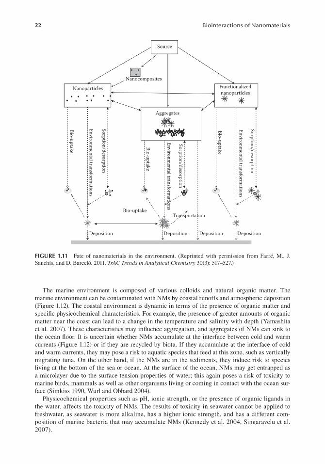
22 Biointeractions of Nanomaterials
The marine environment is composed of various colloids and natural organic matter. The marine environment can be contaminated with NMs by coastal runoffs and atmospheric deposition (Figure 1.12). The coastal environment is dynamic in terms of the presence of organic matter and speci�c physicochemical characteristics. For example, the presence of greater amounts of organic matter near the coast can lead to a change in the temperature and salinity with depth (Yamashita et al. 2007). These characteristics may in�uence aggregation, and aggregates of NMs can sink to the ocean �oor. It is uncertain whether NMs accumulate at the interface between cold and warm currents (Figure 1.12) or if they are recycled by biota. If they accumulate at the interface of cold and warm currents, they may pose a risk to aquatic species that feed at this zone, such as vertically migrating tuna. On the other hand, if the NMs are in the sediments, they induce risk to species living at the bottom of the sea or ocean. At the surface of the ocean, NMs may get entrapped as a microlayer due to the surface tension properties of water; this again poses a risk of toxicity to marine birds, mammals as well as other organisms living or coming in contact with the ocean sur-face (Simkiss 1990, Wurl and Obbard 2004).
Physicochemical properties such as pH, ionic strength, or the presence of organic ligands in the water, affects the toxicity of NMs. The results of toxicity in seawater cannot be applied to freshwater, as seawater is more alkaline, has a higher ionic strength, and has a different com-position of marine bacteria that may accumulate NMs (Kennedy et al. 2004, Singaravelu et al. 2007).
Source
Nanocomposites
Nanoparticles Functionalizednanoparticles
Aggregates
Bio-uptake
Environmental transform
ations
Sorption/desorption
Bio-uptake
Environmental transform
ations
Sorption/desorption
Bio-uptake
Environmental transform
ations
Sorption/desorption
Bio-uptakeTransportation
Deposition Deposition Deposition Deposition
FIGURE 1.11 Fate of nanomaterials in the environment. (Reprinted with permission from Farré, M., J. Sanchís, and D. Barceló. 2011. TrAC Trends in Analytical Chemistry 30(3): 517–527.)

23Introduction—Biointeractions of Nanomaterials
1.6.2 NANOTOXICITY IN THE BODY
NMs effect the human body at multiple levels, broadly differentiated into molecular, cellular, and organular. The interaction of NMs with biomolecules, such as proteins and lipids, is multivari-ate and complex. The nano-biointeractions of NMs with the physiological environment molecules account for most of the toxicological effects induced by NMs.
At the cellular level, NMs may also cause mitochondrial injuries, enter the nucleus and dam-age the DNA, depolarize cell membranes, and also physically damage the membranes by forming nanosized holes. There are different methods by which NMs can interact with the cell membranes, such as via hydrophobic forces, electrostatic forces, van der Waals forces, hydrogen bonding, or receptor–ligand interactions. Once adsorbed on the surface of cells, NMs can be internalized by the cells. Sometimes, the sharp edges of NMs erode the membrane’s surface, leading to perforations. The holes thus formed can act as direct entry points for NMs. Not only do they induce toxicity to the organelles inside the cell, these perforations may also lead the leakage of intracellular �uid into the surrounding medium and vice versa, thus inducing acute toxicity and possibly leading to cell death.
Cellular damage manifests itself at the organular level. As explained earlier, the production of ROS can lead to oxidative stress in biological systems. Their production is believed to be the main cause of induced toxicity in the blood, liver, spleen, kidneys, lungs, and any other organs with which they come into contact. The resultant oxidative stress can produce proin�ammatory cytokines, as it is believed that ROS can affect the calcium-mediated signaling pathways within the cells.
1.6.2.1 Molecular Mechanisms of Nanomaterial ToxicitySeveral different mechanisms have been proposed for the toxicity of NMs in the body (Figure 1.13). The induction of oxidative stress via free radical formation is the prime molecular mechanism of in vivo nanotoxicity (Lanone and Boczkowski 2006). These free radicals cause damage to biological components through the oxidation of lipids, proteins, and DNA. As a consequence of this oxidative
Particulate and organic matterfrom coastal runoffs Atmospheric
inputs Formation of aerosol,risk to seabirds, and mammals
Concentration of NPs in the surface Microlayer Toxicity to embryos and plankton
Dilution and transportto open ocean
Toxicity to pelagic species
Changes in temperatureionic strength, and naturalorganic matter with depth
Coastalsediments
Aggregation
Accumulation of NPsor aggregates at interfaces
Precipitation toocean floor
Mobilization of NPsby microbes
Toxicity to benthos
Ocean floor
FIGURE 1.12 Schematic diagram outlining the possible fate of nanoparticles (NPs) in the marine envi-ronment and the organisms at risk of exposure. (Reprinted with permission from Klaine, S. J. et al. 2009. Environmental Toxicology and Chemistry 27(9): 1825–1851.)

24 Biointeractions of Nanomaterials
stress, the upregulation of various in�ammatory factors, such as redox-sensitive transcription fac-tors (e.g., NF-κB), activator protein-1, and kinases, may induce or enhance in�ammation (Lanone and Boczkowski 2006, Rahman 2000, Rahman et al. 2005). There are several sources of free radical origins, such as phagocytic cell responses to foreign materials, insuf�cient amounts of antioxidants, the presence of transition metals, environmental factors, and physicochemical properties of some NMs (Lanone and Boczkowski 2006). The effect of oxidative stress may extend to organs of the RES, such as the liver, spleen, and organs of high blood �ow, such as the lungs and kidneys, due to the slow clearance and high tissue accumulation of potential free radicals from NMs. Intracellular, NM interactions with cell components, such as mitochondria and nucleus, may result in the cascade of events, such as the creation of ROS, cell cycle arrest, mutagenesis, apoptosis, and nuclear DNA damage, all considered as main sources of toxicity (Aillon et al. 2009, Unfried et al. 2007). NMs may be involved in the upregulation of free radical sources in macrophages and neutrophils (Lanone and Boczkowski 2006). The immediate interaction of NMs with their surrounding environment may result in hemolysis and thrombosis. In addition, NM interactions with the immune system have been known to increase immunotoxicity (Dobrovolskaia and Mcneil 2007).
The adhesion of proteins on the surface of NMs is a normal physiological response in tackling foreign bodies. Once attached to speci�c proteins (opsonization), the NP–protein complex is rec-ognized by phagocytic macrophages. Once engulfed by macrophages, it is taken to the spleen or the liver for its removal from the bloodstream (Owens and Peppas 2006). NMs can also interact with other proteins not intended for opsonization. For example, the binding of human serum albu-min or apolipoproteins promotes a prolonged circulation time in the blood (Ishida et al. 2001). As mentioned earlier in the section, NM surface chemistry determines its interactions with different moieties in the body. Positively charged particles attract proteins, leading to adsorption onto their surfaces, and forming a complex known as a “protein corona.” These coronae can have multiple layers: hard layers composed of proteins strongly attached to the surface of NMs, and dynamic,
Biological fate
Activetargeting
Passive targeting,invisibility to RES/
furtivityOxidativestress
Uncontrolledaggregation
Opsonization andrecognition bymacrophages
Leakageof toxic
components
ROS1O2, HO•, O2
–•
Cell-penetratingpeptide
Antibody
CB
VB
Proteinadsorption
Inorganiccore
Organic orinorganiccoating Antifouling
coating(PEG,
carbohydrates)
Chemical design
FIGURE 1.13 Biological fate of nanomaterials. The top left scenario illustrates some of the effects of the pro-tein adsorption on the nanoparticle surface. The top right scenario represents functionalization of the surface with peptides and antibodies for uptake and cell penetration. The bottom right scenario demonstrates some of the potential coatings for blocking the surface. The bottom left scenario shows dissolution and potential ROS generation by the nanoparticle. (Reprinted with permission from Pelaz, B. et al. 2013. Small 9(9–10):1573–84.)

25Introduction—Biointeractions of Nanomaterials
soft layers containing proteins, which are weakly adsorbed onto the surface (Mahmoudi et al. 2011). This NM–protein interaction can lead to the blockage of the protein’s active site, mild con-formational changes, or even denaturation. This can lead to a failure in the protein’s ability to per-form its normal biological function, such as cell signaling (Lynch and Dawson 2008, Mahmoudi et al. 2011).
Ligand-coated NPs bind to cell receptors and can be endocytosed within the cell or trigger a signaling cascade in the cell. Once inside the cell, the NMs are trapped within endolysosomal vesicles. The exact mechanism of how they escape these vesicles remains unclear. However, once that happens, they can be released into the cytosol where they can interact with other cell organelles, including the nucleus.
1.6.2.2 PharmacokineticsDetermining the pharmacokinetics of NMs is the �rst crucial step in understanding its biological safety and toxicity. Pharmacokinetics is de�ned as the study of the mechanisms of absorption, dis-tribution, metabolism, and excretion of a drug or its metabolite (ADME). A thorough, quantitative, pharmacokinetic analysis of NMs would reveal the target cells, tissues, or organs; the residence time; and the time and dose required to manifest toxicity. This information can be utilized to plan various focused studies that involve only the target cell, eventually helping to decipher the molecu-lar basis of toxicity. The pharmacokinetic behavior of NMs cannot be analyzed at present due to the lack of data and the fact that any difference in the physicochemical properties might affect its pharmacokinetics.
1.6.2.2.1 AbsorptionNMs can enter the human body through different routes of exposure, such as the skin, lungs, and gastrointestinal tract (GIT). As they pass through various parts of the body, they pick up differ-ent biomolecules (Mahmoudi et al. 2011) described in the previous sections (Figure 1.13). The absorption of biomolecules onto their surfaces determines their subsequent biological activities in the body (Lynch and Dawson 2008). As mentioned previously, NMs interact with proteins to form protein coronae, which determine their biodistribution and fate within the body. Protein function is altered by the conformational changes induced by adsorption onto the surfaces of NMs (Darlington et al. 2009, Lundqvist et al. 2004), which, in turn, affects their fate (Ishida et al. 2001, Paciotti et al. 2004).
1.6.2.2.2 DistributionAfter absorption, NMs can be distributed to various organs, tissues, and cells. It is dif�cult to predict the behavior of NMs within living systems, owing to different variants within the systems. The extent of NM distribution within the body depends upon the permeability of blood vessels. Organs such as the liver, spleen, lymph nodes, and bone marrow can take up NMs. These organs contain macrophages and form the RES or mononuclear phagocyte system (MPS). This system is involved in the uptake and metabolism of foreign molecules (Saba 1970). Coatings on NMs can affect their uptake. For example, NMs coated with polyethylene glycol (PEG) resist RES uptake (Paciotti et al. 2004). It is therefore imperative to understand and study the distribution of NMs within the body.
1.6.2.2.3 MetabolismThere are limited reports on NM metabolism. There are some NMs that degrade in the tissues, such as polymer-based NMs and superparamagnetic iron oxide NMs. On the other hand, there are some that show no degradation in vivo, such as QDs, fullerenes, and silica NMs (Ballou et al. 2004, Khan et al. 2005, Singh et al. 2006, Yang et al. 2007). A study �nding that CNTs can be degraded by neutrophil myeloperoxidase provides evidence that suggests enzymatic metabolization of NMs to a certain extent (Kagan et al. 2010). Also, coatings such as proteins used for QDs can be metabolized

26 Biointeractions of Nanomaterials
by proteases in the gut (Hardman 2006). It is still unclear as to how NMs may be metabolized in the body. Therefore, studies need to be conducted to address these unanswered questions.
1.6.2.2.4 EliminationNMs can be eliminated from the body via various routes, such as exhalation; urination (via the kid-neys) (Singh et al. 2006); defecation (via the biliary duct) (Hardonk et al. 1985, Renaud et al. 1989); perspiration; and through the saliva, seminal �uids, and mammary glands. Their fate of elimina-tion, again, depends on their physicochemical properties. For example, hydroxyl functionalized SWCNT accumulate in the liver and kidneys and are excreted in the urine within 18 days (Wang et al. 2004a). On the other hand, ammonium functionalized SWCNT show neither liver uptake not fast, renal excretion (Singh et al. 2006). Contrary to that, QD are not excreted and remain intact in vivo (Fischer et al. 2006, Yang et al. 2007) unless they have a coating. This has been proven in the case of QDs coated with cysteine, which were excreted in mice urine (Choi et al. 2007). Studies should focus on identifying organs that could be stressed by exposure to NMs, possibly providing a molecular basis for the stress response. If there is an association found with speci�c organ cells and NMs characteristics (e.g., size, surface chemistry, aggregation and composition, shape), then it would be possible to establish correlations between the toxic effects of NMs and speci�c NM prop-erties. Demonstrated pharmacokinetic studies of various NMs will be discussed in greater detail in the later chapters.
1.6.3 EFFECTS OF NANOMATERIALS ON ORGAN SYSTEMS
There are numerous, inevitable, exposure routes by which NMs can enter the human body to elicit potential adverse effects. The speci�c routes are the respiratory, reticuloendothelial, cardiovascular, central nervous, and integumentary systems.
1.6.3.1 Pulmonary SystemThe respiratory system serves as a major entry portal for ambient particulate materials. The short-term exposure of inhaled, ultra�ne carbon black, nickel, and TiO2 particles were found to produce an enhanced in�ammatory response in the rat respiratory system, as compared to �ne-sized par-ticles of similar chemical compositions (Grassian et al. 2007, Pettibone et al. 2008, Wani et al. 2011, Warheit et al. 2006). The micron-sized particles are largely trapped and cleared by the upper airway mucociliary escalator system, whereas particles less than 2.5 μm can travel to the alveoli. Inhaled NPs can become deposited in the alveolar region (Arora et al. 2012, Curtis et al. 2006, Hagens et al. 2007). The toxicity of NMs may initiate with the development of exaggerated lung responses, char-acterized by increased and persistent levels of pulmonary in�ammation, subsequently transformed into cellular proliferation, �bro proliferative effects, and in�ammatory-derived mutagenesis, which ultimately results in the development of lung tumors. As previously mentioned, various factors that are likely to in�uence the pulmonary toxicity of NPs are the size and number of particles, surface dose and coating, degree of aggregation, surface charges, and the method of particle synthesis (Lyon et al. 2006, Wani et al. 2011).
1.6.3.2 Gastrointestinal TractNMs can reach the GIT directly through the ingestion of food, water, cosmetics, drugs, and by the use of drug delivery devices, as well as after mucociliary clearance from the respiratory tract through the nasal region (Arora et al. 2012, Hagens et al. 2007). The acute toxicity of ingested nanocopper material was found to be more toxic than bulk copper material in mice. The occurrence of systemic argyria after the ingestion of colloidal nanosilver proves its secondary toxic effects after translocation from the intestinal tract (Arora et al. 2012). Reports were found for the uptake of �uorescently labeled, polystyrene NPs by intestinal lymphatic tissue (Peyer’s patches) (Morishita and Peppas 2006).

27Introduction—Biointeractions of Nanomaterials
1.6.3.3 Reticuloendothelial SystemsSince all of the blood exiting the GIT goes into the hepatic portal vein that directly diffuses through the liver, the RES system in the liver is exposed to all NPs absorbed from the GIT into the car-diovascular system (CVS). NPs such as carbon black and polystyrene exert their toxic effects by enhancing the secretion of proin�ammatory cytokines, such as tumor necrosis factor alpha, fol-lowing the stimulation of macrophages via ROS and calcium signaling (Brown et al. 2004). These proin�ammatory cytokines and oxidative stresses that can potentially damage hepatocyte function and bile formation are also associated with the pathology of liver diseases (Wani et al. 2011). The direct injection of ultra�ne carbon black particles into the blood induces platelet accumulation in the hepatic microvasculature of healthy mice in addition to prothrombotic changes on the endothelial surface of hepatic microvessels.
1.6.3.4 Cardiovascular SystemThe surface charge of NPs plays a vital role in their toxic effects on the CVS. Especially cationic, ultra�ne particles, such as gold and polystyrene, have been shown to cause a lethal effect on RBCs and blood clotting while anionic particles are found to be nontoxic. The exposure to diesel exhaust particles (DEPs) was found to alter heart rates in hypertensive rats, while also inducing direct, nega-tive effects on the heart’s pacemaker activity (Hansen et al. 2007). Exposure to SWCNT has also resulted in adverse cardiovascular effects (Li et al. 2007).
1.6.3.5 Central Nervous SystemThe brain can be exposed to NPs by the means of two different mechanisms after inhalation; namely, trans-synaptic transport after inhalation through the olfactory epithelium and uptake through the blood–brain barrier (Jallouli et al. 2007, Lockman et al. 2004). The adverse patholo-gies, including hypertension and allergic encephalomyelitis, have been found to be associated with the enhanced permeation of NPs to the blood–brain barrier in experimental setups. The production of ROS (Long et al. 2006) and subsequent oxidative stress (Peters et al. 2006) by NPs has been implicated in the pathogenesis of neurodegenerative diseases, such as Parkinson’s and Alzheimer’s. The NP’s surface charges and chemical compositions have been shown to alter blood–brain integ-rity and deserve considerations as to their role in brain toxicity and distribution (Wani et al. 2011).
1.6.3.6 Integumentary SystemThe skin is the largest primary defense organ in our body and comes into direct and indirect con-tact with many toxic agents. The strongly keratinized stratum conium is the rate-limiting barrier to defending against the penetration of most micron-sized particles and harmful exogenetic toxicants. Possible skin exposure to NMs can also occur during the intentional application of cosmetics and other topical drug treatments. In addition, NPs have unique scattering properties due to their small size. They may alter the optical pathway of UV photons entering the upper part of the skin’s horny layer. In this way, more photons can be absorbed by the stratum conium. In vitro studies have shown that MWCNT initiate an irritation response in human epidermal keratinocytes by their localizing effect (Baroli et al. 2007, Zvyagin et al. 2008). QDs with diverse physicochemical properties were found to penetrate the intact stratum conium barrier and get localized within the epidermal and dermal layers (Ryman-Rasmussen et al. 2006).
1.7 NANOTOXICITY: CHALLENGES, SOLUTIONS, AND THE FUTURE
Unfortunately con�ned just to nanomedicine, toxicity assessments are an integral part of NM devel-opment. Even though attempts are being made to assess toxicity of widely used NMs, they are met with various challenges owing to the shortcomings of the methods used for assessment. Even though nanotoxicology is considered a well-de�ned �eld, it faces a lot of challenges even today.

28 Biointeractions of Nanomaterials
1.7.1 PHYSICOCHEMICAL CHARACTERIZATION
As discussed earlier, the physical and chemical properties of NMs play a very important role in their interaction with biological systems. The physicochemical properties of NPs must be scrutinized in detail to interpret any results of NP-induced toxicity by optimizing the NP design. Speci�c NP properties that in�uence cellular toxicity are still not fully understood. Hence, a thorough charac-terization of the NP is essential.
There are diverse approaches that are commonly used to characterize these properties, several of which are described in Table 1.6. Size (distribution) and shape determinations are typically evalu-ated with one or more of the following: dynamic light scattering (DLS) (Murdock et al. 2008), transmission electron microscopy (TEM), scanning electron microscopy (SEM) (Love et al. 2012, Marquis et al. 2009), and AFM. Field �ow fractionation (FFF) is a chromatography-like technique in which the partition of sample species is achieved in a thin, open channel, and the particle size distribution can be calculated directly from the �rst principles (Bohnsack et al. 2012). Crystal struc-ture is generally elucidated by x-ray diffraction and the surface area is determined by the Brunauer–Emmett–Teller (BET) method by nitrogen adsorption and desorption isotherms (Maurer-Jones et al. 2010).
The surface chemical composition can be examined through various techniques, such as induc-tively coupled–mass spectrometry (ICP-MS) and inductively coupled–optic emission spectrometry (ICP-OES), which utilize an inductively coupled plasma as the ion source and can detect metals (and some nonmetals) at concentrations below one part per trillion. ICP-MS and ICP-OES are also capable of monitoring isotopic speciations for the ions of choice (Bohnsack et al. 2012). Other tech-niques are secondary ion mass spectroscopy (SIMS), liquid chromatography–mass spectrometry (LC-MS), matrix-assisted laser desorption/ionization–time of �ight (MALDI-TOF), and are used speci�cally for an elemental analysis along with mass determinations. The elemental composition of NP surfaces can be determined by Auger electron spectroscopy (AES), electron energy loss spec-troscopy (EELS), and x-ray photoelectron spectroscopy (XPS). These are all high-vacuum tech-niques and vary in their capabilities and surface sensitivities (Love et al. 2012, Powers et al. 2012).
NP uptake by cells is a vital factor to the assessment nanotoxicity. Flow cytometry (FCM) has been used in the �eld of biochemistry to analyze thousands of cells in a second, which is advanta-geous over TEM and ICP-MS techniques (Ibuki and Toyooka 2012). Electron paramagnetic reso-nance (EPR)–electron spin resonance (ESR) are specialized methods to measure free radicals either directly or by “spin trapping” them with a reference molecule. They are powerful techniques to quantify oxygen ROS in toxicological evaluations of NMs. The ion abrasion SEM (IA-SEM) and focused ion beam SEM (FIB-SEM), in addition to soft x-ray tomography, are being used to elucidate three-dimensional displays of cells and tissues (Powers et al. 2012). Synchrotron radiation-induced x-ray �uorescence (SRXRF) can provide the local, electronic, and molecular structures around the atom of interest with sub-Angstrom spatial resolutions. It has also been utilized to investigate the uptake of organic mercury in zebra �sh larvae (Bohnsack et al. 2012).
Along with the physical characteristics of NPs, dose characterizations are also critical for the interpretation of results. The calculation of a dosing metric is complicated because little is known about appropriate doses and how aggregation or stability in�uences effective dosings (Love et al. 2012). These characterization challenges are ripe for the study and application of the collective expertise of analytical chemists.
Various combinations of methods may be utilized to validate NM properties, as each method has its own disadvantages and advantages. However, chemical characterization should accompany physical characterization to assess the presence of contaminants in test samples, although exhaus-tive characterization is time consuming as well as very expensive. There is a need to devise a battery of standardized tests to adequately assess these properties, so that the data that are obtained are comparative and reproducible. There is also a need for a standardized reference material for NMs that can be used by toxicologists, so that data can be compared with different studies.

29Introduction—Biointeractions of Nanomaterials
TABLE 1.6Analytical Techniques to Characterize Nanoparticles
Characteristics Analytical Methods Parameters
Particle imaging
TEM—transmission electron microscopy Size, shape, state of aggregation
SEM—scanning electron microscopy Size, shape, state of aggregation topography, elemental composition in combination with other techniques
ESEM—environmental SEM As SEM above
AFM—etomic force microscopy Size, shape, morphology, state of aggregation
SAXS—small-angle x-ray scattering, SANS—small-angle neutron scattering
Number of bilayers, chemical group ordering
Physical property
DLS—dynamic light scatter size of particle, NTA—nanoparticle-tracking analysis, FCS—�uorescence correlation spectroscopy, FFF—�eld �ow fractionation
Particle size distribution
Filtration Size fractionation
Centrifugation
XRD—x-ray diffraction Crystal structure, size
BET—Brunauer–Emmett–Teller method Surface area per unit mass and porosity
SEC—size exclusion chromatography, AFFF–asymmetric �eld fractionation, DLS, electrophoretic method, electroacoustic technique
Size distributionζ potential
Chemical composition
ICP-MS—inductively coupled–mass spectrometry, ICP-OES—inductively coupled–optic emission spectrometry, SIMS—secondary ion mass spectroscopy
Element composition, mass
EDS—x-ray energy dispersive spectroscopy Element composition
LC-MS—liquid chromatography–mass spectrometry, MALDI-TOF—matrix-assisted laser desorption/ionization–time of �ight
Fullerene structure, mass
AES—Auger electron spectroscopy, EELS—electron energy loss spectroscopy, XPS—x-ray photoelectron spectroscopy
Surface chemistry
NMR—nuclear magnetic resonance spectroscopy, FTIR—Fourier transformed infrared spectroscopy, Raman spectroscopy
Chemical functional groups, surface groups
Uptake ICP-AES—inductively coupled plasma atomic emission spectroscopy, SRXRF—synchrotron radiation-induced x-ray �uorescence, NAA—neutron activation analysis, ICP-MS, TEM, �ow cytometry
Cellular uptake
IA-SEM—ion abrasion SEM, FIB-SEM—focused ion beam SEM, soft x-ray tomography
Three-dimensional imaging of nanoparticle distribution in cells and tissues
EPR—electron paramagnetic resonance/ESR—spin resonance
Quanti�cation of reactive oxygen species generation after cellular interaction of nanoparticles
Stability CD—circular dichroism spectroscopy, UV-vis—UV-visible spectroscopy, DLS, ICP-AES, ICP-MS, colorimetric assays
Stability indicating changes, such as particle size distribution, dissolution, surface chemistry, degradant formation

30 Biointeractions of Nanomaterials
Powers et al. (2006) have summarized �ve basic rules for physicochemical characterization:
1. The sample used for characterization assessments should be representative of the material. 2. The size and shape should be measured as dispersed a state as possible. 3. The most appropriate method for measurement should be applied. 4. The particles should be measured in desired amounts to ensure precision and accuracy. 5. As far as possible, the characteristics of the particles should be measured under the same
conditions as its application.
1.7.2 IN VITRO ASSESSMENT
In vitro toxicity studies are valuable in the optimization of NP design. In vitro methods to assess NP toxicities can be classi�ed as mechanistic or viability assays. The mechanistic assays are those that seek to assess the effects of NPs on various cellular processes, whereas viability assays are concerned solely with whether a given NP results in cell death.
Owing to their lower costs and ef�cacy, in vitro studies are the most preferred studies for the assessment of toxicity. In vitro assays consist of subcellular systems, such as macromolecules; organelles; cellular systems, such as individual cells, culture, barrier systems; and whole tissues, such as organs, slices, and explants. Before the administration into biological systems (mice or rabbits), it is very important to ascertain the right dosage or a safe concentration of the drug. That is where in vivo studies play a pivotal role. The toxicity of the drug is �rst tested in cell culture to assess the suitability and dosage for in vitro studies (Geldenhuys et al. 2011).
Although in vitro tests are quick and straightforward, there are some limitations, such as the correlations of the in vitro to the in vivo environments. The process of immortalization alters the cells’ properties and sensitivities. Also, some cells are more sensitive than others to a certain kind to toxin as compared to other cells in a different culture. Because the cells are isolated from their natural environment, they may not be an appropriate model. For example, Lee et al. (2009) reported that 2D cell cultures that are commonly used in in vitro studies may not correctly re�ect the actual toxicity of NPs, as they do not represent functions of 3D tissues that have complex cell-to-cell and cell-to-matrix interactions with different diffusion or transport conditions.
There may also be interferences with speci�c toxicology assays based on the properties of the NM. These are a result of the following unique physical and chemical properties: high surface areas that may lead to increased adsorption capacities, optical properties that may interfere with �uores-cence or visible light absorption detection systems, increased catalytic activities due to enhanced surface energies, and magnetic properties that make them redox active and thus interfere with meth-ods based on redox reactions (Kroll et al. 2009). Interferences with colorimetric assays, such as the MTT 3-(4,5 dimethylthiazol-2-yl)-2-5-diphenyl tetrazolium bromide (MTT) assay, have been reported (Doak et al. 2009, Kroll et al. 2009, Monteiro-Riviere and Inman 2006, Monteiro-Riviere et al. 2009, Pulskamp et al. 2007, Scalf and West 2006, Song et al. 2010). For example, CNTs inter-act with formazan crystals and make them insoluble (Wörle-Knirsch et al. 2006). Consequently, such variables can lead to con�icting results (Doak et al. 2009, Monteiro-Riviere et al. 2009, Wörle-Knirsch et al. 2006). Thus, if such interferences are suspected, additional tests would be required to con�rm the �ndings.
1.7.2.1 DNA Synthesis and DamageDNA synthesis assays are commonly used to assess cell proliferation or to quantify the number of cells in each stage of the cell cycle (which can subsequently reveal cell cycle arrest at a given point). The incorporation of 5-bromo-2-deoxyuridine (BrdU) into newly synthesized DNA has been frequently employed to quantify DNA synthesis in nanotoxicity assays. The genotoxicities of sil-ver (Ag) NPs and PEG-coated cadmium selenide/zinc sul�de (CdSe/ZnS) QDs on lung epithelial cancer (A549) and skin epithelial (HSF-42) cells were assessed by utilizing the aforementioned

31Introduction—Biointeractions of Nanomaterials
method (Oostingh et al. 2011, Zhang et al. 2006). As damage to the DNA is highly correlated with an increased risk of cancer, it is critical to assess such damages by any NP that is likely to come in contact with humans. The comet assay (single-cell gel electrophoresis assay), which is utilized to measure the number of single-strand breaks in DNA, is the most common method to assess DNA damage. This assay has been used to assess DNA damage in cells exposed to cerium oxide (CeO2) (Auffan et al. 2009), Ag (AshaRani et al. 2009), and SiO2 NPs (AshaRani et al. 2009). Other meth-ods to assess DNA damage include checking for the presence of micronuclei or other chromosomal aberrations and measuring the expression of proteins implicated in DNA repair. An increase in the expression and activation of DNA repair-related proteins was found upon cellular exposure to MWCNTs (Zhu et al. 2007).
The general DNA microarray and more speci�c PCR analyses are being utilized to assess the activity of functional genes involved in various cellular processes. They have been used to assess changes in gene expression upon exposure to gold (Au) nanorods (Hauck et al. 2008), SWCNTs (Nygaard et al. 2009), and SiO2-coated CdSe/ZnS QDs (Zhang et al. 2006). PCR was utilized to study the effects of antimony trioxide (Sb2O3) NPs in erythroblasts (Bregoli et al. 2009) and the impact of cesium dioxide (CeO2) NPs on the expression of genes related to oxidative stress and cell structure (Park et al. 2008).
1.7.2.2 ImmunogenicityThe ability of a given NP to evoke an immune response is a vital parameter in demonstrating its toxicity on physiological systems, and it may not be explored by standard cellular toxicity studies. ELISA can accurately detect cytokine levels at picograms levels. Many investigators have studied the formation of proin�ammatory cytokines (e.g., interleukin-6 and -8) following the exposure to metal oxide NPs in various cell types by this technique (Veranth et al. 2007, Schanen et al. 2009).
1.7.2.3 Oxidative StressAn elevated amount of ROS, either due to an innate immune response, to a NP or from the ability of a speci�c NP (e.g., a fullerene or a metal oxide) to autocatalyze ROS formation in the cellular envi-ronment, has the potential to damage or disrupt key cellular processes (Xia et al. 2006). Generally, the presence of ROS can be directly assessed by quantifying the amount of ROS present in a given cell population or indirectly assessed by monitoring the secondary effects of prolonged oxidative stress. The spectro�uorimetry/FCM or spectrophotometry-based system can directly measure and monitor the ROS-induced formation of the �uorescent product, �uorescein, from 2,7-dihydrodichlo-ro�uorescein diacetate (DCFDA), the superoxide-induced conversion of dihydroethidium (DHE) from the blue �uorescent form to the red �uorescent form, or the superoxide-induced conversion of nitroblue tetrazolium (NBT) to blue formazan. The DCFDA and DHE assays have experimentally shown to change ROS levels in MPMCs (Marquis et al. 2011) or human �broblasts (AshaRani et al. 2009), which were exposed to Au or Ag NPs with different surface functionalities. The effects of ultra-small superparamagnetic iron oxide NPs (AshaRani et al. 2009) and cationic lipid-coated Fe3O4 NPs (Soenen et al. 2009) in human monocyte macrophages and 3T3 cells were assessed by NBT assay. The determination of lipid peroxidation or antioxidant depletion is the measurement of the secondary effects of increased cellular ROS levels. These can be done with the detection of 8-hydroxy deoxyguanosine (8-OHdG) and superoxide dismutase (SOD) activities. A green �uores-cent dye, which turns red in the presence of oxidized lipids, was utilized to assess lipid peroxidation in the presence of Cd/Te QDs (Choi et al. 2007).
1.7.2.4 Cell ProliferationThe rate of cell growth is an important indicator of overall cell integrity and of the potential for NPs to interfere with proliferative processes. There are two quantitative assays commonly utilized as the standard for assessing cell proliferation: (a) cell counting by FCM or high-content image ana-lyzers, and (b) the colony-forming ef�ciency (CFE) assay. The effect of SWCNTs (Mu et al. 2009)

32 Biointeractions of Nanomaterials
and PEG-silane modi�ed CdSe/ZnS QDs (Zhang et al. 2006) on the proliferation of HEK293 and human lung and skin epithelial cells has been assessed by FCM. The CFE assay has been used to assess the effects of polymeric-entrapped, thiol-coated Au nanorods (Zhang et al. 2006) on murine �broblasts and human hematopoietic progenitor cells.
1.7.2.5 ExocytosisChanges in exocytosis may be another indicator of nanotoxicity. Carbon-�ber microelectrode amperometry has been employed to study the effects of various NPs on the secretion of small, electro-active molecules (e.g., serotonin and epinephrine). This method allows one to quantify the number of chemical messenger molecules released per vesicle, the speci�c release kinetics, and the frequency of vesicle fusion with a high sensitivity and time resolution. Studies in MPMCs and adre-nal chromaf�n cells have utilized this method to reveal the mutagenic potential of functionalized (with either positive or negative side chains) Au and Ag NP exposure (Marquis et al. 2011).
1.7.2.6 Cell Viability and Metabolic ActivityCell viability studies are perhaps some of the most widely used assays to assess nanotoxicity, as they provide information on the mechanisms or causes of cellular toxicity and death. Any lethal consequences from NP exposure, including membrane lysis, cell cycle arrest, and apoptosis, may stop mitochondrial activity. Many different types of assays that allow for the study of toxicity are used in research. Toxicity can also be assessed by using two or more independent test systems to validate �ndings. The colony formation assay, or the clonogenic assay, is an in vitro cell-survival assay, based on the ability of a single cell to grow into a colony. It is a simple method that can be employed to avoid interference from NPs, as no dye or stain is used (Franken et al. 2006).
Assays of metabolic activity following exposure to NPs are the most common methods used to determine cell viability. The most popular test is the MTT assay in live cells. MTT [3-(4,5-dimeth-ylthiazol-2-yl)-2,5-diphenyltetrazolium bromide] is reduced to purple formazan, which can be detected spectrophotometrically. Several similar assays (MST, MTS, XTT, WST-1) have also been employed to eliminate the possibilities of NM interference with these assays (Stone et al. 2009). Alamar blue (resazurin) is another dye that undergoes reduction by living cells to produce the �uo-rescent product, resoru�n. It has also been extensively utilized to measure cell viability, following exposure to SiO2-coated CdSe QDs (Sharma et al. 2009) and amino acid-functionalized Au (Ghosh et al. 2008).
1.7.2.7 HemolysisThe risk of erythrocytic lysis is especially important for NPs that are intended to be directly intro-duced into the bloodstream. The assessment of hemoglobin (Hb) by spectrophotometric techniques in response to NP exposures can be a measure of both membrane disruption and extreme cellular toxicity (i.e., necrosis). This approach has been utilized to determine the median lethal dose values for functionalized Au NPs (Goodman et al. 2004). Recent studies have focused on the hemolytic potential of functionalized Au NPs while assessing their effects on ROS production in neutrophils and thrombotic capabilities (Love et al. 2012).
The assessment of the indicators of programmed cell death (i.e., apoptosis) and necrosis directly reveal a NP’s ability to induce intracellular, self-destruction mechanisms and destroy cells. Such assays have been developed beyond the measurement of membrane integrity to the quanti�cation of apoptotic protein levels and activation and DNA fragmentation. There are �ve main tech-niques generally used to determine membrane integrity: phosphatidylserine (which migrates to the extracellular surface of apoptotic cells) labeled with annexin V, propidium iodide exclusion by intact membranes (AshaRani et al. 2009), trypan blue exclusion by intact membranes (Goodman et al. 2004, Hauck et al. 2008), neutral red staining (which undergoes a color change due to protonation in intact lysosomes) (Lanone et al. 2009), and the determination of the total lactate

33Introduction—Biointeractions of Nanomaterials
dehydrogenase (LDH) content in extracellular mediums (Papageorgiou et al. 2007, Tkachenko et al. 2004). Another common assay looks for the exclusion of red �uorescent ethidium homodi-mer 1 from live cells, while measuring the uptake of calcein-AM (which �uoresces green after modi�cation by intracellular esterases). Assessing the level of DNA fragmentation with TUNEL (terminal deoxynucleotidyl transferase deoxyuridine triphosphate nick end labeling) can be used to identify apoptosis, as demonstrated by studies on SWCNTs (Mu et al. 2009) and Eu(OH)3 NPs (Patra et al. 2008). It is important to consider the NM’s impedance during the selection of any of the aforementioned methods. For example, in the MTT assay, CNTs can modify the solubility of formazan through the adsorption of the reduced crystals, thereby falsely lowering the viability results (Worle-Knirsch et al. 2006). Such spontaneous reductions may also occur in graphene particles (Liao et al. 2011). The LDH assay has also failed for some NPs, including Cu (LDH was inactivated) and TiO2 (LDH was adsorbed) (Han et al. 2011). Further advancement in this �eld requires the detection and quanti�cation of sensitive toxicological markers that may be unique to nanotoxicity.
1.7.3 IN VIVO ASSESSMENT
The prediction of the safety and toxicity of nanoconstructs has been examined by the extensive testing of in vitro cultured cells along with in silico computational models. In vivo systems are much more complex with interdependent pathways, which are dif�cult to evaluate by in vitro analysis. However, toxicity assays in animal models can provide better correlations with human conditions.
Acute toxicity studies are performed in animal models to identify the maximum tolerated dose (MTD) and no observable effect level (NOEL) in NP dosages. In classical toxicology studies, the dose of NMs is measured by the milligrams of test items per kilogram of animal weight. However, the surface area, size, density, and surface properties of NMs are less common to take under consid-eration in toxicity studies. The true evaluation of nanotoxicity should be based on both the classical mg/kg exposure and the dosage based on surface area to justify the effects of nanoscale reactivities on toxicity. Acute toxicity studies normally span 14 days after a single dose or repeated dose admin-istration, and the evaluation of organ-speci�c toxicity in addition to �nding the right dose. At least two species, one rodent and one nonrodent species, are preferably required to conclude the results from these studies. The following parameters are monitored during the study:
• Responses to the administered dose: Following the administration of NMs, neuronal, hematological, and cardiac responses can occur and, hence, animals should be monitored for at least 30 min postadministration.
• Changes in weight: The overall health of the animal is the simplest parameter to observe for any possible toxic effects of the injected dose. The change in weight (>10%) can sig-ni�cantly indicate the NM’s adverse effect. However, this is a preliminary observation and further investigation is required to �nd out the actual cause of toxicity.
• Clinical observation: The functionality of various organs systems, such as the cardiovas-cular, respiratory, ocular, and gastrointestinal systems, are examined to evaluate clinical changes. Imaging procedures such as ultrasound, x-ray, computed tomography (CT), and MRI are used as supportive elements.
• Clinical pathology: The plasma samples collected from the processed animals are utilized to check liver functionality by the measurement of aspartate aminotransferase (AST), ala-nine aminotransferase (ALT), and total bilirubin and albumin levels. Kidney functions are evaluated by assessing blood urea nitrogen and creatine levels in plasma. Cardiac function is assessed by measuring LDH and creatine phosphokinase (CPK). Amylase levels are indicators of exocrine functions.

34 Biointeractions of Nanomaterials
• Gross necroscopy: Gross necroscopy can reveal valuable information about the speci�c toxicity of NMs. Severe intestinal bleeding was observed after the intravenous adminis-tration of G7 amine-terminated PAMAM dendrimers in CD-1 mice (Greish et al. 2012). The histology of the lung and liver was performed to evaluate the toxicity of SWCNTs (Oberdorster et al. 2005).
1.7.3.1 Absorption, Distribution, Metabolism, Excretion, and Pharmacokinetic StudiesAbsorption, distribution, metabolism, excretion, and pharmacokinetic (ADME/PK) studies are vital tools to evaluate the occupational health impact and potential hazards of NMs to human beings. The pharmacokinetic and biodistribution behaviors of NMs is essential to understand, as it re�ects the in-depth concentration of NMs in each organ, which can be used as the basis of phase I clinical studies in humans. Radiolabeling studies with gamma emitters, such as 125I, is a popular method of in vivo NP quanti�cation. A biodistribution pro�le of PAMAM dendrimers with differential surface charges and sizes was evaluated via 125I labeling in CD-1 mice (Greish et al. 2012). 99Tc was used in the scintigraphy imaging of nanoscale N-(hydroxypropyl) methacrylamide copolymers in mice (Line et al. 2005). The limitation of this method is the stability of the nanoconstruct–radiolabel conjugates, which needs to be evaluated before beginning the imaging study. The application of ICP-MS can eliminate the aforementioned limitations and provide acute quanti�cations of NMs in parts per million levels. The biodistribution pro�le of gold nanorods and spheres in tumor-bearing mice was evaluated and analyzed by ICP-MS (Arnida et al. 2011).
1.7.3.2 Genotoxicity and Carcinogenic StudiesThe mutagenic, teratogenic, and carcinogenic potential of NMs are being evaluated in these stud-ies. Widely accepted in vivo genotoxicity tests are the metaphase chromosomal analysis and the bone marrow micronucleus test. In 1975, Schmid developed the mouse bone marrow micronu-cleus test as an alternative to cytogenic studies on mammalian bone marrow cells (Hayashi et al. 1983). The damage to the chromosome or mitotic apparatus (resulting in micronucleus forma-tion), as a consequence of NMs exposure in animal bone marrow cells, is being detected by the micronucleus test. Micronuclei (MNi) are formed because of acentric fragments of chromosomes or due to chromosomal breakages or mitotic spindle apparatus damages (Sarto et al. 1987). FCM has been used to differentiate bone marrow cells from abnormal, peripheral RBCs (Sarto et al. 1987). Teratogenic studies are not regularly required as part of toxicity studies. Such studies follow the International Conference on Harmonization (ICH) of Technical Requirements for Registration of Pharmaceuticals for Human Use guidelines (Baber 1994). ICH guidelines involve segments of test procedure standardizations for fertility and reproductive performance, embryo–fetal develop-ment, perinatal, and postnatal analyses on maternal and newborn cases. In vivo carcinogenic studies involve long-term observation for the development of tumors following the administration of either single- or repeated-dose NMs. It requires a large number of animals and usually extends up to 30 months.
Apart from the aforementioned studies, chronic and subchronic studies are essential to assess the toxic effects of NMs, which are nonbiodegradable and consist of metal oxides, such as gold, carbon, and silica particles. In such cases, acute and subacute observations are not suf�cient to evaluate the safety of NPs due to their long residence times in the body.
The major challenges, or the most discouraging aspects, of in vivo tests are its length, expense, and ethical issues. Even though in vivo tests may counter most of the limitations faced by in vitro studies, there are still many challenges that impede their use for the assessment of nanotoxicity. First, there is dif�culty in determining the actual dosages of NMs in the environment to which ani-mal and humans are exposed, owing to their small size and quantity. Sometimes, if the concentra-tion of the known dose is high, it may lead to agglomeration. Vehicles are used for dose delivery in order to avoid agglomeration. It is another challenge to ensure that the vehicle used for delivery does

35Introduction—Biointeractions of Nanomaterials
not interfere with the NMs and does not induce any toxicity of its own. For example, some studies found that phosphate-buffered saline, commonly used as a vehicle for dose delivery, is a poor dis-persion agent (Sager et al. 2007, Sayes et al. 2007). Once delivered into the body, NMs may interact with other components of the biological system, such as proteins, different salt concentrations, and variable pH values, to form unstable suspensions, thereby negatively affecting their biodistribution and activity (Buford et al. 2007). Hence, the results thus obtained may be con�icting.
1.7.4 CONSIDERATIONS FOR PREVENTING NANOTOXICITY
As it has been highlighted, the importance of the tight control over NM parameters is crucial for the prevention and control of nanotoxicity. One of the most prominent factors that increases the vari-ability of the properties and activity of NMs is agglomeration. The phenomenon of agglomeration involves the adhesion of particles to each other, mainly because of van der Waal’s forces, which dominate at the nanoscale level due to the increased surface area to volume ratio (Powers et al. 2007). NMs agglomerate after their synthesis in both the dry and suspension forms. The challenge for synthetic chemists is to prevent agglomeration, as it can lead to changes in physical and chemi-cal properties. The major properties affected are size, size distribution, surface-to-volume ratio and, hence, surface reactivity. Since these parameters play a major role in the toxicity of NPs and are altered due to agglomeration, it is prudent to account for these changes in the study design (Borm et al. 2006, Teeguarden et al. 2007).
Agglomeration is in�uenced by several intrinsic and extrinsic factors, such as the composition of NMs and their concentration, size, surface coating, zeta potential, and temperature, among others (Teeguarden et al. 2007). It is well known that NPs can pass through biological barriers due to their small size. Agglomeration can alter their biological responses due to a decrease in the total available surface area, leading to an underestimation of toxic potential, especially in the case of drug delivery and safety and toxicity assessments (Sager et al. 2007). There are differ-ent methods available to deagglomerate NMs. Sonication is the most preferable and widely used method because it disperses NPs in a liquid by cavitation and does not have much of an effect on the properties of the particles. However, the attained deagglomeration is incomplete, as the particles do not reach their primary size and display the tendency to reagglomerate over time (Murdock et al. 2008). Another important method for preventing the agglomeration of NMs is surface modi�cation. The particles can be coated with polymers or dispersed in ionic or nonionic surfactants (Farah et al. 2008, Sager et al. 2007, Skebo et al. 2007, Wick et al. 2007). While surface modi�cation allows the particles to be stabilized and avoids agglomeration, it also raises concerns that they may shield or in�uence the effects of NMs on biological systems (Warheit 2008, Derfus et al. 2004, Warheit et al. 2005). The stability of such surface coatings inside a biological environment is another critical issue.
At the initial synthesis stage, the scientist may need to consider speci�c, physical parameters. However, in order to control the risk of NMs at all lifecycle points, interdisciplinary collabora-tions may be required. Additionally, owing to the increase in the production of NMs, the chances of their release into the environment and their subsequent effects on ecosystems are becoming important issues that need to be addressed. To do that, it is �rst necessary to assess the fate and behavior of NMs in the environment. It is still unclear how, at what concentrations, and in what forms, the NMs will be released into the environment. The answers to these questions will guide the formulation of regulatory guidelines that will protect the ecosystem and will also permit the full industrialization of the bene�ts that nanotechnology offers. It is important to focus current research efforts on the release, behavior (reaction to changes in environments), and fate (aggrega-tion, adsorption, etc.) of NMs. A lifecycle assessment of the release of NMs into the environment is, therefore, imperative as the implementation of effective and protective regulatory policies (Navarro et al. 2008).

36 Biointeractions of Nanomaterials
1.8 FUTURE CONSIDERATIONS
The study of NM toxicity is currently in the initial phases of development. The environmental testing of NMs requires the development of testing guidelines to allow the comparison and interpretation of data from clinical as well as environmental studies, and the close cooperation in interdisciplinary research. Unlike microorganisms and biomolecules, NMs can be engineered in a laboratory. Their physicochemical properties can be modi�ed to enable systematic studies. Monte Carlo simulations have been used to model the effects of NP size and ligand densities on cellular uptake and tumor targeting. This would help to improve NP designs for optimal tumor accumulations in diagnostic and drug delivery applications (Buford et al. 2007, Borm et al. 2006, pp. 68–70, Powers et al. 2007). Such stimulation tools can be developed by conducting studies in a systematic manner and with a properly selected biological system. On that basis, researchers could create a database that would facilitate �nding commonalities in experimental results. The outcomes of these studies can also be entered into a database, which can further help researchers to use computer simulation programs to identify appropriate nanostructure designs for a speci�c application.
There are several regulatory agencies that are looking into the toxicity caused by NMs. NNI was previously mentioned in the chapter; it has a section devoted to identifying the potential exposure, possible toxicity, and the need for personal protective equipments when working with nanoscale materials. Several other U.S. agencies (the National Institute for Occupational Safety and Health, the National Science Foundation Nanoscale Science and Engineering technology, and the Environmental Protection Agency) are working to assess, support, and monitor the impact of nano-technology on health and the environment. The NIH–National Toxicology Program funds research on the toxicity of NMs.
The development of communication activities to enable technical information to be summarized, critiqued, and ultimately synthesized for various interested parties, including decision makers and consumers, would also be bene�cial in tackling toxicity. Applied methods (sample preparation, experimental setup, and toxicity analysis) in current and future studies should be fully documented to enhance the transparency and comparability of obtained data. Finally, a global understanding of nanotechnology-speci�c risks is essential. If the global research community can take cognizance of these issues, then we can surely look forward to the advent of safe nanotechnologies.
1.9 SUMMARY
The �eld of nanotechnology takes root well before the era of peer-reviewed literature when col-loidal gold was used to coat pottery during the Ming dynasty and breathtaking, stained-glass win-dows of seventeenth-century cathedrals. Nanotechnology rose to the interest of science through the famous work of Faraday in 1857 about making a “beautiful ruby �uid” (Faraday 1857). Conceptual explanations of nanotechnology received more publicity in the 1959 presentation by Richard Feyman, a celebrated Nobel Prize winner. However, it was the invention of electron microscopy in 1981 that caused a burst in the growth of nanotechnology. Furthermore, in 2000, the National Nanotechnology Initiative helped the �eld develop into the booming, multitrillion dollar industry that we have today, with many applications from conductors in computer technology to cancer treatments in medicine.
The exciting capabilities of nanotechnology are bringing about its in�ltration into almost every aspect of life. Nanoparticles allow for the design and administration of delivery systems with tar-geted and sustained release capacities and decreased off-target activities and toxicities in the �elds of medicine and pharmacy. New application venues have been discovered in computer technologies where almost every circuitry component can be redesigned with the use of nanotechnology, yielding faster and more ef�cient operations in compact designs. Nanotechnology allows for the remediation and recycling of resources and energy in environmental sustainability efforts. Other uses of nano-technology have been applied in coatings, cosmetics, paints, and lubricants, among others.

37Introduction—Biointeractions of Nanomaterials
The potential health and environmental risks have become more of a concern with the growing, widespread use of nanotechnology. Indeed, for the �eld to move forward, a thorough understanding of nanotoxicology is required. One of the major challenges of nanotoxicity assessments is the vast number of vehicles of exposure and the various pharmacokinetics highly sensitive to even small changes in the physicochemical properties of nanomaterials. Additionally, highly reactive nano-materials may experience a change in properties upon interacting with their environments, such as acquiring or shedding coating, reacting with present compounds, and agglomerating together. Present research methods allow for the detailed characterization of nanomaterials and their action. However, multiple tests may be required to account for the multiple facets of each nanomaterial and its interactions with the environment. As the �eld of nanotechnology continues to develop, a care-ful scrutiny of all the properties of nanomaterials is required. In the future, new protocols will be necessary to improve our capacity to predict the toxicity of nanomaterials.
REFERENCES
Adams, L. K., D. Y. Lyon, and P. J. J. Alvarez. 2006. Comparative eco-toxicity of nanoscale TiO2, SiO2, and ZnO water suspensions. Water Research 40(19):3527–3532. doi: 10.1016/j.watres.2006.08.004.
Aillon, K. L., Y. M. Xie, N. El-Gendy, C. J. Berkland, and M. L. Forrest. 2009. Effects of nanomaterial physicochemical properties on in vivo toxicity. Advanced Drug Delivery Reviews 61(6):457–466. doi: 10.1016/j.addr.2009.03.010.
Alagarasi, A. 2011. Introduction to Nanomaterial. Chennai, India: National Centre for Catalysis Research. Allen, T. M. 2002. Ligand-targeted therapeutics in anticancer therapy. Nature Reviews Cancer 2(10):750–763.
doi: 10.1038/Nrc903.Arnida, M. M. Janat-Amsbury, A. Ray, C. M. Peterson, and H. Ghandehari. 2011. Geometry and surface char-
acteristics of gold nanoparticles in�uence their biodistribution and uptake by macrophages. European Journal of Pharmaceutics and Biopharmaceutics 77(3):417–423. doi: 10.1016/j.ejpb.2010.11.010.
Arora, S., J. M. Rajwade, and K. M. Paknikar. 2012. Nanotoxicology and in vitro studies: The need of the hour. Toxicology and Applied Pharmacology 258(2):151–165. doi: 10.1016/j.taap.2011.11.010.
AshaRani, P. V., G. L. K. Mun, M. P. Hande, and S. Valiyaveettil. 2009. Cytotoxicity and genotoxicity of silver nanoparticles in human cells. ACS Nano 3(2):279–290. doi: 10.1021/Nn800596w.
Astruc, D. 2008. Nanoparticles and Catalysis: Wiley Online Library.Auffan, M., J. Rose, T. Orsiere, M. De Meo, A. Thill, O. Zeyons, O. Proux, A. Masion, P. Chaurand, O. Spalla,
A. Botta, M. R. Wiesner, and J. Y. Bottero. 2009. CeO2 nanoparticles induce DNA damage towards human dermal �broblasts in vitro. Nanotoxicology 3(2):161–171. doi: 10.1080/17435390902788086.
Baber, N. 1994. International conference on harmonisation of technical requirements for registration of phar-maceuticals for human use (ICH). British Journal of Clinical Pharmacology 37(5):401.
Baibarac, M., and P. Gomez-Romero. 2006. Nanocomposites based on conducting polymers and carbon nano-tubes: From fancy materials to functional applications. Journal of Nanoscience and Nanotechnology 6(2):289–302. doi: 10.1166/Jnn.2006.002.
Ballou, B., B. C. Lagerholm, L. A. Ernst, M. P. Bruchez, and A. S. Waggoner. 2004. Noninvasive imaging of quantum dots in mice. Bioconjugate Chemistry 15(1):79–86. doi: 10.1021/bc034153y.
Baroli, B., M. G. Ennas, F. Loffredo, M. Isola, R. Pinna, and M. A. Lopez-Quintela. 2007. Penetration of metal-lic nanoparticles in human full-thickness skin. Journal of Investigative Dermatology 127(7):1701–1712. doi: 10.1038/sj.jid.5700733.
Bell, A. T. 2003. The impact of nanoscience on heterogeneous catalysis. Science 299(5613):1688–1691.Bhushan, B. 2010. Introduction to nanotechnology. In: B. Bhushan (ed.), Springer Handbook of Nanotechnology.
1–13. Berlin Heidelberg: Springer.Bohnsack, J. P., S. Assemi, J. D. Miller, and D. Y. Furgeson. 2012. The primacy of physicochemical charac-
terization of nanomaterials for reliable toxicity assessment: A review of the zebra�sh nanotoxicology model. In: J. Reineke (ed.), Nanotoxicity, 261–316. New York: Springer.
Borm, P., F. C. Klaessig, T. D. Landry, B. Moudgil, J. Pauluhn, K. Thomas, R. Trottier, and S. Wood. 2006. Research strategies for safety evaluation of nanomaterials, part V: Role of dissolution in biological fate and effects of nanoscale particles. Toxicological Sciences 90(1):23–32.
Borup, B., and W. Leuchtenberger. 2002. Soft silanes for scratch-proof surfaces. Materials World 10(3):20–21. Brady, N. C., and R. R. Weil. 1996. The Nature and Properties of Soils. New Jersey: Prentice-Hall Inc.
38 Biointeractions of Nanomaterials
Brar, S. K., M. Verma, R. D. Tyagi, and R. Y. Surampalli. 2010. Engineered nanoparticles in wastewater and wastewater sludge–evidence and impacts. Waste Management 30(3):504–520. doi: 10.1016/j.wasman.2009.10.012 S0956-053X(09)00460-7 [pii].
Bregoli, L., F. Chiarini, A. Gambarelli, G. Sighinol�, A. M. Gatti, P. Santi, A. M. Martelli, and L. Cocco. 2009. Toxicity of antimony trioxide nanoparticles on human hematopoietic progenitor cells and comparison to cell lines. Toxicology 262(2):121–129. doi: 10.1016/j.tox.2009.05.017.
Brown, D. M., K. Donaldson, P. J. Borm, R. P. Schins, M. Dehnhardt, P. Gilmour, L. A. Jimenez, and V. Stone. 2004. Calcium and ROS-mediated activation of transcription factors and TNF-alpha cytokine gene expression in macrophages exposed to ultra�ne particles. American Journal of Physiology—Lung Cellular and Molecular Physiology 286(2):L344–L353. doi: 10.1152/ajplung.00139.2003.
Buford, M. C., R. F. Hamilton Jr., and A. Holian. 2007. A comparison of dispersing media for various engi-neered carbon nanoparticles. Particle and Fibre Toxicology 4:6. doi: 10.1186/1743-8977-4-6.
Buzea, C., I. I. Pacheco, and K. Robbie. 2007. Nanomaterials and nanoparticles: Sources and toxicity. Biointerphases 2(4):MR17–MR71. doi: 10.1116/1.2815690.
Champion, J. A., and S. Mitragotri. 2006. Role of target geometry in phagocytosis. Proceedings of the National Academy of Sciences of the United States of America 103(13):4930–4934. doi: 10.1073/pnas.0600997103.
Choi, A. O., S. J. Cho, J. Desbarats, J. Lovric, and D. Maysinger. 2007. Quantum dot-induced cell death involves Fas upregulation and lipid peroxidation in human neuroblastoma cells. Journal of Nanobiotechnology 5:1. doi: 10.1186/1477-3155-5-1.
Choi, H. S., W. Liu, P. Misra, E. Tanaka, J. P. Zimmer, B. Itty Ipe, M. G. Bawendi, and J. V. Frangioni. 2007. Renal clearance of quantum dots. Nature Biotechnology 25(10):1165–1170. doi: nbt1340 [pii] 10.1038/nbt1340.
Colvin, V. L. 2003. The potential environmental impact of engineered nanomaterials. Nature Biotechnology 21(10):1166–1170. doi: 10.1038/Nbt875.
Curtis, J., M. Greenberg, J. Kester, S. Phillips, and G. Krieger. 2006. Nanotechnology and nanotoxicology: A primer for clinicians. Toxicology Review 25(4):245–260.
Daniel, M. C., and D. Astruc. 2004. Gold nanoparticles: Assembly, supramolecular chemistry, quantum-size-related properties, and applications toward biology, catalysis, and nanotechnology. Chemical Reviews 104(1):293–346. doi: 10.1021/Cr030698+.
Darlington, T. K., A. M. Neigh, M. T. Spencer, O. T. Nguyen, and S. J. Oldenburg. 2009. Nanoparticle charac-teristics affecting environmental fate and transport through soil. Environmental Toxicology & Chemistry 28(6):1191–1199. doi: 10.1897/08-341.1 08-341 [pii].
Derfus, A. M., W. C. W. Chan, and S. N. Bhatia. 2004. Probing the cytotoxicity of semiconductor quantum dots. Nano Letters 4(1):11–18.
Doak, S. H., S. M. Grif�ths, B. Manshian, N. Singh, P. M. Williams, A. P. Brown, and G. J. S. Jenkins. 2009. Confounding experimental considerations in nanogenotoxicology. Mutagenesis 24(4):285–293.
Dobrovolskaia, M. A., and S. E. Mcneil. 2007. Immunological properties of engineered nanomaterials. Nature Nanotechnology 2(8):469–478. doi: 10.1038/nnano.2007.223.
Donaldson, K., V. Stone, C. L. Tran, W. Kreyling, and P. J. A. Borm. 2004. Nanotoxicology. Occupational and Environmental Medicine 61(9):727–728. doi: 10.1136/oem.2004.013243.
Doshi, R., W. Braida, C. Christodoulatos, M. Wazne, and G. O’Connor. 2008. Nano-aluminum: Transport through sand columns and environmental effects on plants and soil communities. Environmental Research 106(3):296–303. doi: S0013-9351(07)00096-5 [pii] 10.1016/j.envres.2007.04.006.
Drexler, K. E. 1986. Engines of Creation. 1st ed. Garden City, NY: Anchor Press/Doubleday.Ellis-Behnke, R. G., Y. X. Liang, S. W. You, D. K. C. Tay, S. G. Zhang, K. F. So, and G. E. Schneider. 2006.
Nano neuro knitting: Peptide nano�ber scaffold for brain repair and axon regeneration with functional return of vision. Proceedings of the National Academy of Sciences of the United States of America 103(19):7530–7530. doi: 10.1073/pnas.0602514103.
Fabrega, J., J. C. Renshaw, and J. R. Lead. 2009. Interactions of silver nanoparticles with Pseudomonas putida bio�lms. Environmental Science & Technology 43(23):9004–9009. doi: 10.1021/es901706j.
Faraday, M. 1857. The bakerian lecture: Experimental relations of gold (and other metals) to light. Philosophical Transactions of the Royal Society of London 147:145–181. doi: 10.2307/108616.
Farah, A. A., R. A. Alvarez-Puebla, and H. Fenniri. 2008. Chemically stable silver nanoparticle-crosslinked polymer microspheres. Journal of Colloid and Interface Science 319(2):572–576.
Farré, M., J. Sanchís, and D. Barceló. 2011. Analysis and assessment of the occurrence, the fate and the behav-ior of nanomaterials in the environment. TrAC Trends in Analytical Chemistry 30(3):517–527.

39Introduction—Biointeractions of Nanomaterials
Ferin, J., G. Oberdorster, D. P. Penney, S. C. Soderholm, R. Gelein, and H. C. Piper. 1990. Increased pulmonary toxicity of ultra�ne particles. 1. Particle clearance, translocation, morphology. Journal of Aerosol Science 21(3):381–384. doi: 10.1016/0021-8502(90)90064-5.
Ferrari, M. 2005. Cancer nanotechnology: Opportunities and challenges. Nature Reviews Cancer 5(3):161–171. doi: 10.1038/Nrc1566.
Fischer, H. C., and W. C. W. Chan. 2007. Nanotoxicity: The growing need for in vivo study. Current Opinion in Biotechnology 18(6):565–571. doi: 10.1016/j.copbio.2007.11.008.
Fischer, H. C., L. Liu, K. S. Pang, and W. C. W. Chan. 2006. Pharmacokinetics of nanoscale quantum dots: In vivo distribution, sequestration, and clearance in the rat. Advanced Functional Materials 16(10):1299–1305. doi: 10.1002/adfm.200500529.
Fleischer, N., M. Genut, I. Rapoport, and R. Tenne. 2003. New nanotechnology solid lubricants for superior dry lubrication, Proceedings of the 10th European Space Mechanisms and Tribology Symposium, pp.65–66.
Franken, N. A. P., H. M. Rodermond, J. Stap, J. Haveman, and C. Van Bree. 2006. Clonogenic assay of cells in vitro. Nature Protocols 1(5):2315–2319.
Garnett, M. C., and P. Kallinteri. 2006. Nanomedicines and nanotoxicology: Some physiological principles. Occupational Medicine 56(5):307–311.
Geldenhuys, W., T. Mbimba, T. Bui, K. Harrison, and V. Sutariya. 2011. Brain-targeted delivery of paclitaxel using glutathione-coated nanoparticles for brain cancers. Journal of Drug Targeting 19(9):837–845. doi: 10.3109/1061186X.2011.589435.
Ghosh, P. S., C. K. Kim, G. Han, N. S. Forbes, and V. M. Rotello. 2008. Ef�cient gene delivery vectors by tun-ing the surface charge density of amino acid-functionalized gold nanoparticles. ACS Nano 2(11):2213–2218. doi: 10.1021/nn800507t.
Glezer, A. M. 2011. Structural classi�cation of nanomaterials. Russian Metallurgy (Metally) 2011(4):263–269.Goodman, C. M., C. D. McCusker, T. Yilmaz, and V. M. Rotello. 2004. Toxicity of gold nanoparticles func-
tionalized with cationic and anionic side chains. Bioconjugate Chemistry 15(4):897–900. doi: 10.1021/bc049951i.
Grabinski, C., S. Hussain, K. Lafdi, L. Braydich-Stolle, J. Schlager. 2007. Effect of particle dimension on bio-compatibility of carbon nanomaterials. Carbon 45(14):2828–2835.
Grassian, V. H., P. T. O’Shaughnessy, A. Adamcakova-Dodd, J. M. Pettibone, and P. S. Thorne. 2007. Inhalation exposure study of titanium dioxide nanoparticles with a primary particle size of 2 to 5 nm. Environmental Health Perspectives 115(3):397–402. doi: 10.1298/Ehp.9469.
Greish, K., G. Thiagarajan, H. Herd, R. Price, H. Bauer, D. Hubbard, A. Burckle, S. Sadekar, T. Yu, A. Anwar, A. Ray, and H. Ghandehari. 2012. Size and surface charge signi�cantly in�uence the toxicity of silica and dendritic nanoparticles. Nanotoxicology 6(7):713–723. doi: 10.3109/17435390.2011.604442.
Gurr, J.-R., A. S. S. Wang, C.-H. Chen, and K.-Y. Jan. 2005. Ultra�ne titanium dioxide particles in the absence of photoactivation can induce oxidative damage to human bronchial epithelial cells. Toxicology 213(1–2):66–73. doi: http://dx.doi.org/10.1016/j.tox.2005.05.007.
Hagens, W. I., A. G. Oomen, W. H. de Jong, F. R. Cassee, and A. J. A. M. Sips. 2007. What do we (need to) know about the kinetic properties of nanoparticles in the body? Regulatory Toxicology and Pharmacology 49(3):217–229. doi: 10.1016/j.yrtph.2007.07.006.
Han, X. L., R. Gelein, N. Corson, P. Wade-Mercer, J. K. Jiang, P. Biswas, J. N. Finkelstein, A. Elder, and G. Oberdorster. 2011. Validation of an LDH assay for assessing nanoparticle toxicity. Toxicology 287(1–3):99–104. doi: 10.1016/j.tox.2011.06.011.
Hansen, C. S., M. Sheykhzade, P. Moller, J. K. Folkmann, O. Amtorp, T. Jonassen, and S. Loft. 2007. Diesel exhaust particles induce endothelial dysfunction in apoE-/- mice. Toxicology and Applied Pharmacology 219(1):24–32. doi: 10.1016/j.taap.2006.10.032.
Hardman, R. 2006. A toxicologic review of quantum dots: Toxicity depends on physicochemical and environ-mental factors. Environmental Health Perspectives 114(2):165–172.
Hardonk, M. J., G. Harms, and J. Koudstaal. 1985. Zonal heterogeneity of rat hepatocytes in the in vivo uptake of 17 nm colloidal gold granules. Histochemistry 83(5):473–477.
Harisinghani, M. G., J. Barentsz, P. F. Hahn, W. M. Deserno, S. Tabatabaei, C. H. van de Kaa, J. de la Rosette, and R. Weissleder. 2003. Noninvasive detection of clinically occult lymph-node metastases in prostate cancer. New England Journal of Medicine 348(25):2491–2499. doi: 10.1056/Nejmoa022749.
Haruta, M. 2002. Catalysis of gold nanoparticles deposited on metal oxides. Cattech 6(3):102–115.Hauck, T. S., A. A. Ghazani, and W. C. W. Chan. 2008. Assessing the effect of surface chemistry on gold
nanorod uptake, toxicity, and gene expression in mammalian cells. Small 4(1):153–159. doi: 10.1002/smll.200700217.

40 Biointeractions of Nanomaterials
Hayashi, M., T. Sofuni, and M. Ishidate Jr. 1983. An application of acridine orange �uorescent staining to the micronucleus test. Mutation Research Letters 120(4):241–247.
He, F., and D. Y. Zhao. 2005. Preparation and characterization of a new class of starch-stabilized bimetallic nanoparticles for degradation of chlorinated hydrocarbons in water. Environmental Science & Technology 39(9):3314–3320. doi: 10.1021/Es048743y.
Hoet, P. H., I. Bruske-Hohlfeld, and O. V. Salata. 2004. Nanoparticles—Known and unknown health risks. Journal of Nanobiotechnology 2(1):12. doi: 1477-3155-2-12 [pii] 10.1186/1477-3155-2-12.
Hrubesh, L. W., and J. F. Poco. 1995. Thin aerogel �lms for optical, thermal, acoustic and electronic applica-tions. Journal of Non-Crystalline Solids 188(1–2):46–53. doi: 10.1016/0022-3093(95)00028-3.
Ibuki, Y., and T. Toyooka. 2012. Nanoparticle uptake measured by �ow cytometry. In: J. Reineke (ed.), Nanotoxicity, 157–166. New York: Springer.
Ishida, T., H. Harashima, and H. Kiwada. 2001. Interactions of liposomes with cells in vitro and in vivo: Opsonins and receptors. Current Drug Metabolism 2(4):397–409.
Jain, K. K. 2005. The role of nanobiotechnology in drug discovery. Drug Discovery Today 10(21):1435–1442. doi: 10.1016/S1359-6446(05)03573-7.
Jaisi, D. P., and M. Elimelech. 2009. Single-walled carbon nanotubes exhibit limited transport in soil columns. Environmental Science & Technology 43(24):9161–9166. doi: 10.1021/es901927y.
Jallouli, Y., A. Paillard, J. Chang, E. Sevin, and D. Betbeder. 2007. In�uence of surface charge and inner composition of porous nanoparticles to cross blood-brain barrier in vitro. International Journal of Pharmaceutics 344(1–2):103–109. doi: 10.1016/j.ijpharm.2007.06.023.
Kagan, V. E., N. V. Konduru, W. Feng, B. L. Allen, J. Conroy, Y. Volkov, Vlasova II, N. A. Belikova et al. 2010. Carbon nanotubes degraded by neutrophil myeloperoxidase induce less pulmonary in�ammation. Nature Nanotechnology 5(5):354–359.
Kennedy, C. B., S. D. Scott, and F. G. Ferris. 2004. Hydrothermal phase stabilization of 2-line ferrihydrite by bacteria. Chemical Geology 212(3–4):269–277. doi: http://dx.doi.org/10.1016/j.chemgeo.2004.08.017.
Khan, M. K., S. S. Nigavekar, L. D. Minc, M. S. Kariapper, B. M. Nair, W. G. Lesniak, and L. P. Balogh. 2005. In vivo biodistribution of dendrimers and dendrimer nanocomposites—Implications for cancer imaging and therapy. Technology in Cancer Research and Treatment 4(6):603–613. doi: d=3022&c=4191&p=13201&do=detail [pii].
Klaine, S. J., P. J. J. Alvarez, G. E. Batley, T. F. Fernandes, R. D. Handy, D. Y. Lyon, S. Mahendra, M. J. McLaughlin, and J. R. Lead. 2009. Nanomaterials in the environment: Behavior, fate, bioavailability, and effects. Environmental Toxicology and Chemistry 27(9):1825–1851.
Kneuer, C., M. Sameti, U. Bakowsky, T. Schiestel, H. Schirra, H. Schmidt, and C. M. Lehr. 2000. A nonvi-ral DNA delivery system based on surface modi�ed silica-nanoparticles can ef�ciently transfect cells in vitro. Bioconjugate Chemistry 11(6):926–932. doi: 10.1021/Bc0000637.
Kroll, A., M. H. Pillukat, D. Hahn, and J. Schnekenburger. 2009. Current in vitro methods in nanoparticle risk assessment: Limitations and challenges. European Journal of Pharmaceutics and Biopharmaceutics 72(2):370–377.
Kubik, T., K. Bogunia-Kubik, and M. Sugisaka. 2005. Nanotechnology on duty in medical applications. Current Pharmaceutical Biotechnology 6(1):17–33.
Kurepa, J., T. Paunesku, S. Vogt, H. Arora, B. M. Rabatic, J. Lu, M. B. Wanzer, G. E. Woloschak, and J. A. Smalle. 2010. Uptake and distribution of ultrasmall anatase TiO2 Alizarin red S nanoconjugates in Arabidopsis thaliana. Nano Lett 10(7):2296–2302. doi: 10.1021/nl903518f.
Lam, C. W., J. T. James, R. McCluskey, and R. L. Hunter. 2004. Pulmonary toxicity of single-wall carbon nanotubes in mice 7 and 90 days after intratracheal instillation. Toxicological Sciences 77(1):126–134. doi: 10.1093/toxsci/kfg243.
Lanone, S., and J. Boczkowski. 2006. Biomedical applications and potential health risks of nanomaterials: Molecular mechanisms. Current Molecular Medicine 6(6):651–663. doi: 10.2174/ 156652406778195026.
Lanone, S., F. Rogerieux, J. Geys, A. Dupont, E. Maillot-Marechal, J. Boczkowski, G. Lacroix, and P. Hoet. 2009. Comparative toxicity of 24 manufactured nanoparticles in human alveolar epithelial and macro-phage cell lines. Particle and Fibre Toxicology 6(1):14.
LaVan, D. A., T. McGuire, and R. Langer. 2003. Small-scale systems for in vivo drug delivery. Nature Biotechnology 21(10):1184–1191. doi: 10.1038/Nbt876.
Lee, C.-W., A. Afzalian, N. D. Akhavan, R. Yan, I. Ferain, and J.-P. Colinge. 2009. Junctionless multigate �eld-effect transistor. Applied Physics Letters 94(5):053511–053511-2.
Lee, J., G. D. Lilly, R. C. Doty, P. Podsiadlo, and N. A. Kotov. 2009. In vitro toxicity testing of nanoparticles in 3D cell culture. Small 5(10):1213–1221.

41Introduction—Biointeractions of Nanomaterials
Lewin, M., N. Carlesso, C. H. Tung, X. W. Tang, D. Cory, D. T. Scadden, and R. Weissleder. 2000. Tat pep-tide-derivatized magnetic nanoparticles allow in vivo tracking and recovery of progenitor cells. Nature Biotechnology 18(4):410–414.
Li, Y., P. Leung, L. Yao, Q. W. Song, and E. Newton. 2006. Antimicrobial effect of surgical masks coated with nanoparticles. Journal of Hospital Infection 62(1):58–63. doi: 10.1016/i.jhin.2005.04.015.
Li, Z., T. Hulderman, R. Salmen, R. Chapman, S. S. Leonard, S. H. Young, A. Shvedova, M. I. Luster, and P. P. Simeonova. 2007. Cardiovascular effects of pulmonary exposure to single-wall carbon nanotubes. Environmental Health Perspectives 115(3):377–382. doi: 10.1289/ehp.9688.
Liao, K. H., Y. S. Lin, C. W. Macosko, and C. L. Haynes. 2011. Cytotoxicity of graphene oxide and graphene in human erythrocytes and skin �broblasts. ACS Applied Materials & Interfaces 3(7):2607–2615. doi: 10.1021/Am200428v.
Lin, D., and B. Xing. 2008. Root uptake and phytotoxicity of ZnO nanoparticles. Environmental Science & Technology 42(15):5580–5585.
Lin, S., J. Reppert, Q. Hu, J. S. Hudson, M. L. Reid, T. A. Ratnikova, A. M. Rao, H. Luo, and P. C. Ke. 2009. Uptake, translocation, and transmission of carbon nanomaterials in rice plants. Small 5(10):1128–1132. doi: 10.1002/smll.200801556.
Line, B. R., A. Mitra, A. Nan, and H. Ghandehari. 2005. Targeting tumor angiogenesis: Comparison of peptide and polymer-peptide conjugates. Journal of Nuclear Medicine 46(9):1552–1560.
Lippmann, M. 1990. Effects of �ber characteristics on lung deposition, retention, and disease. Environmental Health Perspectives 88:311–317.
Liu, M. J., K. Kono, and J. M. J. Frechet. 2000. Water-soluble dendritic unimolecular micelles: Their potential as drug delivery agents. Journal of Controlled Release 65(1–2):121–131. doi: 10.1016/S0168-3659(99)00245-X.
Liu, H., R. Ramnarayanan, and B. E. Logan. 2004. Production of electricity during wastewater treatment using a single chamber microbial fuel cell. Environmental Science & Technology 38(7):2281–2285.
Lockman, P. R., J. M. Koziara, R. J. Mumper, and D. D. Allen. 2004. Nanoparticle surface charges alter blood-brain barrier integrity and permeability. Journal of Drug Targeting 12(9–10):635–641. doi: 10.1080/10611860400015936.
Logan, B. E., B. Hamelers, R. Rozendal, U. Schröder, J. Keller, S. Freguia, P. Aelterman, W. Verstraete, and K. Rabaey. 2006. Microbial fuel cells: Methodology and technology. Environmental Science & Technology 40(17):5181–5192. doi: 10.1021/es0605016.
Long, T. C., N. Saleh, R. D. Tilton, G. V. Lowry, and B. Veronesi. 2006. Titanium dioxide (P25) produces reac-tive oxygen species in immortalized brain microglia (BV2): Implications for nanoparticle neurotoxicity. Environmental Science & Technology 40(14):4346–4352.
Love, S. A., M. A. Maurer-Jones, J. W. Thompson, Y. S. Lin, and C. L. Haynes. 2012. Assessing nanoparticle toxic-ity. Annual Review of Analytical Chemistry 5(5):181–205. doi: 10.1146/annurev-anchem-062011-143134.
Lundqvist, M., I. Sethson, and B. H. Jonsson. 2004. Protein adsorption onto silica nanoparticles: Conformational changes depend on the particles’ curvature and the protein stability. Langmuir 20(24):10639–10647. doi: 10.1021/la0484725.
Luo, X., A. Morrin, A. J. Killard, and M. R. Smyth. 2006. Application of nanoparticles in electrochemical sen-sors and biosensors. Electroanalysis 18(4):319–326.
Lynch, I., and K. A. Dawson. 2008. Protein-nanoparticle interactions. Nano Today 3(1–2):40–47. doi: http://dx.doi.org/10.1016/S1748-0132(08)70014-8.
Lyon, D. Y., L. K. Adams, J. C. Falkner, and P. J. J. Alvarez. 2006. Antibacterial activity of fullerene water suspensions: Effects of preparation method and particle size. Environmental Science & Technology 40(14):4360–4366. doi: 10.1021/Es0603655.
Lyon, D. Y., J. D. Fortner, C. M. Sayes, V. L. Colvin, and J. B. Hughes. 2005. Bacterial cell association and anti-microbial activity of a C-60 water suspension. Environmental Toxicology and Chemistry 24(11):2757–2762. doi: 10.1897/04-649r.1.
Mahmoudi, M., I. Lynch, M. R. Ejtehadi, M. P. Monopoli, F. B. Bombelli, and S. Laurent. 2011. Protein-nanoparticle interactions: Opportunities and challenges. Chemical Reviews 111(9):5610–5637. doi: 10.1021/cr100440 g.
Makarucha, A. J., N. Todorova, and I. Yarovsky. 2011. Nanomaterials in biological environment: A review of computer modelling studies. European Biophysics Journal with Biophysics Letters 40(2):103–115. doi: 10.1007/s00249-010-0651-6.
Marquis, B. J., Z. Liu, K. L. Braun, and C. L. Haynes. 2011. Investigation of noble metal nanoparticle zeta-potential effects on single-cell exocytosis function in vitro with carbon-�ber microelectrode amperom-etry. Analyst 136(17):3478–3486. doi: 10.1039/c0an00785d.

42 Biointeractions of Nanomaterials
Marquis, B. J., M. A. Maurer-Jones, K. L. Braun, and C. L. Haynes. 2009. Amperometric assessment of func-tional changes in nanoparticle-exposed immune cells: Varying Au nanoparticle exposure time and con-centration. Analyst 134(11):2293–2300. doi: 10.1039/B913967b.
Martin, C. R., and P. Kohli. 2003. The emerging �eld of nanotube biotechnology. Nature Reviews Drug Discovery 2(1):29–37. doi: 10.1038/Nrd988.
Matsusaki, M., K. Larsson, T. Akagi, M. Lindstedt, M. Akashi, and C. A. K. Borrebaeck. 2005. Nanosphere induced gene expression in human dendritic cells. Nano Letters 5(11):2168–2173. doi: 10.1021/Nl050541s.
Maurer-Jones, M. A., Y. S. Lin, and C. L. Haynes. 2010. Functional assessment of metal oxide nanoparticle toxicity in immune cells. ACS Nano 4(6):3363–3373. doi: 10.1021/Nn9018834.
Metz, K. M., A. N. Mangham, M. J. Bierman, S. Jin, R. J. Hamers, and J. A. Pedersen. 2009. Engineered nanomaterial transformation under oxidative environmental conditions: Development of an in vitro bio-mimetic assay. Environmental Science & Technology 43(5):1598–1604. doi: 10.1021/Es802217y.
Miller, T. F., and J. D. Herr. 2004. Green rocket propulsion by reaction of Al and Mg powders and water. AIAA paper 4037:2004.
Moghimi, S. M., A. C. Hunter, and J. C. Murray. 2001. Long-circulating and target-speci�c nanoparticles: Theory to practice. Pharmacological Reviews 53(2):283–318.
Monteiro-Riviere, N. A., and A. O. Inman. 2006. Challenges for assessing carbon nanomaterial toxicity to the skin. Carbon 44(6):1070–1078.
Monteiro-Riviere, N. A., A. O. Inman, and L. W. Zhang. 2009. Limitations and relative utility of screen-ing assays to assess engineered nanoparticle toxicity in a human cell line. Toxicology and Applied Pharmacology 234(2):222–235.
Morishita, M., and N. A. Peppas. 2006. Is the oral route possible for peptide and protein drug delivery? Drug Discovery Today 11(19):905–910.
Morris, J., J. Willis, and K. Gallagher. 2007. Nanotechnology White Paper. US Environmental Protection Agency. Washington, DC, www.epa.gov/osa/pdfs/nanotech/epa-nanotechnology-whitepaper-0207.pdf (February 2007).
Mu, Q., G. Du, T. Chen, B. Zhang, and B. Yan. 2009. Suppression of human bone morphogenetic protein signaling by carboxylated single-walled carbon nanotubes. ACS Nano 3(5):1139–1144. doi: 10.1021/nn900252j.
Murdock, R. C., L. Braydich-Stolle, A. M. Schrand, J. J. Schlager, and S. M. Hussain. 2008. Characterization of nanomaterial dispersion in solution prior to in vitro exposure using dynamic light scattering technique. Toxicological Sciences 101(2):239–253. doi: 10.1093/toxsci/kfm240.
Navarro, E., A. Baun, R. Behra, N. B. Hartmann, J. Filser, A. J. Miao, A. Quigg, P. H. Santschi, and L. Sigg. 2008. Environmental behavior and ecotoxicity of engineered nanoparticles to algae, plants, and fungi. Ecotoxicology 17(5):372–386. doi: 10.1007/s10646-008-0214-0.
Niazi, J. H., and M. B. Gu. 2009. Toxicity of metallic nanoparticles in microorganisms-a review. In Y. J. Kim, U. Platt, M. B. Gu, and H. Iwahashi (eds.), Atmospheric and Biological Environmental Monitoring, 193–206. Heidelberg, Germany: Springer.
Nohynek, G. J., E. K. Dufour, and M. S. Roberts. 2008. Nanotechnology, cosmetics and the skin: Is there a health risk? Skin Pharmacology and Physiology 21(3):136–149.
Nohynek, G. J., J. Lademann, C. Ribaud, and M. S. Roberts. 2007. Grey goo on the skin? Nanotechnology, cosmetic and sunscreen safety. CRC Critical Reviews in Toxicology 37(3):251–277.
Nowack, B., and T. D. Bucheli. 2007. Occurrence, behavior and effects of nanoparticles in the environment. Environmental Pollution 150(1):5–22.
Nygaard, U. C., J. S. Hansen, M. Samuelsen, T. Alberg, C. D. Marioara, and M. Lovik. 2009. Single-walled and multi-walled carbon nanotubes promote allergic immune responses in mice. Toxicological Sciences: An Official Journal of the Society of Toxicology 109(1):113–123. doi: 10.1093/toxsci/kfp057.
Oberdorster, G., J. Ferin, G. Finkelstein, P. Wade, and N. Corson. 1990. Increased pulmonary toxic-ity of ultra�ne particles .2. Lung lavage studies. Journal of Aerosol Science 21(3):384–387. doi: 10.1016/0021-8502(90)90065-6.
Oberdorster, G., J. Ferin, and B. E. Lehnert. 1994. Correlation between particle-size, in-vivo particle persis-tence, and lung injury. Environmental Health Perspectives 102:173–179. doi: 10.2307/3432080.
Oberdorster, G., E. Oberdorster, and J. Oberdorster. 2005. Nanotoxicology: An emerging discipline evolving from studies of ultra�ne particles. Environmental Health Perspectives 113(7):823–839.
Oostingh, G. J., E. Casals, P. Italiani, R. Colognato, R. Stritzinger, J. Ponti, T. Pfaller et al. 2011. Problems and challenges in the development and validation of human cell-based assays to deter-

43Introduction—Biointeractions of Nanomaterials
mine nanoparticle-induced immunomodulatory effects. Particle and Fibre Toxicology 8(1):8. doi: 10.1186/1743-8977-8-8.
Owens, D. E. 3rd, and N. A. Peppas. 2006. Opsonization, biodistribution, and pharmacokinetics of polymeric nanoparticles. International Journal of Pharmaceutics 307(1):93–102. doi: S0378-5173(05)00668-X [pii] 10.1016/j.ijpharm.2005.10.010.
Paciotti, G. F., L. Myer, D. Weinreich, D. Goia, N. Pavel, R. E. McLaughlin, and L. Tamarkin. 2004. Colloidal gold: A novel nanoparticle vector for tumor directed drug delivery. Drug Delivery 11(3):169–183. doi: 10.1080/10717540490433895 2PLY5CG1FR8GVUVF [pii].
Papageorgiou, I., C. Brown, R. Schins, S. Singh, R. Newson, S. Davis, J. Fisher, E. Ingham, and C. P. Case. 2007. The effect of nano- and micron-sized particles of cobalt-chromium alloy on human �broblasts in vitro. Biomaterials 28(19):2946–2958. doi: 10.1016/j.biomaterials.2007.02.034.
Park, E. J., J. Choi, Y. K. Park, and K. Park. 2008. Oxidative stress induced by cerium oxide nanoparticles in cultured BEAS-2B cells. Toxicology 245(1–2):90–100. doi: 10.1016/j.tox.2007.12.022.
Patra, C. R., R. Bhattacharya, S. Patra, N. E. Vlahakis, A. Gabashvili, Y. Koltypin, A. Gedanken, P. Mukherjee, and D. Mukhopadhyay. 2008. Pro-angiogenic properties of europium(III) hydroxide nanorods. Advanced Materials 20(4):753-+. doi: 10.1002/adma.200701611.
Pelaz, B.z, G. Charron, C. Pfeiffer, Y. Zhao, J. M. de la Fuente, X.-J. Liang, W. J. Parak, and P. del Pino. 2013. Interfacing engineered nanoparticles with biological systems: Anticipating adverse nano–bio interac-tions. Small. 9(9–10):1573–84. doi: 10.1002/smll.201201229. Epub 2012 Oct 30.
Peppas, N. A. 2004. Intelligent therapeutics: Biomimetic systems and nanotechnology in drug delivery. Advanced Drug Delivery Reviews 56(11):1529–1531. doi: 10.1016/j.addr.2004.07.001.
Peters, A., B. Veronesi, L. Calderon-Garciduenas, P. Gehr, L. C. Chen, M. Geiser, W. Reed, B. Rothen-Rutishauser, S. Schurch, and H. Schulz. 2006. Translocation and potential neurological effects of �ne and ultra�ne particles a critical update. Particle and Fibre Toxicology 3:13. doi: 10.1186/1743-8977-3-13.
Pettibone, J. M., A. Adamcakova-Dodd, P. S. Thorne, P. T. O’Shaughnessy, J. A. Weydert, and V. H. Grassian. 2008. In�ammatory response of mice following inhalation exposure to iron and copper nanoparticles. Nanotoxicology 2(4):189–204. doi: 10.1080/17435390802398291.
Poland, C. A., R. Duf�n, I. Kinloch, A. Maynard, W. A. Wallace, A. Seaton, V. Stone, S. Brown, W. Macnee, and K. Donaldson. 2008. Carbon nanotubes introduced into the abdominal cavity of mice show asbestos-like pathogenicity in a pilot study. Nature Nanotechnology 3(7):423–428. doi: 10.1038/nnano.2008.111 nnano.2008.111 [pii].
Powers, K. W., S. C. Brown, V. B. Krishna, S. C. Wasdo, B. M. Moudgil, and S. M. Roberts. 2006. Research strategies for safety evaluation of nanomaterials. Part VI. Characterization of nanoscale particles for toxicological evaluation. Toxicological Sciences 90(2):296–303.
Powers, K. W., P. L. Carpinone, and K. N. Siebein. 2012. Characterization of nanomaterials for toxicological studies. Methods in Molecular Biology 926:13–32. doi: 10.1007/978-1-62703-002-1_2.
Powers, K. W., M. Palazuelos, B. M. Moudgil, and S. M. Roberts. 2007. Characterization of the size, shape, and state of dispersion of nanoparticles for toxicological studies. Nanotoxicology 1(1):42–51. doi: 10.1080/17435390701314902.
Pulskamp, K., S. Diabaté, and H. F. Krug. 2007. Carbon nanotubes show no sign of acute toxicity but induce intracellular reactive oxygen species in dependence on contaminants. Toxicology Letters 168(1):58.
Rabaey, K., and W. Verstraete. 2005. Microbial fuel cells: Novel biotechnology for energy generation. Trends in Biotechnology 23(6):291–298.
Rahman, I. 2000. Regulation of nuclear factor-kappa B, activator protein-1, and glutathione levels by tumor necrosis factor-alpha and dexamethasone in alveolar epithelial cells. Biochemical Pharmacology 60(8):1041–1049. doi: 10.1016/S0006-2952(00)00392-0.
Rahman, I., S. K. Biswas, L. A. Jimenez, M. Torres, and H. J. Forman. 2005. Glutathione, stress responses, and redox signaling in lung in�ammation. Antioxidants & Redox Signaling 7(1–2):42–59. doi: 10.1089/ars.2005.7.42.
Reguera, G., K. D. McCarthy, T. Mehta, J. S. Nicoll, M. T. Tuominen, and D. R. Lovley. 2005. Extracellular electron transfer via microbial nanowires. Nature 435(7045):1098–1101. doi: 10.1038/nature03661.
Rejman, J., V. Oberle, I. S. Zuhorn, and D. Hoekstra. 2004. Size-dependent internalization of particles via the pathways of clathrin- and caveolae-mediated endocytosis. Biochemical Journal 377:159–169. doi: 10.1042/Bj20031253.
Renaud, G., R. L. Hamilton, and R. J. Havel. 1989. Hepatic metabolism of colloidal gold-low-density lipo-protein complexes in the rat: Evidence for bulk excretion of lysosomal contents into bile. Hepatology 9(3):380–392. doi: S0270913989000649 [pii].

44 Biointeractions of Nanomaterials
Renehan, E. M., F. K. Enneking, M. Varshney, R. Partch, D. M. Dennis, and T. E. Morey. 2005. Scavenging nanoparticles: An emerging treatment for local anesthetic toxicity. Regional Anesthesia and Pain Medicine 30(4):380–384. doi: 10.1016/j.rapm.2005.04.004.
Risha, G., E. Boyer, R. Wehrman, and K. Kuo. 2002. Performance comparison of HTPB-based solid fuels containing nano-sized energetic powder in a cylindrical hybrid rocket motor. Paper read at 38th AIAA/ASME/SAE/ASEE Joint Propulsion Conference and Exhibit, Indianapolis, IN.
Roberts, A. P., A. S. Mount, B. Seda, J. Souther, R. Qiao, S. J. Lin, P. C. Ke, A. M. Rao, and S. J. Klaine. 2007. In vivo biomodi�cation of lipid-coated carbon nanotubes by Daphnia magna. Environmental Science & Technology 41(8):3025–3029. doi: 10.1021/Es062572a.
Rosi, N. L., and C. A. Mirkin. 2005. Nanostructures in biodiagnostics. Chemical Reviews 105(4):1547–1562. doi: 10.1021/Cr030067f.
Ryman-Rasmussen, J. P., J. E. Riviere, and N. A. Monteiro-Riviere. 2006. Penetration of intact skin by quan-tum dots with diverse physicochemical properties. Toxicological Sciences 91(1):159–165. doi: 10.1093/toxsci/kfj122.Saba, T. M. 1970. Physiology and physiopathology of the reticuloendothelial system. Archives of Internal Medicine 126(6):1031–1052.
Sager, T. M., D. W. Porter, V. A. Robinson, W. G. Lindsley, D. E. Schwegler-Berry, and V. Castranova. 2007. Improved method to disperse nanoparticles for in vitro and in vivo investigation of toxicity. Nanotoxicology 1(2):118–129.
Sahoo, S. K., and V. Labhasetwar. 2003. Nanotech approaches to drug delivery and imaging. Drug Discovery Today 8(24):1112–1120.
Saleh, N., H. J. Kim, T. Phenrat, K. Matyjaszewski, R. D. Tilton, and G. V. Lowry. 2008. Ionic strength and composition affect the mobility of surface-modi�ed Fe0 nanoparticles in water-saturated sand columns. Environmental Science & Technology 42(9):3349–3355.
Santamaria, A. 2012. Historical overview of nanotechnology and nanotoxicology. Methods in Molecular Biology 926:1–12. doi: 10.1007/978-1-62703-002-1_1.
Saraf, S. 2006. Recent developments in pharmaceutical nanotechnology. The Pharmaceutical Magazine, 1–3.Sarto, F., S. Finotto, L. Giacomelli, D. Mazzotti, R. Tomanin, and A. G. Levis. 1987. The micronucleus assay
in exfoliated cells of the human buccal mucosa. Mutagenesis 2(1):11–17. doi: 10.1093/mutage/2.1.11.Sayes, C. M., K. L. Reed, and D. B. Warheit. 2007. Assessing toxicity of �ne and nanoparticles: Comparing
in vitro measurements to in vivo pulmonary toxicity pro�les. Toxicological Sciences: An Official Journal of the Society of Toxicology 97(1):163–180. doi: 10.1093/toxsci/kfm018.
Scalf, J., and P. West. 2006. Part I: Introduction to nanoparticle characterization with AFM. Application Note-Paci�c Nanotechnologies, www.nanoparticles.org/pdf/scalf-west.pdf (last accessed November 23, 2009).
Schanen, B. C., A. S. Karakoti, S. Seal, D. R. Drake, 3rd, W. L. Warren, and W. T. Self. 2009. Exposure to tita-nium dioxide nanomaterials provokes in�ammation of an in vitro human immune construct. ACS Nano 3(9):2523–2532. doi: 10.1021/nn900403 h.
Schilling, K., B. Bradford, D. Castelli, E. Dufour, J. F. Nash, W. Pape, S. Schulte, I. Tooley, J. van den Bosch, and F. Schellauf. 2010. Human safety review of “nano” titanium dioxide and zinc oxide. Photochemical & Photobiological Sciences 9(4):495–509.
Sharma, H., S. N. Sharma, U. Kumar, V. N. Singh, B. R. Mehta, G. Singh, S. M. Shivaprasad, and R. Kakkar. 2009. Formation of water-soluble and biocompatible TOPO-capped CdSe quantum dots with ef�cient photoluminescence. The Journal of Materials Science: Materials in Medicine 20 Suppl 1:S123–S130. doi: 10.1007/s10856-008-3494-2.
Shvedova, A. A., E. Kisin, N. Keshava, A. R. Murray, O. Gorelik, and S. Arepalli. 2004. Exposure of human bronchial cells to carbon nanotubes caused oxidative stress and cytotoxicity. In: Galaris D. (ed.), Proceedings Meeting of the Society for Free Radical Research European Section. Philadelphia: Taylor & Francis Group.
Shvedova, A. A., E. R. Kisin, R. Mercer, A. R. Murray, V. J. Johnson, A. I. Potapovich, Y. Y. Tyurina et al. 2005. Unusual in�ammatory and �brogenic pulmonary responses to single-walled carbon nanotubes in mice. American Journal of Physiology—Lung Cellular and Molecular Physiology 289(5):L698–L708. doi: 10.1152/ajplung.00084.2005.
Siegel, R. W., and G. E. Fougere. 1995. Mechanical properties of nanophase metals. Nanostructured Materials 6(1):205–216.
Simkiss, K. 1990. Surface effects in ecotoxicology. Functional Ecology 4(3):303–308. doi: 10.2307/2389590.Singaravelu, G., J. S. Arockiamary, V. G. Kumar, and K. Govindaraju. 2007. A novel extracellular synthesis of
monodisperse gold nanoparticles using marine alga, Sargassum wightii Greville. Colloids and Surfaces B: Biointerfaces 57(1):97–101. doi: S0927-7765(07)00025-2 [pii] 10.1016/j.colsurfb.2007.01.010.

45Introduction—Biointeractions of Nanomaterials
Singh, R., D. Pantarotto, L. Lacerda, G. Pastorin, C. Klumpp, M. Prato, A. Bianco, and K. Kostarelos. 2006. Tissue biodistribution and blood clearance rates of intravenously administered carbon nanotube radio-tracers. Proceedings of the National Academy of Sciences of the United States of America 103(9):3357–3362. doi: 0509009103 [pii] 10.1073/pnas.0509009103.
Skebo, J. E., C. M. Grabinski, A. M. Schrand, J. J. Schlager, and S. M. Hussain. 2007. Assessment of metal nanoparticle agglomeration, uptake, and interaction using high-illuminating system. International Journal of Toxicology 26(2):135–141.
Smith, C. J., B. J. Shaw, and R. D. Handy. 2007. Toxicity of single walled carbon nanotubes to rainbow trout (Oncorhynchus mykiss): Respiratory toxicity, organ pathologies, and other physiological effects. Aquatic Toxicology 82(2):94–109. doi: 10.1016/j.aquatox.2007.02.003.
Soenen, S. J. H., A. R. Brisson, and M. De Cuyper. 2009. Addressing the problem of cationic lipid- mediated toxicity: The magnetoliposome model. Biomaterials 30(22):3691–3701. doi: 10.1016/j.biomaterials.2009.03.040.
Solovitch, N., J. Labille, J. Rose, P. Chaurand, D. Borschneck, M. R. Wiesner, and J. Y. Bottero. 2010. Concurrent aggregation and deposition of TiO2 nanoparticles in a sandy porous media. Environmental Science & Technology 44(13):4897–4902. doi: 10.1021/es1000819.
Song, M.-M., W.-J. Song, H. Bi, J. Wang, W.-L. Wu, J. Sun, and M. Yu. 2010. Cytotoxicity and cellular uptake of iron nanowires. Biomaterials 31(7):1509–1517.
Stone, V., H. Johnston, and R. P. Schins. 2009. Development of in vitro systems for nanotoxicology: Methodological considerations. Critical Reviews in Toxicology 39(7):613–626. doi: 10.1080/10408440903120975.
Taniguchi, N. 1974. On the basic concept of nanotechnology. Paper read at Proc. Intl. Conf. Prod. Eng. Tokyo, Part II, Japan Society of Precision Engineering.
Teeguarden, J. G., P. M. Hinderliter, G. Orr, B. D. Thrall, and J. G. Pounds. 2007. Particokinetics in vitro: Dosimetry considerations for in vitro nanoparticle toxicity assessments. Toxicological Sciences 95(2):300–312.
Tervonen, T., I. Linkov, J. R. Figueira, J. Steevens, M. Chappell, and M. Merad. 2009. Risk-based classi-�cation system of nanomaterials. Journal of Nanoparticle Research 11(4):757–766. doi: 10.1007/s11051-008-9546-1.
Thassu, D., Y. Pathak, and M. Deleers (eds.). 2007. Nanoparticulate drug-delivery systems: An overview. In: Nanoparticulate Drug Delivery Systems, 1–32. New York: Informa.
Thomas, K., and P. Sayre. 2005. Research strategies for safety evaluation of nanomaterials, Part I: Evaluating the human health implications of exposure to nanoscale materials. Toxicological Sciences 87(2):316–321. doi: k�270 [pii] 10.1093/toxsci/k�270.
Tkachenko, A. G., H. Xie, Y. L. Liu, D. Coleman, J. Ryan, W. R. Glomm, M. K. Shipton, S. Franzen, and D. L. Feldheim. 2004. Cellular trajectories of peptide-modi�ed gold particle complexes: Comparison of nuclear localization signals and peptide transduction domains. Bioconjugate Chemistry 15(3):482–490. doi: 10.1021/Bc034189q.
Tuominen, M., and E. Schultz. 2010. Environmental aspects related to nanomaterials—A literature survey. The Finnish Environment | 26: ISBN 978-952-11-3813-3 (PDF); ISSN 1796–1637 (online).
Unfried, K., C. Albrecht, L. O. Klotz, A. Von Mikecz, S. Grether-Beck, and R. P. F. Schins. 2007. Cellular responses to nanoparticles: Target structures and mechanisms. Nanotoxicology 1(1):52–71. doi: 10.1080/00222930701314932.
Usenko, C. Y., S. L. Harper, and R. L. Tanguay. 2007. In vivo evaluation of carbon fullerene toxicity using embryonic zebra�sh. Carbon 45(9):1891–1898. doi: 10.1016/j.carbon.2007.04.021.
Varshney, M. 2012. “Nanotechnology” current status in pharmaceutical science: A review. IJTA . 6:14–24.Vasir, J. K., M. K. Reddy, and V. D. Labhasetwar. 2005. Nanosystems in drug targeting: Opportunities and chal-
lenges. Current Nanoscience 1(1):47–64. doi: 10.2174/1573413052953110.Veranth, J. M., E. G. Kaser, M. M. Veranth, M. Koch, and G. S. Yost. 2007. Cytokine responses of human lung
cells (BEAS-2B) treated with micron-sized and nanoparticles of metal oxides compared to soil dusts. Particle and Fibre Toxicology 4:2. doi: 10.1186/1743-8977-4-2.
Wang, H., J. Wang, X. Deng, H. Sun, Z. Shi, Z. Gu, Y. Liu, and Y. Zhao. 2004a. Biodistribution of carbon single-wall carbon nanotubes in mice. Journal of Nanoscience and Nanotechnology 4(8):1019–1024.
Wang, L., L. Wang, T. Xia, L. Dong, G. Bian, and H. Chen. 2004b. Direct �uorescence quanti�cation of chro-mium (VI) in wastewater with organic nanoparticles sensor. Analytical Sciences 20(7):1013–1017.
Wani, M. Y., M. A. Hashim, F. Nabi, and M. A. Malik. 2011. Nanotoxicity: Dimensional and morphological concerns. Advances in Physical Chemistry 2011, 1–15 (Article ID 450912) doi: 10.1155/2011/450912.
Warheit, D. B. 2008. How meaningful are the results of nanotoxicity studies in the absence of adequate material characterization? Toxicological Sciences 101(2):183–185.

46 Biointeractions of Nanomaterials
Warheit, D. B., W. J. Brock, K. P. Lee, T. R. Webb, and K. L. Reed. 2005. Comparative pulmonary toxicity inhalation and instillation studies with different TiO2 particle formulations: Impact of surface treatments on particle toxicity. Toxicological Sciences 88(2):514–524.
Warheit, D. B., B. R. Laurence, K. L. Reed, D. H. Roach, G. A. M. Reynolds, and T. R. Webb. 2004. Comparative pulmonary toxicity assessment of single-wall carbon nanotubes in rats. Toxicological Sciences 77(1):117–125. doi: 10.1093/toxsci/kfg228.
Warheit, D. B., T. R. Webb, C. M. Sayes, V. L. Colvin, and K. L. Reed. 2006. Pulmonary instillation studies with nanoscale TiO2 rods and dots in rats: Toxicity is not dependent upon particle size and surface area. Toxicological Sciences 91(1):227–236. doi: 10.1093/toxsci/kfj140.
Weinberg, G., R. Ripper, D. L. Feinstein, and W. Hoffman. 2003. Lipid emulsion infusion rescues dogs from bupivacaine-induced cardiac toxicity. Regional Anesthesia and Pain Medicine 28(3):198–202. doi: 10.1053/rapm.2003.50041.
Wick, P., P. Manser, L. K. Limbach, U. Dettlaff-Weglikowska, F. Krumeich, S. Roth, W. J. Stark, and A. Bruinink. 2007. The degree and kind of agglomeration affect carbon nanotube cytotoxicity. Toxicology Letters 168(2):121–131.
Wickline, S. A., and G. M. Lanza. 2002. Molecular imaging, targeted therapeutics, and nanoscience. Journal of Cellular Biochemistry 87(Suppl 39), 90–97. doi: 10.1002/Jcb.10422.
Wilson, M. A., N. H. Tran, A. S. Milev, G. S. Kannangara, H. Volk, and G. Q. Lu. 2008. Nanomaterials in soils. Geoderma 146(1):291–302.
Wolfgang, L. 2004. Industrial Application of Nanomaterials Chances and Risks. Dusseldorf, Germany: VDI Technologiezentrum.
Worle-Knirsch, J. M., K. Pulskamp, and H. F. Krug. 2006. Oops they did it again! Carbon nanotubes hoax scientists in viability assays. Nano Letters 6(6):1261–1268. doi: 10.1021/Nl060177c.
Wurl, O., and J. P. Obbard. 2004. A review of pollutants in the sea-surface microlayer (SML): A unique habitat for marine organisms. Marine Pollution Bulletin 48(11–12):1016–1030. doi: http://dx.doi.org/10.1016/j.marpolbul.2004.03.016.
Xia, T. A., Y. Zhao, T. Sager, S. George, S. Pokhrel, N. Li, D. Schoenfeld et al. 2011. Decreased dissolution of ZnO by iron doping yields nanoparticles with reduced toxicity in the rodent lung and zebra�sh embryos. ACS Nano 5(2):1223–1235. doi: 10.1021/Nn1028482.
Xia, T., M. Kovochich, J. Brant, M. Hotze, J. Sempf, T. Oberley, C. Sioutas, J. I. Yeh, M. R. Wiesner, and A. E. Nel. 2006. Comparison of the abilities of ambient and manufactured nanoparticles to induce cel-lular toxicity according to an oxidative stress paradigm. Nano Letters 6(8):1794–1807. doi: 10.1021/Nl061025k.
Yamashita, Y., A. Tsukasaki, T. Nishida, and E. Tanoue. 2007. Vertical and horizontal distribution of �uorescent dissolved organic matter in the Southern Ocean. Marine Chemistry 106(3–4):498–509. doi: http://dx.doi.org/10.1016/j.marchem.2007.05.004.
Yang, K., D. Lin, and B. Xing. 2009. Interactions of humic acid with nanosized inorganic oxides. Langmuir 25(6):3571–3576. doi: 10.1021/la803701b 10.1021/la803701b [pii].
Yang, R. S., L. W. Chang, J. P. Wu, M. H. Tsai, H. J. Wang, Y. C. Kuo, T. K. Yeh, C. S. Yang, and P. Lin. 2007. Persistent tissue kinetics and redistribution of nanoparticles, quantum dot 705, in mice: ICP-MS quantita-tive assessment. Environmental Health Perspectives 115(9):1339–1343. doi: 10.1289/ehp.10290.
Yao, Z., J. J. Stiglich, and T. S. Sudarshan. 2002. Nano-grained tungsten carbide–cobalt (WC/Co). Working paper. Fairfax: Materials Modi�cation, Print.
Zhang, T., J. L. Stilwell, D. Gerion, L. Ding, O. Elboudwarej, P. A. Cooke, J. W. Gray, A. P. Alivisatos, and F. F. Chen. 2006. Cellular effect of high doses of silica-coated quantum dot pro�led with high throughput gene expression analysis and high content cellomics measurements. Nano Letters 6(4):800–808. doi: 10.1021/nl0603350.
Zhu, L., D. W. Chang, L. M. Dai, and Y. L. Hong. 2007. DNA damage induced by multiwalled carbon nano-tubes in mouse embryonic stem cells. Nano Letters 7(12):3592–3597. doi: 10.1021/Nl071303v.
Zhu, S. Q., E. Oberdorster, and M. L. Haasch. 2006. Toxicity of an engineered nanoparticle (fullerene, C-60) in two aquatic species, Daphnia and fathead minnow. Marine Environmental Research 62:S5–S9. doi: 10.1016/j.marenvres.2006.04.059.
Zhu, X. S., L. Zhu, Y. Li, Z. H. Duan, W. Chen, and P. J. J. Alvarez. 2007. Developmental toxicity in zebra�sh (Danio rerio) embryos after exposure to manufactured nanomaterials: Buckminsterfullerene aggregates (nC(60)) and fullerol. Environmental Toxicology and Chemistry 26(5):976–979. doi: 10.1897/06–583.1.
Zhu, X. S., L. Zhu, Z. H. Duan, R. Q. Qi, Y. Li, and Y. P. Lang. 2008. Comparative toxicity of several metal oxide nanoparticle aqueous suspensions to Zebra�sh (Danio rerio) early developmental stage. Journal of

47Introduction—Biointeractions of Nanomaterials
Environmental Science and Health Part a—Toxic/Hazardous Substances & Environmental Engineering 43(3):278–284. doi: 10.1080/10934520701792779.
Zvyagin, A. V., X. Zhao, A. Gierden, W. Sanchez, J. A. Ross, and M. S. Roberts. 2008. Imaging of zinc oxide nanoparticle penetration in human skin in vitro and in vivo. Journal of Biomedical Optics 13(6):064031. doi: 10.1117/1.3041492.


References
1 Chapter 1: Introduction—Biointeractionsof Nanomaterials : Challenges andSolutions
Adams, L. K., D. Y. Lyon, and P. J. J. Alvarez. 2006.Comparative eco-toxicity of nanoscale TiO 2 , SiO 2 , andZnO water suspensions. Water Research 40(19):3527–3532.doi: 10.1016/j.watres.2006.08.004.
Aillon, K. L., Y. M. Xie, N. El-Gendy, C. J. Berkland, andM. L. Forrest. 2009. Effects of nanomaterialphysicochemical properties on in vivo toxicity. AdvancedDrug Delivery Reviews 61(6):457–466. doi:10.1016/j.addr.2009.03.010.
Alagarasi, A. 2011. Introduction to Nanomaterial. Chennai,India: National Centre for Catalysis Research.
Allen, T. M. 2002. Ligand-targeted therapeutics inanticancer therapy. Nature Reviews Cancer 2(10):750–763.doi: 10.1038/Nrc903.
Arnida, M. M. Janat-Amsbury, A. Ray, C. M. Peterson, and H.Ghandehari. 2011. Geometry and surface characteristics ofgold nanoparticles in�uence their biodistribution anduptake by macrophages. European Journal of Pharmaceuticsand Biopharmaceutics 77(3):417–423. doi:10.1016/j.ejpb.2010.11.010.
Arora, S., J. M. Rajwade, and K. M. Paknikar. 2012.Nanotoxicology and in vitro studies: The need of the hour.Toxicology and Applied Pharmacology 258(2):151–165. doi:10.1016/j.taap.2011.11.010.
AshaRani, P. V., G. L. K. Mun, M. P. Hande, and S.Valiyaveettil. 2009. Cytotoxicity and genotoxicity ofsilver nanoparticles in human cells. ACS Nano3(2):279–290. doi: 10.1021/Nn800596w.
Astruc, D. 2008. Nanoparticles and Catalysis: Wiley OnlineLibrary.
Auffan, M., J. Rose, T. Orsiere, M. De Meo, A. Thill, O.Zeyons, O. Proux, A. Masion, P. Chaurand, O. Spalla, A.Botta, M. R. Wiesner, and J. Y. Bottero. 2009. CeO 2nanoparticles induce DNA damage towards human dermalbroblasts in vitro. Nanotoxicology 3(2):161–171. doi:10.1080/17435390902788086.

Baber, N. 1994. International conference on harmonisationof technical requirements for registration ofpharmaceuticals for human use (ICH). British Journal ofClinical Pharmacology 37(5):401.
Baibarac, M., and P. Gomez-Romero. 2006. Nanocompositesbased on conducting polymers and carbon nanotubes: Fromfancy materials to functional applications. Journal ofNanoscience and Nanotechnology 6(2):289–302. doi:10.1166/Jnn.2006.002.
Ballou, B., B. C. Lagerholm, L. A. Ernst, M. P. Bruchez,and A. S. Waggoner. 2004. Noninvasive imaging of quantumdots in mice. Bioconjugate Chemistry 15(1):79–86. doi:10.1021/bc034153y.
Baroli, B., M. G. Ennas, F. Loffredo, M. Isola, R. Pinna,and M. A. Lopez-Quintela. 2007. Penetration of metallicnanoparticles in human full-thickness skin. Journal ofInvestigative Dermatology 127(7):1701–1712. doi:10.1038/sj.jid.5700733.
Bell, A. T. 2003. The impact of nanoscience onheterogeneous catalysis. Science 299(5613):1688–1691.
Bhushan, B. 2010. Introduction to nanotechnology. In: B.Bhushan (ed.), Springer Handbook of Nanotechnology. 1–13.Berlin Heidelberg: Springer.
Bohnsack, J. P., S. Assemi, J. D. Miller, and D. Y.Furgeson. 2012. The primacy of physicochemicalcharacterization of nanomaterials for reliable toxicityassessment: A review of the zebra�sh nanotoxicology model.In: J. Reineke (ed.), Nanotoxicity, 261–316. New York:Springer.
Borm, P., F. C. Klaessig, T. D. Landry, B. Moudgil, J.Pauluhn, K. Thomas, R. Trottier, and S. Wood. 2006.Research strategies for safety evaluation of nanomaterials,part V: Role of dissolution in biological fate and effectsof nanoscale particles. Toxicological Sciences 90(1):23–32.
Borup, B., and W. Leuchtenberger. 2002. Soft silanes forscratch-proof surfaces. Materials World 10(3):20–21.
Brady, N. C., and R. R. Weil. 1996. The Nature andProperties of Soils. New Jersey: Prentice-Hall Inc.
Brar, S. K., M. Verma, R. D. Tyagi, and R. Y. Surampalli.

2010. Engineered nanoparticles in wastewater andwastewater sludge–evidence and impacts. Waste Management30(3):504–520. doi: 10.1016/j. wasman.2009.10.012S0956-053X(09)00460-7 [pii].
Bregoli, L., F. Chiarini, A. Gambarelli, G. Sighinol�, A.M. Gatti, P. Santi, A. M. Martelli, and L. Cocco. 2009.Toxicity of antimony trioxide nanoparticles on humanhematopoietic progenitor cells and comparison to celllines. Toxicology 262(2):121–129. doi:10.1016/j.tox.2009.05.017.
Brown, D. M., K. Donaldson, P. J. Borm, R. P. Schins, M.Dehnhardt, P. Gilmour, L. A. Jimenez, and V. Stone. 2004.Calcium and ROS-mediated activation of transcriptionfactors and TNF-alpha cytokine gene expression inmacrophages exposed to ultra�ne particles. American Journalof Physiology—Lung Cellular and Molecular Physiology286(2):L344–L353. doi: 10.1152/ajplung.00139.2003.
Buford, M. C., R. F. Hamilton Jr., and A. Holian. 2007. Acomparison of dispersing media for various engineeredcarbon nanoparticles. Particle and Fibre Toxicology 4:6.doi: 10.1186/1743-8977-4-6.
Buzea, C., I. I. Pacheco, and K. Robbie. 2007.Nanomaterials and nanoparticles: Sources and toxicity.Biointerphases 2(4):MR17–MR71. doi: 10.1116/1.2815690.
Champion, J. A., and S. Mitragotri. 2006. Role of targetgeometry in phagocytosis. Proceedings of the NationalAcademy of Sciences of the United States of America103(13):4930–4934. doi: 10.1073/ pnas.0600997103.
Choi, A. O., S. J. Cho, J. Desbarats, J. Lovric, and D.Maysinger. 2007. Quantum dot-induced cell death involvesFas upregulation and lipid peroxidation in humanneuroblastoma cells. Journal of Nanobiotechnology 5:1.doi: 10.1186/1477-3155-5-1.
Choi, H. S., W. Liu, P. Misra, E. Tanaka, J. P. Zimmer, B.Itty Ipe, M. G. Bawendi, and J. V. Frangioni. 2007. Renalclearance of quantum dots. Nature Biotechnology25(10):1165–1170. doi: nbt1340 [pii] 10.1038/ nbt1340.
Colvin, V. L. 2003. The potential environmental impact ofengineered nanomaterials. Nature Biotechnology21(10):1166–1170. doi: 10.1038/Nbt875.
Curtis, J., M. Greenberg, J. Kester, S. Phillips, and G.

Krieger. 2006. Nanotechnology and nanotoxicology: A primerfor clinicians. Toxicology Review 25(4):245–260.
Daniel, M. C., and D. Astruc. 2004. Gold nanoparticles:Assembly, supramolecular chemistry, quantum-sizerelatedproperties, and applications toward biology, catalysis, andnanotechnology. Chemical Reviews 104(1):293–346. doi:10.1021/Cr030698+.
Darlington, T. K., A. M. Neigh, M. T. Spencer, O. T.Nguyen, and S. J. Oldenburg. 2009. Nanoparticlecharacteristics affecting environmental fate and transportthrough soil. Environmental Toxicology & Chemistry28(6):1191–1199. doi: 10.1897/08-341.1 08-341 [pii].
Derfus, A. M., W. C. W. Chan, and S. N. Bhatia. 2004.Probing the cytotoxicity of semiconductor quantum dots.Nano Letters 4(1):11–18.
Doak, S. H., S. M. Grif�ths, B. Manshian, N. Singh, P. M.Williams, A. P. Brown, and G. J. S. Jenkins. 2009.Confounding experimental considerations innanogenotoxicology. Mutagenesis 24(4):285–293.
Dobrovolskaia, M. A., and S. E. Mcneil. 2007. Immunologicalproperties of engineered nanomaterials. NatureNanotechnology 2(8):469–478. doi: 10.1038/nnano.2007.223.
Donaldson, K., V. Stone, C. L. Tran, W. Kreyling, and P. J.A. Borm. 2004. Nanotoxicology. Occupational andEnvironmental Medicine 61(9):727–728. doi:10.1136/oem.2004.013243.
Doshi, R., W. Braida, C. Christodoulatos, M. Wazne, and G.O’Connor. 2008. Nano-aluminum: Transport through sandcolumns and environmental effects on plants and soilcommunities. Environmental Research 106(3):296–303. doi:S0013-9351(07)00096-5 [pii] 10.1016/j.envres.2007.04.006.
Drexler, K. E. 1986. Engines of Creation. 1st ed. GardenCity, NY: Anchor Press/Doubleday.
Ellis-Behnke, R. G., Y. X. Liang, S. W. You, D. K. C. Tay,S. G. Zhang, K. F. So, and G. E. Schneider. 2006. Nanoneuro knitting: Peptide nano�ber scaffold for brain repairand axon regeneration with functional return of vision.Proceedings of the National Academy of Sciences of theUnited States of America 103(19):7530–7530. doi:10.1073/pnas.0602514103.

Fabrega, J., J. C. Renshaw, and J. R. Lead. 2009.Interactions of silver nanoparticles with Pseudomonasputida bio�lms. Environmental Science & Technology43(23):9004–9009. doi: 10.1021/es901706j.
Faraday, M. 1857. The bakerian lecture: Experimentalrelations of gold (and other metals) to light.Philosophical Transactions of the Royal Society of London147:145–181. doi: 10.2307/108616.
Farah, A. A., R. A. Alvarez-Puebla, and H. Fenniri. 2008.Chemically stable silver nanoparticle-crosslinked polymermicrospheres. Journal of Colloid and Interface Science319(2):572–576.
Farré, M., J. Sanchís, and D. Barceló. 2011. Analysis andassessment of the occurrence, the fate and the behavior ofnanomaterials in the environment. TrAC Trends in AnalyticalChemistry 30(3):517–527.
Ferin, J., G. Oberdorster, D. P. Penney, S. C. Soderholm,R. Gelein, and H. C. Piper. 1990. Increased pulmonarytoxicity of ultra�ne particles. 1. Particle clearance,translocation, morphology. Journal of Aerosol Science21(3):381–384. doi: 10.1016/0021-8502(90)90064-5.
Ferrari, M. 2005. Cancer nanotechnology: Opportunities andchallenges. Nature Reviews Cancer 5(3):161– 171. doi:10.1038/Nrc1566.
Fischer, H. C., and W. C. W. Chan. 2007. Nanotoxicity: Thegrowing need for in vivo study. Current Opinion inBiotechnology 18(6):565–571. doi:10.1016/j.copbio.2007.11.008.
Fischer, H. C., L. Liu, K. S. Pang, and W. C. W. Chan.2006. Pharmacokinetics of nanoscale quantum dots: In vivodistribution, sequestration, and clearance in the rat.Advanced Functional Materials 16(10):1299– 1305. doi:10.1002/adfm.200500529.
Fleischer, N., M. Genut, I. Rapoport, and R. Tenne. 2003.New nanotechnology solid lubricants for superior drylubrication, Proceedings of the 10th European SpaceMechanisms and Tribology Symposium, pp.65–66.
Franken, N. A. P., H. M. Rodermond, J. Stap, J. Haveman,and C. Van Bree. 2006. Clonogenic assay of cells in vitro.Nature Protocols 1(5):2315–2319.

Garnett, M. C., and P. Kallinteri. 2006. Nanomedicines andnanotoxicology: Some physiological principles.Occupational Medicine 56(5):307–311.
Geldenhuys, W., T. Mbimba, T. Bui, K. Harrison, and V.Sutariya. 2011. Brain-targeted delivery of paclitaxelusing glutathione-coated nanoparticles for brain cancers.Journal of Drug Targeting 19(9):837–845. doi:10.3109/1061186X.2011.589435.
Ghosh, P. S., C. K. Kim, G. Han, N. S. Forbes, and V. M.Rotello. 2008. Ef�cient gene delivery vectors by tuning thesurface charge density of amino acid-functionalized goldnanoparticles. ACS Nano 2(11):2213– 2218. doi:10.1021/nn800507t.
Glezer, A. M. 2011. Structural classi�cation ofnanomaterials. Russian Metallurgy (Metally)2011(4):263–269.
Goodman, C. M., C. D. McCusker, T. Yilmaz, and V. M.Rotello. 2004. Toxicity of gold nanoparticlesfunctionalized with cationic and anionic side chains.Bioconjugate Chemistry 15(4):897–900. doi: 10.1021/bc049951i.
Grabinski, C., S. Hussain, K. Lafdi, L. Braydich-Stolle, J.Schlager. 2007. Effect of particle dimension onbiocompatibility of carbon nanomaterials. Carbon45(14):2828–2835.
Grassian, V. H., P. T. O’Shaughnessy, A. Adamcakova-Dodd,J. M. Pettibone, and P. S. Thorne. 2007. Inhalationexposure study of titanium dioxide nanoparticles with aprimary particle size of 2 to 5 nm. Environmental HealthPerspectives 115(3):397–402. doi: 10.1298/Ehp.9469.
Greish, K., G. Thiagarajan, H. Herd, R. Price, H. Bauer, D.Hubbard, A. Burckle, S. Sadekar, T. Yu, A. Anwar, A. Ray,and H. Ghandehari. 2012. Size and surface chargesigni�cantly in�uence the toxicity of silica and dendriticnanoparticles. Nanotoxicology 6(7):713–723. doi:10.3109/17435390.2011.604442.
Gurr, J.-R., A. S. S. Wang, C.-H. Chen, and K.-Y. Jan.2005. Ultra�ne titanium dioxide particles in the absenceof photoactivation can induce oxidative damage to humanbronchial epithelial cells. Toxicology 213(1– 2):66–73.doi: http://dx.doi.org/10.1016/j.tox.2005.05.007.

Hagens, W. I., A. G. Oomen, W. H. de Jong, F. R. Cassee,and A. J. A. M. Sips. 2007. What do we (need to) knowabout the kinetic properties of nanoparticles in the body?Regulatory Toxicology and Pharmacology 49(3):217–229. doi:10.1016/j.yrtph.2007.07.006.
Han, X. L., R. Gelein, N. Corson, P. Wade-Mercer, J. K.Jiang, P. Biswas, J. N. Finkelstein, A. Elder, and G.Oberdorster. 2011. Validation of an LDH assay for assessingnanoparticle toxicity. Toxicology 287(1– 3):99–104. doi:10.1016/j.tox.2011.06.011.
Hansen, C. S., M. Sheykhzade, P. Moller, J. K. Folkmann, O.Amtorp, T. Jonassen, and S. Loft. 2007. Diesel exhaustparticles induce endothelial dysfunction in apoE-/- mice.Toxicology and Applied Pharmacology 219(1):24–32. doi:10.1016/j.taap.2006.10.032.
Hardman, R. 2006. A toxicologic review of quantum dots:Toxicity depends on physicochemical and environmentalfactors. Environmental Health Perspectives 114(2):165–172.
Hardonk, M. J., G. Harms, and J. Koudstaal. 1985. Zonalheterogeneity of rat hepatocytes in the in vivo uptake of17 nm colloidal gold granules. Histochemistry83(5):473–477.
Harisinghani, M. G., J. Barentsz, P. F. Hahn, W. M.Deserno, S. Tabatabaei, C. H. van de Kaa, J. de la Rosette,and R. Weissleder. 2003. Noninvasive detection ofclinically occult lymph-node metastases in prostatecancer. New England Journal of Medicine 348(25):2491–2499.doi: 10.1056/Nejmoa022749.
Haruta, M. 2002. Catalysis of gold nanoparticles depositedon metal oxides. Cattech 6(3):102–115.
Hauck, T. S., A. A. Ghazani, and W. C. W. Chan. 2008.Assessing the effect of surface chemistry on gold nanoroduptake, toxicity, and gene expression in mammalian cells.Small 4(1):153–159. doi: 10.1002/ smll.200700217.
Hayashi, M., T. Sofuni, and M. Ishidate Jr. 1983. Anapplication of acridine orange �uorescent staining to themicronucleus test. Mutation Research Letters120(4):241–247.
He, F., and D. Y. Zhao. 2005. Preparation andcharacterization of a new class of starch-stabilizedbimetallic nanoparticles for degradation of chlorinated

hydrocarbons in water. Environmental Science & Technology39(9):3314–3320. doi: 10.1021/Es048743y.
Hoet, P. H., I. Bruske-Hohlfeld, and O. V. Salata. 2004.Nanoparticles—Known and unknown health risks. Journal ofNanobiotechnology 2(1):12. doi: 1477-3155-2-12 [pii]10.1186/1477-3155-2-12.
Hrubesh, L. W., and J. F. Poco. 1995. Thin aerogel �lms foroptical, thermal, acoustic and electronic applications.Journal of Non-Crystalline Solids 188(1–2):46–53. doi:10.1016/0022-3093(95)00028-3.
Ibuki, Y., and T. Toyooka. 2012. Nanoparticle uptakemeasured by �ow cytometry. In: J. Reineke (ed.),Nanotoxicity, 157–166. New York: Springer.
Ishida, T., H. Harashima, and H. Kiwada. 2001. Interactionsof liposomes with cells in vitro and in vivo: Opsonins andreceptors. Current Drug Metabolism 2(4):397–409.
Jain, K. K. 2005. The role of nanobiotechnology in drugdiscovery. Drug Discovery Today 10(21):1435–1442. doi:10.1016/S1359-6446(05)03573-7.
Jaisi, D. P., and M. Elimelech. 2009. Single-walled carbonnanotubes exhibit limited transport in soil columns.Environmental Science & Technology 43(24):9161–9166. doi:10.1021/es901927y.
Jallouli, Y., A. Paillard, J. Chang, E. Sevin, and D.Betbeder. 2007. In�uence of surface charge and innercomposition of porous nanoparticles to cross blood-brainbarrier in vitro. International Journal of Pharmaceutics344(1–2):103–109. doi: 10.1016/j.ijpharm.2007.06.023.
Kagan, V. E., N. V. Konduru, W. Feng, B. L. Allen, J.Conroy, Y. Volkov, Vlasova II, N. A. Belikova et al. 2010.Carbon nanotubes degraded by neutrophil myeloperoxidaseinduce less pulmonary in�ammation. Nature Nanotechnology5(5):354–359.
Kennedy, C. B., S. D. Scott, and F. G. Ferris. 2004.Hydrothermal phase stabilization of 2-line ferrihydriteby bacteria. Chemical Geology 212(3–4):269–277. doi:http://dx.doi.org/10.1016/j. chemgeo.2004.08.017.
Khan, M. K., S. S. Nigavekar, L. D. Minc, M. S. Kariapper,B. M. Nair, W. G. Lesniak, and L. P. Balogh. 2005. In vivobiodistribution of dendrimers and dendrimer

nanocomposites—Implications for cancer imaging andtherapy. Technology in Cancer Research and Treatment4(6):603–613. doi: d=3022&c=4191&p=13201&do=detail [pii].
Klaine, S. J., P. J. J. Alvarez, G. E. Batley, T. F.Fernandes, R. D. Handy, D. Y. Lyon, S. Mahendra, M. J.McLaughlin, and J. R. Lead. 2009. Nanomaterials in theenvironment: Behavior, fate, bioavailability, and effects.Environmental Toxicology and Chemistry 27(9):1825–1851.
Kneuer, C., M. Sameti, U. Bakowsky, T. Schiestel, H.Schirra, H. Schmidt, and C. M. Lehr. 2000. A nonviral DNAdelivery system based on surface modi�edsilica-nanoparticles can ef�ciently transfect cellsin vitro. Bioconjugate Chemistry 11(6):926–932. doi:10.1021/Bc0000637.
Kroll, A., M. H. Pillukat, D. Hahn, and J. Schnekenburger.2009. Current in vitro methods in nanoparticle riskassessment: Limitations and challenges. European Journal ofPharmaceutics and Biopharmaceutics 72(2):370–377.
Kubik, T., K. Bogunia-Kubik, and M. Sugisaka. 2005.Nanotechnology on duty in medical applications. CurrentPharmaceutical Biotechnology 6(1):17–33.
Kurepa, J., T. Paunesku, S. Vogt, H. Arora, B. M. Rabatic,J. Lu, M. B. Wanzer, G. E. Woloschak, and J. A. Smalle.2010. Uptake and distribution of ultrasmall anatase TiO 2Alizarin red S nanoconjugates in Arabidopsis thaliana.Nano Lett 10(7):2296–2302. doi: 10.1021/nl903518f.
Lam, C. W., J. T. James, R. McCluskey, and R. L. Hunter.2004. Pulmonary toxicity of single-wall carbon nanotubesin mice 7 and 90 days after intratracheal instillation.Toxicological Sciences 77(1):126–134. doi:10.1093/toxsci/kfg243.
Lanone, S., and J. Boczkowski. 2006. Biomedicalapplications and potential health risks of nanomaterials:Molecular mechanisms. Current Molecular Medicine6(6):651–663. doi: 10.2174/ 156652406778195026.
Lanone, S., F. Rogerieux, J. Geys, A. Dupont, E.Maillot-Marechal, J. Boczkowski, G. Lacroix, and P. Hoet.2009. Comparative toxicity of 24 manufactured nanoparticlesin human alveolar epithelial and macrophage cell lines.Particle and Fibre Toxicology 6(1):14.
LaVan, D. A., T. McGuire, and R. Langer. 2003. Small-scale

systems for in vivo drug delivery. Nature Biotechnology21(10):1184–1191. doi: 10.1038/Nbt876.
Lee, C.-W., A. Afzalian, N. D. Akhavan, R. Yan, I. Ferain,and J.-P. Colinge. 2009. Junctionless multigate �eldeffecttransistor. Applied Physics Letters 94(5):053511–053511-2.
Lee, J., G. D. Lilly, R. C. Doty, P. Podsiadlo, and N. A.Kotov. 2009. In vitro toxicity testing of nanoparticles in3D cell culture. Small 5(10):1213–1221.
Lewin, M., N. Carlesso, C. H. Tung, X. W. Tang, D. Cory, D.T. Scadden, and R. Weissleder. 2000. Tatpeptide-derivatized magnetic nanoparticles allow in vivotracking and recovery of progenitor cells. NatureBiotechnology 18(4):410–414.
Li, Y., P. Leung, L. Yao, Q. W. Song, and E. Newton. 2006.Antimicrobial effect of surgical masks coated withnanoparticles. Journal of Hospital Infection 62(1):58–63.doi: 10.1016/i.jhin.2005.04.015.
Li, Z., T. Hulderman, R. Salmen, R. Chapman, S. S. Leonard,S. H. Young, A. Shvedova, M. I. Luster, and P. P.Simeonova. 2007. Cardiovascular effects of pulmonaryexposure to single-wall carbon nanotubes. EnvironmentalHealth Perspectives 115(3):377–382. doi: 10.1289/ehp.9688.
Liao, K. H., Y. S. Lin, C. W. Macosko, and C. L. Haynes.2011. Cytotoxicity of graphene oxide and graphene in humanerythrocytes and skin �broblasts. ACS Applied Materials &Interfaces 3(7):2607–2615. doi: 10.1021/Am200428v.
Lin, D., and B. Xing. 2008. Root uptake and phytotoxicityof ZnO nanoparticles. Environmental Science & Technology42(15):5580–5585.
Lin, S., J. Reppert, Q. Hu, J. S. Hudson, M. L. Reid, T. A.Ratnikova, A. M. Rao, H. Luo, and P. C. Ke. 2009. Uptake,translocation, and transmission of carbon nanomaterials inrice plants. Small 5(10):1128–1132. doi:10.1002/smll.200801556.
Line, B. R., A. Mitra, A. Nan, and H. Ghandehari. 2005.Targeting tumor angiogenesis: Comparison of peptide andpolymer-peptide conjugates. Journal of Nuclear Medicine46(9):1552–1560.
Lippmann, M. 1990. Effects of �ber characteristics on lungdeposition, retention, and disease. Environmental Health

Perspectives 88:311–317.
Liu, M. J., K. Kono, and J. M. J. Frechet. 2000.Water-soluble dendritic unimolecular micelles: Theirpotential as drug delivery agents. Journal of ControlledRelease 65(1–2):121–131. doi: 10.1016/S0168-3659(99)00245-X.
Liu, H., R. Ramnarayanan, and B. E. Logan. 2004. Productionof electricity during wastewater treatment using a singlechamber microbial fuel cell. Environmental Science &Technology 38(7):2281–2285.
Lockman, P. R., J. M. Koziara, R. J. Mumper, and D. D.Allen. 2004. Nanoparticle surface charges alterblood-brain barrier integrity and permeability. Journal ofDrug Targeting 12(9–10):635–641. doi:10.1080/10611860400015936.
Logan, B. E., B. Hamelers, R. Rozendal, U. Schröder, J.Keller, S. Freguia, P. Aelterman, W. Verstraete, and K. Rabaey. 2006. Microbial fuel cells: Methodology andtechnology. Environmental Science & Technology40(17):5181–5192. doi: 10.1021/es0605016.
Long, T. C., N. Saleh, R. D. Tilton, G. V. Lowry, and B.Veronesi. 2006. Titanium dioxide (P25) produces reactiveoxygen species in immortalized brain microglia (BV2):Implications for nanoparticle neurotoxicity. EnvironmentalScience & Technology 40(14):4346–4352.
Love, S. A., M. A. Maurer-Jones, J. W. Thompson, Y. S. Lin,and C. L. Haynes. 2012. Assessing nanoparticle toxicity.Annual Review of Analytical Chemistry 5(5):181–205. doi:10.1146/annurev-anchem-062011-143134.
Lundqvist, M., I. Sethson, and B. H. Jonsson. 2004. Proteinadsorption onto silica nanoparticles: Conformationalchanges depend on the particles’ curvature and the proteinstability. Langmuir 20(24):10639–10647. doi:10.1021/la0484725.
Luo, X., A. Morrin, A. J. Killard, and M. R. Smyth. 2006.Application of nanoparticles in electrochemical sensors andbiosensors. Electroanalysis 18(4):319–326.
Lynch, I., and K. A. Dawson. 2008. Protein-nanoparticleinteractions. Nano Today 3(1–2):40–47. doi: http://dx.doi.org/10.1016/S1748-0132(08)70014-8.

Lyon, D. Y., L. K. Adams, J. C. Falkner, and P. J. J.Alvarez. 2006. Antibacterial activity of fullerene watersuspensions: Effects of preparation method and particlesize. Environmental Science & Technology 40(14):4360–4366.doi: 10.1021/Es0603655.
Lyon, D. Y., J. D. Fortner, C. M. Sayes, V. L. Colvin, andJ. B. Hughes. 2005. Bacterial cell association andantimicrobial activity of a C-60 water suspension.Environmental Toxicology and Chemistry 24(11):2757– 2762.doi: 10.1897/04-649r.1.
Mahmoudi, M., I. Lynch, M. R. Ejtehadi, M. P. Monopoli, F.B. Bombelli, and S. Laurent. 2011. Proteinnanoparticleinteractions: Opportunities and challenges. ChemicalReviews 111(9):5610–5637. doi: 10.1021/cr100440 g.
Makarucha, A. J., N. Todorova, and I. Yarovsky. 2011.Nanomaterials in biological environment: A review ofcomputer modelling studies. European Biophysics Journalwith Biophysics Letters 40(2):103–115. doi:10.1007/s00249-010-0651-6.
Marquis, B. J., Z. Liu, K. L. Braun, and C. L. Haynes.2011. Investigation of noble metal nanoparticlezetapotential effects on single-cell exocytosis function invitro with carbon-�ber microelectrode amperometry. Analyst136(17):3478–3486. doi: 10.1039/c0an00785d.
Marquis, B. J., M. A. Maurer-Jones, K. L. Braun, and C. L.Haynes. 2009. Amperometric assessment of functional changesin nanoparticle-exposed immune cells: Varying Aunanoparticle exposure time and concentration. Analyst134(11):2293–2300. doi: 10.1039/B913967b.
Martin, C. R., and P. Kohli. 2003. The emerging �eld ofnanotube biotechnology. Nature Reviews Drug Discovery2(1):29–37. doi: 10.1038/Nrd988.
Matsusaki, M., K. Larsson, T. Akagi, M. Lindstedt, M.Akashi, and C. A. K. Borrebaeck. 2005. Nanosphere inducedgene expression in human dendritic cells. Nano Letters5(11):2168–2173. doi: 10.1021/ Nl050541s.
Maurer-Jones, M. A., Y. S. Lin, and C. L. Haynes. 2010.Functional assessment of metal oxide nanoparticle toxicityin immune cells. ACS Nano 4(6):3363–3373. doi:10.1021/Nn9018834.
Metz, K. M., A. N. Mangham, M. J. Bierman, S. Jin, R. J.

Hamers, and J. A. Pedersen. 2009. Engineered nanomaterialtransformation under oxidative environmental conditions:Development of an in vitro biomimetic assay. EnvironmentalScience & Technology 43(5):1598–1604. doi:10.1021/Es802217y.
Miller, T. F., and J. D. Herr. 2004. Green rocketpropulsion by reaction of Al and Mg powders and water. AIAApaper 4037:2004.
Moghimi, S. M., A. C. Hunter, and J. C. Murray. 2001.Long-circulating and target-speci�c nanoparticles: Theoryto practice. Pharmacological Reviews 53(2):283–318.
Monteiro-Riviere, N. A., and A. O. Inman. 2006. Challengesfor assessing carbon nanomaterial toxicity to the skin.Carbon 44(6):1070–1078.
Monteiro-Riviere, N. A., A. O. Inman, and L. W. Zhang.2009. Limitations and relative utility of screening assaysto assess engineered nanoparticle toxicity in a human cellline. Toxicology and Applied Pharmacology 234(2):222–235.
Morishita, M., and N. A. Peppas. 2006. Is the oral routepossible for peptide and protein drug delivery? DrugDiscovery Today 11(19):905–910.
Morris, J., J. Willis, and K. Gallagher. 2007.Nanotechnology White Paper. US Environmental ProtectionAgency. Washington, DC,
Mu, Q., G. Du, T. Chen, B. Zhang, and B. Yan. 2009.Suppression of human bone morphogenetic protein signalingby carboxylated single-walled carbon nanotubes. ACS Nano3(5):1139–1144. doi: 10.1021/ nn900252j.
Murdock, R. C., L. Braydich-Stolle, A. M. Schrand, J. J.Schlager, and S. M. Hussain. 2008. Characterization ofnanomaterial dispersion in solution prior to in vitroexposure using dynamic light scattering technique.Toxicological Sciences 101(2):239–253. doi:10.1093/toxsci/kfm240.
Navarro, E., A. Baun, R. Behra, N. B. Hartmann, J. Filser,A. J. Miao, A. Quigg, P. H. Santschi, and L. Sigg. 2008.Environmental behavior and ecotoxicity of engineerednanoparticles to algae, plants, and fungi. Ecotoxicology17(5):372–386. doi: 10.1007/s10646-008-0214-0.
Niazi, J. H., and M. B. Gu. 2009. Toxicity of metallic

nanoparticles in microorganisms-a review. In Y. J. Kim, U.Platt, M. B. Gu, and H. Iwahashi (eds.), Atmospheric andBiological Environmental Monitoring, 193–206. Heidelberg,Germany: Springer.
Nohynek, G. J., E. K. Dufour, and M. S. Roberts. 2008.Nanotechnology, cosmetics and the skin: Is there a healthrisk? Skin Pharmacology and Physiology 21(3):136–149.
Nohynek, G. J., J. Lademann, C. Ribaud, and M. S. Roberts.2007. Grey goo on the skin? Nanotechnology, cosmetic andsunscreen safety. CRC Critical Reviews in Toxicology37(3):251–277.
Nowack, B., and T. D. Bucheli. 2007. Occurrence, behaviorand effects of nanoparticles in the environment.Environmental Pollution 150(1):5–22.
Nygaard, U. C., J. S. Hansen, M. Samuelsen, T. Alberg, C.D. Marioara, and M. Lovik. 2009. Single-walled andmulti-walled carbon nanotubes promote allergic immuneresponses in mice. Toxicological Sciences: An OfficialJournal of the Society of Toxicology 109(1):113–123. doi:10.1093/toxsci/kfp057.
Oberdorster, G., J. Ferin, G. Finkelstein, P. Wade, and N.Corson. 1990. Increased pulmonary toxicity of ultra�neparticles .2. Lung lavage studies. Journal of AerosolScience 21(3):384–387. doi: 10.1016/0021-8502(90)90065-6.
Oberdorster, G., J. Ferin, and B. E. Lehnert. 1994.Correlation between particle-size, in-vivo particlepersistence, and lung injury. Environmental HealthPerspectives 102:173–179. doi: 10.2307/3432080.
Oberdorster, G., E. Oberdorster, and J. Oberdorster. 2005.Nanotoxicology: An emerging discipline evolving fromstudies of ultra�ne particles. Environmental HealthPerspectives 113(7):823–839.
Oostingh, G. J., E. Casals, P. Italiani, R. Colognato, R.Stritzinger, J. Ponti, T. Pfaller et al. 2011. Problemsand challenges in the development and validation of humancell-based assays to determine nanoparticle-inducedimmunomodulatory effects. Particle and Fibre Toxicology8(1):8. doi: 10.1186/1743-8977-8-8.
Owens, D. E. 3rd, and N. A. Peppas. 2006. Opsonization,biodistribution, and pharmacokinetics of polymericnanoparticles. International Journal of Pharmaceutics

307(1):93–102. doi: S0378-5173(05)00668-X [pii]10.1016/j.ijpharm.2005.10.010.
Paciotti, G. F., L. Myer, D. Weinreich, D. Goia, N. Pavel,R. E. McLaughlin, and L. Tamarkin. 2004. Colloidal gold: Anovel nanoparticle vector for tumor directed drug delivery.Drug Delivery 11(3):169–183. doi:10.1080/10717540490433895 2PLY5CG1FR8GVUVF [pii].
Papageorgiou, I., C. Brown, R. Schins, S. Singh, R. Newson,S. Davis, J. Fisher, E. Ingham, and C. P. Case. 2007. Theeffect of nano- and micron-sized particles ofcobalt-chromium alloy on human �broblasts in vitro.Biomaterials 28(19):2946–2958. doi:10.1016/j.biomaterials.2007.02.034.
Park, E. J., J. Choi, Y. K. Park, and K. Park. 2008.Oxidative stress induced by cerium oxide nanoparticles incultured BEAS-2B cells. Toxicology 245(1–2):90–100. doi:10.1016/j.tox.2007.12.022.
Patra, C. R., R. Bhattacharya, S. Patra, N. E. Vlahakis, A.Gabashvili, Y. Koltypin, A. Gedanken, P. Mukherjee, and D.Mukhopadhyay. 2008. Pro-angiogenic properties ofeuropium(III) hydroxide nanorods. Advanced Materials20(4):753-+. doi: 10.1002/adma.200701611.
Pelaz, B.z, G. Charron, C. Pfeiffer, Y. Zhao, J. M. de laFuente, X.-J. Liang, W. J. Parak, and P. del Pino. 2013.Interfacing engineered nanoparticles with biologicalsystems: Anticipating adverse nano–bio interactions. Small.9(9–10):1573–84. doi: 10.1002/smll.201201229. Epub 2012 Oct30.
Peppas, N. A. 2004. Intelligent therapeutics: Biomimeticsystems and nanotechnology in drug delivery. Advanced DrugDelivery Reviews 56(11):1529–1531. doi:10.1016/j.addr.2004.07.001.
Peters, A., B. Veronesi, L. Calderon-Garciduenas, P. Gehr,L. C. Chen, M. Geiser, W. Reed, B. RothenRutishauser, S.Schurch, and H. Schulz. 2006. Translocation and potentialneurological effects of �ne and ultra�ne particles acritical update. Particle and Fibre Toxicology 3:13. doi:10.1186/1743-8977-3-13.
Pettibone, J. M., A. Adamcakova-Dodd, P. S. Thorne, P. T.O’Shaughnessy, J. A. Weydert, and V. H. Grassian. 2008.In�ammatory response of mice following inhalation exposureto iron and copper nanoparticles. Nanotoxicology

2(4):189–204. doi: 10.1080/17435390802398291.
Poland, C. A., R. Duf�n, I. Kinloch, A. Maynard, W. A.Wallace, A. Seaton, V. Stone, S. Brown, W. Macnee, and K.Donaldson. 2008. Carbon nanotubes introduced into theabdominal cavity of mice show asbestoslike pathogenicity ina pilot study. Nature Nanotechnology 3(7):423–428. doi:10.1038/nnano.2008.111 nnano.2008.111 [pii].
Powers, K. W., S. C. Brown, V. B. Krishna, S. C. Wasdo, B.M. Moudgil, and S. M. Roberts. 2006. Research strategiesfor safety evaluation of nanomaterials. Part VI.Characterization of nanoscale particles for toxicologicalevaluation. Toxicological Sciences 90(2):296–303.
Powers, K. W., P. L. Carpinone, and K. N. Siebein. 2012.Characterization of nanomaterials for toxicologicalstudies. Methods in Molecular Biology 926:13–32. doi:10.1007/978-1-62703-002-1_2.
Powers, K. W., M. Palazuelos, B. M. Moudgil, and S. M.Roberts. 2007. Characterization of the size, shape, andstate of dispersion of nanoparticles for toxicologicalstudies. Nanotoxicology 1(1):42–51. doi:10.1080/17435390701314902.
Pulskamp, K., S. Diabaté, and H. F. Krug. 2007. Carbonnanotubes show no sign of acute toxicity but induceintracellular reactive oxygen species in dependence oncontaminants. Toxicology Letters 168(1):58.
Rabaey, K., and W. Verstraete. 2005. Microbial fuel cells:Novel biotechnology for energy generation. Trends inBiotechnology 23(6):291–298.
Rahman, I. 2000. Regulation of nuclear factor-kappa B,activator protein-1, and glutathione levels by tumornecrosis factor-alpha and dexamethasone in alveolarepithelial cells. Biochemical Pharmacology60(8):1041–1049. doi: 10.1016/S0006-2952(00)00392-0.
Rahman, I., S. K. Biswas, L. A. Jimenez, M. Torres, and H.J. Forman. 2005. Glutathione, stress responses, and redoxsignaling in lung in�ammation. Antioxidants & RedoxSignaling 7(1–2):42–59. doi: 10.1089/ ars.2005.7.42.
Reguera, G., K. D. McCarthy, T. Mehta, J. S. Nicoll, M. T.Tuominen, and D. R. Lovley. 2005. Extracellular electrontransfer via microbial nanowires. Nature435(7045):1098–1101. doi: 10.1038/nature03661.

Rejman, J., V. Oberle, I. S. Zuhorn, and D. Hoekstra. 2004.Size-dependent internalization of particles via thepathways of clathrin- and caveolae-mediated endocytosis.Biochemical Journal 377:159–169. doi: 10.1042/Bj20031253.
Renaud, G., R. L. Hamilton, and R. J. Havel. 1989. Hepaticmetabolism of colloidal gold-low-density lipoproteincomplexes in the rat: Evidence for bulk excretion oflysosomal contents into bile. Hepatology 9(3):380–392.doi: S0270913989000649 [pii].
Renehan, E. M., F. K. Enneking, M. Varshney, R. Partch, D.M. Dennis, and T. E. Morey. 2005. Scavengingnanoparticles: An emerging treatment for local anesthetictoxicity. Regional Anesthesia and Pain Medicine30(4):380–384. doi: 10.1016/j.rapm.2005.04.004.
Risha, G., E. Boyer, R. Wehrman, and K. Kuo. 2002.Performance comparison of HTPB-based solid fuelscontaining nano-sized energetic powder in a cylindricalhybrid rocket motor. Paper read at 38th AIAA/ ASME/SAE/ASEEJoint Propulsion Conference and Exhibit, Indianapolis, IN.
Roberts, A. P., A. S. Mount, B. Seda, J. Souther, R. Qiao,S. J. Lin, P. C. Ke, A. M. Rao, and S. J. Klaine. 2007. Invivo biomodi�cation of lipid-coated carbon nanotubes byDaphnia magna. Environmental Science & Technology41(8):3025–3029. doi: 10.1021/Es062572a.
Rosi, N. L., and C. A. Mirkin. 2005. Nanostructures inbiodiagnostics. Chemical Reviews 105(4):1547–1562. doi:10.1021/Cr030067f.
Ryman-Rasmussen, J. P., J. E. Riviere, and N. A.Monteiro-Riviere. 2006. Penetration of intact skin byquantum dots with diverse physicochemical properties.Toxicological Sciences 91(1):159–165. doi: 10.1093/toxsci/kfj122.Saba, T. M. 1970. Physiology andphysiopathology of the reticuloendothelial system.Archives of Internal Medicine 126(6):1031–1052.
Sager, T. M., D. W. Porter, V. A. Robinson, W. G. Lindsley,D. E. Schwegler-Berry, and V. Castranova. 2007. Improvedmethod to disperse nanoparticles for in vitro and in vivoinvestigation of toxicity. Nanotoxicology 1(2):118–129.
Sahoo, S. K., and V. Labhasetwar. 2003. Nanotech approachesto drug delivery and imaging. Drug Discovery Today8(24):1112–1120.

Saleh, N., H. J. Kim, T. Phenrat, K. Matyjaszewski, R. D.Tilton, and G. V. Lowry. 2008. Ionic strength andcomposition affect the mobility of surface-modi�ed Fe0nanoparticles in water-saturated sand columns.Environmental Science & Technology 42(9):3349–3355.
Santamaria, A. 2012. Historical overview of nanotechnologyand nanotoxicology. Methods in Molecular Biology 926:1–12.doi: 10.1007/978-1-62703-002-1_1.
Saraf, S. 2006. Recent developments in pharmaceuticalnanotechnology. The Pharmaceutical Magazine, 1–3.
Sarto, F., S. Finotto, L. Giacomelli, D. Mazzotti, R.Tomanin, and A. G. Levis. 1987. The micronucleus assay inexfoliated cells of the human buccal mucosa. Mutagenesis2(1):11–17. doi: 10.1093/mutage/2.1.11.
Sayes, C. M., K. L. Reed, and D. B. Warheit. 2007.Assessing toxicity of �ne and nanoparticles: Comparingin vitro measurements to in vivo pulmonary toxicitypro�les. Toxicological Sciences: An Official Journal ofthe Society of Toxicology 97(1):163–180. doi:10.1093/toxsci/kfm018.
Scalf, J., and P. West. 2006. Part I: Introduction tonanoparticle characterization with AFM. ApplicationNotePaci�c Nanotechnologies,www.nanoparticles.org/pdf/scalf-west.pdf (last accessedNovember 23, 2009).
Schanen, B. C., A. S. Karakoti, S. Seal, D. R. Drake, 3rd,W. L. Warren, and W. T. Self. 2009. Exposure to titaniumdioxide nanomaterials provokes in�ammation of an in vitrohuman immune construct. ACS Nano 3(9):2523–2532. doi:10.1021/nn900403 h.
Schilling, K., B. Bradford, D. Castelli, E. Dufour, J. F.Nash, W. Pape, S. Schulte, I. Tooley, J. van den Bosch,and F. Schellauf. 2010. Human safety review of “nano”titanium dioxide and zinc oxide. Photochemical &Photobiological Sciences 9(4):495–509.
Sharma, H., S. N. Sharma, U. Kumar, V. N. Singh, B. R.Mehta, G. Singh, S. M. Shivaprasad, and R. Kakkar. 2009.Formation of water-soluble and biocompatible TOPO-cappedCdSe quantum dots with ef�cient photoluminescence. TheJournal of Materials Science: Materials in Medicine 20Suppl 1:S123–S130. doi: 10.1007/s10856-008-3494-2.

Shvedova, A. A., E. Kisin, N. Keshava, A. R. Murray, O.Gorelik, and S. Arepalli. 2004. Exposure of humanbronchial cells to carbon nanotubes caused oxidative stressand cytotoxicity. In: Galaris D. (ed.), ProceedingsMeeting of the Society for Free Radical Research EuropeanSection. Philadelphia: Taylor & Francis Group.
Shvedova, A. A., E. R. Kisin, R. Mercer, A. R. Murray, V.J. Johnson, A. I. Potapovich, Y. Y. Tyurina et al. 2005.Unusual in�ammatory and �brogenic pulmonary responses tosingle-walled carbon nanotubes in mice. American Journalof Physiology—Lung Cellular and Molecular Physiology289(5):L698–L708. doi: 10.1152/ajplung.00084.2005.
Siegel, R. W., and G. E. Fougere. 1995. Mechanicalproperties of nanophase metals. Nanostructured Materials6(1):205–216.
Simkiss, K. 1990. Surface effects in ecotoxicology.Functional Ecology 4(3):303–308. doi: 10.2307/2389590.
Singaravelu, G., J. S. Arockiamary, V. G. Kumar, and K.Govindaraju. 2007. A novel extracellular synthesis ofmonodisperse gold nanoparticles using marine alga,Sargassum wightii Greville. Colloids and Surfaces B:Biointerfaces 57(1):97–101. doi: S0927-7765(07)00025-2[pii] 10.1016/j.colsurfb.2007.01.010.
Singh, R., D. Pantarotto, L. Lacerda, G. Pastorin, C.Klumpp, M. Prato, A. Bianco, and K. Kostarelos. 2006.Tissue biodistribution and blood clearance rates ofintravenously administered carbon nanotube radiotracers.Proceedings of the National Academy of Sciences of theUnited States of America 103(9):3357– 3362. doi: 0509009103[pii] 10.1073/pnas.0509009103.
Skebo, J. E., C. M. Grabinski, A. M. Schrand, J. J.Schlager, and S. M. Hussain. 2007. Assessment of metalnanoparticle agglomeration, uptake, and interaction usinghigh-illuminating system. International Journal ofToxicology 26(2):135–141.
Smith, C. J., B. J. Shaw, and R. D. Handy. 2007. Toxicityof single walled carbon nanotubes to rainbow trout(Oncorhynchus mykiss): Respiratory toxicity, organpathologies, and other physiological effects. AquaticToxicology 82(2):94–109. doi:10.1016/j.aquatox.2007.02.003.
Soenen, S. J. H., A. R. Brisson, and M. De Cuyper. 2009.Addressing the problem of cationic lipid- mediatedtoxicity: The magnetoliposome model. Biomaterials30(22):3691–3701. doi: 10.1016/j. biomaterials.2009.03.040.
Solovitch, N., J. Labille, J. Rose, P. Chaurand, D.Borschneck, M. R. Wiesner, and J. Y. Bottero. 2010.Concurrent aggregation and deposition of TiO 2nanoparticles in a sandy porous media. EnvironmentalScience & Technology 44(13):4897–4902. doi:10.1021/es1000819.
Song, M.-M., W.-J. Song, H. Bi, J. Wang, W.-L. Wu, J. Sun,and M. Yu. 2010. Cytotoxicity and cellular uptake of ironnanowires. Biomaterials 31(7):1509–1517.
Stone, V., H. Johnston, and R. P. Schins. 2009. Developmentof in vitro systems for nanotoxicology: Methodologicalconsiderations. Critical Reviews in Toxicology39(7):613–626. doi: 10.1080/10408440903120975.
Taniguchi, N. 1974. On the basic concept of nanotechnology.Paper read at Proc. Intl. Conf. Prod. Eng. Tokyo, Part II,Japan Society of Precision Engineering.
Teeguarden, J. G., P. M. Hinderliter, G. Orr, B. D. Thrall,and J. G. Pounds. 2007. Particokinetics in vitro: Dosimetryconsiderations for in vitro nanoparticle toxicityassessments. Toxicological Sciences 95(2):300–312.
Tervonen, T., I. Linkov, J. R. Figueira, J. Steevens, M.Chappell, and M. Merad. 2009. Risk-based classi�cationsystem of nanomaterials. Journal of Nanoparticle Research11(4):757–766. doi: 10.1007/ s11051-008-9546-1.
Thassu, D., Y. Pathak, and M. Deleers (eds.). 2007.Nanoparticulate drug-delivery systems: An overview. In:Nanoparticulate Drug Delivery Systems, 1–32. New York:Informa.
Thomas, K., and P. Sayre. 2005. Research strategies forsafety evaluation of nanomaterials, Part I: Evaluating thehuman health implications of exposure to nanoscalematerials. Toxicological Sciences 87(2):316– 321. doi:k�270 [pii] 10.1093/toxsci/k�270.
Tkachenko, A. G., H. Xie, Y. L. Liu, D. Coleman, J. Ryan,W. R. Glomm, M. K. Shipton, S. Franzen, and D. L.Feldheim. 2004. Cellular trajectories of peptide-modi�edgold particle complexes: Comparison of nuclear

localization signals and peptide transduction domains.Bioconjugate Chemistry 15(3):482–490. doi:10.1021/Bc034189q.
Tuominen, M., and E. Schultz. 2010. Environmental aspectsrelated to nanomaterials—A literature survey. The FinnishEnvironment | 26: ISBN 978-952-11-3813-3 (PDF); ISSN1796–1637 (online).
Unfried, K., C. Albrecht, L. O. Klotz, A. Von Mikecz, S.Grether-Beck, and R. P. F. Schins. 2007. Cellularresponses to nanoparticles: Target structures andmechanisms. Nanotoxicology 1(1):52–71. doi:10.1080/00222930701314932.
Usenko, C. Y., S. L. Harper, and R. L. Tanguay. 2007. Invivo evaluation of carbon fullerene toxicity usingembryonic zebra�sh. Carbon 45(9):1891–1898. doi:10.1016/j.carbon.2007.04.021.
Varshney, M. 2012. “Nanotechnology” current status inpharmaceutical science: A review. IJTA . 6:14–24.
Vasir, J. K., M. K. Reddy, and V. D. Labhasetwar. 2005.Nanosystems in drug targeting: Opportunities andchallenges. Current Nanoscience 1(1):47–64. doi:10.2174/1573413052953110.
Veranth, J. M., E. G. Kaser, M. M. Veranth, M. Koch, and G.S. Yost. 2007. Cytokine responses of human lung cells(BEAS-2B) treated with micron-sized and nanoparticles ofmetal oxides compared to soil dusts. Particle and FibreToxicology 4:2. doi: 10.1186/1743-8977-4-2.
Wang, H., J. Wang, X. Deng, H. Sun, Z. Shi, Z. Gu, Y. Liu,and Y. Zhao. 2004a. Biodistribution of carbon single-wallcarbon nanotubes in mice. Journal of Nanoscience andNanotechnology 4(8):1019–1024.
Wang, L., L. Wang, T. Xia, L. Dong, G. Bian, and H. Chen.2004b. Direct �uorescence quanti�cation of chromium (VI) inwastewater with organic nanoparticles sensor. AnalyticalSciences 20(7):1013–1017.
Wani, M. Y., M. A. Hashim, F. Nabi, and M. A. Malik. 2011.Nanotoxicity: Dimensional and morphological concerns.Advances in Physical Chemistry 2011, 1–15 (Article ID450912) doi: 10.1155/2011/450912.
Warheit, D. B. 2008. How meaningful are the results of

nanotoxicity studies in the absence of adequate materialcharacterization? Toxicological Sciences 101(2):183–185.
Warheit, D. B., W. J. Brock, K. P. Lee, T. R. Webb, and K.L. Reed. 2005. Comparative pulmonary toxicity inhalationand instillation studies with different TiO 2 particleformulations: Impact of surface treatments on particletoxicity. Toxicological Sciences 88(2):514–524.
Warheit, D. B., B. R. Laurence, K. L. Reed, D. H. Roach, G.A. M. Reynolds, and T. R. Webb. 2004. Comparativepulmonary toxicity assessment of single-wall carbonnanotubes in rats. Toxicological Sciences 77(1):117–125.doi: 10.1093/toxsci/kfg228.
Warheit, D. B., T. R. Webb, C. M. Sayes, V. L. Colvin, andK. L. Reed. 2006. Pulmonary instillation studies withnanoscale TiO 2 rods and dots in rats: Toxicity is notdependent upon particle size and surface area.Toxicological Sciences 91(1):227–236. doi:10.1093/toxsci/kfj140.
Weinberg, G., R. Ripper, D. L. Feinstein, and W. Hoffman.2003. Lipid emulsion infusion rescues dogs frombupivacaine-induced cardiac toxicity. Regional Anesthesiaand Pain Medicine 28(3):198–202. doi:10.1053/rapm.2003.50041.
Wick, P., P. Manser, L. K. Limbach, U.Dettlaff-Weglikowska, F. Krumeich, S. Roth, W. J. Stark,and A. Bruinink. 2007. The degree and kind ofagglomeration affect carbon nanotube cytotoxicity.Toxicology Letters 168(2):121–131.
Wickline, S. A., and G. M. Lanza. 2002. Molecular imaging,targeted therapeutics, and nanoscience. Journal ofCellular Biochemistry 87(Suppl 39), 90–97. doi:10.1002/Jcb.10422.
Wilson, M. A., N. H. Tran, A. S. Milev, G. S. Kannangara,H. Volk, and G. Q. Lu. 2008. Nanomaterials in soils.Geoderma 146(1):291–302.
Wolfgang, L. 2004. Industrial Application of NanomaterialsChances and Risks. Dusseldorf, Germany: VDITechnologiezentrum.
Worle-Knirsch, J. M., K. Pulskamp, and H. F. Krug. 2006.Oops they did it again! Carbon nanotubes hoax scientistsin viability assays. Nano Letters 6(6):1261–1268. doi:

10.1021/Nl060177c.
Wurl, O., and J. P. Obbard. 2004. A review of pollutants inthe sea-surface microlayer (SML): A unique habitat formarine organisms. Marine Pollution Bulletin48(11–12):1016–1030. doi: http://dx.doi.org/10.1016/j.marpolbul.2004.03.016.
Xia, T. A., Y. Zhao, T. Sager, S. George, S. Pokhrel, N.Li, D. Schoenfeld et al. 2011. Decreased dissolution ofZnO by iron doping yields nanoparticles with reducedtoxicity in the rodent lung and zebra�sh embryos. ACS Nano5(2):1223–1235. doi: 10.1021/Nn1028482.
Xia, T., M. Kovochich, J. Brant, M. Hotze, J. Sempf, T.Oberley, C. Sioutas, J. I. Yeh, M. R. Wiesner, and A. E.Nel. 2006. Comparison of the abilities of ambient andmanufactured nanoparticles to induce cellular toxicityaccording to an oxidative stress paradigm. Nano Letters6(8):1794–1807. doi: 10.1021/ Nl061025k.
Yamashita, Y., A. Tsukasaki, T. Nishida, and E. Tanoue.2007. Vertical and horizontal distribution of �uorescentdissolved organic matter in the Southern Ocean. MarineChemistry 106(3–4):498–509. doi: http://dx.doi.org/10.1016/j.marchem.2007.05.004.
Yang, K., D. Lin, and B. Xing. 2009. Interactions of humicacid with nanosized inorganic oxides. Langmuir25(6):3571–3576. doi: 10.1021/la803701b 10.1021/la803701b[pii].
Yang, R. S., L. W. Chang, J. P. Wu, M. H. Tsai, H. J. Wang,Y. C. Kuo, T. K. Yeh, C. S. Yang, and P. Lin. 2007.Persistent tissue kinetics and redistribution ofnanoparticles, quantum dot 705, in mice: ICP-MSquantitative assessment. Environmental Health Perspectives115(9):1339–1343. doi: 10.1289/ehp.10290.
Yao, Z., J. J. Stiglich, and T. S. Sudarshan. 2002.Nano-grained tungsten carbide–cobalt (WC/Co). Workingpaper. Fairfax: Materials Modi�cation, Print.
Zhang, T., J. L. Stilwell, D. Gerion, L. Ding, O.Elboudwarej, P. A. Cooke, J. W. Gray, A. P. Alivisatos, andF. F. Chen. 2006. Cellular effect of high doses ofsilica-coated quantum dot pro�led with high throughputgene expression analysis and high content cellomicsmeasurements. Nano Letters 6(4):800–808. doi:10.1021/nl0603350.

Zhu, L., D. W. Chang, L. M. Dai, and Y. L. Hong. 2007. DNAdamage induced by multiwalled carbon nanotubes in mouseembryonic stem cells. Nano Letters 7(12):3592–3597. doi:10.1021/Nl071303v.
Zhu, S. Q., E. Oberdorster, and M. L. Haasch. 2006.Toxicity of an engineered nanoparticle (fullerene, C-60)in two aquatic species, Daphnia and fathead minnow. MarineEnvironmental Research 62:S5–S9. doi:10.1016/j.marenvres.2006.04.059.
Zhu, X. S., L. Zhu, Y. Li, Z. H. Duan, W. Chen, and P. J.J. Alvarez. 2007. Developmental toxicity in zebra�sh(Danio rerio) embryos after exposure to manufacturednanomaterials: Buckminsterfullerene aggregates (nC(60))and fullerol. Environmental Toxicology and Chemistry26(5):976–979. doi: 10.1897/06–583.1.
Zhu, X. S., L. Zhu, Z. H. Duan, R. Q. Qi, Y. Li, and Y. P.Lang. 2008. Comparative toxicity of several metal oxidenanoparticle aqueous suspensions to Zebra�sh (Danio rerio)early developmental stage. Journal of EnvironmentalScience and Health Part a—Toxic/Hazardous Substances &Environmental Engineering 43(3):278–284. doi:10.1080/10934520701792779.
Zvyagin, A. V., X. Zhao, A. Gierden, W. Sanchez, J. A.Ross, and M. S. Roberts. 2008. Imaging of zinc oxidenanoparticle penetration in human skin in vitro and invivo. Journal of Biomedical Optics 13(6):064031. doi:10.1117/1.3041492.

2 Chapter 2: Nanoparticle Exposures inOccupational Environments
ACGIH (American Conference of Industrial Hygienists). 2010.Industrial Ventilation: A Manual of Recommended Practice,27th ed., Cincinnati, OH: ACGIH, USA.
Adeleye, A., Huang, D., Layton, Z., Paladugu, S., Twining,J. 2011. CERNS: A condensed environmental health & safetyreference for nanotechnology startups. Master’s thesis,University of California-Santa Barbara, CA, USA.
ANSES (French Agency for Environmental and OccupationalHealth Safety). 2010. Development of a Control BandingTool for Nano Materials. France: ANSES. Request no.2008-SA-0407.
Behrens, A., Brandner, S., Genoud, N., Aguzzi, A. 2001.Normal neurogenesis and scrapie pathogenesis in neuralgrafts lacking the prion protein homologue Doppel. EMBORep. 2:347–352.
Bello, D., Hart, A.J., Ahn, K., Hallock, M., Yamamoto, N.,Garcia, E.J., Ellenbecker, M.J., Wardle, B.L. 2008.Particle exposure levels during CVD growth and subsequenthandling of vertically-aligned carbon nanotube �lms.Carbon. 46:974–981.
Bello, D., Wardie, B.L., Yamamoto, N., deVilloria, R.G.,Garcia, E.J., Hart, A.J., Ahn, K., Ellenbecker, M.J.,Hallock, M. 2009. Exposure to nanoscale particles and �bersduring machining of hybrid advanced composites containingcarbon nanotubes. J. Nanopart. Res. 5:231–249.
Bollinger, N. 2004. NIOSH Respirator Selection Logic.Cincinnati, OH: U.S. DHHS, CDC, NIOSH. DHHS (NIOSH)Publication No. 2005–100.
Brouwer, D.H. 2012. Control banding approaches fornanomaterials. Ann. Occup. Hyg. 56:506–514.
Brown, J.S., Zeman, K.L., Bennett, W.D. 2002. Ultra�neparticle deposition and clearance in the healthy andobstructed lung. Am. J. Resp. Crit. Care Med.166:1240–1247.
Cheng, Y.H., Chao, Y.C., Wu, C.H., Tsai, C.J., Uang, S.N.,Shih, T.S. 2008. Measurements of ultra�ne particleconcentrations and size distribution in an iron foundary.J. Hazard. Mater. 158:124–130.

COMEST (World Commission on the Ethics of Scienti�cKnowledge and Technology). 2005. The PrecautionaryPrinciple. Paris, France: United Nations Educational,Scienti�c and Cultural Organization.
Cornelissen, R., Jongeneelen, F., van Broekhuizen, P., vanBroekhuizen, F. 2011. Guidance Working Safely withNanomaterials and Nanoproducts. Amsterdam, Netherlands:IVAM.
Dasch, J., D’Arcy, J. 2008. Physical and chemicalcharacterization of airborne particles from weldingoperations in automotive plants. J. Occup. Environ. Hyg.5:444–454.
Demou, E., Peter, P., Hellweg, S. 2008. Exposure tomanufactured nanostructured particles in an industrialpilot plant. Ann. Occup. Hyg. 52:695–706.
Dockery, D.W., Pope, C.A., Xu, X., Spengler, J.D., Ware,J.H., Fay, M.E. 1993. An association between air pollutionand mortality in six U.S. cities. N. Engl. J. Med.329:1753–1759.
Donaldson, K., Brown, D., Clouter, A., Duf�n, R., MacNee,W., Renwick, L., Tran, L., Stone, V. 2002. The pulmonarytoxicology of ultra�ne particles. J. Aerosol. Med.15:213–220.
Ellenbecker, M., Tsai, S.-J.C. 2008. Interim Best Practicesfor Working with Nanoparticles. Lowell, MA: Center forHigh-Rate Nanomanufacturing, University ofMassachusetts-Lowell (rev.1).
Elihn, K., Berg, P. 2009. Ultra�ne particle characteristicsin seven industrial plants. Ann. Occup. Hyg. 53:475–484.
Evans, D.E., Heitbrink, W.A., Slavin, T.J., Peters, T.M.2008. Ultra�ne and Respirable particles in an automotivegrey iron foundry. Ann. Occup. Hyg. 52:9–21.
Fissan, H., Trampe, A., Neunman, S., Pui, D.Y.H., Shin,W.G. 2007. Rationale and principle of an instrumentmeasuring lung deposition area. J. Nanopart. Res. 9:53–59.
Fujitani, Y., Kobayashi, T., Arashidani, K., Kunugita, N.,Suemura, K. 2008. Measurement of physical properties ofaerosols in a fullerene factory for inhalation exposureassessment. J. Occup. Environ. Hyg. 5:380–389.

Han, J.H., Lee, E.J., Lee, J.H., So, K.P., Lee, Y.H., Bae,G.N., Lee, S.-B., Ji, J.H., Cho, M.H., Yu, I.J. 2008.Monitoring multiwalled carbon nanotube exposure in carbonnanotube research facility. Inhal. Toxicol. 20:741–749.
Halperin, W.E. 1996. The role of surveillance in thehierarchy of prevention. Am. J. Ind. Med. 29:321–323.
Hinds, W.C. 1999. Condensation and evaporation, in: AerosolTechnology Properties, Behavior, and Measurement ofAirborne Particles. 2nd ed., New York, USA: John Wiley andSons, Inc, 278–303.
Höck, J., Hofmann, H., Krug, H., Lorenz, C. 2008.Guidelines on the Precautionary Matrix for SyntheticNanomaterials. Berne, Switzerland: Federal Of�ce of PublicHealth and Federal Of�ce for the Environment, 2011 Version2.1.
HSE (Health and Safety Executive). 2010. Fire and ExplosionProperties of Nanopowders. UK: Health and SafetyExecutive. Research Report RR782.
Huang, R.F., Wu, Y.D., Chen, H.D., Chen, C.C., Chen, C.W.,Chang, C.P., Shih, T.S. 2007. Development and evaluationof an air-curtain fume cabinet with considerations of itsaerodynamics. Ann. Occup. Hyg. 51:189–206.
ICRP (International Commission on Radiological Protection)1994. Human Respiratory Tract Model for RadiologicalProtection, Publication 66, Annals of ICRP. London, UK:Oxford, Pergamon.
ISO. 2007. Workplace Atmospheres-Ultrafine, Nanoparticleand Nano-Structured Aerosols-Inhalation ExposureCharacterization and Assessment. ISO/TR 27628. Geneva,Switzerland: International Organization forStandardization.
Kreyling, W.G., Semmler, M., Erbe, F., Mayer, P., Takenaka,S., Schulz, H. 2002. Translocation of ultra�ne insolubleiridium particles from lung epithelium to extrapulmonaryorgans is size dependent but very low. J. Toxicol.Environ. Health. 65:1513–1530.
Kreyling, W.G., Semmler, M., Moller, W. 2006. Healthimplications of nanoparticles. J. Nanopart. Res.8:543–562.
Kuhlbusch, T.A.J., Asbach, C., Fissan, H., Göhler, D.,Stintz, M. 2011. Nanoparticle exposure at nanotechnologyworkplaces: A review. Part. Fibre Toxicol. 8:22.
Kuhlbusch, T.A.J., Neumann, S., Fissan, H. 2004. Numbersize distribution, mass concentration, and particlecomposition of PM1, PM2.5, and PM10 in bag �lling areas ofcarbon black production. J. Occup. Environ. Hyg.1:660–671.
Kuhlbusch, T.A.J., Neumann, S., Fissan, H. 2006. Particlecharacteristics in the reactor and pelletizing areas ofcarbon black production. J. Occup. Environ. Hyg. 3:558–567.
Lademann, J., Weigmann, H.J., Rickmeyer, C., Barthelmes,H., Schaefer, H., Mueller, G. et al. 1999. Penetration oftitanium dioxide microparticles in a sunscreen formulationinto the horny layer and the follicular ori�ce. SkinPharmacol. Appl. Skin Physiol. 12:247–256.
Li, L., Chen, D.R., Tsai, P.J. 2009a. Use of an electricalaerosol detector (EAD) for nanoparticle size distributionmeasurement. J. Nanopart. Res. 11:111–120.
Li, L., Chen, D.R., Tsai, P.J. 2009b. Evaluation of anelectrical aerosol detector (EAD) for the aerosol integralparameter measurement. J. Electrostatics 67:765–773.
Maynard, A.D. 2003. Estimating aerosol surface area fornumber and mass concentration measurements. Ann. Occup.Hyg. 42:123–144.
Maynard, A.D., Aitken, R.J. 2007. Assessing exposure toairborne nanomaterials: Current abilities and futurerequirements. Nanotoxicology 1:26–41.
McCawley, M.A., Kent, M.S., Berakis, M.T. 2001. Ultra�neberyllium number concentration as a possible metric forchronic beryllium disease risk. Appl. Occup. Environ. Hyg.16:631–638.
Methner, M., Hodson, L., Dames, A., Geraci, C. 2010.Nanoparticle emission assessment technique (NEAT) Part B.J. Occup. Environ. Hyg. 7:163–176.
Methner, M.M. 2008. Effectiveness of local exhaustventilation (LEV) in controlling engineered nanomaterialemissions during reactor cleanout operations. J. Occup.Environ. Hyg. 5:D63–D69.

Möhlmann, C. 2005. Vorkommen ultrafeiner Aerosole an Arbeitsplätzen. Gefahrstoffe-Reinhaltung der Luft. 65:469–471.
Monteiro-Riviere, N.A., Ryman-Rasmussen, J.P. 2009.Mechanisms of quantum dot nanoparticle cellular uptake.Toxicol. Sci. 110:138–155.
NIOSH (National Institute for Occupational Safety andHealth). 2009a. Approaches to Safe Nanotechnology:Managing the Health and Safety Concerns Associated withEngineered Nanomaterials. Cincinnati, OH: U.S. Departmentof Health and Human Services (DHHS), Centers for DiseaseControl and Prevention (CDC), NIOSH. DHHS (NIOSH)Publication No. 200–25.
NIOSH. 2009b. Current Intelligence Bulletin 60: InterimGuidance for Medical Screening and Hazard Surveillance forWorkers Potentially Exposed to Engineered Nanoparticles.Cincinnati, OH: U.S. DHHS, CDC, NIOSH. DHHS (NIOSH)Publication No. 2009–116.
NIOSH. 2009c. Qualitative Risk Characterization andManagement of Occupational Hazards: Control Banding (CB).Cincinnati, OH: U.S. DHHS, CDC, NIOSH. DHHS (NIOSH)Publication No. 200–52.
NIOSH. 2010a. (Draft) NIOSH Current Intelligence Bulletin:Occupational Exposure to Carbon Nanotubes and CarbonNanofibers. Cincinnati, OH: U.S. DHHS, CDC, NIOSH.http://www.cdc.gov/niosh/topics/ ptd (accessed March 1,2013).
NIOSH. 2010b. Control Banding. Cincinnati, OH: U.S. DHHS,CDC, NIOSH. http://www.cdc.gov/niosh/topics/ctrlbanding(accessed March 1, 2013).
NIOSH. 2011a. Prevention through Design. Cincinnati, OH:U.S. DHHS, CDC, NIOSH. http://www.cdc.gov/ niosh/topics/ptd(accessed March 1, 2013).
NIOSH, 2011b. Current Intelligence Bulletin 63:Occupational Exposure to Titanium Dioxide. Cincinnati, OH:U.S. DHHS, CDC, NIOSH. DHHS (NIOSH) Publication No.2011–160.
NIOSH, 2012. General Safe Practices for Working withEngineered Nanomaterials in Research Laboratories.Cincinnati, OH: U.S. DHHS, CDC, NIOSH. DHHS (NIOSH) Pub.No. 2012–147.

NRC (National Research Council). 2011. Prudent Practices inthe Laboratory: Handling and Management of ChemicalHazards, Updated Version. Washington, DC: The NationalAcademies Press.
Oberdörster, E. 2004. Manufactured nanomaterials(Fullerenes, C60) induce oxidative stress in the brain ofjuvenile largemouth bass. Environ. Health Perspect.112:1058–1062.
Oberdörster, G. 2000. Toxicology of ultra�ne particles: Invivo studies. Philos. Trans. R. Soc. Lond. A.358:2719–2740.
Oberdörster, G. 2001. Pulmonary effects of inhaled ultra�neparticles. Int. Arch. Occup. Environ. Health 74: 1–8.
Oberdörster, G., Oberdörster, E., Oberdörster, J. 2005.Nanotoxicology: An emerging discipline evolving fromstudies of ultra�ne particles. Environ. Health Perspect.113:823–839.
OSHA (Occupational Safety and Health Administration).2009a. Screening and Surveillance: A Guide to OSHAStandards. Washington, DC: OSHA, Department of Labor. OSHA3162–12R.
OSHA. 2009b. Assigned Protection Factors for the RevisedRespiratory Protection Standard. Washington, DC: OSHA,Department of Labor. OSHA 3352-02.
Ostiguy, C., Roberge, B., Ménard, L., Endo, C.-A. 2009.Best Practices Guide to Synthetic Nanoparticle RiskManagement. Montréal, Québec, Canada: IRSST. Report R-599.
Ostiguy, C., Roberge, B., Woods, C., Soucy, B. 2010.Engineered Nanoparticles: Current Knowledge aboutOccupational Health and Safety Risks and PreventionMeasures. Montréal, Québec, Canada: IRSST. Report R-656.
Paik, S.Y., Zalk, D.M., Swuste, P. 2008. Application of apilot control banding tool for risk level assessment andcontrol of nanoparticle exposures. Ann. Occup. Hyg.52:419–428.
Park, J., Kwak, B.K., Bae, E., Lee, J., Kim, Y., Choi, K.,Yi, J. 2009. Characterization of exposure to silvernanoparticles in a manufacturing facility. J. Nanopart.Res. 11:1705–1712.

Park, J.Y., Ramachandran, G., Raynor, P.C., Olson, Jr.,G.M. 2010. Determination of particle concentrationrankings by spatial mapping of particle surface area,number, and mass concentrations in a restaurant and a diecasting plant. J. Occup. Environ. Hyg. 7:466–476.
Peters, T., Elzey, S., Johnson, R., Park, H., Grassian, V.,Maher, T., O’Shaughnessy, P. 2009. Airborne monitoring todistinguish engineered nanomaterials from incidentalparticles for environmental health and safety. J. Occup.Environ. Hyg. 6:73–81.
P�ücker, F., Wendel, V., Hohenberg, H., Gartner, E., Will,T., Pfeiffer, S., Wepf, R., Gers-Barlag, H. 2001. Thehuman stratum corneum layer: An effective barrier againstdermal uptake of different forms of topically appliedmicronised titanium dioxide. Skin Pharmacol. Appl. SkinPhysiol. 14:92–97.
Plitzko, S. 2009. Workplace exposure to engineerednanoparticles. Inhal. Toxicol. 21:25–29.
Plog, B.A., Quinlan, P.J., Villarreal, J. 2012.Fundamentals of Industrial Hygiene, 6th ed., USA: NationalSafety Council.
Ramachandran, G., Paulsen, D., Watts, W., Kittelson, D.2005. Mass, surface area and number metrics in dieseloccupational exposure assessment. J. Environ. Monit.728–735.
Riediker, M., Ostiguy, C., Triolet, J., Troisfontaine, P.,Vernez, D., Bourdel, G., Thieriet, N., Cadène, A. 2012.Development of a control banding tool for nanomaterials. J.Nanomaterials. 1:1–8.
Ryman-Rasmussen, J.P., Riviere, J.E., Monteiro-Riviere,N.A. 2006. Penetration of intact skin by quantum dots withdiverse physicochemical properties. Toxicol. Sci.91:159–165.
Steinkrauss, M., Fierz, H., Lerena, P., Suter, G. 2010.Fire and Explosion Properties of Synthetic Nanomaterials.Berne, Switzerland: Federal Of�ce for the Environment(FOEN). Environmental studies no. 1011.
Stephenson, D., Seshadri, G., Veranth, J.M. 2003. Workplaceexposure to submicron particle mass and numberconcentrations from manual arc welding of carbon steel.

AIHA J. 64:516–521.
Tinkle, S.S., Antonini, J.M., Rich, B.A., Roberts, J.R.,Salmen, R., DePree, K., Adkins, E.J. 2003. Skin as a routeof exposure and sensitization in chronic beryllium disease.Environ. Health Perspect. 111:1202–1208.
Tran, C.L., Buchanan, D., Cullen, R.T., Searl, A., Jones,A.D., Donaldson, K. 2000. Inhalation of poorly solubleparticles. II. In�uence of particle surface area onin�ammation and clearance. Inhal. Toxicol. 12:1113–1126.
Tsai, C.J., Liu, C.N., Hung, S.M., Chen, S.C., Uang, S.N.,Cheng, Y.S., Zhou, Y. 2012. Novel active personalnanoparticle sampler for the exposure assessment ofnanoparticles in workplaces. Environ. Sci. Technol.46:4546–4552.
Tsai, S.J., Ada, E., Isaacs, J., Ellenbecker, M.J. 2009.Airborne nanoparticle exposures associated with the manualhandling of nanoaluminia and nanosilver in fume hoods. J.Nanopart. Res. 11:147–161.
Tsai, S.J., Huang, R.F., Ellenbecker, M.J. 2010. Airbornenanoparticle exposures while using constant-�ow,constant-velocity, and air-curtain-isolated fume hoods.Ann. Occup. Hyg. 54:78–87.
Van Duuren-Stuurman, B., Vink, S.R., Verbist, K.J.M.,Heussen, H.G.A., Brouwer, D.H., Kroese, D.E.D., Tielemans,E., Fransman, W. 2012. Stoffenmanager nano version 1.0: Aweb-based tool for risk prioritization of airbornemanufactured nano objects. Ann. Occup. Hyg. 56:525–541.
Wang, Y.F., Tsai, P.J., Chen, C.W., Chen, D.R., Hsu, D.J.2010. Using a modi�ed electrical aerosol detector topredict nanoparticle exposure to different regions of therespiratory tract for workers in carbon blackmanufacturing industry. Environ. Sci. Technol.44:6767–6774.
Wang, Y.F., Tsai, P.J., Chen, C.W., Chen, D.R., Dai, Y.T.2011. Size distributions and exposure concentrations ofnanoparticles associated with the emissions of oil mistsfrom fastener manufacturing processes. J. Hazard. Mater.198:182–187.
Wake, D. 2001. Ultra�ne particles in the workplace. HSLReport number ECO/00/18.

Wegner, K., Schimmoeller, B., Thiebaut, B., Fernandez,C.N., Rao, T. 2011. Pilot plants for industrialnanoparticle production by �ame spray pyrolysis. KONAPowder Part. J. 29:251–263.
Yeganeh, B., Kull, C.M., Hull, M.S., Marr, L.C. 2008.Characterization of airborne particles during productionof carbonaceous nanomaterials. Environ. Sci. Technol.42:4600–4606.
Zalk, D.M., Nelson, D.I. 2008. History and evolution ofcontrol banding: A review. J. Occup. Environ. Hyg.5:330–346.
Zalk, D.M., Paik, S.Y., Swuste, P. 2009. Evaluating thecontrol banding nanotool: A qualitative risk assessmentmethod for controlling nanoparticle exposures. J.Nanoparticle Res. 11:1685–1704.
Zimmer, A.T., Biswas, P. 2001. Characterization of theaerosols resulting from arc welding processes. J. AerosolSci. 32:993–1008.

3 Chapter 3: PhysicochemicalCharacterization–Dependent Toxicity ofNanoparticles
Alkilany, A.M., Nagaria, P.K., Hexel, C.R., Shaw, T.J.,Murphy, C.J., Wyatt, M.D. 2009. Cellular uptake andcytotoxicity of gold nanorods: Molecular origin ofcytotoxicity and surface effects. Small 5:701–708.
Asharani, P.V., LowKah, M.G., Hande, M.P., Valiyaveettil,S. 2009. Cytotoxicity and genotoxicity of silvernanoparticles in human cells. Am Chem Soc Nanotechnol3:279–290.
Badea, I., Wettig, S., Verrall, R., Foldvari, M. 2007.Topical non-invasive gene delivery using gemininanoparticles in interferon-gamma-de�cient mice. Eur JPharm Biopharm 65(3):414–422.
TABLE 3.16
Nanomaterials, Their Size and Relative Cytotoxicity Indexon Macrophage Cell
Material Mean Aggregate Size (μm) Mean Particle Size (nm)RCI (at 5 μg/mL) RCI (at 10 μg/mL)
Ag (silver) 1 30 1.5 0.8
Ag (silver) 0.4 30 1.8 0.1
Al 2 O 3 0.7 50 0.7 0.4
Fe 2 O 3 0.7 50 0.9 0.1
ZrO 2 0.7 20 0.7 0.6
TiO 2 1 Fibers 5–15 nm dia. 0.3 0.05
TiO 2 2.5 20 nm 0.4 0.1
Si 3 N 4 1 60 0.4 0.06
Asbestos chrysolite 7 Fibers 20 nm dia. up to 500 aspectratio 1 1
Carbon black 0.5 20 0.8 0.6
Single-walled carbon

nanotubes 10 100 nm dia. 1.1 0.9
Multi-walled carbon
nanotubes 2 15 nm dia. 0.9 0.8
Source: Data from Soto, K.F. et al. 2005. J NanoparticleRes 7: 145–169.
TABLE 3.17
Possible Mechanisms of Nanotoxicity Caused by CationicNanoparticles
Membrane Damage/Leakage/Thinning Cationic Nanoparticles
Protein binding/unfolding responses/loss offunction/�brillation TiO 2 , carbon nanoparticles
DNA cleavage and mutation Ag nanoparticles
Mitochondrial damage: electron transfer/ATP/apoptosis Agand gold nanoparticles
Lysosomal damage: proton pump activity/lysis/frustrated
phagocytosis Ag, gold nanoparticles and carbon nanotubes(CNTs)
In�ammation: signalingcascade/cytokines/chemokines/adhesion Metal oxidenanoparticles (e.g., TiO 2 ) and CNTs
Fibrogenesis and tissue remodeling injury CNTs
Blood platelet, vascular endothelial, and clottingabnormalities SiO 2
Oxidative stress injury, radical production, GSH(glutathione)
depletion, lipid peroxidation, membrane oxidation, protein
oxidation. CNTs, metal oxide nanoparticles, cationicnanoparticles
Source: Data from Ramakrishna, D. and Pragna, R. 2011. J.Int. Fed. Clin. Chem. Lab. Med 22:1.
Barillet, S., Simon-Deckers, A., Herlin-Boime, N., Mayne-L’

H., Reynaud, C. et al. 2010. Toxicological consequences ofTiO 2 , SiC nanoparticles and multiwalled carbon nanotubesexposure in several mammalian cell types: An in-vitrostudy. J Nanoparticle Res 12:61–73.
BeruBe, K., Balharry, D., Sexton, K., Koshy, L., Jones, T.2007. Combustion derived nanoparticles: Mechanisms ofpulmonary toxicity. Clin Exp Pharmacol Physiol34(10):1044–1050.
Bhadra, D., Bhadra, S., Jain, P., Jain, N.K. 2002.Pegnology: A review of PEG-ylated systems. Pharmazie57:5–29.
Bottini, M., Bruckner, S., Nika, K., Bottini, N., Bellucci,S., Magrini, A. et al. 2006. Multi-walled carbon nanotubesinduce T lymphocyte apoptosis. Toxicol Lett 160(2):121–126.
Bowden, L.P., Royer, M.C. et al. 2011. Rapid onset ofargyria induced by a silver-containing dietary supplement.J Cutaneous Pathol 38(10):832–835.
Brandt, D., Park, B. et al. 2005. Argyria secondary toingestion of homemade silver solution. J Am Acad Dermatol53(2):S105-1–S105-7.
Braydich-Stolle, L., Hussain, S., Schlager, J., Hofmann,M.C. 2005. In vitro cytotoxicity of nanoparticles inmammalian germ-line stem cells. Toxicol Sci 88(2):412–419.
Braydich-Stolle, L.K., Schaeublin, N.M., Murdock, R.C.,Jiang, J., Biswas, P. et al. 2009. Crystal structuremediates mode of cell death in TiO 2 nanotoxicity. JNanoparticle Res 11:1361–1374.
Brunner, T.J., Wick, P., Manser, P., Spohn, P., Grass,R.N., Limbach, L.K. et al. 2006. In vitro cytotoxicity ofoxide nanoparticles: Comparison to asbestos, silica, andeffects of particle solubility. Environ Sci Technol44:4373–4381.
Buzea, C., Blandino, I.I.P., Robbi, K. 2007. Nanomaterialsand nanoparticles: Sources and toxicity. Biointerphases2(4):MR 17–MR 172.
Chan, W.C. 2006. Bionanotechnology progress and advances.Biol Blood Marrow Transplant 12:87–91.
Chang, J.S, Chang, K.L.B, Hwang, D.F. et al. 2007. In vitrotoxicity of silica nanoparticles at high concentrations

strongly depends on the metabolic activity type of the cellline. Environ Sci Technol 41:2064–2080.
Chen, H.W., Su, S.F., Chein, C.T., Lin, W.H., Yu, S.L.,Chou, C.C., Chen, J.J.W., Yang, P.C. 2006a. Titaniumdioxide nanoparticles induce emphysema-like lung injury inmice. J Fed Am Soc Exp Biol 20:2393–2395.
Cheng, M.M., Cuda, G., Bunimovich, Y.L. et al. 2006.Nanotechnologies for biomolecular detection and medicaldiagnostics. Curr Opin Chem Biol 10:11–19.
Choi, A.O., Cho, S.J., Desbarats, J. et al. 2007. Quantumdot-induced cell death involves Fas upregulation and lipidperoxidation in human neuroblastoma cells. J Nanobiotechnol12:1.
Churg, A., Stevend, B., Wright, J.L. 1998. Comparison ofthe uptake of �ne and ultra�ne TiO 2 in a trachealexplant system. Am J Physiol 274:L81–L86.
Colvin, V.L. 2003. The potential environmental impact ofengineered nanomaterials. Nat Biotechnol 21:1166– 1170.doi: 10.1038/nbt875.
Conner, S.D., Schmid, S.L. 2003. Regulated portals of entryinto the cell. Nature 422:37–44. doi: 10.1038/nature01451ER.
Connor, E.E., Mwamuka, J., Gole, A., Murphy, C.J., Wyatt,M.D. 2005. Gold nanoparticles are taken up by human cellsbut do not cause acute cytotoxicity. Small 1:325–327.
Cui, D., Tian, F., Ozkan, C.S., Wang, M., Gao, H. 2005.Effect of single wall carbon nanotubes on human HEK293cells. Toxicol Lett 155:73–85.
Curtis, J., Greenberg, M., Kester, J., Phillips, S.,Krieger, G. 2006. Nanotechnology and nanotoxicology: Aprimer for clinicians. Toxicol Sci 25(4): 245–260.
Dai, H. 2002. Carbon nanotubes: Opportunities andchallenges. Surf Sci 500: 218–241.
Derfus, A.M., Chan, W.C.W., Bhatia, S.N. 2004. Probing thecytotoxicity of semiconductor quantum dots. Nano Lett4:11–18.
Des Rieux, A., Fievez, V. et al. 2007. An improved in vitromodel of human intestinal follicle-associated epithelium to

study nanoparticle transport by M cells. Eur J PharmaceutSci 30(5):380–391.
Desai, M.P., Labhasetwar, V., Walter, E., Levy, R.J.,Amidon, G.L. 1997. The mechanism of uptake of biodegradablemicroparticles in Caco-2 cells is size dependent. Pharm Res14, 1568–1573.
Di-Giorgio, M.L., Di Bucchianico, S., Ragnelli, A.M.,Aimola, P., Santucci, S., Poma, A. 2011. Effects of singleand multiwalled carbon nanotubes on macrophages:Cytotoxicity and genotoxicity and electron microscopy.Mutation Res: Gene Toxicol Environ Mutagen 722:20–31.
Donaldson, K., Stone, V., Tran, C.L., Kreyling, W., Borm,P.J.A. 2004. Nanotoxicology. Occup Environ Med 61:727–728.
Duf�n, R., Mills, N.L., Donaldson, K. 2007. Nanoparticles—Athoracic toxicology perspective. Yonsei Med J48(4):561–572.
Emerich, D.F. 2005. Nanomedicine—Prospective therapeuticand diagnostic applications. Expert Opin Biol Ther 5:1–5.
Emerich, D.F., Thanos, C.G. 2003. Nanotechnology andmedicine. Expert Opin Biol Ther 3:655–663.
Ferin, J., Oberdörster, G., Penney, D.P. 1992. Pulmonaryretention of ultra�ne and fone particles in rats. Am JRespir Cell Mol Biol 6:535–552.
Frohlich, E. 2012. The roll of surface charge in cellularuptake and cytotoxicity of medical nanoparticles. Int JNanomed 7:5577–5591.
Garnett, M.C., Kallinteri, P. 2006. Nanomedicines andnanotoxicology: Some physiological principles. Occup Med(Lond) 56(5):307–311.
Ge, H., Hu, Y., Jiang, X. et al. 2002. Preparation,characterization, and drug release behaviors of drugnimodipine-loaded poly
Ge, Y., Bruno, M., Wallace, K., Winnik, W., Prasad, R.Y.2011. Proteome pro�ling reveals potential toxicity anddetoxi�cation pathways following exposure of BEAS-2B cellsto engineered nanoparticle titanium dioxide. Proteomics11:2406–2422.
Goodman, C.M., McCusker, C.D., Yilmaz, T., Rotello, V.

2004. Toxicity of gold nanoparticles functionalized withcationic and anionic side chains. Bioconjugate Chem15:897–900.
Gupta, A.K., Gupta, M. 2005. Cytotoxicity suppression andcellular uptake enhancement of surface modi�ed magneticnanoparticles. Biomaterials 26:1565–1573.
Gurr, J. R., Wang, A.S.S., Chen, C.H., Jan, K.Y. 2005.Ultra�ne titanium dioxide particles in the absence ofphotoactivation can induce oxidative damage to humanbronchial epithelial cells. Toxicology 213:66–73.
Hackenberg, S., Friehs, G., Froelich, K., Ginzkey, C.,Koehler, C. et al. 2010. Intracellular distribution, genand cytotoxic effects of nanosized titanium dioxideparticles in the anatase crystal phase on human nasalmucosa cells. Toxicol Lett 195:9–14.
Hagens, W.I., Oomen, A.G., De Jong, W.H., Cassee, F.R.,Sips, A.J. 2007. What do we (need to) know about thekinetic properties of nanoparticles in the body? RegulToxicol Pharmacol 49(3):217–219.
Harush-Frenkel, O., Rozentur, E., Benita, S., Altschuler,Y. 2008. Surface charge of nanoparticles determines theirendocytic and transcytotic pathway in polarized MDCK cells.Biomacromolecules 9:435–443.
Helmus, M. 2007. The need for rules and regulations. NatNanotechnol 2:333–334. doi:10.1038/ nnano.2007.165.
Hillyer, J.F., Albrecht, R.M. 2001. Gastrointestinalpersorption and tissue distribution of differently sizedcolloidal gold nanoparticles. J Pharmaceut Sci90:1927–1936.
Hoet, P.H.M., Bruske-Hohlfeld, I., Salata, O.V. 2004.Nanoparticles—Known and unknown health risks.J Nanobiotechnol 2:12–27.
Hsiao, L.H., Huang, Y.J. 2011. Effects of variousphysicochemical characteristics on the toxicities of ZnOand TiO 2 nanoparticles toward human lung epithelialcells. Sci Total Environ 409:1219–1228.
Hussain, S., Boland, S., Baeza-Squiban, A., Hamel, R.,Thomassen, L.C.J., Martens, J.A. et al. 2009. Oxidativestress and proin�ammatory effects of carbon black andtitanium dioxide nanoparticles: Role of particle surface

area and internalized amount. Toxicology 260:142–149.
Hussain, S., Thomassen, L.C.J, Ferecatu, I., Borot, M.-C.,Andreau, K. et al. 2010. Carbon black and titanium dioxideelicit distinct apoptotic pathways in bronchial epithelialcells. Particle Fibre Toxicol 7:10.
Jani, P., Halbert, G.W. et al. 1990. Nanoparticle uptake bythe rat gastrointestinal mucosa—Quantitation and particlesize dependency. J Pharmacy Pharmacol 42(12):821–826.
Jia, G., Wang, H.F., Yan, L., Wang, X., Pei, R.J. et al.2005. Cytotoxicity of carbon nanotube, multiwall nanotubeand fullerene. Environ Sci Technol 39:1378–1383.
Jiang, J., Oberdorster, G., Elder, A., Gelein, R., Mercer,P., Biswas, P. 2008. Does nanoparticle activity dependupon size and crystal phase? Nanotoxicology 2:33–42.
Jong, W.H.D., Borm, P.J. 2008. Drug delivery andnanoparticles: Applications and hazards. Int J Nanomed3(2):133–149.
Karakoti, A.S., Hench, L.L., Seal, S. 2006. The potentialtoxicity of nanomaterials—The role of surfaces. JOM58:77–82.
Kim, Y.S., Song, M.Y. et al. 2010. Subchronic oral toxicityof silver nanoparticles. Particle Fibre Toxicol 7:1–11.
Kirchner, C., Liedl, T., Kudera, S. et al. 2005.Cytotoxicity of colloidal CdSe and CdSe/ZnS nanoparticles.Nano Letters 5:331–338.
Kommareddy, S., Tiwari, S.B., Amiji, M.M. 2005.Long-circulating polymeric nanovectors for tumor-selectivegene delivery. Technol Cancer Res Treat 4:615–625.
Kreuter, J. 2007. Nanoparticles—A historical perspective.Int J Pharm 331:1–10.
Kurath, M., Maasen, S. 2006. Toxicology as ananoscience?—Disciplinary identities reconsidered. PartFibre Toxicol 3(6) doi: 10.1186/1743-8977-3-6.
Lam, C.W., James, J.T., McCluskey, R., Hunter, R.L. 2004.Pulmonary toxicity of single-wall carbon nanotubes in mice7 and 90 days after intratracheal instillation. Toxicol Sci77:126–134.

Langer, R. 2002. Biomaterials in drug delivery and tissueengineering: One laboratory’s experience. Acc Chem Res33:94–101.
Lanone, S., Boczkowski, J. 2006. Biomedical applicationsand potential health risks of nanomaterials: Molecularmechanisms. Curr Mol Med 6(6):651–663.
Lee, M., Kim, S.W. 2005. Polyethylene glycol-conjugatedcopolymers for plasmid DNA delivery. Pharm Res 22:1–10.
Lewinski, N., Colvin, V., Drezek, R. 2008. Cytotoxicity ofnanoparticles. Small 4:26–49. doi:10.1002/smll.2007 00595.
Lin, W., Huang, Y.W., Zhou, X.D. et al. 2006. In vitrotoxicity of silica nanoparticles in human lung cancercells. Toxicol Appl Pharmacol 217:252–259.
Lippmann, M. 1990. Effects of �ber characteristics on lungdeposition, retention, and disease. Environ Health Perspec88:311–317.
Loeschner, K., Hadrup, N. et al. 2011. Distribution ofsilver in rats following 28 days of repeated oral exposureto silver nanoparticles or silver acetate. Particle FiberTechnol 8. doi: 10.1186/1743-8977-8-18.
Lovric, J., Cho, S.J., Winnik, F.M. et al. 2005. Unmodi�edcadmium teluride quantum dots induce reactive oxygenspecies formation leading to multiple organelle damage andcell death. Chem Biol 12:1159–1161.
Matsudai, M., Hunt, G. 2005. Nanotechnology and publichealth. Nippon Koshu Eisei Zasshi 52:923–927.
Maynard, A.D., Aitken, R.J., Butz, T. et al. 2006. Safehandling of nanotechnology. Nature 444:267–269. doi:10.1038/444267a
Medina, C., Santos-Martinez, M.J., Radomski, A., Corrigan,O.I., Radomski, M.W. 2007. Nanoparticles: Pharmacologicaland toxicological signi�cance. Br J Pharmacol150(5):552–558.
Moghimi, S.M., Hunter, A.C., Murray, J.C. 2005.Nanomedicine: Current status and future prospects. FASEB J19:311–330.
Mohanraj, V.J., Chen, Y. 2006. Nanoparticles—A review. TropJ Pharm Res 5(1):561–573.

Monteiro-Riviere, N.A., Nemanich, R.J., Inman, A.O., Wang,Y.Y., Riviere, J. E. 2005. Multi-walled carbon nanotubeinteractions with human epidermal keratinocytes. ToxicolLett 155:377–384.
Muller, J., Huaux, F., Moreau, N., Misson, P., Heiliea,J.F., Delos, M., Arras, M., Fonseca, A., Nagyb, J.B.,Lison, D. 2005. Respiratory toxicity of multi-wall carbonnanotubes. Toxicol Appl Pharmacol 207:221–231.
Nahar, M., Dutta, T., Murugesan, S. et al. 2006. Functionalpolymeric nanoparticles: An ef�cient and promising toolfor active delivery of bioactives. Crit Rev Ther DrugCarrier Syst 23(4):259–318.
Nel, A., Xia, T., Madler, L., Li, N. 2006. Toxic potentialof materials at the nanolevel. Science 311:622–627. doi:10.1126/science.1114397.
Niidome, T., Yamagata, M., Okamoto, Y. et al. 2006.PEG-modi�ed gold nanorods with a stealth character for invivo applications. J Control Release 114:343–347.
Oberdörster, G. 2002. Toxicokinetics and effects of �brousand non�brous particles. Inhalation Toxicol 14:29–56.
Oberdörster, G., Ferin, J., Lehnert, B.E. 1994. Correlationbetween particle size, in vivo particle persistence, andlung injury. Environ Health Perspect 102(5):173–179.
Oberdörster, G., Oberdorster, E., Oberdorster, J. 2005.Nanotoxicology: An emerging discipline evolving fromstudies of ultra�ne particles. Environ Health Perspect113(7):823–839.
Oberdörster, G., Sharp, Z., Atudorei, V. et al. 2004.Translocation of inhaled ultra�ne particles to the brain.Inhal Toxicol 16:437–445. doi: 10.1080/08958370490439597.
Osaka, T., Nakanishi, T., Shanmugam, S., Takahama, S.,Zhang, H. 2009. Effect of surface charge of magnetitenanoparticles on their internalization into breast cancerand umbilical vein endothelial cells. Colloids Surf B:Biointerfaces 71:325–330.
Pan, Y., Leifert, A., Ruau, D. et al. 2009. Goldnanoparticles of diameter 1.4 mm trigger necrosis byoxidative stress and mitochondrial damage. Small5(18):2067–2076.

Panyam, J., Labhasetwar, V. 2003. Biodegradablenanoparticles for drug and gene delivery to cells andtissue. Adv. Drug Delivery Rev 55:329–347.
Panyam, J., Sahoo, S.K., Prabha, S., Bargar, T.,Labhasetwar, V., 2003b. Fluorescence and electronmicroscopy probes for cellular and tissue uptake ofpoly(D,L-lactide-coglycolide) nanoparticles. Int J Pharm262, 1–11.
Parveen, S., Misra, R., Sahoo, S.K. 2012. Nanoparticles: Aboon to drug delivery, therapeutics, diagnostics andimaging. Nanomedicine: NBM 8:147–166. doi:10.1016/j.nano.2011.05.016.
Powers, K., Brown, S., Krishna, V., Wasdo, S., Moudgil, B.,Roberts, S. 2006. Research strategies for safetyevaluation of nanomaterials. Part VI. Characterization ofnanoscale particles for toxicological evaluation. ToxicolSci 90:296–303.
Ramakrishna, D. and Pragna, R. 2011. Nanoparticles: Istoxicity a concern? J. Int. Fed. Clin. Chem. Lab. Med 22:1.
Rejeski, D. 2009. Nanotechnology and consumer products.Project on Emerging Nanotechnologies: WoodrowWilsonInternational Center for Scholars.
Renwick, L.C., Brown, D., Clouter, A., Donaldson, K. 2004.Increased in�ammation and altered macrophage chemotacticresponses caused by two ultra�ne particle types. OccupEnviron Med 61:442–446.
Risom, L., Moller, P., Loft, S. 2005. Oxidativestress-induced DNA damage by particulate air pollution.Mutat Res 592:119–137.
Roduner, E. 2006. Size matters: Why nanomaterials aredifferent. Chem Soc Rev 35:583–592.
Ronconi, L., Marzano, C. et al. 2006. Gold (III)dithiocarbamate derivatives for the treatment of cancer:Solution chemistry, DNA binding, and hemolytic properties.J Med Chem 49:1648–1657.
Sahoo, S.K., Labhasetwar, V. 2003. Nanotech approaches todrug delivery and imaging. Drug Discov Today 8:1112–1120.
Sahoo, S.K., Parveen, S., Panda, J.J. 2007. The present and

future of nanotechnology in human health care.Nanomedicine: Nanotechnol Biol Med 3:20–31.
Sanchez, V.C., Pietruska, J.R., Miselis, N.R., Hurt, R.H.,Kane, A.B. 2009. Biopersistence and potential adversehealth impacts of �brous nanomaterials: What have welearned from asbestos? Wiley Interdiscip Rev NanomedNanobiotechnol 1(5):511–529.
Sara, A.L., Melissa, A.M., John, W.T., Yu-Shen, L.,Christy, L.H. 2012. Assessing nanoparticle toxicity. AnnuRev Anal Chem 5:181–205.
Sarin, H., Kanevsky, A.S., Wu, H.T. et al. 2008. Effectivetransvascular delivery of nanoparticles across theblood–brain tumor barrier into malignant glioma cells. JTransl Med 6:1–15. doi: 10.1186/ 1479-5876-6-80.
Sayes, C.M., Fortner, J.D., Guo, W., Lyon, D., Boyd, A.M.,Ausman, K.D., Tao, Y.J. et al. 2004. The differentialcytotoxicity of water-soluble fullerenes Nano Letters4:1881–1887.
Sayes, C.M., Gobin, A.M., Ausman, K.D. et al. 2006a.Functionalization density dependence of single-walledcarbon nanotubes cytotoxicity in-vitro. Toxicol Lett161:135–142.
Sayes, C.M., Wahi, R., Kurian, P.A., Liu Y.P., West, J.L.,Ausman, K.D. et al. 2006b. A cytotoxicity and in�ammatoryresponse study with human dermal �broblasts and human lungepithelial cells. Toxicol Sci 92:174–185.
Sayes, M.C., Wahi, R., Kurian, P.A., Liu, Y., West, J.L.,Ausman, K.D. et al. 2006c. Correlating nanoscale titaniastructure with toxicity: A cytotoxicity and in�ammatoryresponse study with human dermal �broblasts and human lungepithelial cells. Toxicol Sci 92:174–85.
Scienti�c committee on emerging and newly identi�ed healthrisks (SCENIHR) modi�ed opinion (after publicconsultation) on the appropriateness of existingmethodologies to assess the potential risks associatedwith engineered and adventitious products ofnanotechnologies 2006. http://ec.europa.eu/health/ph_risk/documents/synth_report.pdf
Shaffer, C. 2005. Nanomedicine transforms drug delivery.Drug Discov Today 10:1581–1582.

Shvedova, A.A., Castranova, V., Kisin, E.R. et al. 2003.Exposure to carbon nanotube material: Assessment ofnanotube cytotoxicity using human keratinocyte cells. JToxicol, Environ Health 66, no. Part A: 1909–1926.
Singh, R., Lillard, J.W. Jr. 2009. Nanoparticle-basedtargeted drug delivery. Exp Mol Pathol 86:215–223.
Sohaebuddin, S. K., Thevenot, P. T., Baker, D., Eaton, J.W., Tang, L. 2010. Nanomaterial cytotoxicity iscomposition, size and cell type dependent. Part FibreToxicol 7:22.
Sonavane, G., Tomoda, K., Makino, K. 2008. Biodistributionof colloidal gold nanoparticles after intravenousadministration: Effect of particle size. Colloids Surf B66:274–280. doi: 10.1016/j.colsurfb.2008.07.004.
Soto, K.F., Carrasco, A., Powell, T.G., Garza, K.M., Murr,L.E. 2005. Comparative in vitro cytotoxicity assessment ofsome manufactured nanoparticulate materials characterizedby transmission electron microscopy. J Nanoparticle Res7:145–169.
Takenaka, S., Karg, E., Roth, C., Schulz, H., Ziesenis, A.,Heinzmann, U., Schramel, P., Heyder, J. 2001. Pulmonaryand systemic distribution of inhaled ultra�ne silverparticles in rats. Environ Health Persp 109(Suppl.4):547–551.
Tsukahara, T., Haniu, H. 2011. Cellular cytotoxic responseinduced by highly puri�ed multi-wall carbon nanotube inhuman lung cells. Mol Cell Biochem 352:57–63.
Turner, M., Golovko, V.B., Vaughan, O.P.H. et al. 2008.Selective oxidation with dioxygen by gold nanoparticlecatalysts derived from 55-atom clusters. Nature454:U31-981.
Varner, K.E., El-badawy, A., Feldhake, D., Venkatapathy, R.2010. State-of-the-Science Review: Everything Nanosilverand More. U.S. Environmental Protection Agency. Washington,DC, EPA/600/R-10/084.
Vecchio, G., Antonio, G., Virgilio, B., Gabriele, M.,Stefenia, S., Roberto, C., Pier, P.P. 2012.Concentrationdependent, size-independent toxicity ofcitrate capped AuNPs in Drosophila melanogaster. PLoS ONE7(1):e29980. doi: 10.1371/journal.pone.0029980.

Wagner, A.J., Bleckmann, C.A., Murdock, R.C., Schrand,A.M., Schlager, J.J., Hussain, S.M. 2007. Cellularinteraction of different forms of aluminum nanoparticles inrat alveolar macrophages. J Phys Chem 111(25):7353–7359.
Wang, R.H., Mikoryak, C., Li, S.Y., Bushdiecker, D.,Musselman, I.H. et al. 2011. Cytotoxicity screening ofsingle-walled carbon nanotubes: Detection and removal ofcytotoxic contaminants from carboxylated carbon nanotubes.Mol Pharmacol 8:1351–1361.
Warheit, D.B., Laurence, B.R., Reed, K.L., Roach, D.H.,Reynolds, G.A.M., Webb, T.R. 2004. Comparative toxicityassessment of single wall carbon nanotubes in rats. ToxicolSci 77:117–125.
Warheit, D.B., Webb, T.R., Reed, K.L., Frerichs, S., Sayes,C.M. 2007. Pulmonary toxicity study in rats with threeforms of ultra�ne-TiO 2 particles: Differential responsesrelated to surface properties. Toxicology 230(1):90–104.
Wick, P., Manser, P., Limbach, L.K., Dettlaff, W.U.,Krumeich, F. et al. 2007. The degree and kind ofagglomeration affect carbon nanotube cytotoxicity. ToxicolLett 168:121–131.
Williams, D. 2004. Nanotechnology: A new look. Med DeviceTechnol 15:9–10.
Wilson, M.R., Lightbody, J.H., Donaldson, K., Sales, J.,Stone, V. 2002. Interactions between ultra�ne particlesand transition metals in vivo and in vitro. Toxicol ApplPharmacol 184:172–179.
Wu, J., Sun, J., Xue, Y. 2010. Involvement of JNK and P53activation in G2/M cell cycle arrest and apoptosis inducedby titanium dioxide nanoparticles in neuron cells. ToxicolLett 199:269–276.
www.bccresearch.com (accessed on March, 2013)
Xia, T., Kovochich, M., Brant, J., Hotze, M., Sempf, J.,Oberley, T., Sioutas, C., Yeh, J.I., Wiesner, M.R., Nel,A.E. 2006. Comparison of the abilities of ambient andmanufactured nanoparticles to induce cellular toxicityaccording to an oxidative stress paradigm. Nano Letters6:1794–1807.
Xia, T., Kovochich, M., Nel, A.E. 2007. Impairment ofmitochondrial function by particulate matter (PM) and

their toxic components: Implications for PM-inducedcardiovascular and lung disease. Front Biosci12:1238–1246.
Yamamoto, A., Honma, R., Sumita, M., Hanawa, T. 2004.Cytotoxicity evaluation of ceramic. J Biomed Mater Res68A:244–256.
Yang, H., Liu, C., Yang, D.F., Zhang, H.S., Xi, Z.G. 2009.Comparative study of cytotoxicity, oxidative stress andgenotoxicity induced by four typical nanomaterials: Therole of particle size, shape and composition. J ApplToxicol 29:69–78.
Yin, H., Too, H.P., Chow, G.M. 2005. The effect of particlesize and surface coating on the cytotoxicity of nickelferrite. Biomaterials 26:5818–5826.
Yoshikawa, T., Tsutsumi, Y., Nakagawa, S. 2006. Developmentof nanomedicine using intracellular DDS. Nippon Rinsho64:247–252.
Zhang, L.W., Monteiro-Riviere, N.A. 2009. Mechanisms ofquantum dot nanoparticle cellular uptake. Toxicol Sci110:138–155.
Zhang, X.D., Wu, H.Y. et al. 2010c. Toxicologic effects ofgold nanoparticles in vivo by different administrationroutes. Int J Nanomed 5:771–781.

4 Chapter 4: Cytotoxicity ofStimuli-Responsive Nanomaterials :Predicting Clinical Viability throughRobust Biocompatibility Profiles
Boca SC, Potara M, Gabudean AM, Juhem A, Baldeck PL,Astilean S. Chitosan-coated triangular silvernanoparticles as a novel class of biocompatible, highlyeffective photothermal transducers for in vitro cancercell therapy. Cancer Lett. 2011, 311:131–140.
Brown JM, Wilson WR. Exploiting tumor hypoxia in cancertreatment. Nat. Rev. Cancer. 2004, 4:437–447.
Carstensen H, Muller RH, Muller BW. Particle size, surfacehydrophobicity and interaction with serum of parenteralfat emulsions and model drug carriers as parameters relatedto RES uptake. Clin. Nutr. 1992, 11:289–297.
Checcot F, Lecommandoux S, Klok HA, Gnanou Y. Fromsupramolecular polymersomes to stimuli-responsivenano-capsules based on poly(diene-b-peptide) diblockcopolymers. Eur. Phys. J. E. Soft Matter. 2003,10(1):25–35.
Chen CC, Lin YP, Wang CW, Tzeng HC, Wu CH, Chen YC, ChenCP, Chen LC, Wu YC. DNA-gold nanorod conjugates for remotecontrol of localized gene expression by near infraredirradiation. J. Am. Chem. Soc. 2006, 128:3709–3715.
Chen CJ, Liu GY, She YT, Zhu CS, Pang SP, Liu XS, Ji J.Biocompatible micelles based on comb-like PEG derivates:Formation, characterization, and photo-responsiveness.Macromol. Rapid Commun. 2011, 32:1077–1081.
Chen X, Gooding J, Zou G, Su W, Zhang Q. Polydiacetylenevesicles containing αα-cyclodextrin and azobenzene asphotocontrolled nanocarriers. Chem. Phys. Chem. 2011,12:2714–2718.
Chong H, Nie C, Zhu C, Yang Q, Liu L, Lv F, Wang S.Conjugated polymer nanoparticles for light-activatedanticancer and antibacterial activity with imagingcapability. Langmuir. 2012, 28:2091–2098.
Chou FY, Lai JY, Shih CM, Tsai MC, Lue SJ. In vitrobiocompatibility of magnetic thermo-responsive nanohydrogelparticles of poly(N-isopropylacrylamide-co-acrylic acid)with Fe 3 O 4 cores: Effect of particle size and chemicalcomposition. Colloid. Surface. B. 2013, 104:66–74.

Dai Y, Ma P, Cheng Z, Kang X, Zhang X, Hou Z, Li C, Yang D,Zhai X, Lin J. Up-conversion cell imaging and pH-inducedthermally controlled drug release from NaYF 4 :Yb 3+ /Er 3+at hydrogel core-shell hybrid microspheres. ACS Nano.2012, 6(4):3327–3338.
Fomina N, McFearin CL, Sermsakdi M, Morachis JM, AlmutairiA. Low power biologically benign NIR light triggerspolymer disaasembly. Macromolecules. 2011, 44:8590–8597.
Forsyth PA, Wong H, Laing TD, Rewcastle NB, Morris DG,Muzik H et al. Gelatinase-a (MMP-2), gelatinase-B (MMP-9)and membrane type matrix metalloproteinase-1 (MT1-Mmp) areinvolved in different aspects of the pathophysiology ofmalignant gliomas. Br. J. Cancer. 1999, 79(11–12):1828–35.
Franz S, Rammelt S, Scharnweber D, Simon JC. Immuneresponse to implants—A review of the implications for thedesign of immunomodulatory biomaterials. Biomaterials.2011, 32:6692–6709.
Garcia-Amoros J, Diaz-Lobo M, Nonell S, Velasco D. Fastestthermal isomerization of an azobenzene for nanosecondphotoswitching applications under physiological conditions.Angew. Chem. Int. Ed. 2012, 51:12820–12823.
Guan B, Zou F, Zhi J. Nanodiamond as the pH-responsivevehicle for an anticancer drug. Small. 2010,6(14):1514–1519.
Keefe DM. 2011. FDA Agency Response Letter GRAS Notice No.GRN 000397, http://www.fda.gov/Food/
Koschwanez HE, Reichert WM. In vitro, in vivo and postexplantation testing of glucose-detecting biosensors:Current methods and recommendations. Biomaterials. 2007,28(25):3687–36703.
Liberman M, Marks AD. Relationship between cell biology andbiochemistry. Basic Medical Biochemistry A ClinicalApproach, 3rd edition. Wolters Kluwer Health, Philadelphia,PA, 2009.
Liu J, Pang Y, Huang W, Zhu Z, Zhu X, Zhou Y, Yan D.Redox-responsive polyphosphate nanosized assemblies: Asmart drug delivery platform for cancer therapy.Biomacromolecules. 2011, 12:2407–2415.
Li J, Zhu Y, Li W, Zhang X, Peng Y, Huang Q. Nanodiamonds

as intracellular transporters of chemotherapeutic drug.Biomaterials. 2010, 31:8410–8418.
Lu J, Choi E, Tamanoi F, Zink JI. Light-activatednanoimpeller-controlled drug release in cancer cells.Small. 2008, 4(4):421–426.
Lux CG, McFearin CL, Joshi-Barr S, Sankaranarayanan J,Fomina N, Almutairi A. Single UV or near IR triggeringevent leads to polymer degradation into small molecules.ACS Macro. Lett. 2012, 1:922–926.
McMillan TJ, Leatherman E, Ridley A, Shorrocks J, Tobi SE,Whiteside JR. Cellular effects of long wavelength UV light(UVA) in mammalian cells. J. Pharm. Pharmacol. 2008,60:969–976.
Mithieus SM, Rasko JEJ, Weiss AS. Synthetic elastinhydrogels derived from massive elastic assemblies ofself-organized human protein monomers. Biomaterials. 2004,25:4921–4927.
Mrakovcic M, Absenger M, Reidl R, Smole C, Roblegg E,Frohlich LF, Frohlich E. Assessment of long-term effectsof nanoparticles in a microcarrier cell culture system.PLoS ONE. 2013, 8(2):e56791. doi:10.1371/journal.pone.0056791.
Muller RH, Wallis KH, Troster SD, Kreuter J. In vitrocharacterization of poly(methyl-methacrylate) nanoparticlesand correlation to their in vivo fate. J. Control. Release.1992, 20:237–246.
Maddipatla MVSN, Wehrung D, Tang C, Fan W, Oyewumi, MO,Miyoshi T, Joy A. Photoresponsive coumarin polyesters thatshow crosslinking and polymer chain scission properties.Macromolecules. 2013, 46: 5133–5140.
Nimeri G, Ohman L, Elwing H, Wettero J, Bengtsson T. Thein�uence of plasma proteins and platelets on oxygen radicalproduction and F-actin distribution in neutrophils adheringto polymer surfaces. Biomaterials. 2002, 23:185–1795.
Onuki Y, Bhardwaj U, Papadimitrakopoulos F, Burgess DJ. Areview of the biocompatibility of implantable devices:Current challenges to overcome foreign body response. J.Diabetes Sci. Technol. 2008, (2)6:1003–1015.
Ortis-Gil G, Natte K, Thiermann R, Girod M, Rades S, KalbeH, Thunemann AF, Maskos M, Osterle W. On the role of

surface composition and curvature on biointerface formationand colloidal stability of nanoparticles in a protein-richmodel system. Colloid. Surface. B. 2013, 108:110–119.
Owens DE, Peppas NA. Opsonization, biodistribution andpharmacokinetics of polymeric nanoparticles. Int. J.Pharm. 2006, 307:93–102.
Pan YJ, Chen YY, Wang DR, Wei C, Guo J, Lu DR, Chu CC, WangCC. Redox/pH dual stimuli-responsive biodegradablenanohydrogels with varying responses to dithiothreitol andglutathione for controlled drug release. Biomaterials.2012, 33:6570–6579.
Pedersen PJ, Adolph SK, Subramanian AK, Arouri A, AndresenTL, Mouritsen OG, Madsen R, Madsen MW, Peters GH, ClausenMD. Liposomal formulation of retinoids designed for enzymetriggered release. J. Med. Chem. 2010, 53:3782–3792.
Poon Z, Chang D, Zhao X, Hammond PT. Layer-by-layernanoparticles with a pH-sheddable layer for in vivotargeting of tumor hypoxia. J. Am. Chem. Soc. 2011,5(6):4284–4292.
Rakhmatullina E, Braun T, Chami M, Malinova V, Meier W.Self-organization behavior of methacrylate-basedamphiphilic di and triblock copolymers. Langmuir. 2007,23(24):12371–12379.
Sankaranarayanan J, Mahmoud EA, Kim G, Morachis JM,Almutairi A. Multiresponse strategies to modulate burstdegradation and release from nanoparticles. J. Am. Chem.Soc. 2010, 4(10):5930–5936.
Sauer M, Haefele T, Graff A, Nardin C, Meier W. Ion-carriercontrolled precipitation of calcium phosphate in giant ABAtriblock copolymer vesicles. Chem Commun. 2001, 2452–2453.
Schmers JM, Fustin CA, Gohy JF. Light-responsive blockcopolymers. Macromolecules. Rapid Commun. 2010,31:1588–1607.
Sivadas N, Cryan SA. Inhalable bioresponsive microparticlesfor targeted drug delivery in the lungs. J. Pharm.Pharmacol. 2011, 63:369–375.
Sun S, Chamsaz EA, Joy A 2012. Photoinduced polymer chainscission of alkoxylphenacyl based polycarbonates. ACSMacro. Lett. 1:1184–1188.
Tauro JR, Lee BS, Lateef SS, Gemeinhart RA. Matrixmetalloprotease selective peptide substrates cleavagewithin hydrogel matrices for cancer chemotherapyactivation. Peptides. 2008, 29:1965–1973.
Tice RR, Argurell E, Anderson D, Burlinson B, Hartmann A,Kobayashi H et al. Single cell gel/Comet assay: Guidelinesfor in vitro and in vivo genetic toxicology testing.Environ. Mol. Mutagen. 2000, 35:206–221.
Vihola H, Laukkanen A, Valtola L, Tenhu H, Hirvonen J.Cytotoxicity of thermosensitive polymers poly(N-isopropylacrylamide), poly(N-vinylcaprolactam) andamphiphilically modi�ed poly(N-vinylcaprolactam).Biomaterials. 2005, 26:3055–3064.
Wang YC, Li Y, Yang XZ, Yuan YY, Yan LF, Wang J. Tunablethermosensitivity of biodegradable polymer micelles ofpoly(€-caprolactone) and polyphosphoester block copolymers.Macromolecules. 2009, 42:3026–3032.
Wantanabe W, Arkawa N, Matsunaga S, Higashi T, Fukui K,Isobe K, Itoh K. Femtosecond laser disruption ofsubcellular organelles in a living cell. Opt. Express.2004, 12(18):4203–4213.
Wehrung D, Geldenhuys WJ, Oyewumi MO. Effects of gelucirecontent on stability, macrophage interaction and bloodcirculation of nanoparticles engineered from nanoemulsions.Colloid. Surface. B. 2012a, 94:259–265.
Wehrung D, Oyewumi MO. Antitumor effect of novel galliumcompounds and ef�cacy of nanoparticle- mediated galliumdelivery in lung cancer. J. Biomed. Nanotechnol. 2012b,8:1–11.
Wehrung D, Geldenhuys WJ, Bi L, Oyewumi MO.Biocompatibility, ef�cacy and biodistribution ofGelucire-stabilized nanoparticles engineered for docetaxeldelivery. J. Nanosci. Nanotechnol. 2012c, 12(3):2901–2911.
Wehrung D, Sun S, Chamsaz EA, Joy A, Oyewumi MO.Biocompatibility and in vivo tolerability of a new classof photoresponsive alkoxylphenacyl-based polycarbonates. J.Pharm. Sci. 2013, 102(5):1650–1660.
Wells LA, Brook MA, Sheardown H. Generic anthracene-basedhydrogel crosslinkers for photo-controllable drugdelivery. Macromol. Biosci. 2011, 11:988–998.

Yuan Q, Zhang Y, Chen T, Lu D, Zhao Z, Zhang X, Li Z, YanCH, Tan W. Photo-manipulated drug release from mesoporousnanocontainers controlled by azobenezene-modi�ed nucleicacid. ACS Nano. 2012, 6(7):6337–6344.
Zhao Y. Rational design of light-controllable polymermicelles. Chem. Rec. 2007, 7:286–294.

5 Chapter 5: Biosensing Devices forToxicity Assessment of Nanomaterials
Akhtar, M. J., M. Ahamed, M. Fareed, S. A. Alrokayan, andS. Kumar. 2012. Protective effect of sulphoraphane againstoxidative stress mediated toxicity induced by CuOnanoparticles in mouse embryonic �broblasts BALB 3T3. JToxicol Sci 37 (1):139–148.
Asphahani, F. and M. Zhang. 2007. Cellular impedancebiosensors for drug screening and toxin detection. Analyst132 (9):835–841.
Bard, A. J. and L. R. Faulkner. 2001. ElectrochemicalMethods: Fundamentals and Applications. 2nd ed. John Wiley& Sons: New York.
Battaglia, T. M., J. F. Masson, M. R. Sierks, S. P.Beaudoin, J. Rogers, K. N. Foster, G. A. Holloway, and K.S. Booksh. 2005. Quanti�cation of cytokines involved inwound healing using surface plasmon resonance. Anal Chem77 (21):7016–7023.
Bhattacharya, K., M. Davoren, J. Boertz, R. P. Schins, E.Hoffmann, and E. Dopp. 2009. Titanium dioxidenanoparticles induce oxidative stress and DNA-adductformation but not DNA-breakage in human lung cells. PartFibre Toxicol 6:17.
Bianco, A., K. Kostarelos, and M. Prato. 2005. Applicationsof carbon nanotubes in drug delivery. Curr Opin Chem Biol9 (6):674–679.
Chow, J. C., J. G. Watson, N. Savage, C. J. Solomon, Y. S.Cheng, P. H. McMurry, L. M. Corey et al. 2005.Nanoparticles and the environment. J Air Waste Manag Assoc55 (10):1411–1417.
Clark, L. C. Jr. 1956. Monitor and control of blood andtissue oxygen tensions. ASAIO J 2 (1):41–48.
Clemens, A. H., P. H. Chang, and R. W. Myers. 1976.Development of an automatic system of insulin infusioncontrolled by blood sugar, its system for the determinationof glucose and control algorithms. J Annu Diabetol HotelDieu May :269–278.
Colvin, V. L. 2003. The potential environmental impact ofengineered nanomaterials. Nat Biotechnol 21(10):1166–1170.

Connolly, P., P. Clark, A. S. G. Curtis, J. A. T. Dow, andC. D. W. Wilkinson. 1990. An extracellular microelectrodearray for monitoring electrogenic cells in culture. BiosensBioelectron 5 (3):223–234.
Davoren, M., E. Herzog, A. Casey, B. Cottineau, G.Chambers, H. J. Byrne, and F. M. Lyng. 2007. In vitrotoxicity evaluation of single walled carbon nanotubes onhuman A549 lung cells. Toxicol in Vitro 21 (3): 438–448.
DeBusschere, B. D. and G. T. A. Kovacs. 2001. Portablecell-based biosensor system using integrated CMOScell-cartridges. Biosens Bioelectron 16 (7–8):543–556.
Duf�n, R., L. Tran, D. Brown, V. Stone, and K. Donaldson.2007. Proin�ammogenic effects of low-toxicity and metalnanoparticles in vivo and in vitro: Highlighting the roleof particle surface area and surface reactivity. InhalToxicol 19 (10):849–856.
Dungchai, W., O. Chailapakul, and C. S. Henry. 2010. Use ofmultiple colorimetric indicators for paper-basedmicro�uidic devices. Anal Chim Acta 674 (2):227–233.
Durocher, S., A. Rezaee, C. Hamm, C. Rangan, S. Mittler,and B. Mutus. 2009. Disul�de-linked, gold nanoparticlebased reagent for detecting small molecular weight thiols.J Am Chem Soc 131 (7):2475–2477.
Elder, A., S. Vidyasagar, and L. DeLouise. 2009.Physicochemical factors that affect metal and metal oxidenanoparticle passage across epithelial barriers. WileyInterdiscip Rev—Nanomed Nanobiotechnol 1 (4): 434–450.
Fahmy, B. and S. A. Cormier. 2009. Copper oxidenanoparticles induce oxidative stress and cytotoxicity inairway epithelial cells. Toxicol in Vitro 23 (7):1365–1371.
Faraji, A. H. and P. Wipf. 2009. Nanoparticles in cellulardrug delivery. Bioorg Med Chem 17 (8):2950–2962.
Giaever, I. and C. R. Keese. 1984. Monitoring �broblastbehavior in tissue-culture with an applied electric�eld.Proc Nat Acad Sci USA—Biol Sci 81 (12):3761–3764.
Giaever, I. and C. R. Keese. 1986. Use of electric �elds tomonitor the dynamical aspect of cell behavior in tissueculture. IEEE Trans Biomed Eng 33 (2):242–247.

Giaever, I. and C. R. Keese. 1989. Fractal motion ofmammalian-cells. Physica D 38 (1–3):128–133.
Giaever, I. and C. R. Keese. 1991. Micromotion of mammaliancells measured electrically. Proc Natl Acad Sci USA 88(17):7896–7900.
Giaever, I. and C. R. Keese. 1992. Toxic—Cells can tell.Chemtech 22 (2):116–125.
Giaever, I. and C. R. Keese. 1993. A morphologicalbiosensor for mammalian-cells. Nature 366 (6455):591–592.
Graf, T. and M. Stadtfeld. 2008. Heterogeneity of embryonicand adult stem cells. Cell Stem Cell 3 (5):480–483.
Gross, G. W., B. K. Rhoades, D. L. Reust, and F. U.Schwalm. 1993. Stimulation of monolayer networks inculture through thin-�lm indium–tin oxide recordingelectrodes. J Neurosci Methods 50 (2):131–143.
Gross, G. W., E. Rieske, G. W. Kreutzberg, and A. Meyer.1977. A new �xed-array multi-microelectrode system designedfor long-term monitoring of extracellular single unitneuronal activity in vitro. Neurosci Lett 6 (2–3):101–105.
Gross, G. W., A. N. Williams, and J. H. Lucas. 1982.Recording of spontaneous activity with photoetchedmicroelectrode surfaces from mouse spinal neurons inculture. J Neurosci Methods 5 (1–2):13–22.
Guilbault, G. G. and J. G. Montalvo, Jr. 1969. Aurea-speci�c enzyme electrode. J Am Chem Soc 91 (8):2164–2165.
Harrison, B. S. and A. Atala. 2007. Carbon nanotubeapplications for tissue engineering. Biomaterials 28 (2):344–353.
Hillegass, J. M., A. Shukla, S. A. Lathrop, M. B.MacPherson, N. K. Fukagawa, and B. T. Mossman. 2010.Assessing nanotoxicity in cells in vitro. Wiley InterdiscipRev—Nanomed Nanobiotechnol 2 (3):219–231.
Hussain, S. M., K. L. Hess, J. M. Gearhart, K. T. Geiss,and J. J. Schlager. 2005. In vitro toxicity ofnanoparticles in BRL 3A rat liver cells. Toxicol in Vitro19 (7):975–983.
Ida, N., S. Sakurai, K. Hosoi, and T. Kunitomo. 1992. A

highly sensitive enzyme-linked-immunosorbent-assay for themeasurement of interleukin-8 in biological-�uids. J ImmunolMethods 156 (1):27–38.
Irish, J. M., N. Kotecha, and G. P. Nolan. 2006. Mappingnormal and cancer cell signalling networks: Towardssingle-cell proteomics. Nat Rev Cancer 6 (2):146–155.
Jia, H. Y., Y. Liu, X. J. Zhang, L. Han, L. B. Du, Q. Tian,and Y. C. Xu. 2009. Potential oxidative stress of goldnanoparticles by induced-NO releasing in serum. J Am ChemSoc 131 (1):40–41.
Jones, C. F. and D. W. Grainger. 2009. In vitro assessmentsof nanomaterial toxicity. Adv Drug Deliv Rev 61(6):438–456.
Kajikawa, O., R. B. Goodman, M. C. Johnson, K. Konishi, andT. R. Martin. 1996. Sensitive and speci�c immunoassays todetect rabbit IL-8 and MCP-1: Cytokines that mediateleukocyte recruitment to the lungs. J Immunol Methods 197(1–2):19–29.
Karlsson, H. L., P. Cronholm, J. Gustafsson, and L. Moller.2008. Copper oxide nanoparticles are highly toxic: Acomparison between metal oxide nanoparticles and carbonnanotubes. Chem Res Toxicol 21 (9):1726–1732.
Kasai, H. 2002. Chemistry-based studies on oxidative DNAdamage: Formation, repair, and mutagenesis. Free RadicBiol Med 33 (4):450–456.
Kasai, H., N. Iwamoto-Tanaka, T. Miyamoto, K. Kawanami, S.Kawanami, R. Kido, and M. Ikeda. 2001. Life style andurinary 8-hydroxydeoxyguanosine, a marker of oxidative DNAdamage: Effects of exercise, working conditions, meatintake, body mass index, and smoking. Jpn J Cancer Res 92(1):9–15.
Katz, E. and I. Willner. 2003. Probing biomolecularinteractions at conductive and semiconductive surfaces byimpedance spectroscopy: Routes to impedimetricimmunosensors, DNA-sensors, and enzyme biosensors.Electroanalysis 15 (11):913–947.
Kawde, A.-N., X. Mao, H. Xu, Q. Zeng, Y. He, and G. Liu.2010. Moving enzyme-linked immunosorbent assay to thepoint-of-care dry reagent strip biosensors. Am J Biomed Sci2 (1):23–32.

Kemmler, M., B. Koger, G. Sulz, U. Sauer, E. Schleicher, C.Preininger, and A. Brandenburg. 2009. Compactpoint-of-care system for clinical diagnostics. Sens ActuatB—Chemical 139 (1):44–51.
Kim, J., J. E. Lee, S. H. Lee, J. H. Yu, J. H. Lee, T. G.Park, and T. Hyeon. 2008. Designed fabrication of amultifunctional polymer nanomedical platform forsimultaneous cancer-targeted imaging and magneticallyguided drug delivery. Adv Mater 20 (3):478–483.
Kim, T. H., S. R. Kang, B. K. Oh, and J. W. Choi. 2011.Cell chip for detection of silica nanoparticle-inducedcytotoxicity. Sensor Lett 9 (2):861–865.
Kreyling, W. G., M. Semmler-Behnke, and W. Moller. 2006.Health implications of nanoparticles. J Nanoparticle Res 8(5):543–562.
Kroll, A., M. H. Pillukat, D. Hahn, and J. Schnekenburger.2009. Current in vitro methods in nanoparticle riskassessment: Limitations and challenges. Eur J PharmBiopharm 72 (2):370–377.
Lai, J. C., M. B. Lai, S. Jandhyam, V. V. Dukhande, A.Bhushan, C. K. Daniels, and S. W. Leung. 2008. Exposure totitanium dioxide and other metallic oxide nanoparticlesinduces cytotoxicity on human neural cells and �broblasts.Int J Nanomed 3 (4):533–545.
Landsiedel, R., M. D. Kapp, M. Schulz, K. Wiench, and F.Oesch. 2009. Genotoxicity investigations on nanomaterials:Methods, preparation and characterization of test material,potential artifacts and limitations— Many questions, someanswers. Mutat Res 681 (2–3):241–258.
Lequin, R. M. 2005. Enzyme immunoassay (EIA)/enzyme-linkedimmunosorbent assay (ELISA). Clin Chem 51 (12):2415–2418.
Lewinski, N., V. Colvin, and R. Drezek. 2008. Cytotoxicityof nanoparticles. Small 4 (1):26–49.
Li, S. Q., R. R. Zhu, H. Zhu, M. Xue, X. Y. Sun, S. D. Yao,and S. L. Wang. 2008. Nanotoxicity of TiO(2) nanoparticlesto erythrocyte in vitro. Food Chem Toxicol 46(12):3626–3631.
Lin, Y. Y., J. Wang, G. D. Liu, H. Wu, C. M. Wai, and Y. H.Lin. 2008. A nanoparticle label/immunochromatographicelectrochemical biosensor for rapid and sensitive detection

of prostate-speci�c antigen. Biosens Bioelectron 23(11):1659–1665.
Liu, G., X. Mao, J. A. Phillips, H. Xu, W. Tan, and L.Zeng. 2009. Aptamer–nanoparticle strip biosensor forsensitive detection of cancer cells. Anal Chem 81(24):10013–10018.
Liu, G., J. Wang, R. Barry, C. Petersen, C. Timchalk, P. L.Gassman, and Y. Lin. 2008. Nanoparticle-basedelectrochemical immunosensor for the detection ofphosphorylated acetylcholinesterase: An exposure biomarkerof organophosphate pesticides and nerve agents. Chemistry14 (32):9951–9959.
Liu, M. Y., A. M. Xydakis, R. C. Hoogeveen, P. H. Jones, E.O. B. Smith, K. W. Nelson, and C. M. Ballantyne. 2005.Multiplexed analysis of biomarkers related to obesity andthe metabolic syndrome in human plasma, using theluminex-100 system. Clin Chem 51 (7):1102–1109.
Love, S. A. and C. L. Haynes. 2010. Assessment offunctional changes in nanoparticle-exposed neuroendocrinecells with amperometry: Exploring the generalizability ofnanoparticle–vesicle matrix interactions. Anal BioanalChem 398 (2):677–688.
Love, S. A., Z. Liu, and C. L. Haynes. 2012. Examiningchanges in cellular communication in neuroendocrine cellsafter noble metal nanoparticle exposure. Analyst 137(13):3004–3010.
Lu, F., K. H. Wang, and Y. H. Lin. 2005. Rapid,quantitative and sensitive immunochromatographic assaybased on stripping voltammetric detection of a metal ionlabel. Analyst 130 (11):1513–1517.
Luong, J. H. T., K. B. Male, and J. D. Glennon. 2008.Biosensor technology: Technology push versus market pull.Biotechnol Adv 26 (5):492–500.
MacNee, W. and K. Donaldson. 2003. Mechanism of lung injurycaused by PM10 and ultra�ne particles with specialreference to COPD. Eur Respir J Suppl 40:47s–51s.
Mao, X., M. Baloda, A. S. Gurung, Y. H. Lin, and G. D. Liu.2008. Multiplex electrochemical immunoassay using goldnanoparticle probes and immunochromatographic strips.Electrochem Commun 10 (10): 1636–1640.

Mao, X., Y. Q. Ma, A. G. Zhang, L. R. Zhang, L. W. Zeng,and G. D. Liu. 2009. Disposable nucleic acid biosensorsbased on gold nanoparticle probes and lateral �ow strip.Anal Chem 81 (4):1660–1668.
Marquis, B. J., Z. Liu, K. L. Braun, and C. L. Haynes.2011. Investigation of noble metal nanoparticlezetapotential effects on single-cell exocytosis function invitro with carbon-�ber microelectrode amperometry. Analyst136 (17):3478–3486.
Marquis, B. J., M. A. Maurer-Jones, K. L. Braun, and C. L.Haynes. 2009. Amperometric assessment of functionalchanges in nanoparticle-exposed immune cells: Varying Aunanoparticle exposure time and concentration. Analyst 134(11):2293–2300.
Marquis, B. J., A. D. McFarland, K. L. Braun, and C. L.Haynes. 2008. Dynamic measurement of altered chemicalmessenger secretion after cellular uptake of nanoparticlesusing carbon-�ber microelectrode amperometry. Anal Chem 80(9):3431–3437.
Maurer-Jones, M. A., Y. S. Lin, and C. L. Haynes. 2010.Functional assessment of metal oxide nanoparticle toxicityin immune cells. ACS Nano 4 (6):3363–3373.
Mcconnell, H. M., J. C. Owicki, J. W. Parce, D. L. Miller,G. T. Baxter, H. G. Wada, and S. Pitchford. 1992. Thecytosensor microphysiometer—Biological applications ofsilicon technology. Science 257 (5078):1906–1912.
Mei, N., Y. Zhang, Y. Chen, X. Guo, W. Ding, S. F. Ali, A.S. Biris, P. Rice, M. M. Moore, and T. Chen. 2012. Silvernanoparticle-induced mutations and oxidative stress inmouse lymphoma cells. Environ Mol Mutagen 53 (6):409–419.
Moller, P., N. R. Jacobsen, J. K. Folkmann, P. H.Danielsen, L. Mikkelsen, J. G. Hemmingsen, L. K. Vesterdal,L. Forchhammer, H. Wallin, and S. Loft. 2010. Role ofoxidative damage in toxicity of particulates. Free RadicRes 44 (1):1–46.
Monteiro-Riviere, N. A. and A. O. Inman. 2006. Challengesfor assessing carbon nanomaterial toxicity to the skin.Carbon 44 (6):1070–1078.
Mosbach, K. and B. Danielsson. 1974. An enzyme thermistor.Biochim Biophys Acta 364 (1):140–145.

Mossman, B. T., P. J. Borm, V. Castranova, D. L. Costa, K.Donaldson, and S. R. Kleeberger. 2007. Mechanisms ofaction of inhaled �bers, particles and nanoparticles inlung and cardiovascular diseases. Part Fibre Toxicol 4:4.
Mroz, R. M., R. P. Schins, H. Li, L. A. Jimenez, E. M.Drost, A. Holownia, W. MacNee, and K. Donaldson. 2008.Nanoparticle-driven DNA damage mimics irradiation-relatedcarcinogenesis pathways. Eur Respir J 31 (2):241–251.
Napierska, D., L. C. Thomassen, V. Rabolli, D. Lison, L.Gonzalez, M. Kirsch-Volders, J. A. Martens, and P. H.Hoet. 2009. Size-dependent cytotoxicity of monodispersesilica nanoparticles in human endothelial cells. Small 5(7):846–853.
Nowak, K., E. Mix, J. Gimsa, U. Strauss, K. K.Sriperumbudur, R. Benecke, and U. Gimsa. 2011. Optimizing arodent model of Parkinson’s disease for exploring theeffects and mechanisms of deep brain stimulation.Parkinsons Dis 2011:414682.
Oberdorster, G., A. Maynard, K. Donaldson, V. Castranova,J. Fitzpatrick, K. Ausman, J. Carter et al. 2005.Principles for characterizing the potential human healtheffects from exposure to nanomaterials: Elements of ascreening strategy. Part Fibre Toxicol 2:8.
Oh, S. W., Y. M. Kim, H. J. Kim, S. J. Kim, J. S. Cho, andE. Y. Choi. 2009. Point-of-care �uorescence immunoassay forprostate speci�c antigen. Clinica Chim Acta 406(1–2):18–22.
Owen, R. and M. Depledge. 2005. Nanotechnology and theenvironment: Risks and rewards. Mar Pollut Bull 50(6):609–612.
Pan, Y., A. Leifert, D. Ruau, S. Neuss, J. Bornemann, G.Schmid, W. Brandau, U. Simon, and W. Jahnen-Dechent. 2009.Gold nanoparticles of diameter 1.4 nm trigger necrosis byoxidative stress and mitochondrial damage. Small 5(18):2067–2076.
Pancrazio, J. J., J. P. Whelan, D. A. Borkholder, W. Ma,and D. A. Stenger. 1999. Development and application ofcell-based biosensors. Ann Biomed Eng 27 (6):697–711.
Pattarawarapan, M., S. Nangola, T. R. Cressey, and C.Tayapiwatana. 2007. Development of a one-stepimmunochromatographic strip test for the rapid detection of

nevirapine (NVP), a commonly used antiretroviral drug forthe treatment of HIV/AIDS. Talanta 71 (1):462–470.
Pfaf�in, A. and E. Schleicher. 2009. In�ammation markers inpoint-of-care testing (POCT). Anal Bioanal Chem 393(5):1473–1480.
Pfaller, T., V. Puntes, E. Casals, A. Duschl, and G. J.Oostingh. 2009. In vitro investigation of immunomodulatoryeffects caused by engineered inorganic nanoparticles—Theimpact of experimental design and cell choice.Nanotoxicology 3 (1):46–59.
Poonam, T. and S. Mahant. 2011. In vitro methods fornanotoxicity assessment: Advantages and applications. ArchAppl Sci Res 3 (2):389–403.
Pujalte, I., I. Passagne, B. Brouillaud, M. Treguer, E.Durand, C. Ohayon-Courtes, and B. L’Azou. 2011.Cytotoxicity and oxidative stress induced by differentmetallic nanoparticles on human kidney cells. Part FibreToxicol 8:10.
Pulido, M. D. and A. R. Parrish. 2003. Metal-inducedapoptosis: Mechanisms. Mutat Res 533 (1–2):227–241.
Rao, K. M., D. W. Porter, T. Meighan, and V. Castranova.2004. The sources of in�ammatory mediators in the lungafter silica exposure. Environ Health Perspect 112(17):1679–1686.
Risom, L., P. Moller, and S. Loft. 2005. Oxidativestress-induced DNA damage by particulate air pollution.Mutat Res 592 (1–2):119–137.
Ryman-Rasmussen, J. P., J. E. Riviere, and N. A.Monteiro-Riviere. 2007. Surface coatings determinecytotoxicity and irritation potential of quantum dotnanoparticles in epidermal keratinocytes. J Invest Dermatol127 (1):143–153.
Saquib, Q., A. A. Al-Khedhairy, M. A. Siddiqui, F. M.Abou-Tarboush, A. Azam, and J. Musarrat. 2012. Titaniumdioxide nanoparticles induced cytotoxicity, oxidativestress and DNA damage in human amnion epithelial (WISH)cells. Toxicol in Vitro 26 (2):351–361.
Shi, D. L. 2009. Integrated multifunctional nanosystems formedical diagnosis and treatment. Adv Funct Mater 19(21):3356–3373.

Singh, N., B. Manshian, G. J. Jenkins, S. M. Grif�ths, P.M. Williams, T. G. Maffeis, C. J. Wright, and S. H. Doak.2009. Nanogenotoxicology: The DNA damaging potential ofengineered nanomaterials. Biomaterials 30(23–24):3891–3914.
Slowing, I. I., B. G. Trewyn, S. Giri, and V. S. Y. Lin.2007. Mesoporous silica nanoparticles for drug deliveryand biosensing applications. Adv Funct Mater 17(8):1225–1236.
Soenen, S. J., P. Rivera-Gil, J. M. Montenegro, W. J.Parak, S. C. De Smedt, and K. Braeckmans. 2011. Cellulartoxicity of inorganic nanoparticles: Common aspects andguidelines for improved nanotoxicity evaluation. NanoToday 6 (5):446–465.
Song, M. F., Y. S. Li, H. Kasai, and K. Kawai. 2012. Metalnanoparticle-induced micronuclei and oxidative DNA damagein mice. J Clin Biochem Nutr 50 (3):211–216.
Song, S., H. Xu, and C. H. Fan. 2006. Potential diagnosticapplications of biosensors: Current and future directions.Int J Nanomed 1 (4):433–440.
Soto, K. F., A. Carrasco, T. G. Powell, K. M. Garza, and L.E. Murr. 2005. Comparative in vitro cytotoxicity assessmentof some manufactured nanoparticulate materialscharacterized by transmission electron microscopy. JNanoparticle Res 7 (2–3):145–169.
Takhar, P. and S. Mahant. 2011. In vitro methods fornanotoxicity assessment: Advantages and applications. ArchAppl Sci Res 3 (2):389–403.
Tan, W., L. Sabet, Y. Li, T. Yu, P. R. Klokkevold, D. T.Wong, and C. M. Ho. 2008. Optical protein sensor fordetecting cancer markers in saliva. Biosens Bioelectron 24(2):266–271.
Tang, D., J. C. Sauceda, Z. Lin, S. Ott, E. Basova, I.Goryacheva, S. Biselli, J. Lin, R. Niessner, and D. Knopp.2009. Magnetic nanogold microspheres-based lateral-�owimmunodipstick for rapid detection of a�atoxin B2 in food.Biosens Bioelectron 25 (2):514–518.
Tao, F. and L. Kobzik. 2002. Lung macrophage–epithelialcell interactions amplify particle-mediated cytokinerelease. Am J Respir Cell Mol Biol 26 (4):499–505.

Thomas, C. A. Jr., P. A. Springer, G. E. Loeb, Y.Berwald-Netter, and L. M. Okun. 1972. A miniaturemicroelectrode array to monitor the bioelectric activity ofcultured cells. Exp Cell Res 74 (1):61–66.
Updike, S. J. and G. P. Hicks. 1967. The enzyme electrode.Nature 214 (5092):986–988.
Valavanidis, A., T. Vlachogianni, and C. Fiotakis. 2009.8-Hydroxy-2′-deoxyguanosine (8-OHdG): A critical biomarkerof oxidative stress and carcinogenesis. J Environ SciHealth C Environ Carcinog Ecotoxicol Rev 27 (2):120–139.
Valko, M., H. Morris, and M. T. Cronin. 2005. Metals,toxicity and oxidative stress. Curr Med Chem 12(10):1161–1208.
Valko, M., C. J. Rhodes, J. Moncol, M. Izakovic, and M.Mazur. 2006. Free radicals, metals and antioxidants inoxidative stress-induced cancer. Chem Biol Interact 160(1):1–40.
Volkl, K. P., N. Opitz, and D. W. Lubbers. 1980. Continuousmeasurement of concentrations of alcohol using auorescence–photometric enzymatic method. FreseniusZeitschrift Fur Analytische Chemie 301 (2):162–163.
Wang, L., W. Ma, W. Chen, L. Liu, Y. Zhu, L. Xu, H. Kuang,and C. Xu. 2011. An aptamer-based chromatographic stripassay for sensitive toxin semi-quantitative detection.Biosens Bioelectron 26 (6):3059–3062.
Weaver, J. C., C. L. Cooney, S. P. Fulton, P. Schuler, andS. R. Tannenbaum. 1976. Experiments and calculationsconcerning a thermal enzyme probe. Biochim Biophys Acta 452(2):285–291.
Wilhelm, C., F. Gazeau, J. Roger, J. N. Pons, and J. C.Bacri. 2002. Interaction of anionic superparamagneticnanoparticles with cells: Kinetic analyses of membraneadsorption and subsequent internalization. Langmuir 18(21):8148–8155.
Wottrich, R., S. Diabate, and H. F. Krug. 2004. Biologicaleffects of ultra�ne model particles in human macrophagesand epithelial cells in mono- and co-culture. Int J HygEnviron Health 207 (4):353–361.
Wu, Y., T. Yu, T. A. Gilbertson, A. Zhou, H. Xu, and K. T.

Nguyen. 2012. Biophysical assessment of single cellcytotoxicity: Diesel exhaust particle-treated human aorticendothelial cells. PLoS One 7 (5):e36885.
Xia, T., N. Li, and A. E. Nel. 2009. Potential healthimpact of nanoparticles. Annu Rev Public Health 30:137–150.
Yang, C. Y., E. Brooks, Y. Li, P. Denny, C. M. Ho, F. X.Qi, W. Y. Shi et al. 2005. Detection of picomolar levelsof interleukin-8 in human saliva by SPR. Lab Chip 5(10):1017–1023.
Yang, Q. H., X. Q. Gong, T. Song, J. M. Yang, S. J. Zhu, Y.H. Li, Y. Cui, Y. X. Li, B. B. Zhang, and J. Chang. 2011.Quantum dot-based immunochromatography test strip forrapid, quantitative and sensitive detection of alphafetoprotein. Biosens Bioelectron 30 (1):145–150.
Zeng, Q., X. Mao, H. Xu, S. Wang, and G. Liu. 2009.Quantitative immunochromatographic strip biosensor for thedetection of carcinoembryonic antigen tumor biomarker inhuman plasma. Am J Biomed Sci: (1): 70–79.
Zhu, X., E. Hondroulis, W. Liu, and C. Z. Li. 2013.Biosensing approaches for rapid genotoxicity andcytotoxicity assays upon nanomaterial exposure. Small 9:1821–1830.
Zysk, J. R., and W. R. Baumbach. 1998. Homogeneouspharmacologic and cell-based screens provide diversestrategies in drug discovery: Somatostatin antagonists as acase study. Comb Chem High Throughput Screen 1(4):171–183.

6 Chapter 6: Carbon Nanotubes andPulmonary Toxicity
Aiso, S., Yamazaki, K., Umed, Y., Asakura, M., Takaya, M.,Toya, T., Koda, S., Nagano, K., Arito, H., and Fukushima,S., 2010. Pulmonary toxicity of intratracheally instilledmultiwall carbon nanotubes in male Fischer 344 rats. IndHealth 48(6):783–95.
Ajayan, P. M. and Ebbesen, T. W., 1997. Nanometre-sizetubes of carbon. Rep Prog Phys 60:1025–62.
Alan, B. D., Steve, C., Edgar, M., Joselito, M. R., Von, H.E., John, P. F., Jonathan, N. C., Bog, G. K., and Ray, H.B., 2003. Super-tough carbon-nanotube �bres. Nature423:703. doi:10.1038/423703a
Ali, E. A., Jiyoung, O. H., Mikhail, E. K., Alexander, A.K., Shaoli, F., Alexandre et al., 2009. Giant-stroke,superelastic carbon nanotube aerogel muscles. Science323(5921):1575–8. DOI: 10.1126/science.1168312.
Balaji, S., Xinfeng, S., Walboomers, X. F., Hongbing, L.,Vincent, C., Lon, J. W., Antonios, G. M., and John, A. J.,2008. In vivo biocompatibility of ultra-short single-walledcarbon nanotube/biodegradable polymer nanocomposites forbone tissue engineering. Bone 43(2):362–70.
Ball, P., 1999. Focus carbon nanotubes. Nat Sci update.http://www.nature.com/nsu/991202/991202–1.html.
Bronikowski, M. J., Willis, P. A., Colbert, D. T., Smith,K. A., and Smalley, R. E., 2001. Gas-phase production ofcarbon single-walled nanotubes from carbon monoxide via theHiPco process: A parametric study. J Vac Sci Technol A 19:1800–5.
Casey, A., Herzog, E., Lyng, F. M., Byrne, H. J., Chambers,G., and Davoren, M., 2008. Single walled carbon nanotubesinduce indirect cytotoxicity by medium depletion in A549lung cells. Toxicol Lett 179:78–84.
Cherukuri, P., Bachilo, S. M., Litovsky, S. H., andWeisman, R. B., 2004. Near-infrared �uorescence microscopyof single-walled carbon nanotubes in phagocytic cells. J AmChem Soc 126:15638–9.
Coccini, T., Roda, E., Sarigiannis, D. A., Mustarelli, P.,Quartarone, E., Profumo, A., and Manzo, L., 2010. Effectsof water-soluble functionalized multi-walled carbon

nanotubes examined by different cytotoxicity methods inhuman astrocyte D384 and lung A549 cells. Toxicology269(1):41–53.
Craig, A. P., Rodger, D., Ian, K., Andrew, M., William, A.H., Wallace, A. S., Vicki, S., Simon, B., William, M., andDonaldson, K., 2008. Carbon nanotubes introduced into theabdominal cavity of mice show asbestoslike pathogenicity ina pilot study. Nat Nanotechnol 3: 423–8.
Dalton, A., 2005. Nanotubes may heal broken bones.http://www.wired.com/medtech/health/news/2005/08/ 68512.
David, S. S. and Karl, J. H., 2006. Making high-�uxmembranes with carbon nanotubes. Science 312(5776):1003–4. DOI: 10.1126/science.1127261.
Dekker, C., 1999. Carbon nanotubes as molecular quantumwires. Phys Today 52(5):22–8. doi:10.1063/1.882658.
Donaldson, K., Aitken, R., Tran, L., Stone, V., Duf�n, R.,Forrest, G. et al., 2006. Carbon nanotubes: A review oftheir properties in relation to pulmonary toxicology andworkplace safety. Toxicol Sci 92:5–22.
Firme, C. P. and Bandaru, P. R., 2010. Toxicity issues inthe application of carbon nanotubes to biological systems.Nanomedicine: Nanotechnol Biol Med 6:245–56.
Flahaut, E., Bacsa, R., Peigney, A., and Laurent, C., 2003.Gram-scale CCVD synthesis of double-walled carbonnanotubes. Chem Commun 12(12):1442–3. doi:10.1039/b301514a.PMID 12841282.
Gullapalli, S. and Wong, M. S., 2011. Nanotechnology: Aguide to nano-objects. Chem Eng Prog 107(5):28–32.
Haddon, R. C., Zanello, L. P., Zhao, B., and Hu, H., 2006.Bone cell proliferation on carbon nanotubes. Nano Lett6(3):562–7. doi:10.1021/nl051861e. PMID 16522063.
Han, S. G., Andrews, R., Gairola, C. G., and Bhalla, D. K.,2008. Acute pulmonary effects of combined exposure tocarbon nanotubes and ozone in mice. Inhal Toxicol 20:391–8.
Han, S. G., Andrews, R., and Gairola, C. G., 2010. Acutepulmonary response of mice to multi-wall carbon nanotubes.Inhal Toxicol 22:340–47.
http://www.wired.com/medtech/health/news/2005/08/68512.
Iijima, S., 1991. Helical microtubules of graphitic carbon.Nature 354:56–8.
Jacobsen, N. R., Pojana, G., White, P., Møller, P., Cohn,C. A., Korsholm, K. S., Vogel, U., Marcomini, A., Loft,S., and Wallin, H., 2008. Genotoxicity, cytotoxicity, andreactive oxygen species induced by singlewalled carbonnanotubes and C 60 fullerenes in the FE1-Muta™ mouse lungepithelial cells. Environ Mol Mutagen 49:476–87.
Jacobsen, N. R., Moller, P., Jensen, K. A., Vogel, U.,Ladefoged, O., Loft, S., and Wallin, H., 2009. Lungin�ammation and genotoxicity following pulmonary exposureto nanoparticles in ApoE −/– mice. Part Fibre Toxicol6:2.
Jelena, K., Szwarc, H., and Moussa, F., 2007. Toxicitystudies of carbon nanotubes. Adv Exp Med Biol 620:181–204.
Kagan, V. E., Tyurina, Y. Y., Tyurin, V. A., Konduru, N.V., Potapovich, A. I., Osipov, A. N. et al., 2006. Directand indirect effects of single walled carbon nanotubes onRAW. Toxicol Lett 165:88–100.
Kam, N. S., Jessop, T. C., Wender, P. A., and Dai, H.,2004. Nanotube molecular transporters: Internalization ofcarbon nanotube–protein conjugates into mammalian cells. JAm Chem Soc 126:6850–1.
Karlsson, H. L., Cronholm, P., Gustafsson, J., and Moller,L., 2008. Copper oxide nanoparticles are highly toxic: Acomparison between metal oxide nanoparticles and carbonnanotubes. Chem Res Toxicol 21:1726–32.
Kayat, J., Gajbhiye, V., Tekade, R. K., and Jain, N. K.,2011. Pulmonary toxicity of carbon nanotubes: A systematicreport. Nanomedicine: Nanotechnol Biol Med 7:40–9.
Kelly, Y. and Kim, M. A., 2007. Nanotechnology platformsand physiological challenges for cancer therapeutics.Nanomed Nanotechnol Biol Med 3:103–10.
Ken, G., Sae-Khow, O., and Mitra, S., 2011. Waterdesalination using carbon-nanotube-enhanced membranedistillation. ACS Appl Mater Interfaces 3(2):110. DOI:10.1021/am100981s.
Kisin, E. R., Murray, A. R., Sargent, L., Lowry, D.,

Chirila, M., Siegrist, K. J. et al., 2011. Genotoxicity ofcarbon nano�bers: Are they potentially more or lessdangerous than carbon nanotubes or asbestos? Toxicol ApplPharmacol 252:1–10.
Kuhn, C., 1995. Basement membrane (type IV) collagen.Matrix Biol 14:439–45.
Lam, C. W., James, J. T., McCluskey, R., and Hunter, R. L.,2004. Pulmonary toxicity of single-wall carbon nanotubesin mice 7 and 90 days after intratracheal instillation.Toxicol Sci 77:126–34.
Li, J. G., Li, W. X., Xu, J. Y., Cai, X. Q., Liu, R. L.,Li, Y. J., Zhao, Q. F., and Li, Q. N., 2007. Comparativestudy of pathological lesions induced by multiwalled carbonnanotubes in lungs of mice by intratracheal instillationand inhalation. Environ Toxicol 22:415–21.
Li, R., Wang, X., Ji, Z., Sun, B., Zhang, H., Chang, C. H.et al., 2013. The surface charge and cellular processing ofcovalently functionalized multiwall carbon nanotubesdetermine pulmonary toxicity. ACS Nano 7(3):2352–2368.
Magrez, A., Kasas, S., Salicio, V., Pasquier, N., Seo, J.W., Celio, M., Catsicas, S., Schwaller, B., and Forró, L.,2006. Cellular toxicity of carbon-based nanomaterials. NanoLett. 6:1121–5.
Ma-Hock, L., Treumann, S., Strauss, V., Brill, S., Luizi,F., Mertler, M., Wiench, K., Garner, A. O., Ravenzwaay,B., and Landsiedle, R., 2009. Inhalation toxicity ofmultiwall carbon nanotubes in rats exposed for 3 months.Toxicol Sci 112:468–81.
Majumder, M., Chopra, N., Andrews, R., and Hinds, B. J.,2005. Nanoscale hydrodynamics: Enhanced �ow in carbonnanotubes. Nature 438(3): 44. doi:10.1038/438044a.
Martel, R., Derycke, V., Lavoie, C., Appenzeller, J., Chan,K., Tersoff, J., and Avouris, P. H., 2001. Ambipolarelectrical transport in semiconducting single-wall carbonnanotubes. Phys Rev Lett 87(25):256805.doi:10.1103/PhysRevLett.87.256805. PMID 11736597.
Mercer, R. R. and Crapo, J. D., 1990. Spatial distributionof collagen and elastin �bers in the lungs. J Appl Physiol69:756–65.
Mercer, R. R., Scabilloni, J. F., Wang, L., Kisin, E.,

Murray, A. R., Schwegler-Berry, D. S. A. A., andCastranova, V., 2008. Alteration of deposition pattern andpulmonary response as a result of improved dispersion ofaspirated single-walled carbon nanotubes in a mouse model.Am J Physiol Lung Cell Mol Physiol 294:L87–97.
Mercer, R. R., Hubbs, A. F., Scabilloni, J. F., Wang, L.,Battelli, L. A., Friend, S., Castranova, V., and Porter,D. W., 2011. Pulmonary �brotic response to aspiration ofmulti-walled carbon nanotubes. Part Fibre Toxicol 8:21.
Mintmire, J. W., Dunlap, B. I., and White, C. T., 1992. Arefullerene tubules metallic? Phys Rev Lett 68(5):631– 4.doi:10.1103/PhysRevLett.68.631. PMID 10045950.
MIT Institute for Soldier Nanotechnologies. Web: www.mit.edu
Monteiller, C., Macnee, W., Faur, S., Jones, A., Miller,B., and Donaldson, K., 2007. The proin�ammatory effect oflow toxicity, low solubility particles on epithelial cellsin vitro, the role of surface area. Occup Environ Med64:609–15.
Muller, J., Huauxa, F., Moreaub, N., Missona, P., Heiliera,J. F., Delosc, M., et al., 2005. Respiratory toxicity ofmulti-wall carbon nanotubes. Toxicol Appl Pharmacol207:221–31.
Nanotube Tips. NanoScience Instruments(www.nanoscience.com).
Nygaard, U. C., Hansen, J. S., Samuelsen, M., Alberg, T.,Marioara, C. D., and Lovik, M., 2009. Single-walled andmulti-walled carbon nanotubes promote allergic immuneresponses in mice. Toxicol Sci 109:113–23.
Oberdorster, G., Oberdorster, E., and Oberdorster, J.,2005. Nanotoxicology: An emerging discipline evolving fromstudies of ultra�ne particles. Environ Health Perspect113:823–39.
O’Connell, M. J., Bachilo, S. M., Huffman, C. B., Moore, V.C., Strano, M. S., Haroz, E. H. et al., 2002. Band gapuorescence from individual single-walled carbon nanotubes.Science 297:593–6.
Porter, D. W., Hubbs, A. F., Mercer, R. R., Wu, N.,Wolfarth, M. G., Sriram, K. et al., 2010. Mouse pulmonarydose- and time course-responses induced by exposure tomulti-walled carbon nanotubes. Toxicology 269:136–47.

Pulskamp, K., Diabate, S., and Krug, H. F., 2007. Carbonnanotubes show no sign of acute toxicity but induceintracellular reactive oxygen species in dependence oncontaminants. Toxicol Lett 168:58–74.
Rina Tannenbaum, I. T., Kim, Nunnery, G. A., Jacob, K. I.,Schwartz, J., and Liu, X., 2010. Synthesis,characterization, and alignment of magnetic carbonnanotubes tethered with magnemite nanoparticles. J PhysChem C 114(15):6944–51. http://dx.doi.org/10.1021/jp9118925
Rina Tannenbaum, I. T., and Kim Jacob, K. I., 2011.Anisotropic conductivity of magnetic carbon nanotubesembedded in epoxy matrices. Carbon 49(1):54–61.http://dx.doi.org/10.1016/j.carbon.2010.08.041
Says, C. M., Liang, F., Hudson, J. L., Mendez, J., Guo, W.,Beach, J. M. et al., 2006. Functionalization densitydependence of single wall carbon nanotubes cytotoxicity invitro. Toxicol Lett 161:135–42.
Shi, X., Sitharaman, B., Pham, Q. P., Liang, F., Wu, K. W.,Billups, W.E., Wilson, L. J., and Mikosa, A.G., 2007.Fabrication of porous ultra-short single-walled carbonnanotube nanocomposite scaffolds for bone tissueengineering. Biomaterials 28(28):4078–4090.
Sholl, D. S. and Johnson, J. K., 2006. Making high-�uxmembranes with carbon nanotubes. Science 312(5776):1003–4.DOI: 10.1126/science.1127261.
Shvedova, A. A., Castranova, V., Kisin, K. R.,Schwegler-Berry, D., Murray, A. R., and Gandelsman, V. Z.,2003. Exposure to carbon nanotube material: Assessment ofnanotube cytotoxicity using human keratinocyte cells. JToxicol Environ Health A 66:1909–26.
Shvedova, A. A., Kisin, E. R., Mercer, R. R., Murray, A.R., Johnson, V. J., Potapovich, A. I. et al., 2005.Unusual in�ammatory and �brogenic pulmonary responses tosingle-walled carbon nanotubes in mice. Am J Physiol LungCell Mol Physiol 289:L698–708.
Shvedova, A. A., Castranova, V., Kisin, E. R.,Schwegler-Berry, D., Murray, A. R., Gandelsman, V. Z.,Maynard, A., and Baron, P., 2003. Exposure to carbonnanotube material: Assessment of nanotube cytotoxicityusing human keratinocyte cells. J Toxicol Environ Health66:1909–26.

Shvedova, A. A., Kisin, E., Murray, A. R., Johnson, V. J.,Gorelik, O., Arepalli, S. et al., 2008a. Inhalation vs.aspiration of single-walled carbon nanotubes in C57BL/6mice: In�ammation, �brosis, oxidative stress, andmutagenesis. Am J Physiol Lung Cell Mol Physiol295:L552–65.
Shvedova, A. A., Fabisiak, J. P., Kisin, E. R., Murray, A.R., Roberts, J. R., Antonini, J. A. et al., 2008b.Sequential exposure to carbon nanotubes and bacteriaenhances pulmonary in�ammation and infectivity. Am JRespir Cell Mol Biol 38:579–90.
Singh, R., Pantarotto, D., Lacerda, L., Pastorin, G.,Klumpp, C., Prato, M. et al., 2006. Tissue biodistributionand blood clearance rates of intravenously administeredcarbon nanotube radiotracers. Proc Natl Acad Sci USA103:3357–62.
Tang, Z. K., Zhang, L., Wang, N., Zhang, X. X., Wen, G. H.,Li, G. D., Wang, J. N., Chan, C. T., and Sheng, P., 2001.Superconductivity in 4 angstrom single-walled carbonnanotubes. Science 292(5526):2462–5. DOI:10.1126/science.1060470.
Tong, H., McGee, J. K., Saxena, R. K., Kodavanti, U. P.,Devlin, R. B., Gilmour, M. I. et al., 2009. In�uence ofacid functionalization on the cardiopulmonary toxicity ofcarbon nanotubes and carbon black particles in mice.Toxicol Appl Pharmacol 239:224–32.
Ursini, C. L., Cavallo, D., Fresegna, A. M., Ciervo, A.,Maiello, R., Buresti, G., Casciardi, S., Tombolini, F.,Bellucci, S., and Iavicoli, S., 2012, Comparativecyto-genotoxicity assessment of functionalized andpristine multiwalled carbon nanotubes on human lungepithelial cells. Toxicol in Vitro 26:831–40.
Wang, L., Mercer, R. R., Rojanasakul, Y., Qiu, A., Lu, Y.,Scabilloni, J. F., Wu, N., and Castranova, V., 2010.Direct �brogenic effects of dispersed single-walled carbonnanotubes on human lung �broblasts. J Toxicol EnvironHealth A 73:410–22.
Wang, X., Katwa, P., Podila, R., Chen, P., Ke, P. C., Rao,A. M., Walters, D. M., Wingard, C. J., and Brown, J. M.,2011. Multi-walled carbon nanotube instillation impairspulmonary function in C57BL/6 mice. Part Fibre Technol8(24):1–13.

Wang, X., Li, Q., Xie, J., Jin, Z., Wang, J., Li, Y.,Jiang, K., and Fan, S., 2009. Fabrication of ultralong andelectrically uniform single-walled carbon nanotubes onclean substrates. Nano Lett 9(9):3137–41.doi:10.1021/nl901260b. PMID 19650638.
Web address: www.newscientist.com/section/tech
White House. 2000. National Nanotechnology Initiative:Leading to the next industrial revolution. The WhiteHouse, Of�ce of the Press Secretary, Washington, DC.http://clinton4.nara.gov/textonly/WH/New/html/20000121_4.html.
Yu, M.-F., Lourie, O., Dyer, M. J., Moloni, K., Kelly, T.F., and Ruoff, R. S., 2000. Strength and breaking mechanismof multiwalled carbon nanotubes under tensile load. Science287(5453):637–40. doi:10.1126/science.287.5453.637. PMID10649994.

7 Chapter 7: Nanotoxicity of Polymericand Solid Lipid Nanoparticles
1. L. Zhang, F.X. Gu, J.M. Chan, A.Z. Wang, R.S. Langer,O.C. Farokhzad, Nanoparticles in medicine: Therapeuticapplications and developments, Clinical Pharmacology andTherapeutics, 83, 2007, 761–769.
2. W. Mehnert, K. Mäder, Solid lipid nanoparticles:Production, characterization and applications, AdvancedDrug Delivery Reviews, 47, 2001, 165–196.
3. M. Hamidi, A. Azadi, P. Ra�ei, Hydrogel nanoparticles indrug delivery, Advanced Drug Delivery Reviews, 60, 2008,1638–1649.
4. K. Cho, X. Wang, S. Nie, Z. Chen, D.M. Shin, Therapeuticnanoparticles for drug delivery in cancer, Clinical CancerResearch, 14, 2008, 1310–1316.
5. C. Pinto Reis, R.J. Neufeld, A.J. Ribeiro, F. Veiga,Nanoencapsulation I. Methods for preparation ofdrug-loaded polymeric nanoparticles, Nanomedicine:Nanotechnology, Biology and Medicine, 2, 2006, 8–21.
6. M. Rawat, D. Singh, S. Saraf, nanocarriers: Promisingvehicle for bioactive drugs, Biological & PharmaceuticalBulletin, 29, 2006, 1790–1798.
7. C. Vauthier, K. Bouchemal, Methods for the preparationand manufacture of polymeric nanoparticles, PharmaceuticalResearch, 26, 2009, 1025–1058.
8. A. Grana, A. Limpach, H. Chauhan, Formulationconsiderations and applications of solid lipidnanoparticles, American Pharmaceutical Review, 16, 2013.
9. G. Wang, B. Yu, Y. Wu, B. Huang, Y. Yuan, C.S. Liu,Controlled preparation and antitumor ef�cacy of vitamin ETPGS-functionalized PLGA nanoparticles for delivery ofpaclitaxel, International Journal of Pharmaceutics, 446,2013, 24–33.
10. Y.-C. Chen, W.-Y. Hsieh, W.-F. Lee, D.-T. Zeng, Effectsof surface modi�cation of PLGA-PEG-PLGA nanoparticles onloperamide delivery ef�ciency across the blood–brainbarrier, Journal of Biomaterials Applications, 27, 2013,909–922.
11. H. Wang, S. Han, J. Sun, T. Fan, C. Tian, Y. Wu,

Amphiphilic dextran derivatives nanoparticles for thedelivery of mitoxantrone, Journal of Applied PolymerScience, 126, 2012, E35–E43.
12. S.J. Kshirsagar, M.R. Bhalekar, J.N. Patel, S.K.Mohapatra, N.S. Shewale, Preparation and characterizationof nanocapsules for colon-targeted drug delivery system,Pharmaceutical Development and Technology, 17, 2012,607–613.
13. A. Giarmoukakis, G. Labiris, H. Sideroudi, Z. Tsimali,N. Koutsospyrou, K. Avgoustakis, V. Kozobolis,Biodegradable nanoparticles for controlled subconjunctivaldelivery of latanoprost acid: In vitro and in vivoevaluation. Preliminary Results, Experimental Eye Research,112, 2013, 29–36.
14. L.H. Guerreiro, D. Da Silva, E. Ricci-Junior, W.Girard-Dias, C.M. Mascarenhas, M. Sola-Penna, K. Miranda,L.M.T. Lima, Polymeric particles for the controlled releaseof human amylin, Colloids and Surfaces B: Biointerfaces,94, 2012, 101–106.
15. L.T.M. Hoa, N.T. Chi, L.H. Nguyen, D.M. Chien,Preparation and characterisation of nanoparticlescontaining ketoprofen and acrylic polymers prepared byemulsion solvent evaporation method, Journal ofExperimental Nanoscience, 7, 2012, 189–197.
16. W.S. Cheow, M.W. Chang, K. Hadinoto, Antibacterialef�cacy of inhalable levo�oxacin-loaded polymericnanoparticles against E. coli bio�lm cells: The effect ofantibiotic release pro�le, Pharmaceutical Research, 27,2010, 1597–1609.
17. J. Liu, Z. Qiu, S. Wang, L. Zhou, S. Zhang, A modi�eddouble-emulsion method for the preparation ofdaunorubicin-loaded polymeric nanoparticle with enhanced invitro anti-tumor activity, Biomedical Materials, 5, 2010,065002.
18. C. Yan, D. Chen, J. Gu, J. Qin, Nanoparticles of5-�uorouracil (5-FU) loaded N-succinyl-chitosan (SucChi)for cancer chemotherapy: Preparation,characterization—In-vitro drug release and anti-tumouractivity, Journal of Pharmacy and Pharmacology, 58, 2006,1177–1181.
19. J. Anais, N. Razzouq, M. Carvalho, C. Fernandez, A.Astier, M. Paul, H. Fessi, A. Lorino, Development of

α-tocopherol acetate nanoparticles: In�uence of preparativeprocesses, Drug Development and Industrial Pharmacy, 35,2009, 216–223.
20. S.A. Galindo-Rodríguez, F. Puel, S. Briançon, E.Allémann, E. Doelker, H. Fessi, Comparative scale-up ofthree methods for producing ibuprofen-loaded nanoparticles,European Journal of Pharmaceutical Sciences, 25, 2005,357–367.
21. P.A. McCarron, R.F. Donnelly, W. Marouf,Celecoxib-loaded poly (d,l-lactide-co-glycolide)nanoparticles prepared using a novel and controllablecombination of diffusion and emulsi�cation steps as partof the salting-out procedure, Journal ofMicroencapsulation, 23, 2006, 480–498.
22. E. Allémann, J.-C. Leroux, R. Gurny, E. Doelker, invitro extended-release properties of drug-loaded poly(dl-lactic acid) nanoparticles produced by a salting-outprocedure, Pharmaceutical Research, 10, 1993, 1732–1737.
23. M. de Matos, A. Piedade, C. Alvarez-Lorenzo, A.Concheiro, M. Braga, H. de Sousa, SCF-assisted processingof dexamethasone-loaded poly (ε-caprolactone)/MCM-41materials for biomedical applications, in: Bioengineering(ENBENG), 2012 IEEE 2nd Portuguese Meeting in, IEEE, 2012,pp. 1–4.
24. A.S. Mayo, B.K. Ambati, U.B. Kompella, Gene deliverynanoparticles fabricated by supercritical �uid extractionof emulsions, International Journal of Pharmaceutics, 387,2010, 278–285.
25. A. Sane, J. Limtrakul, Formation of retinylpalmitate-loaded poly (l-lactide) nanoparticles using rapidexpansion of supercritical solutions into liquid solvents(RESOLV), The Journal of Supercritical Fluids, 51, 2009,230–237.
26. A. Kumar, K. Sawant, Encapsulation of exemestane inpolycaprolactone nanoparticles: Optimization,characterization, and release kinetics, CancerNanotechnology, 4, 2013, 57–71.
27. T. Tencomnao, K. Klangthong, N. Pimpha, S.Chaleawlert-umpon, S. Saesoo, N. Woramongkolchai, N.Saengkrit, Acceleration of gene transfection ef�ciency inneuroblastoma cells through polyethyleneimine/poly(methylmethacrylate) core-shell magnetic nanoparticles,

International Journal of Nanomedicine, 7, 2012, 2783.
28. A.A. Date, M.D. Joshi, V.B. Patravale, Parasiticdiseases: Liposomes and polymeric nanoparticles versuslipid nanoparticles, Advanced Drug Delivery Reviews, 59,2007, 505–521.
29. Y. Li, L. Dong, A. Jia, X. Chang, H. Xue, Preparationand characterization of solid lipid nanoparticles loadedtraditional chinese medicine, International Journal ofBiological Macromolecules, 38, 2006, 296–299.
30. R.H. Muller, C.M. Keck, Challenges and solutions forthe delivery of biotech drugs—A review of drug nanocrystaltechnology and lipid nanoparticles, Journal ofBiotechnology, 113, 2004, 151–170.
31. Q.-B. Han, M.-L. Li, S.-H. Li, Y.-K. Mou, Z.-W. Lin,H.-D. Sun, Ent-kaurane diterpenoids from Isodon rubescensvar. lushanensis, Chemical and Pharmaceutical Bulletin, 51,2003, 790–793.
32. K. Jores, W. Mehnert, M. Drechsler, H. Bunjes, C.Johann, K. Mäder, Investigations on the structure of solidlipid nanoparticles (SLN) and oil-loaded solid lipidnanoparticles by photon correlation spectroscopy, �eld-�owfractionation and transmission electron microscopy, Journalof Controlled Release, 95, 2004, 217–227.
33. V. Venkateswarlu, K. Manjunath, Preparation,characterization and in vitro release kinetics of clozapinesolid lipid nanoparticles, Journal of Controlled Release,95, 2004, 627–638.
34. S. Hatziantoniou, G. Deli, Y. Nikas, C. Demetzos, G.T.Papaioannou, Scanning electron microscopy study onnanoemulsions and solid lipid nanoparticles containing highamounts of ceramides, Micron, 38, 2007, 819–823.
35. J. Liu, T. Gong, H. Fu, C. Wang, X. Wang, Q. Chen, Q.Zhang, Q. He, Z. Zhang, Solid lipid nanoparticles forpulmonary delivery of insulin, International Journal ofPharmaceutics, 356, 2008, 333–344.
36. B. Du, Y. Yan, Y. Li, S. Wang, Z. Zhang, Preparationand passive target of 5-�uorouracil solid lipidnanoparticles, Pharmaceutical Development and Technology,15, 2010, 346–353.
37. C.M. Keck, R.H. Müller, Nanotoxicological classi�cation

system (NCS)—A guide for the risk- bene�t assessment ofnanoparticulate drug delivery systems, European Journal ofPharmaceutics and Biopharmaceutics, 84, 2013, 445–448.
38. J. Pardeike, A. Hommoss, R.H. Müller, Lipidnanoparticles (SLN, NLC) in cosmetic and pharmaceuticaldermal products, International Journal of Pharmaceutics,366, 2009, 170–184.
39. R. Müller, S. Maaβen, H. Weyhers, F. Specht, J. Lucks,Cytotoxicity of magnetite-loaded polylactide,polylactide/glycolide particles and solid lipidnanoparticles, International Journal of Pharmaceutics,138, 1996, 85–94.
40. P. Somasundaran, X. Fang, S. Ponnurangam, B. Li,Nanoparticles: Characteristics, mechanisms and modulationof biotoxicity, KONA Powder and Particle Journal, 28, 2010,38–49.
41. R.H. Müller, E.B. Souto, T. Göppert, S. Gohla,Production of biofunctionalized solid lipid nanoparticlesfor site-speci�c drug delivery. In: Nanotechnologies forthe Life Sciences, Challa S.S.R. Kumar (ed.), Wiley-VCHVerlag GmbH & Co. KGaA: Weinheim, 2007.
42. S. Dey, V. Bakthavatchalu, M.T. Tseng, P. Wu, R.L.Florence, E.A. Grulke, R.A. Yokel, S.K. Dhar, H.-S. Yang,Y. Chen, Interactions between SIRT1 and AP-1 reveal amechanistic insight into the growth promoting properties ofalumina (Al 2 O 3 ) nanoparticles in mouse skin epithelialcells, Carcinogenesis, 29, 2008, 1920–1929.
43. M. Monthioux, B. Smith, B. Burteaux, A. Claye, J.Fischer, D. Luzzi, Sensitivity of single-wall carbonnanotubes to chemical processing: An electron microscopyinvestigation, Carbon, 39, 2001, 1251–1272.
44. L. Ding, J. Stilwell, T. Zhang, O. Elboudwarej, H.Jiang, J.P. Selegue, P.A. Cooke, J.W. Gray, F.F. Chen,Molecular characterization of the cytotoxic mechanism ofmultiwall carbon nanotubes and nano-onions on human skin�broblast, Nano Letters, 5, 2005, 2448–2464.
45. W. Tansey, S. Ke, X.-Y. Cao, M.J. Pasuelo, S. Wallace,C. Li, Synthesis and characterization of branched poly(l-glutamic acid) as a biodegradable drug carrier, Journalof Controlled Release, 94, 2004, 39–51.
46. S. Hong, A.U. Bielinska, A. Mecke, B. Keszler, J.L.

Beals, X. Shi, L. Balogh, B.G. Orr, J.R. Baker, M.M.Banaszak Holl, Interaction of poly (amidoamine) dendrimerswith supported lipid bilayers and cells: Hole formationand the relation to transport, Bioconjugate Chemistry, 15,2004, 774–782.
47. T. Xia, M. Kovochich, J. Brant, M. Hotze, J. Sempf, T.Oberley, C. Sioutas, J.I. Yeh, M.R. Wiesner, A.E. Nel,Comparison of the abilities of ambient and manufacturednanoparticles to induce cellular toxicity according to anoxidative stress paradigm, Nano Letters, 6, 2006,1794–1807.
48. T.S. Hauck, A.A. Ghazani, W.C. Chan, Assessing theeffect of surface chemistry on gold nanorod uptake,toxicity, and gene expression in mammalian cells, Small, 4,2008, 153–159.
49. E. Navarro, A. Baun, R. Behra, N.B. Hartmann, J.Filser, A.-J. Miao, A. Quigg, P.H. Santschi, L. Sigg,Environmental behavior and ecotoxicity of engineerednanoparticles to algae, plants, and fungi, Ecotoxicology,17, 2008, 372–386.
50. H.L. Karlsson, P. Cronholm, J. Gustafsson, L. Mo¨ller,Copper oxide nanoparticles are highly toxic: A comparisonbetween metal oxide nanoparticles and carbon nanotubes,Chemical Research in Toxicology, 21, 2008, 1726–1732.
51. W.F. Vevers, A.N. Jha, Genotoxic and cytotoxicpotential of titanium dioxide (TiO 2 ) nanoparticles on �shcells in vitro, Ecotoxicology, 17, 2008, 410–420.
52. T. Xia, M. Kovochich, M. Liong, L. Ma¨dler, B. Gilbert,H. Shi, J.I. Yeh, J.I. Zink, A.E. Nel, Comparison of themechanism of toxicity of zinc oxide and cerium oxidenanoparticles based on dissolution and oxidative stressproperties, ACS Nano, 2, 2008, 2121–2134.
53. E. Oesterling, N. Chopra, V. Gavalas, X. Arzuaga, E.J.Lim, R. Sultana, D.A. Butter�eld, L. Bachas, B. Hennig,Alumina nanoparticles induce expression of endothelial celladhesion molecules, Toxicology Letters, 178, 2008,160–166.
54. I. Slowing, B.G. Trewyn, V.S.-Y. Lin, Effect of surfacefunctionalization of MCM-41-type mesoporous silicananoparticles on the endocytosis by human cancer cells,Journal of the American Chemical Society, 128, 2006,14792–14793.

55. K. Fujiwara, H. Suematsu, E. Kiyomiya, M. Aoki, M.Sato, N. Moritoki, Size-dependent toxicity of silicanano-particles to Chlorella kessleri, Journal ofEnvironmental Science and Health, Part A, 43, 2008,1167–1173.
56. S.C. Brown, M. Kamal, N. Nasreen, A. Baumuratov, P.Sharma, V.B. Antony, B.M. Moudgil, In�uence of shape,adhesion and simulated lung mechanics on amorphous silicananoparticle toxicity, Advanced Powder Technology, 18,2007, 69–80.
57. D. Guo, C. Wu, X. Li, H. Jiang, X. Wang, B. Chen, Invitro cellular uptake and cytotoxic effect offunctionalized nickel nanoparticles on leukemia cancercells, Journal of Nanoscience and Nanotechnology, 8, 2008,2301–2307.
58. C. Kirchner, T. Liedl, S. Kudera, T. Pellegrino, A.Muñoz Javier, H.E. Gaub, S. Stölzle, N. Fertig, W.J.Parak, Cytotoxicity of colloidal CdSe and CdSe/ZnSnanoparticles, Nano Letters, 5, 2005, 331–338.
59. P. Zou, L. Helson, A. Maitra, S.T. Stern, S.E. McNeil,Polymeric curcumin nanoparticle pharmacokinetics andmetabolism in bile duct cannulated rats, MolecularPharmaceutics, 10, 2013, 1977–1987.
60. H.N. Woo, H.K. Chung, E.J. Ju, J. Jung, H.-W. Kang,S.-W. Lee, M.-H. Seo, J.S. Lee, J.S. Lee, H.J. Park,Preclinical evaluation of injectable sirolimus formulatedwith polymeric nanoparticle for cancer therapy,International Journal of Nanomedicine, 7, 2012, 2197.
61. Y.-P. Fang, P.-C. Wu, Y.-B. Huang, C.-C. Tzeng, Y.-L.Chen, Y.-H. Hung, M.-J. Tsai, Y.-H. Tsai, Modi�cation ofpolyethylene glycol onto solid lipid nanoparticlesencapsulating a novel chemotherapeutic agent (PK-L4) toenhance solubility for injection delivery, InternationalJournal of Nanomedicine, 7, 2012, 4995.
62. H.S. Jeon, J.E. Seo, M.S. Kim, M.H. Kang, D.H. Oh, S.O.Jeon, J. Seong Hoon, Y.W. Choi, S. Lee, A retinylpalmitate-loaded solid lipid nanoparticle system: Effect ofsurface modi�cation with dicetyl phosphate on skinpermeation in vitro and anti-wrinkle effect in vivo,International Journal of Pharmaceutics, 452, 2013,311–320.

63. A.D. Maynard, D.B. Warheit, M.A. Philbert, The newtoxicology of sophisticated materials: Nanotoxicology andbeyond, Toxicological Sciences, 120, 2011, S109–S129.
64. G. Oberdörster, Safety assessment for nanotechnologyand nanomedicine: Concepts of nanotoxicology, Journal ofInternal Medicine, 267, 2010, 89–105.
65. J. Ferin, G. Oberdörster, D.P. Penney, S.C. Soderholm,R. Gelein, H.C. Piper, Increased pulmonary toxicity ofultra�ne particles? I. Particle clearance, translocation,morphology, Journal of Aerosol Science, 21, 1990, 381–384.
66. G. Oberdörster, J. Ferin, G. Finkelstein, P. Wade, N.Corson, Increased pulmonary toxicity of ultra�neparticles? II. Lung lavage studies, Journal of AerosolScience, 21, 1990, 384–387.
67. G. Oberdürster, Toxicology of ultra�ne particles: Invivo studies, Philosophical Transactions of the RoyalSociety of London. Series A: Mathematical, Physical andEngineering Sciences, 358, 2000, 2719–2740.
68. S. Xiong, S. George, H. Yu, R. Damoiseaux, B. France,K. Ng, J.-C. Loo, Size in�uences the cytotoxicity of poly(lactic-co-glycolic acid) (PLGA) and titanium dioxide (TiO2 ) nanoparticles, Archives of Toxicology, 87, 2013,1075–1086.
69. S. Akhtar, B. Chandrasekhar, S. Attur, M.H.M. Yousif,I.F. Benter, On the nanotoxicity of PAMAM dendrimers:Superfect ® stimulates the EGFR–ERK1/2 signal transductionpathway via an oxidative stressdependent mechanism in HEK293 cells, International Journal of Pharmaceutics, 448,2013, 239–246.
70. M.S. Lee, N.W. Kim, K. Lee, H. Kim, J.H. Jeong,Enhanced transfection by antioxidative polymeric genecarrier that reduces polyplex-mediated cellular oxidativestress, Pharmaceutical Research, 2013, 1–10.
71. T.G. Park, J.H. Jeong, S.W. Kim, Current status ofpolymeric gene delivery systems, Advanced Drug DeliveryReviews, 58, 2006, 467–486.
72. G. Grandinetti, N.P. Ingle, T.M. Reineke, Interactionof poly (ethylenimine)–DNA polyplexes with mitochondria:Implications for a mechanism of cytotoxicity, MolecularPharmaceutics, 8, 2011, 1709–1719.

73. N. Li, C. Sioutas, A. Cho, D. Schmitz, C. Misra, J.Sempf, M. Wang, T. Oberley, J. Froines, A. Nel, Ultra�neparticulate pollutants induce oxidative stress andmitochondrial damage, Environmental Health Perspectives,111, 2003, 455.
74. P. Borm, F.C. Klaessig, T.D. Landry, B. Moudgil, J.Pauluhn, K. Thomas, R. Trottier, S. Wood, Researchstrategies for safety evaluation of nanomaterials, part V:Role of dissolution in biological fate and effects ofnanoscale particles, Toxicological Sciences, 90, 2006,23–32.
75. A. Nel, T. Xia, L. Mädler, N. Li, Toxic potential ofmaterials at the nanolevel, Science, 311, 2006, 622–627.
76. D.R. Boverhof, R.M. David, Nanomaterialcharacterization: Considerations and needs for hazardassessment and safety evaluation, Analytical andBioanalytical Chemistry, 396, 2010, 953–961.
77. R. Zini, P. Riant, J. Barré, J.-P. Tillement,disease-induced variations in plasma protein levels,Clinical Pharmacokinetics, 19, 1990, 218–229.
78. J. McElnay, P. D’Arcy, Protein binding displacementinteractions and their clinical importance, Drugs, 25,1983, 495–513.
79. T.R. Nurkiewicz, D.W. Porter, A.F. Hubbs, J.L.Cumpston, B.T. Chen, D.G. Frazer, V. Castranova,Nanoparticle inhalation augments particle-dependentsystemic microvascular dysfunction, Particle and FibreToxicology, 5, 2008, 1–12.
80. N. Schöler, H. Hahn, R. Müller, O. Liesenfeld, Effectof lipid matrix and size of solid lipid nanoparticles(SLN) on the viability and cytokine production ofmacrophages, International Journal of Pharmaceutics, 231,2002, 167–176.
81. E. Bermudez, J.B. Mangum, B.A. Wong, B. Asgharian, P.M.Hext, D.B. Warheit, J.I. Everitt, Pulmonary responses ofmice, rats, and hamsters to subchronic inhalation ofultra�ne titanium dioxide particles, ToxicologicalSciences, 77, 2004, 347–357.
82. J. Ferin, G. Oberdorster, D. Penney, Pulmonaryretention of ultra�ne and �ne particles in rats, AmericanJournal of Respiratory Cell and Molecular Biology, 6, 1992,

535–542.
83. D.B. Warheit, W.J. Brock, K.P. Lee, T.R. Webb, K.L.Reed, Comparative pulmonary toxicity inhalation andinstillation studies with different TiO 2 particleformulations: Impact of surface treatments on particletoxicity, Toxicological Sciences, 88, 2005, 514–524.
84. D.B. Warheit, T.R. Webb, K.L. Reed, S. Frerichs, C.M.Sayes, Pulmonary toxicity study in rats with three formsof ultra�ne-TiO 2 particles: Differential responsesrelated to surface properties, Toxicology, 230, 2007,90–104.
85. D.B. Warheit, How meaningful are the results ofnanotoxicity studies in the absence of adequate materialcharacterization? Toxicological Sciences, 101, 2008,183–185.
86. R.S. Yang, L.W. Chang, J.P. Wu, M.H. Tsai, H.J. Wang,Y.C. Kuo, T.K. Yeh, C.S. Yang, P. Lin, Persistent tissuekinetics and redistribution of nanoparticles, quantum dot705, in mice: ICP-MS quantitative assessment, EnvironmentalHealth Perspectives, 115, 2007, 1339–1343.
87. R. Singh, D. Pantarotto, L. Lacerda, G. Pastorin, C.Klumpp, M. Prato, A. Bianco, K. Kostarelos, Tissuebiodistribution and blood clearance rates of intravenouslyadministered carbon nanotube radiotracers, Proceedings ofthe National Academy of Sciences of the United States ofAmerica, 103, 2006, 3357–3362.
88. H.C. Fischer, W.C.W. Chan, nanotoxicity: The growingneed for in vivo study, Current Opinion in Biotechnology,18, 2007, 565–571.
89. H.C. Fischer, L. Liu, K.S. Pang, W.C.W. Chan,Pharmacokinetics of nanoscale quantum dots: In vivodistribution, sequestration, and clearance in the rat,Advanced Functional Materials, 16, 2006, 1299–1305.
90. B. Ballou, B.C. Lagerholm, L.A. Ernst, M.P. Bruchez,A.S. Waggoner, noninvasive imaging of quantum dots inmice, Bioconjugate Chemistry, 15, 2004, 79–86.
91. A. Heydenreich, R. Westmeier, N. Pedersen, H. Poulsen,H. Kristensen, Preparation and puri�cation of cationicsolid lipid nanospheres—Effects on particle size, physicalstability and cell toxicity, International Journal ofPharmaceutics, 254, 2003, 83–87.

92. R.C. Murdock, L. Braydich-Stolle, A.M. Schrand, J.J.Schlager, S.M. Hussain, Characterization of nanomaterialdispersion in solution prior to in vitro exposure usingdynamic light scattering technique, ToxicologicalSciences, 101, 2008, 239–253.
93. C.F. Jones, D.W. Grainger, In vitro assessments ofnanomaterial toxicity, Advanced Drug Delivery Reviews, 61,2009, 438–456.
94. W. Lin, Y.-W. Huang, X.-D. Zhou, Y. Ma, In vitrotoxicity of silica nanoparticles in human lung cancercells, Toxicology and Applied Pharmacology, 217, 2006,252–259.
95. L. Dong, K.L. Joseph, C.M. Witkowski, M.M. Craig,Cytotoxicity of single-walled carbon nanotubes suspendedin various surfactants, Nanotechnology, 19, 2008, 255702.
96. R.P. Schins, R. Duf�n, D. Höhr, A.M. Knaapen, T. Shi,C. Weishaupt, V. Stone, K. Donaldson, P.J. Borm, Surfacemodi�cation of quartz inhibits toxicity, particle uptake,and oxidative DNA damage in human lung epithelial cells,Chemical Research in Toxicology, 15, 2002, 1166–1173.
97. S.-J. Chiu, S. Liu, D. Perrotti, G. Marcucci, R.J. Lee,Ef�cient delivery of a Bcl-2-speci�c antisenseoligodeoxyribonucleotide (G3139) via transferrinreceptor-targeted liposomes, Journal of ControlledRelease, 112, 2006, 199–207.

8 Chapter 8: Analytical Characterizationof Nanomaterials in Biological Matricesfor Hazard Assessment
1. Xu, M. S., Li, J., Iwai, H. et al. 2012. Formation ofnano-bio-complex as nanomaterials dispersed in abiological solution for understanding nanobiologicalinteractions. Sci. Rep. 2: 406.
2. Walkey, C. D. and Chan, W. C. W. 2012. Understanding andcontrolling the interaction of nanomaterials with proteinsin a physiological environment. Chem. Soc. Rev. 41:2780–2799.
3. Xu, M. S., Fujita, D., Kajiwara, S. et al 2010.Contribution of physicochemical characteristics ofnanooxides to cytotoxicity. Biomaterials 31: 8022–8031.
4. Hanagata N., Zhuang, F., Connolly, S., Li, J., Ogawa,N., and Xu, M. S. 2011. Molecular responses of human lungepithelial cells to the toxicity of copper oxidenanoparticles inferred from whole genome expressionanalysis. ACS Nano 5: 9326–9338.
5. Reddy, L. H., Arias, J. L., Nicolas, J., and Couvreur,P. 2012. Magnetic nanoparticles: Design andcharacterization, toxicity and biocompatibility,pharmaceutical and biomedical applications. Chem. Rev. 112:5818–5878.
6. Shari�, S., Behzadi, S., Laurent, S., Forrest, M. L.,Stroeve, P., and Mahmoudi, M. 2012. Toxicity ofnanomaterials. Chem. Soc. Rev. 41: 2323–2343.
7. Albanese, A., Tang, P. S., and Chan, W. C. W. 2012. Theeffect of nanoparticle size, shape, and surface chemistryon biological systems. Annu. Rev. Biomed. Eng. 14: 1–16.
8. Grif�tt, R. J., Luo, J., Gao, J., Bonzongo, J. C., andBarber, D. S. 2008. Effects of particle composition andspecies on toxicity of metallic nanomaterials in aquaticorganisms. Environ. Toxicol. Chem. 27: 1972–1978.
9. Vandebriel, R. J. and De Jong, W. H. 2012. A review ofmammalian toxicity of ZnO nanoparticles. Nanotech. Sci.Appl. 5: 61–71.
10. Moos, P. J., Chung, K., Woessner, D., Honeggar, M.,Shane Cutler, N., and Veranth, J. M. 2010. ZnO particulatematter requires cell contact for toxicity in human colon

cancer cells. Chem. Res. Toxicol. 23: 733–739.
11. Bouwmeester, H., Lynch, I., Marvin, H. J. P. et al.2011. Minimal analytical characterization of engineerednanomaterials needed for hazard assessment in biologicalmatrices. Nanotoxicology 5: 1–11.
12. Zhang, H. Y., Ji, Z. X., Xia, T. et al. 2012. Use ofmetal oxide nanoparticle band gap to develop a predictiveparadigm for oxidative stress and acute pulmonaryin�ammation. ACS Nano 6: 4349–4368.
13. Xu, M. S., Fujita, D., and Onishi, K. 2009.Reconstruction of atomic force microscopy image by usingnanofabricated tip characterizer toward the actual samplesurface topography. Rev. Sci. Instrum. 80: 043703.
14. Xu, M. S., Fujita, D., Sagisaka, S., Watanabe, E., andHanagata, N. 2011. Production of extended singlelayergraphene. ACS Nano 5: 1522–1528.
15. Ramachandran, S., Arce, F. T., and Lal, R. 2011.Potential role of atomic force microscopy in systemsbiology. Wiley Interdisciplinary Reviews: Systems Biologyand Medicine 3: 702–716.
16. Dudkiewicz, A., Tiede, K., Loeschner, K. et al. 2011.Characterization of nanomaterials in food by electronmicroscopy. Trend. Anal. Chem. 30: 28–43.
17. Xu, M. S., Fujita, D., Gao, J. H., and Hanagata, N.2010. Auger electron spectroscopy: A rational method fordetermining thickness of graphene �lms. ACS Nano 4:2937–2945.
18. Lesniak, A., Fenaroli, F., Monopoli, M. P., Aberg, C.,Dawson, K. A., and Salvati, A. 2012. Effects of thepresence or absence of a protein corona on silicananoparticle uptake and impact on cells. ACS Nano 6:5845–5857.
19. Lovestam, G., Rauscher, H., Roebben, G. et al. 2010.Considerations on a Definition of Nanomaterial forRegulatory Purposes. Luxembourg: Publications Of�ce of theEuropean Union. DOI:10.2788/98686.
20. http://www.iso.org/iso/home.htm.
21. https://www.iso.org/obp/ui/.

22. Vecitis, C. D., Zodrow, K. R., Kang, S., and Elimelech,M. 2010. Electronic-structure-dependent bacterialcytotoxicity of single-walled carbon nanotubes. ACS Nano 4:5471–5479.
23. Burello, E. and Worth, A. P. 2011. A theoreticalframework for predicting the oxidative stress potential ofoxide nanoparticles. Nanotoxicology 5: 228–235.
24. Nel, A., Xia, T., Madler, L., and Li, N. 2006. Toxicpotential of materials at the nanolevel. Science 311:622–627.
25. Moller, P., Jacobsen, N. R., Folkmann, J. K. et al.2010. Free Radical Res. 44: 1–46.
26. Kohen, R. and Nyska, A. 2002. Oxidation of biologicalsystems: Oxidative stress phenomena, antioxidants, redoxreactions, and methods for their quanti�cation. Toxicol.Pathol. 30:620–650.
27. Giessibl, F. J. 2003. Advances in atomic forcemicroscopy. Rev. Mod. Phys. 75: 949–983.
28. Hinterdorfer, P. and Dufrene, Y. F. 2006. Detection andlocalization of single molecular recognition events usingatomic force microscopy. Nat. Meth. 3: 347–355.
29. Xu, M. S., Pathak, Y., Fujita, D., Ringor, C., andMiyazawa, K. 2008. Covered conduction of individual C 60nanowhiskers. Nanotechnology 19: 075712.
30. Mohamed, H. D. A., Watson, S. M. D., Horrocks, B. R.,and Houlton, A. 2012. Magnetic and conductive magnetitenanowires by DNA-templating. Nanoscale 4: 5936–5945.
31. Hasseloev, M. and Kaegi, R. 2009. Analysis andcharacterization of manufactured nanoparticles in aquaticenvironments. Environmental and Human Health Impact ofNanotechnology, Lead, J. R. Ed., Smith, E. BlackwellPublishing Ltd.
32. Domingos, R., Ballousha, M. A., Nam, Y. et al. 2009.Characterizing manufactured nanoparticles in theenvironment: Multimethod determination of particle sizes.Environ. Sci. Technol. 43: 7277–7284.
33. Brar, S. K. and Verma, M. 2011. Measurement ofnanoparticles by light-scattering techniques. Trend. Anal.Chem. 30: 4–17.

34. Monopoli, M. P., Walczyk, D., Campbell, A. et al.2011. Physical-chemical aspects of protein corona:Relevance to in vitro and in vivo biological impacts ofnanoparticles. J. Am. Chem. Soc. 133: 2525–2534.
35. Filipe, V., Hawe, A., and Jiskoot, W. 2010. Criticalevaluation of nanoparticle tracking analysis (NTA) byNanoSight for the measurement of nanoparticles and proteinaggregates. Pharm. Res. 27: 796–810.
36. Walker, J. G. 2012. Improved nano-particle trackinganalysis. Meas. Sci. Technol. 23: 065605.
37. Hassellov, M., Readman, J. M., Ranville, J. F., andTiede, K. 2008. Nanoparticle analysis and characterizationmethodologies in environmental risk assessment ofengineered nanoparticles. Ecotoxicology 17: 344–361.
38. Laidlaw, I. and Steinmetz, M. 2005. Introduction todifferential sedimentation. AnalyticalUltracentrifugation: Techniques and Methods, Scott D. J.,Harding S. E., Rowe A.J., Eds. 1st ed. Cambridge: TheRoyal Society of Chemistry, pp 270–290.
39. Thomas, J. C., Middelberg, A. P., Hamel, J. F., andSnoswell, M. A. 1991. High-resolution particle sizeanalysis in biotechnology process control. Biotechnol.Prog. 7: 377–379.
40. Zolls, S., Tantipolphan, R., Wiggenhorn, M., Winter,G., Jiskoot, W., Friess, W., and Hawe, A. 2012. Particlesin therapeutic protein formulations, Part 1: Overview ofanalytical methods. J. Pharm. Sci. 101: 914–935.
41. von der Kammer, F., Ferguson, P. L., Holden, P. A.et al. 2012. Analysis of engineered nanomaterials incomplex matrices (environment and biota): Generalconsiderations and conceptual case studies. Environ.Toxicol. Chem. 31: 32–49.
42. Reed, R. B., Higgins, C. P., Westerhoff, P., Tadjiki,S., and Ranville, J. F. 2012. Overcoming challenges inanalysis of polydisperse metal-containing nanoparticles bysingle particle inductively coupled plasma massspectrometry. J. Anal. At. Spectrom. 27: 1093–1100.
43. Gray, E. P., Bruton, T. A., Higgins, C. P., Halden, R.U., Westerhoff, P., and Ranville, J. F. 2012. Analysis ofgold nanoparticle mixtures: A comparison of hydrodynamic

chromatography (HDC) and asymmetrical �ow �eld-�owfractionation (AF4) coupled to ICP-MS. J. Anal. At.Spectrom. 27: 1532–1539.
44. SCCS/1448/12 (Scienti�c Committee on Consumer Safety),Guidance on safety assessment of nanomaterials incosmetics, June 26–27, 2012.
45. Walczyk, D., Bombelli, F. B., Monopoli, M. P., Lynch,I., and Dawson, K. A. 2010. What the cell “see” inbionanoscience. J. Am. Chem. Soc. 132: 5761–5768.
46. Azizeh-Mitra Youse�, A. M., Zhou, Y. L.,Querejeta-Fernández, A., Sun, K., and Kotov, N. A. 2012.Streptavidin inhibits self-assembly of CdTe nanoparticles.J. Phys. Chem. Lett. 3: 3249–3256.
47. Gratton, S. E. A., Ropp, P. A., Pohlhaus, P. D. et al.2008. The effect of particle design on cellularinternalization pathways. Proc. Natl. Acad. Sci. USA 105:11613–11618.
48. Belade, E., Armand, L., Matinon, L. et al. 2012. Acomparative transmission electron microscopy study oftitanium dioxide and carbon black nanoparticles uptake inhuman lung epithelial and �broblast cell lines. Toxicol.In Vitro. 26: 57–66.
49. Javier, A. M., Kreft, O., Semmling, M. et al. 2008.Uptake of colloidal polyelectrolyte-coated particles andpolyelectrolyte multilayer capsules by living cells. Adv.Mater. 20: 4281–4287.
50. Kuntsche, J., Horst, J. C., and Bunjes, H. 2011.Cryogenic transmission electron microscopy (cryo-TEM) forstudying the morphology of colloidal drug delivery systems.Int. J. Pharm. 417: 120–137.
51. Hondow, N., Brydson, R., Wang, P. Y. et al. 2012.Quantitative characterization of nanoparticle agglomerationwithin biological media. J. Nanopart. Res. 14: 977.
52. Win, K. Y. and Feng, S. S. 2005. Effects of particlesize and surface coating on cellular uptake of polymericnanoparticles for oral delivery of anticancer drugs.Biomaterials 26: 2713–2722.
53. Drescher, D. and Kneipp, J. 2012. Nanomaterials incomplex biological systems: Insights from Ramanspectroscopy. Chem. Soc. Rev. 41: 5780–5799.

54. Downes, A. and El�ck, A. 2010. Raman spectroscopy andrelated techniques in biomedicine. Sensors 10: 1871–1889.
55. Braun, G., Lee, S. J., Dante, M. et al. 2007. Surfaceenhanced Raman spectroscopy for DNA detection bynanoparticle assembly onto smooth metal �lms. J. Am. Chem.Soc. 129: 6378–6379.
56. Bell, S. E. J. and Sirimuthu, N. M. S. 2006.Surface-enhanced Raman spectroscopy (SERS) forsubmicromolar detection of DNA/RNA mononucleotides. J. Am.Chem. Soc. 128: 15580–15581.
57. Bizzarri, A. R. and Cannistraro, S. 2007. SERSdetection of thrombin by protein recognition usingfunctionalized gold nanoparticles. Nanomed. Nanotechnol.Biol. Med. 3: 306–310.
58. Kim, J. H., Kim, J. S., Choi, H. et al. 2006.Nanoparticle probes with surface enhanced Ramanspectroscopic tags for cellular cancer targeting. Anal.Chem. 78: 6967–6973.
59. Sathuluri, R. R., Yoshikawa, H., Shimizu, E., Saito,M., and Tamiya, E. 2011 Gold nanoparticle-basedsurface-enhanced Raman scattering for noninvasive molecularprobing of embryonic stem cell differentiation. PLoS ONE 6:e22802.
60. Sun L., Sung K. B., Dentinger C., Lutz B., Nguyen L.et al. 2007. Composite organic-inorganic nanoparticles asRaman labels for tissue analysis. Nano Lett. 7: 351–356.
61. Yu K. N., Lee S. M., Han J. Y. et al. 2007. Multiplextargeting, tracking, and imaging of apoptosis byuorescent surface enhanced Raman spectroscopic dots.Bioconjug. Chem. 18: 1155–1162.
62. Keren, S., Zavaleta, C., Cheng, Z., dela Zerda, A.,Gheysens, O., and Gambhir, S. S. 2008. Noninvasivemolecular imaging of small living subjects using Ramanspectroscopy. Proc. Natl. Acad. Sci. USA 105: 5844–5849.
63. Kang, J. W., Nguyen, F. T., Lue, N., Dasari, R., andHeller, D. A. 2012. Measuring uptake dynamics of multipleidenti�able carbon nanotube species via high-speed confocalRaman imaging of live cells. Nano Lett.DOI:10.1021/nl302991y.

64. Liu, Z., Davis, C., Cai, W. B., He, L., Chen, X. Y.,and Dai, H. J. 2008. Circulation and long-term fate offunctionalized, biocompatible single-walled carbonnanotubes in mice probed by Raman spectroscopy. Proc.Natl. Acad. Sci. USA 105: 1410–1415.
65. Dorney, J. Bonnier, F., Garcia, A., Casey, A.,Chambers, G., and Byrne, H. J. 2012. Identifying andlocalizing intracellular nanoparticles using Ramanspectroscopy. Analyst 137: 1111–1119.
66. Zhang, Y. B., Xu, Y., Li, Z. G. et al. 2011.Mechanistic toxicity evaluation of uncoated and PEGylatedsingle-walled carbon nanotubes in neuronal PC12 cells. ACSNano 5: 7020–7033.
67. Andersson, P. A., Lejon, C., Ekstrand-Hammarstrom, B.et al. 2011. Polymorph- and size-dependent uptake andtoxicity of TiO 2 nanoparticles in living lung epithelialcells. Small 7: 514–523.
68. Zavaleta, C., dela Zerda, A., Liu, Z. et al. 2008.Noninvasive Raman spectroscopy in living mice forevaluation of tumor targeting the carbon nanotubes. NanoLett. 8: 2800–2805.
69. Luyts, K., Napierska, D., Nemery, B., and Hoet, P. H.M. 2013. How physico-chemical characteristics ofnanoparticles cause their toxicity: Complex and unresolvedinterrelations. Environ. Sci. Processes Impacts 15: 23–38.
70. Xia, T., Kovochich, M., Liong, M. et al. 2008.Comparison of the mechanism of toxicity of zinc oxide andcerium oxide nanoparticles based on dissolution andoxidative stress properties. ACS Nano 2: 2121–2134.
71. Brunner, T. J., Wick, P., Manser, P. et al. 2006. Invitro cytotoxicity of oxide nanoparticles: Comparison toasbestos, silica, and the effect of particle solubility.Environ. Sci. Technol. 40: 4374–4381.
72. Franklin, N. N., Rogers, N. J., Apte, S. C., Batley, G.E., Gadd, G. E., and Casey, P. S. 2007. Comparativetoxicity of nanoparticulate ZnO, bulk ZnO, and ZnCl 2 to afreshwater microalga (Pseudokirchneriella subcapitata):The importance of particle solubility. Environ. Sci.Technol. 41: 8484–8490.
73. Gilbert, B., Fakra, S. C., Xia, T., Pokhrel, S.,Madler, L., and Nel, A. E. 2012. The fate of ZnO

nanoparticles administered to human bronchial epithelialcells. ACS Nano 6: 4921–4930.
74. Magdolenova, Z., Collins, A. R., Kumar, A., Dhawam, A.,Stone, V., and Dusinska, M. 2014. Mechanisms ofgenotoxicity: Review of recent in vitro and in vivo studieswith engineered nanoparticles. Nanotoxicology. 8:233–237.
75. Studer, A. M., Limbach, L. K., Van Duc, L. et al.2010. Comparison of stabilized copper metal and degradablecopper oxide nanoparticles. Toxicol. Lett. 197: 169–174.
76. Gunawan, C., Teoh, W. Y., Marquis, C. P., and Amal, R.2011. Cytotoxic origin of copper(II) oxide nanoparticles:Comparative studies with micron-sized particles, leachate,and metal Salts. ACS Nano 5: 7214–7225.
77. Volker, C., Oetken, M., and Oehlmann, J. 2013. Thebiological effects and possible modes of action ofnanosilver. In Rev. Environ. Contam. Toxi. Vol. 223. D. M.Whitacre, Ed. Springer, New York.DOI:10.1007/978-1-4614-5577-6_4. P.81-106.
78. Mortimer, M., Kasemets, K., and Kahru, A. 2010.Toxicity of ZnO and CuO nanoparticles to ciliated protozoaTetrahymena thermophila. Toxicology 269: 182–189.
79. Midander, K., Cronholm, P., Karlsson, H. L. et al.2009. Surface characteristics, copper release, andtoxicity of nano- and micrometer-sized copper andcopper(II) oxide particles: A cross-disciplinary study.Small 5: 389–399.
80. Wang, Z. Y., Li, N., Zhao, J., White, J. C., Qu, P.,and Xing, B. S. 2012. CuO nanoparticle interaction withhuman epithelial cells: Cellular uptake, location, export,and genotoxicity. Chem. Res. Toxicol. 25: 1512–1521.
81. Ammann, A. A. 2007. Inductively coupled plasma massspectrometry (ICP MS): A versatile tool. J. Mass Spectrum.42: 419–427.
82. Manning, T. J. and Grow, W. R. 1997. Inductivelycoupled plasma—atomic emission spectrometry. ChemicalEducator 2: 1–19.
83. Xu, M. S., Li, J., Hanagata, N., Chen, H. Z., andFujita, D. 2013. Challenge to assess the toxic contributionof metal cation released from nanomaterials fornanotoxicology—The case of ZnO nanoparticles. Nanoscale

5: 4763–4769.
84. Servin, A. D., Castillo-Michel, H., Hernandez-Viezcas,J. A., Corral Diaz, B., Peralta-Videa, J. R., andGardea-Torresdey, J. L. 2012. Synchrotron micro-XRF andmicro-XANES con�rmation of the uptake translocation of TiO2 nanoparticles in cucumber (Cucumis sativus) plants.Environ. Sci. Technol. 46: 7637–7643.
85. Zhao, L., Peralta-Videa, J. R., Varela-Ramirez, A.et al. 2012. Effect of surface coating and organic matteron the uptake of CeO 2 NPs by corn plants grown in soil:Insight into the uptake mechanism. J. Hazard. Mater.225–226: 131–138.
86. Hernandez-Viezcas, J. A., Castillo-Michel, H., Andrews,J. C. et al. 2013. In situ synchrotron X-ray �uorescencemapping and speciation of CeO 2 and ZnO nanoparticles insoil cultivated soybean (glycine mas). ACS Nano 7:1415–1423.
87. Cook, J. R., Frey, W., and Emelianov, S. 2013.Quantitative photoacoustic imaging of nanoparticles incells and tissues. ACS Nano 7: 1277–1280.
88. van den Broek, B., Ashcroft, B., Oosterkamp, T. H., andvan Noort, J. 2013. Parallel nanometric 3D tracking ofintracellular gold nanorods using multifocal two-photonmicroscopy. Nano Lett. 13: 980–986.

9 Chapter 9: Nanoparticles and HumanHealth : A Review of EpidemiologicalStudies
1. Donaldson K, Stone V, Tran CL, Kreyling W, Borm PJ.Nanotoxicology. Occupational and Environmental Medicine.2004;61(9):727–8. Epub 2004/08/20.
2. Handy RD, Shaw BJ. Toxic effects of nanoparticles andnanomaterials: Implications for public health, riskassessment and the public perception of nanotechnology.Health Risk & Society. 2007;9(2): 125–44.
3. Merri�eld DL, Shaw BJ, Harper GM, Saoud IP, Davies SJ,Handy RD et al. Ingestion of metal- nanoparticlecontaminated food disrupts endogenous microbiota inzebra�sh (Danio rerio). Environmental Pollution.2013;174:157–63. Epub 2012/12/25.
4. Chernousova S, Epple M. Silver as antibacterial agent:Ion, nanoparticle, and metal. Angewandte Chemie. 2012.Epub 2012/12/21.
5. Miller CN, Newall N, Kapp SE, Lewin G, Karimi L,Carville K et al. A randomized-controlled trial comparingcadexomer iodine and nanocrystalline silver on the healingof leg ulcers. Wound Repair and Regeneration.2010;18(4):359–67. Epub 2010/07/20.
6. Chen MY, Millwood IY, Wand H, Poynten M, Law M, KaldorJM et al. A randomized controlled trial of the safety ofcandidate microbicide SPL7013 gel when applied to thepenis. Journal of Acquired Immune Deficiency Syndrome.2009;50(4):375–80. Epub 2009/02/14.
7. Rigo C, Ferroni L, Tocco I, Roman M, Munivrana I, GardinC et al. Active silver nanoparticles for wound healing.International Journal of Molecular Sciences.2013;14(3):4817–40. Epub 2013/03/05.
8. World Health Organization. Health Criteria and OtherSupporting Information. 2nd ed. Geneva: World HealthOrganization; 1996.
9. Alexander JW. History of the medical use of silver.Surgical Infections. 2009;10(3):289–92. Epub 2009/07/02.
10. Alexander JW, Rahn R, Goodman HR. Prevention ofsurgical site infections by an infusion of topicalantibiotics in morbidly obese patients. Surgical

Infections. 2009;10(1):53–7. Epub 2009/02/28.
11. Fung MC, Bowen DL. Silver products for medicalindications: Risk-bene�t assessment. Journal of ToxicologyClinical Toxicology. 1996;34(1):119–26. Epub 1996/01/01.
12. Plack W, Bellizzi R. Generalized argyria secondary tochewing photographic �lm. Report of a case. Oral Surgery,Oral Medicine, and Oral Pathology. 1980;49(6):504–6. Epub1980/06/01.
13. Kimura E, Kawano Y, Todo H, Ikarashi Y, Sugibayashi K.Measurement of skin permeation/penetration ofnanoparticles for their safety evaluation. Biological &Pharmaceutical Bulletin. 2012;35(9):1476–86. Epub2012/09/15.
14. Samberg ME, Oldenburg SJ, Monteiro-Riviere NA.Evaluation of silver nanoparticle toxicity in skin in vivoand keratinocytes in vitro. Environmental HealthPerspectives. 2010;118(3):407–13. Epub 2010/01/13.
15. Brabez N, Lynch RM, Xu L, Gillies RJ, Chassaing G,Lavielle S et al. Design, synthesis, and biologicalstudies of ef�cient multivalent melanotropin ligands: Toolstoward melanoma diagnosis and treatment. Journal ofMedicinal Chemistry. 2011;54(20):7375–84. Epub 2011/09/21.
16. Nguyen TH, Kim YH, Song HY, Lee BT. Nano Ag loaded PVAnano-�brous mats for skin applications. Journal ofBiomedical Materials Research B Application Biomaterials.2011;96(2):225–33. Epub 2011/01/07.
17. Kokura S, Handa O, Takagi T, Ishikawa T, Naito Y,Yoshikawa T. Silver nanoparticles as a safe preservativefor use in cosmetics. Nanomedicine. 2010;6(4):570–4. Epub2010/01/12.
18. Monteiro MS, Ozzetti RA, Vergnanini AL, deBrito-Gitirana L, Volpato NM, de Freitas ZM et al.Evaluation of octyl p-methoxycinnamate included inliposomes and cyclodextrins in anti-solar preparations:Preparations, characterizations and in vitro penetrationstudies. International Journal of Nanomedicine.2012;7:3045–58. Epub 2012/07/13.
19. Dragicevic-Curic N, Fahr A. Liposomes in topicalphotodynamic therapy. Expert Opinion on Drug Delivery.2012;9(8):1015–32. Epub 2012/06/27.

20. Golmohammadzadeh S, Jaafarixx MR, Khalili N. Evaluationof liposomal and conventional formulations of octylmethoxycinnamate on human percutaneous absorption using thestripping method. Journal of Cosmetic Science.2008;59(5):385–98. Epub 2008/10/09.
21. Papakostas D, Rancan F, Sterry W, Blume-Peytavi U, VogtA. Nanoparticles in dermatology. Archives ofDermatological Research. 2011;303(8):533–50. Epub2011/08/13.
22. Pasricha A, Jangra SL, Singh N, Dilbaghi N, Sood KN,Arora K et al. Comparative study of leaching of silvernanoparticles from fabric and effective ef�uent treatment.Journal of Environmental Sciences. 2012;24(5):852–9. Epub2012/08/17.
23. Whitlow TH, Hall A, Zhang KM, Anguita J. Impact oflocal traf�c exclusion on near-road air quality: Findingsfrom the New York City “Summer Streets” campaign.Environmental Pollution. 2011;159(8– 9):2016–27. Epub2011/03/25.
24. Li R, Ning Z, Cui J, Khalsa B, Ai L, Takabe W et al.Ultra�ne particles from diesel engines induce vascularoxidative stress via JNK activation. Free Radical Biology &Medicine. 2009;46(6):775–82. Epub 2009/01/22.
25. Harrison RM, Yin J. Particulate matter in theatmosphere: Which particle properties are important for itseffects on health? The Science of the Total Environment.2000;249(1–3):85–101. Epub 2000/05/17.
26. Kreyling W, Semmler-Behnke M, M√∂ller W. Healthimplications of nanoparticles. Journal of NanoparticleResearch. 2006;8(5):543–62.
27. Warheit DB, Sayes CM, Reed KL. Nanoscale and �ne zincoxide particles: Can in vitro assays accurately forecastlung hazards following inhalation exposures? EnvironmentalScience & Technology. 2009;43(20):7939–45. Epub2009/11/20.
28. Dhawan A, Sharma V. Toxicity assessment ofnanomaterials: Methods and challenges. Analytical andBioanalytical Chemistry. 2010;398(2):589–605. Epub2010/07/24.
29. Arora S, Rajwade JM, Paknikar KM. Nanotoxicology and invitro studies: The need of the hour. Toxicology and

Applied Pharmacology. 2012;258(2):151–65. Epub 2011/12/20.
30. National Research Council (US). Toxicity Testing in the21st Century: A Vision and a Strategy. The NationalAcademies Press; 2007.
31. Monteiro-Riviere NA, Inman AO, Zhang LW. Limitationsand relative utility of screening assays to assessengineered nanoparticle toxicity in a human cell line.Toxicology and Applied Pharmacology. 2009;234(2):222–35.Epub 2008/11/06.
32. Grif�ths SM, Singh N, Jenkins GJ, Williams PM, OrbaekAW, Barron AR et al. Dextran coated ultra�nesuperparamagnetic iron oxide nanoparticles: Compatibilitywith common �uorometric and colorimetric dyes. AnalyticalChemistry. 2011;83(10):3778–85. Epub 2011/04/08.
33. Karlsson HL. The comet assay in nanotoxicologyresearch. Analytical and Bioanalytical Chemistry.2010;398(2):651–66. Epub 2010/07/20.
34. Singh N, Manshian B, Jenkins GJ, Grif�ths SM, WilliamsPM, Maffeis TG et al. NanoGenotoxicology: The DNA damagingpotential of engineered nanomaterials. Biomaterials.2009;30(23–24):3891–914. Epub 2009/05/12.
35. Lai ZW, Yan Y, Caruso F, Nice EC. Emerging techniquesin proteomics for probing nano-bio interactions. ACS Nano.2012;6(12):10438–48. Epub 2012/12/12.
36. Buford MC, Hamilton RF, Jr., Holian A. A comparison ofdispersing media for various engineered carbonnanoparticles. Particle and Fibre Toxicology. 2007;4:6.Epub 2007/07/28.
37. Sayes CM, Reed KL, Warheit DB. Assessing toxicity ofne and nanoparticles: Comparing in vitro measurements toin vivo pulmonary toxicity pro�les. Toxicological Sciences:An Official Journal of the Society of Toxicology.2007;97(1):163–80. Epub 2007/02/16.
38. Chen C, Xing G, Wang J, Zhao Y, Li B, Tang J et al.Multihydroxylated [Gd Antineoplastic activity of highef�ciency and low toxicity. Nano Letters 2005;5(10):2050–7.Epub 2005/10/13.
39. Gonzalez L, Sanderson BJ, Kirsch-Volders M. Adaptationsof the in vitro MN assay for the genotoxicity assessmentof nanomaterials. Mutagenesis. 2011;26(1):185–91. Epub

2010/12/18.
40. Sayes C, Ivanov I. Comparative study of predictivecomputational models for nanoparticle-induced cytotoxicity.Risk Analysis: An official publication of the Society forRisk Analysis. 2010;30(11):1723–34. Epub 2010/06/22.
41. North M, Vulpe CD. Functional toxicogenomics:Mechanism-centered toxicology. International Journal ofMolecular Sciences. 2010;11(12):4796–813. Epub 2010/01/01.
42. Curtis J, Greenberg M, Kester J, Phillips S, Krieger G.Nanotechnology and nanotoxicology: A primer forclinicians. Toxicological Reviews. 2006;25(4):245–60. Epub2007/02/10.
43. Peters A, Wichmann HE, Tuch T, Heinrich J, Heyder J.Respiratory effects are associated with the number ofultra�ne particles. American Journal of Respiratory andCritical Care Medicine. 1997;155(4):1376–83.
44. Kane AB, Hurt RH. Nanotoxicology: The asbestos analogyrevisited. Nature Nanotechnology. 2008;3(7):378–9. Epub2008/07/26.
45. Donaldson K, Stone V, Duf�n R, Clouter A, Schins R,Borm P. The quartz hazard: Effects of surface and matrixon in�ammogenic activity. Journal of EnvironmentalPathology, Toxicology and Oncology: Official Organ of theInternational Society for Environmental Toxicology andCancer. 2001;20(Suppl 1):109–18. Epub 2001/09/26.
46. Alfaro-Moreno E, Nawrot TS, Nemmar A, Nemery B.Particulate matter in the environment: Pulmonary andcardiovascular effects. Current Opinion in PulmonaryMedicine. 2007;13(2):98–106. Epub 2007/01/27.
47. Gillissen A, Gessner C, Hammerschmidt S, Hoheisel G,Wirtz H. [Health signi�cance of inhaled particles].Deutsche medizinische Wochenschrift. 2006;131(12):639–44.Epub 2006/03/18. Gesundheitliche Bedeutung inhalierterStaube.
48. Warheit DB, Reed KL, Delorme MP. Embracing aweight-of-evidence approach for establishing NOAELs fornanoparticle inhalation toxicity studies. ToxicologicPathology. 2012. Epub 2012/12/18.
49. Beckett WS, Chalupa DF, Pauly-Brown A, Speers DM,Stewart JC, Frampton MW et al. Comparing inhaled ultra�ne

versus �ne zinc oxide particles in healthy adults: A humaninhalation study. American Journal of Respiratory andCritical Care Medicine. 2005;171(10):1129–35.
50. Mossman BT, Bignon J, Corn M, Seaton A, Gee JB.Asbestos: Scienti�c developments and implications forpublic policy. Science (New York, NY).1990;247(4940):294–301.
51. Brown RC, Hoskins JA, Miller K, Mossman BT.Pathogenetic mechanisms of asbestos and other mineral�bres. Molecular Aspects of Medicine. 1990;11(5):325–49.
52. Peters A, Veronesi B, Calderón-Garcidueñas L, Gehr P,Chen LC, Geiser M et al. Translocation and potentialneurological effects of �ne and ultra�ne particles acritical update. Particle and Fibre Toxicology. 2006;3:13.
53. Geiser M, Rothen-Rutishauser B, Kapp N, Schürch S,Kreyling W, Schulz H et al. Ultra�ne particles crosscellular membranes by nonphagocytic mechanisms in lungs andin cultured cells. Environmental Health Perspectives.2005;113(11):1555–60.
54. Hagens WI, Oomen AG, de Jong WH, Cassee FR, Sips AJ.What do we (need to) know about the kinetic properties ofnanoparticles in the body? Regulatory Toxicology andPharmacology: RTP. 2007;49(3):217– 29. Epub 2007/09/18.
55. Oberdorster G, Oberdorster E, Oberdorster J.Nanotoxicology: An emerging discipline evolving fromstudies of ultra�ne particles. Environmental HealthPerspectives. 2005;113(7):823–39. Epub 2005/07/09.
56. Chen Z, Meng H, Xing G, Chen C, Zhao Y, Jia G et al.Acute toxicological effects of copper nanoparticles invivo. Toxicology Letters. 2006;163(2):109–20.
57. Yamago S, Tokuyama H, Nakamura E, Kikuchi K, KananishiS, Sueki K et al. In vivo biological behavior of awater-miscible fullerene: 14C labeling, absorption,distribution, excretion and acute toxicity. Chemistry &Biology. 1995;2(6):385–9.
58. Alvarez-Rom√°n R, Naik A, Kalia YN, Guy RH, Fessi H.Skin penetration and distribution of polymericnanoparticles. Journal of Controlled Release.2004;99(1):53–62.
59. Tinkle SS, Antonini JM, Rich BA, Roberts JR, Salmen R,

DePree K et al. Skin as a route of exposure andsensitization in chronic beryllium disease. EnvironmentalHealth Perspectives. 2003;111(9):1202–8. Epub 2003/07/05.
60. Monteiro-Riviere NA, Nemanich RJ, Inman AO, Wang YY,Riviere JE. Multi-walled carbon nanotube interactions withhuman epidermal keratinocytes. Toxicology Letters.2005;155(3):377–84. Epub 2005/01/15.
61. Rouse JG, Yang J, Ryman-Rasmussen JP, Barron AR,Monteiro-Riviere NA. Effects of mechanical �exion on thepenetration of fullerene amino acid-derivatized peptidenanoparticles through skin. Nano Letters.2007;7(1):155–60. Epub 2007/01/11.
62. Baroli B, Ennas MG, Loffredo F, Isola M, Pinna R,Lopez-Quintela MA. Penetration of metallic nanoparticles inhuman full-thickness skin. The Journal of InvestigativeDermatology. 2007;127(7):1701–12. Epub 2007/03/24.
63. Larese FF, D,ÄôAgostin F, Crosera M, Adami G, Renzi N,Bovenzi M et al. Human skin penetration of silvernanoparticles through intact and damaged skin. Toxicology.2009;255(1Äì2):33–7.
64. Mostafalou S, Mohammadi H, Ramazani A, Abdollahi M.Different biokinetics of nanomedicines linking to theirtoxicity; An overview. Daru: Journal of Faculty ofPharmacy, Tehran University of Medical Sciences.2013;21(1):14. Epub 2013/02/26.
65. Albanese A, Tang PS, Chan WC. The effect ofnanoparticle size, shape, and surface chemistry onbiological systems. Annual Review of BiomedicalEngineering. 2012;14:1–16. Epub 2012/04/25.
66. Gratton SE, Ropp PA, Pohlhaus PD, Luft JC, Madden VJ,Napier ME et al. The effect of particle design on cellularinternalization pathways. Proceedings of the NationalAcademy of Sciences of the United States of America.2008;105(33):11613–8. Epub 2008/08/14.
67. Qiu Y, Liu Y, Wang L, Xu L, Bai R, Ji Y et al. Surfacechemistry and aspect ratio mediated cellular uptake of Aunanorods. Biomaterials. 2010;31(30):7606–19. Epub2010/07/27.
68. Chithrani BD, Ghazani AA, Chan WC. Determining the sizeand shape dependence of gold nanoparticle uptake intomammalian cells. Nano Letters. 2006;6(4):662–8. Epub

2006/04/13.
69. Chithrani BD, Chan WC. Elucidating the mechanism ofcellular uptake and removal of protein-coated goldnanoparticles of different sizes and shapes. Nano Letters.2007;7(6):1542–50. Epub 2007/05/01.
70. Hutter E, Boridy S, Labrecque S, Lalancette-Hebert M,Kriz J, Winnik FM et al. Microglial response to goldnanoparticles. ACS Nano. 2010;4(5):2595–606. Epub2010/03/25.
71. Yuan H, Li J, Bao G, Zhang S. Variablenanoparticle-cell adhesion strength regulates cellularuptake. Physical Review Letters. 2010;105(13):138101. Epub2011/01/15.
72. Lu F, Wu SH, Hung Y, Mou CY. Size effect on cell uptakein well-suspended, uniform mesoporous silicananoparticles. Small. 2009;5(12):1408–13. Epub 2009/03/20.
73. Jin H, Heller DA, Sharma R, Strano MS. Size-dependentcellular uptake and expulsion of single-walled carbonnanotubes: Single particle tracking and a generic uptakemodel for nanoparticles. ACS Nano. 2009;3(1):149–58. Epub2009/02/12.
74. Wang J, Tian S, Petros RA, Napier ME, Desimone JM. Thecomplex role of multivalency in nanoparticles targeting thetransferrin receptor for cancer therapies. Journal of theAmerican Chemical Society. 2010;132(32):11306–13. Epub2010/08/12.
75. Thorek DL, Tsourkas A. Size, charge and concentrationdependent uptake of iron oxide particles by nonphagocyticcells. Biomaterials. 2008;29(26):3583–90. Epub 2008/06/06.
76. Slowing I, Trewyn BG, Lin VS. Effect of surfacefunctionalization of MCM-41-type mesoporous silicananoparticles on the endocytosis by human cancer cells.Journal of the American Chemical Society.2006;128(46):14792–3. Epub 2006/11/16.
77. Arvizo RR, Miranda OR, Thompson MA, Pabelick CM,Bhattacharya R, Robertson JD et al. Effect of nanoparticlesurface charge at the plasma membrane and beyond. NanoLetters. 2010;10(7):2543–8. Epub 2010/06/11.
78. Wang B, Zhang L, Bae SC, Granick S.Nanoparticle-induced surface reconstruction of phospholipid

membranes. Proceedings of the National Academy of Sciencesof the United States of America. 2008;105(47):18171–5.Epub 2008/11/18.
79. Giljohann DA, Seferos DS, Patel PC, Millstone JE, RosiNL, Mirkin CA. Oligonucleotide loading determines cellularuptake of DNA-modi�ed gold nanoparticles. Nano Letters.2007;7(12):3818–21. Epub 2007/11/14.
80. Cedervall T, Lynch I, Lindman S, Berggard T, Thulin E,Nilsson H et al. Understanding the nanoparticleproteincorona using methods to quantify exchange rates andaf�nities of proteins for nanoparticles. Proceedings ofthe National Academy of Sciences of the United States ofAmerica. 2007;104(7):2050–5. Epub 2007/02/03.
81. Lynch I, Cedervall T, Lundqvist M, Cabaleiro-Lago C,Linse S, Dawson KA. The nanoparticle-protein complex as abiological entity; a complex �uids and surface sciencechallenge for the 21st century. Advances in Colloid andInterface Science. 2007;134–135:167–74. Epub 2007/06/19.
82. Lynch I, Salvati A, Dawson KA. Protein-nanoparticleinteractions: What does the cell see? NatureNanotechnology. 2009;4(9):546–7. Epub 2009/09/08.
83. Hellstrand E, Lynch I, Andersson A, Drakenberg T,Dahlback B, Dawson KA et al. Complete high-densitylipoproteins in nanoparticle corona. The FEBS Journal.2009;276(12):3372–81. Epub 2009/05/15.
84. Elsaesser A, Howard CV. Toxicology of nanoparticles.Advanced Drug Delivery Reviews. 2012;64(2):129– 37. Epub2011/09/20.
85. Cross SE, Innes B, Roberts MS, Tsuzuki T, Robertson TA,McCormick P. Human skin penetration of sunscreennanoparticles: In-vitro assessment of a novel micronizedzinc oxide formulation. Skin Pharmacology and Physiology.2007;20(3):148–54. Epub 2007/01/19.
86. Madan J, Pandey RS, Jain V, Katare OP, Chandra R,Katyal A. Poly (ethylene)-glycol conjugated solid lipidnanoparticles of noscapine improve biological half-life,brain delivery and ef�cacy in glioblastoma cells.Nanomedicine (London). 2013;9(4):492–503. Epub 2012/11/03.
87. Geng Y, Dalhaimer P, Cai S, Tsai R, Tewari M, Minko Tet al. Shape effects of �laments versus sphericalparticles in �ow and drug delivery. Nature Nanotechnology.

2007;2(4):249–55. Epub 2008/07/26.
88. Choi HS, Liu W, Misra P, Tanaka E, Zimmer JP, Itty IpeB et al. Renal clearance of quantum dots. NatureBiotechnology. 2007;25(10):1165–70. Epub 2007/09/25.
89. Nel AE, Madler L, Velegol D, Xia T, Hoek EM,Somasundaran P et al. Understanding biophysicochemicalinteractions at the nano-bio interface. Nature Materials.2009;8(7):543–57. Epub 2009/06/16.
90. Donaldson K, Tran L, Jimenez LA, Duf�n R, Newby DE,Mills N et al. Combustion-derived nanoparticles: A reviewof their toxicology following inhalation exposure. Particleand Fibre Toxicology. 2005;2:10. Epub 2005/10/26.
91. Walczyk D, Bombelli FB, Monopoli MP, Lynch I, DawsonKA. What the cell “sees” in bionanoscience. Journal of theAmerican Chemical Society. 2010;132(16):5761–8. Epub2010/04/02.
92. Yang ST, Liu Y, Wang YW, Cao A. Biosafety andbioapplication of nanomaterials by designingproteinnanoparticle interactions. Small. 2013. Epub2013/01/24.
93. Ryman-Rasmussen JP, Riviere JE, Monteiro-Riviere NA.Surface coatings determine cytotoxicity and irritationpotential of quantum dot nanoparticles in epidermalkeratinocytes. The Journal of Investigative Dermatology.2007;127(1):143–53. Epub 2006/08/12.
94. Su Y, He Y, Lu H, Sai L, Li Q, Li W et al. Thecytotoxicity of cadmium based, aqueous phase—synthesized,quantum dots and its modulation by surface coating.Biomaterials. 2009;30(1):19–25. Epub 2008/10/14.
95. Tang M, Xing T, Zeng J, Wang H, Li C, Yin S et al.Unmodi�ed CdSe quantum dots induce elevation of cytoplasmiccalcium levels and impairment of functional properties ofsodium channels in rat primary cultured hippocampalneurons. Environmental Health Perspectives.2008;116(7):915–22. Epub 2008/07/17.
96. Wang L, Nagesha DK, Selvarasah S, Dokmeci MR, CarrierRL. Toxicity of CdSe Nanoparticles in Caco-2 CellCultures. Journal of Nanobiotechnology. 2008;6:11. Epub2008/10/25.
97. Zhang Y, Chen W, Zhang J, Liu J, Chen G, Pope C. In

vitro and in vivo toxicity of CdTe nanoparticles. Journalof Nanoscience and Nanotechnology. 2007;7(2):497–503. Epub2007/04/25.
98. Jones P, Sugino S, Yamamura S, Lacy F, Biju V.Impairments of cells and genomic DNA by environmentallytransformed engineered nanomaterials. Nanoscale. 2013. Epub2013/07/23.
99. Chu M, Wu Q, Yang H, Yuan R, Hou S, Yang Y et al.Transfer of quantum dots from pregnant mice to pups acrossthe placental barrier. Small. 2010;6(5):670–8. Epub2010/02/10.
100. Farkas J, Christian P, Urrea JA, Roos N, Hassellov M,Tollefsen KE et al. Effects of silver and goldnanoparticles on rainbow trout (Oncorhynchus mykiss)hepatocytes. Aquatic Toxicology. 2010;96(1):44– 52. Epub2009/10/27.
101. Li JJ, Hartono D, Ong CN, Bay BH, Yung LY. Autophagyand oxidative stress associated with gold nanoparticles.Biomaterials. 2010;31(23):5996–6003. Epub 2010/05/15.
102. Pan Y, Leifert A, Ruau D, Neuss S, Bornemann J, SchmidG et al. Gold nanoparticles of diameter 1.4 nm triggernecrosis by oxidative stress and mitochondrial damage.Small. 2009;5(18):2067–76. Epub 2009/07/31.
103. Tarantola M, Schneider D, Sunnick E, Adam H, PierratS, Rosman C et al. Cytotoxicity of metal and semiconductornanoparticles indicated by cellular micromotility. ACSNano. 2009;3(1):213–22. Epub 2009/02/12.
104. Deng J, Sun M, Zhu J, Gao C. Molecular interactions ofdifferent size AuNP-COOH nanoparticles with human�brinogen. Nanoscale. 2013. Epub 2013/07/26.
105. Liu CP, Lin FS, Chien CT, Tseng SY, Luo CW, Chen CHet al. In-situ formation and assembly of goldnanoparticles by gum arabic as ef�cient photothermal agentfor killing cancer cells. Macromolecule Bioscience. 2013.Epub 2013/07/19.
106. Cho WS, Cho M, Jeong J, Choi M, Cho HY, Han BS et al.Acute toxicity and pharmacokinetics of 13 nmsizedPEG-coated gold nanoparticles. Toxicology and AppliedPharmacology. 2009;236(1):16–24. Epub 2009/01/24.
107. Hoshino A, Fujioka K, Oku T, Nakamura S, Suga M,

Yamaguchi Y et al. Quantum dots targeted to the assignedorganelle in living cells. Microbiology and Immunology.2004;48(12):985–94. Epub 2004/12/22.
108. Pante N, Kann M. Nuclear pore complex is able totransport macromolecules with diameters of about 39 nm.Molecular Biology of the Cell. 2002;13(2):425–34. Epub2002/02/21.
109. Dobson CM. Protein folding and misfolding. Nature.2003;426(6968):884–90. Epub 2003/12/20.
110. Lasagna-Reeves C, Gonzalez-Romero D, Barria MA, OlmedoI, Clos A, Sadagopa Ramanujam VM et al. Bioaccumulationand toxicity of gold nanoparticles after repeatedadministration in mice. Biochemical and BiophysicalResearch Communications. 2010;393(4):649–55. Epub2010/02/16.
111. Wiwanitkit V, Sereemaspun A, Rojanathanes R. Effect ofgold nanoparticles on spermatozoa: The �rst world report.Fertility and Sterility. 2009;91(1):e7–8. Epub 2007/12/07.
112. Sengupta J, Datta P, Patra HK, Dasgupta AK, Gomes A.In vivo interaction of gold nanoparticles after acute andchronic exposures in experimental animal models. Journal ofNanoscience and Nanotechnology. 2013;13(3):1660–70. Epub2013/06/13.
113. Asharani PV, Hande MP, Valiyaveettil S.Anti-proliferative activity of silver nanoparticles. BMCCell Biology. 2009;10:65. Epub 2009/09/19.
114. AshaRani PV, Low Kah Mun G, Hande MP, Valiyaveettil S.Cytotoxicity and genotoxicity of silver nanoparticles inhuman cells. ACS Nano. 2009;3(2):279–90. Epub 2009/02/25.
115. Foldbjerg R, Dang DA, Autrup H. Cytotoxicity andgenotoxicity of silver nanoparticles in the human lungcancer cell line, A549. Archives of Toxicology.2011;85(7):743–50. Epub 2010/04/30.
116. Hsin YH, Chen CF, Huang S, Shih TS, Lai PS, Chueh PJ.The apoptotic effect of nanosilver is mediated by a ROS-and JNK-dependent mechanism involving the mitochondrialpathway in NIH3T3 cells. Toxicology Letters.2008;179(3):130–9. Epub 2008/06/13.
117. Kawata K, Osawa M, Okabe S. In vitro toxicity ofsilver nanoparticles at noncytotoxic doses to HepG2 human

hepatoma cells. Environmental Science & Technology.2009;43(15):6046–51. Epub 2009/09/08.
118. Miura N, Shinohara Y. Cytotoxic effect and apoptosisinduction by silver nanoparticles in HeLa cells.Biochemical and Biophysical Research Communications.2009;390(3):733–7. Epub 2009/10/20.
119. Yang W, Shen C, Ji Q, An H, Wang J, Liu Q et al. Foodstorage material silver nanoparticles interfere with DNAreplication �delity and bind with DNA. Nanotechnology.2009;20(8):085102. Epub 2009/05/07.
120. Noh HJ, Kim HS, Jun SH, Kang YH, Cho S, Park Y.Biogenic silver nanoparticles with chlorogenic acid as abioreducing agent. Journal of Nanoscience andNanotechnology. 2013;13(8):5787–93. Epub 2013/07/26.
121. Tang J, Xiong L, Wang S, Wang J, Liu L, Li J et al.Distribution, translocation and accumulation of silvernanoparticles in rats. Journal of Nanoscience andNanotechnology. 2009;9(8):4924–32. Epub 2009/11/26.
122. Rahman MF, Wang J, Patterson TA, Saini UT, RobinsonBL, Newport GD et al. Expression of genes related tooxidative stress in the mouse brain after exposure tosilver-25 nanoparticles. Toxicology Letters.2009;187(1):15–21. Epub 2009/05/12.
123. Ringwood AH, McCarthy M, Bates TC, Carroll DL. Theeffects of silver nanoparticles on oyster embryos. MarineEnvironmental Research. 2010;69(Suppl):S49–51. Epub2009/11/17.
124. Scown TM, Santos EM, Johnston BD, Gaiser B, BaaloushaM, Mitov S et al. Effects of aqueous exposure to silvernanoparticles of different sizes in rainbow trout.Toxicological Sciences: An Official Journal of the Societyof Toxicology. 2010;115(2):521–34. Epub 2010/03/12.
125. Wise JP, Sr., Goodale BC, Wise SS, Craig GA, PonganAF, Walter RB et al. Silver nanospheres are cytotoxic andgenotoxic to �sh cells. Aquatic Toxicology.2010;97(1):34–41. Epub 2010/01/12.
126. Ahamed M, Posgai R, Gorey TJ, Nielsen M, Hussain SM,Rowe JJ. Silver nanoparticles induced heat shock protein70, oxidative stress and apoptosis in Drosophilamelanogaster. Toxicology and Applied Pharmacology.2010;242(3):263–9. Epub 2009/10/31.

127. Roh JY, Sim SJ, Yi J, Park K, Chung KH, Ryu DY et al.Ecotoxicity of silver nanoparticles on the soil nematodeCaenorhabditis elegans using functional ecotoxicogenomics.Environmental Science & Technology. 2009;43(10):3933–40.Epub 2009/06/24.
128. Yilma AN, Singh SR, Dixit S, Dennis VA.Anti-in�ammatory effects of silver-polyvinyl pyrrolidone(Ag-PVP) nanoparticles in mouse macrophages infected withlive Chlamydia trachomatis. International Journal ofNanomedicine. 2013;8:2421–32. Epub 2013/07/25.
129. Gnanadhas DP, Thomas MB, Thomas R, Raichur AM,Chakravortty D. Interaction of silver nanoparticles withserum proteins affects their antimicrobial activity invivo. Antimicrobial Agents and Chemotherapy. 2013. Epub2013/07/24.
130. Cveticanin J, Joksic G, Leskovac A, Petrovic S, SobotAV, Neskovic O. Using carbon nanotubes to inducemicronuclei and double strand breaks of the DNA in humancells. Nanotechnology. 2010;21(1):015102. Epub 2009/12/01.
131. Kirchner C, Liedl T, Kudera S, Pellegrino T, MunozJavier A, Gaub HE et al. Cytotoxicity of colloidal CdSeand CdSe/ZnS nanoparticles. Nano Letters. 2005;5(2):331–8.Epub 2005/03/30.
132. Jain TK, Morales MA, Sahoo SK, Leslie-Pelecky DL,Labhasetwar V. Iron oxide nanoparticles for sustaineddelivery of anticancer agents. Molecular Pharmaceutics.2005;2(3):194–205. Epub 2005/06/07.
133. Walker VG, Li Z, Hulderman T, Schwegler-Berry D,Kashon ML, Simeonova PP. Potential in vitro effects ofcarbon nanotubes on human aortic endothelial cells.Toxicology and Applied Pharmacology. 2009;236(3):319–28.Epub 2009/03/10.
134. Lindberg HK, Falck GC, Suhonen S, Vippola M, VanhalaE, Catalan J et al. Genotoxicity of nanomaterials: DNAdamage and micronuclei induced by carbon nanotubes andgraphite nano�bres in human bronchial epithelial cells invitro. Toxicology Letters. 2009;186(3):166–73. Epub2008/12/31.
135. Herzog E, Byrne HJ, Casey A, Davoren M, Lenz AG, MaierKL et al. SWCNT suppress in�ammatory mediator responses inhuman lung epithelium in vitro. Toxicology and Applied

Pharmacology. 2009;234(3):378–90. Epub 2008/12/02.
136. Migliore L, Saracino D, Bonelli A, Colognato R,D’Errico MR, Magrini A et al. Carbon nanotubes induceoxidative DNA damage in RAW 264.7 cells. Environmental andMolecular Mutagenesis. 2010;51(4):294–303. Epub2010/01/22.
137. Murray AR, Kisin E, Leonard SS, Young SH, Kommineni C,Kagan VE et al. Oxidative stress and in�ammatory responsein dermal toxicity of single-walled carbon nanotubes.Toxicology. 2009;257(3):161–71. Epub 2009/01/20.
138. Pacurari M, Yin XJ, Zhao J, Ding M, Leonard SS,Schwegler-Berry D et al. Raw single-wall carbon nanotubesinduce oxidative stress and activate MAPKs, AP-1,NF-kappaB, and Akt in normal and malignant humanmesothelial cells. Environmental Health Perspectives.2008;116(9):1211–7. Epub 2008/09/17.
139. Wang L, Mercer RR, Rojanasakul Y, Qiu A, Lu Y,Scabilloni JF et al. Direct �brogenic effects of dispersedsingle-walled carbon nanotubes on human lung �broblasts.Journal of Toxicology and Environmental Health Part A.2010;73(5):410–22. Epub 2010/02/16.
140. Kang S, Mauter MS, Elimelech M. Microbial cytotoxicityof carbon-based nanomaterials: Implications for riverwater and wastewater ef�uent. Environmental Science &Technology. 2009;43(7):2648–53. Epub 2009/05/21.
141. Nygaard UC, Hansen JS, Samuelsen M, Alberg T, MarioaraCD, Lovik M. Single-walled and multiwalled carbon nanotubespromote allergic immune responses in mice. ToxicologicalSciences: An Official Journal of the Society ofToxicology. 2009;109(1):113–23. Epub 2009/03/19.
142. Bihari P, Holzer M, Praetner M, Fent J, LerchenbergerM, Reichel CA et al. Single-walled carbon nanotubesactivate platelets and accelerate thrombus formation in themicrocirculation. Toxicology. 2010;269(2–3):148–54. Epub2009/08/25.
143. Bari S, Chu PP, Lim A, Fan X, Gay FP, Bunte RM et al.Protective role of functionalized single walled carbonnanotubes enhance ex vivo expansion of hematopoietic stemand progenitor cells in human umbilical cord blood.Nanomedicine (London). 2013. Epub 2013/06/05.
144. Cheng C, Muller KH, Koziol KK, Skepper JN, Midgley PA,

Welland ME et al. Toxicity and imaging of multi-walledcarbon nanotubes in human macrophage cells. Biomaterials.2009;30(25):4152–60. Epub 2009/05/29.
145. Patlolla A, Patlolla B, Tchounwou P. Evaluation ofcell viability, DNA damage, and cell death in normal humandermal �broblast cells induced by functionalizedmultiwalled carbon nanotube. Molecular and CellularBiochemistry. 2010;338(1–2):225–32. Epub 2009/12/18.
146. Ravichandran P, Periyakaruppan A, Sadanandan B, RameshV, Hall JC, Jejelowo O et al. Induction of apoptosis inrat lung epithelial cells by multiwalled carbon nanotubes.Journal of Biochemical and Molecular Toxicology.2009;23(5):333–44. Epub 2009/10/15.
147. Reddy AR, Reddy YN, Krishna DR, Himabindu V. Multiwall carbon nanotubes induce oxidative stress andcytotoxicity in human embryonic kidney (HEK293) cells.Toxicology. 2010;272(1–3):11–6. Epub 2010/04/08.
148. Mattson MP, Haddon RC, Rao AM. Molecularfunctionalization of carbon nanotubes and use as substratesfor neuronal growth. Journal of Molecular Neuroscience.2000;14(3):175–82.
149. Moraes AS, Paula RF, Pradella F, Santos MP, OliveiraEC, von Glehn F et al. The suppressive effect of IL-27 onencephalitogenic Th17 cells induced by multiwalled carbonnanotubes reduces the severity of experimental autoimmuneencephalomyelitis. CNS Neuroscience & Therapeutics. 2013.Epub 2013/06/05.
150. Toyokuni S. Genotoxicity and carcinogenicity risk ofcarbon nanotubes. Advanced Drug Delivery Reviews. 2013.Epub 2013/06/12.
151. Xia T, Kovochich M, Brant J, Hotze M, Sempf J, OberleyT et al. Comparison of the abilities of ambient andmanufactured nanoparticles to induce cellular toxicityaccording to an oxidative stress paradigm. Nano Letters.2006 Aug;6(8):1794–807.
152. Dhawan A, Taurozzi JS, Pandey AK, Shan W, Miller SM,Hashsham SA et al. Stable colloidal dispersions of C 60fullerenes in water: Evidence for genotoxicity.Environmental Science and Technology.2006;40(23):7394–401. Epub 2006/12/22.
153. Sera N, Tokiwa H, Miyata N. Mutagenicity of the

fullerene C 60 -generated singlet oxygen dependentformation of lipid peroxides. Carcinogenesis. 1996Oct;17(10):2163–9.
154. van Berlo D, Clift M, Albrecht C, Schins R. Carbonnanotubes: An insight into the mechanisms of theirpotential genotoxicity. Swiss Medical Weekly.2012;142:w13698.
155. Du J, Wang S, You H, Zhao X. Understanding thetoxicity of carbon nanotubes in the environment is crucialto the control of nanomaterials in producing and processingand the assessment of health risk for human: A review.Environmental Toxicology and Pharmacology. 2013;36:451–462.
156. Yang H, Liu C, Yang D, Zhang H, Xi Z. Comparativestudy of cytotoxicity, oxidative stress and genotoxicityinduced by four typical nanomaterials: The role of particlesize, shape and composition. Journal of AppliedToxicology. 2009;29(1):69–78.
157. Patlolla AK, Hussain SM, Schlager JJ, Patlolla S,Tchounwou PB. Comparative study of the clastogenicity offunctionalized and nonfunctionalized multiwalled carbonnanotubes in bone marrow cells of Swiss-Webster mice.Environmental Toxicology. 2010;25(6):608–21.
158. Folkmann JK, Risom L, Jacobsen NR, Wallin H, Loft S,Møller P. Oxidatively damaged DNA in rats exposed by oralgavage to C 60 fullerenes and single-walled carbonnanotubes. Environmental Health Perspectives.2009;117(5):703.
159. Jacobsen NR, Moller P, Jensen KA, Vogel U, LadefogedO, Loft S et al. Lung in�ammation and genotoxicityfollowing pulmonary exposure to nanoparticles inApoE-/-mice. Part Fibre Toxicology. 2009;6(2):2.
160. Kato T, Totsuka Y, Ishino K, Matsumoto Y, Tada Y,Nakae D et al. Genotoxicity of multi-walled carbonnanotubes in both in vitro and in vivo assay systems.Nanotoxicology. 2013;7(4):452–61.
161. Kim JS, Sung JH, Song KS, Lee JH, Kim SM, Lee GHet al. Persistent DNA damage measured by Comet Assay ofSprague Dawley rat lung cells after �ve days of inhalationexposure and 1 month post-exposure to dispersed multi-wallcarbon nanotubes (MWCNTs) generated by new MWCNT aerosolgeneration system. Toxicological Sciences.2012;128(2):439–48.

162. Muller J, Huaux Fo, Fonseca A, Nagy JB, Moreau N,Delos M et al. Structural defects play a major role in theacute lung toxicity of multiwall carbon nanotubes:Toxicological aspects. Chemical Research in Toxicology.2008;21(9):1698–705.
163. Gonzalez L, Lison D, Kirsch-Volders M. Genotoxicity ofengineered nanomaterials: A critical review.Nanotoxicology. 2008;2(4):252–73.
164. Sargent LM, Reynolds SH, Castranova V. Potentialpulmonary effects of engineered carbon nanotubes: In vitrogenotoxic effects. Nanotoxicology. 2010;4(4):396–408.
165. Sargent L, Shvedova A, Hubbs A, Salisbury J, BenkovicS, Kashon M et al. Induction of aneuploidy bysingle-walled carbon nanotubes. Environmental and MolecularMutagenesis. 2009;50(8):708–17.
166. Asakura M, Sasaki T, Sugiyama T, Takaya M, Koda S,Nagano K et al. Genotoxicity and cytotoxicity ofmulti-wall carbon nanotubes in cultured Chinese hamsterlung cells in comparison with chrysotile A �bers. Journalof Occupational Health. 2010(0):1003290122.
167. Muller J, Decordier I, Hoet PH, Lombaert N, ThomassenL, Huaux F et al. Clastogenic and aneugenic effects ofmulti-wall carbon nanotubes in epithelial cells.Carcinogenesis. 2008;29(2):427–33.
168. Shvedova AA, Kisin E, Murray AR, Johnson VJ, GorelikO, Arepalli S et al. Inhalation vs. aspiration ofsingle-walled carbon nanotubes in C57BL/6 mice:In�ammation, �brosis, oxidative stress, and mutagenesis.American Journal of Physiology—Lung Cellular and MolecularPhysiology. 2008;295(4):L552–L65.
169. Pauluhn J. Subchronic 13-week inhalation exposure ofrats to multiwalled carbon nanotubes: Toxic effects aredetermined by density of agglomerate structures, notbrillar structures. Toxicological Sciences.2010;113(1):226–42.
170. Koyama S, Kim YA, Hayashi T, Takeuchi K, Fujii C,Kuroiwa N et al. In vivo immunological toxicity in mice ofcarbon nanotubes with impurities. Carbon.2009;47(5):1365–72.
171. Lam C-W, James JT, McCluskey R, Hunter RL. Pulmonary

toxicity of single-wall carbon nanotubes in mice 7 and 90days after intratracheal instillation. ToxicologicalSciences. 2004;77(1):126–34.
172. Muller J, Huaux F, Lison D. Respiratory toxicity ofcarbon nanotubes: How worried should we be? Carbon.2006;44(6):1048–56.
173. Shvedova AA, Kisin ER, Mercer R, Murray AR, JohnsonVJ, Potapovich AI et al. Unusual in�ammatory and �brogenicpulmonary responses to single-walled carbon nanotubes inmice. American Journal of Physiology—Lung Cellular andMolecular Physiology. 2005;289(5):L698–L708.
174. Carrero-Sanchez J, Elias A, Mancilla R, Arrellin G,Terrones H, Laclette J et al. Biocompatibility andtoxicological studies of carbon nanotubes doped withnitrogen. Nano Letters. 2006;6(8):1609–16.
175. Zhu M-T, Feng W-Y, Wang B, Wang T-C, Gu Y-Q, Wang Met al. Comparative study of pulmonary responses tonano-and submicron-sized ferric oxide in rats. Toxicology.2008;247(2):102–11.
176. Brown T. Silica exposure, smoking, silicosis and lungcancer—complex interactions. Occupational Medicine.2009;59(2):89–95.
177. De Jong WH, Borm PJ. Drug delivery and nanoparticles:Applications and hazards. International Journal ofNanomedicine. 2008;3(2):133.
178. Bhattacharya K, Davoren M, Boertz J, Schins R,Hoffmann E, Dopp E. Titanium dioxide nanoparticles induceoxidative stress and DNA-adduct formation but notDNA-breakage in human lung cells. Part Fibre Toxicology.2009;6:17.
179. AshaRani P, Low Kah Mun G, Hande MP, Valiyaveettil S.Cytotoxicity and genotoxicity of silver nanoparticles inhuman cells. ACS Nano. 2009;3(2):279–90. Epub 2009/02/25.
180. Blasi P, Giovagnoli S, Schoubben A, Ricci M, Rossi C.Solid lipid nanoparticles for targeted brain drugdelivery. Advanced Drug Delivery Reviews.2007;59(6):454–77.
181. Grabowski N, Hillaireau H, Vergnaud J, Aragao LS,Kerdine-Romer S, Pallardy M et al. Toxicity ofsurface-modi�ed plga nanoparticles towards lung alveolar

epithelial cells. International Journal of Pharmaceutics.2013;454:686–694.
182. McClements DJ. Edible lipid nanoparticles: Digestion,absorption, and potential toxicity. Progress in LipidResearch. 2013;52:409–423.
183. Thakore S, Valodkar M, Soni JY, Vyas K, Jadeja RN,Devkar RV et al. Synthesis and cytotoxicity evaluation ofnovel acylated starch nanoparticles. Bioorganic Chemistry.2013;46:26–30.
184. Termsarasab U, Cho H-J, Kim DH, Chong S, Chung S-J,Shim C-K et al. Chitosan oligosaccharide– arachidicacid–based nanoparticles for anti-cancer drug delivery.International Journal of Pharmaceutics. 2012;441:373–380.
185. Xu Q, Wang C-H, Pack DW. Polymeric carriers for genedelivery: Chitosan and poly (amidoamine) dendrimers.Current Pharmaceutical Design. 2010;16(21):2350.
186. Vishu Kumar A, Varadaraj MC, Lalitha RG, TharanathanR. Low molecular weight chitosans: Preparation with theaid of papain and characterization. Biochimica etBiophysica Acta (BBA)—General Subjects.2004;1670(2):137–46.
187. Prow TW. Toxicity of nanomaterials to the eye. WileyInterdisciplinary Reviews: Nanomedicine andNanobiotechnology. 2010;2(4):317–33.
188. Vadlapudi AD, Mitra AK. Nanomicelles: An emergingplatform for drug delivery to the eye. TherapeuticDelivery. 2013;4(1):1–3.
189. Lu Y, Zhou, Y., Wang, J., Peng, F., Zhong, Y., Huang,Q., Fan, C., He, Y. In vivo behavior of nearinfraredemitting quantum dots. Biomaterials. 2013;34(17).Epub 2013 Mar 13.
190. Nurunnabi M, Khatun Z, Huh KM, Park SY, Lee DY, Cho KJet al. In vivo biodistribution and toxicology ofcarboxylated graphene quantum dots. ACS Nano.2013;7:6858–6867.
191. Nguyen KC, Willmore WG, Tayabali AF. Cadmium telluridequantum dots cause oxidative stress leading to extrinsicand intrinsic apoptosis in hepatocellular carcinoma HepG2cells. Toxicology. 2013;306:114–123.
192. Bruneau A, Fortier M, Gagne F, Gagnon C, Turcotte P,Tayabali A et al. In vitro immunotoxicology of quantumdots and comparison with dissolved cadmium and tellurium.Environmental Toxicology. 2013. DOI: 10.1002/tox.21890.
193. Roberts JR, Antonini JM, Porter DW, Chapman RS,Scabilloni JF, Young S-H et al. Lung toxicity andbiodistribution of Cd/Se-ZnS quantum dots with differentsurface functional groups after pulmonary exposure inrats. Particle and Fibre Toxicology. 2013;10(1).
194. Wiecinski PN, Metz KM, King Heiden TC, Louis KM,Mangham AN, Hamers RJ, Heideman W, Peterson RE, PedersenJA. Toxicity of oxidatively degraded quantum dots todeveloping zebra�sh (Danio rerio). Environmental Science &Technology. 2013;47(16): 9132–9139.
195. Jacobs R. Argyria: My life story. Clinics inDermatology. 2006;24(1):66–9; discussion 9. Epub2006/01/24.
196. Buzea C, Pacheco, II, Robbie K. Nanomaterials andnanoparticles: Sources and toxicity. Biointerphases.2007;2(4):MR17–71. Epub 2007/12/01.
197. Cho SJ, Maysinger D, Jain M, Roder B, Hackbarth S,Winnik FM. Long-term exposure to CdTe quantum dots causesfunctional impairments in live cells. Langmuir: The ACSJournal of Surfaces and Colloids. 2007;23(4):1974–80. Epub2007/02/07.
198. Li KG, Chen JT, Bai SS, Wen X, Song SY, Yu Q et al.Intracellular oxidative stress and cadmium ions releaseinduce cytotoxicity of unmodi�ed cadmium sul�de quantumdots. Toxicology In Vitro: An International JournalPublished in Association with BIBRA. 2009;23(6):1007–13.Epub 2009/06/23.
199. Elgrabli D, Abella-Gallart S, Robidel F, Rogerieux F,Boczkowski J, Lacroix G. Induction of apoptosis andabsence of in�ammation in rat lung after intratrachealinstillation of multiwalled carbon nanotubes. Toxicology.2008;253(1–3):131–6. Epub 2008/10/07.
200. Han SG, Andrews R, Gairola CG. Acute pulmonaryresponse of mice to multi-wall carbon nanotubes.Inhalation Toxicology. 2010;22(4):340–7. Epub 2010/01/13.
201. Ma-Hock L, Treumann S, Strauss V, Brill S, Luizi F,Mertler M et al. Inhalation toxicity of multiwall carbon

nanotubes in rats exposed for 3 months. ToxicologicalSciences: An Official Journal of the Society ofToxicology. 2009;112(2):468–81. Epub 2009/07/09.
202. Porter DW, Hubbs AF, Mercer RR, Wu N, Wolfarth MG,Sriram K et al. Mouse pulmonary dose- and timecourse-responses induced by exposure to multi-walled carbonnanotubes. Toxicology. 2010;269(2– 3):136–47. Epub2009/10/28.
203. Poland CA, Duf�n R, Kinloch I, Maynard A, Wallace WA,Seaton A et al. Carbon nanotubes introduced into theabdominal cavity of mice show asbestos-like pathogenicityin a pilot study. Nature Nanotechnology. 2008;3(7):423–8.Epub 2008/07/26.
204. Ji Z, Zhang D, Li L, Shen X, Deng X, Dong L et al. Thehepatotoxicity of multi-walled carbon nanotubes in mice.Nanotechnology. 2009;20(44):445101. Epub 2009/10/06.
205. Park EJ, Cho WS, Jeong J, Yi J, Choi K, Park K.Pro-in�ammatory and potential allergic responses resultingfrom B cell activation in mice treated with multi-walledcarbon nanotubes by intratracheal instillation. Toxicology.2009;259(3):113–21. Epub 2009/05/12.
206. Hsieh MS, Shiao NH, Chan WH. Cytotoxic effects of CdSequantum dots on maturation of mouse oocytes,fertilization, and fetal development. International Journalof Molecular Sciences. 2009;10(5):2122–35. Epub2009/07/01.
207. Mortensen LJ, Oberdorster G, Pentland AP, Delouise LA.In vivo skin penetration of quantum dot nanoparticles inthe murine model: The effect of UVR. Nano Letters.2008;8(9):2779–87. Epub 2008/08/09.
208. Kim J, Park Y, Yoon TH, Yoon CS, Choi K. Phototoxicityof CdSe/ZnSe quantum dots with surface coatings of3-mercaptopropionic acid or tri-n-octylphosphine oxide/gumArabic in Daphnia magna under environmentally relevantUV-B light. Aquatic Toxicology. 2010;97(2):116–24. Epub2010/01/20.
209. Patlolla AK, Hussain SM, Schlager JJ, Patlolla S,Tchounwou PB. Comparative study of the clastogenicity offunctionalized and nonfunctionalized multiwalled carbonnanotubes in bone marrow cells of Swiss-Webster mice.Environmental Toxicology. 2010;25(6):608–21. Epub2010/06/16.

210. Asharani PV, Serina NG, Nurmawati MH, Wu YL, Gong Z,Valiyaveettil S. Impact of multi-walled carbon nanotubeson aquatic species. Journal of Nanoscience andNanotechnology. 2008;8(7):3603–9. Epub 2008/12/05.
211. Kang S, Mauter MS, Elimelech M. Physicochemicaldeterminants of multiwalled carbon nanotube bacterialcytotoxicity. Environmental Science & Technology.2008;42(19):7528–34. Epub 2008/10/23.
212. Ma H, Bertsch PM, Glenn TC, Kabengi NJ, Williams PL.Toxicity of manufactured zinc oxide nanoparticles in thenematode Caenorhabditis elegans. Environmental Toxicologyand Chemistry/SETAC. 2009;28(6):1324–30. Epub 2009/02/06.
213. Miller RJ, Lenihan HS, Muller EB, Tseng N, Hanna SK,Keller AA. Impacts of metal oxide nanoparticles on marinephytoplankton. Environmental Science & Technology.2010;44(19):7329–34. Epub 2010/05/18.
214. Zhu X, Wang J, Zhang X, Chang Y, Chen Y. The impact ofZnO nanoparticle aggregates on the embryonic developmentof zebra�sh (Danio rerio). Nanotechnology.2009;20(19):195103. Epub 2009/05/08.
215. Chen YC, Hsiao JK, Liu HM, Lai IY, Yao M, Hsu SC et al. The inhibitory effect of superparamagnetic iron oxidenanoparticle (Ferucarbotran) on osteogenic differentiationand its signaling mechanism in human mesenchymal stemcells. Toxicology and Applied Pharmacology.2010;245(2):272–9. Epub 2010/03/27.
216. Choi SJ, Oh JM, Choy JH. Toxicological effects ofinorganic nanoparticles on human lung cancer A549 cells.Journal of Inorganic Biochemistry. 2009;103(3):463–71. Epub2009/02/03.
217. Eom HJ, Choi J. Oxidative stress of CeO 2nanoparticles via p38-Nrf-2 signaling pathway in humanbronchial epithelial cell, Beas-2B. Toxicology Letters.2009;187(2):77–83. Epub 2009/05/12.
218. Fahmy B, Cormier SA. Copper oxide nanoparticles induceoxidative stress and cytotoxicity in airway epithelialcells. Toxicology In Vitro: An International JournalPublished in Association with BIBRA. 2009;23(7):1365–71.Epub 2009/08/25.
219. Falck GC, Lindberg HK, Suhonen S, Vippola M, Vanhala

E, Catalan J et al. Genotoxic effects of nanosized and �neTiO 2 . Human & Experimental Toxicology.2009;28(6–7):339–52. Epub 2009/09/17.
220. Huang CC, Aronstam RS, Chen DR, Huang YW. Oxidativestress, calcium homeostasis, and altered gene expressionin human lung epithelial cells exposed to ZnOnanoparticles. Toxicology In Vitro: An InternationalJournal Published in Association with BIBRA.2010;24(1):45–55. Epub 2009/09/17.
221. Hussain S, Thomassen LC, Ferecatu I, Borot MC, AndreauK, Martens JA et al. Carbon black and titanium dioxidenanoparticles elicit distinct apoptotic pathways inbronchial epithelial cells. Particle and Fibre Toxicology.2010;7:10. Epub 2010/04/20.
222. Karlsson HL, Gustafsson J, Cronholm P, Moller L.Size-dependent toxicity of metal oxide particles—Acomparison between nano- and micrometer size. ToxicologyLetters. 2009;188(2):112–8. Epub 2009/05/19.
223. Midander K, Cronholm P, Karlsson HL, Elihn K, MollerL, Leygraf C et al. Surface characteristics, copperrelease, and toxicity of nano- and micrometer-sized copperand copper(II) oxide particles: A crossdisciplinary study.Small. 2009;5(3):389–99. Epub 2009/01/17.
224. Ogami A, Morimoto Y, Myojo T, Oyabu T, Murakami M,Todoroki M et al. Pathological features of different sizesof nickel oxide following intratracheal instillation inrats. Inhalation Toxicology. 2009;21(10):812–8. Epub2009/02/20.
225. Tsaousi A, Jones E, Case CP. The in vitro genotoxicityof orthopaedic ceramic (Al2O3) and metal (CoCr alloy)particles. Mutation Research. 2010;697(1–2):1–9. Epub2010/02/09.
226. Hauck TS, Ghazani AA, Chan WC. Assessing the effect ofsurface chemistry on gold nanorod uptake, toxicity, andgene expression in mammalian cells. Small. 2008;4(1):153–9.Epub 2007/12/18.
227. Feick JD, Chukwumah N, Noel AE, Velegol D. Alteringsurface charge nonuniformity on individual colloidalparticles. Langmuir: The ACS Journal of Surfaces andColloids. 2004;20(8):3090–5. Epub 2005/05/07.
228. Decuzzi P, Ferrari M. The role of speci�c and

non-speci�c interactions in receptor-mediated endocytosisof nanoparticles. Biomaterials. 2007;28(18):2915–22. Epub2007/03/17.
229. Sharma HS, Hussain S, Schlager J, Ali SF, Sharma A.In�uence of nanoparticles on blood-brain barrierpermeability and brain edema formation in rats. ActaNeurochirurgica Supplement. 2010;106:359–64. Epub2009/12/05.
230. Choi JE, Kim S, Ahn JH, Youn P, Kang JS, Park K et al.Induction of oxidative stress and apoptosis by silvernanoparticles in the liver of adult zebra�sh. AquaticToxicology. 2010;100(2):151–9. Epub 2010/01/12.
231. Gao H, Shi W, Freund LB. Mechanics ofreceptor-mediated endocytosis. Proceedings of the NationalAcademy of Sciences of the United States of America.2005;102(27):9469–74. Epub 2005/06/24.
232. Dawson KA, Salvati A, Lynch I. Nanotoxicology:Nanoparticles reconstruct lipids. Nature Nanotechnology.2009;4(2):84–5. Epub 2009/02/07.
233. Verma A, Uzun O, Hu Y, Han HS, Watson N, Chen S et al.Surface-structure-regulated cell- membrane penetration bymonolayer-protected nanoparticles. Nature Materials.2008;7(7):588–95. Epub 2008/05/27.
234. Leroueil PR, Berry SA, Duthie K, Han G, Rotello VM,McNerny DQ et al. Wide varieties of cationic nanoparticlesinduce defects in supported lipid bilayers. Nano Letters.2008;8(2):420–4. Epub 2008/01/26.
235. Navarro E, Baun A, Behra R, Hartmann NB, Filser J,Miao AJ et al. Environmental behavior and ecotoxicity ofengineered nanoparticles to algae, plants, and fungi.Ecotoxicology. 2008;17(5):372–86. Epub 2008/05/08.
236. Ovrevik J, Lag M, Schwarze P, Refsnes M. p38 andSrc-ERK1/2 pathways regulate crystalline silicainducedchemokine release in pulmonary epithelial cells.Toxicological Sciences: An Official Journal of the Societyof Toxicology. 2004;81(2):480–90. Epub 2004/07/09.
237. Greulich C, Diendorf J, Simon T, Eggeler G, Epple M,Koller M. Uptake and intracellular distribution of silvernanoparticles in human mesenchymal stem cells. ActaBiomaterialia. 2011;7(1):347–54. Epub 2010/08/17.

238. Al-Rawi M, Diabate S, Weiss C. Uptake andintracellular localization of submicron and nano-sizedSiO(2) particles in HeLa cells. Archives of Toxicology.2011;85(7):813–26. Epub 2011/01/18.
239. Parfenov AS, Salnikov V, Lederer WJ, Lukyanenko V.Aqueous diffusion pathways as a part of the ventricularcell ultrastructure. Biophysical Journal.2006;90(3):1107–19. Epub 2005/11/15.
240. Williams Y, Sukhanova A, Nowostawska M, Davies AM,Mitchell S, Oleinikov V et al. Probing cell-typespeci�cintracellular nanoscale barriers using size-tuned quantumdots. Small. 2009;5(22):2581–8. Epub 2009/08/18.
241. Nabiev I, Mitchell S, Davies A, Williams Y, KelleherD, Moore R et al. Nonfunctionalized nanocrystals canexploit a cell’s active transport machinery delivering themto speci�c nuclear and cytoplasmic compartments. NanoLetters. 2007;7(11):3452–61. Epub 2007/10/24.
242. Mahmoudi M, Shokrgozar MA, Sardari S, Moghadam MK,Vali H, Laurent S et al. Irreversible changes in proteinconformation due to interaction with superparamagnetic ironoxide nanoparticles. Nanoscale. 2011;3(3):1127–38. Epub2011/01/07.
243. Shang W, Nuffer JH, Dordick JS, Siegel RW. Unfoldingof ribonuclease A on silica nanoparticle surfaces. NanoLetters. 2007;7(7):1991–5. Epub 2007/06/15.
244. Peng ZG, Hidajat K, Uddin MS. Adsorption of bovineserum albumin on nanosized magnetic particles. Journal ofColloid and Interface Science. 2004;271(2):277–83. Epub2004/02/20.
245. Chen M, von Mikecz A. Formation of nucleoplasmicprotein aggregates impairs nuclear function in response toSiO 2 nanoparticles. Experimental Cell Research.2005;305(1):51–62. Epub 2005/03/22.
246. Yang ST, Wang H, Guo L, Gao Y, Liu Y, Cao A.Interaction of fullerenol with lysozyme investigated byexperimental and computational approaches. Nanotechnology.2008;19(39):395101. Epub 2008/10/01.
247. Cao A, Ye Z, Cai Z, Dong E, Yang X, Liu G et al. Afacile method to encapsulate proteins in silicananoparticles: Encapsulated green �uorescent protein as arobust �uorescence probe. Angewandte Chemie.

2010;49(17):3022–5. Epub 2010/03/24.
248. Cai Z, Ye Z, Yang X, Chang Y, Wang H, Liu Y et al.Encapsulated enhanced green �uorescence protein in silicananoparticle for cellular imaging. Nanoscale.2011;3(5):1974–6. Epub 2011/03/04.
249. Folkmann JK, Risom L, Jacobsen NR, Wallin H, Loft S,Moller P. Oxidatively damaged DNA in rats exposed by oralgavage to C 60 fullerenes and single-walled carbonnanotubes. Environmental Health Perspectives.2009;117(5):703–8. Epub 2009/05/30.
250. Yang ST, Wang X, Jia G, Gu Y, Wang T, Nie H et al.Long-term accumulation and low toxicity of singlewalledcarbon nanotubes in intravenously exposed mice. ToxicologyLetters. 2008;181(3):182–9. Epub 2008/09/02.
251. De M, Rotello VM. Synthetic ‘chaperones’:Nanoparticle-mediated refolding of thermally denaturedproteins. Chemical Communications. 2008(30):3504–6. Epub2008/07/26.
252. Marano F, Hussain S, Rodrigues-Lima F, Baeza-SquibanA, Boland S. Nanoparticles: Molecular targets and cellsignalling. Archives of Toxicology. 2011;85(7):733–41. Epub2010/05/27.
253. Li N, Xia T, Nel AE. The role of oxidative stress inambient particulate matter-induced lung diseases and itsimplications in the toxicity of engineered nanoparticles.Free Radical Biology & Medicine. 2008;44(9):1689–99. Epub2008/03/04.
254. Li N, Sioutas C, Cho A, Schmitz D, Misra C, Sempf Jet al. Ultra�ne particulate pollutants induce oxidativestress and mitochondrial damage. Environmental HealthPerspectives. 2003;111(4):455–60. Epub 2003/04/05.
255. Foley S, Crowley C, Smaihi M, Bon�ls C, Erlanger BF,Seta P et al. Cellular localisation of a water- solublefullerene derivative. Biochemical and Biophysical ResearchCommunications. 2002;294(1):116–9. Epub 2002/06/11.
256. Zhu Y, Zhao Q, Li Y, Cai X, Li W. The interaction andtoxicity of multi-walled carbon nanotubes with Stylonychiamytilus. Journal of Nanoscience and Nanotechnology.2006;6(5):1357–64. Epub 2006/06/24.
257. Uchino T, Tokunaga H, Ando M, Utsumi H. Quantitative

determination of OH radical generation and itscytotoxicity induced by TiO(2)-UVA treatment. Toxicology InVitro: An International Journal Published in Associationwith BIBRA. 2002;16(5):629–35. Epub 2002/09/11.
258. Nel A, Xia T, Madler L, Li N. Toxic potential ofmaterials at the nanolevel. Science. 2006;311(5761):622–7.Epub 2006/02/04.
259. Lao F, Chen L, Li W, Ge C, Qu Y, Sun Q et al.Fullerene nanoparticles selectively enter oxidation-damagedcerebral microvessel endothelial cells and inhibitJNK-related apoptosis. ACS Nano. 2009;3(11):3358– 68. Epub2009/10/21.
260. Oberdorster G. Safety assessment for nanotechnologyand nanomedicine: Concepts of nanotoxicology. Journal ofInternal Medicine. 2010;267(1):89–105. Epub 2010/01/12.
261. Wittmaack K. In search of the most relevant parameterfor quantifying lung in�ammatory response to nanoparticleexposure: Particle number, surface area, or what?Environmental Health Perspectives. 2007;115(2):187–94.Epub 2007/03/27.
262. Elsaesser A, Barnes CA, McKerr G, Salvati A, Lynch I,Dawson KA et al. Quanti�cation of nanoparticle uptake bycells using an unbiased sampling method and electronmicroscopy. Nanomedicine. 2011;6(7):1189–98. Epub2011/09/21.
263. Elsaesser A, Taylor A, de Yanes GS, McKerr G, Kim EM,O’Hare E et al. Quanti�cation of nanoparticle uptake bycells using microscopical and analytical techniques.Nanomedicine. 2010;5(9):1447–57. Epub 2010/12/07.
264. Xia T, Kovochich M, Liong M, Madler L, Gilbert B, ShiH et al. Comparison of the mechanism of toxicity of zincoxide and cerium oxide nanoparticles based on dissolutionand oxidative stress properties. ACS Nano.2008;2(10):2121–34. Epub 2009/02/12.
265. Khan JA, Pillai B, Das TK, Singh Y, Maiti S. Moleculareffects of uptake of gold nanoparticles in HeLa cells.Chembiochem: A European Journal of Chemical Biology.2007;8(11):1237–40. Epub 2007/06/15.
266. Kostarelos K. The long and short of carbon nanotubetoxicity. Nature Biotechnology. 2008;26(7):774–6. Epub2008/07/10.

267. Lacerda L, Herrero MA, Venner K, Bianco A, Prato M,Kostarelos K. Carbon-nanotube shape and individualizationcritical for renal excretion. Small. 2008;4(8):1130–2. Epub2008/07/31.
268. Kroto HW, Heath JR, O’Brien SC, Curl RF, Smalley RE. C60 : Buckminsterfullerene. Nature. 1985;318(6042):162–3.
269. Powell MC, Kanarek MS. Nanomaterial healtheffects—Part 1: Background and current knowledge. WMJ 2006Mar;105(2):16–20.
270. Gaiser BK, Fernandes TF, Jepson M, Lead JR, Tyler CR,Stone V. Assessing exposure, uptake and toxicity of silverand cerium dioxide nanoparticles from contaminatedenvironments. Environmental Health: A Global AccessScience Source. 2009;8(Suppl 1):S2. Epub 2010/02/05.
271. Kasemets K, Ivask A, Dubourguier HC, Kahru A. Toxicityof nanoparticles of ZnO, CuO and TiO 2 to yeastSaccharomyces cerevisiae. Toxicology In Vitro: AnInternational Journal Published in Association with BIBRA.2009;23(6):1116–22. Epub 2009/06/03.
272. Halford B. Fullerene for the face. Chemical andEngineering News. 2006;84:47.
273. Warheit DB. How Meaningful are the Results ofNanotoxicity Studies in the Absence of Adequate MaterialCharacterization? Toxicological Sciences.2008;101(2):183–5.
274. Donaldson K, Stone V. Current hypotheses on themechanisms of toxicity of ultra�ne particles. Annalidell’Istituto superiore di sanita. 2003;39(3):405–10.
275. Donaldson K, Tran L, Jimenez LA, Duf�n R, Newby DE,Mills N et al. Combustion-derived nanoparticles: A reviewof their toxicology following inhalation exposure. PartFibre Toxicology. 2005 Oct 21;2:10.
276. Kagan VE, Bayir H, Shvedova AA. Nanomedicine andnanotoxicology: Two sides of the same coin. Nanomedicine2005 Dec;1(4):313–6.
277. Rouse JG, Yang J, Barron AR, Monteiro-Riviere NA.Fullerene-based amino acid nanoparticle interactions withhuman epidermal keratinocytes. Toxicology In Vitro. 2006Dec;20(8):1313–20 Epub 2006 May 3.

278. Harhaji L, Isakovic A, Vucicevic L, Janjetovic K,Misirkic M, Markovic Z et al. Modulation of tumor necrosisfactor-mediated cell death by fullerenes. PharmaceuticalResearch 2008 Jun;25(6):1365–76.
279. Roursgaard M, Poulsen SS, Kepley CL, Hammer M, NielsenGD, Larsen ST. Polyhydroxylated C 60 fullerene (fullerenol)attenuates neutrophilic lung in�ammation in mice. BasicClinical Pharmacology and Toxicology. 2008Oct;103(4):386–8 doi: 101111/j1742–7843200800315x.
280. Huang ST, Liao JS, Fang HW, Lin CM. Synthesis andanti-in�ammation evaluation of new C 60fulleropyrrolidines bearing biologically active xanthine.Bioorganic and Medicinal Chemistry Letters. 2008 Jan1;18(1):99–103 Epub 2007 Nov 5.
281. Tsao N, Kanakamma PP, Luh TY, Chou CK, Lei HY.Inhibition of Escherichia coli-induced meningitis bycarboxyfullerence. Antimicrobial Agents and Chemotherapy.1999 Sep;43(9):2273–7.
282. Stone V, Shaw J, Brown DM, Macnee W, Faux SP,Donaldson K. The role of oxidative stress in the prolongedinhibitory effect of ultra�ne carbon black on epithelialcell function. Toxicology In Vitro. 1998 Dec;12(6):649–59.
283. Sayes CM, Gobin AM, Ausman KD, Mendez J, West JL,Colvin VL. Nano-C 60 cytotoxicity is due to lipidperoxidation. Biomaterials. 2005 Dec;26(36):7587–95.
284. Kamat JP, Devasagayam TP, Priyadarsini KI, Mohan H.Reactive oxygen species mediated membrane damage inducedby fullerene derivatives and its possible biologicalimplications. Toxicology. 2000 Nov 30;155(1–3):55–61.
285. Zhao J, Xu L, Zhang T, Ren G, Yang Z. In�uences ofnanoparticle zinc oxide on acutely isolated rat hippocampalCA3 pyramidal neurons. Neurotoxicology. 2009;30(2):220–30.Epub 2009/01/17.
286. Zhang LW, Yang J, Barron AR, Monteiro-Riviere NA.Endocytic mechanisms and toxicity of a functionalizedfullerene in human cells. Toxicology Letters.2009;191(2–3):149–57. Epub 2009/09/03.
287. Jacobsen NR, Pojana G, White P, Moller P, Cohn CA,Korsholm KS et al. Genotoxicity, cytotoxicity, andreactive oxygen species induced by single-walled carbon

nanotubes and C(60) fullerenes in the FE1-MutatrademarkMouse lung epithelial cells. Environmental andMolecular Mutagenesis. 2008;49(6):476–87. Epub 2008/07/12.
288. Brunet L, Lyon DY, Hotze EM, Alvarez PJ, Wiesner MR.Comparative photoactivity and antibacterial properties ofC60 fullerenes and titanium dioxide nanoparticles.Environmental Science & Technology. 2009;43(12):4355–60.Epub 2009/07/17.
289. Cho M, Fortner JD, Hughes JB, Kim JH. Escherichia coliinactivation by water-soluble, ozonated C 60 derivative:Kinetics and mechanisms. Environmental Science &Technology. 2009;43(19):7410–5. Epub 2009/10/24.
290. Canesi L, Ciacci C, Vallotto D, Gallo G, Marcomini A,Pojana G. In vitro effects of suspensions of selectednanoparticles (C 60 fullerene, TiO 2 , SiO 2 ) on Mytilushemocytes. Aquatic Toxicology. 2010;96(2):151–8. Epub2009/11/11.
291. Ringwood AH, Levi-Polyachenko N, Carroll DL. Fullereneexposures with oysters: Embryonic, adult, and cellularresponses. Environmental Science & Technology.2009;43(18):7136–41. Epub 2009/10/08.
292. Tao X, Fortner JD, Zhang B, He Y, Chen Y, Hughes JB.Effects of aqueous stable fullerene nanocrystals (nC 60 )on Daphnia magna: Evaluation of sub-lethal reproductiveresponses and accumulation. Chemosphere.2009;77(11):1482–7. Epub 2009/11/10.
293. Yang XY, Edelmann RE, Oris JT. Suspended C 60nanoparticles protect against short-term UV and �uoranthenephoto-induced toxicity, but cause long-term cellular damagein Daphnia magna. Aquatic Toxicology. 2010;100(2):202–10.Epub 2009/10/27.
294. Zhu X, Zhu L, Lang Y, Chen Y. Oxidative stress andgrowth inhibition in the freshwater �sh Carassius auratusinduced by chronic exposure to sublethal fullereneaggregates. Environmental Toxicology and Chemistry/SETAC.2008;27(9):1979–85. Epub 2008/12/17.

10 Chapter 10: Toxicogenomic Approachesto Understanding the Toxicity ofNanoparticles
Ahamed, M., Karns, M., Goodson, M. et al. 2008. DNA damageresponse to different surface chemistry of silvernanoparticles in mammalian cells. Toxicol. Appl. Pharmacol.233, 404–410.
Arora, S., Rajwade, J. M., Paknikar, K. M. 2012.Nanotoxicology and in vitro studies: The need for the hour.Toxicol. Appl. Pharmacol. 258, 151–165.
AshaRani, P. V., Mun, G. L. K., Hande, M. P. andValiyaveettil, S. 2008. Cytotoxicity and genotoxicity ofsilver nanoparticles in human cells. ACS. Nano. 24,279–290.
Barnard, A. S. 2009. Computational strategies forpredicting the potential risks associated withnanotechnology. Nanoscale. 1, 89–95.
Burello, E. and Worth, A. 2011. Predicting toxicity ofnanoparticles. Nat. Nanotechnol. 6, 138–139.
Cohen, Y., Rallo, R., Liu, R. and Liu, H. H. 2013. Insilico analysis of nanomaterials hazard and risk. Acc.Chem. Res. 46(3), 802–812.
Cornin, M. T. D., Enoch, S. J., Hewitt, M., and Madden, J.C. 2011. Formation of mechanistic categories and localmodels to facilitate the predication of toxicity. ALTEX.28, 45–49.
Cronin, M. T. D., Enoch, S. J., Hewitt, M., and Madden, J.C. 2009. Formation of Mechanistic Categories and LocalModels to Facilitate the Prediction of Toxicity. Highlightsof WC7—7th Worldcongress in Rome 2009. ALTEX. 28, 45–49.
Cronin, M. T. D. 2002. The current status and futureapplicability of quantitative structure–activityrelationships (QSARs) in predicting toxicity. Altern. Lab.Anim. 30, 81–84.
Dua, P., Chaudhari, K. N., Lee, C. H. et al. 2011.Evaluation of toxicity and gene expression changestriggered by oxide nanoparticles. Bull. Korean. Chem. Soc.32, 2051–2057.
Fischer, H. C. and Chan, W. C. W. 2007. Nanotoxicity: The

growing need for in vivo study. Curr. Opin. Biotechnol.18, 565–571.
Gagne, F., Andre, C., Skirrow, R. et al. 2012. Toxicity ofsilver nanoparticles to rainbow trout: A toxicogenomicapproach. Chemosphere. 89, 615–622.
Gopinath, P., Gogoi, S. K., Sanpui, P. et al. 2010.Signaling gene cascade in silver nanoparticle inducedapoptosis. Colloids Surf. B. Biointerfaces. 77, 240–245.
Grif�tt, R. J., Weild, R., Hyndman, K. A. et al. 2007.Exposure to copper nanoparticles causes gill injury andacute lethality in zebra�sh. Environ. Sci. Technol. 41(23),8178–8186.
Hackenberg, S., Scherzed, A., Kessler, M. et al. 2011.Silver nanoparticles: Evaluation of DNA damage, toxicityand functional impairment in human mesenchymal stem cells.Toxicol. Lett. 201, 27–33.
Heinrich, U., Fuhst, R., Rittinghausen, S. et al. 1995.Chronic inhalation exposure of Wistar rats and twodifferent strains of mice to diesel engine exhaust, carbonblack, and titanium dioxide. Inhal. Toxicol. 7, 533–556.
Houck, K. A. and Kavlock, R. J. 2008. Understandingmechanisms of toxicity: Insights from drug discoverresearch. Toxicol. Appl. Pharmacol. 227(2), 163–178.
Karlsson, H. L. 2010. The comet assay in nanotoxicologyresearch. Anal. Bioanal. Chem. 398, 651–666.
Kim, H. R., Kim, M. J., Lee, S. Y. et al. 2011. Genotoxiceffects of sliver nanoparticles stimulated by oxidativestress in human normal bronchial epithelial (BEAS-2B)cells. Mut. Res. 736, 129–135.
Kim, S., Choi, J. E., Choi, J. et al. 2009. Oxidativestress-dependent toxicity of silver nanoparticles in humanhepatoma cells. Toxicol. In Vitro. 23, 1076–1084.
Kim, Y. S., Kim, J. S., Cho, H. S. et al. 2008.Twenty-eight-day oral toxicity, genotoxicity, andgender-related tissue distribution of silver nanoparticlesin Sprague-Dawley rats. Inhal. Toxicol. 20(6), 575–583.
Kyung Eun, C. and Myung, H. 2007. Cytotoxic effects ofnanoparticles assessed in vitro and in vivo. J. Microbiol.Biotechnol. 17, 1573–1578

Limbach, L. K., Wick, P., Manser, P. et al. 2007. Exposureof engineered nanoparticles to human lung epithelialcells: In�uence of chemical composition and catalyticactivity on oxidative stress. Environ. Sci. Technol. 41,4158–4163.
Liu, H., Ma, L., Zhao J. et al. 2009. Biochemical toxicityof nano-nantase TiO2 particles in mice. Biol. Trace. Elem.Res. 129, 170–180.
Liu, S., Xu, L., Zhang, T. et al. 2010. Oxidative stressand apoptosis induced by nanosized titanium dioxide inPC12 cells. Toxicology. 267, 172–177.
Maojo, V., Fritts, M., de la lglesia, D. et al. 2012.Nanoinformatics: A new area of research in nanomedicine.Int. J. Nanomedicine. 7, 3867–3890.
Mazzatorta, P., Benfenati, E., Neagu, C. D., and Gini, G.2003. Tunning neural and fuzzy-neural networks fortoxicity modeling. J. Chen. Inf. Comput. Sci. 43, 513–518.
Papageorgiou, I., Brown, C., Schins, R. et al. 2007. Theeffect of nano- and micro-sized particles of cobaltchromiumalloy on human �broblasts in vitro. Biomaterials. 28,1946–1958.
Park, M. V., Neigh, A. M., Vermeulen, J. P. et al. 2011.The effect of particle size on cytotoxicity, in�ammation,developmental toxicity and genotoxicity of silvernanoparticles. Biomaterials. 32(36), 9810–9817.
Puzyn, T., Rasulev, B., Gajewicz, A. et al. 2011. Usingnano-QSAR to predict the cytotoxicity of metal oxidenanoparticles. Nat. Nanotechnol. 6, 175–178.
Rothen-Rutishauser. B. M., Kiama, S. G., and Gehr, P. 2005.A three-dimensional cellular model of the humanrespiratory tract to study the interaction with particles.Am. J. Respir. Cell. Mol. Biol. 32, 281–289.
Rushton, E. K., Jiang, J., Leonard, S. S. et al. 2010.Concept of assessing nanoparticle hazards consideringnanoparticle dosimetric and chemical/biological responsemetrics. J. Toxicol. Environ. Health. 73, 445–461.
Rusyn, I. and Daston, G. P. 2010. Computational toxicology:Realizing the promise of the toxicity testing in the 21stcentury. Environ. Health Perspect. 118, 1047–1050.

Sakamoto, Y., Nakae, D., Fukumori, N. et al. 2009.Induction of mesothelioma by a single intrascrotaladministration of multi-wall carbon nanotube in intact maleFischer 344 rats. J. Toxicol. Sci. 34, 65–76.
Sayes, C. and Ivanov, I. 2010. Comparative study ofpredictive computational models for nanoparticle-inducedcytotoxicity. Risk. Anal. 30, 1723–1734.
Sayes, C. M., Reed, K. L., and Warheit, D. B. 2007.Assessing toxicity of �ne and nanoparticles: Comparing invitro measurements to in vivo pulmonary toxicity pro�les.Toxicol. Sci. 97, 163–180.
Singh, R. P. and Ramarao, P. 2012. Cellular uptake,intracellular traf�cking and cytotoxicity of silvernanoparticles. Toxicol. Lett. 213, 249–259.
Stone, V., Johnston, H. and Schins, R. P. F. 2009.Development of in vitro systems for nanotoxicology:Methodological considerations. Crit. Rev. Toxicol. 39,613–626.
Warheit, D. B., Laurence, B. R., Reed, K. L. et al. 2004.Comparative pulmonary toxicity assessment of singlewallcarbon nanotubes in rats. Toxicol. Sci. 77, 117–125.
Waters, M. D., Olden, K., and Tennant, R. W. 2003.Toxicogenomic approach for assessing toxicant-relateddisease. Mut. Res. 544, 415–424.
Worle-Knirsch, J. H., Pulskamp, K., and Krug, H. F. 2006.Oops they did it again! Carbon nanotubes hoax scientistsin viability assays. Nano. Lett. 6, 1261–1268.
Yang, X., Gondikas, A. P., Marinakos, S. M. et al. 2011.Mechanism of silver nanoparticle toxicity is dependent ondissolved silver and surface coating in Caenorbabditiselegans. Environ. Sci. Technol. 46, 1119–1127.

11 Chapter 11: Nanomaterial-Based Geneand Drug Delivery : Pulmonary ToxicityConsiderations
1. Cefalu WT. Concept, strategies, and feasibility ofnoninvasive insulin delivery. Diabetes Care.2004;27:239–46.
2. Illum L. Nasal drug delivery: New developments andstrategies. Drug Discovery Today. 2002;7:1184–9.
3. Watts AB, Wang YB, Johnston KP, Williams RO, 3rd.Respirable low-density microparticles formed in situ fromaerosolized brittle matrices. Pharmaceutical Research.2013;30:813–25.
4. Son YJ, McConville JT. Advancements in dry powderdelivery to the lung. Drug Development and IndustrialPharmacy. 2008;34:948–59.
5. Djupesland PG. Nasal drug delivery devices:Characteristics and performance in a clinical perspective—A review. Drug Delivery and Translational Research.2013;3:42–62.
6. Watts AB, McConville JT, Williams RO, 3rd. Currenttherapies and technological advances in aqueous aerosoldrug delivery. Drug Development and Industrial Pharmacy.2008;34:913–22.
7. Dolovich M. New propellant-free technologies underinvestigation. Journal of Aerosol Medicine: The OfficialJournal of the International Society for Aerosols inMedicine. 1999;12 Suppl 1:S9–17.
8. Krajnik M, Podolec Z, Siekierka M, Sykutera M, Pufal E,Sobanski P et al. Morphine inhalation by cancer patients: Acomparison of different nebulization techniques usingpharmacokinetic, spirometric, and gasometric parameters.Journal of Pain and Symptom Management. 2009;38:747–57.
9. Scheuch G, Siekmeier R. Novel approaches to enhancepulmonary delivery of proteins and peptides. Journal ofPhysiology and Pharmacology: An Official Journal of thePolish Physiological Society. 2007;58 Suppl 5:615–25.
10. Beauchesne PR, Chung NS, Wasan KM. Cyclosporine A. Areview of current oral and intravenous delivery systems.Drug Development and Industrial Pharmacy. 2007;33:211–20.

11. Degim IT, Celebi N. Controlled delivery of peptides andproteins. Current Pharmaceutical Design. 2007;13:99–117.
12. Labiris NR, Dolovich MB. Pulmonary drug delivery. PartI: Physiological factors affecting therapeuticeffectiveness of aerosolized medications. British Journalof Clinical Pharmacology. 2003;56:588–99.
13. Wolff RK. Safety of inhaled proteins for therapeuticuse. Journal of Aerosol Medicine: The Official Journal ofthe International Society for Aerosols in Medicine.1998;11:197–219.
14. Mygind N. Structure and function of the respiratorytract: In Aerosols in Medicine. New York: Elsevier; 1985.p. 52.
15. Wiebel. E. The Lung: Scientific Foundations. New York:Raven Press; 1991.
16. Yu J, Chien YW. Pulmonary drug delivery: Physiologicand mechanistic aspects. Critical Reviews in TherapeuticDrug Carrier Systems. 1997;14:395–453.
17. Parks WR. Morphology of the respiratory tract.Occupational Lung Disorders. Oxford: ButterworthHeinemann;1994.
18. Thompson AJHaDC. Physiology of the Airways.Pharmaceutical Inhalation Technology. New York: MarcelDekker; 2004. p. 1.
19. Stone KC, Mercer RR, Gehr P, Stockstill B, Crapo JD.Allometric relationships of cell numbers and size in themammalian lung. American Journal of Respiratory Cell andMolecular Biology. 1992;6:235–43.
20. Lai SK, Wang YY, Wirtz D, Hanes J. Micro- andmacrorheology of mucus. Advanced Drug Delivery Reviews.2009;61:86–100.
21. Finkbeiner WE. Physiology and pathology oftracheobronchial glands. Respiration Physiology.1999;118:77–83.
22. Schutte BC, McCray PB, Jr. [Beta]-defensins in lunghost defense. Annual Review of Physiology. 2002;64:709–48.
23. Chow AH, Tong HH, Chattopadhyay P, Shekunov BY.Particle engineering for pulmonary drug delivery.

Pharmaceutical Research. 2007;24:411–37.
24. Patton JS, Byron PR. Inhaling medicines: Deliveringdrugs to the body through the lungs. Nature Reviews DrugDiscovery. 2007;6:67–74.
25. Van der Schans CP. Bronchial mucus transport.Respiratory Care. 2007;52:1150–6; discussion 6–8.
26. Geiser M. Update on macrophage clearance of inhaledmicro- and nanoparticles. Journal of Aerosol Medicine andPulmonary Drug Delivery. 2010;23:207–17.
27. Ross MH, Romrell LJ, Kaye GI. Respiratory system: InHistology—A Text and Atlas. Philadelphia: Wiliams &Wilkins; 1995.
28. Panyam J, Labhasetwar V. Biodegradable nanoparticlesfor drug and gene delivery to cells and tissue. AdvancedDrug Delivery Reviews. 2003;55:329–47.
29. Kubik T, Bogunia-Kubik K, Sugisaka M. Nanotechnology onduty in medical applications. Current PharmaceuticalBiotechnology. 2005;6:17–33.
30. Tekade RK, Kumar PV, Jain NK. Dendrimers in oncology:An expanding horizon. Chemical Reviews. 2009;109:49–87.
31. Tekade RK, Dutta T, Gajbhiye V, Jain NK. Exploringdendrimer towards dual drug delivery: pH responsivesimultaneous drug-release kinetics. Journal ofMicroencapsulation. 2009;26:287–96.
32. Tekade RK, Dutta T, Tyagi A, Bharti AC, Das BC, JainNK. Surface-engineered dendrimers for dual drug delivery:A receptor up-regulation and enhanced cancer targetingstrategy. Journal of Drug Targeting. 2008;16:758–72.
33. Panyam J, Labhasetwar V. Sustained cytoplasmic deliveryof drugs with intracellular receptors using biodegradablenanoparticles. Molecular Pharmaceutics. 2004;1:77–84.
34. Prabha S, Labhasetwar V. Nanoparticle-mediatedwild-type p53 gene delivery results in sustainedantiproliferative activity in breast cancer cells.Molecular Pharmaceutics. 2004;1:211–9.
35. Sahoo SK, Ma W, Labhasetwar V. Ef�cacy oftransferrin-conjugated paclitaxel-loaded nanoparticles ina murine model of prostate cancer. International Journal of

Cancer Journal International du Cancer. 2004;112:335–40.
36. Moghimi SM, Hunter AC. Capture of stealth nanoparticlesby the body’s defences. Critical Reviews in TherapeuticDrug Carrier Systems. 2001;18:527–50.
37. Bala I, Hariharan S, Kumar MN. PLGA nanoparticles indrug delivery: The state of the art. Critical Reviews inTherapeutic Drug Carrier Systems. 2004;21:387–422.
38. Williams D. Nanotechnology: A new look. Medical DeviceTechnology. 2004;15:9–10.
39. Yuan X, Naguib S, Wu Z. Recent advances of siRNAdelivery by nanoparticles. Expert Opinion on DrugDelivery. 2011;8:521–36.
40. Farokhzad OC, Langer R. Impact of nanotechnology ondrug delivery. ACS Nano. 2009;3:16–20.
41. Borm PJ, Kreyling W. Toxicological hazards of inhalednanoparticles—Potential implications for drug delivery.Journal of Nanoscience and Nanotechnology. 2004;4:521–31.
42. Nel A, Xia T, Madler L, Li N. Toxic potential ofmaterials at the nanolevel. Science (New York, NY).2006;311:622–7.
43. Makarucha AJ, Todorova N, Yarovsky I. Nanomaterials inbiological environment: A review of computer modellingstudies. European Biophysics Journal: EBJ. 2011;40:103–15.
44. Bergamaschi E, Bussolati O, Magrini A, Bottini M,Migliore L, Bellucci S et al. Nanomaterials and lungtoxicity: Interactions with airways cells and relevance foroccupational health risk assessment. International Journalof Immunopathology and Pharmacology. 2006;19:3–10.
45. Donaldson K, Aitken R, Tran L, Stone V, Duf�n R,Forrest G et al. Carbon nanotubes: A review of theirproperties in relation to pulmonary toxicology andworkplace safety. Toxicological Sciences: An OfficialJournal of the Society of Toxicology. 2006;92:5–22.
46. Baker SE, Sawvel AM, Fan J, Shi Q, Strandwitz N, StuckyGD. Blood clot initiation by mesocellular foams: Dependenceon nanopore size and enzyme immobilization. Langmuir: TheACS Journal of Surfaces and Colloids. 2008;24:14254–14260.
47. Lewinski J, Suwala K, Kaczorowski T, Galezowski M,

Gryko DT, Justyniak I et al. Oxygenation of alkylzinccomplexes with pyrrolylketiminate ligand: Access toalkylperoxide versus oxo-encapsulated complexes. Chemicalcommunications (Cambridge, England). 2009;215–217.
48. Li Z, Hulderman T, Salmen R, Chapman R, Leonard SS,Young SH et al. Cardiovascular effects of pulmonaryexposure to single-wall carbon nanotubes. EnvironmentalHealth Perspectives. 2007;115:377–82.
49. Shvedova AA, Castranova V, Kisin ER, Schwegler-Berry D,Murray AR, Gandelsman VZ et al. Exposure to carbonnanotube material: Assessment of nanotube cytotoxicityusing human keratinocyte cells. Journal of Toxicology andEnvironmental Health Part A. 2003;66:1909–26.
50. Cha KE, Myung H. Cytotoxic effects of nanoparticlesassessed in vitro and in vivo. Journal of Microbiology andBiotechnology. 2007;17:1573–8.
51. Firme CP, 3rd, Bandaru PR. Toxicity issues in theapplication of carbon nanotubes to biological systems.Nanomedicine: Nanotechnology, Biology, and Medicine.2010;6:245–56.
52. Kelly KF, Billups WE. Synthesis of soluble graphite andgraphene. Accounts of Chemical Research. 2013;46:4–13.
53. Daenen M, de Fouw RD, Hamers B, Janssen PGA, SchoutedenK, and Veld MAJ. The wondrous world of carbon nanotubes.Chemistry and Applied Physics, 2003;1–63.
54. Kostarelos K, Lacerda L, Pastorin G, Wu W, WieckowskiS, Luangsivilay J et al. Cellular uptake of functionalizedcarbon nanotubes is independent of functional group andcell type. Nature Nanotechnology. 2007;2:108–13.
55. Cai D, Mataraza JM, Qin ZH, Huang Z, Huang J, Chiles TCet al. Highly ef�cient molecular delivery into mammaliancells using carbon nanotube spearing. Nature Methods.2005;2:449–54.
56. Pantarotto D, Singh R, McCarthy D, Erhardt M, BriandJP, Prato M et al. Functionalized carbon nanotubes forplasmid DNA gene delivery. Angewandte Chemie (Internationaled in English). 2004;43:5242–6.
57. Meredith JR, Jin C, Narayan RJ, Aggarwal R. Biomedicalapplications of carbon-nanotube composites. Frontiers inBioscience (Elite edition). 2013;5:610–21.
58. Kayat J, Gajbhiye V, Tekade RK, Jain NK. Pulmonarytoxicity of carbon nanotubes: A systematic report.Nanomedicine: Nanotechnology, Biology, and Medicine.2011;7:40–9.
59. O’Connell MJ, Bachilo SM, Huffman CB, Moore VC, StranoMS, Haroz EH et al. Band gap �uorescence from individualsingle-walled carbon nanotubes. Science (New York, NY).2002;297:593–6.
60. Shvedova AA, Kisin ER, Porter D, Schulte P, Kagan VE,Fadeel B et al. Mechanisms of pulmonary toxicity andmedical applications of carbon nanotubes: Two faces ofJanus? Pharmacology & Therapeutics. 2009;121:192–204.
61. Bottini M, Bruckner S, Nika K, Bottini N, Bellucci S,Magrini A et al. Multi-walled carbon nanotubes induce Tlymphocyte apoptosis. Toxicology Letters. 2006;160:121–6.
62. Maynard AD, Aitken RJ, Butz T, Colvin V, Donaldson K,Oberdorster G et al. Safe handling of nanotechnology.Nature. 2006;444:267–9.
63. Cui D, Tian F, Ozkan CS, Wang M, Gao H. Effect ofsingle wall carbon nanotubes on human HEK293 cells.Toxicology Letters. 2005;155:73–85.
64. Monteiller C, Tran L, MacNee W, Faux S, Jones A, MillerB et al. The pro-in�ammatory effects of lowtoxicitylow-solubility particles, nanoparticles and �ne particles,on epithelial cells in vitro: The role of surface area.Occupational and Environmental Medicine. 2007;64:609–15.
65. Vardharajula S, Ali SZ, Tiwari PM, Eroglu E, Vig K,Dennis VA et al. Functionalized carbon nanotubes:Biomedical applications. International Journal ofNanomedicine. 2012;7:5361–74.
66. Muller J, Huaux F, Moreau N, Misson P, Heilier JF,Delos M et al. Respiratory toxicity of multi-wall carbonnanotubes. Toxicology and Applied Pharmacology.2005;207:221–31.
67. Han SG, Andrews R, Gairola CG, Bhalla DK. Acutepulmonary effects of combined exposure to carbon nanotubesand ozone in mice. Inhalation Toxicology. 2008;20:391–8.
68. Jacobsen NR, Moller P, Jensen KA, Vogel U, Ladefoged O,Loft S et al. Lung in�ammation and genotoxicity following

pulmonary exposure to nanoparticles in ApoE-/- mice.Particle and Fibre Toxicology. 2009;6:2.
69. Warheit DB, Laurence BR, Reed KL, Roach DH, ReynoldsGA, Webb TR. Comparative pulmonary toxicity assessment ofsingle-wall carbon nanotubes in rats. ToxicologicalSciences: An Official Journal of the Society ofToxicology. 2004;77:117–25.
70. Chou CC, Hsiao HY, Hong QS, Chen CH, Peng YW, Chen HWet al. Single-walled carbon nanotubes can induce pulmonaryinjury in mouse model. Nano Letters. 2008;8:437–45.
71. Lam CW, James JT, McCluskey R, Hunter RL. Pulmonarytoxicity of single-wall carbon nanotubes in mice 7 and 90days after intratracheal instillation. ToxicologicalSciences: An Official Journal of the Society ofToxicology. 2004;77:126–34.
72. Shvedova AA, Kisin E, Murray AR, Johnson VJ, Gorelik O,Arepalli S et al. Inhalation vs. aspiration ofsingle-walled carbon nanotubes in C57BL/6 mice:In�ammation, �brosis, oxidative stress, and mutagenesis.American Journal of Physiology Lung Cellular and MolecularPhysiology. 2008;295:L552–65.
73. Smith CJ, Shaw BJ, Handy RD. Toxicity of single walledcarbon nanotubes to rainbow trout, (Oncorhynchus mykiss):Respiratory toxicity, organ pathologies, and otherphysiological effects. Aquatic Toxicology (Amsterdam,Netherlands). 2007;82:94–109.
74. Shvedova AA, Kisin ER, Mercer R, Murray AR, Johnson VJ,Potapovich AI et al. Unusual in�ammatory and �brogenicpulmonary responses to single-walled carbon nanotubes inmice. American Journal of Physiology Lung Cellular andMolecular Physiology. 2005;289:L698–708.
75. Muller J, Huaux F, Fonseca A, Nagy JB, Moreau N, DelosM et al. Structural defects play a major role in the acutelung toxicity of multiwall carbon nanotubes: Toxicologicalaspects. Chemical Research in Toxicology.2008;21:1698–705.
76. Fenoglio I, Greco G, Tomatis M, Muller J,Raymundo-Pinero E, Beguin F et al. Structural defects playa major role in the acute lung toxicity of multiwallcarbon nanotubes: Physicochemical aspects. ChemicalResearch in Toxicology. 2008;21:1690–7.

77. Li JG, Li QN, Xu JY, Cai XQ, Liu RL, Li YJ et al. Thepulmonary toxicity of multi-wall carbon nanotubes in mice30 and 60 days after inhalation exposure. Journal ofNanoscience and Nanotechnology. 2009;9:1384–7.
78. Jia G, Wang H, Yan L, Wang X, Pei R, Yan T et al.Cytotoxicity of carbon nanomaterials: Single-wallnanotube, multi-wall nanotube, and fullerene. EnvironmentalScience & Technology. 2005;39:1378–83.
79. Klumpp C, Kostarelos K, Prato M, Bianco A.Functionalized carbon nanotubes as emerging nanovectorsfor the delivery of therapeutics. Biochimica et BiophysicaActa. 2006;1758:404–12.
80. Burghardt S, Hirsch A, Schade B, Ludwig K, Bottcher C.Switchable supramolecular organization of structurallyde�ned micelles based on an amphiphilic fullerene.Angewandte Chemie (International ed in English).2005;44:2976–9.
81. Schoenhagen P, Conyers JL. Nanotechnology andatherosclerosis imaging: Emerging diagnostic andtherapeutic applications. Recent Patents on CardiovascularDrug Discovery. 2008;3:98–104.
82. Foley S, Crowley C, Smaihi M, Bon�ls C, Erlanger BF,Seta P et al. Cellular localisation of a watersolublefullerene derivative. Biochemical and Biophysical ResearchCommunications. 2002;294:116–9.
83. Nakamura E, Isobe H. Functionalized fullerenes inwater. The �rst 10 years of their chemistry, biology, andnanoscience. Accounts of Chemical Research. 2003;36:807–15.
84. Zakharian TY, Seryshev A, Sitharaman B, Gilbert BE,Knight V, Wilson LJ. A fullerene-paclitaxelchemotherapeutic: Synthesis, characterization, and study ofbiological activity in tissue culture. Journal of theAmerican Chemical Society. 2005;127:12508–9.
85. Mori T, Takada H, Ito S, Matsubayashi K, Miwa N,Sawaguchi T. Preclinical studies on safety of fullereneupon acute oral administration and evaluation for nomutagenesis. Toxicology. 2006;225:48–54.
86. Yamago S, Tokuyama H, Nakamura E, Kikuchi K, KananishiS, Sueki K et al. In vivo biological behavior of awater-miscible fullerene: 14C labeling, absorption,distribution, excretion and acute toxicity. Chemistry &

Biology. 1995;2:385–9.
87. Zhu S, Oberdorster E, Haasch ML. Toxicity of anengineered nanoparticle (fullerene, C60) in two aquaticspecies, Daphnia and fathead minnow. Marine EnvironmentalResearch. 2006;62 Suppl:S5–9.
88. Blickley TM, McClellan-Green P. Toxicity of aqueousfullerene in adult and larval Fundulus heteroclitus.Environmental Toxicology and Chemistry/SETAC.2008;27:1964–71.
89. Markovic Z, Trajkovic V. Biomedical potential of thereactive oxygen species generation and quenching byfullerenes (C60). Biomaterials. 2008;29:3561–73.
90. Kolosnjaj J, Szwarc H, Moussa F. Toxicity studies offullerenes and derivatives. Advances in ExperimentalMedicine and Biology. 2007;620:168–80.
91. Lens M, Medenica L, Citernesi U. Antioxidative capacityof C(60) (buckminster fullerene) and newly synthesizedfulleropyrrolidine derivatives encapsulated in liposomes.Biotechnology and Applied Biochemistry. 2008;51:135–40.
92. Poland CA, Duf�n R, Kinloch I, Maynard A, Wallace WA,Seaton A et al. Carbon nanotubes introduced into theabdominal cavity of mice show asbestos-like pathogenicityin a pilot study. Nature Nanotechnology. 2008;3:423–8.
93. Oberdorster E. Manufactured nanomaterials (fullerenes,C60) induce oxidative stress in the brain of juvenilelargemouth bass. Environmental Health Perspectives.2004;112:1058–62.
94. Sayes CM, Gobin AM, Ausman KD, Mendez J, West JL,Colvin VL. Nano-C60 cytotoxicity is due to lipidperoxidation. Biomaterials. 2005;26:7587–95.
95. Gharbi N, Pressac M, Hadchouel M, Szwarc H, Wilson SR,Moussa F. [60]fullerene is a powerful antioxidant in vivowith no acute or subacute toxicity. Nano Letters.2005;5:2578–85.
96. Yu W, Zhang Y, Xu L, Sun S, Jiang X, Zhang F.Microarray-based bioinformatics analysis of osteoblasts onTiO 2 nanotube layers. Colloids and Surfaces B,Biointerfaces. 2012;93:135–42.
97. De Marni ML, Monegal A, Venturini S, Vinati S, Carbone

R, de Marco A. Antibody puri�cation-independent microarrays(PIM) by direct bacteria spotting on TiO 2 -treated slides.Methods (San Diego, California). 2012;56:317–25.
98. Doong RA, Shih HM. Array-based titanium dioxidebiosensors for ratiometric determination of glucose,glutamate and urea. Biosensors & Bioelectronics.2010;25:1439–46.
99. Paunesku T, Rajh T, Wiederrecht G, Maser J, Vogt S,Stojicevic N et al. Biology of TiO 2 -oligonucleotidenanocomposites. Nature Materials. 2003;2:343–6.
100. Afaq F, Abidi P, Matin R, Rahman Q. Cytotoxicity,pro-oxidant effects and antioxidant depletion in rat lungalveolar macrophages exposed to ultra�ne titanium dioxide.Journal of Applied Toxicology: JAT. 1998;18:307–12.
101. Churg A, Gilks B, Dai J. Induction of �brogenicmediators by �ne and ultra�ne titanium dioxide in rattracheal explants. The American Journal of Physiology.1999;277:L975–82.
102. Oberdorster G, Ferin J, Gelein R, Soderholm SC,Finkelstein J. Role of the alveolar macrophage in lunginjury: Studies with ultra�ne particles. EnvironmentalHealth Perspectives. 1992;97:193–9.
103. Jin CY, Zhu BS, Wang XF, Lu QH. Cytotoxicity oftitanium dioxide nanoparticles in mouse �broblast cells.Chemical Research in Toxicology. 2008;21:1871–7.
104. Liu S, Xu L, Zhang T, Ren G, Yang Z. Oxidative stressand apoptosis induced by nanosized titanium dioxide inPC12 cells. Toxicology. 2010;267:172–7.
105. Grassian VH, O’Shaughnessy PT, Adamcakova-Dodd A,Pettibone JM, Thorne PS. Inhalation exposure study oftitanium dioxide nanoparticles with a primary particle sizeof 2 to 5 nm. Environmental Health Perspectives.2007;115:397–402.
106. Hamilton RF, Wu N, Porter D, Buford M, Wolfarth M,Holian A. Particle length-dependent titanium dioxidenanomaterials toxicity and bioactivity. Particle and FibreToxicology. 2009;6:35.
107. Sun Q, Tan D, Ze Y, Sang X, Liu X, Gui S et al.Pulmotoxicological effects caused by long-term titaniumdioxide nanoparticles exposure in mice. Journal of

Hazardous Materials. 2012;235–236:47–53.
108. Chen HW, Su SF, Chien CT, Lin WH, Yu SL, Chou CCet al. Titanium dioxide nanoparticles induceemphysema-like lung injury in mice. FASEB Journal: OfficialPublication of the Federation of American Societies forExperimental Biology. 2006;20:2393–5.
109. Warheit DB, Webb TR, Sayes CM, Colvin VL, Reed KL.Pulmonary instillation studies with nanoscale TiO 2 rodsand dots in rats: Toxicity is not dependent upon particlesize and surface area. Toxicological Sciences: An OfficialJournal of the Society of Toxicology. 2006;91:227–36.
110. Warheit DB, Webb TR, Reed KL, Frerichs S, Sayes CM.Pulmonary toxicity study in rats with three forms ofultra�ne-TiO 2 particles: Differential responses relatedto surface properties. Toxicology. 2007;230:90–104.
111. Chen J, Dong X, Zhao J, Tang G. In vivo acute toxicityof titanium dioxide nanoparticles to mice afterintraperitioneal injection. Journal of Applied Toxicology:JAT. 2009;29:330–7.
112. Rossi EM, Pylkkanen L, Koivisto AJ, Vippola M, JensenKA, Miettinen M et al. Airway exposure to silica-coatedTiO 2 nanoparticles induces pulmonary neutrophilia inmice. Toxicological Sciences: An Official Journal of theSociety of Toxicology. 2010;113:422–33.
113. Leppanen M, Korpi A, Miettinen M, Leskinen J, TorvelaT, Rossi EM et al. Nanosized TiO(2) caused minor air�owlimitation in the murine airways. Archives of Toxicology.2011;85:827–39.
114. Koike E, Kobayashi T. Chemical and biologicaloxidative effects of carbon black nanoparticles.Chemosphere. 2006;65:946–51.
115. Muhlfeld C, Geiser M, Kapp N, Gehr P,Rothen-Rutishauser B. Re-evaluation of pulmonary titaniumdioxide nanoparticle distribution using the “relativedeposition index”: Evidence for clearance throughmicrovasculature. Particle and Fibre Toxicology. 2007;4:7.
116. Olmedo D, Guglielmotti MB, Cabrini RL. An experimentalstudy of the dissemination of titanium and zirconium inthe body. Journal of Materials Science Materials inMedicine. 2002;13:793–6.

117. Berry JP, Arnoux B, Stanislas G, Galle P, Chretien J.A microanalytic study of particles transport across thealveoli: Role of blood platelets. Biomedicine/[publiee pourl’AAICIG]. 1977;27:354–7.
118. Nemmar A, Vanbilloen H, Hoylaerts MF, Hoet PH,Verbruggen A, Nemery B. Passage of intratracheallyinstilled ultra�ne particles from the lung into thesystemic circulation in hamster. American Journal ofRespiratory and Critical Care Medicine. 2001;164:1665–8.
119. Shimada M, Wang WN, Okuyama K, Myojo T, Oyabu T,Morimoto Y et al. Development and evaluation of an aerosolgeneration and supplying system for inhalation experimentsof manufactured nanoparticles. Environmental Science &Technology. 2009;43:5529–34.
120. Christensen FM, Johnston HJ, Stone V, Aitken RJ,Hankin S, Peters S et al. Nano-TiO(2)—Feasibility andchallenges for human health risk assessment based on openliterature. Nanotoxicology. 2011;5:110–24.
121. Moebus K, Siepmann J, Bodmeier R. Cubic phase-formingdry powders for controlled drug delivery on mucosalsurfaces. Journal of Controlled Release: Official Journalof the Controlled Release Society. 2012;157:206–15.
122. Li FQ, Hu JH, Lu B, Yao H, Zhang WG.Cipro�oxacin-loaded bovine serum albumin microspheres:Preparation and drug-release in vitro. Journal ofMicroencapsulation. 2001;18:825–9.
123. Sugio S, Kashima A, Mochizuki S, Noda M, Kobayashi K.Crystal structure of human serum albumin at 2.5 Aresolution. Protein Engineering. 1999;12:439–46.
124. He XM, Carter DC. Atomic structure and chemistry ofhuman serum albumin. Nature. 1992;358:209–15.
125. Hawkins MJ, Soon-Shiong P, Desai N. Proteinnanoparticles as drug carriers in clinical medicine.Advanced Drug Delivery Reviews. 2008;60:876–85.
126. Paal K, Muller J, Hegedus L. High af�nity binding ofpaclitaxel to human serum albumin. European Journal ofBiochemistry/FEBS. 2001;268:2187–91.
127. Purcell M, Neault JF, Tajmir-Riahi HA. Interaction oftaxol with human serum albumin. Biochimica et BiophysicaActa. 2000;1478:61–8.

128. Wartlick H, Michaelis K, Balthasar S, Strebhardt K,Kreuter J, Langer K. Highly speci�c HER2mediated cellularuptake of antibody-modi�ed nanoparticles in tumour cells.Journal of Drug Targeting. 2004;12:461–71.
129. Weber C, Coester C, Kreuter J, Langer K. Desolvationprocess and surface characterisation of proteinnanoparticles. International Journal of Pharmaceutics.2000;194:91–102.
130. Langer K, Balthasar S, Vogel V, Dinauer N, von BriesenH, Schubert D. Optimization of the preparation process forhuman serum albumin (HSA) nanoparticles. InternationalJournal of Pharmaceutics. 2003;257:169–80.
131. Minshall RD, Sessa WC, Stan RV, Anderson RG, Malik AB.Caveolin regulation of endothelial function. AmericanJournal of Physiology Lung Cellular and MolecularPhysiology. 2003;285:L1179–83.
132. Miele E, Spinelli GP, Miele E, Tomao F, Tomao S.Albumin-bound formulation of paclitaxel (Abraxane ABI-007)in the treatment of breast cancer. International Journal ofNanomedicine. 2009;4:99–105.
133. Desai N, Trieu V, Yao Z, Louie L, Ci S, Yang A et al.Increased antitumor activity, intratumor paclitaxelconcentrations, and endothelial cell transport ofcremophor-free, albumin-bound paclitaxel, ABI007, comparedwith cremophor-based paclitaxel. Clinical Cancer Research:An Official Journal of the American Association for CancerResearch. 2006;12:1317–24.
134. Sparreboom A, Scripture CD, Trieu V, Williams PJ, DeT, Yang A et al. Comparative preclinical and clinicalpharmacokinetics of a cremophor-free, nanoparticlealbumin-bound paclitaxel (ABI-007) and paclitaxelformulated in Cremophor (Taxol). Clinical Cancer Research:An Official Journal of the American Association for CancerResearch. 2005;11:4136–43.
135. Wang C, Wu QH, Li CR, Wang Z, Ma JJ, Zang XH et al.Interaction of tetrandrine with human serum albumin: Auorescence quenching study. Analytical Sciences: TheInternational Journal of the Japan Society for AnalyticalChemistry. 2007;23:429–33.
136. Peterson C, Neal JH, Cotman CW. Development ofN-methyl-d-aspartate excitotoxicity in cultured hippocampal

neurons. Brain Research Developmental Brain Research.1989;48:187–95.
137. Taplin GV, Johnson DE, Dore EK, Kaplan HS. Suspensionsof radioalbumin aggregates for photoscanning the liver,spleen, lung and other organs. Journal of Nuclear Medicine:Official Publication, Society of Nuclear Medicine.1964;5:259–75.
138. Vallet-Regi M. Ordered mesoporous materials in thecontext of drug delivery systems and bone tissueengineering. Chemistry (Weinheim an der Bergstrasse,Germany). 2006;12:5934–43.
139. Charnay C, Begu S, Tourne-Peteilh C, Nicole L, LernerDA, Devoisselle JM. Inclusion of ibuprofen in mesoporoustemplated silica: Drug loading and release property.European Journal of Pharmaceutics and Biopharmaceutics:Official Journal of Arbeitsgemeinschaft fur PharmazeutischeVerfahrenstechnik eV. 2004;57:533–40.
140. Lu J, Liong M, Zink JI, Tamanoi F. Mesoporous silicananoparticles as a delivery system for hydrophobicanticancer drugs. Small (Weinheim an der Bergstrasse,Germany). 2007;3:1341–6.
141. Slowing I, Trewyn BG, Lin VS. Effect of surfacefunctionalization of MCM-41-type mesoporous silicananoparticles on the endocytosis by human cancer cells.Journal of the American Chemical Society.2006;128:14792–3.
142. Fubini B, Hubbard A. Reactive oxygen species (ROS) andreactive nitrogen species (RNS) generation by silica inin�ammation and �brosis. Free Radical Biology & Medicine.2003;34:1507–16.
143. Peters K, Unger RE, Kirkpatrick CJ, Gatti AM, MonariE. Effects of nano-scaled particles on endothelial cellfunction in vitro: Studies on viability, proliferation andin�ammation. Journal of Materials Science Materials inMedicine. 2004;15:321–5.
144. Chen M, von Mikecz A. Formation of nucleoplasmicprotein aggregates impairs nuclear function in response toSiO 2 nanoparticles. Experimental Cell Research.2005;305:51–62.
145. Warheit DB, Brock WJ, Lee KP, Webb TR, Reed KL.Comparative pulmonary toxicity inhalation and instillation
studies with different TiO 2 particle formulations: Impactof surface treatments on particle toxicity. ToxicologicalSciences: An Official Journal of the Society of Toxicology.2005;88:514–24.
146. Lin W, Huang YW, Zhou XD, Ma Y. In vitro toxicity ofsilica nanoparticles in human lung cancer cells.Toxicology and Applied Pharmacology. 2006;217:252–9.
147. Kaewamatawong T, Kawamura N, Okajima M, Sawada M,Morita T, Shimada A. Acute pulmonary toxicity caused byexposure to colloidal silica: Particle size dependentpathological changes in mice. Toxicologic Pathology.2005;33:743–9.
148. D’Angelis CA, Coalson JJ, Ryan RM, Fuhrman B,Zimmerman J. Structure of the respiratory system. Lowerrespiratory tract: In Pediatric Critical Care. 14 ed.Philadelphia: Elsevier Saunders; 2011.
149. Hirsch A. The era of carbon allotropes. NatureMaterials. 2010;9:868–71.
12 Chapter 12: Cardiovascular Toxicity ofNanomaterials
1. Oberdorster, G. 2010, Safety assessment fornanotechnology and nanomedicine: Concepts ofnanotoxicology. J Intern Med 267:89–105.
2. Oberdorster, G., Oberdorster, E., Oberdorster, J. 2005,Nanotoxicology: An emerging discipline evolving fromstudies of ultra�ne particles. Environ Health Perspect113:823–39.
3. Kroto, H. W., Heath, J. R., O’Brien, S. C., Curl, R. F.,Smalley, R. E. 1985, C 60 : Buckminsterfullerene. Nature318:162.
4. Lam, C. W., James, J. T., McCluskey, R., Arepalli, S.,Hunter, R. L. 2006, A review of carbon nanotube toxicityand assessment of potential occupational and environmentalhealth risks. Crit Rev Toxicol 36:189–217.
5. Li, Z., Salmen, R., Huldermen, T., Kisin, E., Shvedova,A., Luster, M. I., Simeonova, P. P. 2005, Pulmonaryexposure to carbon nanotubes induces vascular toxicity.Toxicol CD—Off J Soc Toxicol 84:213 (abstr. 1045).
6. Li, Z., Hulderman, T., Salmen, R., Chapman, R., Leonard,S. S., Young, S.-H., Shvedova, A., Luster, M. I.,Simeonova, P. P. 2007, Cardiovascular effects of pulmonaryexposure to single-wall carbon nanotubes. Environ HealthPerspect 115:377.
7. Li, Z. J., Chapman, R., Hulderman, T., Salmen, R.,Shvedova, A., Luster, M. I., Simeonova, P. P. 2006,Relationship between pulmonary exposure to multiple dosesof single wall carbon nanotubes and atherosclerosis in ApoE− / − mouse model. Toxicologist–Suppl Toxicol Sci 90:213(abstract # 1555).
8. Erdely, A., Hulderman, T., Salmen, R., Liston, A.,Zeidler-Erdely, P. C., Schwegler-Berry, D., Castranova,V., Koyama, S., Kim, Y.-A., Endo, M. 2008, Cross-talkbetween lung and systemic circulation during carbonnanotube respiratory exposure. Potential biomarkers. NanoLetts 9:36–43.
9. Erdely, A., Liston, A., Salmen-Muniz, R., Hulderman, T.,Young, S. H., Zeidler-Erdely, P. C., Castranova, V.,Simeonova, P. P. 2011, Identi�cation of systemic markersfrom a pulmonary carbon nanotube exposure. J Occup Environ

Med 53:S80–6.
10. Mutlu, G. M., Green, D., Bellmeyer, A., Baker, C. M.,Burgess, Z., Rajamannan, N., Christman, J. W. et al. 2007,Ambient particulate matter accelerates coagulation via anIL-6-dependent pathway. J Clin Invest 117:2952–61.
11. Koskivirta, I., Rahkonen, O., Mäyränpää, M., Pakkanen,S., Husheem, M., Sainio, A., Hakovirta, H. et al. 2006,Tissue inhibitor of metalloproteinases 4 (TIMP4) isinvolved in in�ammatory processes of human cardiovascularpathology. Histochem Cell Biol 126:335–42.
12. Mitchell, L. A., Lauer, F. T., Burchiel, S. W., MacDonald, J. D. 2009, Mechanisms for how inhaled multiwalledcarbon nanotubes suppress systemic immune function in mice.Nat Nanotechnol 4:451–6.
13. Valentini, F., Carbone, M., Palleschi, G. 2013, Carbonnanostructured materials for applications in nanomedicine,cultural heritage, and electrochemical biosensors. AnalBioanal Chem 405:451–65.
14. Urankar, R. N., Lust, R. M., Mann, E., Katwa, P., Wang,X., Podila, R., Hilderbrand, S. C. et al. 2012, Expansionof cardiac ischemia/reperfusion injury after instillationof three forms of multi-walled carbon nanotubes. PartFibre Toxicol 9:38.
15. Katwa, P., Wang, X., Urankar, R. N., Podila, R.,Hilderbrand, S. C., Fick, R. B., Rao, A. M., Ke, P. C.,Wingard, C. J., Brown, J. M. 2012, A carbon nanotubetoxicity paradigm driven by mast cells and theIL-(3)(3)/ST(2) axis. Small 8:2904–12.
16. Jain, S., Thakare, V. S., Das, M., Godugu, C., Jain, A.K., Mathur, R., Chuttani, K., Mishra, A. K. 2011, Toxicityof multiwalled carbon nanotubes with end defects criticallydepends on their functionalization density. Chem ResToxicol 24:2028–39.
17. Lacerda, L., Ali-Boucetta, H., Herrero, M. A.,Pastorin, G., Bianco, A., Prato, M., Kostarelos, K. 2008,Tissue histology and physiology following intravenousadministration of different types of functionalizedmultiwalled carbon nanotubes. Nanomedicine (London)3:149–61.
18. Tang, S., Tang, Y., Zhong, L., Murat, K., Asan, G., Yu,J., Jian, R., Wang, C., Zhou, P. 2012, Short- and

long-term toxicities of multi-walled carbon nanotubes invivo and in vitro. J Appl Toxicol 32:900–12.
19. Delogu, L. G., Vidili, G., Venturelli, E.,Ménard-Moyon, C., Zoroddu, M. A., Pilo, G., Nicolussi, P.et al. 2012, Functionalized multiwalled carbon nanotubesas ultrasound contrast agents. Proc Natl Acad Sci USA109:16612–7.
20. Jain, S., Thakare, V. S., Das, M., Godugu, C., Jain, A.K., Mathur, R., Chuttani, K., Mishra, A. K. 2011, Toxicityof multiwalled carbon nanotubes with end defects criticallydepends on their functionalization density. Chem ResToxicol 24:2028–39.
21. Cheng, W. W., Lin, Z. Q., Ceng, Q., Wei, B. F., Fan, X.J., Zhang, H. S., Zhang, W. et al. 2012, Singlewall carbonnanotubes induce oxidative stress in rat aortic endothelialcells. Toxicol Mech Methods 22:268–76.
22. Martinelli, V., Cellot, G., Toma, F. M., Long, C. S.,Caldwell, J. H., Zentilin, L., Giacca, M. et al. 2012,Carbon nanotubes promote growth and spontaneous electricalactivity in cultured cardiac myocytes. Nano Lett12:1831–8.
23. Novoselov, K. S., Geim, A. K., Morozov, S. V., Jiang,D., Zhang, Y., Dubonos, S. V., Grigorieva, I. V., Firsov,A. A. 2004, Electric �eld effect in atomically thin carbon�lms. Science 306:666–9.
24. Gollavelli G., Ling, Y. C. 2012, Multi-functionalgraphene as an in vitro and in vivo imaging probe.Biomaterials 33:2532–45.
25. Duch, M. C., Budinger, G. R., Liang, Y. T., Soberanes,S., Urich, D., Chiarella, S. E., Campochiaro, L. A. et al.2011, Minimizing oxidation and stable nanoscale dispersionimproves the biocompatibility of graphene in the lung. NanoLett 11:5201–7.
26. Barnes, C. A., Elsaesser, A., Arkusz, J., Smok, A.,Palus, J., Les´niak, A., Salvati, A., Hanrahan, J. P.,Jong, W. H. D., Dziubałtowska, E. B. 2008, Reproduciblecomet assay of amorphous silica nanoparticles detects nogenotoxicity. Nano Lett 8:3069–74.
27. Brunner, T. J., Wick, P., Manser, P., Spohn, P., Grass,R. N., Limbach, L. K., Bruinink, A., Stark, W. J. 2006, Invitro cytotoxicity of oxide nanoparticles: Comparison to

asbestos, silica, and the effect of particle solubility.Environ Sci Technol 40:4374–81.
28. Galagudza, M. M., Korolev, D. V., Sonin, D. L.,Postnov, V. N., Papayan, G. V., Uskov, I. S., Belozertseva,A. V., Shlyakhto, E. V. 2010, Targeted drug delivery intoreversibly injured myocardium with silica nanoparticles:Surface functionalization, natural biodistribution, andacute toxicity. Int J Nanomed 5:231–7.
29. Nishimori, H., Kondoh, M., Isoda, K., Tsunoda, S.-I.,Tsutsumi, Y., Yagi, K. 2009, Histological analysis of70-nm silica particles-induced chronic toxicity in mice.Eur J Pharm Biopharm 72:626–9.
30. He, Q., Zhang, Z., Gao, F., Li, Y., Shi, J. 2011, Invivo biodistribution and urinary excretion of mesoporoussilica nanoparticles: Effects of particle size andPEGylation. Small 7:271–80.
31. Huang, X., Li, L., Liu, T., Hao, N., Liu, H., Chen, D.,Tang, F. 2011, The shape effect of mesoporous silicananoparticles on biodistribution, clearance, andbiocompatibility in vivo. ACS Nano 5:5390–9.
32. Kumar, R., Roy, I., Ohulchanskky, T. Y., Vathy, L. A.,Bergey, E. J., Sajjad, M., Prasad, P. N. 2010, In vivobiodistribution and clearance studies using multimodalorganically modi�ed silica nanoparticles. ACS Nano4:699–708.
33. Liu, X., Sun, J. 2010, Endothelial cells dysfunctioninduced by silica nanoparticles through oxidative stressvia JNK/P53 and NF-κB pathways. Biomaterials 31:8198–209.
34. Corbalan, J. J., Medina, C., Jacoby, A., Malinski, T.,Radomski, M. W. 2012, Amorphous silica nanoparticlesaggregate human platelets: Potential implications forvascular homeostasis. Int J Nanomed 7:631–9.
35. Kasper, J., Hermanns, M. I., Bantz, C., Maskos, M.,Stauber, R., Pohl, C., Unger, R. E., Kirkpatrick, J. C.2011, In�ammatory and cytotoxic responses of analveolar–capillary coculture model to silica nanoparticles:Comparison with conventional monocultures. Part FibreToxicol 8:6.
36. Khlebtsov, N., Dykman, L. 2011, Biodistribution andtoxicity of engineered gold nanoparticles: A review of invitro and in vivo studies. Chem Soc Rev 40:1647–71.

37. Johnston, H. J., Hutchison, G., Christensen, F. M.,Peters, S., Hankin, S., Stone, V. 2010, A review of the invivo and in vitro toxicity of silver and gold particulates:Particle attributes and biological mechanisms responsiblefor the observed toxicity. Crit Rev Toxicol 40:328–46.
38. Abdelhalim, M. A. 2011, Gold nanoparticlesadministration induces disarray of heart muscle,hemorrhagic, chronic in�ammatory cells in�ltrated by smalllymphocytes, cytoplasmic vacuolization and congested anddilated blood vessels. Lipids Health Dis 10:233.
39. Abdelhalim, M. A. 2011, The effects of size and periodof administration of gold nanoparticles on rheologicalparameters of blood plasma of rats over a wide range ofshear rates: In vivo. Lipids Health Dis 10:191.
40. Abdelhalim, M. A. 2011, Exposure to gold nanoparticlesproduces cardiac tissue damage that depends on the sizeand duration of exposure. Lipids Health Dis 10:205.
41. Abdelhalim, M. A., Jarrar, B. M. 2011, Goldnanoparticles administration induced prominent in�ammatory,central vein intima disruption, fatty change and Kupffercells hyperplasia. Lipids Health Dis 10:133.
42. Sonavane, G., Tomoda, K., Makino, K. 2008,Biodistribution of colloidal gold nanoparticles afterintravenous administration: Effect of particle size.Colloids Surf B Biointerfaces 66:274–80.
43. De Jong, W. H., Hagens, W. I., Krystek, P., Burger, M.C., Sips, A. J., Geertsma, R. E. 2008, Particlesize-dependent organ distribution of gold nanoparticlesafter intravenous administration. Biomaterials 29:1912–9.
44. Thakor, A. S., Luong, R., Paulmurugan, R., Lin, F. I.,Kempen, P., Zavaleta, C., Chu, P., Massoud,T. F., Sinclair, R., Gambhir, S. S. 2011, The fate andtoxicity of Raman-active silica–gold nanoparticles inmice. Sci Transl Med 3:79ra33.
45. Asharani, P. V., Lianwu, Y., Gong, Z., Valiyaveettil,S. 2011, Comparison of the toxicity of silver, gold andplatinum nanoparticles in developing zebra�sh embryos.Nanotoxicology 5:43–54.
46. Wang, Y., Seebald, J. L., Szeto, D. P., Irudayaraj, J.2010, Biocompatibility and biodistribution of

surfaceenhanced Raman scattering nanoprobes in zebra�shembryos: In vivo and multiplex imaging. ACS Nano4:4039–53.
47. Freese, C., Gibson, M. I., Klok, H.-A., Unger, R. E.,Kirkpatrick, C. J. 2012, Size- and coating-dependentuptake of polymer-coated gold nanoparticles in primaryhuman dermal microvascular endothelial cells.Biomacromolecules 13:1533–43.
48. Liu, G., Gao, J., Ai, H., Chen, X. 2013, Applicationsand potential toxicity of magnetic iron oxidenanoparticles. Small 9:1533–45.
49. Gojova, A., Guo, B., Kota, R. S., Rutledge, J. C.,Kennedy, I. M., Barakat, A. I. 2007, Induction ofin�ammation in vascular endothelial cells by metal oxidenanoparticles: Effect of particle composition. EnvironHealth Perspect 115:403.
50. Sun, J., Wang, S., Zhao, D., Hun, F. H., Weng, L., Liu,H. 2011, Cytotoxicity, permeability, and in�ammation ofmetal oxide nanoparticles in human cardiac microvascularendothelial cells. Cell Biol Toxicol 27:333–42.
51. Au, K.-W., Liao, S.-Y., Lee, Y.-K., Lai, W.-H., Ng,K.-M., Chan, Y.-C., Yip, M.-C., Ho, C.-Y., Wu, E. X., Li,R. A. 2009, Effects of iron oxide nanoparticles on cardiacdifferentiation of embryonic stem cells. Biochem BiophysRes Commun 379:898–903.
52. Mahmoudi, M., Laurent, S., Shokrgozar, M. A.,Hosseinkhani, M. 2011, Toxicity evaluations ofsuperparamagnetic iron oxide nanoparticles: Cell visionversus physicochemical properties of nanoparticles. ACSNano 5:7263–76.
53. Hanini, A., Schmitt, A., Kacem, K., Chau, F., Ammar,S., Gavard, J. 2011, Evaluation of iron oxide nanoparticlebiocompatibility. Int J Nanomed 6:787–94.
54. Iversen, N. K., Frische, S., Thomsen, K., Laustsen, C.,Pedersen, M., Hansen, P. B., Bie, P., Fresnais, J.,Berret, J.-F., Baatrup, E. 2012, Superparamagnetic ironoxide polyacrylic acid coated γ-Fe 2 O 3 nanoparticlesdoes not affect kidney function but causes acute effect onthe cardiovascular function in healthy mice. Toxicol ApplPharmacol 266:276–88.
55. Pelley, J. L., Daar, A. S., Saner, M. A. 2009, State of

academic knowledge on toxicity and biological fate ofquantum dots. Toxicol Sci 112:276–96.
56. Chen, N., He, Y., Su, Y., Li, X., Huang, Q., Wang, H.,Zhang, X., Tai, R., Fan, C. 2012, The cytotoxicity ofcadmium-based quantum dots. Biomaterials 33:1238–44.
57. Bakalova, R., Zhelev, Z., Aoki, I., Masamoto, K.,Mileva, M., Obata, T., Higuchi, M., Gadjeva, V., Kanno, I.2008, Multimodal silica-shelled quantum dots: Directintracellular delivery, photosensitization, toxic, andmicrocirculation effects. Bioconjug Chem 19:1135–42.
58. Yong, K. T., Roy, I., Ding, H., Bergey, E. J., Prasad,P. N. 2009, Biocompatible near-infrared quantum dots asultrasensitive probes for long-term in vivo imagingapplications. Small 5:1997–2004.
59. Haque, M., Im, H. Y., Seo, J. E., Hasan, M., Woo, K.,Kwon, O. S. 2013, Acute toxicity and tissue distribution ofCdSe/CdS-MPA quantum dots after repeated intraperitonealinjection to mice. J Appl Toxicol 33:940–50.
60. Zhang, W., Lin, K., Miao, Y., Dong, Q., Huang, C.,Wang, H., Guo, M., Cui, X. 2012, Toxicity assessment ofzebra�sh following exposure to CdTe QDs. J Hazard Mater213–214:413–20.
61. Gao, J., Liang, G., Zhang, B., Kuang, Y., Zhang, X.,Xu, B. 2007, FePt@ CoS 2 yolk-shell nanocrystals as apotent agent to kill HeLa cells. J Am Chem Soc 129:1428–33.
62. Kim, J. S., Kuk, E., Yu, K. N., Kim, J.-H., Park, S.J., Lee, H. J., Kim, S. H., Park, Y. K., Park, Y. H.,Hwang, C.-Y. 2007, Antimicrobial effects of silvernanoparticles. Nanomedicine: NBM 3:95–101.
63. Lee, K. J., Nallathamby, P. D., Browning, L. M.,Osgood, C. J., Xu, X.-H. N. 2007, In vivo imaging oftransport and biocompatibility of single silvernanoparticles in early development of zebra�sh embryos.ACS Nano 1:133–43.
64. Kennedy, I. M., Wilson, D., Barakat, A. I. 2009, Uptakeand in�ammatory effects of nanoparticles in a humanvascular endothelial cell line. Res Rep Health Eff Inst136:3–32.
65. Niu, J., Azfer, A., Rogers, L. M., Wang, X.,Kolattukudy, P. E. 2007, Cardioprotective effects of cerium

oxide nanoparticles in a transgenic murine model ofcardiomyopathy. Cardiovasc Res 73:549–59.
13 Chapter 13: Toxicity of Nanomaterialson the Gastrointestinal Tract
Albrecht, D.S., Lee, J.T., Molby, N. et al. 2009.Functionalized porous silicon in a simulatedgastrointestinal tract: Modeling the biocompability of amonolayer protected nanostructured material. In MaterialsResearch Society Symposia Proceedings, Zaragoza, Spain,vol. 1063, pp. 39–44.
Atuma, C., Strugala, V., Allen, A. and Holm, L. 2001. Theadherent gastrointestinal mucus gel layer: Thickness andphysical state in vivo. Am J Physiol Gastrointest LiverPhysiol 280:G922–G929.
Avdeef, A. and Testa, B. 2002. Physicochemical pro�ling indrug research: A brief survey of the state-of-the-art ofexperimental techniques. Cell Mol Life Sci 59:1681–1689.
Ballestri, M., Baraldi, A., Gatti, A.M. et al. 2001. Liverand kidney foreign bodies granulomatosis in a patient withmalocclusion, bruxism, and worn dental prostheses.Gastroenterology 121:1234–1238.
Balogh, L., Nigavekar, S.S., Nair, B.M. et al. 2007.Signi�cant effect of size on the in vivo biodistribution ofgold composite nanodevices in mouse tumor models.Nanomedicine 3(4):281–296.
Becker, S., Mundandhara, S., Devlin, R.B. and Madden, M.2005. Regulation of cytokine production in human alveolarmacrophages and airway epithelial cells in response toambient air pollution particles: Further mechanisticstudies. Toxicol Appl Pharmacol 207:269–275.
Bhaskar, K.R., Gong, D.H., Bansil, R. et al. 1991. Profoundincrease in viscosity and aggregation of pig gastric mucinat low pH. Am J Physiol 261:G827–G832.
Bimbo, L.M., Mäkilä, E., Laaksonen, T. et al. 2011. Drugpermeation across intestinal epithelial cells using poroussilicon nanoparticles. Biomaterials 32:2625–2633.
Borm, P.J., Robbins, D., Haubold, S. et al. 2006. Thepotential risks of nanomaterials: A review carried out forECETOC. Part Fibre Toxicol 3:11.
Bykov, V.L. 1996. The tissue and cell defense mechanisms ofthe oral mucosa. Morfologiia 110:14–24.

Bykov, V.L. 1997. The functional morphology of theepithelial barrier of the oral mucosa. Stomatologiia(Mosk) 76:12–17.
Carr, K.E., Hazzard, R.A., Reid, S. and Hodges, G.M. 1996.The effect of size on uptake of orally administered latexmicroparticles in the small intestine and transport tomesenteric lymph nodes. Pharm Res 13(8):1205–1209 .
Carrero-Sanchez, J.C., Elias, A.L., Mancilla, R. et al.2006. Biocompatibility and toxicological studies of carbonnanotubes doped with nitrogen. Nano Lett 6:1609–1616.
Chen, E.Y., Garnica, M., Wang, Y.C., Chen, C.S. and Chin,W.C. 2011. Mucin secretion induced by titanium dioxidenanoparticles. PLoS One 6:e16198.
Cheng, S.-H., Liao, W.-N., Chen, L.-M. and Lee, C.-H. 2011.pH-controllable release using functionalized mesoporoussilica nanoparticles as an oral drug delivery system. JMater Chem 21:7130–7137.
Chen, Z., Meng, H., Xing, G.M. et al. 2006. Acutetoxicological effect of copper nanoparticle in vivo.Toxicol Lett 163:109–120.
Chen, E.Y., Wang, Y.C., Chen, C.S. and Chin, W.C. 2010.Functionalized positive nanoparticles reduce mucinswelling and dispersion. PLoS One 5:e15434.
Chu, Z., Huang, Y., Tao, Q. and Li, Q. 2011. Cellularuptake, evolution, and excretion of silica nanoparticles inhuman cells. Nanoscale 3:3291–3299.
Chung, T.H., Wu, S.H., Yao, M. et al. 2007. The effect ofsurface charge on the uptake and biological function ofmesoporous silica nanoparticles in 3T3-L1 cells and humanmesenchymal stem cells. Biomaterials 28:2959–2966.
Clift, M.J., Rothen-Rutishauser, B., Brown, D.M. et al.2008. The impact of different nanoparticle surfacechemistry and size on uptake and toxicity in a murinemacrophage cell line. Toxicol Appl Pharmacol 232:418–427.
Collins, L.M. and Dawes, C. 1987. The surface area of theadult human mouth and thickness of the salivary �lmcovering the teeth and oral mucosa. J Dent Res66:1300–1302.
Cone, R.A. 2009. Barrier properties of mucus. Adv Drug

Deliv Rev 61:75–85.
Cor�eld, A.P., Wagner, S.A., Clamp, J.R., Kriaris, M.S. andHoskins, L.C. 1992. Mucin degradation in the human colon:Production of sialidase, sialate O-acetylesterase,N-acetylneuraminate lyase, arylesterase, andglycosulfatase activities by strains of fecal bacteria.Infect Immun 60:3971–3978.
Daniel, L.M., Benjamin, J.S., Glenn, M.H. et al. 2013.Ingestion of metal–nanoparticle contaminated food disruptsendogenous microbiota in zebra�sh (Danio rerio). EnvironPollut 174:157e163.
Dawson, M., Wirtz, D. and Hanes, J. 2003. Enhancedviscoelasticity of human cystic �brotic sputum correlateswith increasing microheterogeneity in particle transport. JBiol Chem 278:50393–50401.
Des Rieux, A., Fievez, V., Garinot, M., Schneider, Y.J. andPreat, V. 2006. Nanoparticles as potential oral deliverysystems of proteins and vaccines: A mechanistic approach. JControl Release 1:1–27.
Desai, M.P., Labhasetwar, V., Amidon, G.L. and Levy, R.J.1996. Gastrointestinal uptake of biodegradablemicroparticles: Effect of particle size. Pharm Res13:1838–1845.
Diaz del Consuelo, I., Falson, F., Guy, R.H. and Jacques,Y. 2005. Transport of fentanyl through pig buccal andesophageal epithelia in vitro. In�uence of concentrationand vehicle pH. Pharm Res 22: 1525–1529.
Doherty, G.J. and McMahon, H.T. 2009. Mechanisms ofendocytosis. Ann Rev Biochem 78:857–902.
Donaldson, K., Stone, V., Tran, C.L., Kreyling, W. andBorm, P.J. 2004. Nanotoxicology. Occup Environ Med61:727–728.
Eom, H.J. and Choi, J. 2009. Oxidative stress of silicananoparticles in human bronchial epithelial cell, Beas-2B.Toxicol in Vitro 23:1326–1332.
European Food Safety Authority (EFSA). 2009. Scienti�copinion of the scienti�c committee on the potential risksarising from nanoscience and nanotechnologies on food andfeed safety. http://www.efsa.europa.eu/

Florence, A.T. 1997. The oral absorption of micro- andnanoparticulates: Neither exceptional nor unusual. PharmRes 14(3):259–266.
Florence, A.T. 2005. Nanoparticle uptake by the oral route:Ful�lling its potential? Drug Discov Today Technol 2(1):75–81.
Food and Agriculture Organization of the United Nations(FAO) and World Health Organization (WHO). 2009. Report ofjoint FAO/WHO expert meeting on the application ofnanotechnologies in the food and agriculture sectors:Potential food safety implications.http://www.fao.org/ag/agn/agns/�les/FAO_WHO_Nano_Expert_Meeting_Report_Final.pdf
Foraker, A.B., Walczak, R.J., Cohen, M.H., Boiarski, T.A.,Grove, C.F. and Swaan, P.W. 2003. Microfabricated poroussilicon particles enhance paracellular delivery of insulinacross intestinal Caco-2 cell monolayers. Pharm Res20:110–116.
Gatti, A.M. 2004. Biocompatibility of micro- andnano-particles in the colon. Part II. Biomaterials25:385–392.
Gaumet, M., Gurny, R. and Delie, F. 2009. Localization andquanti�cation of biodegradable particles in an intestinalcell model: The in�uence of particle size. Eur J Pharm Sci36:465–473.
Geiser, M., Rothen-Rutishauser, B., Kapp, N., Schurch, S.,Kreyling, W. and Schulz, H. 2005. Ultra�ne particles crosscellular membranes by nonphagocytic mechanisms in lungs andin cultured cells. Environ Health Perspect113(11):1555–1560.
Gerbert, A., Rothkotter, H.J. and Pabst, R. 1996. M-cellsin Peyer’s patches of the intestine. Int Rev Cytol167:91–159.
Greulich, C., Diendorf, J., Simon, T., Eggeler, G., Epple,M. and Koller, M. 2011. Uptake and intracellulardistribution of silver nanoparticles in human mesenchymalstem cells. Acta Biomater 7:347–354.
Grif�tt, R.J., Weil, R., Hyndman, K.A. et al. 2007.Exposure to copper nanoparticles causes gill injury andacute lethality in zebra�sh (Danio rerio). Environ SciTechnol 41 (23):8178–8186.

Harris, D. and Robinson, J.R. 1992. Drug delivery via themucous membranes of the oral cavity. J Pharm Sci 81:1–10.
Harush-Frenkel, O., Rozentur, E., Benita, S., andAltschuler, Y. 2008. Surface charge of nanoparticlesdetermines their endocytic and transcytotic pathway inpolarized MDCK cells. Biomacromolecules 9:435–443.
He, Q., Zhang, Z., Gao, Y., Shi, J. and Li, Y. 2009.Intracellular localization and cytotoxicity of sphericalmesoporous silica nano- and microparticles. Small5:2722–2729.
Heikkila, T., Salonen, J., Tuura, J. et al. 2007.Evaluation of mesoporous TCPSi, MCM-41, SBA-15, and TUD-1materials as API carriers for oral drug delivery. DrugDeliv 14:337–347.
Heikkilä, T., Santos, H.A., Kumar, N. et al. 2010.Cytotoxicity study of ordered mesoporous silica MCM-41 andSBA-15 microparticles on Caco-2 cells. Eur J Pharm Biopharm74:483–494.
Henry, T.B., Menn, F., Fleming, J.T., Wilgus, J., Compton,R.L. and Sayler, G.S. 2007. Attributing the toxicity ofaqueous C 60 nano-aggregates to tetrahydrofurandecomposition products in larval zebra�sh by assessment ofgene expression. Environ Health Perspect 115(7):1059–1065.
Hillery, A.M., Jani, P.U. and Florence, A.T. 1994.Comparative, quantitative study of lymphoid andnon-lymphoid uptake of 60 nm polystyrene particles. J DrugTarget 2(2):151–156.
Hillyer, J.F. and Albrecht, R.M. 2001. Gastrointestinalpersorption and tissue distribution of differently sizedcolloidal gold nanoparticles. J Pharm Sci 90(12):1927–1936.
Hoet, P.H.M., Bruske-Hohlfeld, I. and Salata, O.V. 2004.Nanoparticles—Known and unknown health risks.J Nanobiotechnol 2:12–27.
Hoskins, L.C. and Boulding, E.T. 1981. Mucin degradation inhuman colon ecosystems. Evidence for the existence androle of bacterial subpopulations producing glycosidases asextracellular enzymes. J Clin Invest 67:163–172.
Huang, D.M., Hung, Y., Ko, B.S. et al. 2005. Highlyef�cient cellular labeling of mesoporous nanoparticles in

human mesenchymal stem cells: Implication for stem celltracking. FASEB J 19:2014–2016.
Huang, M., Khor, E. and Lim, L.Y. 2004. Uptake andcytotoxicity of chitosan molecules and nanoparticles:Effects of molecular weight and degree of deacetylation.Pharm Res 21:344–353.
Huh, C.H., Bhutani, M.S., Farfan, E.B. and Bolch, W.E.2003. Individual variations in mucosa and total wallthickness in the stomach and rectum assessed via endoscopicultrasound. Physiol Meas 24: N15–N22.
Hussain, N., Jaitley, V. and Florence, A.T. 2001. Recentadvances in the understanding of uptake of microparticulateacross the gastrointestinal lymphatics. Adv Drug Deliv Rev50:107–142.
Jaganathan, H. and Godin, B. 2012. Biocompatibilityassessment of Si-based nano- and micro-particles. Adv DrugDeliv Rev 64:1800–1819.
Janes, K.A., Fresneau, M.P., Marazuela, A., Fabra, A. andAlonso, M.J. 2001. Chitosan nanoparticles as deliverysystems for doxorubicin. J Control Release 73(2–3):255–267.
Jani, P., Halbert, G.W., Langridge, J. and Florence, A.T.1990. Nanoparticle uptake by the rat gastrointestinalmucosa: Quantitation and particle size dependency. J PharmPharmacol 42 (12):821–826.
Jani, P.U., Mccarthy, D.E. and Florence, A.T. 1994.Titanium dioxide (rutile) particle uptake from the rat GItract and translocation to systemic organs afteroral-administration. Int J Pharm 105:157–168.
Jeong, G.N., Jo, U.B., Ryu, H.Y., Kim, Y.S., Song, K.S. andYu, I.J. 2010. Histochemical study of intestinal mucinsafter administration of silver nanoparticles inSprague–Dawley rats. Arch Toxicol 84:63–69.
Jiang, X., Rocker, C., Hafner, M., Brandholt, S., Dorlich,R.M. and Nienhaus, G.U. 2010. Endo- and exocytosis ofzwitterionic quantum dot nanoparticles by live HeLa cells.ACS Nano 4:6787–6797.
Knowles, M.R. and Boucher, R.C. 2002. Mucus clearance as aprimary innate defense mechanism for mammalian airways. JClin Invest 109:571–577.

Koeneman, B.A., Zhang, Y., Westerhoff, P., Chen, Y.,Crittenden, J.C. and Capco, D.G. 2010. Toxicity andcellular responses of intestinal cells exposed to titaniumdioxide. Cell Biol Toxicol 26:225–238.
Kreyling, W.G., Semmler, M., Erbe, F. et al. 2002.Translocation of ultra�ne insoluble iridium particles fromlung epithelium to extrapulmonary organs is size dependentbut very low. J Toxicol Environ Health Part A65:1513–1530.
Lagerlof, F. and Dawes, C. 1984. The volume of saliva inthe mouth before and after swallowing. J Dent Res63:618–621.
Lai, S.K., O’Hanlon, D.E., Harrold, S. et al. 2007. Rapidtransport of large polymeric nanoparticles in freshundiluted human mucus. Proc Nat Acad Sci USA 104:1482–1487.
Lanone, S. and Boczkowski, J. 2006. Biomedical applicationsand potential health risks of nanomaterials: Molecularmechanisms. Curr Mol Med 6:651–663.
Loh, J.W., Yeoh, G., Saunders, M. and Lim, L.-Y. 2010.Uptake and cytotoxicity of chitosan nanoparticles in humanliver cells. Toxicol Appl Pharmacol 249:148–157.
Lomer, M.C.E., Hutchinson, C., Volkert, S. et al. 2004.Dietary sources of inorganic microparticles and theirintake in healthy subjects and patients with Crohn’sdisease. Brit J Nutr 92:947–955.
Lomer, M.C., Thompson, R.P. and Powell, J.J. 2002. Fine andultra�ne particles of the diet: In�uence on the mucosalimmune response and association with Crohn’s disease. ProcNutr Soc 61(1):123–130.
Long, J.D. and Orlando, R.C. 1999. Esophageal submucosalglands: Structure and function. Am J Gastroenterol94:2818–2824.
Ma, Z. and Lim, L.Y. 2003. Uptake of chitosan andassociated insulin in Caco-2 cell monolayers: A comparisonbetween chitosan molecules and chitosan nanoparticles.Pharm Res 20:1812–1819.
Matsuo, K., Ota, H., Akamatsu, T., Sugiyama, A. andKatsuyama, T. 1997. Histochemistry of the surface mucousgel layer of the human colon. Gut 40:782–789.

Maynard, A.D. and Michelson, E. 2005. The nanotechnologyconsumer products inventory. Woodrow Wilson Int CenterScholars 1–8.
Meng, H., Chen, Z., Xing, G. et al. 2007. Ultrahighreactivity provokes nanotoxicity: Explanation of oraltoxicity of nano-copper particles. Toxicol Lett175(1–3):102–110.
Myllynen, P.K., Loughran, M.J., Vyvyan, H.C., Sormunen, R.,Walsh, A.A. and Vähäkangas, K.H. 2008. Kinetics of goldnanoparticles in the human placenta. Reprod Toxicol26(2):130–137.
Nel, A., Xia, T., Mädler, L. and Li, N. 2006. Toxicpotential of materials at the nanolevel. Science311:622–627.
Oberdörster, E. 2004. Manufactured nanomaterials(fullerenes, C 60 ) induce oxidative stress in the brain ofjuvenile largemouth bass. Environ Health Persp112:1058–1062.
Oberdorster, G. 2000. Toxicology of ultra�ne particles: Invivo studies. Philos Trans Roy Soc Lond Series 358(1775):2719–2739.
Oberdörster, G., Ferin, J. and Lehnert, B.E. 1994.Correlation between particle-size, in-vivo particlepersistence, and lung injury. Environ Health Perspect102:173–179.
Oberdorster, G., Oberdorster, E. and Oberdorster, J. 2005.Nanotoxicology: An emerging discipline evolving fromstudies of ultra�ne particles. Environ Health Perspect113:823–839.
Olmsted, S.S., Padgett, J.L., Yudin, A.I., Whaley, K.J.,Moench, T.R. and Cone, R.A. 2001. Diffusion ofmacromolecules and virus-like particles in human cervicalmucus. Biophys J 81:1930–1937.
Park, J.W., Henry, T.B., Ard, S., Menn, F.M., Compton, R.N.and Sayler, G.S. 2011. Mixtures of 17α-ethinylestradiol(EE2) and aqueous C 60 aggregates decrease bioavailabilityof EE2 and change C 60 aggregate characteristics.Nanotoxicology 5(3):406–416.
Park, J.W., Henry, T.B., Menn, F.M., Compton, R.N. and

Sayler, G.S. 2010. No bioavailability of17α-ethinylestradiol (EE2) when associated with nC 60aggregates during dietary exposure in adult male zebra�shDanio rerio. Chemosphere 81:1227–1232.
Paul, J.A.B., David, R., Stephan, H. et al. 2006. Thepotential risks of nanomaterials: A review carried out forECETOC. Part Fibre Toxicol 3:11
Penn, A., Murphy, G., Barker, S., Henk, W. and Penn, L.2005. Combustion-derived ultra�ne particles transportorganic toxicants to target respiratory cells. EnvironHealth Perspect 113:956–963.
Pietzonka, P., Rothen-Rutishauser, B., Langguth, P.,Wunderli-Allenspach, H., Walter, E. and Merkle, H.P. 2002.Transfer of lipophilic markers from PLGA and polystyrenenanoparticles to Caco-2 monolayers mimics particle uptake.Pharm Res 19:595–601.
Pompa, P.P., Vecchio, G., Galeone, A. et al. 2011. In vivotoxicity assessment of gold nanoparticles in Drosophilamelanogaster. Nano Res 4(4):405–413.
Powell, J.J., Ainley, C.C. and Harvey, R.S. 1996.Characterization of inorganic microparticles in pigmentcells of human gut associated lymphoid tissue. Gut38:390–395.
Price, O.T., Asgharian, B., Miller, F.J., Cassee, F.R. andde Winter-Sorkina, R. 2002. Multiple Path ParticleDosimetry Model (MPPD v1.0): A Model for Human and RatAirway Particle Dosimetry (Report No. 650010030).[CD-ROM]. Bilthoven, The Netherlands: National Institute ofPublic Health and the Environment (RIVM).
Qian, K.K. and Bogner, R.H. 2012. Application of mesoporoussilicon dioxide and silicate in oral amorphous drugdelivery systems. J Pharm Sci 101:444–463.
Rae, C.S., Khor, I.W. and Wang, Q. 2005. Systemictraf�cking of plant virus nanoparticles in mice via theoral route. Virology 343(2):224–235.
Ramesan, R.M. and Sharma, C.P. 2009. Challenges andadvances in nanoparticle-based oral insulin delivery.Expert Rev Med Dev 6.6:665.
Rawls, J.F., Mahowald, M.A., Goodman, A.L., Trent, C.M. andGordon, J.I. 2007. In vivo imaging and genetic analysis

link bacterial motility and symbiosis in the zebra�sh gut.Proc Nat Acad Sci USA 104(18): 7622–7627.
Rawls, J.F., Samuel, B.S. and Gordon, J.I. 2004.Gnotobiotic zebra�sh reveal evolutionarily conservedresponses to the gut microbiota. Proc Nat Acad Sci USA101(13):4596–4601.
Sahay, G., Alakhova, D.Y. and Kabanov, A.V. 2010.Endocytosis of nanomedicines. J Control Release 145:182–195.
Saltzman, W.M., Radomsky, M.L., Whaley, K.J. and Cone, R.A.1994. Antibody diffusion in human cervical mucus. BiophysJ 66:508–515.
Sayes, C.M., Fortner, J.D., Guo, W. et al. 2004. Thedifferential cytotoxicity of water soluble fullerenes. NanoLett 4:1881–1887.
Schmidt, G., Decker, M., Ernst, H. et al. 2003. Smalldimensions and material properties. Europaische AkademieGraue Reihe in a Definition of Nanotechnology Bad Neuenahr134.
Schreiber, S. and Scheid, P. 1997. Gastric mucus of theguinea pig: Proton carrier and diffusion barrier. Am JPhysiol 272:G63–G70.
Shaw, B.J., Al-Bairuty, G. and Handy, R.D. 2012. Effects ofwaterborne copper nanoparticles and copper sulfate onrainbow trout (Oncorhynchus mykiss): Physiology andaccumulation. Aquat Toxicol 116–117:90–101.
Singh, S., Shi, T., Duf�n, R. et al. 2007. Endocytosis,oxidative stress and IL-8 expression in human lungepithelial cells upon treatment with �ne and ultra�ne TiO 2: Role of the speci�c surface area and of surfacemethylation of the particles. Toxicol Appl Pharmacol222:141–151.
Smith, C.J., Shaw, B.J. and Handy, R.D. 2007. Toxicity ofsingle walled carbon nanotubes to rainbow trout,(Oncorhynchus mykiss): Respiratory toxicity, organpathologies, and other physiological effects. AquatToxicol 82:94–109.
Squier, C.A. and Kremer, M.J. 2001. Biology of oral mucosaand esophagus. Oxf J: JNCI Monogr 2001(29):7–15.

Stoeger, T., Reinhard, C., Takenaka, S. et al. 2006.Instillation of six different ultra�ne carbon particlesindicates a surface area threshold dose for acute lungin�ammation in mice. Environ Health Perspect 114:328–333.
Sudhakar, Y., Kuotsu, K. and Bandyopadhyay, A.K. 2006.Buccal bioadhesive drug delivery—A promising option fororally less ef�cient drugs. J Control Release 114:15–40.
Sun, W., Fang, N., Trewyn, B.G. et al. 2008. Endocytosis ofa single mesoporous silica nanoparticle into a human lungcancer cell observed by differential interference contrastmicroscopy. Anal Bioanal Chem 391:2119–2125.
Szentkuri, L. 1997. Light microscopical observations onluminally administered dyes, dextrans, nanospheres andmicrospheres in the pre-epithelial mucus gel layer of therat distal colon. J Control Release 46(3): 233–242.
Takenaka, S., Karg, E., Roth, C. et al. 2001. Pulmonary andsystemic distribution of inhaled ultra�ne silver particlesin rats. Environ Health Persp 109(4):547–551.
Takizawa, H., Ohtoshi, T., Ohta, K. et al. 1992.Interleukin 6/B cell stimulatory factor-II is expressed andreleased by normal and transformed human bronchialepithelial cells. Biochem Biophys Res Commun 187:596–602.
Takubo, K. 2009. Pathology of the Esophagus: An Atlas andTextbook. Springer: Tokyo, Berlin, Heidelberg. .
Tan, A., Simovic, S., Davey, A.K., Rades, T., Boyd, B.J.and Prestidge, C.A. 2010. Silica nanoparticles to controlthe lipase-mediated digestion of lipid-based oral deliverysystems. Mol Pharm 7:522–532.
Tran, C.L., Buchanan, D., Cullen, R.T., Searl, A., Jones,A.D. and Donaldson, K. 2000. Inhalation of poorly solubleparticles. In�uence of particle surface area on in�ammationand clearance. Inhal Toxicol 12(12): 1113–1126.
Tsuchiya, T., Oguri, I., Yamakoshi, Y.N. and Miyata, N.1996. Novel harmful effects of fullerene on mouse embryosin vitro and in vivo. FEBS Lett 393(1):139–145.
Vallhov, H., Qin, J., Johansson, S.M. et al. 2006. Theimportance of an endotoxin-free environment during theproduction of nanoparticles used in medical applications.Nano Lett 6:1682–1686.

Volkheimer, G. 1974. Passage of particles through the wallof the gastrointestinal tract. Environ Health Perspect9:215–225.
Volkheimer, G., Schulz, F.H., Aurich, I., Strauch, S.,Beuthin, K. and Wendlandt, H. 1968. Persorption ofparticles. Digestion 1(2):78–80.
Wang, B., Feng, W.-Y., Wang, T.-C. et al. 2006. Acutetoxicity of nano- and micro-scale zinc powder in healthyadult mice. Toxicol Lett 161:115–123.
Wang, B., Feng, W.Y., Wang, M. et al. 2008a. Acutetoxicological impact of nano- and submicron-scaled zincoxide powder on adult mice. J Nanopart Res 10(2):263–276.
Wang, L., Nagesha, D.K., Selvarasah, S., Dokmeci, M.R. andCarrier, R.L. 2008b. Toxicity of CdSe nanoparticles inCaco-2 cell cultures. J Nanobiotechnol 6:11.
Wang, J.J., Sanderson, B.J. and Wang, H. 2007a. Cyto- andgenotoxicity of ultra�ne TiO 2 particles in culturedhuman lymphoblastoid cells. Mutat Res 628:99–106.
Wang, Z., Tiruppathi, C., Minshall, R.D. and Malik, A.B.2009. Size and dynamics of caveolae studied usingnanoparticles in living endothelial cells. ACS Nano3:4110–4116.
Wang, J., Zhou, G., Chen, C. et al. 2007b. Acute toxicityand biodistribution of different sized titanium dioxideparticles in mice after oral administration. Toxicol Lett168:176–185.
Werner, I.H., Agnes, G.O., Wim, H.J., Flemming, R.C. andAdrienne, J.A.M.S. 2007. What do we (need to) know aboutthe kinetic properties of nanoparticles in the body? RegulToxicol Pharmacol 49:217–229.
Xia, T., Kovochich, M., Brant, J. et al. 2006. Comparisonof the abilities of ambient and manufactured nanoparticlesto induce cellular toxicity according to an oxidativestress paradigm. Nano Lett 6:1794–1807.
Yacobi, N.R., Demaio, L., Xie, J. et al. 2008. Polystyrenenanoparticle traf�cking across alveolar epithelium.Nanomed Nanotechnol Biol Med 4:139–145.
Yoshifumi, T. 2002. Lipid formulation as a drug carrier fordrug delivery. Curr Pharm Des 8(6):467–474 .

Zhang, X., Sun, H., Zhang, Z., Niu, Q., Chen, Y. andCrittenden, J.C. 2007. Enhanced bioaccumulation of cadmiumin carp in the presence of titanium dioxide nanoparticles.Chemosphere 67:160–166.
Zhao, Y., Wang, B., Feng, W. and Bai, C. 2010. Nanosciencesand nanotechnologies—Nanotoxicology: Toxicology andbiological activities of nanomaterials, Encyclopedia ofLife Support System (EOLSS), UNESCO-EOLSS Publishers,USA:1–68.
14 Chapter 14: Toxicity of Nanomaterialson the Liver, Kidney, and Spleen
Akgun, H., Gonlusen, G., Cartwright, J., Suki, W.N., andTruong, L.D. 2006. Are gadolinium-based contrast medianephrotoxic? Arch Pathol Lab Med 130:1354–1357.
Allen, T.M. and Hansen, C. 1991. Pharmacokinetics ofstealth versus conventional liposomes: Effect of dose.Biochim Biophys Acta 1068:133–141.
Anderson, J.T. and Sandlie, I. 2009. The versatile MHCClass 1-related FcRn protects IgG and albumin fromdegradation: Implications for the development of newdiagnostics and therapeutics. Drug Metab Pharmacokinet24:318–332.
Arora, S., Jain, J., Rajwade, J.M., and Paknikar, K.M.2008. Cellular responses induced by silver nanoparticles:In vitro studies. Toxicol Lett 179(2):93–100.
Arora, S., Jain, J., Rajwade, J.M., and Paknikar, K.M.2009. Interactions of silver nanoparticles with primarymouse �broblasts and liver cells. Toxicol Appl Pharmacol236(3):310–318.
Arslan, Z., Ates, M., McDuffy, W. et al. 2011. Probingmetabolic stability of CdSe nanoparticles: Alkalineextraction of free cadmium from liver and kidney samples ofrats exposed to CdSe nanoparticles. J Hazard Mater192:192–199.
Auffan, M., Rose, J., Bottero, J.Y., Lowry, G.V., Jolivet,J.P., and Wiesner, M.R. 2009a. Towards a de�nition ofinorganic nanoparticles from an environmental, health andsafety perspective. Nat Nanotechnol 4:634–641.
Babier, O., Jacquillet, G., Tauc, M., Cougnon, M., andPoujeol, P. 2005. Effect of heavy metals on, and handlingby, the kidney. Nephron Physiol 99:105–110.
Bachmann, M.F. and Jennings, G.T. 2010. Vaccine delivery: Amatter of size, geometry, kinetics and molecular patterns.Nat Rev Immunol 10:787–796.
Ballestri, M., Baraldi, A., Gatti, A.M. et al. 2001. Liverand kidney foreign bodies granulomatosis in a patient withmalocclusion, bruxism, and worn dental prostheses.Gastroenterology 121:1234–1238.

Ballou, B., Lagerholm, B.C., Ernst, L.A., Bruchez, M.P.,and Waggoner, A.S. 2004. Noninvasive imaging of quantumdots in mice. Bioconjug Chem 15:79–86.
Barceloux, D.G. 1999. Copper. J Toxicol Clin Toxicol37(2):217–230.
Bendele, A., Seely, J., Richey, C., Sennello, G., andShopp, G. 1998. Renal tubular vacuolation in animalstreated with polyethylene-glycol-conjugated proteins.Toxicol Sci 42:152–157.
Bertrand, N., Fleischer, J.G., Wasan, K.M., and Leroux,J.C. 2009. Pharmacokinetics and biodistribution ofN-isopropylacrylamide copolymers for the design ofpH-sensitive liposomes. Biomaterials 30:2598–2605.
Bremner, I. 1998. Manifestations of copper excess. Am JClin Nutr 67(5):1069S–1073S.
Brown, D.M., Donaldson, K., Borm, P.J. et al. 2004. Calciumand ROS-mediated activation of transcription factors andTNF-α cytokine gene expression in macrophages exposed toultra�ne particles. Am J Physiol 286(2):L344–L353.
Cannon, G.J. and Swanson, J.A. 1992. The macrophagecapacity for phagocytosis. J Cell Sci 101:907–913.
Chakraborty, S.P., Mahapatra, S.K., Sahu, S.K. et al. 2011.Antioxidative effect of folate-modi�ed chitosannanoparticles. Asian Pac J Trop Biomed 1(1):29–38.
Champion, J.A. and Mitragotri, S. 2006. Role of targetgeometry in phagocytosis. Proc Natl Acad Sci USA103:4930–4934.
Champion, J.A. and Mitragotri, S. 2009. Shape inducedinhibition of phagocytosis of polymer particles. Pharm Res26:244–249.
Champion, J.A., Walker, A., and Mitragotri, S. 2008. Roleof particle size in phagocytosis of polymeric microspheres.Pharm Res 25:1815–1821.
Chen, J., Dong, X., Zhao, J. et al. 2009. In vivo acutetoxicity of titanium dioxide nanoparticles to mice afterintraperitoneal injection. J Appl Toxicol 29:330–337.
Chen, F.Q. and Gerion, D. 2004. Fluorescent CdSe/ZnSnanocrystal-peptide conjugates for long-term, nontoxic

imaging and nuclear targeting in living cells. Nano Letters4:1827–1832.
Chen, Z., Meng, H., Xing, G. et al. 2006. Acutetoxicological effects of copper nanoparticles in vivo.Toxicol Lett 163:109–120.
Chen, Y., Yang, L., Feng, C., and Wen, L.P. 2005. Nanoneodymium oxide induces massive vacuolization andautophagic cell death in non-small cell lung cancerNCI-H460 cells. Biochem Biophys Res Commun 337:52–60.
Chen, H.H., Yu, C., Ueng, T.H., Chen, S. et al. 1998. Acuteand subacute toxicity study of water-solublepolyalkylsulfonated C60 in rats. Toxicol Pathol 26:143–151.
Cherukuri, P., Gannon, C.J., Leeuw, T.K. et al. 2006.Mammalian pharmacokinetics of carbon nanotubes usingintrinsic near-infrared �uorescence. Proc Natl Acad Sci USA103:18882–18886.
Chitta, R.P., Soha, S.A.M., Enfeng, W. et al. 2009. In vivotoxicity studies of europium hydroxide nanorods in mice.Toxicol Appl Pharmacol 240:88–98.
Cho, M., Cho, W.S., Choi, M. et al. 2009a. The impact ofsize on tissue distribution and elimination by singleintravenous injection of silica nanoparticles. Toxicol Lett189:177–183.
Cho, W.S., Cho, M., Jeong, J., Choi, M., Cho, H.Y., Han,B.S. et al. 2009b. Acute toxicity and pharmacokinetics of13 nm-sized PEG-coated gold nanoparticles. Toxicol ApplPharmacol 236:16–24.
Cho, W.S., Cho, M., Jeong, J., Choi, M., Han, B.S., Shin,H.S. et al. 2010. Size-dependent tissue kinetics ofPEG-coated gold nanoparticles. Toxicol Appl Pharmacol245:116–123.
Choi, J.E., Kim, S., Ahn, J.H. et al. 2010b. Induction ofoxidative stress and apoptosis by silver nanoparticles inthe liver of adult zebra�sh. Aquatic Toxicol 100:151–159.
Choi, H.S., Liu, W., Misra, P. et al. 2007. Renal clearanceof nanoparticles. Nat Biotechnol 25:1165–1170.
Choi, H.S., Liu, W., Liu, F. et al. 2010a. Designconsiderations for tumour-targeted nanoparticles. NatNanotechnol 5:42–47.

Choi, C.H.J., Zuckerman, J.E., Webster, P., and Davis, M.E.2011. Targeting kidney mesangium by nanoparticles of de�nedsize. Proc Natl Acad Sci USA 108:6656–6661.
Clapham, D.E., 2007. Calcium signaling. Cell 131:1047–1058.
Cooper, J.R., Stradling, G.N., Smith, H., and Breadmore,S.E. 1979. The reactions of 1.0 nanometre diameterplutonium-238 dioxide particles with rat lung �uid. Int JRadiat Biol Relat Stud Phys Chem Med 36:453–466.
Cristina, B., Ivan, I.P.B., and Kevin, R. 2007.Nanomaterials and nanoparticles: Sources and toxicity.Biointerphases 2(4):MR17–MR172.
Dan, M., Tseng, M.T., Florence, R.L. et al. 2010. Short-and long-term biodistribution and oxidative stress effectsof a systemically-introduced 5 nm ceria engineerednanomaterial. 49th Annual Meeting (March 7–11, 2010) ofthe Society of Toxicology, Salt Lake City, UT.
DeJesus, O.L.P., Ihre, H.R., Gagne, L., Frechet, J.M.J.,and Szoka, F.C. 2002. Polyester dendritic systems for drugdelivery applications: In vitro and in vivo evaluation.Bioconjug Chem 13:453–461.
Del, M.U. 2005. Swelling of hepatocytes injured byoxidative stress suggests pathological changes related tomacromolecular crowding. Med Hypotheses 64(4):818–825.
Demoy, M., Andreux, J.P., Weingarten, C., Gouritin, B.,Guilloux, V., and Couvreur, P. 1999. Spleen capture ofnanoparticles: In�uence of animal species and surfacecharacteristics. Pharm Res 16:37–41.
Deng, X., Jia, G., Wang, H. et al. 2007. Translocation andfate of multi-walled carbon nanotubes in vivo. Carbon45(7):1419–1424.
Deng, X.Y., Yang, S.T., Nie, H.Y., Wang, H.F., and Liu,Y.F. 2008. A generally adoptable radiotracing method fortracking carbon nanotubes in animals. Nanotechnology19:075101.
Derfus, A.M., Chan, W.C.W., and Bhatia, S.N. 2004. Probingthe cytotoxicity of semiconductor quantum dots. NanoLetters 4(1):11–18.
Dobrovolskaia, M.A., and McNeil, S.E. 2007. Immunological

properties of engineered nanomaterials. Nat Nanotechnol2:469–478.
Donaldson, K., Tran, L., Jimenez, L.A. et al. 2005.Combustion-derived nanoparticles: A review of theirtoxicology following inhalation exposure part. FibreToxicol 2:10.
Doshi, N. and Mitragotri, S. 2010. Macrophages recognizesize and shape of their targets. PLoS One 5:e10051.
Dunford, R., Salinaro, A., Cai, L. et al. 1995. Chemicaloxidation and DNA damage catalyzed by inorganic sunscreeningredients. Toxicol Lett 80: 61–67.
El-Ansarty, A. and Al-Daihan, S. 2009. On the toxicity oftherapeutically used nanoparticles: An overview. J Toxicol1–9, Article ID 754810.
Fabian, E., Landsiedel, R., Ma-Hock, L., Wiench, K.,Wohlleben, W., and Van, R.B. 2008. Tissue distribution andtoxicity of intravenously administered titanium dioxidenanoparticles in rats. Arch Toxicol 82:151–157.
Fischer, H.C., Liu, L., Pang, K.S., and Chan, W.C.W. 2006.Pharmacokinetics of nanoscale quantum dots: In vivodistribution, sequestration, and clearance in the rat. AdvFunct Mater 16:1299–1305.
Fox, M.E., Szoka, F.C., and Fréchet, J.M.J. 2009. Solublepolymer carriers for the treatment of cancer: Theimportance of the molecular architecture. Acc Chem Res42:1141–1151.
Fridovich, I. 1978. The biology in oxygen radical. Science201:875–888.
Galhardi, C.M., Diniz, Y.S., Faine, L.A. et al. 2004.Toxicity of copper intake: Lipid pro�le, oxidative stressand susceptibility to renal dysfunction. Food Chem Toxicol42(12):2053–2060.
Gao, W., Liu, W., Christensen, T., Zalutsky, M.R., andChilkoti, A. 2010. In situ growth of a PEG like polymerfrom the C terminus of an intein fusion protein improvespharmacokinetics and tumor accumulation. Proc Natl AcadSci USA 107:16432–16437.
Gatti, A.M. 2004. Biocompatibility of micro- andnano-particles in the colon. Part II. Biomaterials

25:385–392.
Gatti, A.M. and Rivasi, F. 2002. Biocompatibility of micro-and nanoparticles. Part I: In liver and kidney.Biomaterials 23:2381–2387.
Gatti, A.M., Montanari, S., Monari, E. et al. 2004.Detection of micro- and nano-sized biocompatible particlesin the blood. J Mater Sci Mater Med 15:469–472.
Gauthier, M.A. and Klok, H.A. 2010. Polymer–proteinconjugates: An enzymatic activity perspective. Polym Chem1:1352–1373.
Gillian, F., Benjamin, J.S., and Richard, D.H. 2007.Aquatic Toxicol 84:415–430.
Gong, C., Tao, G., Yang, L., Liu, J., He, H., and Zhuang,Z. 2012. The role of reactive oxygen species in silicondioxide nanoparticle-induced cytotoxicity and DNA damage inHaCaT cells. Mol Biol Rep 39:4915–4925.
Gopinath, P., Gogoi, S.K., Sanpui, P., Paul, A.,Chattopadhyay, A., and Ghosh, S.S. 2010. Signaling genecascade in silver nanoparticle induced apoptosis. ColloidsSurf B 77(2):240–245.
Gratton, S.E., Ropp, P.A., Pohlhaus, P.D. et al. 2008. Theeffect of particle design on cellular internalizationpathways. Proc Natl Acad Sci USA 105:11613–11618.
Haraldsson, B., Nyström, J., and Deen, W.M. 2008.Properties of the glomerular barrier and mechanisms ofproteinuria. Physiol Rev 88:451–487.
Harashima, H., Sakata, K., Funato, K., and Kiwada, H. 1994.Enhanced hepatic uptake of liposomes through complementactivation depending on the size of the liposomes. PharmRes 11:402–406.
Hardman, R. 2006. A toxicologic review of quantum dots:Toxicity depends on physicochemical and environmentalfactors. Environ Health Perspect 114:165–172.
Harris, M.J. and Chess, R.B. 2003. Effect of pegylation ofpharmaceuticals. Nat Rev Drug Discov 2:214–221.
Helinor, J.J., Gary, R.H., Frans, M.C., Karin, A., andVicki, S. 2010. The biological mechanisms andphysicochemical characteristics responsible for driving

fullerene toxicity. Toxicol Sci 114:162–182.
Hockenbery, D.M., Oltvai, Z.N., Yin, X.M., Milliman, C.L.,and Korsmeyer, S.J. 1993. Bcl-2 functions in anantioxidant pathway to prevent apoptosis. Cell 75:241–251.
Hsin, Y.H., Chen, C.F., Huang, S., Shih, T.S., Lai, P.S.,and Chueh, P.J. 2008. The apoptotic effect of nanosilveris mediated by a ROS- and JNK-dependent mechanism involvingthe mitochondrial pathway in NIH3T3 cells. Toxicol Lett179(3):130–139.
Hussain, N., Jaitley, V., and Florence, A.T. 2001. Recentadvances in the understanding of uptake of microparticulateacross the gastrointestinal lymphatics. Adv Drug DeliveryRev 50:107–142.
Hussain, S. M., Hess, K. L., Gearhart, J. M., Geiss, K. T.,and Schlager, J. J. 2005. In vitro toxicity ofnanoparticles in BRL 3A rat liver cells. Toxicol In Vitro19(7):975–983.
Isabelle, P., Marie, M., Marine, R., Igor, P., andBéatrice, L.A. 2012. Implication of oxidative stress insizedependent toxicity of silica nanoparticles in kidneycells. Toxicology 299:112–124.
Ishida, T., Atobe, K., Wang, X.Y., and Kiwada, H. 2006.Accelerated blood clearance of PEGylated liposomes uponrepeated injections: Effect of doxorubicin encapsulationand high dose �rst injection. J Control Release115:251–258.
Ishida, T., Harada, M., Wang, X.Y., Ichihara, M., Irimura,K., and Kiwada, H. 2005. Accelerated blood clearance ofPEGylated liposomes following preceding liposome injection:Effects of lipid dose and PEG surface-density and chainlength of the �rst-dose liposomes. J Control Release105:305–317.
Ishida, T., Ichikawa, T., Ichihara, M., Sadsuka, Y., andKiwada, H. 2004. Effect of physicochemical properties ofinitially injected liposomes on the clearance ofsubsequently injected PEGylated liposomes in mice.J Control Release 95:403–412.
Ishihara, T., Takeda, M., and Sakamoto, H. 2009.Accelerated blood clearance phenomenon upon repeatedinjection of PEG-modi�ed PLA nanoparticles, Pharm Res26:2270–2279.

Jacobs, F., Wisse, E., and De Geest, B. 2010. The role ofthe sinusoidal cells in hepatocyte directed gene transfer.Am J Pathol 176:14–21.
Jiang, W., Kim, B.Y., Rutka, J.T., and Chan, W.C. 2008.Nanoparticle-mediated cellular response is size-dependent.Nat Nanotechnol 3:145–150.
Jong, W.H.D., Hagens, W.I., Krystek, P., Burger, M.C.,Sips, A.J.A.M., and Geertsma, R.E. 2008. Particlesize-dependent organ distribution of gold nanoparticlesafter intravenous administration. Biomaterials29:1912–1919.
Judge, A., McClintock, K., Phelps, J.R., and MacLachlan, I.2006. Hypersensitivity and loss of disease site targetingcaused by antibody responses to PEGylated liposomes. MolTher 13:328–337.
Kandasamy, K., Srinivasula, S.M., Alnemri, E.S. et al.2003. Involvement of proapoptotic molecules Bax and Bak intumor necrosis factor-related apoptosis-inducing ligand(TRAIL)—Induced mitochondrial disruption and apoptosis:Differential regulation of cytochrome c and Smac/DIABLOrelease. Cancer Res 63:1712–1721.
Kawata, K., Osawa, M., and Okabe, S. 2009. In vitrotoxicity of silver nanoparticles at noncytotoxic doses toHepG2 human hepatoma cells. Environ Sci Technol43(15):6046–6051.
Khandoga, A., Stamp�, A., and Takenaka, S. 2004. Ultra�neparticles exert prothrombotic but not in�ammatory effectson the hepatic microcirculation in healthy mice in vivo.Circulation 109:1320–1325.
Kopecek, J., Kopeckova-Rejmanova, P., Minko, T., Lu, Z.R.,and Peterson, C.M. 2001. Water soluble polymers in tumortargeted delivery. J Control Release 74:147–158.
Kostarelos, K., Lacerda, L., and Pastorin, G. 2007.Functionalized carbon nanotube cellular uptake andinternalisation mechanism is independent of functionalgroup and cell type. Nat Nanotechnol 2:108–113.
Kumar, V., Abbas, A., Nelson, F., and Mitchell, R. 2007.Robbins basic pathology. Robbins Basic Pathol 6:9–10.
Lacerda, L., Herrero, M.A., Venner, K., Bianco, A., Prato,

M., and Kostarelos, K. 2008. Carbon nanotube shape andindividualization critical for renal excretion. Small4:1130–1132.
Lankveld, D.P.K., Oomen, A.G., Krystek, P. et al. 2010. Thekinetics of the tissue distribution of silver nanoparticlesof different sizes. Biomaterials 31:8350–8361.
Lanone, S. and Boczkowski, J. 2006. Biomedical applicationsand potential health risks of nanomaterials: Molecularmechanisms. Curr Mol Med 6:651–663.
Laverman, P., Carstens, M.G., Boerman, O.C. et al. 2001.Factors affecting the accelerated blood clearance ofpolyethylene glycol-liposomes upon repeated injection. JPharmacol Exp Ther 298:607–612.
Lei, R., Wu, C., and Yang, B. 2008. Integrated metabolomicanalysis of the nano-sized copper particle-inducedhepatotoxicity and nephrotoxicity in rats: A rapid in vivoscreening method for nanotoxicity. Toxicol Appl Pharmacol232(2):292–301.
Li, N., Ma, L.L., Wang, J. et al. 2010. Interaction betweennano-anatase TiO 2 and liver DNA from mice in vivo.Nanoscale Res Lett 5:108–115.
Li, C., Price, J.E., Milas, L. et al. 1999. Antitumoractivity of poly (l-glutamic acid)-paclitaxel on syngeneicand xenografted tumors. Clin Cancer Res 5:891–897.
Li, N., Yanmei, D., Mengmeng, H. et al. 2010. Spleen injuryand apoptotic pathway in mice caused by titanium dioxidenanoparticles. Toxicol Lett 195:161–168.
Lin, P., Chen, J.W., Chang, L.W. et al. 2008. Computationaland ultra structural toxicology of a nanoparticle, QuantumDot 705, in mice. Environ Sci Technol 42:6264–6270.
Lipka, J., Semmler-Behnke, M., Sperling, R.A. et al. 2010.Biodistribution of PEG-modi�ed gold nanoparticlesfollowing intratracheal instillation and intravenousinjection. Biomaterials 31:6574–6581.
Liu, Z., Davis, C., Cai, W., He, L., Chen, X., and Dai, H.2008. Circulation and long-term fate of functionalized,biocompatible single-walled carbon nanotubes in mice probedby Raman spectroscopy. Proc Natl Acad Sci USA105:1410–1415.

Liu, G., Li, X., Qin, B., Xing, D., Guo, Y., and Fan, R.2004. Investigation of the mending effect and mechanism ofcopper nano-particles on a tribologically stressed surface.Tribol Lett 17(4):961–966.
Long, T.C., Saleh, N., Tilton, R.D., Lowry, G., andVeronesi, B. 2006. Titanium dioxide (P25) produces reactiveoxygen species in immortalized brain microglia (BV2):Implications for nanoparticle neurotoxicity. Environ SciTechnol 40:4346–4352.
Long, T.C., Tajuba, J., Sama, P. et al. 2007. Nano-TiO 2stimulates ROS in brain microglia and damages neurons invitro. Environ Health Perspect 115:1631–1637.
Ma, X., Wu, Y., Jin, S. et al. 2011. Gold nanoparticlesinduce autophagosome accumulation through sizedependentnanoparticle uptake and lysosome impairment. ACS Nano 5:8629–8639.
Ma, L.L., Zhao, J.F., Wang, J. et al. 2009. The acute liverinjury in mice caused by nano-anatase TiO 2 . NanoscaleRes Lett 4:1275–2128.
Maack, T., Johnson, V., Kau, S.T., Figueiredo, J., andSigulem, D. 1979. Renal �ltration, transport, andmetabolism of low molecular-weight proteins: A review.Kidney Int 16:251–270.
Malik, N., Wiwattanapatapee, R., Klopsch, R. et al. 2000.Dendrimers: Relationship between structure andbiocompatibility in vitro, and preliminary studies on thebiodistribution of I-125-labelled polyamidoaminedendrimers in vivo. J Control Release 65:133–148.
Mancini, M.C., Kairdolf, B.A., Smith, A.M., and Nie, S.2008. Oxidative quenching and degradation of polymerencapsulated quantum dots: New insights into the long-termfate and toxicity of nanocrystals in vivo. J Am Chem Soc130:10836–10837.
Mebius, R.E. and Kraal, G. 2005. Structure and function ofthe spleen. Nat Rev Immunol 5:606–616.
Merkel, T.J., Jones, S.W., Herlihy, K.P. et al. 2011. Usingmechanobiological mimicry of red blood cells to extendcirculation times of hydrogel microparticles. Proc NatlAcad Sci USA 108:586–591.
Michael, A.F., Keane, W.F., Raij, L., Vernier, R.L., and

Mauer, S.M. 1980. Glomerular mesangium. Kidney Int17:141–154.
Mitchell, L.A., Gao, J., Wal, R.V., Gigliotti, A.,Burchiel, S.W., and McDonald, J.D. 2007. Pulmonary andsystemic immune response to inhaled multiwalled carbonnanotubes. Toxicol Sci 100(1):203–214.
Mitchell, L.A., Lauer, F.T., Burchiel, S.W., and McDonald,J.D. 2009. Mechanisms for how inhaled multiwalled carbonnanotubes suppress systemic immune function in mice. NatNanotechnol 4(7):451–456.
Moghimi, S.M. 1995. Mechanisms of splenic clearance ofblood cells and particles: Towards development of newsplenotropic agents. Adv Drug Delivery Rev 17:103–115.
Moghimi, S.M., Hunter, A.C., and Murray, J.C. 2001.Long-circulating and target-speci�c nanoparticles: Theoryto practice. Pharmacol Rev 53:283–318.
Mohamed, A.K.A. and Bashir, M.J. 2011. Renal tissuealterations were size-dependent with smaller ones inducedmore effects and related with time exposure of goldnanoparticles. Lipids Health Dis 10:163.
Nabeshi, H., Yoshikawa, T., Matsuyama, K. et al. 2011.Amorphous nanosilica induce endocytosis-dependent ROSgeneration and DNA damage in human keratinocytes. PartFibre Toxicol 8:1.
Napierska, D., Thomassen, L.C., Lison, D., Martens, J.A.,and Hoet, P.H. 2010. The nanosilic hazard: Anothervariable entity. Part Fibre Toxicol 7:39.
Nasongkla, N., Chen, B., Macaraeg, N., Fox, M.E., Fréchet,J.M.J., and Szoka, F.C. 2009. Dependence ofpharmacokinetics and biodistribution on polymerarchitecture: Effect of cyclic versus linear polymers. JAm Chem Soc 131:3842–3843.
Neerman, M.F., Zhang, W., Parrish, A.R., and Simanek, E.E.2004. In vitro and in vivo evaluation of a melaminedendrimer as a vehicle for drug delivery. Int J Pharm281:129–132.
Oberdörster, G., Maynard, A., Donaldson, K. et al. 2005.Principles for characterizing the potential human healtheffects from exposure to nanomaterials: Elements of ascreening strategy. Part Fibre Toxicol 2:1–35.

Omar, L., Julie, L., Lüt�ye, A. et al. 2012. Effects of SiCnanoparticles orally administered in a rat model:Biodistribution, toxicity and elemental composition changesin faeces and organs. Toxicol Appl Pharmacol 264:232–245.
Owens III, D.E. and Peppas, N.A. 2006. Opsonization,biodistribution, and pharmacokinetics of polymericnanoparticles. Int J Pharm 307:93–102.
Peracchia, M.T., Fattal, E., and Desmaële, D. 1999.Stealth(R) PEGylated polycyanoacrylate nanoparticles forintravenous administration and splenic targeting. J ControlRelease 60:121–128.
Petros, R.A. and DeSimone, J.M. 2010. Strategies in thedesign of nanoparticles for therapeutic applications. NatRev Drug Discov 9:615–627.
Poon, V.K.M. and Burd, A. 2004. In vitro cytotoxity ofsilver: Implication for clinical wound care. Burns30(2):140–147.
Rahman, I. 2000. Regulation of nuclear factor-κB, activatorprotein-1, and glutathione levels by tumor necrosisfactor-α and dexamethasone in alveolar epithelial cells.Biochem Pharmacol 60:1041–1049.
Rahman, I., Biswas, S.K., Jimenez, L.A. et al. 2005Glutathione, stress responses, and redox signaling in lungin�ammation. Antioxid Redox Signaling 7:42–59.
Reddy, Y.N., Reddy, A.R.N., Krishnaa, D.R., and Himabindu,V. 2010. Multi wall carbon nanotubes induce oxidativestress and cytotoxicity in human embryonic kidney (HEK293)cells. Toxicology 272:11–16.
Roberts, J.C., Bhalgat, M.K., and Zera, R.T. 1996.Preliminary biological evaluation of polyamidoamine(PAMAM) Starburst (TM) dendrimers. J Biomed Mater Res A30:53–65.
Roberts, R.A., Ganey, P.E., Ju, C., Kamendulis, L.M.,Rusyn, I., and Klaunig, J.E. 2007. Role of Kupffer cell inmediating hepatic toxicity and carcinogenesis. Toxicol Sci96 (1):2–15.
Ruggiero, A., Villa, C.H., Bander, E. et al. 2010.Paradoxical glomerular �ltration of carbon nanotubes. ProcNatl Acad Sci USA 107:12369–12374.

Schellenberger, V., Wang, C.-W., Geething, N.C. et al.2009. A recombinant polypeptide extends the in vivohalf-life of peptides and proteins in a tunable manner. NatBiotechnol 27:1186–1191.
Schipper, M.L., Nakayama-Ratchford, N., Davis, C.R. et al.2008. A pilot toxicology study of single-walled carbonnanotubes in a small sample of mice. Nat Nanotechnol3:216–221.
Schwab, A.J. and Pang, K.S. 2000. The multiple indicatordilution method and its utility in risk assessment.Environ Health Perspect 108:861–872.
Schwartz, J. 2001. Air pollution and blood markers ofcardiovascular risks. Environ Health Perspects 109(suppl.3):405–409.
Seleverstov, O., Zabirnyk, O., Zscharnack, M. et al. 2006.Quantum dots for human mesenchymal stem cells labeling. Asize-dependent autophagy activation. Nano Letters6:2826–2832.
Sereemaspun, A., Rojanathanes, R., and Wiwanitkit, V. 2008.Effect of gold nanoparticle on renal cell: An implicationfor exposure risk. Ren Fail 30:323–325.
Sergey, I., Sergey, Z., Galina, Y., Vladimir, T., Dmitry,K., and Michael, G. 2012. In vivo toxicity of intravenouslyadministered silica and silicon nanoparticles. Materials5:1873–1889.
Seymour, L.W., Duncan, R., Strohalm, J., and Kopecek, J.1987. Effect of molecular weight (Mw) on N(2-hydroxypropyl) methacrylamide copolymers on bodydistribution and rate of excretion after subcutaneous,intraperitoneal, and intravenous administration to rats. JBiomed Mater Res 21:1341–1358.
Shang, Y., Zhu, T., Li, T., and Zhao, J. 2009.Size-dependent hydroxyl radicals generation induced by SiO2 ultra-�ne particles: The role of surface iron. Sci ChinaSer B Chem 52:1033–1041.
Sharma, G., Valenta, D.T., and Altman, Y. 2010. Polymerparticle shape independently in�uences binding andinternalization by macrophages. J Control Release147:408–412.

Sharma, V., Singh, P., Pandey, A.K., and Dhawan, A. 2012.Induction of oxidative stress, DNA damage and apoptosis inmouse liver after sub-acute oral exposure to zinc oxidenanoparticles. Mutat Res 745:84–91.
Sil, P.C., Sarkar, A., Das, J., and Manna, P. 2011.Nano-copper induces oxidative stress and apoptosis inkidney via both extrinsic and intrinsic pathways.Toxicology 290:208–217.
Sopjani, M., Föller, M., and Lang, F. 2008. Gold stimulatesCa 2+ entry into and subsequent suicidal death oferythrocytes. Toxicology 244:271–279.
Steenland, K., Rosenman, K., Socie, E., and Valiante, D.2002. Silicosis and end-stage renal disease. Scand J WorkEnviron Health 28:439–442.
Stern, S.T., Zolnik, B.S., McLeland, C.B., Clogston, J.,Zheng, J., and McNeil, S.E. 2008. Induction of autophagy inporcine kidney cells by quantum dots: A common cellularresponse to nanomaterials? Toxicol Sci 106:140–152.
Suliman, A., Lam, A., Datta, R., and Srivastava, R.K. 2001.Intracellular mechanisms of TRAIL: Apoptosis throughmitochondrial-dependent and -independent pathways. Oncogene20:2122–2133.
Sumit, A., Jyutika, M.R., and Kishore, M.P. 2012.Nanotoxicology and in vitro studies: The need of the hour.Toxicol Appl Pharmacol 258:151–165.
Sung, J.H., Ji, J.H., Yoon, J.U. et al. 2008. Lung functionchanges in Sprague–Dawley rats after prolonged inhalationexposure to silver nanoparticles. Inhalation Toxicol20(6):567–574.
Suxing, G., Zengli, Z., Lei, Z. et al. 2011. Molecularmechanism of kidney injury of mice caused by exposure totitanium dioxide nanoparticles. J Hazardous Mater195:365–370.
Tagami, T., Nakamura, K., Shimizu, T., Ishida, T., andKiwada, H. 2009. Effect of siRNA in PEGcoated siRNAlipoplexon anti-PEG IgM production. J Control Release 137:234–240.
Takenaka, S., Karg, E., Roth, C. et al. 2001. Pulmonary andsystemic distribution of inhaled ultra�ne silver particlesin rats. Environ Health Perspect 109(4):547–551.

Terentyuk, G., Maslyyakova, G., Suleymanova, L. et al.2009. Tracking gold nanoparticles in the body. J BiomedOpt 14:19–16.
Unfried, K., Albrecht, C., Klotz, L-O. et al. 2007.Cellular response to nanoparticles: Target structures andmechanisms. Nanotoxicology 1:52–71.
Ushio-Fukai, M. 2006. Localizing NADPH oxidase-derived ROS.Sci STKE 349:re8.
Van, R.B., Landsiedel, R., Fabian, E., Burkhardt, S.,Strauss, V., and Ma-Hock, L. 2009. Comparing fate andeffects of three particles of different surface properties:Nano-TiO 2 , pigmentary TiO 2 and quartz. Toxicol Lett186:152–159.
Wang, B., Feng, W., Wang, M. et al. 2008. Acutetoxicological impact of nano- and submicro-scaled zincoxide powder on healthy adult mice. J Nanopart Res10:263–276.
Wang, B., Feng, W.-Y., Wang, T.-C. et al. 2006. Acutetoxicity of nano- and micro-scale zinc powder in healthyadult mice. Toxicol Lett 161:115–123.
Wang, F., Gao, F., Lan, M., Yuan, H., Huang, Y., and Liu,J. 2009a. Oxidative stress contributes to silicananoparticle-induced cytotoxicity in human embryonic kidneycells. Toxicol In Vitro 23:808–815.
Wang, F., Jiao, C., Liu, J., Yuan, H., Lan, M., and Gao, F.2009b. Oxidative mechanisms contribute to nanosize silicandioxide-induced developmental neurotoxicity in PC12 cells.Toxicol In Vitro 23:808–815.
Wang, J.X., Liu, Y., Jiao, F. et al. 2008. Time-dependenttranslocation and potential impairment on central nervoussystem by intranasally instilled TiO 2 nanoparticles.Toxicology 254:82–90.
Wang, H.F., Wang, J., Deng, X.Y. et al. 2004.Biodistribution of carbon single-wall nanotubes in mice.J Nanosci Nanotechnol 4:1019–1024.
Wang, J.X., Zhou, G.Q., Chen, C.Y. et al. 2007. Acutetoxicity and biodistribution of different sized titaniumdioxide particles in mice after oral administration.Toxicol Lett 168:176–185.

Wilbur, D.S., Pathare, P.M., Hamlin, D.K., Buhler, K.R.,and Vessella, R.L. 1998. Biotin reagents for antibodypretargeting. 3. Synthesis, radioiodination, and evaluationof biotinylated starburst dendrimers. Bioconjug Chem9:813–825.
William, K.B., Rui, C., Chunying, C., and Robert, A.Y.2012. The neurotoxic potential of engineered nanomaterials.NeuroToxicology 33:902–910.
Winge, D.R. and Mehra, R.K. 1990. Host defenses againstcopper toxicity. Int Rev Exp Pathol 31:47–83.
Wisse, E., Jacobs, F., Topal, B., Frederik, P., and DeGeest, B. 2008. The size of endothelial fenestrae in humanliver sinusoids: Implications for hepatocyte-directed genetransfer. Gene Ther 15:1193–1199.
Xiao-Dong, Z., Di, W., Xiu, S., Pei-Xun, L., Fei-Yue, F.,and Sai-Jun, F. 2012. In vivo renal clearance,biodistribution, toxicity of gold nanoclusters.Biomaterials 33:4628–4638.
Xiaoyong, D., Fei, W., Zhen, L. et al. 2009. The splenictoxicity of water soluble multi-walled carbon nanotubes inmice. Carbon 47:1421–1428.
Xiong, L.Q., Yang, T.S., Yang, Y., Xu, C.J., and 2Li, F.Y.2010. Long-term in vivo biodistribution imaging andtoxicity of polyacrylic acid-coated upconversionnanophosphors. Biomaterials 31:7078–7085.
Yang, R.S., Chang, L.W., Wu, J.P. et al. 2007. Persistenttissue kinetics and redistribution of nanoparticles,quantum dot 705, in mice: ICP-MS quantitative assessment.Environ Health Perspect 115:1339–1343.
Yang, S., Guo, W., Lin, Y. et al. 2007. Biodistribution ofpristine single-walled carbon nanotubes in vivo. J PhysChem C 111:17761–17764.
Yang, Z., Liu, Z.W., Allaker, R.P. et al. 2010. A review ofnanoparticles functionality and toxicity on the centralnervous system. J R Soc Interface S411–S422.
Yang, S., Wang, X., Jia, G. et al. 2008. Long-termaccumulation and low toxicity of single-walled carbonnanotubes in intravenously exposed mice. Toxicol Lett181:182–189.

Yang, Y., Yun, S., Ying, L. et al. 2013. Long-term in vivobiodistribution and toxicity of Gd (OH) 3 nanorods.Biomaterials 34:508–515.
Yao, J.C., Jiang, Z.Z., Duan, W.G. et al. 2008. Involvementof mitochondrial pathway in triptolide inducedcytotoxicity in human normal liver L-02 cells. Biol PharmBull 31:592–597.
Ye, Y., Liu, J., Xu, J., Sun, L., Chen, M., and Lan, M.2010. Nano-SiO 2 induces apoptosis via activation of p53and Bax mediated by oxidative stress in human hepatic cellline. Toxicol In Vitro 24:751–758.
Yokel, R.A., Florence, R.M.D., Unrine, J. et al. 2009.Safety/toxicity assessment of ceria (a model engineeredNP) to the brain. In Presentation: InteragencyNanotechnology Implications Grantees Workshop-EPA, NSF,NIEHS, NIOSH, and DOE; November 9–11. Las Vegas, NV.
Zalevsky, J., Chamberlain, A.K., Horton, H.M. et al. 2010.Enhanced antibody half-life improves in vivo activity. NatBiotechnol 28:157–159.
Zhang, T., Stilwell, J.L., Gerion, D. et al. 2006.Cellular effect of high doses of silica-coated quantum dotpro�led with high throughput gene expression analysis andhigh content cellomics measurements. Nano Letters6:800–808.
Zhao, J.F., Wang, J., Wang, S.S. et al. 2010. The mechanismof oxidative damage in nephrotoxicity of mice caused bynano-anatase TiO 2 . J Exp Nanosci 5:447–462.
Zhou, C., Long, M., Qin, Y., Sun, X., and Zheng, J. 2011.Luminescent gold nanoparticles with ef�cient renalclearance. Angew Chem Int 123:3226–3230.
Zusman, I., Kozlenko, M., and Zimber, A. 1991. Nuclearpolymorphism and nuclear size in precarcinomatous andcarcinomatous lesions in rat colon and liver. Cytometry12(4):302–307.

16 Chapter 16: Ocular Toxicity ofNanoparticles
1. Vadlapudi AD, Mitra AK. Nanomicelles: An emergingplatform for drug delivery to the eye. TherapeuticDelivery. 2013;4(1):1–3.
2. Prow TW. Toxicity of nanomaterials to the eye. WileyInterdisciplinary Reviews: Nanomedicine andNanobiotechnology. 2010;2(4):317–33.
3. Christoforidis JB, Chang S, Jiang A, Wang J, Cebulla CM.Intravitreal devices for the treatment of vitreousin�ammation. Mediators of Inflammation. 2012.
4. Raju HB, Hu Y, Vedula A, Dubovy SR, Goldberg JL.Evaluation of magnetic micro- and nanoparticle toxicity toocular tissues. PLoS One. 2011;6(5):e17452.
5. Prow TW, Bhutto I, Kim SY, Grebe R, Merges C, McLeod DSet al. Ocular nanoparticle toxicity and transfection ofthe retina and retinal pigment epithelium. Nanomedicine:Nanotechnology, Biology and Medicine. 2008;4(4):340–9.
6. Wong LL, Hirst SM, Pye QN, Reilly CM, Seal S, McGinnisJF. Catalytic nanoceria are preferentially retained in therat retina and are not cytotoxic after intravitrealinjection. PLoS One. 2013;8(3):e58431.
7. Jessica E. Nanoceria exhibit redox state-dependentcatalase mimetic activity. Chemical Communications.2010;46(16):2736–8.
8. Self WT, Seal S. Nanoparticles of cerium oxide havingsuperoxide dismutase activity. Google Patents; 2009.
9. Asati A, Santra S, Kaittanis C, Perez JM.Surface-charge-dependent cell localization and cytotoxicityof cerium oxide nanoparticles. ACS Nano.2010;4(9):5321–31.
10. Verma A, Stellacci F. Effect of surface properties onnanoparticle–cell interactions. Small. 2010;6(1):12–21.
11. Han Z, Conley SM, Makkia R, Guo J, Cooper MJ, Naash MI.Comparative analysis of DNA nanoparticles and AAVs forocular gene delivery. PLoS One. 2012;7(12):e52189.
12. Amado D, Mingozzi F, Hui D, Bennicelli JL, Wei Z, ChenY et al. Safety and ef�cacy of subretinal readministration

of a viral vector in large animals to treat congenitalblindness. Science Translational Medicine.2010;2(21):21ra16.
13. Ding X-Q, Quiambao AB, Fitzgerald JB, Cooper MJ, ConleySM, Naash MI. Ocular delivery of compactedDNA-nanoparticles does not elicit toxicity in the mouseretina. PLoS One. 2009;4(10):e7410.
14. Liu G, Li D, Pasumarthy MK, Kowalczyk TH, Gedeon CR,Hyatt SL et al. Nanoparticles of compacted DNA transfectpostmitotic cells. Journal of Biological Chemistry.2003;278(35):32578–86.
15. Fink T, Klepcyk P, Oette S, Gedeon C, Hyatt S,Kowalczyk T et al. Plasmid size up to 20 kbp does notlimit effective in vivo lung gene transfer using compactedDNA nanoparticles. Gene Therapy. 2006;13(13):1048–51.
16. Cai X, Conley SM, Nash Z, Fliesler SJ, Cooper MJ, NaashMI. Gene delivery to mitotic and postmitoticphotoreceptors via compacted DNA nanoparticles results inimproved phenotype in a mouse model of retinitispigmentosa. The FASEB Journal. 2010;24(4):1178–91.
17. Declercq SS, Meredith P, Rosenthal AR. Experimentalsiderosis in the rabbit: Correlation betweenelectroretinography and histopathology. Archives ofOphthalmology. 1977;95(6):1051–8.
18. Calvo P, Vila-Jato JL, Alonso MJ. Comparative in vitroevaluation of several colloidal systems, nanoparticles,nanocapsules, and nanoemulsions, as ocular drug carriers.Journal of Pharmaceutical Sciences. 1996;85(5):530–6.
19. Pitt C. Poly-e-caprolactone and its copolymers. In:Chasin M, Langer R, eds. Biodegradable Polymers as DrugDelivery Systems. New York, NY: Marcel Dekker; 1990. p. 71.
20. Silva-Cunha A, Fialho SL, Naud MC, Behar-Cohen F.Poly-e-caprolactone intravitreous devices: An in vivostudy. Investigative Ophthalmology Visual Science.2009;50(5):2312–8.
21. Trivedi R, Kompella UB. Nanomicellar formulations forsustained drug delivery: Strategies and underlyingprinciples. Nanomedicine. 2010;5(3):485–505.

17 Chapter 17: Genotoxicity ofNanoparticles
Azqueta, A., Shaposhnikov, S., and A.R. Collins. 2009. DNAoxidation: Investigating its key role in environmentalmutagenesis with the comet assay. Mutat. Res. 674:101–8.
Bawa, R., Bawa, S.R., Maebius, S.B., Flynn, T., and C. Wei.2005. Protecting new ideas and inventions in nanomedicinewith patents. Nanomedicine 1:150–8.
Borm, P.J., Tran, L., and K. Donaldson. 2011. Thecarcinogenic action of crystalline silica: A review of theevidence supporting secondary in�ammation-drivengenotoxicity as a principal mechanism. Crit. Rev. Toxicol.41:756–70.
Brayner, R., Ferrari-Iliou, R., Brivois, N., Djediat, S.,Benedetti, M.F., and F. Fievet. 2006. Toxicological impactstudies based on Escherichia coli bacteria in ultra�ne ZnOnanoparticles colloidal medium. Nano Lett. 6:866–70.
Calarco, A., Bosetti, M., Margarucci, S. et al. 2013. Thegenotoxicity of PEI-based nanoparticles is reduced byacetylation of polyethylenimine amines in human primarycells. Toxicol. Lett. 218:10–7.
Collins, A.R. 2009. Investigating oxidative DNA damage andits repair using the comet assay. Mutat. Res. 681:24–32.
Collins, A.R. and A. Azqueta. 2012. Single cell gelelectrophoresis combined with lesion-speci�c enzymes tomeasure oxidative damage to DNA. Methods Cell Biol.112:69–92.
Collins, A.R., Oscoz, A.A., Brunborg, G. et al. 2008. Thecomet assay: Topical issues. Mutagenesis 23:143–51.
Doak, S.H., Grif�ths, S.M., Manshian, B. et al. 2009.Confounding experimental considerations innanogenotoxicology. Mutagenesis 24:285–93.
Doak, S.H., Manshian, B., Jenkins, G.J., and N. Singh.2012. In vitro genotoxicity testing strategy fornanomaterials and the adaptation of current OECDguidelines. Mutat. Res. 745:104–11.
Donaldson, K., Poland, C.A., and R.P. Schins. 2010.Possible genotoxic mechanisms of nanoparticles: Criteriafor improved test strategies. Nanotoxicology 4:414–20.

Downs, T.R., Crosby, M.E., Hu, T. et al. 2012. Silicananoparticles administered at the maximum tolerated doseinduce genotoxic effects through an in�ammatory reactionwhile gold nanoparticles do not. Mutat. Res. 745:38–50.
Durnev, A.D. 2008. Toxicology of nanoparticles. Bull. Exp.Biol. Med. 145:72–4.
Friedberg, E.C., Walker, G.C., Siede, W., Wood, R.D.,Schultz, R.A., and T. Ellenberger. 2006. DNA Repair andMutagenesis. ASM Press, Washington.
Ghosh, M., Bandyopadhyay, M., and A. Mukherjee. 2010.Genotoxicity of titanium dioxide (TiO 2 ) nanoparticles attwo trophic levels: Plant and human lymphocytes.Chemosphere 81:1253–62.
Gonzalez, L., Sanderson, B.J., and M. Kirsch-Volders. 2011.Adaptations of the in vitro MN assay for the genotoxicityassessment of nanomaterials. Mutagenesis 26:185–91.
Gratton, S.E., Ropp, P.A., Pohlhaus, P.D. et al. 2008. Theeffect of particle design on cellular internalizationpathways. Proc. Natl. Acad. Sci. U.S.A. 105:11613–8.
Greim, H., Borm, P., Schins, R. et al. 2001. Toxicity ofbers and particles. Report of the workshop held in Munich,Germany, October 26–27, 2000. Inhal. Toxicol. 13:737–54.
Hartmann, A., Agurell, E., Beevers, C. et al. 2003.Recommendations for conducting the in vivo alkaline cometassay. 4th International Comet Assay Workshop. Mutagenesis18:45–51.
Hong, S.C., Lee, J.H., Lee, J. et al. 2011. Subtlecytotoxicity and genotoxicity differences insuperparamagnetic iron oxide nanoparticles coated withvarious functional groups. Int. J. Nanomedicine 6:3219–31.
International Conference on Harmonisation (ICH). 2011.Harmonised Tripartite guideline S2 (R1). Guidance ongenotoxicity testing and data interpretation forpharmaceuticals intended for human use.
Jomini, S., Lebille, J., Bauda, P., and C. Pagnout. 2012.Modi�cations of the bacterial reverse mutation testreveals mutagenicity of TiO 2 nanoparticles and byproductsfrom a sunscreen TiO 2 -based nanocomposite. Toxicol.Lett. 215:54–61.

Jugan, M.-L., Barillet, S., Simon-Deckers, A. et al. 2012.Titanium dioxide nanoparticles exhibit genotoxicity andimpair DNA repair activity in A549 cells. Nanotoxicology6:501–13.
Kim, J.S., Song, K.S., Joo, H.J., Lee, J.H., and I.J. Yu.2010a. Determination of cytotoxicity attributed tomultiwall carbon nanotubes (MWCNT) in normal humanembryonic lung cell (WI-38) line. J. Toxicol. Environ.Health. 73:1521–9.
Kim, S., Seong, K., Kim, O. et al. 2010b. Polyoxalatenanoparticles as a biodegradable and biocompatible drugdelivery vehicle. Biomacromolecules 11:555–60.
Kim, Y.S., Kim, J.S., Cho, H.S. et al. 2008.Twenty-eight-day oral toxicity, genotoxicity, andgender-related tissue distribution of silver nanoparticlesin Sprague-Dawley rats. Inhal. Toxicol. 20:575–83.
Konczol, M., Ebeling, S., Goldenberg, E. et al. 2011.Cytotoxicity and genotoxicity of size-fractionated ironoxide (magnetite) in A549 human lung epithelial cells: Roleof ROS, JNK, and NF-jB. Chem. Res. Toxicol. 24:1460–75.
Landsiedel, R., Kapp, M.D., Schulz, M., Wiench, K., and F.Oesch. 2009. Genotoxicity investigations on nanomaterials:Methods, preparation and characterization of test material,potential artifacts and limitations— Many questions, someanswers. Mutat. Res. 681:241–58.
Lewis, D.J., Bruce, C., Bohic, S. et al. 2010.Intracellular synchrotron nanoimaging and DNAdamage/genotoxicity screening of novel lanthanide&endash;coated nanovectors. Nanomedicine (Lond). 5:1547–57.
Liu, W., Chaurand, P., Di Giorgio, C. et al. 2012. In�uenceof the length of imogolite-like nanotubes on theircytotoxicity and genotoxicity toward human dermal cells.Chem. Res. Toxicol. 25:2513–22.
Magdolenova, Z., Bilanicova, D., Pojana, G. et al. 2012.Impact of agglomeration and different dispersions oftitanium dioxidenanoparticles on the human related in vitrocytotoxicity and genotoxicity. J. Environ. Monit.14:455–64.
Merhi, M., Dombu, C.Y., Brient, A. et al. 2012. Study ofserum interaction with a cationic nanoparticle:
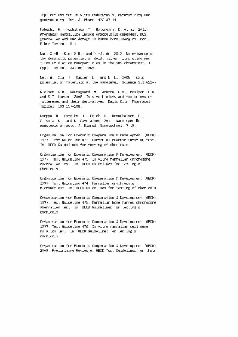
Implications for in vitro endocytosis, cytotoxicity andgenotoxicity. Int. J. Pharm. 423:37–44.
Nabeshi, H., Yoshikawa, T., Matsuyama, K. et al. 2011.Amorphous nanosilica induce endocytosis-dependent ROSgeneration and DNA damage in human keratinocytes. Part.Fibre Toxicol. 8:1.
Nam, S.-H., Kim, S.W., and Y.-J. An. 2013. No evidence ofthe genotoxic potential of gold, silver, zinc oxide andtitanium dioxide nanoparticles in the SOS chromotest. J.Appl. Toxicol. 33:1061–1069.
Nel, A., Xia, T., Madler, L., and N. Li. 2006. Toxicpotential of materials at the nanolevel. Science 311:622–7.
Nielsen, G.D., Roursgaard, M., Jensen, K.A., Poulsen, S.S.,and S.T. Larsen. 2008. In vivo biology and toxicology offullerenes and their derivatives. Basic Clin. Pharmacol.Toxicol. 103:197–208.
Norppa, H., Catalán, J., Falck, G., Hannukainen, K.,Siivola, K., and K. Savolainen. 2011. Nano-speci�cgenotoxic effects. J. Biomed. Nanotechnol. 7:19.
Organisation for Economic Cooperation & Development (OECD).1977. Test Guideline 471: Bacterial reverse mutation test.In: OECD Guidelines for testing of chemicals.
Organisation for Economic Cooperation & Development (OECD).1977. Test Guideline 473. In vitro mammalian chromosomeaberration test. In: OECD Guidelines for testing ofchemicals.
Organisation for Economic Cooperation & Development (OECD).1997. Test Guideline 474. Mammalian erythrocytemicronucleus. In: OECD Guidelines for testing of chemicals.
Organisation for Economic Cooperation & Development (OECD).1997. Test Guideline 475. Mammalian bone marrow chromosomeaberration test. In: OECD Guidelines for testing ofchemicals.
Organisation for Economic Cooperation & Development (OECD).1997. Test Guideline 476. In vitro mammalian cell genemutation test. In: OECD Guidelines for testing ofchemicals.
Organisation for Economic Cooperation & Development (OECD).2009. Preliminary Review of OECD Test Guidelines for their

Applicability to Manufactured Nanomaterials. Series ofSafety of Manufactured Nanomaterials No. 15.
Organisation for Economic Cooperation & Development (OECD).2010. Test Guideline 487. In vitro mammalian cellmicronucleus test. In: OECD Guidelines for testing ofchemicals.
Organisation for Economic Cooperation & Development (OECD).OECD guidelines for the testing of chemicals;
health-effects_20745788.
Paino, I.M., Marangoni, V.S., de Oliveira R. de C.,Antunes, L.M., and V. Zucolotto. 2012. Cyto andgenotoxicity of gold nanoparticles in human hepatocellularcarcinoma and peripheral blood mononuclear cells. Toxicol.Lett. 215:119–25.
Park, M.V., Neigh A.M., Vermeulen, J.P. et al. 2011a. Theeffect of particle size on the cytotoxicity, in�ammation,developmental toxicity and genotoxicity of silvernanoparticles. Biomaterials 32:9810–7.
Park, M.V., Verharen, H.W., Zwart, E. et al. 2011b.Genotoxicity evaluation of amorphous silica nanoparticlesof different sizes using the micronucleus and the plasmidlacZ gene mutation assay. Nanotoxicology 5:168–81.
Petkovic, J., Zegura, B., Stevanovic, M. et al. 2011. DNAdamage and alterations in expression of DNA damageresponsive genes induced by TiO 2 nanoparticles in humanhepatoma HepG2 cells. Nanotoxicology 5:341–53.
Pfaller, T., Colognato, R., Nelissen, I. et al. 2010. Thesuitability of different cellular in vitro immunotoxicityand genotoxicity methods for the analysis ofnanoparticle-induced events. Nanotoxicology 4:52–72.
PubMed database: www.ncbi.nlm.nih.gov/pubmed/
Schins, R.P. 2002. Mechanisms of genotoxicity of particlesand �bers. Inhal. Toxicol. 14, 57–78.
Schins, R.P. and A.M. Knaapen. 2007. Genotoxicity of poorlysoluble particles. Inhal. Toxicol. 19:189–98.
Sergent, J.A., Paget, V., and S. Chevillard. 2012. Toxicityand genotoxicity of nano-SiO2 on human epithelialintestinal HT-29 cell line. Ann. Occup. Hyg. 56:622–30.

Shah, V., Taratula, O., Garbuzenco, O.B. et al. 2012.Genotoxicity of different nanocarriers: Possiblemodi�cations for the delivery of nucleic acids. Curr. DrugDiscov. Technol. 10:8–15.
Singh, N., Jenkins G.J., Nelson, B.C. et al. 2012. The roleof iron redox state in the genotoxicity of ultra�nesuperparamagnetic iron oxide nanoparticles. Biomaterials33:163–70.
Singh, N., Manshian, B., Jenkins, G.J. et al. 2009.Nanogenotoxicology: The DNA damaging potential ofengineered nanomaterials. Biomaterials 30, 3891–914.
Singh, S. and H.S. Nalwa. 2007. Nanotechnology and healthsafety- toxicity and risk assessments of nanostructuredmaterials on human health. J. Nanosci. Nanotechnol.7:3048–70.
Stella, G.M. 2011. Carbon nanotubes and pleural damage:Perspectives of nanosafety in the light of asbestosexperience. Biointerphases 6:1–17.
Stone, V., Johnston, H., and R.P. Schins. 2009. Developmentof in vitro systems for nanotoxicology: Methodologicalconsiderations. Crit. Rev. Toxicol. 39:613–26.
Tang, M., Zhang, T., Xue, Y. et al. 2010. Dose dependent invivo metabolic characteristics of titanium dioxidenanoparticles. J. Nanosci. Nanotechnol. 10:8575–83.
Tice, R.R., Agurell, E., Anderson, D. et al. 2000. Singlecell gel/comet assay: Guidelines for in vitro and in vivogenetic toxicology testing. Environ. Mol. Mutagen.35:206–21.
Toduka, Y., Toyooka, T., and Y. Ibuki. 2012. Flowcytometric evaluation of nanoparticles using side-scatteredlight and reactive oxygen species-mediateduorescence−correlation with genotoxicity. Environ. Sci.Technol. 46:7629–36.
Toyooka, T., Amano, T., and Y. Ibuki. 2012. Titaniumdioxide particles phosphorylate histone H2AX independent ofROS production. Mutat. Res. 742:84–91.
Tsoli, M., Kuhn, H., Brandau, W., Esche, H., and G. Schmid.2005. Cellular uptake and toxicity of Au55 clusters. Small1:841–4.

Vasquez, M.Z. 2012. Recommendations for safety testing withthe in vivo comet assay. Mutat. Res. 747:142–56.
Wang, Z., Li, N., Zhao, J. et al. 2012. CuO nanoparticleinteraction with human epithelial cells: Cellular uptake,location, export, and genotoxicity. Chem. Res. Toxicol.25:1512–21.
Woodruff, R.S., Li, Y., Yan, J. et al. 2012. Genotoxicityevaluation of titanium dioxide nanoparticles using theAmes test and Comet assay. J. Appl. Toxicol. 32:934–43.
Xu, A., Chai, Y., Nohmi, T., and T.K. Hei. 2009. Genotoxicresponses to titanium dioxide nanoparticles and fullerenein gpt delta transgenic MEF cells. Part. Fibre Toxicol.6:3.
Xu, X.H., Brownlow, W.J., Kyriacou, S.V., Wan, Q., andJ.J., Viola. 2004. Real-time probing of membrane transportin living microbial cells using single nanoparticles opticsand living cell imaging. Biochemistry 43:10400–13.
Yang, H., Liu, C., Yang, D., Zhanga, H., and Z. Xi. 2009.Comparative study of cytotoxicity, oxidative stress andgenotoxicity induced by four typical nanomaterials: Therole of particle size, shape and composition. J. Appl.Toxicol. 29:69–78.
Yin, H., Casey, P.S., McCall, M.J., and M. Fenech. 2010.Effects of surface chemistry on cytotoxicity, genotoxicity,and the generation of reactive oxygen species induced byZnO nanoparticles. Langmuir 26:15399–408.
Zhang, Y., Wang, B., Meng, X., Sun, G., and C. Gao. 2011.In�uences of acid-treated multiwalled carbon nanotubes onbroblasts: Proliferation, adhesion, migration, and woundhealing. Ann. Biomed. Eng. 39:414–26.

18 Chapter 18: Interactions ofPolysaccharide-Coated Nanoparticles withProteins
Alexis, F., Pridgen, E., Molnar, L.K., and Farokhzad, O.C.2008. Factors affecting the clearance and biodistributionof polymeric nanoparticles. Mol. Pharm. 5:505–15.
Algar, W.R., Prasuhn, D.E., Stewart, M.H., Jennings, T.L.,Blanco-Canosa, J.B. et al. 2011. The controlled display ofbiomolecules on nanoparticles: A challenge suited tobioorthogonal chemistry. Bioconjug. Chem. 22:825–58.
Anderson, N.L., Polanski, M., Pieper, R., Gatlin, T.,Tirumalai, R.S., Conrads, T.P. et al. 2004. The humanplasma proteome: A nonredundant list developed bycombination of four separate sources. Mol. Cell Proteom.3:311–26.
Andrieux, K. and Couvreur, P. 2009. Polyalkylcyanoacrylatenanoparticles for delivery of drugs across the blood–brainbarrier. Wiley Interdiscip. Rev. Nanomed. Nanobiotechnol.1:463–74.
Ballauff, M. 2007. Spherical polyelectrolyte brushes. Prog.Polym. Sci. 32:1135–51.
Banerjee, I., Pangule, R.C., and Kane, R.S. 2011.Antifouling coatings: Recent developments in the design ofsurfaces that prevent fouling by proteins, bacteria, andmarine organisms. Adv. Mater.23:690–718.
Bangham, A.D., Standish, M.M., and Watkins, J.C. 1965.Diffusion of univalent ions across the lamellae of swollenphospholipids. J. Mol. Biol. 13:238–52.
Bakht, M.K., Sadeghi, M., Pourbaghi-Masouleh, M., andTenreiro, C. 2012. Scope of nanotechnology-based radiationtherapy and thermotherapy methods in cancer treatment.Curr. Cancer Drug Targets 12:998–1015.
Ballauff, M. and Borisov, O.V. 2006. Polyelectrolytebrushes. Curr. Opin. Colloid Interf. Sci. 11:316–23.
Bertholon, I., Vauthier, C., and Labarre, D. 2006a.Complement activation by core-shellpoly(isobutylcyanoacrylate)-polysaccharide nanoparticles:In�uences of surface morphology, length, and type ofpolysaccharide. Pharm. Res. 23:1313–1323.

Bertholon, I., Lesieur, S., Labarre, D., Besnard, M., andVauthier C. 2006b. Characterization ofdextranpoly(isobutylcyanoacrylate) copolymers obtained byredox radical and anionic emulsion polymerization.Macromolecules. 39:3559–67.
Bertholon, I., Hommel, H., Labarre, D., and Vauthier, C.2006c. Properties of polysaccharides grafted onnanoparticles investigated by EPR. Langmuir. 22:5485–90.
Bravo-Osuna, I., Vauthier, C., Farabollini, A., Palmieri,G.F., and Ponchel, G. 2007. Mucoadhesion mechanism ofchitosan and thiolated chitosan-poly(isobutylcyanoacrylate) core-shell nanoparticles. Biomaterials.28:2233–43.
Chauvierre, C., Labarre, D., Couvreur, P., and Vauthier, C.2003. Novel polysaccharide-decorated poly(isobutylcyanoacrylate) nanoparticles. Pharm. Res. 20:1786–93.
Chauvierre, C., Vauthier, C., Labarre, D., and Hommel, H.2004. Evaluation of the surface properties of dextrancoated poly(isobutylcyanocrylate) nanoparticles byspin-labelling coupled with electron resonancespectroscopy. Coll. Polym. Sci. 282:1016–25.
Couvreur, P. and Vauthier, C. 2006. Nanotechnology:Intelligent design to treat complex disease. Pharm. Res.23:1417–50.
Daniel, J.C. 2003. A long history with many futurechallenges to meet in the future—Free-radical emulsionpolymerization and aqueous polymer dispersions. In:Colloidal Polymers, Synthesis and Characterisation,Surfactant Science Series. Ed. A. Elaissari, 115:1–22. NewYork: Marcel Dekker.
de Martimprey, H., Vauthier, C., Malvy, C., and Couvreur,P. 2009. Polymer nanocarriers for the delivery of smallfragments of nucleic acids: Oligonucleotides and siRNA.Eur. J. Pharm. Biopharm. 71(3):490–504.
Dobrovolskaia, M.A., Aggarwal, P., Hall, J.B., and McNeil,S.E. 2008. Preclinical studies to understand nanoparticleinteraction with the immune system and its potentialeffects on nanoparticle biodistribution. Mol. Pharm.5:487–95.
Douglas, S.J., Illum, L., Davis, S.S., and Kreuter, J.1984. Particle size and size distribution of poly(butyl 2

cyanoacrylate) nanoparticles. In�uence of physicochemicalfactors. J. Coll. Interface Sci. 101:149–158.
Etheridge, M.L., Campbell, S.A., Erdman, A.G., Haynes,C.L., Wolf, S.M., and McCullough, J. 2013. The big pictureon nanomedicine: The state of investigational and approvednanomedicine products. Nanomedicine:NBM. 9:1–14.
Estephan, Z.G., Jaber, J.A., and Schlenoff, J.B. 2010,Zwitterion-stabilized silica nanoparticles: Toward nonsticknano. Langmuir. 26:16884–9.
Farokhzad, O.C. and Langer, R. 2009. Impact ofnanotechnology on drug delivery. ACS Nano. 1:16–20.
Gref, R., Lück, M., Quellec, P., Marchand, M., Dellacherie,E., Harnisch, S. et al. 2000. Stealth corona-corenanoparticles surface modi�ed by polyethylene glycol (PEG):In�uences of the corona (PEG chain length and surfacedensity) and of the core composition on phagocytic uptakeand plasma protein adsorption. Colloids Surf BBiointerfaces. 18:301–13.
Gref, R., Minamitake, Y., Peracchia, M.T., Domb, A.,Trubetskoy, V., Torchilin, V. et al. 1997. Poly(ethyleneglycol)-coated nanospheres: Potential carriers forintravenous drug administration. Pharm. Biotechnol.10:167–98.
Gref, R., Minamitake, Y., Peracchia, M.T., Trubetskoy, V.,Torchilin, V., and Langer, R. 1994. Biodegradablelong-circulating polymeric nanospheres. Science 263:1600–3.
Gregoriadis, G. 1976. The carrier potential of liposomes inbiology and medicine. N. Eng. J. Med. 295:704–10.
Gregoriadis, G., Wills, E.J., Swain, C.P., and Tavill, A.S.1974. Drug-carrier potential of liposomes in cancerchemotherapy. Lancet. 1:1313–6.
Hirsh, S.L., McKenzie, D.R., Nosworthy, N.J., Denman, J.A.,Sezerman, O.U., and Bilek, M.M.M. 2013. The Vroman effect:Competitive protein exchange with dynamic multilayerprotein aggregates. Coll. Surf. B: Biointerfaces103:395–404.
Jeon, S.I. and Andrade, J.D. 1991. Protein–surfaceinteractions in the presence of polyethylene oxide: II.Effect of protein size. J. Coll. Interf. Sci. 142:159–166.

Jeon, S.I., Lee, J.H., Andrade, J.D., and De Gennes P.G.1991. Protein–surface interactions in the presence ofpolyethylene oxide: I. Simpli�ed theory. J. Coll. Interf.Sci. 142:149–158.
Jiskoot, W., Van Schie, R.M.F., Carstens, M.G., andSchellekens, H. 2009. Immunological risk of injectabledrug delivery systems. Pharm. Res. 26:1303–14.
Juliano, R.L. 1976. The role of drug delivery systems incancer chemotherapy. Prog. Clin. Biol. Res. 9:21–32.
Kreuter, J. 2007. Nanoparticles—A historical perspective.Int. J. Pharm. 331:1–10.
Kreuter, J. and Speiser, P.P. 1976. New adjuvants on apolymethylmethacrylate base. Infect. Immun. 13, 204–210.
Labarre, D., Vauthier, C., Chauvierre, C., Petri, B.,Muller, R.H., and Chehimi, M.M. 2005. Interactions ofblood proteins with poly (isobutylcyanoacrylate)nanoparticles decorated with a polysaccharidic brush.Biomaterials. 26:5075–84.
Lehner, R., Wang, X., Marsch, S., and Hunziker, P. 2013.Intelligent nanomaterials for medicine: Carrier platformsand targeting strategies in the context of clinicalapplication. Nanomedicine:NBM 9:742–757.
Lemarchand, C., Gref, R., and Couvreur, P. 2004.Polysaccharide-decorated nanoparticles. Eur. J. Pharm.Biopharm. 58:327–41.
Li, S.D. and Huang, L. 2008. Pharmacokinetics andbiodistribution of nanoparticles. Mol. Pharm. 5:496–504.
Lira, M.C.B., Santos-Magalhães, N.S., Nicolas, V., Marsaud,V., Silva, M.P., Ponchel, G. et al. 2011. Cytotoxicity andcellular uptake of newly synthesized fucoidan-coatednanoparticles. Eur. J. Pharm. Biopharm. 79:162–70.
Liu, F., Laurent, S., Fattahi, H., Vander Elst, L., andMuller, R.N. 2011. Superparamagnetic nanosystems based oniron oxide nanoparticles for biomedical imaging.Nanomedicine (Lond). 6:519–28.
Lynch, I., Cedervall, T., Lundqvist, M., Cabaleiro-Lago,C., Linse, S. et al. 2007. The nanoparticle–proteincomplex as a biological entity; a complex �uids and surfacescience challenge for the 21st century. Adv. Colloid

Interface Sci. 134–135:167–74.
Moyano, D.F. and Rotello, V.M. 2011. Nano meets biology:Structure and function at the nanoparticle interface.Langmuir. 27:10376–85.
Mura, S. and Couvreur, P. 2012. Nanotheranostics forpersonalized medicine. Adv. Drug Deliv. Rev. 64:1394–416.
Nicolas, J. and Couvreur, P. 2009. Synthesis of poly(alkylcyanoacrylate)-based colloidal nanomedicines. WileyInterdiscip. Rev. Nanomed. Nanobiotechnol. 1:111–27.
Nicolas, J. and Vauthier, C. 2011.Poly(alkylcyanoacrylate) nanosystems. In IntracellularDelivery Fundamental Biomedical Technologies ed. A.Prokov, pp. 225–251. New York: Springer, Volume 5, Part 2.
Nilsson, B., Ekdahl, K.N., Mollnes, T.E., and Lambris, J.D.2007. The role of complement in biomaterialinducedin�ammation. Mol. Immunol. 44:82–94.
Norde, W. 2008. My voyage of discovery to proteins in�atland…and beyond. Coll. Surf. B. Biointerface. 61:1–9.
Owens, D.E. and Peppas, N.A. 2006. Opsonization,biodistribution and pharmacokinetics of polymericnanoparticles. Int. J. Pharm. 307:93–102.
Papahadjopoulos, D., Allen, T.M., Gabizon, A., Mayhew, E.,Matthay, K., Huang, S.K. et al. 1991. Stericallystabilized liposomes: Improvements in pharmacokinetics andantitumor therapeutic ef�cacy. Proc. Natl. Acad. Sci.U.S.A. 88:11460–4.
Passirani, C., Ferrarini, L,. Barratt, G., Devissaguet,J.P., and Labarre, D. 1999. Preparation andcharacterization of nanoparticles bearing heparin ordextran covalently-linked to poly(methyl methacrylate).J. Biomater. Sci. Polym. Ed. 10:47–62.
Peracchia, M.T., Fattal, E., Desmaële, D., Besnard, M.,Noël, J.P., Gomis, J.M. et al. 1999. Stealth PEGylatedpolycyanoacrylate nanoparticles for intravenousadministration and splenic targeting. J. Control. Release.60:121–8.
Szebeni, J., Muggia, F., Gabizon, A., and Barenholz, Y.2011. Activation of complement by therapeutic liposomes andother lipid excipient-based therapeutic products:

Prediction and prevention. Adv. Drug Deliv. Rev.63:1020–30.
Vauthier, C., Labarre, D., and Ponchel, G. 2007. Designaspects on poly(alkylcyanoacrylate) nanoparticles fortargeted drug delivery. J. Drug Targeting. 15:641–63.
Vauthier, C., Lindner, P., and Cabane, B., 2009.Con�guration of bovine serum albumin adsorbed on polymerparticles with grafted dextran corona. Coll. Surf. B:Biointerfaces. 69:207–15.
Vauthier, C., Persson, B., Lindner, P., and Cabane, B.2011. Protein adsorption and complement activation fordi-block copolymer nanoparticles. Biomaterials. 32:1646–56.
Vonarbourg, A., Passirani, C., Saulnier, P., and Benoit,J.P. 2006. Parameters in�uencing the stealthiness ofcolloidal drug delivery systems. Biomaterials. 27:4356–73.
Walczyk, D., Baldelli Bombelli, F., Monopoli, M.P., Lynch,I., and Dawson, K.A. 2010. What the cell sees inbionanoscience. J. Am. Chem. Soc. 132:5761–68.
Walkey, C.D. and Chan, W.C.W. 2012. Understanding andcontrolling the interaction of nanomaterials with proteinsin a physiological environment. Chem. Soc. Chem. 41,2780–99.
Walkey, C.D., Olsen, J.B., Guo, H., Emili, A., and Chan,W.C. 2012. Nanoparticle size and surface chemistrydetermine serum protein adsorption and macrophage uptake.J. Am. Chem. Soc. 134:2139–47.
Wang, R., Billone, P.S., and Mullet, W.M. 2013.Nanomedicine in action: An overview of cancer nanomedicineon the market and in clinical trials. J. Nanomed.DOI:10.1155/2013/629681, Article ID: 629681, 12 pages.
Welsch, N., Lu, Y., Dzubiella, J., and Ballauff, M. 2013.Adsorption of proteins to dunctional polymericnanoparticles. Polymer. 54:2835–49.
Wittemann, A., Haupt, B., and Ballauff, M. 2003. Adsorptionof proteins on spherical polyelectrolyte brushes inaqueous solution. Phys. Chem. Chem. Phys. 5:1671–7.
Zandanel, C. and Vauthier, C. 2012.Poly(isobutylcyanoacrylate) nanoparticles decorated withchitosan: Effect of conformation of chitosan chains at the

surface on complement activation properties. J. Coll. Sci.Biotechnol. 1:68–81.

19 Chapter 19: Models for RiskAssessments of Nanoparticles
1. De Jong WH, Borm PJ. Drug delivery and nanoparticles:Applications and hazards. International Journal ofNanomedicine. 2008;3(2):133.
2. Kwon J-T, Hwang S-K, Jin H, Kim D-S, Minai-Tehrani A,Yoon H-J et al. Body distribution of inhaled �uorescentmagnetic nanoparticles in the mice. Journal of OccupationalHealth. 2008;50(1):1–6.
3. Lewinski N, Colvin V, Drezek R. Cytotoxicity ofnanoparticles. Small. 2008;4(1):26–49.
4. Oberdörster G, Maynard A, Donaldson K, Castranova V,Fitzpatrick J, Ausman K et al. Principles forcharacterizing the potential human health effects fromexposure to nanomaterials: Elements of a screeningstrategy. Particle and Fibre Toxicology. 2005;2(1):8.
5. Oberdörster G, Oberdörster E, Oberdörster J.Nanotoxicology: An emerging discipline evolving fromstudies of ultra�ne particles. Environmental HealthPerspectives. 2005;113(7):823.
6. Service RF. Nanotechnology—Can High-Speed Tests Sort outWhich Nanomaterials Are Safe?: American Association ofAdvancement Science 1200, New York Avenue, NW, Washington,DC 20005, USA; 2008.
7. Park MV, Lankveld DP, van Loveren H, de Jong WH. Thestatus of in vitro toxicity studies in the risk assessmentof nanomaterials. Nanomedicine. 2009;4(6):669–85.
8. Vasir JK, Labhasetwar V. Quanti�cation of the force ofnanoparticle–cell membrane interactions and its in�uenceon intracellular traf�cking of nanoparticles. Biomaterials.2008;29(31):4244–52.
9. Peetla C, Labhasetwar V. Biophysical characterization ofnanoparticle—Endothelial model cell membrane interactions.Molecular Pharmaceutics. 2008;5(3):418–29.
10. Ginzburg VV, Balijepalli S. Modeling the thermodynamicsof the interaction of nanoparticles with cell membranes.Nano Letters. 2007;7(12):3716–22.
11. Foley S, Crowley C, Smaihi M, Bon�ls C, Erlanger BF,Seta P et al. Cellular localisation of a water- soluble

fullerene derivative. Biochemical and Biophysical ResearchCommunications. 2002;294(1):116–9.
12. Zhu Y, Zhao Q, Li Y, Cai X, Li W. The interaction andtoxicity of multi-walled carbon nanotubes with Stylonychiamytilus. Journal of Nanoscience and Nanotechnology.2006;6(5):1357–64.
13. Xia T, Kovochich M, Brant J, Hotze M, Sempf J, OberleyT et al. Comparison of the abilities of ambient andmanufactured nanoparticles to induce cellular toxicityaccording to an oxidative stress paradigm. Nano Letters.2006;6(8):1794–807.
14. Jia G, Wang H, Yan L, Wang X, Pei R, Yan T et al.Cytotoxicity of carbon nanomaterials: Singlewall nanotube,multi-wall nanotube, and fullerene. Environmental Scienceand Technology. 2005;39(5):1378–83.
15. Al-Rawi M, Diabaté S, Weiss C. Uptake and intracellularlocalization of submicron and nano-sized SiO 2 particles inHeLa cells. Archives of Toxicology. 2011;85(7):813–26.
16. Dobson CM. Protein folding and misfolding. Nature.2003;426(6968):884–90.
17. Marano F, Hussain S, Rodrigues-Lima F, Baeza-Squiban A,Boland S. Nanoparticles: Molecular targets and cellsignalling. Archives of Toxicology. 2011;85(7):733–41.
18. Dawson KA, Salvati A, Lynch I. Nanotoxicology:Nanoparticles reconstruct lipids. Nature Nanotechnology.2009;4:84–5.
19. Takahashi H, Sawada S-I, Akiyoshi K. Amphiphilicpolysaccharide nanoballs: A new building block for nanogelbiomedical engineering and arti�cial chaperones. Acs Nano.2010;5(1):337–45.
20. Wagner SC, Roskamp M, Pallerla M, Araghi RR, SchlechtS, Koksch B. Nanoparticle-induced folding and �brilformation of coiled-coil-based model peptides. Small.2010;6(12):1321–8.
21. Karlsson HL. The comet assay in nanotoxicologyresearch. Analytical and Bioanalytical Chemistry.2010;398(2):651–66.
22. Xie W, Wang L, Zhang Y, Su L, Shen A, Tan J et al.Nuclear targeted nanoprobe for single living cell

detection by surface-enhanced Raman scattering.Bioconjugate Chemistry. 2009;20(4):768–73.
23. Myllynen P. Nanotoxicology: Damaging DNA from adistance. Nature Nanotechnology. 2009;4(12):795–6.
24. Stern ST, McNeil SE. Nanotechnology safety concernsrevisited. Toxicological Sciences. 2008;101(1):4–21.
25. Elsaesser A, Vyvyan Howard C. Toxicology ofnanoparticles. Advanced Drug Delivery Reviews.2012;64(2):12–137.
26. Hagens WI, Oomen AG, de Jong WH, Cassee FR, Sips AJ.What do we (need to) know about the kinetic properties ofnanoparticles in the body? Regulatory Toxicology andPharmacology. 2007;49(3):217–29.
27. Nurkiewicz TR, Porter DW, Barger M, Millecchia L, RaoKMK, Marvar PJ et al. Systemic microvascular dysfunctionand in�ammation after pulmonary particulate matterexposure. Environmental Health Perspectives.2006;114(3):412.
28. Yeates DB, Mauderly JL. Inhaledenvironmental/occupational irritants and allergens:Mechanisms of cardiovascular and systemic responses.Introduction. Environmental Health Perspectives.2001;109(Suppl 4):479.
29. Soto K, Garza K, Murr L. Cytotoxic effects ofaggregated nanomaterials. Acta Biomaterialia.2007;3(3):351–8.
30. Crosera M, Bovenzi M, Maina G, Adami G, Zanette C,Florio C et al. Nanoparticle dermal absorption andtoxicity: A review of the literature. InternationalArchives of Occupational and Environmental Health.2009;82(9):1043–55.
31. Tinkle SS, Antonini JM, Rich BA, Roberts JR, Salmen R,DePree K et al. Skin as a route of exposure andsensitization in chronic beryllium disease. EnvironmentalHealth Perspectives. 2003;111(9):1202.
32. Ryman-Rasmussen JP, Riviere JE, Monteiro-Riviere NA.Penetration of intact skin by quantum dots with diversephysicochemical properties. Toxicological Sciences.2006;91(1):159–65.

33. Trop M, Novak M, Rodl S, Hellbom B, Kroell W, GoesslerW. Silver-coated dressing acticoat caused raised liverenzymes and argyria-like symptoms in burn patient. TheJournal of Trauma and Acute Care Surgery.2006;60(3):648–52.
34. Rouse JG, Yang J, Ryman-Rasmussen JP, Barron AR,Monteiro-Riviere NA. Effects of mechanical �exion on thepenetration of fullerene amino acid-derivatized peptidenanoparticles through skin. Nano Letters.2007;7(1):155–60.
35. Gopee NV, Roberts DW, Webb P, Cozart CR, Siitonen PH,Warbritton AR et al. Migration of intradermally injectedquantum dots to sentinel organs in mice. ToxicologicalSciences. 2007;98(1):249–57.
36. Kim S, Lim YT, Soltesz EG, De Grand AM, Lee J, NakayamaA et al. Near-infrared �uorescent type II quantum dots forsentinel lymph node mapping. Nature Biotechnology.2003;22(1):93–7.
37. Monteiro-Riviere NA, Nemanich RJ, Inman AO, Wang YY,Riviere JE. Multi-walled carbon nanotube interactions withhuman epidermal keratinocytes. Toxicology Letters.2005;155(3):377–84.
38. Zhang LW, Zeng L, Barron AR, Monteiro-Riviere NA.Biological interactions of functionalized single-wallcarbon nanotubes in human epidermal keratinocytes.International Journal of Toxicology. 2007;26(2):103–13.
39. Hillyer JF, Albrecht RM. Gastrointestinal persorptionand tissue distribution of differently sized colloidalgold nanoparticles. Journal of Pharmaceutical Sciences.2001;90(12):1927–36.
40. Nemmar A, Hoet PM, Vanquickenborne B, Dinsdale D,Thomeer M, Hoylaerts M et al. Passage of inhaledparticles into the blood circulation in humans.Circulation. 2002;105(4):411–4.
41. Borm PJ, Robbins D, Haubold S, Kuhlbusch T, Fissan H,Donaldson K et al. The potential risks of nanomaterials: Areview carried out for ECETOC. Particle and FibreToxicology. 2006;3(1):11.
42. Nigavekar SS, Sung LY, Llanes M, El-Jawahri A, LawrenceTS, Becker CW et al. 3H dendrimer nanoparticle organ/tumordistribution. Pharmaceutical Research. 2004;21(3):476–83.

43. Curtis J, Greenberg, M, Kester J, Phillips S, KriegerG. Nanotechnology and nanotoxicology: A primer forclinicians. Toxicology Review. 2006;25(4):245–60.
44. Fabian E, Landsiedel R, Ma-Hock L, Wiench K, WohllebenW, van Ravenzwaay B. Tissue distribution and toxicity ofintravenously administered titanium dioxide nanoparticlesin rats. Archives of Toxicology. 2008;82(3):151–7.
45. Huang X-L, Zhang B, Ren L, Ye S-F, Sun L-P, Zhang Q-Qet al. In vivo toxic studies and biodistribution of nearinfrared sensitive Au–Au2S nanoparticles as potential drugdelivery carriers. Journal of Materials Science: Materialsin Medicine. 2008;19(7):2581–8.
46. Moghimi SM, Hunter AC, Murray JC. Nanomedicine: Currentstatus and future prospects. The FASEB Journal.2005;19(3):311–30.
47. Garnett M, Kallinteri P. Nanomedicines andnanotoxicology: Some physiological principles. OccupationalMedicine. 2006;56(5):307–11.
48. BéruBé K, Balharry D, Sexton K, Koshy L, Jones T.Combustion-derived nanoparticles: Mechanisms of pulmonarytoxicity. Clinical and Experimental Pharmacology andPhysiology. 2007;34(10):1044–50.
49. Oberdörster G, Sharp Z, Atudorei V, Elder A, Gelein R,Lunts A et al. Extrapulmonary translocation of ultra�necarbon particles following whole-body inhalation exposureof rats. Journal of Toxicology and Environmental HealthPart A. 2002;65(20):1531–43.
50. Coleman KP, Toscano WA, Wiese TE. QSAR models of the invitro estrogen activity of bisphenol A analogs. QSAR andCombinatorial Science. 2003;22(1):78–88.
51. Warheit D, Reed K, Webb T. Pulmonary toxicity studiesin rats with triethoxyoctylsilane (OTES)-coated,pigment-grade titanium dioxide particles: Bridging studiesto predict inhalation hazard. Experimental Lung Research.2003;29(8):593–606.
52. Fortner J, Lyon D, Sayes C, Boyd A, Falkner J, Hotze Eet al. C 60 in water: Nanocrystal formation and microbialresponse. Environmental Science and Technology.2005;39(11):4307–16.

53. Kreyling W, Semmler M, Erbe F, Mayer P, Takenaka S,Schulz H et al. Translocation of ultra�ne insoluble iridiumparticles from lung epithelium to extrapulmonary organs issize dependent but very low. Journal of Toxicology andEnvironmental Health Part A. 2002;65(20):1513–30.
54. Bermudez E, Mangum JB, Wong BA, Asgharian B, Hext PM,Warheit DB et al. Pulmonary responses of mice, rats, andhamsters to subchronic inhalation of ultra�ne titaniumdioxide particles. Toxicological Sciences.2004;77(2):347–57.
55. Borm PJ, Schins RP, Albrecht C. Inhaled particles andlung cancer, part B: Paradigms and risk assessment.International Journal of Cancer. 2004;110(1):3–14.
56. Sato QZYKK, Donaldson KNNKK. Differences in the extentof in�ammation caused by intratracheal exposure to threeultra�ne metals: Role of free radicals. Journal ofToxicology and Environmental Health Part A.1998;53(6):423–38.
57. Kuschner WG, D’Alessandro A, Wong H, Blanc PD. Earlypulmonary cytokine responses to zinc oxide fumeinhalation. Environmental Research. 1997;75(1):7–11.
58. Chou C-C, Hsiao H-Y, Hong Q-S, Chen C-H, Peng Y-W, ChenH-W et al. Single-walled carbon nanotubes can inducepulmonary injury in mouse model. Nano Letters.2008;8(2):437–45.
59. Baciu CL, Becker J, Janshoff A, Sönnichsen C.Protein–membrane interaction probed by single plasmonicnanoparticles. Nano Letters. 2008;8(6):1724–8.
60. Elder A, Lynch I, Grieger K, Chan-Remillard S, Gatti A,Gnewuch H, Kenaway E et al. Human health risks ofengineered nanomaterials: Critical knowledge gaps innanomaterials risk assessment. In: Linkov I, Steevens J,eds. Nanomaterials: Risks and Benefits. Dordrecht:Springer; 2009. pp. 3–29.
61. Kuhlbusch T, Fissan H, Asbach C. Nanotechnologies andenvironmental risks: Measurement technologies andstrategies. In: Linkov I, Steevens J, eds. Nanomaterials:Risks and Benefits. Dordrecht: Springer; 2009. pp. 233–43.
62. Seipenbusch M, Binder A, Kasper G. Temporal evolutionof nanoparticle aerosols in workplace exposure. Annals ofOccupational Hygiene. 2008;52(8):707–16.

63. Weber AP, Seipenbusch M, Kasper G. Size effects in thecatalytic activity of unsupported metallic nanoparticles.Journal of Nanoparticle Research. 2003;5(3–4):293–8.
64. Long TC, Tajuba J, Sama P, Saleh N, Swartz C, Parker Jet al. Nanosize titanium dioxide stimulates reactive oxygenspecies in brain microglia and damages neurons in vitro.Environmental Health Perspectives. 2007;115(11):1631.
65. Rossi EM, Pylkkänen L, Koivisto AJ, Vippola M, JensenKA, Miettinen M et al. Airway exposure to silica-coatedTiO 2 nanoparticles induces pulmonary neutrophilia inmice. Toxicological Sciences. 2010;113(2):422–33.
66. Pylkkänen L, Alenius H, Tuomi T, Savolainen K, eds. Theeffect of nanoparticles on the expression of mRNA andproteins of chemokines and cytokines in in�ammatory cellsin the lungs. The Sixth Princess Chulabhorn InternationalScience Congress, Bangkok, Thailand; 2007.
67. Nurkiewicz TR, Porter DW, Hubbs AF, Cumpston JL, ChenBT, Frazer DG et al. Nanoparticle inhalation augmentsparticle-dependent systemic microvascular dysfunction. PartFibre Toxicology. 2008;5(1). doi:10.1186/1743-8977-5-1.
68. Dankovic D, Kuempel E, Wheeler M. An approach to riskassessment for TiO 2 . Inhalation Toxicology.2007;19(S1):205–12.
69. Nel AE, Mädler L, Velegol D, Xia T, Hoek EM,Somasundaran P et al. Understanding biophysicochemicalinteractions at the nano–bio interface. Nature Materials.2009;8(7):543–57.
70. Nadagouda M, Varma RS. Risk reduction via greenersynthesis of nober metal nanostructures. In: Linkov I,Steevens J, eds. Nanomaterials: Risks and Benefits.Dordrecht: Springer; 2009. pp. 209–17.
71. Owen R, Crane M, Grieger K, Handy R, Linkov I, DepkedgeM. Strategic approaches for the management ofenvironmental risk uncertainties posed by nanomaterials.In: Linkov I, Steevens J, eds. Nanomaterials: Risks andBenefits. Dordrecht: Springer; 2009. pp. 369–84.
72. Peters TM, Elzey S, Johnson R, Park H, Grassian VH,Maher T et al. Airborne monitoring to distinguishengineered nanomaterials from incidental particles forenvironmental health and safety. Journal of Occupational

and Environmental Hygiene. 2008;6(2):73–81.
73. Sakamoto Y, Nakae D, Fukumori N, Tayama K, Maekawa A,Imai K et al. Induction of mesothelioma by a singleintrascrotal administration of multi-wall carbon nanotubein intact male Fischer 344 rats. The Journal ofToxicological Sciences. 2009;34(1):65–76.
74. Nasibulin AG, Queipo P, Shandakov SD, Brown DP, JiangH, Pikhitsa PV et al. Studies on mechanism of single-walledcarbon nanotube formation. Journal of Nanoscience andNanotechnology. 2006;6:1–14.
75. Maynard AD, Aitken RJ. Assessing exposure to airbornenanomaterials: Current abilities and future requirements.Nanotoxicology. 2007;1(1):26–41.
76. Vincent JH. Aerosol Sampling: Science, Standards,Instrumentation and Applications. Wiley.com; 2007.
77. Prather KA, Hatch CD, Grassian VH. Analysis ofatmospheric aerosols. Annual Review of AnalyticalChemistry. 2008;1:485–514.
78. Bergamaschi E. Occupational exposure to nanomaterials:Present knowledge and future development. Nanotoxicology.2009;3(3):194–201.
79. Monteiro-Riviere NA, Tran CL. Nanotoxicology:Characterization, Dosing and Health Effects. CRC Press;2007.
80. Yoshida R, Kitamura D, Maenosono S. Mutagenicity ofwater-soluble ZnO nanoparticles in Ames test. The Journalof Toxicological Sciences. 2009;34(1):119–22.
81. Landsiedel R, Kapp MD, Schulz M, Wiench K, Oesch F.Genotoxicity investigations on nanomaterials: Methods,preparation and characterization of test material,potential artifacts and limitations—Many questions, someanswers. Mutation Research/Reviews in Mutation Research.2009;681(2):241–58.
82. Li JG, Li WX, Xu JY, Cai XQ, Liu RL, Li YJ et al.Comparative study of pathological lesions induced bymultiwalled carbon nanotubes in lungs of mice byintratracheal instillation and inhalation. EnvironmentalToxicology. 2007;22(4):415–21.
83. Jacobsen NR, Moller P, Jensen KA, Vogel U, Ladefoged O,

Loft S et al. Lung in�ammation and genotoxicity followingpulmonary exposure to nanoparticles in ApoE −/– mice. PartFibre Toxicology. 2009;6(2):2.
84. Shvedova AA, Kisin E, Murray AR, Johnson VJ, Gorelik O,Arepalli S et al. Inhalation vs. aspiration ofsingle-walled carbon nanotubes in C57BL/6 mice:In�ammation, �brosis, oxidative stress, and mutagenesis.American Journal of Physiology—Lung Cellular and MolecularPhysiology. 2008; 295(4):L552–65.
85. Folkmann JK, Risom L, Jacobsen NR, Wallin H, Loft S,Møller P. Oxidatively damaged DNA in rats exposed by oralgavage to C 60 fullerenes and single-walled carbonnanotubes. Environmental Health Perspectives.2009;117(5):703.
86. Yang H, Liu C, Yang D, Zhang H, Xi Z. Comparative studyof cytotoxicity, oxidative stress and genotoxicity inducedby four typical nanomaterials: The role of particle size,shape and composition. Journal of Applied Toxicology.2009;29(1):69–78.
87. Mouchet F, Landois P, Sarremejean E, Bernard G, PuechP, Pinelli E et al. Characterisation and in vivoecotoxicity evaluation of double-wall carbon nanotubes inlarvae of the amphibian Xenopus laevis. AquaticToxicology. 2008;87(2):127–37.
88. Muller J, Decordier I, Hoet PH, Lombaert N, ThomassenL, Huaux F et al. Clastogenic and aneugenic effects ofmulti-wall carbon nanotubes in epithelial cells.Carcinogenesis. 2008;29(2):427–33.
89. Muller J, Huaux FO, Fonseca A, Nagy JB, Moreau N, DelosM et al. Structural defects play a major role in the acutelung toxicity of multiwall carbon nanotubes: Toxicologicalaspects. Chemical Research in Toxicology.2008;21(9):1698–705.
90. Karlsson HL, Cronholm P, Gustafsson J, Möller L. Copperoxide nanoparticles are highly toxic: A comparison betweenmetal oxide nanoparticles and carbon nanotubes. ChemicalResearch in Toxicology. 2008;21(9):1726–32.
91. Pacurari M, Yin XJ, Ding M, Leonard SS, Schwegler-BerryD, Ducatman BS et al. Oxidative and molecular interactionsof multi-wall carbon nanotubes (MWCNT) in normal andmalignant human mesothelial cells. Nanotoxicology.2008;2(3):155–70.

92. Zhu H, Yang H, Owen MR. Combined microarray analysisuncovers self-renewal related signaling in mouse embryonicstem cells. Systems and Synthetic Biology.2007;1(4):171–81.
93. Lindberg HK, Falck GC-M, Suhonen S, Vippola M, VanhalaE, Catalán J et al. Genotoxicity of nanomaterials: DNAdamage and micronuclei induced by carbon nanotubes andgraphite nano�bres in human bronchial epithelial cells invitro. Toxicology Letters. 2009;186(3):166–73.
94. Falck G, Lindberg H, Suhonen S, Vippola M, Vanhala E,Catalan J et al. Genotoxic effects of nanosized and �neTiO 2 . Human and Experimental Toxicology.2009;28(6–7):339–52.
95. Takagi A, Hirose A, Nishimura T, Fukumori N, Ogata A,Ohashi N et al. Induction of mesothelioma in p53± mouse byintraperitoneal application of multi-wall carbon nanotube.The Journal of Toxicological Sciences. 2008;33(1):105–16.
96. Lam C-W, James JT, McCluskey R, Arepalli S, Hunter RL.A review of carbon nanotube toxicity and assessment ofpotential occupational and environmental health risks. CRCCritical Reviews in Toxicology. 2006;36(3):189–217.
97. Maynard A. Experimental determination of ultra�ne TiO 2deagglomeration in a surrogate pulmonary surfactant:Preliminary results. Annals of Occupational Hygiene.2002;46(Suppl 1):197–202.
98. Donaldson K, Borm P, Oberdorster G, Pinkerton K, StoneV, Tran C. Concordance between in vitro and in vivodosimetry in the proin�ammatory effects of low-toxicity,low-solubility particles: The key role of the proximalalveolar region. Inhalation Toxicology. 2008;20(1):53–62.
99. Nel A, Xia T, Mädler L, Li N. Toxic potential ofmaterials at the nanolevel. Science. 2006;311(5761):622–7.
100. Elder A, Gelein R, Silva V, Feikert T, Opanashuk L,Carter J et al. Translocation of inhaled ultra�nemanganese oxide particles to the central nervous system.Environmental Health Perspectives. 2006;114(8):1172.
101. Sonavane G, Tomoda K, Makino K. Biodistribution ofcolloidal gold nanoparticles after intravenousadministration: Effect of particle size. Colloids andSurfaces B: Biointerfaces. 2008;66(2):274–80.

102. Pal S, Tak YK, Song JM. Does the antibacterialactivity of silver nanoparticles depend on the shape ofthe nanoparticle? A study of the Gram-negative bacteriumEscherichia coli. Applied and Environmental Microbiology.2007;73(6):1712–20.
103. Jr RFH, Wu N, Porter D, Buford M, Wolfarth M, HolianA. Particle length-dependent titanium dioxidenanomaterials toxicity and bioactivity. Particle and FibreToxicology. 2009;6:35.
104. Schaeublin NM, Braydich-Stolle LK, Schrand AM, MillerJM, Hutchison J, Schlager JJ et al. Surface charge of goldnanoparticles mediates mechanism of toxicity. Nanoscale.2011;3(2):410–20.
105. Donaldson K, Stone V, Clouter A, Renwick L, MacNee W.Ultra�ne particles. Occupational and EnvironmentalMedicine. 2001;58(3):211–6.
106. Warheit DB, Webb TR, Colvin VL, Reed KL, Sayes CM.Pulmonary bioassay studies with nanoscale and �ne-quartzparticles in rats: Toxicity is not dependent upon particlesize but on surface characteristics. ToxicologicalSciences. 2007;95(1):270–80.
107. Warheit DB, Hoke RA, Finlay C, Donner EM, Reed KL,Sayes CM. Development of a base set of toxicity tests usingultra�ne TiO 2 particles as a component of nanoparticlerisk management. Toxicology Letters. 2007;171(3):99–110.
108. Berridge MV, Herst PM, Tan AS. Tetrazolium dyes astools in cell biology: New insights into their cellularreduction. Biotechnology Annual Review. 2005;11:127–52.
109. Hussain S, Hess K, Gearhart J, Geiss K, Schlager J. Invitro toxicity of nanoparticles in BRL 3A rat liver cells.Toxicology in Vitro. 2005;19(7):975–83.
110. Plumb JA, Milroy R, Kaye S. Effects of the pHdependence of 3-(4,5-dimethylthiazol-2-yl)-2,5-diphenyltetrazolium bromide–formazan absorption onchemosensitivity determined by a novel tetrazolium-basedassay. Cancer Research. 1989;49(16):4435–40.
111. Wörle-Knirsch J, Pulskamp K, Krug H. Oops they did itagain! Carbon nanotubes hoax scientists in viabilityassays. Nano Letters. 2006;6(6):1261–8.

112. Davis R, Lockwood P, Hobbs D, Messer R, Price R, LewisJ et al. In vitro biological effects of sodium titanatematerials. Journal of Biomedical Materials Research Part B:Applied Biomaterials. 2007;83(2):505–11.
113. Yamin T-T, Ayala JM, Miller DK. Activation of thenative 45-kDa precursor form of interleukin-1- convertingenzyme. Journal of Biological Chemistry.1996;271(22):13273–82.
114. Srinivasula SM, Saleh A, Ahmad M, Fernandes-Alnemri T,Alnemri ES. Isolation and assay of caspases. Methods inCell Biology. 2001;66:1–27.
115. Wróblewski F, Ladue JS, eds. Lactic dehydrogenaseactivity in blood. Proceedings of the Society forExperimental Biology and Medicine Society for ExperimentalBiology and Medicine (New York, NY); 1955: Royal Societyof Medicine.
116. Choi SY, Jeong S, Jang SH, Park J, Park JH, Ock KS et al. In vitro toxicity of serum proteinadsorbedcitrate-reduced gold nanoparticles in human lungadenocarcinoma cells. Toxicology in Vitro.2012;26(2):229–37.
117. Babson AL, Phillips G. A rapid colorimetric assay forserum lactic dehyurogenase. Clinica Chimica Acta.1965;12(2):210–5.
118. Suska F, Gretzer C, Esposito M, Tengvall P, Thomsen P.Monocyte viability on titanium and copper coated titanium.Biomaterials. 2005;26(30):5942–50.
119. Strauss GH. Non-random cell killing incryopreservation: Implications for performance of thebattery of leukocyte tests (BLT), I. Toxic and immunotoxiceffects. Mutation Research/Environmental Mutagenesis andRelated Subjects. 1991;252(1):1–15.
120. Park MV, Annema W, Salvati A, Lesniak A, Elsaesser A,Barnes C et al. In vitro developmental toxicity testdetects inhibition of stem cell differentiation by silicananoparticles. Toxicology and Applied Pharmacology.2009;240(1):108–16.
121. Passagne I, Morille M, Rousset M, Pujalté I, L’Azou B.Implication of oxidative stress in size-dependent toxicityof silica nanoparticles in kidney cells. Toxicology.2012;299(2):112–24.

122. Herzog E, Casey A, Lyng FM, Chambers G, Byrne HJ,Davoren M. A new approach to the toxicity testing ofcarbon-based nanomaterials—The clonogenic assay. ToxicologyLetters. 2007;174(1):49–60.
123. Ye Y, Liu J, Chen M, Sun L, Lan M. In vitro toxicityof silica nanoparticles in myocardial cells. EnvironmentalToxicology and Pharmacology. 2010;29(2):131–7.
124. Lee JK, Kim DB, Kim JI, Kim PY. In vitro cytotoxicitytests on cultured human skin �broblasts to predict skinirritation potential of surfactants. Toxicology in Vitro.2000;14(4):345–9.
125. Nemes Z, Dietz R, Lüth J, Gomba S, Hackenthal E, GrossF. The pharmacological relevance of vital staining withneutral red. Experientia. 1979;35(11):1475–6.
126. Spielmann H, Hoffmann S, Liebsch M, Botham P, FentemJH, Eskes C et al. The ECVAM international validation studyon in vitro tests for acute skin irritation: Report on thevalidity of the EPISKIN and EpiDerm assays and on the skinintegrity function test. ATLA—Alternatives to LaboratoryAnimals. 2007;35(6):559.
127. Ramires P, Romito A, Cosentino F, Milella E. Thein�uence of titania/hydroxyapatite composite coatings onin vitro osteoblasts behaviour. Biomaterials.2001;22(12):1467–74.
128. Monteiro-Riviere NA, Inman AO, Zhang L. Limitationsand relative utility of screening assays to assessengineered nanoparticle toxicity in a human cell line.Toxicology and Applied Pharmacology. 2009;234(2):222–35.
129. Huang M, Khor E, Lim L-Y. Uptake and cytotoxicity ofchitosan molecules and nanoparticles: Effects of molecularweight and degree of deacetylation. PharmaceuticalResearch. 2004;21(2):344–53.
130. Horobin RW, Kiernan JA. Conn’s Biological Stains. AHandbook of Dyes and Fluorochromes for Use in Biology andMedicine, 10th edn. Oxford: BIOS Scienti�c Publishers;2002.
131. Shang L, Zou X, Jiang X, Yang G, Dong S.Investigations on the adsorption behavior of neutral red onmercaptoethane sulfonate protected gold nanoparticles.Journal of Photochemistry and Photobiology A: Chemistry.

2007;187(2):152–9.
132. Casey A, Herzog E, Davoren M, Lyng F, Byrne H,Chambers G. Spectroscopic analysis con�rms theinteractions between single walled carbon nanotubes andvarious dyes commonly used to assess cytotoxicity. Carbon.2007;45(7):1425–32.
133. Darolles C, Sage N, Armengaud J, Malard V. In vitroassessment of cobalt oxide particle toxicity: Identifyingand circumventing interference. Toxicology in Vitro.2013;27(6):1699–1710.
134. Saathoff JG, Inman AO, Xia XR, Riviere JE,Monteiro-Riviere NA. In vitro toxicity assessment of threehydroxylated fullerenes in human skin cells. Toxicology inVitro. 2011;25(8):2105–12.
135. Singh RP, Ramarao P. Cellular uptake, intracellulartraf�cking and cytotoxicity of silver nanoparticles.Toxicology Letters. 2012;213(2):249–59.
136. Pokhrel LR, Silva T, Dubey B, El Badawy AM, TolaymatTM, Scheuerman PR. Rapid screening of aquatic toxicity ofseveral metal-based nanoparticles using the MetPLATE™bioassay. Science of the Total Environment.2012;426:414–22.
137. Braydich-Stolle L, Hussain S, Schlager JJ, HofmannM-C. In vitro cytotoxicity of nanoparticles in mammaliangermline stem cells. Toxicological Sciences.2005;88(2):412–9.
138. Van Engeland M, Nieland LJ, Ramaekers FC, Schutte B,Reutelingsperger CP. Annexin V-af�nity assay: A review onan apoptosis detection system based on phosphatidylserineexposure. Cytometry. 1998;31(1):1–9.
139. Aubry JP, Blaecke A, Lecoanet-Henchoz S, Jeannin P,Herbault N, Caron G et al. Annexin V used for measuringapoptosis in the early events of cellular cytotoxicity.Cytometry. 1999;37(3):197–204.
140. Bartkowiak D, Högner S, Baust H, Nothdurft W,Röttinger EM. Comparative analysis of apoptosis in HL60detected by annexin-V and �uorescein-diacetate. Cytometry.1999;37(3):191–6.
141. Aljandali A, Pollack H, Yeldandi A, Li Y, Weitzman SA,Kamp DW. Asbestos causes apoptosis in alveolar epithelial

cells: Role of iron-induced free radicals. Journal ofLaboratory and Clinical Medicine. 2001;137(5):330–9.
142. Isakovic A, Markovic Z, Todorovic-Markovic B, NikolicN, Vranjes-Djuric S, Mirkovic M et al. Distinct cytotoxicmechanisms of pristine versus hydroxylated fullerene.Toxicological Sciences. 2006;91(1):173–83.
143. Chan W-H, Shiao N-H, Lu P-Z. CdSe quantum dots induceapoptosis in human neuroblastoma cells viamitochondrial-dependent pathways and inhibition of survivalsignals. Toxicology Letters. 2006;167(3):191–200.
144. Darzynkiewicz Z, Bedner E, Traganos F. Dif�culties andpitfalls in analysis of apoptosis. Methods in CellBiology. 2001;63:527–46.
145. Shukla S, Priscilla A, Banerjee M, Bhonde RR, GhatakJ, Satyam P et al. Porous gold nanospheres by controlledtransmetalation reaction: A novel material for applicationin cell imaging. Chemistry of Materials.2005;17(20):5000–5.
146. Waterhouse NJ, Green DR. Mitochondria and apoptosis:HQ or high-security prison? Journal of ClinicalImmunology. 1999;19(6):378–87.
147. Harhaji L, Isakovic A, Vucicevic L, Janjetovic K,Misirkic M, Markovic Z et al. Modulation of tumor necrosisfactor-mediated cell death by fullerenes. PharmaceuticalResearch. 2008;25(6):1365–76.
148. Zeni O, Palumbo R, Bernini R, Zeni L, Sarti M, ScarfìMR. Cytotoxicity investigation on cultured human bloodcells treated with single-wall carbon nanotubes. Sensors.2008;8(1):488–99.
149. Thibodeau M, Giardina C, Hubbard AK. Silica-inducedcaspase activation in mouse alveolar macrophages isdependent upon mitochondrial integrity and asparticproteolysis. Toxicological Sciences. 2003;76(1):91–101.
150. Segal MS, Beem E. Effect of pH, ionic charge, andosmolality on cytochromatic-mediated caspase-3 activity.American Journal of Physiology—Cell Physiology.2001;281(4):C1196–204.
151. Jakubowski W, Bartosz G. 2, 7-dichloro�uoresceinoxidation and reactive oxygen species: What does itmeasure? Cell Biology International. 2000;24(10):757–60.

152. Tarpey MM, Wink DA, Grisham MB. Methods for detectionof reactive metabolites of oxygen and nitrogen: In vitroand in vivo considerations. American Journal ofPhysiology—Regulatory, Integrative and ComparativePhysiology. 2004;286(3):R431–44.
153. Royall JA, Ischiropoulos H. Evaluation of2′,7′-dichloro�uorescein and dihydrorhodamine 123 asuorescent probes for intracellular H 2 O 2 in culturedendothelial cells. Archives of Biochemistry and Biophysics.1993;302(2):348–55.
154. Wrona M, Wardman P. Properties of the radicalintermediate obtained on oxidation of2′,7′-dichlorodihydro�uorescein, a probe for oxidativestress. Free Radical Biology and Medicine.2006;41(4):657–67.
155. Aam BB, Fonnum F. Carbon black particles increasereactive oxygen species formation in rat alveolarmacrophages in vitro. Archives of Toxicology.2007;81(6):441–6.
156. Knaapen AM, Borm PJ, Albrecht C, Schins RP. Inhaledparticles and lung cancer. Part A: Mechanisms.International Journal of Cancer. 2004;109(6):799–809.
157. Fahmy B, Cormier SA. Copper oxide nanoparticles induceoxidative stress and cytotoxicity in airway epithelialcells. Toxicology in Vitro. 2009;23(7):1365–71.
158. Fujii T, Hayashi S, Hogg JC, Vincent R, Van Eeden SF.Particulate matter induces cytokine expression in humanbronchial epithelial cells. American Journal of RespiratoryCell and Molecular Biology. 2001;25(3):265–71.
159. Park E-J, Yoon J, Choi K, Yi J, Park K. Induction ofchronic in�ammation in mice treated with titanium dioxidenanoparticles by intratracheal instillation. Toxicology.2009;260(1):37–46.
160. Bonner JC. The epidermal growth factor receptor at thecrossroads of airway remodeling. American Journal ofPhysiology—Lung Cellular and Molecular Physiology.2002;283(3):L528–30.
161. Sime P, Marr RA, Gauldie D, Xing Z, Hewlett BR, GrahamFL, Gauldie J. Transfer of tumor necrosis factor-alpha torat lung induces severe pulmonary in�ammation and patchy

interstitial �brogenesis with induction of transforminggrowth factor-beta1 and myo�broblasts. American Journal ofPathology. 1998;153(3):825–32.
162. Ahamed M, Posgai R, Gorey TJ, Nielsen M, Hussain SM,Rowe JJ. Silver nanoparticles induced heat shock protein70, oxidative stress and apoptosis in Drosophilamelanogaster. Toxicology and Applied Pharmacology.2010;242(3):263–9.
163. Shukla RK, Sharma V, Pandey AK, Singh S, Sultana S,Dhawan A. ROS-mediated genotoxicity induced by titaniumdioxide nanoparticles in human epidermal cells. Toxicologyin Vitro. 2011;25(1):231–41.
164. Srinivas A, Rao PJ, Selvam G, Murthy PB, Reddy PN.Acute inhalation toxicity of cerium oxide nanoparticles inrats. Toxicology Letters. 2011;205(2):105–15.
165. Chairuangkitti P, Lawanprasert S, Roytrakul S,Aueviriyavit S, Phummiratch D, Kulthong K et al. Silvernanoparticles induce toxicity in A549 cells viaROS-dependent and ROS-independent pathways. Toxicology inVitro. 2012;27:330–38.
166. Lequin RM. Enzyme immunoassay (EIA)/enzyme-linkedimmunosorbent assay (ELISA). Clinical Chemistry.2005;51(12):2415–8.
167. Tao F, Kobzik L. Lung macrophage–epithelial cellinteractions amplify particle-mediated cytokine release.American Journal of Respiratory Cell and Molecular Biology.2002;26(4):499–505.
168. Wottrich R, Diabaté S, Krug HF. Biological effects ofultra�ne model particles in human macrophages andepithelial cells in mono- and co-culture. InternationalJournal of Hygiene and Environmental Health.2004;207(4):353–61.
169. Sayes CM, Reed KL, Warheit DB. Assessing toxicity ofne and nanoparticles: Comparing in vitro measurements to invivo pulmonary toxicity pro�les. Toxicological Sciences.2007;97(1):163–80.
170. Duf�n R, Tran L, Brown D, Stone V, Donaldson K.Proin�ammogenic effects of low-toxicity and metalnanoparticles in vivo and in vitro: Highlighting the roleof particle surface area and surface reactivity.Inhalation Toxicology. 2007;19(10):849–56.

171. Davoren M, Herzog E, Casey A, Cottineau B, Chambers G,Byrne HJ et al. In vitro toxicity evaluation of singlewalled carbon nanotubes on human A549 lung cells.Toxicology in Vitro. 2007;21(3):438–48.
172. Sayes CM, Marchione AA, Reed KL, Warheit DB.Comparative pulmonary toxicity assessments of C 60 watersuspensions in rats: Few differences in fullerene toxicityin vivo in contrast to in vitro pro�les. Nano Letters.2007;7(8):2399–406.
173. Rao KMK, Porter DW, Meighan T, Castranova V. Thesources of in�ammatory mediators in the lung after silicaexposure. Environmental Health Perspectives.2004;112(17):1679.
174. Ryman-Rasmussen JP, Riviere JE, Monteiro-Riviere NA.Surface coatings determine cytotoxicity and irritationpotential of quantum dot nanoparticles in epidermalkeratinocytes. Journal of Investigative Dermatology.2006;127(1):143–53.
175. Monteiro-Riviere NA, Inman AO. Challenges forassessing carbon nanomaterial toxicity to the skin.Carbon. 2006;44(6):1070–8.
176. Veranth JM, Kaser EG, Veranth MM, Koch M, Yost GS.Cytokine responses of human lung cells (BEAS-2B) treatedwith micron-sized and nanoparticles of metal oxidescompared to soil dusts. Part Fibre Toxicology. 2007;4(2).doi:10.1186/1743-8977-4-2.
177. Guo L, Von Dem Bussche A, Buechner M, Yan A, Kane AB,Hurt RH. Adsorption of essential micronutrients by carbonnanotubes and the implications for nanotoxicity testing.Small. 2008;4(6):721–7.
178. Singh N, Manshian B, Jenkins GJ, Grif�ths SM, WilliamsPM, Maffeis TG et al. Nanogenotoxicology: The DNA damagingpotential of engineered nanomaterials. Biomaterials.2009;30(23–24):3891–914.
179. AshaRani P, Low Kah Mun G, Hande MP, Valiyaveettil S.Cytotoxicity and genotoxicity of silver nanoparticles inhuman cells. ACS Nano. 2008;3(2):279–90.
180. Trouiller B, Reliene R, Westbrook A, Solaimani P,Schiestl RH. Titanium dioxide nanoparticles induce DNAdamage and genetic instability in vivo in mice. Cancer
Research. 2009;69(22):8784–9.
181. Li JJ, Zou L, Hartono D, Ong CN, Bay BH, Lanry YungLY. Gold nanoparticles induce oxidative damage in lung�broblasts in vitro. Advanced Materials. 2008;20(1):138–42.
182. Pantarotto D, Briand J-P, Prato M, Bianco A.Translocation of bioactive peptides across cell membranesby carbon nanotubes. Chemical Communications.2004;10(1):16–7.
183. Muller J, Delos M, Panin N, Rabolli V, Huaux F, LisonD. Absence of carcinogenic response to multiwall carbonnanotubes in a 2-year bioassay in the peritoneal cavity ofthe rat. Toxicological Sciences. 2009;110(2):442–8.
184. Roller M. Carcinogenicity of inhaled nanoparticles.Inhalation Toxicology. 2009;21(S1):144–57.
185. Onuma K, Sato Y, Ogawara S, Shirasawa N, Kobayashi M,Yoshitake J et al. Nano-scaled particles of titaniumdioxide convert benign mouse �brosarcoma cells intoaggressive tumor cells. The American Journal of Pathology.2009;175(5):2171–83.
186. Hackenberg S, Scherzed A, Kessler M, Hummel S, TechnauA, Froelich K et al. Silver nanoparticles: Evaluation ofDNA damage, toxicity and functional impairment in humanmesenchymal stem cells. Toxicology Letters.2011;201(1):27–33.
187. Arora S, Rajwade JM, Paknikar KM. Nanotoxicology andin vitro studies: The need of the hour. Toxicology andApplied Pharmacology. 2012;258(2):151–65.
188. Kim S, Choi JE, Choi J, Chung K-H, Park K, Yi J et al.Oxidative stress-dependent toxicity of silvernanoparticles in human hepatoma cells. Toxicology in Vitro.2009;23(6):1076–84.
189. Lemaire F, Mandon CA, Reboud J, Papine A, Angulo J,Pointu H et al. Toxicity assays in nanodrops combiningbioassay and morphometric endpoints. PloS One.2007;2(1):e163.
190. Jan E, Byrne SJ, Cuddihy M, Davies AM, Volkov Y,Gun’ko YK et al. High-content screening as a universal toolfor �ngerprinting of cytotoxicity of nanoparticles. ACSNano. 2008;2(5):928–38.

191. Wittmaack K. In search of the most relevant parameterfor quantifying lung in�ammatory response to nanoparticleexposure: Particle number, surface area, or what?Environmental Health Perspectives. 2007;115(2):187.
192. Tang M, Zhang T, Xue Y, Wang S, Huang M, Yang Y et al. Dose dependent in vivo metabolic characteristics oftitanium dioxide nanoparticles. Journal of Nanoscience andNanotechnology. 2010;10(12):8575–83.
193. Leszczynski J. Bionanoscience: Nano meets bio at theinterface. Nature Nanotechnology. 2010;5(9):633–4.
194. Barnard AS. How can ab initio simulations addressrisks in nanotech? Nature Nanotechnology. 2009;4(6):332–5.
195. Lacerda L, Bianco A, Prato M, Kostarelos K. Carbonnanotubes as nanomedicines: From toxicology topharmacology. Advanced Drug Delivery Reviews.2006;58(14):1460–70.
196. Puzyn T, Gajewicz A, Leszczynska D, Leszczynski J.Nanomaterials—The next great challenge for QSAR modelers.In: Puzyn T, Leszczynski J, Cronin M, eds. Recent Advancesin QSAR Studies: Methods and Applications. London, NewYork: M.T. Springer; 2010.
197. Puzyn T, Rasulev B, Gajewicz A, Hu X, Dasari TP,Michalkova A et al. Using nano-QSAR to predict thecytotoxicity of metal oxide nanoparticles. NatureNanotechnology. 2011;6(3):175–8.
198. Bonnefoi MS, Belanger SE, Devlin DJ, Doerrer NG, EmbryMR, Fukushima S et al. Human and environmental healthchallenges for the next decade (2010–2020). CriticalReviews in Toxicology. 2010;40(10):893–911.
199. Lyon DY, Adams LK, Falkner JC, Alvarez PJ.Antibacterial activity of fullerene water suspensions:Effects of preparation method and particle size.Environmental Science and Technology. 2006;40(14):4360–6.
200. Zhu S, Oberdörster E, Haasch ML. Toxicity of anengineered nanoparticle (fullerene, C 60 ) in two aquaticspecies, Daphnia and fathead minnow. Marine EnvironmentalResearch. 2006;62:S5–9.
201. Roberts AP, Mount AS, Seda B, Souther J, Qiao R, Lin Set al. In vivo biomodi�cation of lipid-coated carbonnanotubes by Daphnia magna. Environmental Science and

Technology. 2007;41(8):3025–9.
202. Smith CJ, Shaw BJ, Handy RD. Toxicity of single walledcarbon nanotubes to rainbow trout, (Oncorhynchus mykiss):Respiratory toxicity, organ pathologies, and otherphysiological effects. Aquatic Toxicology.2007;82(2):94–109.
203. Templeton RC, Ferguson PL, Washburn KM, Scrivens WA,Chandler GT. Life-cycle effects of singlewalled carbonnanotubes (SWNTs) on an estuarine meiobenthic copepod.Environmental Science and Technology. 2006;40(23):7387–93.
204. Adams LK, Lyon DY, Alvarez PJ. Comparativeeco-toxicity of nanoscale TiO 2 , SiO 2 , and ZnO watersuspensions. Water Research. 2006;40(19):3527–32.
205. Zhu X, Zhu L, Duan Z, Qi R, Li Y, Lang Y. Comparativetoxicity of several metal oxide nanoparticle aqueoussuspensions to zebra�sh (Danio rerio) early developmentalstage. Journal of Environmental Science and Health Part A.2008;43(3):278–84.
206. Zhu X, Zhu L, Chen Y, Tian S. Acute toxicities of sixmanufactured nanomaterial suspensions to Daphnia magna.Journal of Nanoparticle Research. 2009;11(1):67–75.
207. Scown TM, Goodhead RM, Johnston BD, Moger J, BaaloushaM, Lead JR et al. Assessment of cultured �sh hepatocytesfor studying cellular uptake and (eco) toxicity ofnanoparticles. Environmental Chemistry. 2010;7(1):36–49.
208. Xia T, Zhao Y, Sager T, George S, Pokhrel S, Li Net al. Decreased dissolution of ZnO by iron doping yieldsnanoparticles with reduced toxicity in the rodent lung andzebra�sh embryos. ACS Nano. 2011;5(2):1223–35.
209. Metz KM, Mangham AN, Bierman MJ, Jin S, Hamers RJ,Pedersen JA. Engineered nanomaterial transformation underoxidative environmental conditions: Development of an invitro biomimetic assay. Environmental Science andTechnology. 2009;43(5):1598–604.
210. Makarucha A, Todorova N, Yarovsky I. Nanomaterials inbiological environment: A review of computer modellingstudies. European Biophysics Journal. 2011;40(2):103–15.
211. Hayashi Y. Designing in vitro assay systems for hazardcharacterization. Basic strategies and related technicalissues. Experimental and Toxicologic Pathology.

2005;57:227–32.
212. Sutter TR. Molecular and cellular approaches toextrapolation for risk assessment. Environmental HealthPerspectives. 1995;103(4):386.
213. Anderson D, Russell T, Savage Jr RE. The status ofalternative methods in toxicology. Toxicology Methods.1997;7(2):149–51.
214. Cimino MC. Comparative overview of currentinternational strategies and guidelines for genetictoxicology testing for regulatory purposes. Environmentaland Molecular Mutagenesis. 2006;47(5):362–90.
215. Snodin DJ. An EU perspective on the use of in vitromethods in regulatory pharmaceutical toxicology.Toxicology Letters. 2002;127(1):161–8.
216. Clemedson C, Ekwall B. Overview of the �nal MEICresults: I. The in vitro–in vitro evaluation. Toxicologyin Vitro. 1999;13(4):657–63.
217. Liebsch M, Spielmann H. Currently available in vitromethods used in the regulatory toxicology. ToxicologyLetters. 2002;127(1):127–34.
218. Lilienblum W, Dekant W, Foth H, Gebel T, Hengstler J,Kahl R et al. Alternative methods to safety studies inexperimental animals: Role in the risk assessment ofchemicals under the new European Chemicals Legislation(REACH). Archives of Toxicology. 2008;82(4):211–36.
219. Heijne WH, Kienhuis AS, van Ommen B, Stierum RH,Groten JP. Systems toxicology: Applications oftoxicogenomics, transcriptomics, proteomics andmetabolomics in toxicology. 2005;2(5):767–80.
220. Marquet P, Rappaz B, Magistretti PJ, Cuche E, Emery Y,Colomb T et al. Digital holographic microscopy: Anoninvasive contrast imaging technique allowingquantitative visualization of living cells withsubwavelength axial accuracy. Optics Letters.2005;30(5):468–70.
221. Kemper B, Carl D, Höink A, Von Bally G, Bredebusch I,Schenekenburger J. Modular digital holographic microscopysystem for marker free quantitative phase contrast imagingof living cells. Proc. SPIE 6191, Biophotonics and NewTherapy Frontiers; April 2006;6191:61910T-1–61910T-8.

222. Schnekenburger J, Kemper B. Digital holographyimaging—A 3D view on drug induced apoptosis. ScreeningTrends of Drug Discovery. 2008;9:24–6.
223. Ratanachoo K, Gascoyne PR, Ruchirawat M. Detection ofcellular responses to toxicants by dielectrophoresis.Biochimica et Biophysica Acta (BBA)—Biomembranes.2002;1564(2):449–58.
224. Ceriotti L, Ponti J, Colpo P, Sabbioni E, Rossi F.Assessment of cytotoxicity by impedance spectroscopy.Biosensors and Bioelectronics. 2007;22(12):3057–63.
225. Ressler J, Grothe H, Motrescu E, Wolf B, eds. Newconcepts for chip-supported multi-well-plates: Realizationof a 24-well-plate with integrated impedance-sensors forfunctional cellular screening applications and automatedmicroscope aided cell-based assays. Engineering in Medicineand Biology Society, 2004 IEMBS’04 26th AnnualInternational Conference of the IEEE, San Francisco, CA;2004: IEEE.
226. Ponti J, Ceriotti L, Munaro B, Farina M, Munari A,Whelan M et al. Comparison of impedance-based sensors forcell adhesion monitoring and in vitro methods for detectingcytotoxicity induced by chemicals. ATLA Alternatives toLaboratory Animals. 2006;34(5):515–25.
227. Linkov I, Satterstrom FK. Nanomaterial risk assessmentand risk management: Review of regulatory frameworks. In:Linkov I, Ferguson E, Magar VS, eds. Real Time andDeliberative Decision Making: Application for RiskAssessment for Non-chemical Stressors. Amsterdam: Springer;2008. pp. 129–58.
228. Kandlikar MG, Ramachandran AD, Maynard A, Murdock B,Toscano W. Health risk assessment for nanoparticles: Acase for using expert judgment. Journal of NanoparticleResearch. 2007;9:137–56.
229. Tsuji JS, Maynard AD, Howard PC, James JT, Lam CW,Warheit DB et al. Research strategies for safetyevaluation of nanomaterials, part IV: Risk assessment ofnanoparticles. Toxicology Science 2006;89:42–50.
230. Li JJ, Muralikrishnan S, Ng C, Yung LL, Bay B.Nanoparticle-induced pulmonary toxicity. ExperimentalBiology and Medicine. 2010;235:1025–33.

20 Chapter 20: Immunotoxicity of CarbonNanoparticles
1. Jain S, Singh SR, Pillai S. Toxicity issues related tobiomedical applications of carbon nanotubes. Journal ofNanomedicine & Nanotechnology. 2013;3(5):140.
2. Dolatabadi JEN, Jamali AA, Hasanzadeh M, Omidi Y.Quercetin delivery into cancer cells with single walledcarbon nanotubes. International Journal of Bioscience,Biochemistry and Bioinformatics. 2011;1(1):21–5.
3. Canesi L, Ciacci C, Betti M, Fabbri R, Canonico B,Fantinati A et al. Immunotoxicity of carbon blacknanoparticles to blue mussel hemocytes. EnvironmentInternational. 2008;34(8):1114–9.
4. Hussain S, Vanoirbeek JA, Hoet PH. Interactions ofnanomaterials with the immune system. WileyInterdisciplinary Reviews: Nanomedicine andNanobiotechnology. 2012;4(2):169–83.
5. Ezzati Nazhad Dolatabadi J, Omidi Y, Losic D. Carbonnanotubes as an advanced drug and gene deliverynanosystem. Current Nanoscience. 2011;7(3):297–314.
6. Kim SN, Rusling JF, Papadimitrakopoulos F. Carbonnanotubes for electronic and electrochemical detection ofbiomolecules. Advanced Materials. 2007;19(20):3214–28.
7. Beg S, Rizwan M, Sheikh AM, Hasnain MS, Anwer K, KohliK. Advancement in carbon nanotubes: Basics, biomedicalapplications and toxicity. Journal of Pharmacy andPharmacology. 2011;63(2):141–63.
8. Murr L, Garza K, Soto K, Carrasco A, Powell T, Ramirez Det al. Cytotoxicity assessment of some carbon nanotubes andrelated carbon nanoparticle aggregates and the implicationsfor anthropogenic carbon nanotube aggregates in theenvironment. International Journal of EnvironmentalResearch and Public Health. 2005;2(1):31–42.
9. Donaldson K, Stone V, Tran C, Kreyling W, Borm PJ.Nanotoxicology. Occupational and Environmental Medicine.2004;61(9):727–8.
10. Poland CA, Duf�n R, Kinloch I, Maynard A, Wallace WA,Seaton A et al. Carbon nanotubes introduced into theabdominal cavity of mice show asbestos-like pathogenicityin a pilot study. Nature Nanotechnology. 2008;3(7):423–8.

11. Tian F, Cui D, Schwarz H, Estrada GG, Kobayashi H.Cytotoxicity of single-wall carbon nanotubes on human�broblasts. Toxicology In Vitro. 2006;20(7):1202–12.
12. Tran PA, Zhang L, Webster TJ. Carbon nano�bers andcarbon nanotubes in regenerative medicine. Advanced DrugDelivery Reviews. 2009;61(12):1097–114.
13. Jiang J, Oberdörster G, Biswas P. Characterization ofsize, surface charge, and agglomeration state ofnanoparticle dispersions for toxicological studies. Journalof Nanoparticle Research. 2009;11(1):77–89.
14. Aillon KL, Xie Y, El-Gendy N, Berkland CJ, Forrest ML.Effects of nanomaterial physicochemical properties on invivo toxicity. Advanced Drug Delivery Reviews.2009;61(6):457–66.
15. Wang J, Rahman MF, Duhart HM, Newport GD, Patterson TA,Murdock RC et al. Expression changes of dopaminergicsystem-related genes in PC12 cells induced by manganese,silver, or copper nanoparticles. Neurotoxicology.2009;30(6):926–33.
16. Ye S, Jiang Y, Zhang H, Wang Y, Wu Y, Hou Z et al.Multi-walled carbon nanotubes induce apoptosis in RAW 264.7cell-derived osteoclasts through mitochondria-mediateddeath pathway. Journal of Nanoscience and Nanotechnology.2012;12(3):2101–12.
17. Sachar S, Saxena RK. Cytotoxic effect of poly-dispersedsingle walled carbon nanotubes on erythrocytes in vitroand in vivo. PloS One. 2011;6(7):e22032.
18. Knaapen AM, Borm PJ, Albrecht C, Schins RP. Inhaledparticles and lung cancer. Part A: Mechanisms.International Journal of Cancer. 2004;109(6):799–809.
19. Dumortier H, Lacotte S, Pastorin G, Marega R, Wu W,Bonifazi D et al. Functionalized carbon nanotubes arenon-cytotoxic and preserve the functionality of primaryimmune cells. Nano Letters. 2006;6(7):1522–8.
20. Cui D, Tian F, Ozkan CS, Wang M, Gao H. Effect ofsingle wall carbon nanotubes on human HEK293 cells.Toxicology Letters. 2005;155(1):73–85.
21. Pantarotto D, Partidos CD, Graff R, Hoebeke J, BriandJ-P, Prato M et al. Synthesis, structural characterization,

and immunological properties of carbon nanotubesfunctionalized with peptides. Journal of the AmericanChemical Society. 2003;125(20):6160–4.
22. Pantarotto D, Partidos CD, Hoebeke J, Brown F, KramerE, Briand J-P et al. Immunization withpeptidefunctionalized carbon nanotubes enhancesvirus-speci�c neutralizing antibody responses. Chemistry &Biology. 2003;10(10):961–6.
23. Kagan V, Tyurina Y, Tyurin V, Konduru N, Potapovich A,Osipov A et al. Direct and indirect effects of singlewalled carbon nanotubes on RAW 264.7 macrophages: Role ofiron. Toxicology Letters. 2006;165(1):88–100.
24. Zhou H, Mu Q, Gao N, Liu A, Xing Y, Gao S et al. Anano-combinatorial library strategy for the discovery ofnanotubes with reduced protein-binding, cytotoxicity, andimmune response. Nano Letters. 2008;8(3):859–65.
25. Gustafsson Å, Lindstedt E, Elfsmark LS, Bucht A. Lungexposure of titanium dioxide nanoparticles induces innateimmune activation and long-lasting lymphocyte response inthe Dark Agouti rat. Journal of Immunotoxicology.2011;8(2):111–21.
26. Salvador-Morales C, Flahaut E, Sim E, Sloan J H, GreenML, Sim RB. Complement activation and protein adsorption bycarbon nanotubes. Molecular Immunology. 2006;43(3):193–201.
27. Lam C-w, James JT, McCluskey R, Arepalli S, Hunter RL.A review of carbon nanotube toxicity and assessment ofpotential occupational and environmental health risks. CRCCritical Reviews in Toxicology. 2006;36(3):189–217.
28. Casey A, Herzog E, Lyng F, Byrne H, Chambers G, DavorenM. Single walled carbon nanotubes induce indirectcytotoxicity by medium depletion in A549 lung cells.Toxicology Letters. 2008;179(2):78–84.
29. Byrne JD, Baugh JA. The signi�cance of nanoparticles inparticle-induced pulmonary �brosis. McGill Journal ofMedicine: MJM. 2008;11(1):43.
30. Hussain S, Vanoirbeek JA, Luyts K, De Vooght V,Verbeken E, Thomassen LC et al. Lung exposure tonanoparticles modulates an asthmatic response in a mousemodel. European Respiratory Journal. 2011;37(2):299–309.
31. Hu X, Cook S, Wang P, Hwang H-m, Liu X, Williams QL. In

vitro evaluation of cytotoxicity of engineered carbonnanotubes in selected human cell lines. Science of theTotal Environment. 2010;408(8):1812–7.
32. Bottini M, Bruckner S, Nika K, Bottini N, Bellucci S,Magrini A et al. Multi-walled carbon nanotubes induce Tlymphocyte apoptosis. Toxicology Letters.2006;160(2):121–6.
33. Fiorito S, Sera�no A, Andreola F, Bernier P. Effects offullerenes and single-wall carbon nanotubes on murine andhuman macrophages. Carbon. 2006;44(6):1100–5.
34. Pulskamp K, Diabaté S, Krug HF. Carbon nanotubes showno sign of acute toxicity but induce intracellular reactiveoxygen species in dependence on contaminants. ToxicologyLetters. 2007;168(1):58–74.
35. Ema M, Matsuda A, Kobayashi N, Naya M, Nakanishi J.Evaluation of dermal and eye irritation and skinsensitization due to carbon nanotubes. RegulatoryToxicology and Pharmacology. 2011;61(3):276–81.
36. Murray A, Kisin E, Leonard S, Young S, Kommineni C,Kagan V et al. Oxidative stress and in�ammatory responsein dermal toxicity of single-walled carbon nanotubes.Toxicology. 2009;257(3):161–71.
37. Prow TW, Grice JE, Lin LL, Faye R, Butler M, Becker Wet al. Nanoparticles and microparticles for skin drugdelivery. Advanced Drug Delivery Reviews.2011;63(6):470–91.
38. Witzmann FA, Monteiro-Riviere NA. Multi-walled carbonnanotube exposure alters protein expression in humankeratinocytes. Nanomedicine: Nanotechnology, Biology andMedicine. 2006;2(3):158–68.
39. Takagi A, Hirose A, Nishimura T, Fukumori N, Ogata A,Ohashi N et al. Induction of mesothelioma in p53+/− mouseby intraperitoneal application of multi-wall carbonnanotube. The Journal of Toxicological Sciences.2008;33(1):105–16.
40. Jia’en Li J, Muralikrishnan S, Ng C-T, Yung L-YL, BayB-H. Nanoparticle induced pulmonary toxicity. ExperimentalBiology and Medicine 2010;235:1025–33. Nature NanometersPharmaceutical nanot echnology 10 –1 10 1 10 4 10 6 10 8 1010 Water molecule DNA Virus Erythrocyte Apple DendrimersNanotubes Quantum dots Niosome Polymer Nanoparticles

Micelles Liposome Microparticles Tablet capsule
FIGURE 1.1 Dimensions scale of nanotechnology.Physicochemical determinants Regulatory issue Target ofnanocarriers system Molecular determinants Routes ofexposure G l t r a c t N e c r o s i s A p o p t o s i s DN A c l e a v a g e M u t a t i o n R e s p i r a t o r y tr a c t S k i n I n j e c t i o n Genotoxicity Regulatorybodies Government Academy Industry NanotoxocityBiodistribution Inflammation Oxidative stress OpsonizationBiopersistence Clearance M a c r o p h a g e s T u m o r sE n d o t h e l i u m C e l l n u c l e u s B i o l o g i ca l a c t i v i t y S t a b i l i t y S u r f a c e a r e aD o s e c o n c e n t r a t i o n C r y s t a l l i n e s tr u c t u r e
FIGURE 3.4 Multidimensional issues affecting nanotoxicity.Double emulsionbased method Using supercritical fluidsSolvent emulsification/ evaporation Layer-by-layersynthesis or polymerization Microemulsionbased method Drugrelease studies Drug entrapment Drug loading In vitroanalysis Co-existence of additional structures and dynamicphenomena Degree of crystallinity and lipid/polymermodification Particle size and zeta potential Analyticalcharacterization High-pressure homogenization a. Hothomogenization b. Cold homogenization Ultrasonication/high-speed homogenization Emulsification– salting outEmulsification– solvent diffusion Emulsification– solventevaporation Nanoprecipitation PNs preparation methods SLNspreparation methods Characterization of PNs and SLNsPreparation and characterization of PNs and SLNs
FIGURE 7.1 Preparation and characterization techniques ofPNs and SLNs. Size shape Charge Surface area CompositionSolubility Aggregation Genotoxicity Tumors Liver KidneysBrain Physiochemical parameters Receptors OpsonizationProtein corona Clearance Respiratory Skin Digestive tractIntravenous Molecular interactions Distribution ToxicityMutagenesis Chromosomal Abberations ROS and NOSInflammatory response Neuro-inflammation Routes of exposure
FIGURE 9.2 Concepts in nanotoxicology. The toxicity ofnanomaterials is most commonly determined by
the route of exposure and physiochemical parameters of thenanomaterials. Further, the type of molecular
interactions, based on the properties of the nanoparticlesurface, de�ne the distribution in the body, and the
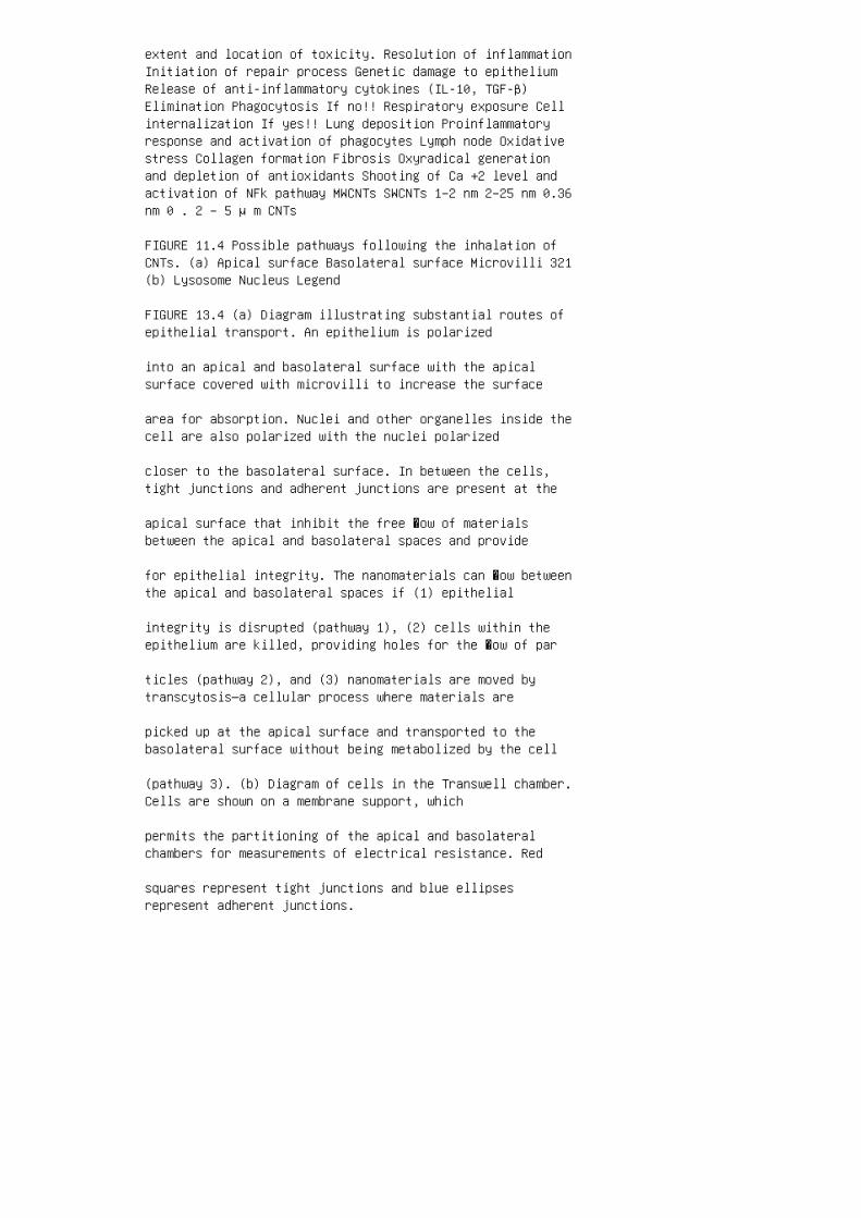
extent and location of toxicity. Resolution of inflammationInitiation of repair process Genetic damage to epitheliumRelease of anti-inflammatory cytokines (IL-10, TGF-β)Elimination Phagocytosis If no!! Respiratory exposure Cellinternalization If yes!! Lung deposition Proinflammatoryresponse and activation of phagocytes Lymph node Oxidativestress Collagen formation Fibrosis Oxyradical generationand depletion of antioxidants Shooting of Ca +2 level andactivation of NFk pathway MWCNTs SWCNTs 1–2 nm 2–25 nm 0.36nm 0 . 2 – 5 μ m CNTs
FIGURE 11.4 Possible pathways following the inhalation ofCNTs. (a) Apical surface Basolateral surface Microvilli 321(b) Lysosome Nucleus Legend
FIGURE 13.4 (a) Diagram illustrating substantial routes ofepithelial transport. An epithelium is polarized
into an apical and basolateral surface with the apicalsurface covered with microvilli to increase the surface
area for absorption. Nuclei and other organelles inside thecell are also polarized with the nuclei polarized
closer to the basolateral surface. In between the cells,tight junctions and adherent junctions are present at the
apical surface that inhibit the free �ow of materialsbetween the apical and basolateral spaces and provide
for epithelial integrity. The nanomaterials can �ow betweenthe apical and basolateral spaces if (1) epithelial
integrity is disrupted (pathway 1), (2) cells within theepithelium are killed, providing holes for the �ow of par
ticles (pathway 2), and (3) nanomaterials are moved bytranscytosis—a cellular process where materials are
picked up at the apical surface and transported to thebasolateral surface without being metabolized by the cell
(pathway 3). (b) Diagram of cells in the Transwell chamber.Cells are shown on a membrane support, which
permits the partitioning of the apical and basolateralchambers for measurements of electrical resistance. Red
squares represent tight junctions and blue ellipsesrepresent adherent junctions.

FIGURE 13.7 Representative confocal microscopy image ofDrosophila midgut in �ies treated with 15 nm
Au NPs (100 pmol/L). Nuclei are stained with Hoechst 33 342(blue) while cells containing DNA strand nicks
are detected by TUNEL assay and �uorescent red (highlightedby the white arrows). R3R1 R2 A1, A2 C3 C3 C3 C3
FIGURE 18.5 A scheme illustrating the interactions ofproteins with a series of nanoparticles having corona
of dextran with different characteristics. The dark spotincluded in C3 indicates that the component C3 of the
complement system is activated. Albumin adsorbed on thesurface of the nanoparticle core appears as a dark
triangle.
Related Documents