The Reproductive Biology Group at the University of Kansas Medical Center hosts the Annual Gilbert S. Greenwald Symposium on Reproduction in honor and as a memorial to the life and research career of Gilbert S. Greenwald, PhD. Professor Greenwald had an illustrious career as a Distinguished Professor at the Medical Center and as an internationally recognized reproductive biologist. Professor Greenwald received his doctorate from the University of California at Berkeley, followed by postdoctoral studies at the Carnegie Institute of Embryology in Baltimore. He then moved to his first faculty appointment in the Department of Anatomy at the University of Washington. He joined the Departments of Obstetrics & Gynecology and Anatomy at the University of Kansas Medical Center in 1961 where he held an endowed chair in Research in Human Reproduction. He also served as chair of the Department of Physiology at the Medical Center for 16 years (1977-1993). Professor Greenwald received numerous awards for his outstanding research accomplishments from several scientific societies. Among these is the Distinguished Service Award from the Society for the Study of Reproduction for his work as one of the founding members and early president of the Society, as well as Editor-in-Chief of its journal, Biology of Reproduction. Professor Greenwald also received the Carl Hartman Award for a career of outstanding scientific contributions to the field of reproductive biology. The National Institutes of Health supported his research over his entire career. Professor Greenwald trained more than 50 graduate students and postdoctoral fellows and was instrumental in the career development of numerous faculty, including several currently holding leadership positions at the University of Kansas Medical Center and at other academic institutions throughout the world. He was a true scholar, a superb mentor, and a generous friend. Professor Greenwald passed away on August 26, 2004. TABLE OF CONTENTS Gilbert S. Greenwald Biography 1 Sponsors & Volunteers 2 Organizing Committee 3 History 4-5 Program Schedule 6-7 KUMC Campus Map 8 Kansas City Map 9 Venue Information 10 Speaker Biographies 11-15 Abstract Titles 16-19 Full Abstracts 20-42 Registrant List 43-45 Notes 45 Biography - Gilbert S. Greenwald 1

Welcome message from author
This document is posted to help you gain knowledge. Please leave a comment to let me know what you think about it! Share it to your friends and learn new things together.
Transcript
The Reproductive Biology Group at the University of Kansas Medical Center hosts the Annual Gilbert S. Greenwald Symposium on Reproduction in honor and as a memorial to the life and research career of Gilbert S. Greenwald, PhD. Professor Greenwald had an illustrious career as a Distinguished Professor at the Medical Center and as an internationally recognized reproductive biologist.
Professor Greenwald received his doctorate from the University of California at Berkeley, followed by postdoctoral studies at the Carnegie Institute of Embryology in Baltimore. He then moved to his first faculty appointment in the Department of Anatomy at the University of Washington. He joined the Departments of Obstetrics & Gynecology and Anatomy at the University of Kansas Medical Center in 1961 where he held an endowed chair in Research in Human Reproduction. He also served as chair of the
Department of Physiology at the Medical Center for 16 years (1977-1993).
Professor Greenwald received numerous awards for his outstanding research accomplishments from several scientific societies. Among these is the Distinguished Service Award from the Society for the Study of Reproduction for his work as one of the founding members and early president of the Society, as well as Editor-in-Chief of its journal, Biology of Reproduction. Professor Greenwald also received the Carl Hartman Award for a career of outstanding scientific contributions to the field of reproductive biology.
The National Institutes of Health supported his research over his entire career. Professor Greenwald trained more than 50 graduate students and postdoctoral fellows and was instrumental in the career development of numerous faculty, including several currently holding leadership positions at the University of Kansas Medical Center and at other academic institutions throughout the world. He was a true scholar, a superb mentor, and a generous friend. Professor Greenwald passed away on August 26, 2004.
TABLE OF CONTENTS Gilbert S. Greenwald Biography 1 Sponsors & Volunteers 2 Organizing Committee 3 History 4-5 Program Schedule 6-7 KUMC Campus Map 8 Kansas City Map 9 Venue Information 10 Speaker Biographies 11-15 Abstract Titles 16-19 Full Abstracts 20-42 Registrant List 43-45 Notes 45
Biography - Gilbert S. Greenwald
1

SponsorsPola GreenwaldDouglas Greenwald
Peter T. Bohan FundDepartment of Molecular & Integrative Physiology
Department of Anatomy & Cell Biology
Institute for Reproductive Health & Regenerative Medicine
KUMC Research Institute, Inc.
Sponsors & Volunteers Sincere thanks to our generous sponsors and volunteers for making this event possible.
VolunteersValentine Agbor, PhD, Postdoctoral FellowPengli Bu, PhD, Postdoctoral FellowDamayanti Chakraborty, MS, Graduate Student (PhD)Biswarup Saha, PhD, Postdoctoral FellowSarika Kshirsagar, PhD, Research Associate Wei-Ting Hung, MS, Graduate Student (PhD) Kaiyu Kubota, PhD, Postdoctoral FellowLei Qiu, MS, Graduate Student (PhD)Jitu George, MS, Graduate Student (PhD) Stephen Renaud, PhD, Postdoctoral FellowGaneshkumar Rajendran, PhD, Research Associate Avishek Ganguly, PhD, Research Associate
FRONT COVER: Gilbert S. Greenwald Composite Image designed by Stanton Fernald, Bio-Imaging and Illustration Center, University of Kansas Medical Center
Donald C. Johnson Scholar Fund
Department of Obstetrics & Gynecology
KUMC School of Medicine Administration
Beth Greenwald Jordan
2

MEMBERS: Michael Wolfe, PhD (Chair) Associate Professor Molecular & Integrative Physiology Gustavo Blanco, MD, PhD Professor Molecular & Integrative Physiology Vargheese Chennathukuzhi, PhD Assistant Professor Molecular & Integrative Physiology Soumen Paul, PhD Associate Professor Pathology & Laboratory Medicine Adam Krieg, PhD Assistant Professor Obstetrics and Gynecology Jay Vivian, PhD Assistant Professor Pathology & Laboratory Medicine
EVENT SUPPORT STAFF: Jackie Jorland, IRHRM Lesley Shriver, IRHRM Stacy McClure, IRHRM
IRHRM: Institute for Reproductive Health & Regenerative Medicine
Organizing Committee
3
Harry Weitlauf, MDTexas Tech University
Osborn Address
James Cross, PhDUniversity of Calgary
B. Anne Croy, DVM, PhDUniversity of Guelph
Mary Hunzicker-Dunn, PhDNorthwestern University
Feinberg School of Medicine
Kevin Osteen, PhDVanderbilt University
Richard Stouffer, PhDOregon Health &
Science University
Neena Schwartz, PhDNorthwestern University
Shyamal K. Roy, PhDUniversity of Nebraska
Osborn Address
Sally Camper, PhDUniversity of Michigan
Thaddeus Golos, PhDWisconsin Regional
Primate Center
Matthew Hardy, PhDPopulation Council
Joy Pate, PhDOhio State University
John Robinson, PhDOhio State University
2005Geula Gibori, PhD
University of Illinois atChicago
Osborn Address
Robert Braun, PhDUniversity of Washington
Susan Fisher, PhDUniversity of California-
San Fransisco
Fred Karsch, PhDUniversity of Michigan
John Schimenti, PhDCornell University
Teresa Woodruff, PhDNorthwestern University
Trainee Poster Award Winners
(2006)
Toshihiro Konno University of Kansas
Medical Center
Lynda McGinnisUniversity of Kansas
Medical Center
Elizabeth TaglauerUniversity of Kansas
Medical Center
2006John J. Eppig, PhD
The Jackson LaboratoryOsborn Address
Indrani Bagchi, PhDUniversity of Illinois-
Champaign
E. Mitchell Eddy, PhDNational Institute of
Environmental Health & Safety
Patricia Hunt, PhDWashington State
University
Mark S. Roberson, PhDCornell University
Carole R. Mendelson, PhDThe University of Texas Southwestern Medical
Center
Bruce D. Murphy, PhDUniversity of Montreal
Trainee Poster Award Winners
(2007)
Damayanti ChakrabortyUniversity of Kansas
Medical Center
Barbara J. LutjemeierKansas State University
Cheng WangUniversity of Nebraska
Medical Center
2007
Plenary Speakers & Poster Award Winners
2004
Symposium History
Ovary
Inst
itute
for R
ep
roductive Health and Regenerative Medicine
Cente
r for
Dev
elop
men
tal O
rigin
s of H
ealth
& Adult Disease Center for Reproductive Sciences Center for Epigenetics & Stem Cell Biology
4
Symposium History
David Page, MDHoward Hughes Medical
InstituteMIT, Boston, MAOsborn Address
Jon Levine, PhDNorthwestern
UniversityEvanston, IL
Ina Dobrinski, M.V.Sc., PhDUniversity of Pennsylvania
Philadelphia, PA
John Peluso, PhDUniversity of Connecticut
Farmington, CT
Miles Wilkinson, PhDMD Anderson Cancer
CenterHouston, Texas
Nasser Chegini, PhDUniversity of Florida
Gainesville, Fl
Trainee Poster Award Winners
(2008)
Stephanie FiedlerUniversity of Kansas
Medical Center
Tamara JimenezUniversity of Kansas
Medical Center
Dulce MaroniUniversity of Nebraska
Medical Center
2008Jerome Strauss III, MD, PhDVirginia Commonwealth
UniversityOsborn Address
Alberto Darszon PhDNational AutonomousUniversity of Mexico
Louis DePaolo, PhDEunice Kennedy Shriver
NICHD, NIH
Keith Latham, PhDTemple University
Ajay Nangia, MD
University of Kansas Medical Center
Stephanie Seminara, MDMassachusetts General
Hospital, Harvard Medical School
Thomas Spencer, PhDTexas A&M University
Trainee Poster Award Winners
(2009)
Jessica CopelandUniversity of Kansas
Medical Center
Pratik HomeUniversity of Kansas
Medical Center
Emily McDonaldUniversity of Kansas
Medical Center
2009 2010Marco Conti, MD
University of California-San Fransisco
Osborn Address
Romana A. Nowak, PhD University of Ilinois
Susan S. Suarez, MS, PhDCornell University
John Davis, PhDUniversity of Nebraska
Medical Center
Sergio R. Ojeda, DVM Oregon National Primate
Research Center
Stephen A. Krawetz, PhDWayne State University
Gil G. Mor, MD, MSc, PhDYale University
Trainee Poster Award Winners
(2010)
Garialisa Caesar University of Missouri
Susmita JastiUniversity of Kansas
Medical Center
Joseph Murray Wichita State University
Plenary Speakers & Poster Award Winners
2011Kenneth S. Korach, PhD
NIEHS/NIHKeynote Lecture
Blanche Capel, PhDDuke University Medical
Center
Aaron J.W. Hsueh, PhDStanford University School of Medicine
Asgi T. Fazleabas, PhDMichigan State University
Yaacov Barak, PhD University of Pittsburgh
Tony M. Plant, PhDUniversity of Pittsburgh
Trainee Poster Award Winners
(2011)
Pengil Bu, PhD Postdoctoral FellowUniversity of Kansas
Medical Center
Debasree Dutta, PhD Postdoctoral FellowUniversity of Kansas
Medical Center
Caitlin Linscheid MD, PhD Student
University of Kansas Medical Center
Amy Desauliniers MS Student
University of Nebraska - Lincoln
5
THURSDAY, OCTOBER 11th University of Kansas Medical Center 3901 Rainbow Blvd., Kansas City, KS 66160
4:30 - 5:00 p.m. Registration, G013 School of Nursing (SON)
5:00 - 5:10 p.m. Welcome/Introductory Remarks - Michael Wolfe, PhD5:10-6:15 p.m. Keynote Address - R. Michael Roberts, PhD
“Trophoblast from Pluripotent Stem Cells: Can Induced Pluripotent Cells Provide a Glimpse into a Past Pregnancy?”
6:30 - 9:00 p.m. Reception, Beller 1005-1009, Hemenway Building7:00 - 9:00 p.m. Poster Session, Beller 1001-1003, Hemenway Building
FRIDAY, OCTOBER 12th Kansas City Public Library - Central (Downtown) 14 West 10th St., Kansas City, MO 64108 Helzberg Auditorium, 5th Floor (Parking garage located on NW corner of 10th & Baltimore, just West of library)
7:30 - 8:00 a.m. Breakfast8:00 - 8:20 a.m. Introductory Remarks - Michael Wolfe, PhD
Session I: Session Chairs - Ajay Nangia, MBBS, Associate Professor of Urology, and Jitu George, MS, Graduate Student
8:20 - 8:55 a.m. Kyle Orwig, PhD (Nangia)“Translating Spermatogonial Stem Cell Transplantation to the Clinic”
9:05 - 9:25 a.m. Fernando Pierucci-Alves, DVM (George)
“Cellular Signaling by Transforming Growth Factor Beta in the Male Excurrent System - Evaluating the Potential for High Levels of Signaling Acitivity and Possible Impacts on Reproductive Function”
9:30 - 9:38 a.m. Trainee Oral Presentation: Pengli Bu, PhD, Postdoctoral Fellow, Department of Pathology and Laboratory Medicine, University of Kansas Medical Center“ Origin of a Species-Specific Rheostat Controlling Testicular Growth and Steroidogenesis”
9:40 - 10:00 a.m. Morning Break (Refreshments)Session II: Session Chairs - Lane Christenson, PhD, Associate Professor of Physiology, and Lei Qiu, MS, Graduate Student
10:00 - 10:35 a.m. Bruce D. Murphy, PhD (Christenson)“Liver Receptor Homolog-1 Rules Reproductive Processes”
Program Schedule
6
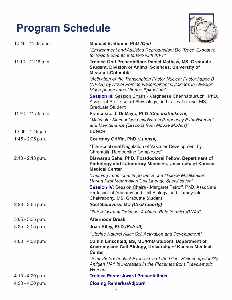
Program Schedule10:45 - 11:05 a.m. Michael S. Bloom, PhD (Qiu)
“Environment and Assisted Reproduction: Do ‘Trace’ Exposure to Toxic Elements Interfere with IVF?”
11:10 - 11:18 a.m. Trainee Oral Presentation: Daniel Mathew, MS, Graduate Student, Division of Animal Sciences, University of Missouri-Columbia“Activation of the Transcription Factor Nuclear Factor kappa B (NFKB) by Novel Porcine Recombinant Cytokines in Alveolar Macrophages and Uterine Epithelium”Session III: Session Chairs - Vargheese Chennathukuzhi, PhD, Assistant Professor of Physiology, and Lacey Luense, MS, Graduate Student
11:20 - 11:55 a.m. Francesco J. DeMayo, PhD (Chennathukuzhi)“Molecular Mechanisms Involved in Pregnancy Establishment and Maintenance (Lessons from Mouse Models)”
12:05 - 1:45 p.m. LUNCH 1:45 - 2:05 p.m. Courtney Griffin, PhD (Luense)
“Transcriptional Regulation of Vascular Development by Chromatin Remodeling Complexes”
2:10 - 2:18 p.m. Biswarup Saha, PhD, Postdoctoral Fellow, Department of Pathology and Laboratory Medicine, University of Kansas Medical Center“Defining Functional Importance of a Histone Modification During First Mammalian Cell Lineage Specification”Session IV: Session Chairs - Margaret Petroff, PhD, Associate Professor of Anatomy and Cell Biology, and Damayanti Chakraborty, MS, Graduate Student
2:20 - 2:55 p.m. Yoel Sadovsky, MD (Chakraborty)“Feto-placental Defense: A Macro Role for microRNAs”
3:05 - 3:35 p.m. Afternoon Break3:35 - 3:55 p.m. Joan Riley, PhD (Petroff)
“Uterine Natural Killer Cell Activation and Development”4:00 - 4:08 p.m. Caitlin Linscheid, BS, MD/PhD Student, Department of
Anatomy and Cell Biology, University of Kansas Medical Center“Syncytiotrophoblast Expression of the Minor Histocompatability Antigen HA1 is Increased in the Placentas from Preeclamptic Women”
4:10 - 4:20 p.m. Trainee Poster Award Presentations4:20 - 4:30 p.m. Closing Remarks/Adjourn
7
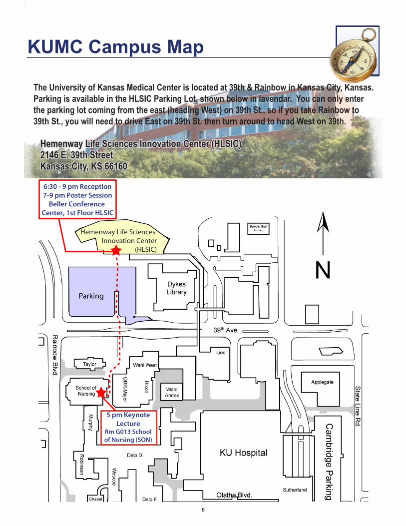
Hemenway Life Sciences Innovation Center
(HLSIC)
6:30 - 9 pm Reception7-9 pm Poster Session
Beller Conference Center, 1st Floor HLSIC
Parking
5 pm Keynote Lecture
Rm G013 School of Nursing (SON)
KUMC Campus Map
The University of Kansas Medical Center is located at 39th & Rainbow in Kansas City, Kansas. Parking is available in the HLSIC Parking Lot, shown below in lavendar. You can only enter the parking lot coming from the east (heading West) on 39th St., so if you take Rainbow to 39th St., you will need to drive East on 39th St. then turn around to head West on 39th.
Hemenway Life Sciences Innovation Center (HLSIC) 2146 E. 39th Street Kansas City, KS 66160
8
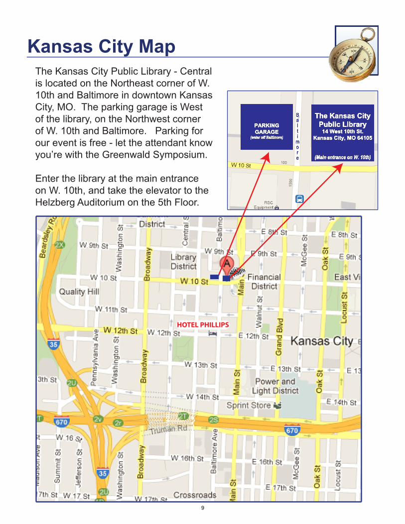
Kansas City MapThe Kansas City Public Library - Central is located on the Northeast corner of W. 10th and Baltimore in downtown Kansas City, MO. The parking garage is West of the library, on the Northwest corner of W. 10th and Baltimore. Parking for our event is free - let the attendant know you’re with the Greenwald Symposium.
Enter the library at the main entrance on W. 10th, and take the elevator to the Helzberg Auditorium on the 5th Floor.
PARKING GARAGE
(enter off Baltimore)
Balti
more
The Kansas City Public Library
14 West 10th St.Kansas City, MO 64105
(Main entrance on W. 10th)
HOTEL PHILLIPS
9

Venue InformationThe Kansas City Public Library - Central
The Kansas City Public Library system consists of a central library, nine branches, and an outreach services program serving a constituency of over 250,000 in Kansas City, Missouri. In addition to providing library services to residents, the Library also serves as a resource for the 1.7 million metropolitan residents of greater Kansas City.
In 2004, the Kansas City Public Library - Central moved into the former First National Bank building at 10th and Baltimore in downtown Kansas City, Missouri. The century-old building, a true masterpiece of craftsmanship with its marble columns, bronze doors and ornate mouldings, required remodeling and a fifth floor addition, but provided the framework for a modern and impressive urban library. The location features state-of-the-art technology, improved and increased services, meeting rooms, a screening room, a coffee shop and much more, all within the walls of a building originally constructed to convey a sense of strength and continuity. It is upon that foundation the Library places its vision for the next century to come.
Facts About the Library
•The Kansas City Public Library has ten locations.•The Central Library is the largest facility, housing resources, special collections and administrative offices.•More than 2,348,408 materials were checked out during the last fiscal year.•The Inter Library Loan department loaned out 115,846 items last year to other libraries. •2,492,118 customers used the Library system last year.•The Library system counts 1,147,278 items in its holdings.
Helzberg AuditoriumThe Library’s most versatile meeting space, the Helzberg Auditorium is located on the 5th floor of the library. Helzberg is also aggressively styled with contemporary and clean lines for an energetic atmosphere, and features performance quality acoustics using cork flooring and specially designed ceiling elements, built-in AV system, and floor-to-ceiling windows on multiple sides that provide natural lighting.
Gladys Feld HelzbergGladys Feld Helzberg was the wife of the late Barnett C. Helzberg, Sr, of Helzberg Diamonds. Helzberg jewelry store was founded in 1915 by the late Morris Helzberg, in Kansas City, Kan., and expanded to a regional market by Barnett C. Helzberg, Sr. Gladys was an active member of the Kansas City Chapter of the Association for Women in Communications and one of the founders of Veterans’ Voices. The Gladys Feld Helzberg Scholarship Fund was established in 1960 for talented journalism students and is administered by the University of Kansas endowment fund. She was also the founder of the Greater Kansas City chapter of the Brandeis Women’s Committee.
10
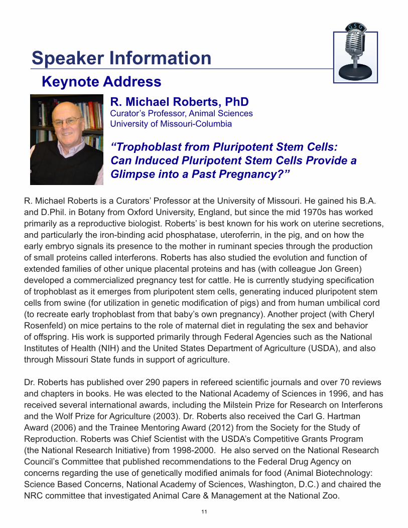
R. Michael Roberts is a Curators’ Professor at the University of Missouri. He gained his B.A. and D.Phil. in Botany from Oxford University, England, but since the mid 1970s has worked primarily as a reproductive biologist. Roberts’ is best known for his work on uterine secretions, and particularly the iron-binding acid phosphatase, uteroferrin, in the pig, and on how the early embryo signals its presence to the mother in ruminant species through the production of small proteins called interferons. Roberts has also studied the evolution and function of extended families of other unique placental proteins and has (with colleague Jon Green) developed a commercialized pregnancy test for cattle. He is currently studying specification of trophoblast as it emerges from pluripotent stem cells, generating induced pluripotent stem cells from swine (for utilization in genetic modification of pigs) and from human umbilical cord (to recreate early trophoblast from that baby’s own pregnancy). Another project (with Cheryl Rosenfeld) on mice pertains to the role of maternal diet in regulating the sex and behavior of offspring. His work is supported primarily through Federal Agencies such as the National Institutes of Health (NIH) and the United States Department of Agriculture (USDA), and also through Missouri State funds in support of agriculture.
Dr. Roberts has published over 290 papers in refereed scientific journals and over 70 reviews and chapters in books. He was elected to the National Academy of Sciences in 1996, and has received several international awards, including the Milstein Prize for Research on Interferons and the Wolf Prize for Agriculture (2003). Dr. Roberts also received the Carl G. Hartman Award (2006) and the Trainee Mentoring Award (2012) from the Society for the Study of Reproduction. Roberts was Chief Scientist with the USDA’s Competitive Grants Program (the National Research Initiative) from 1998-2000. He also served on the National Research Council’s Committee that published recommendations to the Federal Drug Agency on concerns regarding the use of genetically modified animals for food (Animal Biotechnology: Science Based Concerns, National Academy of Sciences, Washington, D.C.) and chaired the NRC committee that investigated Animal Care & Management at the National Zoo.
R. Michael Roberts, PhDCurator’s Professor, Animal SciencesUniversity of Missouri-Columbia
“Trophoblast from Pluripotent Stem Cells: Can Induced Pluripotent Stem Cells Provide a Glimpse into a Past Pregnancy?”
Keynote AddressSpeaker Information
11
Kyle Orwig, PhD Associate Professor of Obstetrics, Gynecology & Reproductive Sciences, and Developmental BiologyUniversity of Pittsburgh “Translating Spermatogonial Stem Cell Transplantation to the Clinic”
Kyle Orwig received his Ph.D. in Biochemistry & Biophysics and Animal Sciences from Oregon State University in 1994 working with Dr. Fred Stormshak on the regulation of
corpus luteum function in livestock species. His postdoctoral studies focused on prolactin family genes in rodents and were conducted in Dr. Mike Soares’ lab at the University of Kansas Medical Center. He developed his current research interests in stem cells and spermatogenic lineage development as a senior fellow and then junior faculty with Dr. Ralph Brinster at the University of Pennsylvania. He moved to the University of Pittsburgh in 2003 where he began translating his discoveries on rodent spermatogonial stem cells (SSCs) to the nonhuman primate and human systems. Dr. Orwig has numerous primary research publications, reviews and book chapters and his research has been continuously funded by NIH other sources.
Dr. Orwig is currently an Associate Professor of Obstetrics, Gynecology and Reproductive Sciences and Director of Research in the Division of Reproductive Endocrinology and Infertility. He is also the founding Director of the Fertility Preservation Program at the University of Pittsburgh Medical Center as well as the Transgenic and Molecular Core at Magee-Womens Research Institute. His laboratory studies the dynamics of male germ lineage development and the mechanisms that regulate spermatogonial stem cell self-renewal and differentiation. In addition, the Orwig lab is actively investigating the feasibility and safety of using SSCs to preserve fertility and treat male infertility.
Session I
Fernando Pierucci-Alves, DVMResearch Assistant Professor, Anatomy and PhysiologyColllege of Veterinary MedicineKansas State University “Cellular Signaling by Transforming Growth Factor Beta in the Male Excurrent System - Evaluating the Potential for High Levels of Signaling Activity & Possible Impacts on Reproductive Function”
Fernando Pierucci-Alves received his D.V.M. from the Federal University of Uberlandia, Brazil, in 1999, where he was a research scholar for most of his program. He was then a visiting research scholar at Southern Illinois University and Duke University. He joined Kansas State University as a postdoctoral fellow in 2004, and is currently a research assistant professor.
The focus of Dr. Pierucci-Alves’ research is to determine the role of transforming growth factor beta (TGFβ) signaling in the male excurrent system, where sperm cells mature and acquire fertilizing capacity. Sperm cells mature in a luminal microenvironment that is modified by the secretory and absorptive activities of various epithelial cell populations lining the excurrent system. His research program, still at early stages of development, has reported that TGFβ-signaling occurs in epithelial cells lining the vas deferens and that signaling upregulation leads to disruptions in tight junction organization and loss of transepithelial resistance. He is currently examining the endogenous levels of this signaling pathway at various segments of the male excurrent system.
12
Bruce D. Murphy, PhDDirector, Center for Research in Animal ReproductionVeterinary BiomedicineUniversity of Montreal “Liver-Receptor Homolog-1 Rules Reproductive Processes”
Bruce D. Murphy was awarded a MSc in physiology (Colorado State University) and a PhD in reproductive biology (University of Saskatchewan). His first academic
appointment was at the University of Washington Medical Program in 1972, after which he joined the University of Saskatchewan in 1973 and rose to the level of Professor. He was the founding director of the University of Saskatachwan Reproductive Biology Research Unit. In 1991, he was recruited to the post of Directeur de Centre de recherche en reproduction animale, Faculté de médicine vétérinaire, Université de Montréal. He holds a joint appointment Département de obstetrique-gynecologie, Faculté de médicine, at the same institution. He has served on numerous granting committees, chairing MRC Endocrinology Committee, NSERC Strategic Biotechnology Committee, Endocrinology Committee CIHR, the NIH Reproductive Biology Study Section and a three-year term on the NSERC Animal Physiology grant panel. He served as Treasurer of the Society for the Study of Reproduction (SSR) for 9 years and is the current Editor-In-Chief of Biology of Reproduction. Dr. Murphy has been continuously funded since 1974 by NSERC for studies of embryo implantation and since 1978 by MRC and CIHR for investigation of embryo implantation. In addition, he has held NSERC Strategic funding to study placental function and the interface between nutrition and reproduction, and investigation of genomic variation associated with reproductive success. He is author of more than 200 scientific publications and has trained more than 50 graduate students and postdoctoral fellows. He has received several awards, including the SSR Distinguished Service Award, the Pfizer Award for Research Excellence and the CFAS Award of Excellence in Reproductive Biology. He was elected to the Argentine Academy of Agricultural Science in 1988 as a Fellow of the Canadian Academy of Health Sciences in 2006 and is Laureate of the Fonds du Québec (2009).
Michael S. Bloom, PhDAssistant Professor, Environmental Health Sciences, Epidemiology and Biostatistics University of Albany School of Public HealthState University of New York (SUNY)
“Environmental and Assisted Reproduction: Do ‘Trace’ Exposures to Toxic Elements Interfere with IVF?”
Michael Bloom received his Ph.D. in Epidemiology from the University at Buffalo, State University of New York and completed a postdoctoral training fellowship in reproductive epidemiology at the Eunice Kennedy Shriver National Institute of Child Health and Human Development (NICHD). Since January, 2008 he has been appointed to the faculty at the University at Albany School of Public Health.
Dr. Bloom’s primary research interest is in the effects of toxic environmental agents on human fecundity and fertility at the population level; including the impact on outcomes following in vitro fertilization. The focus of Dr. Bloom’s work is on the adverse reproductive impacts of widely distributed and non-essential toxic elements including mercury, cadmium and lead. He is also interested in assessing the utility of laboratory measures for use as biomarkers of exposure and effect in epidemiologic studies of human reproductive health.
Session II
13

Francesco J. DeMayo, PhDDan L. Duncan Professor & Gordon Cain Professor, Molecular & Cellular Biology Baylor College of Medicine “Molecular Mechanisms Involved in Pregnancy Establishment and Maintenance (Lessons from Mouse Models)”
Dr. Francesco (Franco) DeMayo is the Dan L Duncan and Gordon Cane Professor of Molecular and Cellular Biology and Co-Director of the newly established Center for Reproductive Medicine at Baylor College of Medicine, Houston TX. Dr. DeMayo received his B.S. degree from Cornell University and M.S and Ph.D. from Michigan State University. He did his postdoctoral training at the Baylor College of Medicine. Dr. DeMayo has served as Director of the Genetically Engineered Mouse Core and is Director of the Specialized Cooperative Center Program Infertility and Reproduction (SCCPIR) at Baylor. Dr. DeMayo’s research career is dedicated to investigating the endocrine regulation of uterine function. Dr. DeMayo has identified numerous molecular pathways in the uterus that are regulated by progesterone receptor and its coactivators. Dr. DeMayo generated an array of genetic tools including the PR-Cre mouse that allows for the conditional ablation of genes in the uterus. Using these tools Dr. DeMayo demonstrated that the Indian hedgehog pathway is a critical regulator of uterine receptivity. Since then he has used this model to investigate the molecular regulation of uterine gland development and differentiation, uterine receptivity and the ability of the uterus to support post implantation embryo development. He has also generated several models to investigate endometrial cancer in mice. Dr. DeMayo’s is the recipient of the Michael E. DeBakey Award for Research Excellence, the Society for the Study of Reproduction Research Award, and the Frontiers in Reproduction Beacon Award for his research in the field of reproduction.
Courtney Griffin, PhDAssistant Member, Cardiovascular Biology Research ProgramOklahoma Medical Research FoundtionAdjunct Assistant Professor, Cell Biology University of Oklahoma Health Sciences Center “Transcriptional Regulation of Vascular Development by Chromatin-Remodeling Complexes”
Courtney Griffin earned her undergraduate degree in Biology at Harvard College in 1995 and her Ph.D. in Biomedical Sciences at the University of California San Francisco in 2001. For her postdoctoral training, Dr. Griffin joined the lab of Terry Magnuson at the University of North Carolina at Chapel Hill. She accepted her first independent position in 2008 at the Oklahoma Medical Research Foundation in Oklahoma City. Dr. Griffin is currently an Assistant Member in the Cardiovascular Biology Research Program at OMRF and holds an Adjunct Assistant Professor position at the University of Oklahoma Health Sciences Center.
During her graduate training with Shaun Coughlin at UCSF, Dr. Griffin became interested in vascular development. She combined that interest with her postdoctoral training in epigenetics to establish her own niche studying the impact of chromatin-remodeling complexes on vascular development. Her lab at OMRF uses mice with mutations in key chromatin-remodeling enzymes to investigate multiple aspects of embryonic vascular development, including transcriptional regulation of venous specification, vascular integrity, and lymphangiogenesis. Her lab is also examining the influence of chromatin-remodeling complexes on postnatal vascular development to determine whether the complexes might serve as therapeutic targets for pathological vascular disorders.
Session III
14
Yoel Sadovsky, MDDirector, Magee-Womens Research InstituteProfessor of OB/GYN, Microbiology and Molecular Genetics University of Pittsburgh School of Medicine “Feto-placental Defense: A Macro Role for microRNAs”
Yoel Sadovsky received his MD degree from the Hebrew University - Hadassah Medical School in Jerusalem in 1986, followed by OBGYN residency at Washington University
in St. Louis and maternal-fetal medicine and postdoctoral research fellowships at the University of California, San Francisco. He then returned to Washington University as a reproductive biologist and specialist in high-risk pregnancy, where he was appointed tenured professor of OBGYN, and Cell Biology and Physiology. Dr. Sadovsky served as Director, Division of Maternal-Fetal Medicine and Ultrasound from 1999-2007. In 2007, he assumed Directorship of Magee-Womens Research Institute (MWRI) at the University of Pittsburgh, and Vice Chair (Research), Department of Obstetrics, Gynecology and Reproductive Sciences.
Dr. Sadovsky’s research centers on feto-placental development and trophoblast function. Using human placental cells as well as mouse models, he studies molecular pathways that govern placental development and adaptive response to stress. Primary areas of research include: Placental uptake and processing of metabolic fuels, the role of microRNA in placental function, and placental injury and adaptation. Dr. Sadovsky’s laboratory is funded via several NIH grants, and his investigation has resulted in the publication of 107 peer-reviewed scientific articles and 17 book chapters and invited publications. Dr. Sadovsky has served as a member or chair on several National Institute of Child Health and Human Development (NICHD) study sections, chaired the NICHD Genomic and Proteomic Network for Preterm Birth Research steering committee, and is currently a member of the NICHD Council and the NICHD Division of Intramural Research Review Panel. He also chairs the March of Dimes Prematurity Research Initiative Advisory Committee, is a member of the March of Dimes Council and a number of other academic advisory boards. He also serves as an Editor for the journal Placenta.
Joan Riley, PhDAssistant Professor, Obstetrics and GynecologyWashington University School of Medicine “Uterine Natural Killer Cell Activation and Development”
Joan K. Riley received her Ph.D. in Immunology from Washington University School of Medicine in 1998. She then completed her postdoctoral training at Genentech, Inc. in 2001 and Washington University School of Medicine in 2006. Since finishing
her postdoctoral training she served first as an Instructor and then as an Assistant Professor at Washington University School of Medicine.
During pregnancy, a functionally unique type of natural killer (NK) cell populates the decidua. These decidual NK cells play an important role in placental development; their dysregulation is associated with recurrent pregnancy loss, implantation failure, and preeclampsia. Dr. Riley’s current research is aimed at gaining a better understanding of: the source of precursor cells that give rise to decidual NK cells, how extensively the uterine environment contributes to NK cell activation and differentiation, and the consequences of impaired decidualization on endometrial NK cell differentiation.
Session IV
15

Abstract Titles *Poster Competition
1. *Development of a novel immunoassay for simultaneous quantitation of endocrine parameters. Elizabeth A. Benavides, Rachel E. Gerrard and Duane H. Keisler. Division of Animal Sciences, University of Missouri, Columbia, MO.
2. Isolation and characterization of pituitary hFSH glycoforms. Viktor Y. Butnev, William K. White, Alice Hwang, Vladimir Y. Butnev, Tarak Sharma, Souraya Alameddine, Patrick Tran, Bin Shuai, Jeffrey V. May, and George R. Bousfield. Department of Biological Sciences, Wichita State University, Wichita, KS.
3. Autophagy and ubiquitin-proteasome pathway are the keystones of sperm mitochondrial degradation following mammalian fertilization. Won-Hee Song1, Young-Joo Yi1, Peter Sutovsky.1, 2
1Division of Animal Sciences, and 2Departments of Obstetrics, Gynecology & Women’s Health, University of Missouri, Columbia, MO.
4. KU-AS-272, a potential single-dose sterilant for cats and dogs, shows safety and ability to block spermatogenesis in testis to Sertoli cells only after a single subcutaneous injection in male rat. Vijayalaxmi Gupta1, 2, Katherine F. Roby3,4 , Brian Kern, 1,2,Todd Hall1,2,Sudhakar Jakkaraj1, 5, 6, Ramappa Chakrasali1, 5, 6, Gunda I. Georg1, 5, 6, Melinda Broward7, Robyn Wood7, Scott Weir7, Joseph S. Tash1, 2 1U54 Interdisciplinary Center for Male Contraceptive Research and Drug Development, 2Dept. of Molecular & Integrative Physiology, 3Institute for Reproductive Health and Regenerative Medicine, 4Department of Anatomy & Cell Biology, University of Kansas Medical Center, Kansas City, KS, 5Dept. Medicinal Chemistry, and Institute for Therapeutics Discovery & Development University of Minnesota, Minneapolis, MN, 6Department of Medicinal Chemistry, University of Kansas, Lawrence, KS, 7Institute for Advancing Medical Innovation, University of Kansas, Lawrence, KS.
5. Glutathione S-transferase Polymorphisms and Mechanisms of Male Infertility. Katherine F. Roby1, Ajay K. Nangia2, Sacha A. Krieg3, Richard C. Hastings4, Renee S. Mijal5 Institute for Reproductive Health and Regenerative Medicine, Departments of 1Anatomy & Cell Biology, 2Urology, 3Obstetrics & Gynecology, 4Microbiology, and 5Preventive Medicine and Public Health, University of Kansas Medical Center, Kansas City, KS.
6. Altering the balance of vascular endothelial growth factor A (VEGFA) isoforms in vivo affects mRNA abundance of genes that regulate the self-renewal of undifferentiated spermatogonia and survival, testis morphogenesis, and germ cell numbers in perinatal rat testes. Kevin M. Sargent, Ningxia Lu, William E. Pohlmeier, Shantille G. Kruse, Meredith L. Bremer, and Andrea Cupp. University of Nebraska-Lincoln, Lincoln, NE.
7. Differential effect of H2-Gamendazole (H2-GMZ) and other small indazole carboxylic acid (ICA) analogs on primary rat Sertoli cells cytoskeletal structure and elongation factor 1 alpha (EEF1A1) expression. Lesya Holets1,2, Terri G Kinzy5, Gunda I Georg1,3,4, and Joseph S Tash1,2. 1Interdisciplinary Center for Male Contraceptive Research & Drug Development. 2Department of Molecular and Integrative Physiology, U. Kansas Medical Center, Kansas City, KS, 3Dept Medicinal Chemistry and Inst for Therapeutics, Discovery & Development, U. Minnesota, Minneapolis, MN. 4Dept Medicinal Chemistry, U. Kansas, Lawrence, KS, U. 5Dept of Molecular Genetics, Microbiology, and Immunology UMDNJ Robert Wood Johnson Medical School.
16
8. Transgenic pig carrying green fluorescent proteasomes reveals interactions of 20S proteasomal core with the sperm-acrosomal membrane proteins. Edward L. Miles1, Chad O’Gorman1, Jianguo Zhao,2, Melissa Samuel1,2, Eric Walters1,2, Young-Joo Yi1, Miriam Sutovsky1, Randall S. Prather1,2, Kevin Wells1,2, Peter Sutovsky1,3,# 1Division of Animal Sciences, 2National Swine Resource and Research Center, and 3Departments of Obstetrics, Gynecology & Women’s Health, University of Missouri, Columbia, MO.
9. Green fluorescence protein driven by the Na,K-ATPase α4 isoform promoter is expressed only in male germ cells of mouse testis. Jeffrey P. McDermott, Gladis Sánchez, Vargheese Chennathukuzhi and Gustavo Blanco. Department of Molecular and Integrative Physiology, University of Kansas Medical Center, Kansas City, KS.
10. *Outer dense fiber protein 2 expression in bull spermatozoa reflects fertility in artificial insemination service. Peter Vargovic1, Abdullah Kaya3, Frans van der Hoorn4, Erdogan Memili5, Peter Sutovsky1,2. Division of Animal Sciences1, and Departments of Obstetrics, Gynecology & Women’s Health2, University of Missouri, Columbia, MO; Alta Genetics, Watertown, WI; Department of Biochemistry and Molecular Biology, Faculty of Medicine, University of Calgary4, Alberta, Canada; Department of Animal and Dairy Sciences5, Mississippi State University, MS.
11. *The autoimmune regulator (AIRE) protects against infertility, reproductive tract inflammation and germ cell loss in male Balb/c mice. BD Warren1, Leslie L Heckert2, Brian K Petroff3, and Margaret G Petroff1 Departments of 1Anatomy and Cell Biology 2Physiology and 3Internal Medicine, University of Kansas Medical Center, Kansas City, KS.
12. *Decoding the functional role of Fast, a long noncoding RNA transcribed at the Nr5A1 locus. Jitu W George1, Brian Hermann2, Lane Christenson1, Leslie Heckert1. 1University of Kansas Medical Center, Kansas City, Kansas. 2 University of Texas at San Antonio, San Antonio, TX.
13. *Origin of a species-specific rheostat controlling testicular growth and steroidogenesis. Pengli Bu1, S.M. Khorshed Alam1, Shintaro Yagi2, Kunio Shiota2, T. Rajendra Kumar3, Ken-ichirou Morohashi4, Jay L. Vivian1, M.A. Karim Rumi1, and Michael J. Soares1. 1Institute for Reproductive Health and Regenerative Medicine, Departments of Pathology and Laboratory Medicine, and 3Molecular and Integrative Physiology, University of Kansas Medical Center, Kansas City, KS; 2Laboratory of Cellular Biochemistry, Veterinary Medical Sciences/Animal Resource Sciences, The University of Tokyo, Tokyo, Japan; 4Graduate School of Medical Sciences, Kyushu University, Higashi-ku, Fukuoka, Japan
14. *Identification of microRNA-21 direct targets in granulosa cells. J. Browning Fitzgerald, L.K. Christenson. University of Kansas Medical Center, Kansas City, KS.
15. *The histone demethylase JMJD2B regulates a core set of cancer associated genes in multiple cancer cell types. Lei Qiu1,2, Judith Chapman1, Amato Giaccia3, Ying Mu4, Jake New1,5, Adam Krieg1,2. 1Department of Obstetrics and Gynecology, 2Department of Pathology and Laboratory Medicine, University of Kansas Medical Center, Kansas City, KS, USA; 3Division of Radiation and Cancer Biology, Department of Radiation Oncology, Stanford University, Stanford, CA, USA; 4Department of Clinical Laboratory Science, University of Kansas Medical Center, Kansas City, KS, USA; 5Kansas State University, Manhattan, KS.
16. *Effect of ovulatory follicle size on steroidogenic capacity and molecular markers of oocyte competence prior to GnRH-induced ovulation in non-lactating beef cows. K. G. Pohler1, M. F. Smith1, E. M. Jinks1, F. M. Abreu2, C. A. Roberts3, J. K. Folger4, G. W. Smith4, and T. W. Geary3 1University of Missouri, 2Ohio State University, 3USDA ARS Fort Keogh, Miles City, MT,4Michigan State University, MI.
17

17. *Identification of microRNA expressed in bovine follicles before and after the LH surge. Wei-Ting Hung1, Xiaoman Hong1, Lacey J. Luense1, Jens Vanselow2, Marion Spitschak2 and Lane K. Christenson1.
1Department of Molecular and Integrative Physiology, University of Kansas Medical Center, Kansas City, Kansas, USA. 2Molecular Biology, Leibniz Institute for Farm Animal Biology (FBN), Dummerstorf, Germany.
18. TGF-beta-related signaling regulates stem cell heterogeneity: self-renewal as a dynamic and regulated equilibrium. Katherine E. Galvin-Burgess, Emily D. Travis, Kelsey E. Pierson, Lauren B. Robertson, and Jay L. Vivian. Institute for Reproductive Health and Regenerative Medicine; Department of Pathology and Laboratory Medicine, University of Kansas Medical Center, Kansas City KS.
19. *Defining functional importance of a histone modification during first mammalian cell lineage specification. Biswarup Saha and Soumen Paul. Institute of Reproductive Health and Regenerative Medicine, Dept. of Pathology and Laboratory Medicine, University of Kansas Medical Center, Kansas City, KS.
20. *Determination of allelic expression of H19 in peri-implantation mouse embryos. Verónica M. Ne-grón Pérez, Franklin D. Echevarría, Sarah R. Huffman and Rocío M. Rivera, Division of Animal Sciences, University of Missouri, Columbia.
21. *Transcription Factor TEAD4: New insight into trophectoderm lineage development. Pratik Home, Biswarup Saha, Soma Ray and Soumen Paul. Inst. for Reproductive Health and Regenerative Medi-cine, Dept. of Pathology and Laboratory Medicine, Univ. of Kansas Medical Center, Kansas City, KS.
22. Global alteration in gene expression profiles of deciduas from women with idiopathic recurrent pregnancy loss. Sacha A. Krieg1, Xiujun Fan2, Yan Hong1, Xing-Qiang Sang4, Amato J. Giaccia3, Lynn M. Westphal2, Ruth B. Lathi2, Nihar R. Nayak2 and Adam J. Krieg1,. Department of Obstetrics and Gynecology, Kansas University Medical Center, Kansas City, KS; 2Department of Obstetrics and Gynecology, Stanford Hospital and Clinics, Stanford, CA; 3Department of Radiation Oncology, Center for Clinical Sciences Research, Stanford University, Stanford, CA; 4Department of Biochemistry, Florida State University, Tallahassee, FL.
23. *Preliminary analysis of uterine leptin receptor (Lepr) knockout mice. Kathleen A. Pennington1, John P. Lydon2, Francesco J. DeMayo2, Laura Clamon Schulz1. 1Department of Ob-GYN and Women’s Health, University of Missouri, Columbia MO, USA 2Department of Molecular and Cellular Biology, Baylor College of Medicine, Houston, TX.
24. Zinc finger nuclease targeted disruption of estrogen receptor alpha signaling in the rat. M.A. Karim Rumi1, Kaiyu Kubota1, Anamika Ratri1, Damayanti Chakraborty1, George Bugarinovic1, Katherine F. Roby2, Melissa A. Larson3, Jay L. Vivian1, Michael W. Wolfe3, and Michael J. Soares1, Institute for Reproductive Health and Regenerative Medicine, 1Departments of Pathology & Laboratory Medicine, 2Anatomy & Cell Biology, and 3Molecular & Integrative Physiology, University of Kansas Medical Center, Kansas City, KS.
25. *Activation of the transcription factor nuclear factor kappa B (NFKB) by novel porcine recombinant cytokines in alveolar macrophages and uterine epithelium. D. J. Mathew, R. D. Geisert, and M. C. Lucy, University of Missouri-Columbia, MO.
26. Expression and regulation of the tumor associated antigen trophoblast glycoprotein (TPGB/5T4) in the human placenta. S.M. Khorshed Alam1, S. Jasti1, T. Fields2, M.G. Petroff1. 1Department of Anatomy and Cell Biology and 2Department of Pathology and Laboratory Medicine, The University of Kansas Medical Center, Kansas City, KS.
18

27. Altered embryonic expression of DESMIN and PPARG is associated with placental insufficiency and increased placental oxidative stress gene expression in a mouse model of maternal obesity. Kristin A. Norwood, Amanda K. Brandt, and Jennifer R. Wood. Department of Animal Science, University of Nebraska-Lincoln, Lincoln, NE.
28. *Role of hypoxia signaling in trophoblast cell lineage development. Damayanti Chakraborty, M.A. Karim Rumi, Adam J. Krieg, and Michael J. Soares, Institute for Reproductive Health and Regenerative Medicine, Departments of Pathology and Laboratory Medicine and Obstetrics & Gynecology, University of Kansas Medical Center, Kansas City, KS.
29. *Cell signaling system directing trophoblast differentiation from human pluripotent stem cells. M. Amita, T. Ezashi, B. P. Telugu, A. Alexenko, K. Adachi, S. Sinha, and R. M. Roberts. Bond Life Sciences Center and Division of Animal Sciences, University of Missouri, Columbia, MO.
30. *FOSL1 regulation of trophoblast cell differentiation: partners and gene targets. Kaiyu Kubota, M.A. Karim Rumi, Lindsey N. Kent, and Michael J. Soares. Institute for Reproductive Health and Regenerative Medicine, Department of Pathology and Laboratory Medicine, University of Kansas Medical Center, Kansas City, KS.
31. Trophoblast differentiation from human induced pluripotent stem cells treated with BMP4. Toshihiko Ezashi1, Mitsuyoshi Amita1, Bhanu P. Telugu4,5, Katsuyuki Adachi3, Danny. J. Schust3, Laura C. Schulz3, and R. Michael Roberts1,2. Division of Animal Sciences & Bond Life Sciences Center1, Departments of Biochemistry2 and Obstetrics, Gynecology & Women’s Health3, University of Missouri-Columbia; Animal Biosciences and Biotechnology Laboratory, USDA-ARS, Beltsville, MD4; and Department of Animal and Avian Sciences, University of Maryland-College Park, MD5.
32. *OVO-like 1 regulates human trophoblast differentiation. Stephen J. Renaud, M.A. Karim Rumi, Michael J. Soares. Institute for Reproductive Health and Regenerative Medicine, Department of Pathology and Laboratory Medicine, University of Kansas Medical Center, Kansas City, KS.
33. *Syncytiotrophoblast expression of the minor histocompatibility antigen HA1 is increased in placentas from preeclamptic women. Caitlin Linscheid1, Paul Singh2, Erica Heitmann2, Elizabeth Wickstrom2, Herbert Hodes3, Traci Nauser3, Lei Qui1 and Margaret Petroff1. 1Department of Anatomy and Cell Biology, University of Kansas Medical Center, KC, KS 2Saint Luke’s Health System, Department of Maternal and Fetal Medicine, KC, MO 3The Center for Women’s Health, Overland Park, KS.
34. Expression, Localization, and Function of Purkinje Cell Protein 4 (PCP4) in Human Myometrium. Clifford W Mason, Lily He, Yafeng Dong, Helen Zhou, and Carl P Weiner. Department of Obstetrics and Gynecology, University of Kansas School of Medicine, Kansas City, KS.
35. *Bovine fetuses with phenotypic characteristics similar to those reported for the human condition Beckwith-Wiedemann Syndrome have biallelic expression of the imprinted gene Kcnq1ot1. Zhiyuan Chen, Katherine Marie Robbins, Kevin Dale Wells and Rocío Melissa Rivera. Division of Animal Sciences, University of Missouri, Columbia, MO.
36. Pharmocologic prevention of neuronal loss during chronic fetal hypoxemia, Yafeng Dong, Weijian Hou, Josh Stites, and Carl P. Weiner, Department of OB/GYN, Kansas University School of Medicine, Kansas City, KS.
19
Full Abstracts *Poster Competition
1. *Development of a novel immunoassay for simultaneous quantitation of endocrine parameters. Elizabeth A. Benavides, Rachel E. Gerrard and Duane H. Keisler. Division of Animal Sciences, University of Missouri, Columbia, MO.
Since the advent of radioimmunoassays in the 1950s, numerous immunologically based methods have been developed for sample analysis. Although each immunological method possess unique assets and liabilities, all share limited abilities in the number of analytes capable of being determined simultaneously – with most limited to the analysis of one analyte per replicate per sample. With the growth in demand for information on more analytes, compounded by limited sample volume in small species, new bio-analytical methodologies are emerging which permit replicate determinations of multiple analytes simultaneously – i.e. multiplexing. Our objective was to determine if tools developed for high throughput genotyping, which have the capacity and specificity for making millions of measurements with a high level of precision, could be used for multiplex analysis of hormones. Specifically, we used the Illumina BeadXpress platform, which consists of a ‘reader machine’ and a bead-set. The bead-set contains fiber-optic ‘beads’ (24mm x 240mm) individually etched with a barcode. One set of beads has the same barcode and 600+ different sets of barcodes are available. The surface of each bead is covalently modified for affixing a known ‘capture’ sequence. Multiple barcoded beads (each with a unique ‘capture’ sequence) are then placed in a sample and the machine reads each bead’s barcode and determines if that antisense sequence also exists with a high level of redundancy. Our hypothesis is that the BeadXpress platform can be optimized to measure hormones in multiplexed format. The objectives of this study were: 1. determine optimum bead labeling and assay design characteristics and 2. determine if the platform can be used to measure a single hormone of interest. We suggest that the technological leap in capabilities provided by successful multiplexing can be used for understanding the complex interaction of endocrine and metabolic signals in the dynamically changing animal.
2. Isolation and characterization of pituitary hFSH glycoforms. Viktor Y. Butnev, William K. White, Alice Hwang, Vladimir Y. Butnev, Tarak Sharma, Souraya Alameddine, Patrick Tran, Bin Shuai, Jeffrey V. May, and George R. Bousfield. Department of Biological Sciences, Wichita State University, Wichita, KS.
Human pituitary FSH (hFSH) consists of two major glycoforms which differ in their glycosylation extent of the hormone-specific β-subunit. The completely glycosylated β-subunit shows up as a 24K band while the carbohydrate-deficient one behaves as 21K band when tested in western blot after SDS-PAGE. Therefore, for simplicity we call fully glycosylated hFSH glycoform hFSH24 and the glycoform with less carbohydrate hFSH21. Previously, our lab demonstrated that hFSH21 displayed ~20-fold higher receptor-binding activity than hFSH24 and proposed it might contribute to age-related decline of female fertility. The crucial separation of the glycoforms was achieved at the stage of pituitary extraction. The hFSH24 glycoform was preferentially extracted with water at pH 5.5 and hFSH21-enriched was subsequently extracted with 0.1 M ammonium sulfate at pH 4.0. Following ammonium sulfate precipitation, dialysis, and lyophilization, each extract was fractionated by Sephacryl 100 chromatography. Immunoreactive hFSH fractions were pooled and lyophilized. The
20

hFSH24-enriched fraction was subjected to QAE-Sepharose and Phenyl-Sepharose chromatography. Contaminating hLH was removed from the resulting hFSH fraction by anti-eLHβ polyclonal antibody immunoaffinity chromatography. The hFSH containing unbound fraction was applied to an anti-FSH monoclonal antibody 4882 affinity column and the bound material purified three Superdex 75 columns connected in series. The hFSH21-enriched Sephacryl 100 fraction was first precipitated with 50% ammonium sulfate, then with 80% ethanol, and hFSH21 purified by the same chromatographic procedures used for hFSH24, except the Phenyl-Sepharose chromatography step was eliminated. Western blotting and SDS-PAGE confirmed isolation of hFSH21 and hFSH 24 glycoforms. Supported by NIH 5PO1-AG029531.
3. Autophagy and Ubiquitin-Proteasome Pathway are the Keystones of Sperm Mitochondrial Degradation following Mammalian Fertilization. Won-Hee Song1, Young-Joo Yi1, Peter Sutovsky1, 2 1Division of Animal Sciences, and 2Departments of Obstetrics, Gynecology & Women’s Health, University of Missouri, Columbia, MO.
Autophagy and the ubiquitin-proteasome system (UPS) are the major protein degradation pathways responsible for the removal of outlived proteins, protein-aggregates, and organelles, including mitochondria. In almost all eukaryotes, mitochondria and mitochondrial DNA (mtDNA) are inherited from the maternal parent. This phenomenon cannot be explained solely through dilution of paternal mtDNA. Autophagy of sperm mitochondria could occur along three distinct degradation routes leading to autophagic clearance: 1) Autophagy-associated ubiquitin-receptor p62/SQSTM1 interacts with autophagosome-binding ubiquitin-like modifiers, such as LC3 or GABARAP; 2) Ubiquitinated proteins are extracted from mitochondria and from aggresomes, the protein aggregates induced by ubiquitin-binding adaptor protein HDAC6 that transports them along the microtubules; 3) Mitophagy receptor BNIP3L binds to sperm mitochondria inside the fertilized oocyte and targets them toward autophagosome. We hypothesized that boar sperm mitochondria are recognized by the above receptors and specifically degraded by autophagic machinery after fertilization. In our results, the GABARAP-positive autophagosomes formed a halo around the sperm nucleus and mitochondrial sheath at 30 hrs post insemination; treatment with MG132 slowed down the degradation of sperm mitochondria and caused the accumulation of the GABARAP around the sperm mitochondria. A change in GABARAP protein band density was observed by Western blotting of MG132-treated oocytes. Other components including LC3, HDAC6, and BNIP3L, were detected and immunolocalized in the boar spermatozoa and porcine zygotes by immunofluorescence and Western blotting. Immunoprecipitation of GABARAP from porcine zygotes identified two potential co-precipitating proteins, FABP3 and Profilin-1. These preliminary results indicate that autophagy-associated ubiquitin-like protein modifiers could cooperate with ubiquitin-proteasome system during the degradation of boar sperm mitochondria after fertilization. 4. KU-AS-272, a Potential Single-dose Sterilant for Cats and Dogs, Shows Safety and Ability to
Block Spermatogenesis in Testis to Sertoli cells only after a Single Subcutaneous Injection in Male Rat. Vijayalaxmi Gupta1, 2, Katherine F. Roby3,4 , Brian Kern, 1,2,Todd Hall1,2,Sudhakar Jakkaraj1, 5, 6, Ramappa Chakrasali1, 5, 6, Gunda I. Georg1, 5, 6, Melinda Broward7, Robyn Wood7, Scott Weir7, Joseph S. Tash1, 2 1U54 Interdisciplinary Center for Male Contraceptive Research and Drug Development, 2Dept. of Molecular & Integrative Physiology, 3Institute for Reproductive Health and Regenerative Medicine, 4Department of Anatomy & Cell Biology, University of Kansas Medical Center, Kansas City, KS, 5Dept. Medicinal Chemistry, and Institute for Therapeutics Discovery & Development University of Minnesota, Minneapolis, MN, 6Department of Medicinal Chemistry, University of Kansas, Lawrence, KS, 7Institute for Advancing Medical Innovation, University of Kansas, Lawrence, KS.
21

According to the United States Humane Society, between 6 and 8 million dogs and cats enter U.S. shelters every year. Only 30% of dogs and less than 5% of cats are reclaimed by their owners. The cost to euthanize homeless animals in shelter costs about $2 billion to the US tax payer annually. The CDC states that more than 90% of all reported rabid animals occur in wildlife, and most people are exposed to rabies due to close contact with feral domestic animals, such as cats or dogs. Many pet owners consider spay/neuter as an expensive and painful procedure. A safe and permanent, easily administered, single dose non-surgical sterilant in both male and females cats and dogs will reduce the number of unwanted pets and the burden to pet owners, animal control and veterinary facilities. We are developing a small molecule compound, KU-AS-272, as single dose injectable pet sterilant that has potent anti-spermatogenic activity. Previous studies in rats showed that a single dose of 6 mg/kg maintained sterility 6 months later in 40% of the animals. Thus, we tested a higher SQ single dose range at 6, 12, 25 and 50 mg/kg of KU-AS-272 (formulated in 0.2M CaptisolTM) in 70 day old male rats, and euthanized for testis and epididymis harvest on day 5, 30 and 60 post-dose. Rats treated with 25 mg/kg KU-AS-272 appeared slightly lethargic, but recovered by next day. However, 50 mg/kg treated rats showed severe lethargy with very limited mobility affecting their food intake and water consumption, but they all recovered by day 2. All rats were weighed on a weekly basis and % change in body weight calculated. An observed small but transient drop in body weight occurred in the 25 mg/kg (0.58%) and 50 mg/kg (4.2%) treated rats on day 2 post-dose. However all growth rates recovered by day 5, and afterwards were similar to the control, 6mg/kg and 12 mg/kg treatment groups. Testis and epididymal weights were significantly lower (P≤ 0.05) at all doses at day 5 post-dose. At 6 mg/kg treatment, two groups of responding animals were identified: 1) many tubules showed lumens, but some retention of spermatogonia & spermatocytes, or 2) all tubules were shedding all spermatogenic cells. All testes at the 12mg/kg and higher doses showed severe disorganization of the seminiferous epithelium with evidence of continued shedding of spermatogenic cells, loss of luminal space, and many tubules showing Sertoli cell only (SCO) morphology. At day 30 post-dose, testis weight continued to drop at all doses above 6 mg/kg (P≤ 0.05) compared to day 5. The 12 mg/kg and above doses became SCO, the 50 mg/kg was calcifying with pyknotic interstitial cells as well. The caput epididymides from all doses were totally devoid of sperm (even the 6 mg/kg). Only rare seminiferous tubules at 6 mg/kg had remnants of gonia or early spermatocytes, the rest were SCO. The data collected thus far indicate that KU-AS-272 at 12 mg/kg and higher may have achieved the desired sterilizing block to spermatogenesis with total loss of spermatogenic cells. Subsequent 60-day data collection will determine data whether a sterilizing dose was likely achieved in preparation for mating trials and proof-of-concept studies in dogs and cats. Pharmacokinetics and dose range finding studies in pre-pubertal rats will also be performed as a model for younger dogs and cats that would normally undergo spay/neuter operations in the veterinary clinic.
5. Glutathione S-transferase Polymorphisms and Mechanisms of Male Infertility. Katherine F. Roby1, Ajay K. Nangia2, Sacha A. Krieg3, Richard C. Hastings4, Renee S. Mijal5 Institute for Reproductive Health and Regenerative Medicine, Departments of 1Anatomy & Cell Biology, 2Urology, 3Obstetrics & Gynecology, 4Microbiology, and 5Preventive Medicine and Public Health, University of Kansas Medical Center, Kansas City, KS.
Male-factor infertility contributes to 50% of infertility cases among couples. In many cases the causes are unknown. Common deletion polymorphisms in glutathione S-transferase (GST) genes may be important susceptibility determinants. Males presenting for infertility evaluation who were GSTT1 null and a history of smoking had increased odds of having low semen parameters. No interaction was reported for GSTM1 null. How GSTs may contribute to male infertility and how they may interact with smoking is unclear.
22

We hypothesize that men who are GSTT1 null or who smoke will have higher levels of DNA fragmentation and reactive oxygen species (ROS), which have been correlated with poorer sperm parameters and possibly IVF outcomes. This Fall we began recruiting men attending the KUMC urology clinic for infertility evaluation as potential cases. Controls were men who had fathered a child and were recruited from pre-vasectomy patients at KUMC urology or men responding to university-wide email announcements. Cases and controls provided semen samples, blood samples for genotyping and completed identical questionnaires about demographics, lifestyle, tobacco use, medical and reproductive history. To date 16 cases and controls have been recruited and had DNA fragmentation/ROS levels determined. Participants were between their mid-twenties and mid-forties and predominantly non-Hispanic white. Thirty-one percent had smoked >100 cigarettes in their lifetime and 13% currently smoked. Forty-one percent had ever used tobacco products other than cigarettes, with 19% of the sample reporting current use. Genotyping will soon begin and allow comparisons between GST genotypes and levels of sperm ROS and DNA fragmentation.
6. Altering the balance of Vascular Endothelial Growth Factor A (VEGFA) isoforms in vivo affects mRNA abundance of genes that regulate the self-renewal of undifferentiated spermatogonia and survival, testis morphogenesis, and germ cell numbers in perinatal rat testes. Kevin M. Sargent, Ningxia Lu, William E. Pohlmeier, Shantille G. Kruse, Meredith L. Bremer, and Andrea Cupp. University of Nebraska-Lincoln, Lincoln, NE.
Proangiogenic and antiangiogenic VEGFA isoforms injected into perinatal mice after spermatogonial stem cell (SSC) formation, postnatal days 3-5 (P3-5), had divergent effects on SSC colonization of recipient testes. Therefore, we hypothesized that treatment of male rat pups with VEGFA isoforms as gonocytes resume mitosis and prior to SSC formation from P0-P2 would alter testis composition and expression of genes regulating the SSC niche. Pups received IP injections of either 0.5 or 1μg VEGFA164 or VEGFA165b or 1μg antiVEGFAxxxb with PBS or IgG as controls. At P8, seminiferous cord area was reduced by VEGFA165b and increased by VEGFA164 (P < 0.02), and the reverse occurred for the interstitium (P < 0.01). VEGFA164 (P < 0.01) increased the number of DDX4-positive germ cells per cord. Genes important in undifferentiated spermatogonial self-renewal were or tended to be increased by VEGFA164: Gdnf (P < 0.06), Nanos2 (P < 0.03), and Bcl6b (P < 0.1). Nanos2 was (P < 0.02) and Ret (P < 0.1) tended to be decreased by antiVEGFAxxxb. Treatment with VEGFA164 or VEGFA165b increased Bcl2 expression, a survival gene (P < 0.02). Pro-apoptotic Bax was reduced by antiVEGFAxxxb (P < 0.02) while the ratio of Bcl2:Bax was increased by VEGFA164 (P < 0.04) and tended to be increased by antiVEGFAxxxb (P < 0.08). Treatment with VEGFA165b increased expression of two genes in the apoptosis pathway- Casp3 (P < 0.002) and Casp9 (P < 0.02). Taken together, VEGFA isoforms affect testis composition and expression of genes important in regulation of the SSC niche and in cell survival when rats were transiently treated prior to SSC formation in vivo.
7. Differential effect of H2-Gamendazole (H2-GMZ) and other small indazole carboxylic acid (ICA) analogs on primary rat Sertoli cells cytoskeletal structure and elongation factor 1 alpha (EEF1A1) expression. Lesya Holets1,2, Terri G Kinzy5, Gunda I Georg1,3,4, and Joseph S Tash1,2. 1Interdisciplinary Center for Male Contraceptive Research & Drug Development. 2Department of Molecular and Integrative Physiology, U. Kansas Medical Center, Kansas City, KS, 3Dept Medicinal Chemistry and Inst for Therapeutics, Discovery & Development, U. Minnesota, Minneapolis, MN. 4Dept Medicinal Chemistry, U. Kansas, Lawrence, KS, U. 5Dept of Molecular Genetics, Microbiology, and Immunology UMDNJ Robert Wood Johnson Medical School.
23

H2-GMZ was recently identified as an effective orally anti-spermatogenic agent . H2-GMZ as well as other ICA compounds exerts its contraceptive effect by eliciting premature release of spermatids via disruption of the Sertoli cell-spermatid junctions. Rapid changes in rat testicular morphology were identified after the lowest single oral dose of H2-GMZ in vivo that causes 100% infertility (6 mg/kg). Altered transient expression of important spermatogenic genes was established in 1-3 hours after H2-GMZ treatment of primary Sertoli cells in vitro. To determine whether these rapid changes are associated with disruption of cytoskeletal structure of Sertoli cells, we determined the effects of H2-Gamendazole (H2-GMZ) and other ICAs analogs – adjudin (AD), AF 2785, gamendazole (GMZ), and lonidamine (LND) on primary rat Sertoli cells cytoskeletal proteins F-actin and vinculin distribution in primary rat Sertoli cells. Cultured cells were treated with each compound (10µM) for 1, 3 and 6 h, and immunofluorescent staining for F- actin, vinculin and EEF1A1 was performed. Untreated primary Sertoli cells displayed well organized parallel bundles of actin filaments, with linearly organized ES type focal adhesion points as indicated by vinculin staining. Starting 1h after AF2785 and H2-GMZ, F-actin bundles became disorganized. LND and GMZ disruption of actin bundle organization 3h after exposure, and AD after 6h. 3h post-treatment with H2-GMZ, the intensity of cytoplasmic vinculin staining appeared to increase and redistributed with actin to the periphery of many cells, and around the cell nucleus. EEF1A1 was confirmed as a direct binding target for FITC-H2-GMZ. The tyrosine phosphorylation of EEF1A1 between 2-5 min of H2-GMZ exposure has been shown in primary Sertoli cells, and H2-GMZ disrupts bundling of purified mammalian F-actin with purified mammalian EEF1A1. Both, Src and Fyn kinases phosphorylate purified EEF1A1. We found altered EEF1A distribution and signal level in Sertoli cells after 3h of H2-GMZ. The appearance and disappearance of the nectin staining at apical ES were coincident to the assembly and disassembly of Sertoli–spermatid junctions. We indicated H2-GMZ cause a dramatic loss of nectin- 3 signal in rat testis and re-assemble of nectin- 2 localization in Sertoli cells. Taken together, our data suggest that H2-GMZ causes premature loss of spermatids via its effect on F-actin associations and interactions with the ES. Furthermore, our results demonstrate differential effects of ICA analogues on Sertoli cell cytoskeletal structure. This results may be important for study the in vivo mechanisms regulating Sertoli cell-Sertoli cell and Sertoli cell-spermatogenic cell interactions.
8. Transgenic Pig Carrying Green Fluorescent Proteasomes Reveals Interactions of 20S Proteasomal Core with the Sperm-Acrosomal Membrane Proteins. Edward L. Miles1, Chad O’Gorman1, Jianguo Zhao,2, Melissa Samuel1,2, Eric Walters1,2, Young-Joo Yi1, Miriam Sutovsky1, Randall S. Prather1,2, Kevin Wells1,2, Peter Sutovsky1,3,# 1Division of Animal Sciences, 2National Swine Resource and Research Center, and 3Departments of Obstetrics, Gynecology & Women’s Health, University of Missouri, Columbia, MO.
During mammalian fertilization, the ubiquitin-proteasome system (UPS) participates in sperm capacitation, sperm-zona pellucida (ZP) binding and penetration, and in the degradation of paternal, sperm-borne mitochondria and mtDNA. However, the mechanisms behind these proteasome-dependent events are not completely understood. We have created a transgenic boar with green fluorescent protein (GFP) tagged 20S proteasomal core subunit alpha type-1 (GFP-PSMA1), that allows us to investigate the localization, subunit composition and function of the sperm proteasomes during fertilization. We hypothesize that GFP-PSMA1 is incorporated into the functional sperm proteasomes of a GFP-PSMA1 transgenic boar and that the subunits of the sperm acrosome-borne proteasomes interact with structural acrosomal membrane proteins that anchor proteasomes to the acrosomal structures and/or depend on proteasomes for their function. Using direct epifluorescence imaging and indirect immunofluorescence detection we have confirmed the presence of GFP-PSMA1 in the transgenic sperm acrosome. Western blotting revealed a protein band corresponding to the predicted mass of GFP-PSMA1 (57 kDa) in transgenic boar spermatozoa. The
24

transgenic boar’s fertility was confirmed by in vitro fertilization resulting in development to blastocyst and by mating resulting in healthy transgenic offspring. We determined, through immunoprecipitation and proteomic analysis that GFP-PSMA1 co-purifies through other proteasomal subunits, with a number of acrosomal membrane proteins (e.g. lactadherin/MFGE8, spermadhesins AWN and PSP1). The identified acrosomal proteins may regulate sperm proteasomal activity during fertilization or may be the substrates of proteasomal proteolysis during capacitation and zona-induced acrosomal exocytosis. Proteomic analysis also confirmed the interaction/co-immunoprecipitation of GFP-PSMA1 with 20S proteasomal core subunits and isoforms. These results demonstrate that the GFP tagged proteasomes can be immunopurified from the transgenic boar spermatozoa. In the future, isolated GFP-proteasomes with high enzymatic activity could be used for studies of sperm-oocyte interactions and wherever UPS plays a role in cellular physiology or pathologies such as Alzheimer’s, Parkinson’s disease and liver cirrhosis.
9. Green Fluorescence Protein driven by the Na,K-ATPase α4 isoform promoter is expressed only in male germ cells of mouse testis. Jeffrey P. McDermott, Gladis Sánchez, Vargheese Chennathukuzhi and Gustavo Blanco. Department of Molecular and Integrative Physiology, University of Kansas Medical Center, Kansas City, KS.
The catalytic α4 isoform of the Na,K-ATPase exhibits the most limited pattern of expression, being present in testis. At present the precise tissue, cell type and developmental pattern of expression of α4 remains unclear. We have investigated this here by inserting the green fluorescent protein (GFP) downstream of the endogenous Atp1a4 promoter, in place of the Na,K-ATPase α4 gene (Atp1a4), used it as a marker for α4 expression in mice (Atp1a4null(GFP) mice). We show that replacement of α4 by GFP completely disrupted α4 expression and activity, produced sperm morphological and functional abnormalities, and caused infertility similar to that of the Atp1a4null(GFP) male mice. Immunoblot analysis of Atp1a4null(GFP) mouse tissues showed GFP expression only in testis and epididymis. This particular expression pattern was found in adult, but not in mouse embryos or in 7, 18 day old mice. In agreement with expression of GFP, adult Atp1a4null(GFP) mouse testis and epididymis displayed the typical fluorescence of GFP. Immunocytochemistry of testis and epididymis identified GFP in more differentiated male germ cells, but not in spermatogonia, Leydig or Sertoli cells. Further analysis, using immunoblot of fluorescently sorted testis cells with cell specific markers, detected GFP only in spermatocytes, spermatids and spermatozoa. Altogether, these results show that the Atp1a4 promoter drives protein expression exclusively in male germ cells of the testis, where it restricts it to post-meiotic stages of spermatogenesis. These findings highlight the exquisite spatial and temporal control of expression exerted by the Atp1a4 promoter on Na,K-ATPase α4, which is particularly well suited to fulfill the special functions of spermatozoa.
10. *Outer dense fiber protein 2 expression in bull spermatozoa reflects fertility in artificial insemination service. Peter Vargovic1, Abdullah Kaya3, Frans van der Hoorn4, Erdogan Memili5, Peter Sutovsky1,2. Division of Animal Sciences1, and Departments of Obstetrics, Gynecology & Women’s Health2, University of Missouri, Columbia, MO; Alta Genetics, Watertown, WI; Department of Biochemistry and Molecular Biology, Faculty of Medicine, University of Calgary4, Alberta, Canada; Department of Animal and Dairy Sciences5, Mississippi State University, MS.
Protein biomarkers have recently become useful tools for evaluating sperm quality and fertility in livestock animals. Particular, the phenotypes of proteins associated with sperm accessory structures may reflect sperm quality. Outer dense fibers are a major constituent/accessory structure of the sperm tail, and outer dense fiber protein 2 (ODF2) is one of the major components. We analyzed ODF2
25
protein expression in sperm samples from 108 Holstein sires of acceptable but varied fertility, used in artificial insemination (AI) service. They were ranked by fertility and divided into two groups: Bulls with below-average fertility (negative %Diff value - % difference from average conception rate of the cohort and above-average (positive % Diff value). ODF2 expression was analyzed by immunocytochemistry and flow cytometry. In normal spermatozoa, ODF2 was localized exclusively in the sperm tail principal piece. Defective spermatozoa displayed various anomalies of ODF2 labeling. Spermatozoa with bent or coiled tails and with altered or missing mitochondrial sheath exhibited higher fluorescence level compared to normal spermatozoa. Based on flow cytometry, all spermatozoa in samples were divided into three populations: Low (marker M1), normal (M2) and elevated (M3) ODF2-induced fluorescence. The group of 54 bulls with negative %Diff values showed ~30% higher ODF2-induced fluorescence than positive % Diff (P=0.012). Furthermore, low fertility group exhibited ~46 % higher number of spermatozoa in M3 population compared to high fertility group (P=0.024). Significant positive correlation was found between % sperm in M2 population and %Diff (R=0.60, P=0.0012), while negative correlations were found between %Diff and total median fluorescence (R=-0.54, P=0.0038), % sperm in M3 (R=-0.61, P=7.1E-4), and M3 median fluorescence. These data show that abnormally high ODF2 protein expression in sperm cells reflects reduced fertility in AI. Therefore, ODF2 might be useful as a potential negative biomarker for evaluation of bull fertility in AI industry.
11. *The Autoimmune Regulator (AIRE) protects against infertility, reproductive tract inflammation and germ cell loss in male Balb/c mice. Warren BD1, Leslie L Heckert2, Brian K Petroff3, and Margaret G Petroff1 Departments of 1) Anatomy and Cell Biology 2) Physiology and 3) Internal Medicine, University of Kansas Medical Center, Kansas City, KS.
Male specific factors contribute to approximately 50% of all cases of infertility, however the etiology of male infertility remains largely classified as idiopathic. Male mice deficient in the autoimmune regulator (Aire) gene, which have impaired central immune tolerance following a lack of thymic self-antigen expression, display strain-dependent but elevated rates of autoimmune-mediated infertility. In this study, we found reduced fertility (21.5% n=14) and litter sizes (2.33 pups/litter n=3) compared to wild type (WT) littermate controls (84% and 5.9 pups/litter, respectively n=12) in six week old Aire-deficient (KO) male mice on the Balb/c background. We show that infertility in Aire-KO males is likely multifactorial with contributions from a significant decrease in testosterone levels (p=0.03 n=18), as well as CD3+ immune infiltration into the testis, epididymis, seminal vesicle, and prostate (5%, 75%, 84% and 90% respectively n=20). In addition, Aire-KO male mice generate autoreactive antibodies in an age-dependent manner against various components of the male reproductive tract, including sperm, epididymis, prostate gland and seminal vesicle. Moreover, transfer of splenocytes from Aire-KO into wild-type mice recapitulated the disease. Finally, 18% of Aire-KO males developed oligospermia characterized by reduced testis weight, depletion of germ cells and morphologic alterations to the stroma and luminal epithelia of the epididymis. These results provide evidence that central immune tolerance provided by the Aire gene plays an integral role in maintaining fertility in male mice by stemming autoimmunity against multiple tissues within the male reproductive tract.
12. *Decoding the functional role of Fast, a long noncoding RNA transcribed at the Nr5A1 locus. Jitu W. George1, Brian Hermann2, Lane Christenson1, Leslie Heckert1. 1University of Kansas Medical Center, Kansas City, KS. 2 University of Texas at San Antonio, San Antonio, TX.
An evolutionarily conserved expressed sequence tag lacking an identifiable open reading frame was identified and named Ftz-F1 associated transcript (Fast) for its proximity and functional associations with Ftz-f1 (Nr5a1), the gene encoding steroidogenic factor 1 (SF-1). SF-1 is a nuclear receptor and a key determinant and regulator of the adrenal and reproductive axes. Mammals with SF-1 mutations
26
display a range of phenotypes, including absence of adrenal glands and gonads, diminished pituitary gonadotropins, disruption of the ventromedial hypothalamus, ovarian failure, and XY-sex reversal. Directional RT-PCR demonstrated Fast is transcribed in opposite direction to that of Ftz-F1 and 5’ and 3’ RACE showed Fast is composed of three exons, polyadenylated, and derived from multiple transcriptional start sites that border and extends into exon 1g of Ftz-F1. In addition, sequence analysis revealed two possible spliced variants that lacked part of exon 3. RT-PCR, using RNA from multiple mouse tissues and cell lines, revealed Fast and Ftz-f1 share the same expression profile. Fast transcript was also located predominantly in cytoplasm, when evaluated by RT-PCR using RNA from MA-10 cells, a Leydig cell line that was separated into nuclear and cytoplasmic fractions. In addition, in P-19 embryonal-carcinoma cells, SF-1 and Fast transcript levels were similarly attenuated in response to retinoic acid, while in granulosa cells, they were both induced by treatment with pregnant mare’s serum gonadotropin in vivo. Thus, Fast is a long non-coding RNA that is regulated like Ftz-F1 with respect to its tissue profile and hormone response, suggesting it acts within the same biological pathway and in conjunction with SF-1 to regulate development and function of adrenal glands and gonads.
13. *Origin of a Species-Specific Rheostat Controlling Testicular Growth and Steroidogenesis. Pengli Bu1, S.M. Khorshed Alam1, Shintaro Yagi2, Kunio Shiota2, T. Rajendra Kumar3, Ken-ichirou Morohashi4, Jay L. Vivian1, M.A. Karim Rumi1, and Michael J. Soares1. 1Institute for Reproductive Health and Regenerative Medicine, Departments of Pathology and Laboratory Medicine, and 3Molecular and Integrative Physiology, University of Kansas Medical Center, Kansas City, KS 66160; 2Laboratory of Cellular Biochemistry, Veterinary Medical Sciences/Animal Resource Sciences, The University of Tokyo, Tokyo, 113-8657, Japan; 4Graduate School of Medical Sciences, Kyushu University, Higashi-ku, Fukuoka 812-8582, Japan
The prolactin (PRL) family of hormones/cytokines participates in the regulation of reproduction. Mouse mutagenesis has been effectively used to characterize the biology of a subset of PRL family ligands. To date, PRL family members have been implicated in female reproduction. In this study, we used mouse mutagenesis to investigate the biology of PRL family 3, subfamily c, member 1 (PRL3C1), a heparin binding decidual cell cytokine previously implicated in the establishment of pregnancy. Male and female mice possessing a Prl3c1 null mutant allele on a C57BL/6 genetic background exhibited severe subfertility and a breeding colony was difficult to maintain. Moving the Prl3c1 mutation to a mixed genetic background (C57BL/6 X CD1) improved fertility and colony maintenance. Reproductive abnormalities remained in females and males possessing the Prl3c1 null mutation. In the remainder of this study, we focused on the unexpected male reproductive phenotype. Relative testis and seminal vesicle weights were increased in Prl3c1 null versus wild type (WT) mice (P<0.005 for both tissues). Consistent with the hypertrophy of the androgen responsive seminal vesicles, serum testosterone and LH levels were also elevated in the Prl3c1 null versus WT mice (P<0.001). These observations prompted an assessment of Prl3c1 expression in the male reproductive tract. Prl3c1 transcripts were identified in the testis, localized to Leydig cells, and showed a dramatic developmental increase between postnatal days 15 and 21, correlating with an increase in the expression of the inducible 3-beta hydroxysteroid dehydrogenase isoform, Hsd3b6. In Prl3c1 null testis, Hsd3b6 transcript levels were decreased (P<0.001), whereas, transcript levels for the constitutive isoform, Hsd3b1, were increased (P<0.001). 5-alpha reductase transcripts (Srd5a2, Srd5a3) were also decreased in the Prl3c1 null versus WT mouse testes (P<0.01). These differences in the expression of genes encoding proteins involved in steroid metabolism may in part explain the hyperandrogenic phenotype of the Prl3c1 null males. Upon examination, the 5’ portion of the Prl3c1 transcript expressed by decidua (V1) differed from the transcript expressed by Leydig cells (V2), resulting in the transformation of a transcript (V1) encoding a secreted protein to a transcript (V2)
27
encoding an intracellular protein. A mouse-specific transposable element (RMER17D) is situated within the first intron of the mouse Prl3c1 gene, immediately upstream of the first exon contributing to the V2 transcript. The presence of a transposable element at this site was not conserved in the rat nor was Prl3c1 expression detected in the rat testis. The Prl3c1-associated transposable element exhibited differential DNA methylation. In decidua, CpG islands within the intronic transposable element were hyper-methylated, whereas the converse was true in Leydig cells. Taken together we have discovered a novel species-specific rheostat controlling testicular growth and steroidogenesis. The experimental evidence suggests that the evolutionary origin of this regulatory mechanism can be traced to the insertion of a transposable element into the first intron of the mouse Prl3c1 gene. This event resulted in the expression of a unique Prl3c1 transcript at a unique site, encoding for a protein with unique biological properties controlling testicular function. (Supported by NIH HD20676)
14. *Identification of microRNA-21 direct targets in granulosa cells. J. Browning Fitzgerald, L. K. Christenson. University of Kansas Medical Center, Kansas City, KS.
MicroRNA-21 (miR-21) is important for maintaining optimal ovulation rates and for preventing cell death in cultured granulosa cells. What this project seeks to identify are the genes that miR-21 regulates to elicit these effects. Briefly, Mouse granulosa cells from 25 day old CF-1 female mice were cultured and transfected with an inhibitory locked nucleic acid specific for miR-21 (LNA-21) or a non-specific LNA-NS control oligonucleotide. At 2, 4 and 8 hours post-transfection cells RNA was isolated and hybridized to the Affymetrix 430E 2.0 arrays (n=3, for each time point). MiR-21 inhibition caused overexpression of 3,824 genes, implicating them as potential miR-21 direct targets. Bioinformatic analysis using algorithms microR, miRanda, PicTar, PITA and TargetScan, revealed 263 genes that had potential miR-21 site/s. Nine genes were chosen, due to their role in apoptosis, to screen as miR-21 direct targets. These included apolipoprotein B mRNA editing enzyme catalytic polypeptide 3 (apobec3), calcium-sensing receptor, cyclin E1, tetraspanin CD151, intestinal transcription factor (ISX), galectin 3, T-lymphoma invasion and metastasis inducing protein, ubiquitin-specific protease 30 (USP30) and WD40-repeat protein. The 3’UTRs of each was cloned into siCHECK2 (Promega). Granulosa cells were transfected with siCHECK2 vectors containing 3’UTR of one of the genes and pre-miR-21, the precursor form of mature miR-21. Control treatments used a non-specific pre-miR. 24 hours after transfection, cells were prepared for luciferase assay using the dual-luciferase reporter assay system (Promega). Preliminary findings show that overexpression of miR-21 caused a 34.8%, 24.3%, and 27.8% decrease in renilla/firefly ratio in ISX, apobec3 and USP30 constructs, respectively (n=3, p<.05). Western analysis will be performed to confirm these are miR-21 direct targets in granulosa cells. Identification of these and potentially other miR-21 direct targets in granulosa cells is an essential first step to elucidating the mechanism through which miR-21 carries out its functions.
15. *The Histone Demethylase JMJD2B Regulates a Core Set of Cancer Associated Genes in Multiple Cancer Cell Types. Lei Qiu1,2, Judith Chapman1, Amato Giaccia3, Ying Mu4, Jake New1,5, Adam Krieg1,2. 1Department of Obstetrics and Gynecology, 2Department of Pathology and Laboratory Medicine, University of Kansas Medical Center, Kansas City, KS, USA; 3Division of Radiation and Cancer Biology, Department of Radiation Oncology, Stanford University, Stanford, CA, USA; 4Department of Clinical Laboratory Science, University of Kansas Medical Center, Kansas City, KS, USA; 5Kansas State University, Manhattan, KS.
Since tumors often outgrow their blood supply, the inner portions of tumors usually experience decreased oxygen concentration and become hypoxic. The hypoxic microenvironment promotes
28

tumor progression in multiple ways, such as by shifting metabolism to anaerobic glycolysis and by promoting angiogenesis. A master regulator in this hypoxic regulation is a group of transcription factors called hypoxia-inducible factors (HIFs). The alpha subunits of HIFs are stabilized under hypoxic conditions and regulate the expression of several genes to promote survival. These genes include glycolytic enzymes and angiogenic factors like vascular endothelial growth factor (VEGF). Histone H3 lysine 9 demethylase JMJD2B is also a known HIF target, indicating indirect epigenetic regulation by HIF in the hypoxic tumor microenvironment. In a microarray analysis using transient knockdown of JMJD2B in 3 different cancer cell types (colon, ovarian and renal), we identified sets of potential JMJD2B targets with clear associations to tumor cell growth, migration, and metastasis. We also identified 17 putative JMJD2B targets common to all three cell lines. Through Ingenuity Pathway Analysis, we found that more than half of these 17 potential targets contribute to tumor progression, raising the possibility of developing a general therapy for multiple cancer types with a hypoxic phenotype. Through Ingenuity Pathway Analysis, we found that JMJD2B is involved in different pathways under different oxygen levels: Cancer and cell cycle predominated under atmospheric conditions while inflammation and cellular movement were predominant in hypoxia. Functional analysis confirmed JMJD2B contributed to SKOV3ip.1 cell proliferation under normoxia and to cell invasion in both atmospheric oxygen and hypoxia, raising the possibility of developing a general therapy for multiple cancer types with a hypoxic phenotype. Our results suggest that JMJD2B activity in response to hypoxia contributes to tumor progression through general and cell-type-specific mechanisms.
16. *Effect of ovulatory follicle size on steroidogenic capacity and molecular markers of oocyte competence prior to GnRH-induced ovulation in non-lactating beef cows. K. G. Pohler1, M. F. Smith1, E. M. Jinks1, F. M. Abreu2, C. A. Roberts3, J. K. Folger4, G. W. Smith4, and T. W. Geary3
1University of Missouri, 2Ohio State University, 3USDA ARS Fort Keogh, Miles City, MT,4Michigan State University.
Gonadotropin releasing hormone (GnRH)-induced ovulation of small dominant follicles decreased pregnancy rates and increased late embryonic/fetal mortality in beef cows. Inadequate oocyte competence, as affected by the physiological status of the dominant follicle, is a potential explanation for the reduction in pregnancy rates and late embryonic/fetal survival. Molecular markers of oocyte competence in both the oocyte (i.e. inhibin beta A [INHBA], inhibin beta B [INHBB] and their binding protein follistatin [FST]) and the surrounding cumulus cells (cathepsins B [CTSB], S [CTSS], and Z [CTSZ]) have been reported. The objective was to examine the relationship between preovulatory follicle diameter, steroidogenic capacity of the preovulatory follicle, and the following measures of oocyte competence: relative abundance of FST, INHBA, and INHBB mRNA in the oocyte and relative abundance of cathepsin (CTSB, CTSS, CTSK, and CTSZ) mRNA in cumulus cells. Non-lactating beef cows (n = 40) were administered GnRH on d -9, prostaglandin F2α (PG) on d -2, and ovaries harvested 48 hr after PG administration (d 0). Dominant follicle diameter ranged from 9 to 14 mm at 48 hr after PG and the correlation between dominant follicle size and serum estradiol and follicular fluid estradiol was 0.54 (P < 0.05) and 0.50 (P < 0.04), respectively. As dominant follicle diameter increased, oocyte mRNA abundance (n = 16) of FST (P < 0.02), INHBA (P < 0.01) and INHBB (P < 0.03) decreased. However, as dominant follicle diameter increased cumulus cells (n = 12) had lower mRNA abundance of CTSB (P < 0.04), and tended to have lower abundance of CTSZ (P < 0.07). Furthermore, as follicular fluid concentrations of estradiol increased, CTSS abundance increased (P < 0.04) and CTSZ abundance tended to decrease (P < 0.06). Supported by USDA NRI grant 2006-35203-17284 from USDA-CSREES.
29
17. *Identification of microRNA expressed in bovine follicles before and after the LH surge. Wei-Ting Hung1, Xiaoman Hong1, Lacey J. Luense1, Jens Vanselow2, Marion Spitschak2 and Lane K. Christenson1. 1Department of Molecular and Integrative Physiology, University of Kansas Medical Center, Kansas City, Kansas 66160, USA. 2Molecular Biology, Leibniz Institute for Farm Animal Biology (FBN), 18196 Dummerstorf, Germany.
The luteinizing hormone (LH) surge induces numerous events in the dominant pre-ovulatory follicle that are essential for ovulation and corpus luteum formation. The mediation of these events requires tight regulation of gene expression in pre-ovulatory granulosa cells. MicroRNA play an important role in cell proliferation and maturation and are ideal candidates to play a role in ovarian development. To identify LH induced miRNA expression changes in pre-ovulatory bovine granulosa cells, large dominant follicles were collected before and 21 hrs after an exogenous GnRH induced pre-ovulatory LH surge. Ultrasonography and follicular fluid steroid concentrations were used to assess the physiological status of individual follicles. RNA was extracted from granulosa cells and quality was assessed prior to reverse transcription. MicroRNA expression profiles were then determined by quantitative real-time PCR. Of the 742 primer sets for miRNA, 367 miRNA were detected as present in granulosa cells before and/or after the LH surge. Thirteen miRNA were up-regulated while nine miRNA were down-regulated. Differentially expressed miRNA were evaluated with several target prediction programs to identify potential target genes in granulosa cells. Interestingly, miR-132, a miRNA recently identified by our laboratory as LH regulated in mural granulosa cells, was highly regulated after the LH surge (174.2 fold). Bioinformatics analysis of potential targets of miR-132 revealed a number of members involved in the regulation of Raf signaling. Additionally, miR-132 as well as miR-155 (9.5 fold), are predicted to target the G-protein coupled receptor pathway and TGF-β signaling which are known to be involved in ovarian function. The present findings identify for the first time miRNA differentially expressed in the bovine follicle before and after the LH surge. Additionally, this study further supports our hypothesis of the importance of LH-mediated miRNA post-transcriptional gene regulation in ovarian function.
18. TGF-beta-related signaling regulates stem cell heterogeneity: self-renewal as a dynamic and regulated equilibrium. Katherine E. Galvin-Burgess, Emily D. Travis, Kelsey E. Pierson, Lauren B. Robertson, and Jay L. Vivian. Institute for Reproductive Health and Regenerative Medicine; Department of Pathology and Laboratory Medicine, University of Kansas Medical Center, Kansas City KS.
Embryonic stem (ES) cells dynamically fluctuate between phenotypic states, as defined by expression levels of genes such as Nanog, while remaining pluripotent. The signaling pathways and transcriptional regulators that influence the metastable heterogeneous phenotype of ES cells in culture are not well-defined. Members of the Transforming Growth Factor-beta superfamily play essential roles in the self-renewal and differentiation of ES cells. Our previous work has shown that the two active arms of the TGF-beta superfamily in ES cells are Nodal and BMP signaling, and that Nodal-Smad2 signaling attenuates BMP activity in mouse ES cells via regulation of Smad7 expression. However, the function of these pathways in controlling the dynamic changes in gene expression in undifferentiated ES cells is not known. To define the functional relevance of Nodal and BMP signaling in the regulating the metastable stem cell state, we used reporter cell lines to follow the heterogeneous subpopulations expressing distinct levels of Nanog. Subpopulations of undifferentiated ES cells exhibited quantitative and qualitatively distinct downstream targets of BMP and Nodal signaling. Long-term inhibition of either of these pathways resulted in alteration of the profiles of heterogeneous populations of cells. Inhibition of Nodal-related signaling enhanced
30

the capacity of undifferentiated ES cells to remain in the Nanog-high epigenetic state, and that this activity is due to enhanced BMP signaling. Attenuation of BMP signaling negated the enhanced Nanog status in response to Nodal inhibition, whereas forced expression of Id1, a downstream target of BMP signaling, mimicked the effects of Nodal inhibition. The combined loss of Nodal and BMP signaling resulted the accumulation of Nanog-negative cells, even in the presence of LIF, uncovering a shared role for BMP and Nodal signaling in maintaining Nanog expression and repression of differentiation. These results demonstrate a complex requirement for both arms of TGF-beta-related signaling to influence the dynamic cellular phenotype of undifferentiated ES cells in serum-based media. Several pathways, including BMP, Nodal, and FGF signaling, have important regulatory function in defining the steady-state distribution of heterogeneity of stem cells.
19. *Defining Functional Importance of a Histone Modification During First Mammalian Cell Lineage Specification. Biswarup Saha and Soumen Paul. Institute of Reproductive Health and Regenerative Medicine, Dept. of Pathology and Laboratory Medicine, University of Kansas Medical Center, Kansas City, KS.
Successful reproduction in mammals requires proper development of the trophectoderm (TE) lineages. Defective TE specification results in either impaired preimplantation development or failure in embryo implantation, the leading causes of early pregnancy failure. Proper development and function of TE is dependent upon spatial expression of key transcription factors, including CDX2 and GATA3. However, epigenetic mechanisms that are functionally important in this process are poorly understood. Here, we show that spatial expression of TE-specific master regulators during preimplantation development is dependent upon a differential deposition of histone H3 lysine 27 trimethylation (H3K27Me3) mark at their chromatin domains. Our mechanistic analyses revealed that reduced expression levels of embryonic ectoderm development (EED) and function of lysine-specific demethylase 6B (KDM6B) ensure loss of H3K27Me3 mark from the chromatin domains of TE regulators. Ectopic expression of EED along with depletion of KDM6B in preimplantation mouse embryo abrogates expression of key regulators in TE lineage, and results in impaired development of the TE leading to failure in embryo implantation. Our study delineates molecular mechanism and functional importance of chromatin domain-specific H3K27Me3 modification during preimplantation development and indicates that alteration in histone modification could be targeted to regulate development, quality, and implantation of a mammalian embryo.
20. *Determination of allelic expression of H19 in peri-implantation mouse embryos. Verónica M. Negrón Pérez, Franklin D. Echevarría, Sarah R. Huffman and Rocío M. Rivera, Division of Animal Sciences, University of Missouri-Columbia, MO.
H19 is a maternally-expressed imprinted non-coding RNA with tumor suppressor activity. During mouse preimplantation development, H19 is primarily expressed in the trophectoderm cells. It has been documented that preimplantation embryo manipulation causes H19 to become biallelically expressed. We have observed biallelic expression of H19 in embryos in vivo-developed leading us to hypothesize that H19 biallelic expression might be a normal occurrence in mouse embryos prior to implantation. H19 expression was determined in C7xB6 F1 embryos derived from C57BL/6J(Cast-7) (C7) females mated to C57BL/6J (B6) males. In vivo-produced control embryos were collected from the uterus. In vitro-cultured embryos were collected from the oviduct at the 2-cell stage and cultured in KSOM supplemented with amino acids or Whitten’s media. Embryo collection times were 84, 96, and 108 hours post-presumed ovulation. Allele-specific gene expression in single embryos was determined by qRT-PCR with subsequent FRET or RT-PCR with subsequent RFLP
31

and PAGE. Biallelic H19 expression was defined as ≥10% of the total H19 RNA of paternal in origin. So far results show that there is an increase of the percentage of embryos with biallelic expression in relation to the time of collection regardless of the treatment. DNA methylation assays are being performed to ascertain if the observed loss of imprinting correlates with a decrease in DNA methylation at the H19 regulatory region. Peri-implantation mouse embryos are composed of at least five cell types, namely, inner cell mass, primitive endoderm, mural trophectoderm, polar trophectoderm, and trophoblast giant cells (TGC). We speculate that biallelic H19 expression is the result of the differentiation of the TGCs and that this may be relevant for implantation. Using a combination of TaqMan probes for H19, Igf2, Pou5f1, Cdx2, α7int, Dek we intend to ascertain the enrichment of TGCs in our mural trophectoderm sample after microdissecting the embryo in two-three parts. 21. *Transcription Factor TEAD4: New insight into trophectoderm lineage development.
Pratik Home, Biswarup Saha, Soma Ray and Soumen Paul. Inst. for Reproductive Health and Regenerative Medicine, Dept. of Pathology and Laboratory Medicine, Univ. of Kansas Medical Center, Kansas City, KS.
In mice, transcription factor TEAD4 is critical for segregating the first two cell lineages, the trophectoderm (TE) and the inner cell mass (ICM). Interestingly, TEAD4 is expressed both in the TE and ICM. Thus, differential function of TEAD4 rather than expression itself regulates specification of the first two cell lineages. We used ChIP-seq to define genome-wide TEAD4 target genes in mouse trophoblast stem (TS) cells and asked how transcriptions of TEAD4 target genes are specifically maintained in TE vs. ICM. Our analyses revealed an evolutionary conserved mechanism, in which lack of nuclear localization of TEAD4 selectively impair TE-specific transcriptional program in inner blastomeres, thereby allowing their maturation towards ICM lineage. Forced restoration of TEAD4 nuclear localization maintains TE-specific transcriptional program in the inner blastomeres of mouse embryos and prevents segregation of the TE and ICM lineages to form blastocyst. We propose that altered sub-cellular localization of TEAD4 dictates first mammalian cell fate specification.
22. Global alteration in gene expression profiles of deciduas from women with idiopathic recurrent pregnancy loss. Sacha A. Krieg1, Xiujun Fan2, Yan Hong1, Xing-Qiang Sang4, Amato J. Giaccia3, Lynn M. Westphal2, Ruth B. Lathi2, Nihar R. Nayak2 and Adam J. Krieg1, Department of Obstetrics and Gynecology, Kansas University Medical Center, Kansas City, KS; 2Department of Obstetrics and Gynecology, Stanford Hospital and Clinics, Stanford, CA; 3Department of Radiation Oncology, Center for Clinical Sciences Research, Stanford University, Stanford, CA; 4Department of Biochemistry, Florida State University, Tallahassee, FL.
Recurrent pregnancy loss (RPL) occurs in approximately 5% of pregnancies. In about half of these cases the etiology is unknown. Defects in the process of decidualization of the endometrium during early pregnancy are known to contribute to several pregnancy complications, such as preeclampsia and IUGR, and are believed to be important in the pathogenesis of idiopathic RPL. We performed microarray analysis to identify gene expression alterations in the decidua of idiopathic RPL patients. Control patients had one antecedent term delivery, but were undergoing dilation and curettage for current aneuploid miscarriage. Gene expression differences were evaluated using both pathway and gene ontology (GO) analysis. Selected genes were validated using quantitative reverse transcription polymerase chain reaction (qRT-PCR). A total of 155 genes were found to be significantly dysregulated in the deciduas of RPL patients (greater than 2-fold change, p<0.05), with 22 genes upregulated and 133 genes downregulated. Gene ontology analysis linked a large percentage of
32
genes to discrete biological functions, including immune response (23%), cell signaling (18%), and cell invasion (17.1%), and pathway analysis revealed consistent changes in both the IL-1 and IL-8 pathways. All genes in the IL-8 pathway were upregulated, while genes in the IL-1 pathway were downregulated. Although both pathways can promote inflammation, IL-1 pathway activity is important for normal implantation. Matrix metalloproteinase 26 (MMP26) showed significant upregulation by both QPCR and immunohistochemistry, potentially impacting normal trophoblastic invasion. In this first microarray approach to decidual gene expression in RPL patients, our data suggest that dysregulation of genes associated with cell invasion and immunity, particularly for IL-1 and MMP26, may contribute significantly to idiopathic recurrent miscarriage.
23. *Preliminary Analysis of Uterine Leptin Receptor (Lepr) Knockout Mice. Kathleen A. Pennington1, John P. Lydon2, Francesco J. DeMayo2, Laura Clamon Schulz1. 1Department of Ob-GYN and Women’s Health, University of Missouri, Columbia, MO 2Department of Molecular and Cellular Biology, Baylor College of Medicine, Houston, TX.
Leptin, an adipocytokine produced by adipose tissue and, in some species the placenta, regulates both energy homeostasis and reproduction. Undernourished women lack serum leptin, while obese women are often leptin resistant; these women also share elevated rates of infertility. LEPR is found in the uterus and placenta in mouse and human, implying a local function of leptin. Mice and humans lacking expression of leptin (Lepob/ob) or Lepr are obese, diabetic, and infertile. Leptin replacement through pregnancy day 6.5, but not 3.5, in Lepob/ob mice reverses infertility, indicating leptin signaling has an essential role in establishment of pregnancy, but it does not indicate which signal - that derived from the hypothalamus, ovary, uterus or fetus - is required. Evidence is mixed, as mice with Lepr expressed only in the nervous system are at least partially fertile, yet injection of a leptin antagonist into one uterine horn completely blocks implantation in that horn in normal mice. We hypothesize that leptin signaling in the uterus is essential for optimal fertility. To test this we generated Lepr uterine knockout mice by crossing mice expressing cre recombinase from the progesterone receptor gene (Pgr) with Lepr flox mice. Fertility is being assessed in knockouts (Pgrcre/+Leprflox/flox) and controls (Pgrcre/+Leprflox/+). Preliminary data indicate that knockouts have significantly increased (p=0.04) days to first litter (25.25±1.6 days, n=4) compared to controls (22.5±0.6 days, n=7). Knockouts also trend (p=0.09) towards decreased litter size (7±0.6 pups) compared to controls (8.6±0.5 pups). Immunohistochemistry is being performed to confirm ablation of Lepr expression in the uterus. Future studies will evaluate defects in decidualization, implantation and placental formation in the Lepr uterine knockout mice. Preliminary results indicate that ablation of leptin signaling in the uterus can lead to suboptimal fertility, a phenotype often observed in undernourished and obese women who also have perturbed leptin signaling.
24. Zinc finger nuclease targeted disruption of estrogen receptor alpha signaling in the rat. M.A. Karim Rumi1, Kaiyu Kubota1, Anamika Ratri1, Damayanti Chakraborty1, George Bugarinovic1, Katherine F. Roby2, Melissa A. Larson3, Jay L. Vivian1, Michael W. Wolfe3, and Michael J. Soares1, Institute for Reproductive Health and Regenerative Medicine, 1Departments of Pathology & Laboratory Medicine, 2Anatomy & Cell Biology, and 3Molecular & Integrative Physiology, University of Kansas Medical Center, Kansas City, KS.
Estrogens are multifunctional hormones that have pivotal regulatory roles in the development and function of many organ systems, including those associated with the reproductive axis. Classical estrogen signaling is mediated by nuclear steroid hormone receptors: estrogen receptor alpha (ESR1) and beta (ESR2). Much has been learned about estrogen signaling from mice with null mutations at
33
the Esr1 and Esr2 loci. However, the mouse has limitations for some fields of investigation, especially those related to physiology and pharmacology where the rat is a more effective model. In this project we have generated mutations at the Esr1 locus of the rat using zinc finger nuclease (ZFN) genome editing and provide preliminary characterization of the reproductive phenotype of the mutant rats. mRNAs encoding ZFNs targeted to exon 3 of Esr1 (DNA binding domain) were injected into 1-cell embryos and transferred to oviducts of pseudopregnant recipients. Of 17 live offspring, six founders with monoallelic or biallelic mutations at the Esr1 locus were identified from tail biopsies using PCR and DNA sequencing. Deletions were found ranging from 7 bp to ~4000 bp. Although, the founders were viable into adulthood, those with biallelic Esr1 mutations (2 males and 3 females) were infertile. Expression of ESR1 mRNA and protein was not detected in uteri, ovaries, testes and livers of biallelic mutant rats. Female rats with biallelic Esr1 mutations possessed thread-like uteri and large polycystic ovaries, whereas the testes of males with biallelic Esr1 mutations were small, contained excessive amounts of fluid, and possessed distended and dysplastic seminiferous tubules. These features resemble the reproductive tracts of mice with null mutations at the Esr1 locus. The monoallelic Esr1 mutant female was backcrossed to a wild type male. She delivered eleven pups, six possessing an allele with an Esr1 mutation and demonstrating germline transmission. These heterozygous offspring have been used to generate rats with homozygous Esr1 mutations. Our findings indicate that ZFN genome editing is an effective research strategy for generating rat models for sex steroid hormone action. Rats possessing disruptions in estrogen signaling will provide powerful new tools for biomedical scientists in a range of disciplines, including cancer biology, reproduction, women’s health, environmental health, metabolism, immunology, neurosciences, and cardiovascular biology. (Supported by pilot funds from the IRHRM and NIH HD066406)
25. *Activation of the transcription factor Nuclear Factor kappa B (NFKB) by novel porcine recombinant cytokines in alveolar macrophages and uterine epithelium. D. J. Mathew, R. D. Geisert, and M. C. Lucy, University of Missouri-Columbia, MO.
Based on pig genomic sequences and expressed sequence tags (EST) within GenBank, two Interleukin 1 beta (IL1B) genes are expressed in the pig. The prototypical cytokine, IL1B, is expressed by macrophages and the novel embryonic IL1B (IL1BE) is expressed by the conceptus during elongation. IL1BE is believed to activate NFKB, a transcription factor that may be important for establishment of pregnancy. To test the capacity of IL1BE to activate NFKB, porcine alveolar macrophages and porcine endometrium were treated in vitro. The tissues were left untreated (negative control) or treated with: 1) LPS (positive control); 2) immature and mature forms of recombinant porcine IL1B and IL1BE proteins; 3) recombinant human IL1B; or 4) recombinant beta galactosidase (expression negative control). The tissues were then fixed and stained for NFKB using immunofluorescence. Cells were photographed and the percentage of activated cells (nuclear NFKB staining) was assessed by using ImageJ. Untreated and beta galactosidase-treated macrophages had the lowest activation (.03% and .06% nuclear staining, respectively; P<.05). Macrophages treated with mature IL1BE and LPS had the greatest activation (8.11% and 18.54% nuclear staining, respectively). Less than 2% of macrophages were activated in all other treatments. Within the endometrial epithelium, untreated and beta galactosidase-treated endometrium epithelium had the least (0.44 and 0.48 respectively) and mature IL1B, human IL1B and mature IL1BE treated had the greatest NFkB activation (0.92, 0.72 and 0.71 respectively) (P<.01). The observation that recombinant IL1BE can activate NFKB in alveolar macrophages and uterine epithelium indicates that this novel cytokine secreted exclusively by the conceptus can activate NFKB within the porcine uterus during the establishment of pregnancy.
34
26. Expression and regulation of the tumor associated antigen Trophoblast Glycoprotein (TPGB/5T4) in the human placenta. S.M. Khorshed Alam1, S. Jasti1, T. Fields2, M.G. Petroff1
1Department of Anatomy and Cell Biology and 2Department of Pathology and Laboratory Medicine, The University of Kansas Medical Center, Kansas City, KS.
5T4 is an oncofetal antigen highly expressed in trophoblasts and by a wide variety of human carcinomas, but shows restricted expression in normal adult tissues. The shared expression of 5T4 in placentas and tumor tissues may reflect similarity between trophoblast invasion into the endometrium and tumor cells metastasis. 5T4 expression has been shown to influence epithelial-mesenchymal transition, cytoskeletal organization and motility, properties that might account for its association with both embryogenesis and epithelial cell metastasis. In this study we have demonstrated further characterization of 5T4 expression and regulation in human placenta.
Methods: First, second and third trimester tissues, as well as placentas from matched healthy and preeclamptic pregnancies were procured from the Research Centre for Women’s and Infant’s Health BioBank at the Samuel Lunenfeld Research Institute (Mount Sinai Hospital, Toronto, Ontario, Canada). For extraction of RNA and protein, villous placental tissues were snap-frozen in liquid nitrogen and stored at -80°C until used. For immunohistochemistry, placenta was prepared by fixation in 4% paraformaldehyde overnight, dehydrated through increasing concentrations of ethanol, and embedded in paraffin. For isolation of exosomes, first trimester and term placental explants were cultured for 24 hours. The culture supernatant was collected, cleared of cellular debris, and subjected to ultracentrifugation to purify small membrane vesicles including exosomes. Pellets were subjected to immunoblot analysis for 5T4 and CD63, a marker for exosomes. The pellets were further analyzed the integrity of the exosome vesicles as opposed to membrane fragments and protein aggregates by using density gradients.
Results: In first trimester placenta, 5T4 localized strongly to both villous and extravillous trophoblast. Specifically, 5T4 expression was prominently expressed at the apical membrane of the syncytiotrophoblast. On trophoblast cell columns, 5T4 expression was low proximal to villi, but higher as cells progressed distal to the villi. In addition, prominent expression could be seen on interstitial trophoblast cells within the decidua. Although low expression was occasionally observed on glandular epithelium of the uterus, no other cell type within the decidual exhibited positive staining. The 5T4 expression in placentas were substantiated by western blot analysis and mRNA expression, but not protein found significantly increased in preeclampsia in comparison to its matched healthy control. This antigen was also determined in different trophoblast cell lines, SW71, BeWo and JEG3, and in primary cytotrophoblasts (CTB). Furthermore, both hypoxia and EGF significantly increased the expression of 5T4 mRNA in CTB. The trophoblasts have been reported to secrete exosmes and this protein was appeared in exosome derived from first trimester and term placentas.
Conclusion: The results suggest that 5T4 oncofetal antigens are expressed in trophoblasts throughout gestation and raise the possibility of association with preeclampsia. This protein may exit the placenta via exosomes, thereby accessing the maternal circulation. The results have important implications for the further elucidation the role of 5T4 in placentation.
27. Altered Embryonic Expression of DESMIN and PPARG is Associated with Placental Insufficiency and Increased Placental Oxidative Stress Gene Expression in a Mouse Model of Maternal Obesity. Kristin A. Norwood, Amanda K. Brandt, and Jennifer R. Wood. Department of Animal Science, University of Nebraska-Lincoln, Lincoln, NE.
35
Maternal obesity has been correlated to decreased skeletal muscle mass and increased risk for metabolic syndrome in adult offspring. In the current study, maternal obesity-dependent differences in the prenatal programming of skeletal muscle were examined. Specifically, markers of placental and embryonic oxidative stress and skeletal muscle differentiation were compared in embryos derived from obese Lethal Yellow (LY) and normal weight C57BL/6 (B6) littermates. As expected, the body weight of LY dams was heavier than age-matched B6 dams and the average wet weight and tail somite number of LY-derived embryos was decreased. The LY dams also exhibited characteristics of placental insufficiency including increased expression of genes associated with oxidative stress. The mRNA abundance of whole embryos showed decreased expression of the antioxidant Gpx1 and the transcriptional regulators Sirt1 and Foxo3a indicating that oxidative stress from the dam may be translated to the embryo. To determine the impact of placental insufficiency and embryonic oxidative stress on mesodermal cell differentiation, we examined the expression of the myogenic marker DESMIN and adipogenic marker PPARG. Interestingly, DESMIN expression was increased in somite-associated cells of LY-derived embryos. Furthermore, PPARG expression was predominately localized to the neural tube and was decreased in LY-derived embryos. We also identified a significant decrease in Snai2 and Zeb2, regulators of epithelial mesenchymal transition, in LY-derived embryos. Collectively, these data suggest that increased oxidative stress associated with maternal obesity may precociously activate myogenesis resulting in depletion of the skeletal muscle stem cell niche and reduced skeletal muscle mass in the adult animal.
28. *Role of hypoxia signaling in trophoblast cell lineage development. Damayanti Chakraborty, M.A. Karim Rumi, Adam J. Krieg, and Michael J. Soares, Institute for Reproductive Health and Regenerative Medicine, Departments of Pathology and Laboratory Medicine and Obstetrics & Gynecology, University of Kansas Medical Center, Kansas City, KS.
The placenta develops as a result of a coordinated expansion and differentiation of trophoblast stem (TS) cells. As pregnancy progresses, specific trophoblast cell lineages develop and are organized within the placentation site. The invasive lineage remodels uterine spiral arteries to convert them to flaccid low resistance vessels, facilitating the flow of nutrients to the placenta and fetus. Failure of trophoblast invasion and vascular remodeling is associated with pathological conditions such as preeclampsia, intrauterine growth restriction, and preterm birth. The maternal environment has an instructive role in directing placentation. Delivery of oxygen is a key signal influencing both trophoblast cell differentiation and organization of the placentation site. Hypoxia promotes development of the invasive trophoblast lineage. In this study, we evaluated the impact of hypoxia on TS cell differentiation. Initially, DNA microarray analyses were performed in rat TS cells exposed to ambient or low oxygen (0.5%). Upregulation of genes characteristic of an invasive/vascular remodeling phenotype and a marked downregulation of stem state-associated genes were observed. Hypoxia upregulated Mmp9, Mmp12 (matrix metalloproteases), and Kdm3a (a histone H3K9 demethylase) transcript levels; while downregulating E-cadherin (Cdh1) expression. These responses were dependent upon the hypoxia inducible factor (HIF) signaling. Hypoxia-induced global changes in histone H3K9 methylation marks were observed in trophoblast cells developing in vitro and in vivo. Several of the HIF targets were determined to be KDM3A targets. Knockdown of KDM3A in rat TS cells inhibited Mmp12 gene expression and disrupted histone H3K9 methylation status at the Mmp12 locus. Conversely ectopic expression of KDM3A in rat TS cells, upregulated Mmp12 expression in ambient conditions. KDM3A knockdown decreased hypoxia-induced blastocyst outgrowth and reduced invasion through Matrigel-coated transwell chambers. In summary, hypoxia/HIF-directed epigenetic remodeling contributes to the control of TS cell adaptations, modulating trophoblast cell lineage development. (Supported by American Heart Association Predoctoral Fellowship and NIH HD020676)
36

29. *Cell signaling system directing trophoblast differentiation from human pluripotent stem cells. M. Amita, T. Ezashi, B. P. Telugu, A. Alexenko, K. Adachi, S. Sinha, and R. M. Roberts. Bond Life Sciences Center and Division of Animal Sciences, University of Missouri, Columbia, MO.
Maintaining pluripotency of prime type embryonic stem cells (ESC) and induced pluripotent stem cells (iPSC) is dependent on fibroblast growth factor (FGF) and TGFb/activin signaling. Perturbing this cell signaling network leads to exit from the pluripotent state. Treating human ESC with bone morphogenic protein 4 (BMP4) is a powerful means for driving differentiation and provides an initial step from pluripotency to trophoblast but possibly also mesoderm and endoderm, a process not well understood. Here we have improved the differentiation procedures by including inhibitors of activin A signaling (A83-01) and FGF receptor inhibitor (PD173074) to the BMP4 system. Human ESC were treated with either BMP4 alone or BMP4 plus the two inhibitors (BAP). A morphological switch to an epithelial phenotype started by day 2 under both treatment regimens, although the process occurred more rapidly under BAP than with BMP4 alone. Consistent with the morphological differences, production of markers of the advanced trophoblast sub-lineage, syncytial trophoblast, including CG, P4 and PGF, were also higher by day 9 in cells treated with BAP. BMP4 treatment progressively led to the appearance of KRT7 positive cells by day 4 when about 95 % of the cells had converted. In the case of BAP, over 40 % had converted within 24 h and almost all cells were KRT7 positive by 48 h. While BMP4 treatment led to expression of HLAG, which is an extravillous trophoblast marker, brachyury (T), a mesoderm marker, was not detectable in the cells treated with BAP. In summary, we have developed a protocol for the unidirectional differentiation of hESC to trophoblast. Our longer term goal is to generate iPSC from umbilical cords of new-born babies. Our experiments may open ways to study connections between genetic/epigenetic background and pregnancy disorders associated with a placental phenotype, such as preeclampsia.
30. *FOSL1 regulation of trophoblast cell differentiation: partners and gene targets. Kaiyu Kubota, M.A. Karim Rumi, Lindsey N. Kent, and Michael J. Soares. Institute for Reproductive Health and Regenerative Medicine, Department of Pathology and Laboratory Medicine, University of Kansas Medical Center, Kansas City, KS.
Precise regulation of trophoblast cell proliferation and differentiation is required for establishment of the maternal-fetal interface and successful pregnancy. Humans and rodents possess a hemochorial placenta. In this type of placentation, trophoblast cells invade into uterine vasculature and breach the endothelium facilitating the direct flow of nutrients and oxygen to the placenta and then on to the fetus. Dysfunctional placentation, especially failures in endovascular trophoblast cell invasion and uterine spiral artery remodeling, causes pregnancy related disorders (preeclampsia, intrauterine fetal growth restriction, preterm birth), leading to mortality, morbidity and postnatal diseases of both mother and fetus. Therefore, understanding mechanisms regulating endovascular trophoblast cell invasion and endovascular trophoblast-directed uterine spiral artery remodeling is crucial. We have demonstrated that Fos like antigen 1 (FOSL1), a component of AP-1 basic leucine-zipper transcription factor complexes, is a mediator of PI3K/AKT regulation of the invasive trophoblast cell lineage (Mol Cell Biol 31:4801-13, 2011). In the present study, we characterized the FOSL1 regulatory pathway in differentiating rat trophoblast cells. In order to activate cellular responses, FOSL1 requires JUN family members as heterodimerizing partners. This led to an evaluation of potential FOSL1 partners in trophoblast cells. JUN, JUNB, and JUND were expressed in differentiating trophoblast cells and co-localized with FOSL1 in midgestational trophoblast cells. Co-immunoprecipitation experiments demonstrated that FOSL1 interacts with JUN and JUNB. The impact of FOSL1 on trophoblast cell
37

differentiation was further examined in trophoblast cell populations expressing control or FOSL1 shRNAs. DNA microarray analysis was performed on these genetically manipulated trophoblast cells to identify potential FOSL1 gene targets. Seventy-two transcripts exhibited a >2 fold increase, while 23 transcripts showed a >2 fold decrease. Transcript levels were verified by qRT-PCR. Several of the affected transcripts encode proteins linked to the endocrine and invasive functions of differentiated trophoblast cells. For example, expression of prolactin family 3, subfamily d, member 1 (Prl3d1), matrix metalloproteinase 9 (Mmp9), grainyhead like 1 (Grhl1), and phosphatidic acid phosphatase type 2B (Ppap2b) were significantly inhibited by FOSL1 knockdown, whereas the opposite was true for adrenomedullin (Adm). FOSL1 was also shown to occupy important regulatory regions for several target genes in differentiating trophoblast cells, including Prl3d1, Mmp9, and Grhl1, as determined by chromatin immunoprecipitation. Knockdown of downstream targets, such as GRHL1 or PPAP2B affected trophoblast cell differentiation but did not mimic the behavior of trophoblast cells following FOSL1 disruption. Analysis of gene expression in trophoblast cells following JUNB knockdown indicated that JUNB and FOSL1 share downstream gene targets. In summary, these results indicate that FOSL1 interacts with JUN family members to regulate the phenotype of differentiated trophoblast cells. The gene network regulated by FOSL1 includes genes characteristic of the endocrine and invasive trophoblast phenotypes. Collectively, these findings provide new insights into regulatory pathways controlling trophoblast cell differentiation and development of the invasive trophoblast cell lineage. (Supported by HD020676)
31. Trophoblast Differentiation from Human Induced Pluripotent Stem Cells Treated with BMP4. Toshihiko Ezashi1, Mitsuyoshi Amita1, Bhanu P. Telugu4,5, Katsuyuki Adachi3, Danny. J. Schust3, Laura C. Schulz3, and R. Michael Roberts1,2. Division of Animal Sciences & Bond Life Sciences Center1, Departments of Biochemistry2 and Obstetrics, Gynecology & Women’s Health3, University of Missouri-Columbia; Animal Biosciences and Biotechnology Laboratory, USDA-ARS, Beltsville, MD4; and Department of Animal and Avian Sciences, University of Maryland-College Park, MD5.
Models appropriate for studying human trophoblast development are limited, especially ones to study transition between the undifferentiated cell and early trophoblast stages. Treating human embryonic stem cells (ESC) with the growth factor, BMP4, is a powerful means for generating trophoblast. Attempts to improve differentiation to trophoblast can be achieved by blocking the signaling systems essential for maintaining pluripotency such as activin A and FGF signaling simultaneously. This involves exposure to A83-01 (an inhibitor of activin A signaling) and PD173074, a fibroblast growth factor receptor inhibitor. If the hESC/BMP4 system can be applied to induced pluripotent stem cells (iPSCs), it opens ways to study connections between genetic/epigenetic background and pregnancy disorders associated with trophoblast phenotype that have been difficult to address. In our work, iPSC are routinely established by either retroviral transduction of reprogramming factors. However, as continued expression of the transgenes may interfere with downstream differentiation processes, alternative methods have been used to generate integration-free iPSC. Here we have developed a dozen lines of human iPSC from mesenchyme outgrowths of umbilical cords collected at birth either by integrating virus or by an episomal plasmid-based approach to establish integration-free iPSC. Four iPSC lines were established by infecting with lentivirus vectors. Eight other primary cultures were transfected with episomal plasmids. Both types of cell lines were tested for differentiation towards trophoblast in response to addition of BMP4 and the activin A/FGF inhibitors. Each cell line produced CG, P4, and PGF, indicators of advanced trophoblast sub-lineage emergence, as efficiently as H1 and H9 hESC. Our data demonstrate that iPSC derived from umbilical cords can be converted efficiently and unidirectionally to trophoblast. This approach will allow us to examine features of sub-lineage differentiation, including invasiveness, hormone production, oxygen sensitivity, and response to various stressors implicated in placental diseases such as preeclampsia.
38

32. *OVO-like 1 regulates human trophoblast differentiation. Stephen J. Renaud, M.A. Karim Rumi, Michael J. Soares. Institute for Reproductive Health and Regenerative Medicine, Department of Pathology and Laboratory Medicine, University of Kansas Medical Center, Kansas City, KS.
Trophoblast cells comprise the structural and functional components of the placenta. All trophoblast cells differentiate from common progenitor cells; however, stable culture of human progenitor trophoblast cells has not been achieved. BeWo trophoblast cells are a human choriocarcinoma cell line that can differentiate into trophoblast lineages following 3’-5’-cyclic adenosine monophosphate (cAMP) stimulation. Therefore, cAMP-induced BeWo trophoblast cell differentiation is used as a model to gain insights into placental development in humans. We performed a microarray analysis comparing BeWo trophoblast cells cultured in the presence or absence of 8-Br-cAMP. Analysis was performed in triplicate; in total 232 genes increased >2-fold following a 24-h 8-Br-cAMP exposure, whereas 163 genes decreased >2-fold. In addition to observing an increase in classical markers of trophoblast differentiation (CGA, CGB, MMP12, HOPX, ERVFRD1) following stimulation of BeWo cells with 8-Br-cAMP, we observed a dramatic induction of OVO-like 1 (OVOL1), a highly conserved zinc finger transcription factor. These results were verified by qRT-PCR on independent samples. The role of OVOL1 in trophoblast development is not clear. To address this, trophoblast cells were stably transduced with lentivirus carrying PLKO.1 vectors encoding OVOL1-specific shRNAs. Control cells were infected with lentivirus carrying empty PLKO.1 vectors. Following 8-Br-cAMP exposure, OVOL1 shRNA-expressing trophoblast cells exhibited knockdown of OVOL1, as well as functionally deficient responses of differentiated trophoblast cells including reduced invasion through Matrigel. Moreover, transcript levels of CGA, CGB, MMP12, HOPX, and ERVFRD1 were blunted in 8-Br-cAMP-stimulated trophoblast cells expressing OVOL1-specific shRNAs. In conclusion, OVOL1 plays a key role in 8-Br-cAMP-induced BeWo trophoblast differentiation and may be involved in human placental development. This work was funded by NIH HD20676. SJR is supported by a Canadian Institutes of Health Research Post-Doctoral Fellowship.
33. *Syncytiotrophoblast expression of the minor histocompatibility antigen HA1 is increased in placentas from preeclamptic women. Caitlin Linscheid1, Paul Singh2, Erica Heitmann2, Elizabeth Wickstrom2, Herbert Hodes3, Traci Nauser3, Lei Qui1 and Margaret Petroff1. 1Department of Anatomy and Cell Biology, University of Kansas Medical Center, KC, KS 2Saint Luke’s Health System, Department of Maternal and Fetal Medicine, KC, MO 3The Center for Women’s Health, Overland Park, KS.
Cohorts of maternal T cells reactive to fetal minor histocompatibility antigens, including the autosomally encoded antigen, HA1, expand during pregnancy. We have shown that at HA1 and at least five additional minor antigens are expressed in human placental trophoblast cells. Because the placenta may be a source of fetal antigens to which mothers are sensitized, we sought to determine how placental HA1 is regulated. Specifically, we asked whether HA1 expression is altered in preeclampsia, and whether its expression in trophoblast cells is regulated by oxygen. HA1 mRNA and protein expression levels were evaluated in placentas of preeclamptic and healthy matched control women (n=8/group). Real time RT-PCR was used to determine relative expression of HA1 mRNA, and semi-quantitative immunohistochemical analysis was used to evaluate HA1 protein expression in the syncytiotrophoblast. Lastly, purified term cytotrophoblast cells were cultured in 2%, 8% and 21% oxygen for 24 hours, and HA1 mRNA levels were determined using real time RT-PCR. When compared to normal controls, HA1 mRNA expression in preeclamptic placentas was increased by 3.3-fold (P=0.015). HA1 protein expression was increased in the syncytiotrophoblast of preeclamptic placentas as compared to their matched controls (mean H-score, 35.4 v. 3.0 in
39

preeclamptic and control placentas, respectively; P=0.03). HA1 mRNA was increased in purified term cytotrophoblast cells cultured under 2% oxygen (p<0.001) as compared to 21% oxygen. Collectively, these data reveal a novel mechanism by which the maternal immune system may be exposed to enhanced levels of fetal antigens. Increased expression of HA1 in the placenta, together with increased placental deportation of syncytiotrophoblast-derived microvesicles, during preeclampsia may increase fetal antigenic load in the mother. This could potentially alter the maternal adaptive immune response to the fetus in the existing and/or subsequent pregnancies. Supported by NIH grants R01 HD045611 and P20 RR016475.
34. Expression, Localization, and Function of Purkinje Cell Protein 4 (PCP4) in Human Myometrium. Clifford W. Mason, Lily He, Yafeng Dong, Helen Zhou, and Carl P. Weiner. Department of Obstetrics and Gynecology, University of Kansas School of Medicine, Kansas City, KS.
Introduction: PCP4 is a small calmodulin-binding protein that regulates calcium/calmodulin-mediated signaling. In a comprehensive genomic analysis of myometrium from 4 groups of women (n=6/group) consisting of term not in labor (TNL), preterm not in labor (PTNL), term in labor (TL), and preterm in labor (PTL), we determined the expression of this molecule is significantly decreased in PTL. There is little known of the regulation and function of PCP4 in myometrial smooth muscle cells (MSMC) during pregnancy and labor. Objectives: Characterize PCP4 expression, localization, and function in human myometrium. Study Design: PCP4 mRNA and protein was quantified in human myometrium from women at term and preterm with or without labor (n=6 subjects/group) using Q-rtPCR and Western blot. Double stained indirect immunofluorescence was used to determine the cell-specific localization of PCP4. MSMC were identified with an anti-desmin antibody and the sarcoplasmic reticulum by staining for the sarco-endoplasmic reticulum Ca2+-ATPase pump (SERCA). PCP4 was then expressed in immortalized pregnant and non-pregnant human myometrial smooth muscle cell lines, which were initially void of PCP4 expression. Results: PCP4 mRNA and protein were significantly decreased in myometrium from PTL compared to TL, TNL, and PTNL, confirming the microarray findings. PCP4 was found only at the periphery of smooth muscle bundles and localized primarily to the sarcoplasmic reticulum. Transfection of PCP4 into pregnant and non-pregnant human MSMC was confirmed by Q-rtPCR and Western blot analysis. Conclusion: PCP4 is uniquely altered in association with spontaneous PTL. It co-localizes to the sarcoplasmic reticulum of MSMC suggesting PCP4 may regulate intracellular Ca2+ levels. Transfection of PCP4 expression into human MSMC will provide an in vitro model to better understand PCP4 function during pregnancy. These studies promise new insights into the role of PCP4 in the regulation of myometrial contraction and may lead to the design of new approaches for controlling myometrial activity during spontaneous preterm labor.
35. *Bovine fetuses with phenotypic characteristics similar to those reported for the human condition Beckwith-Wiedemann Syndrome have biallelic expression of the imprinted gene Kcnq1ot1. Zhiyuan Chen, Katherine Marie Robbins, Kevin Dale Wells and Rocío Melissa Rivera. Division of Animal Sciences, University of Missouri, Columbia, MO.
Beckwith-Wiedemann Syndrome (BWS; OMIM 130650) is a human loss-of-imprinting syndrome primarily characterized by overgrowth, macroglossia, and abdominal wall defects. BWS has been
40
associated with loss-of-imprinting of Kcnq1ot1, Cdkn1c, H19/Igf2, Plagl1, Mest, Phlda2, and Hyma1. Children conceived with the use of assisted reproductive technologies (ART) appear to have an increased incidence of BWS. As in human, ART can also induce a similar overgrowth syndrome in bovine which is referred to as large offspring syndrome (LOS). The main aim of our study is to determine if LOS shows similar loss-of-imprinting at the BWS-associated loci as in human. Bos taurus indicus X Bos taurus taurus F1 conceptuses (~day105) were generated by artificial insemination (control) or by in vitro maturation, fertilization, culture and embryo transfer (IVP group). Seven of the 27 fetuses in the IVP group were in the > 97% body weight when compared to controls. Further, other characteristics reported in BWS were observed in the IVP group such as large tongue, ear malformations, and umbilical hernia. We have analyzed imprinted gene expression of Kcnq1ot1, Cdkn1c, Phlda2, Kcnq1, H19, and Igf2 in the liver, muscle, brain, tongue, heart, lung, kidney, and placenta in several of the F1 conceptuses. In humans, 50% of BWS patients show loss of methylation on the maternal allele of the differentially methylated region known as KvDMR1 and this loss-of-imprinting is correlated with biallelic expression of Kcnq1ot1. Kcnq1ot1 is biallelically expressed in 2 out of 7 overgrown fetuses from the IVP group but shows monoallelic expression in all tissues of the AI fetuses. H19/Igf2 and Cdkn1c are imprinted for both conditions in all tissues except for brain where Cdkn1c and Igf2 have biallelic expression in both treatments. It was also found that biallelic Kcnq1ot1 expression is associated with loss of methylation of the KvDMR1 on the normally methylated maternal allele. 36. Pharmacologic Prevention of Neuronal Loss During Chronic Fetal Hypoxemia, Yafeng
Dong, Weijian Hou, Josh Stites, Carl P. Weiner, Department of OB/GYN, Kansas University School of Medicine, Kansas City, KS.
Objective: The costs of perinatal brain injury to society, affected individuals and their families are extraordinary. For example, cerebral palsy complicates 1-2/1000 deliveries with little variation in prevalence among Western industrialized countries, and is independently associated with both preterm birth and fetal growth restriction, a possible surrogate for chronic fetal hypoxemia.
The most common underlying cause of perinatal brain injury is hypoxia/ischemia presumably causing excessive Reactive Oxygen Species (ROS) generation. However, the precise etiological fac-tors for the development of the fetal chronic hypoxic brain injury are unknown. Perhaps not surpris-ingly, there is no effective treatment, even delivery before the onset of fetal distress. We have shown that chronic hypoxemia activates cellular and molecular cascades causing ROS. Glutathione (GSH) is the principle cellular antioxidant. Herein, we test the hypothesis that N-acetyl cysteine (NAC), a GSH precursor with additional intrinsic antioxidant activities, attenuates ROS and protects the fetal brain from oxidative damage. We sought to determine whether the effect of chronic fetal hypoxemia could be prevented by targeted pharmacologic intervention despite the continuation of fetal hypox-emia.
Study Design: Animal Model: Female Duncan-Hartley guinea pigs (term= ~65days) were time-mat-ed. Dams (n=4 per group) were housed in a chamber containing 10.5% O2 for 14d (HPX) beginning on day 46-50 (term 64d). Normoxic controls (n=4) were housed adjacent to the chamber in room air (approximately 21% O2, NMX). Dams were selected at random to receive NAC (1g/kg/d) in their drinking water. After 14d, the dams were anesthetized without eradication of spontaneous respiration (ketamine [80mg/kg] and xylazine [1mg/kg], a hysterotomy performed aseptically, and the fetal brains collected and rapidly frozen in liquid nitrogen for later study.
Glutathione Assay: Total glutathione (GSH) was measured using a commercially available kit (Glu-tathione Assay Kit, Cat. No.703002, Cayman Chemical Company, Ann Arbor, MI). 200mg of frozen
41

sample was placed into test tubes containing 1ml cold buffer (50mM MES, 1mM EDTA, pH 6-7), homogenized, and centrifuged at 10,000 x g for 15 min at 4˚C. The supernatant was aspirated and treated with an equal volume of metaphosphoric acid (5g/50ml H2O) for deproteination, and then centrifuged at 3000xg for 3min. The supernatant was mixed with the assay buffer cocktail (MES buf-fer, reconstituted Cofactor Mixture, reconstituted Enzyme Mixture, water and reconstituted DTNB) for 10min, and absorbance measured at 405nm using a 96 well plate reader at 5 min intervals for 30 minutes (a total of 6 measurements). GSH concentration of the samples was determined by the Ki-netic method (reference).
ROS Measurement: Tissue ROS was measured using ROS KinEASE™ FP Fluorescein Green Assay from Millipore.
Quantitative Reverse Transcriptase Polymerase Chain Reaction (Q-rtPCR): mRNA quantification of fetal brain samples utilized quantitative real-time (rt) PCR based on SYBR Green I labeling (BioRad Laboratories, Hercules, CA). Total RNA was isolated using RNeasy Mini Kit and RNase-Free DNase Set (Qiagen, Valencia, CA) and then reverse-transcribed (Omniscript and Sensiscript RT Kits, Qia-gen, Valencia, CA). A 1:10 dilution of the resulting cDNA was used as the template utilizing the iCyler iQ Real-time PCR Detection System (BioRad Laboratories, Hercules, CA). The primer sequence for each gene was designed with the Beacon Designer 5.0 (BIO-RAD, Laboratories, Hercules, CA). PCR parameters included an initial denaturation at 95ºC for 180sec, followed by 40 cycles at 95ºC for 30sec, annealing at 60ºC for 25sec, extension at 72 ºC for 30sec, and 1 cycle at 72 ºC for 7min. A melt analysis was performed to confirm the specificity of the PCR amplification. PCR efficiency was demonstrated by the standard curve slope. Target gene mRNA was normalized to the mRNA sample generated by the 18S subunit of rRNA (Applied Biosystems, Foster City, CA) as the input control and quantified by the delta-delta CT (2-DDCt) method.
Nissl Staining and Quantification of Neuronal Density: Frozen sections were prepared using a cryo-stat and conserved in TRIS buffer saline. Brain sections were mounted onto 2% gelatin coated slides and stained according to the Nissl technique as standard procedure. Neuronal density (neurons per mm2) was determined using light microscopy (400x) by an investigator blind to the experimental group.
Statistical Analyses: All results are presented as mean ± SEM. Comparisons between the control and study groups were calculated by t-test. A value of P<0.05 was considered to indicated statistically significant differences between or among groups.
Results: Chronic hypoxemia increased apoptosis (Bax and P53, Figure 1), GSH decreased (Figure 2) and ROS increased (Figure 3). Chronic hypoxemia also significantly decreased neuronal density (Figure 4 and picture 1). Despite continued hypoxemia, NAC normalized fetal brain GSH and ROS (Figure 2 & 3), attenuated the Bax and P53 increase (Figure 1), and dramatically eliminated fetal brain neuronal loss (Figure 4, Picture 1).
Conclusion: Chronic hypoxemia results in fetal brain damage by excess ROS generation resulting in apoptosis gene activation and fetal neuronal loss. NAC negates the impact of the increased ROS and prevents neuronal loss despite continued chronic hypoxemia. As NAC is used during pregnancy, these studies provide a basis for pharmacological intervention to prevent fetal brain damage. Considering the availability of this agent for use during pregnancy and the absence of effective clinical therapy, it is time to consider a clinical trial in fetuses identified to be hypoxemic.
This study was supported by grants from the PHS (R01 HL049041-12, CPW) and CDC grant (U7DP00187-04, CPW).
42

Registrants
First Name Last Name Institution Email AddressKatsuyuki Adachi, PhD University of Missouri-Columbia [email protected] Agbor, PhD University of Kansas Medical Center [email protected] Alam, PhD University of Kansas Medical Center [email protected] Alameddine, MD Wichita State University [email protected] Alexenko, PhD University of Missouri-Columbia [email protected] Algaier, BS University of Kansas Medical Center [email protected] Amita, PhD University of Missouri-Columbia [email protected] Benavides, MS University of Missouri-Columbia [email protected] Blanco, MD, PhD University of Kansas Medical Center [email protected] Bloom, PhD University of Albany (SUNY) [email protected] George Bousfield, PhD Wichita State University [email protected] Bu, PhD University of Kansas Medical Center [email protected] Butnev, PhD Wichita State University [email protected] Caesar, BS University of Missouri-Columbia [email protected] Carslon, PhD University of Kansas Medical Center [email protected] Chakraborty, MS University of Kansas Medical Center [email protected] Chen , BS University of Missouri-Columbia [email protected] Cheney, PhD University of Kansas Medical Center [email protected] Chennathukuzhi, PhD University of Kansas Medical Center [email protected] Christenson, PhD University of Kansas Medical Center [email protected] Cupp, PhD University of Nebraska-Lincoln [email protected] Dai, MD University of Missouri-Columbia [email protected] Davis, PhD University of Nebraska Medical Center [email protected] DeMayo, PhD Baylor College of Medicine [email protected] Dong, PhD University of Kansas Medical Center [email protected] Ezashi, PhD University of Missouri-Columbia [email protected] Fitzgerald, BS University of Kansas Medical Center [email protected] Ganguly, PhD University of Kansas Medical Center [email protected] Geisert, PhD University of Missouri-Columbia [email protected] George, MS University of Kansas Medical Center [email protected] Griffin, PhD Oklahoma Medical Research Foundation [email protected] Vijayalaxmi Gupta, PhD University of Kansas Medical Center [email protected] Holets, PhD University of Kansas Medical Center [email protected] Home, PhD University of Kansas Medical Center [email protected] Hong, MS University of Kansas Medical Center [email protected] Hung, MS University of Kansas Medical Center [email protected] Koohestani, PhD University of Kansas Medical Center [email protected] Kopf, PhD University of Kansas Medical Center [email protected] Krieg, PhD University of Kansas Medical Center [email protected]
43

Registrants
Adam Krieg, PhD University of Kansas Medical Center [email protected] Kshirsagar, PhD University of Kansas Medical Center [email protected] Kubota, PhD University of Kansas Medical Center [email protected]. Rajendra Kumar, PhD University of Kansas Medical Center [email protected] Larson, PhD University of Kansas Medical Center [email protected] Linscheid, BS University of Kansas Medical Center [email protected] Lucy, PhD University of Missouri-Columbia [email protected] Luense, MS University of Kansas Medical Center [email protected] Mahato, PhD University of Kansas Medical Center [email protected] Mason, PhD University of Kansas Medical Center [email protected] Mathew, MS University of Missouri-Columbia [email protected] Matsuyama, BS University of Missouri-Columbia [email protected] McDermott, BS University of Kansas Medical Center [email protected] McWilliams, BS University of Kansas Medical Center [email protected] Miles, BS University of Missouri-Columbia [email protected] Murphy, PhD University of Montreal [email protected] Nangia, MBBS University of Kansas Medical Center [email protected] Navakanitworakul, MS University of Kansas Medical Center Veronica M. Negron Perez, MS University of Missouri-Columbia [email protected] Nothnick, PhD University of Kansas Medical Center [email protected] Orwig, PhD University of Pittsburgh [email protected] Paul, PhD University of Kansas Medical Center [email protected] Paul, PhD University of Kansas Medical Center [email protected] Pennington, PhD University of Missouri-Columbia [email protected] Petroff, DVM, PhD University of Kansas Medical Center [email protected] Petroff, PhD University of Kansas Medical Center [email protected] Pierson, BS University of Kansas Medical Center [email protected] Pierucci-Alves, DVM Kansas State University [email protected] Ky Pohler, MS University of Missouri-Columbia [email protected] Pollock, BS University of Missouri-Columbia [email protected] Qiu, MS University of Kansas Medical Center [email protected] Rajendran, PhD University of Kansas Medical Center [email protected] Renaud, PhD University of Kansas Medical Center [email protected] Riley, PhD Washington University [email protected] R. Michael Roberts, PhD University of Missouri-Columbia [email protected] Robertson, BS University of Kansas Medical Center [email protected] Roby, PhD University of Kansas Medical Center [email protected]. Karim Rumi, MD, PhD University of Kansas Medical Center [email protected] Sadovsky, MD University of Pittsburgh [email protected] Saha, PhD University of Kansas Medical Center [email protected]
44

Registrants
NOTES
____________________________________________________________________________________________________________________________________________________________________________________________________________________________________________________________________________________________________________________________________________________________________________________________________________________________________________________________________________________________________________________________________________________________________________________
Laura Schulz, PhD University of Missouri-Columbia [email protected] J. Soares, PhD University of Kansas Medical Center [email protected] Song, MS University of Missouri-Columbia [email protected] Tash, PhD University of Kansas Medical Center [email protected] F. Terranova, PhD University of Kansas Medical Center [email protected] Vargovic, PhD University of Missouri-Columbia [email protected] Vivian, PhD University of Kansas Medical Center [email protected] Warren, BS University of Kansas Medical Center [email protected] Wertenberger, BS University of Kansas Medical Center [email protected] Wilson, MA University of Kansas Medical Center [email protected] Wolfe, PhD University of Kansas Medical Center [email protected] Wood, PhD University of Nebraska-Lincoln [email protected] Yang, PhD University of Missouri-Columbia [email protected] Yuan, PhD University of Missouri-Columbia [email protected]
45
Related Documents