The Reproductive Biology Group at the University of Kansas Medical Center hosts the Annual Gilbert S. Greenwald Symposium on Reproduction in honor and as a memorial to the life and research career of Gilbert S. Greenwald, PhD. Professor Greenwald had an illustrious career as a Distinguished Professor at the Medical Center and as an internationally recognized reproductive biologist. Professor Greenwald received his doctorate from the University of California at Berkeley, followed by postdoctoral studies at the Carnegie Institute of Embryology in Baltimore. He then moved to his first faculty appointment in the Department of Anatomy at the University of Washington. He joined the Departments of Obstetrics & Gynecology and Anatomy at the University of Kansas Medical Center in 1961 where he held an endowed chair in Research in Human Reproduction. He also served as chair of the Department of Physiology at the Medical Center for 16 years (1977-1993). Professor Greenwald received numerous awards for his outstanding research accomplishments from several scientific societies. Among these is the Distinguished Service Award from the Society for the Study of Reproduction for his work as one of the founding members and early president of the Society, as well as Editor-in-Chief of its journal, Biology of Reproduction. Professor Greenwald also received the Carl Hartman Award for a career of outstanding scientific contributions to the field of reproductive biology. The National Institutes of Health supported his research over his entire career. Professor Greenwald trained more than 50 graduate students and postdoctoral fellows and was instrumental in the career development of numerous faculty, including several currently holding leadership positions at the University of Kansas Medical Center and at other academic institutions throughout the world. He was a true scholar, a superb mentor, and a generous friend. Professor Greenwald passed away on August 26, 2004. TABLE OF CONTENTS Gilbert S. Greenwald Biography 1 Organizing Committee 2 History 3 Program Schedule 4-5 KUMC Campus Map 6 Kansas City Map 7 Venue Information 8 Speaker Biographies 9-14 Abstract Titles 15-18 Full Abstracts 19-36 Registrants 37-38 Biography - Gilbert S. Greenwald 1

Welcome message from author
This document is posted to help you gain knowledge. Please leave a comment to let me know what you think about it! Share it to your friends and learn new things together.
Transcript
The Reproductive Biology Group at the University of Kansas Medical Center hosts the Annual Gilbert S. Greenwald Symposium on Reproduction in honor and as a memorial to the life and research career of Gilbert S. Greenwald, PhD. Professor Greenwald had an illustrious career as a Distinguished Professor at the Medical Center and as an internationally recognized reproductive biologist.
Professor Greenwald received his doctorate from the University of California at Berkeley, followed by postdoctoral studies at the Carnegie Institute of Embryology in Baltimore. He then moved to his first faculty appointment in the Department of Anatomy at the University of Washington. He joined the Departments of Obstetrics
& Gynecology and Anatomy at the University of Kansas Medical Center in 1961 where he held an endowed chair in Research in Human Reproduction. He also served as chair of the Department of Physiology at the Medical Center for 16 years (1977-1993).
Professor Greenwald received numerous awards for his outstanding research accomplishments from several scientific societies. Among these is the Distinguished Service Award from the Society for the Study of Reproduction for his work as one of the founding members and early president of the Society, as well as Editor-in-Chief of its journal, Biology of Reproduction. Professor Greenwald also received the Carl Hartman Award for a career of outstanding scientific contributions to the field of reproductive biology.
The National Institutes of Health supported his research over his entire career. Professor Greenwald trained more than 50 graduate students and postdoctoral fellows and was instrumental in the career development of numerous faculty, including several currently holding leadership positions at the University of Kansas Medical Center and at other academic institutions throughout the world. He was a true scholar, a superb mentor, and a generous friend. Professor Greenwald passed away on August 26, 2004.
TABLE OF CONTENTS Gilbert S. Greenwald Biography 1 Organizing Committee 2 History 3 Program Schedule 4-5 KUMC Campus Map 6 Kansas City Map 7 Venue Information 8 Speaker Biographies 9-14 Abstract Titles 15-18 Full Abstracts 19-36 Registrants 37-38
Biography - Gilbert S. Greenwald
1

MEMBERS: Michael Wolfe, PhD (Chair) Associate Professor Molecular & Integrative Physiology David Albertini, PhD Professor Molecular & Integrative Physiology Warren Nothnick, PhD Associate Professor Obstetrics & Gynecology Soumen Paul, PhD Assistant Professor Pathology & Laboratory Medicine Katherine Roby, PhD Research Associate Professor Anatomy & Cell Biology Jay Vivian, PhD Assistant Professor Pathology & Laboratory Medicine
EVENT SUPPORT STAFF: Jackie Jorland, IRHRM Lesley Shriver, IRHRM Stacy McClure, IRHRM Stanton Fernald, ICMCRDD
IRHRM: Institute for Reproductive Health & Regenerative MedicineICMCRDD: Interdisciplinary Center for Male Contraceptive Research & Drug Development
Organizing Committee
2
Harry Weitlauf, MDTexas Tech University
Osborn Address
James Cross, PhDUniversity of Calgary
B. Anne Croy, DVM, PhDUniversity of Guelph
Mary Hunzicker-Dunn, PhDNorthwestern University
Feinberg School of Medicine
Kevin Osteen, PhDVanderbilt University
Richard Stouffer, PhDOregon Health & Science
University
Neena Schwartz, PhDNorthwestern University
Shyamal K. Roy, PhDUniversity of Nebraska
Osborn Address
Sally Camper, PhDUniversity of Michigan
Thaddeus Golos, PhDWisconsin Regional
PrimateCenter
Matthew Hardy, PhDPopulation Council
Joy Pate, PhDOhio State University
John Robinson, PhDOhio State University
2005
Geula Gibori, PhDUniversity of Illinois at
ChicagoOsborn Address
Robert Braun, PhDUniversity of Washington
Susan Fisher, PhDUniversity of California-
San Fransisco
Fred Karsch, PhDUniversity of Michigan
John Schimenti, PhDCornell University
Teresa Woodruff, PhDNorthwestern University
Trainee Poster Award Winners
(2006)
Toshihiro Konno University of Kansas
Medical Center
Lynda McGinnisUniversity of Kansas
Medical Center
Elizabeth TaglauerUniversity of Kansas
Medical Center
2006John J. Eppig, PhD
The Jackson LaboratoryOsborn Address
Indrani Bagchi, PhDUniversity of Illinois-
Champaign
E. Mitchell Eddy, PhDNational Institute of
Environmental Health & Safety
Patricia Hunt, PhDWashington State
University
Mark S. Roberson, PhDCornell University
Carole R. Mendelson PhDThe University of Texas Southwestern Medical
Center
Bruce D. Murphy, PhDUniversity of Montreal
Trainee Poster Award Winners
(2007)
Damayanti ChakrabortyUniversity of Kansas
Medical Center
Barbara J. LutjemeierKansas State University
Cheng WangUniversity of Nebraska
Medical Center
2007David Page, MD
Howard Hughes Medical Institute
MIT, Boston, MAOsborn Address
Jon Levine, PhDNorthwestern
UniversityEvanston, IL
Ina Dobrinski, M.V.Sc., PhDUniversity of Pennsylvania
Philadelphia, PA
John Peluso, PhDUniversity of Connecticut
Farmington, CT
Miles Wilkinson, PhDMD Anderson Cancer Center
Houston, Texas
Nasser Chegini, PhDUniversity of Florida
Gainesville, Fl
Trainee Poster Award Winners
(2008)
Stephanie FiedlerUniversity of Kansas
Medical Center
Tamara JimenezUniversity of Kansas
Medical Center
Dulce MaroniUniversity of Nebraska
Medical Center
2008Jerome Strauss III, MD, PhD
Virginia Commonwealth University
Osborn Address
Alberto Darszon PhDNational AutonomousUniversity of Mexico
Louis DePaolo, PhDEunice Kennedy Shriver
NICHD, NIH
Keith Latham, PhDTemple University
Ajay Nangia, MD
University of Kansas Medical Center
Stephanie Seminara, MDMassachusetts General
Hospital, Harvard Medical School
Thomas Spencer, PhDTexas A&M University
Trainee Poster Award Winners
(2009)
Jessica CopelandUniversity of Kansas
Medical Center
Pratik HomeUniversity of Kansas
Medical Center
Emily McDonaldUniversity of Kansas
Medical Center
2009 2010Marco Cotni, MD
University of California-San Fransisco
Osborn Address
Romana A. Nowak, PhD University of Ilinois
Susan S. Suarez, MS, PhDCornell University
John Davis, PhDUniversity of Nebraska
Medical Center
Sergio R. Ojeda, DVM Oregon National Primate
Research Center
Stephen A. Krawetz, PhDWayne State University
Gil G. Mor, MD, MSc, PhDYale University
Trainee Poster Award Winners
(2010)
Garialisa Caesar University of Missouri
Susmita JastiUniversity of Kansas
Medical Center
Joseph Murray Wichita State University
Plenary Speakers & Poster Award Winners
2004
Symposium History
3
THURSDAY, SEPTEMBER 22nd University of Kansas Medical Center 3901 Rainbow Blvd., Kansas City, KS 66160
4:30 - 5:00 p.m. Registration, G013 School of Nursing (SON)
5:00 - 6:00 p.m. Welcome/Introductory Remarks - Michael Wolfe, PhDKeynote Address - Kenneth S. Korach, PhD“Biological Consequences Associated with Estrogen Receptor Insensitivity”
6:30 - 7:30 p.m. Dinner Banquet, Beller 1005-1009, Hemenway Building
7:30 - 9:00 p.m. Poster Session, Beller 1001-1003, Hemenway Building
FRIDAY, SEPTEMBER 23rd Screenland Theatre at the Crossroads 1656 Washington St., Kansas City, MO 64108
7:00 - 8:00 a.m. Breakfast, Dining Room
8:00 - 8:20 a.m. Introductory Remarks - Michael Wolfe, PhD, Theatre
8:20 - 9:20 a.m. Session I: Uterus (Session Chair: Warren Nothnick, PhD)Asgi T. Fazleabas, PhD“The Impact of Endometriosis on Uterine Receptivity”
9:20 - 10:20 a.m. Session II: Placenta (Session Chair: Soumen Paul, PhD)Yaccov Barak, PhD“Molecular Insights into the Placental Functions of PPARgamma”
10:20 - 10:40 a.m. Quinton A. Winger, PhD“LIN28 Controls Proliferation and Differentiation of Trophoblast Progenitor Cells”
10:40 - 11:00 a.m. Morning Break (Refreshments Available in the Lobby)
Program Schedule
4
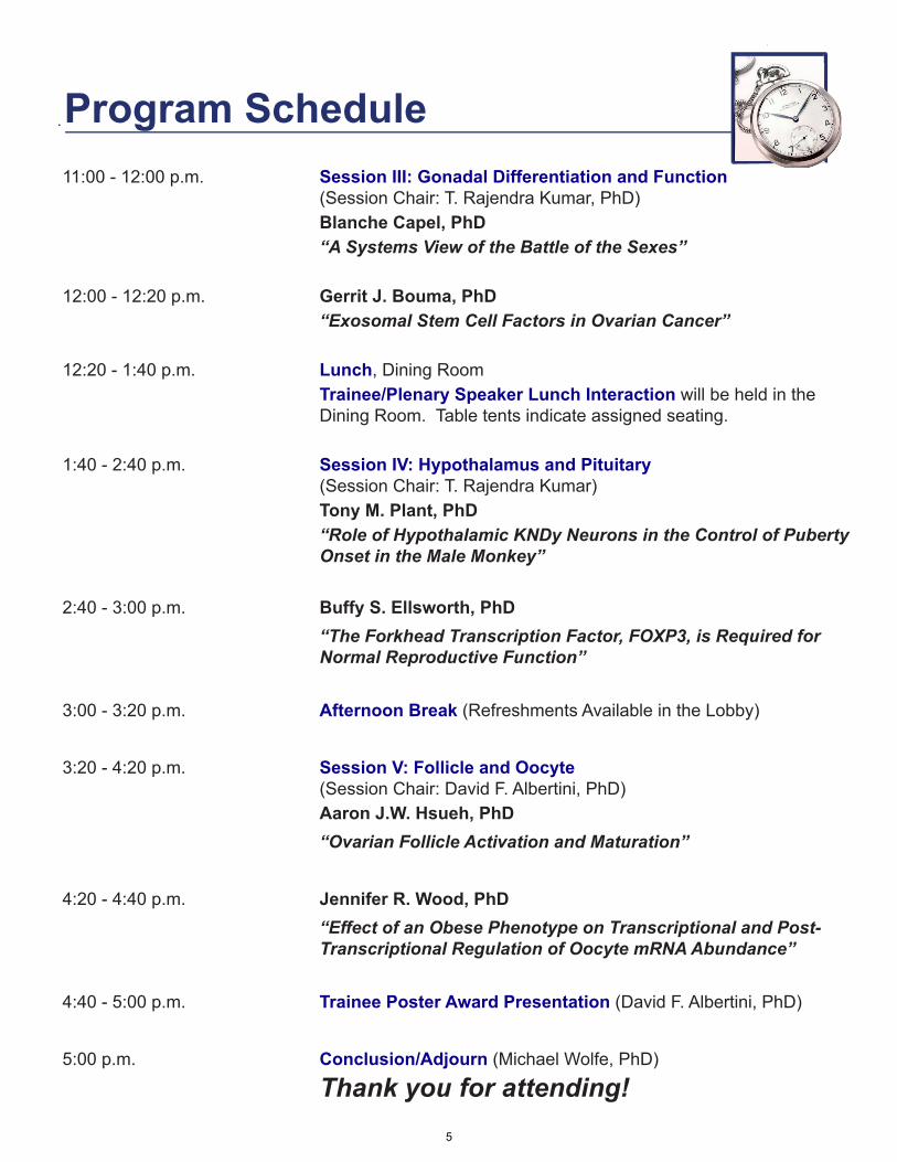
11:00 - 12:00 p.m. Session III: Gonadal Differentiation and Function (Session Chair: T. Rajendra Kumar, PhD)Blanche Capel, PhD“A Systems View of the Battle of the Sexes”
12:00 - 12:20 p.m. Gerrit J. Bouma, PhD“Exosomal Stem Cell Factors in Ovarian Cancer”
12:20 - 1:40 p.m. Lunch, Dining RoomTrainee/Plenary Speaker Lunch Interaction will be held in the Dining Room. Table tents indicate assigned seating.
1:40 - 2:40 p.m. Session IV: Hypothalamus and Pituitary (Session Chair: T. Rajendra Kumar)Tony M. Plant, PhD“Role of Hypothalamic KNDy Neurons in the Control of Puberty Onset in the Male Monkey”
2:40 - 3:00 p.m. Buffy S. Ellsworth, PhD“The Forkhead Transcription Factor, FOXP3, is Required for Normal Reproductive Function”
3:00 - 3:20 p.m. Afternoon Break (Refreshments Available in the Lobby)
3:20 - 4:20 p.m. Session V: Follicle and Oocyte (Session Chair: David F. Albertini, PhD)Aaron J.W. Hsueh, PhD“Ovarian Follicle Activation and Maturation”
4:20 - 4:40 p.m. Jennifer R. Wood, PhD“Effect of an Obese Phenotype on Transcriptional and Post-Transcriptional Regulation of Oocyte mRNA Abundance”
4:40 - 5:00 p.m. Trainee Poster Award Presentation (David F. Albertini, PhD)
5:00 p.m. Conclusion/Adjourn (Michael Wolfe, PhD)
Thank you for attending!
Program Schedule
5
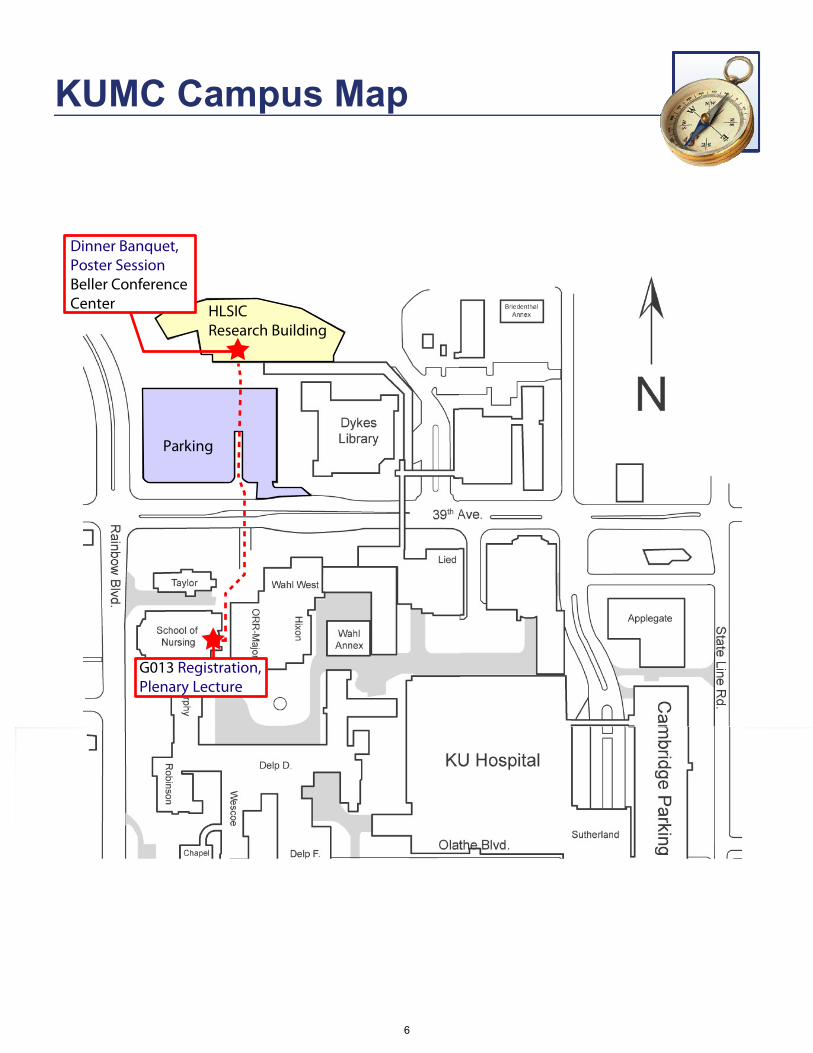
KUMC Campus Map
G013 Registration,Plenary Lecture
HLSICResearch Building
Dinner Banquet,Poster SessionBeller Conference Center
Parking
KUMC Campus Map
6
EXIT 1C
W. Pennway St
Was
hing
ton
St
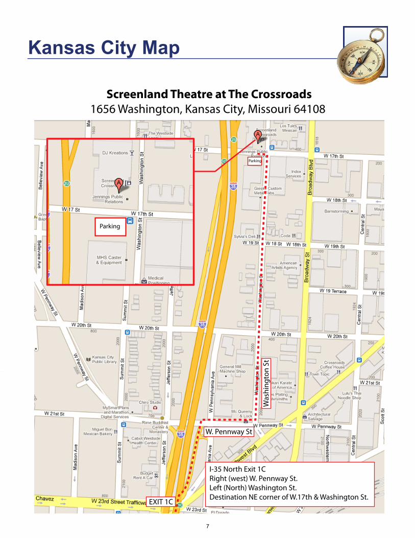
I-35 North Exit 1C Right (west) W. Pennway St.Left (North) Washington St.Destination NE corner of W.17th & Washington St.
Screenland Theatre at The Crossroads1656 Washington, Kansas City, Missouri 64108
Parking
Parking
Kansas City Map
7

Screenland at the Crossroads Located on the edge of the Crossroads and Film Row Districts at 17th & Washington in Kansas City, Missouri, Screenland is a fully renovated creative office center, movie
theatre and event center. You can’t miss their Marquee, which was rescued from the historic Isis Theatre where a very young Walt Disney showed some of his earliest cartoons made right here in Kansas City.
Built in 1913 as a cold storage facility, the building has been transformed into one of the most distinctive buildings in the Crossroads Arts District. After a year of refurbishment, Screenland holds a variety of tenants from advertising agencies to consultants and attorneys. In addition to office space, our special event facility measures over 9,000 square feet in floor space. Screenland has one very important feature that no other venue in Kansas City has - a full service motion picture theatre, capable of both film and digital video projection. The building is innovative, inspirational and a perfect merger of the Film Arts and Kansas City nostalgia.
We hope you enjoy this year’s unique symposium venue!
This year’s lectures will be presented in the Theatre, which seats up to 150 people and features a 26 foot screen. Restrooms are conveniently located near the entrance to the Theatre.
Breakfast and lunch will be served in the Gallery. Restrooms are available adjacent to the gallery.
Venue Information
8
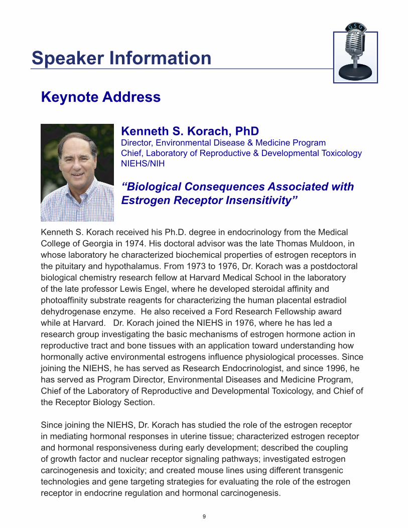
Kenneth S. Korach received his Ph.D. degree in endocrinology from the Medical College of Georgia in 1974. His doctoral advisor was the late Thomas Muldoon, in whose laboratory he characterized biochemical properties of estrogen receptors in the pituitary and hypothalamus. From 1973 to 1976, Dr. Korach was a postdoctoral biological chemistry research fellow at Harvard Medical School in the laboratory of the late professor Lewis Engel, where he developed steroidal affinity and photoaffinity substrate reagents for characterizing the human placental estradiol dehydrogenase enzyme. He also received a Ford Research Fellowship award while at Harvard. Dr. Korach joined the NIEHS in 1976, where he has led a research group investigating the basic mechanisms of estrogen hormone action in reproductive tract and bone tissues with an application toward understanding how hormonally active environmental estrogens influence physiological processes. Since joining the NIEHS, he has served as Research Endocrinologist, and since 1996, he has served as Program Director, Environmental Diseases and Medicine Program, Chief of the Laboratory of Reproductive and Developmental Toxicology, and Chief of the Receptor Biology Section.
Since joining the NIEHS, Dr. Korach has studied the role of the estrogen receptor in mediating hormonal responses in uterine tissue; characterized estrogen receptor and hormonal responsiveness during early development; described the coupling of growth factor and nuclear receptor signaling pathways; investigated estrogen carcinogenesis and toxicity; and created mouse lines using different transgenic technologies and gene targeting strategies for evaluating the role of the estrogen receptor in endocrine regulation and hormonal carcinogenesis.
Kenneth S. Korach, PhDDirector, Environmental Disease & Medicine Program Chief, Laboratory of Reproductive & Developmental Toxicology NIEHS/NIH
“Biological Consequences Associated with Estrogen Receptor Insensitivity”
Keynote Address
Speaker Information
9
Asgi T. Fazleabas, PhD Professor & Associate Chair for ResearchDepartment of Obstetrics & Gynecology & Reproductive BiologyMichigan State University “The Impact of Endometriosis on Uterine Receptivity”
Asgi Fazleabas received his BS degree from California State University, Fresno and his Ph.D. in Reproductive Physiology from the University of Illinois at Urbana-Champaign. Following his post-doctoral training in Reproductive Biology/Cell and Molecular Biology at the University of Florida in Gainesville, he joined the Department of Obstetrics and Gynecology at the University of Illinois at Chicago where he held the rank of Professor and Director of the Center for Women’s Health and Reproduction until October 2009. He currently holds the following positions at Michigan State University: Professor and Associate Chair in the Department of Obstetrics, Gynecology and Reproductive Biology; Director of the Center for Women’s Health and Reproduction; Professor and Associate Chair in the Department of Obstetrics, Gynecology and Reproductive Biology; Director of the Center for Women’s Health Research and Co-Director of the Reproductive and Developmental Sciences Program.
Studies in the Fazleabas’ laboratory are at the leading edge of research into understanding the critical cellular events that define synchrony between the developing embryo and the maternal uterus in a species that is phylogenetically related to humans. His laboratory was the first to conclusively demonstrate that signals from the primate embryo, like those of other species, induce cell specific changes in uterine gene expression. These changes are thought to play critical roles in establishing a synchrony between the maternal environment and the developing embryo that is a pre requisite for a successful pregnancy. These studies have clearly elucidated the mechanisms by which apoptosis is inhibited within the uterus in the presence of a conceptus, the fundamental hormonal and cellular requirements associated with the process of decidualization and potential functions of uterine proteins in the establishment of pregnancy. A hallmark of all the studies from his laboratory is the ability to confirm all their in vitro findings in vivo as a fundamental application of true physiology in the appropriate tissue context. In addition, his laboratory has established a baboon model for endometriosis. The focus of these studies is to understand the etiology and pathophysiology of Endometriosis. The unique nature of the primate model that he has developed to study endometriosis and the strong multi-disciplinary group that he has established has led to important and fundamental findings regarding the causative effects of endometriosis on aberrant gene expression in the eutopic endometrium that may contribute to infertility. Furthermore, studies from the Fazleabas laboratory have also identified the genes that may be involved with the process of angiogenesis and cell adhesion during the establishment of lesions in the peritoneal environment.
Dr. Fazleabas has published 155 scientific articles, secured 18 NIH grants, and has received awards from the CONRAD/Mellon Foundation, University of Illiinois, Ares Advanced Technologies, Inc., Ernst Schering Research Foundation, British Council, Lola Wilson Research Fund, and the University of Sidney.
Session I
10

Yaacov Barak, PhDAssociate Professor Department of Obstetrics, Gynecology & Reproductive BiologyUniversity of Pittsburgh “Molecular Insights into the Placental Functions of PPARgamma”
Dr. Yaacov Barak received his PhD in Molecular Biology from The Weizmann Institute of Science in 1994, followed by postdoctoral training at The Salk Institute in La Jolla, CA, with Dr. Ron Evans. In 2001, Dr Barak accepted an assistant professorship at The Jackson Laboratory, Bar Harbor, ME, and in 2008, he joined Magee-Womens Research Institute, Pittsburgh, PA, as an Associate Professor of OBGYN and Reproductive Sciences. Dr. Barak’s team studies the molecular genetics of both placental development and adipose tissue dynamics. The major technology platforms in his laboratory are mouse gene targeting, stem and primary cell culture, and molecular analysis of gene expression and regulation, integrated with systems approaches for broader and deeper understanding of both organ systems. Dr. Barak has authored numerous peer reviewed research and review manuscripts, and has been awarded grants by the March of Dimes, American Heart Association, NICHD and NIDDK.
Quinton A. Winger, PhDAssistant ProfessorDepartment of Biomedical Sciences, Animal Reproduction & Biotechnology Lab Colorado State University “LIN28 Controls Proliferation & Differentiation of Trophoblast Progenitor Cells”
Quinton Winger received his Ph.D. in Veterinary Physiology and Pharmacology from Texas A&M University in 2000. His postdoctoral training was completed at the Colorado Center for Reproductive Medicine and the Department of Craniofacial Biology and Cellular and Developmental Biology at the University of Colorado Health Sciences Center. Following postdoctoral training, he served as Assistant Professor in the Department of Animal, Dairy and Veterinary Sciences at Utah State University and since 2008 as Assistant Professor in the Department of Biomedical Sciences at Colorado State University.
The main focus of Dr. Winger’s research is investigating the genes that regulate mammalian reproduction. One approach he utilizes is to study genetic regulation of reproduction is gene “knock-out” models that present an abnormal reproductive phenotype. Characterizing these mutant models has resulted in two main areas of research investigating germ cell development and the initiation of meiosis during embryonic development and regulation of the placental trophoblast stem cell lineage during pregnancy.
Session II
11
Blanche Capel, PhDJames B. Duke Distinguished Professor Department of Cell BiologyDuke University Medical Center “A Systems View of the Battle of the Sexes”
Blanche Capel received a BA from Hollins University, and completed her Ph.D. in Genetics from the University of Pennsylvania in 1989. Her post-doctoral work was conducted in the laboratory of Dr. Robin Lovell-Badge at the National Institute for Medical Research in London, where she was involved in the identification and initial characterization of the Y chromosome-linked gene, Sry, that regulates mammalian sex determination. In 1993, she joined the Department of Cell Biology at Duke University Medical Center as an Assistant Professor. In 1999 she was promoted to Associate Professor, and to Full Professor in 2005. She was a recipient of the Langford Prize from Duke University in 1999, and the Hammes Excellence in Teaching award in 2006. In 2010, she was named a James B. Duke Distinguished Professor, and elected as a fellow to the American Association for the Advancement of Science in 2011. She currently is a member of the Board of Scientific Overseers at the Jackson Laboratory and the Board of the Society for the Study of Reproduction.
The Capel laboratory is very interested in the biology of sex determination, and the basic questions it raises about how patterning decisions are made during organ development. Other research in the lab is focused on the morphological reorganization of the cells in the gonad into testis or ovarian structure. Dr. Capel has published 85 scientific articles.
Gerrit J. Bouma, PhDAssistant ProfessorDepartment of Biomedical Sciences, Animal Reproduction & Biotechnology Lab Colorado State University “Exosomal Stem Cell Factors in Ovarian Cancer”
Gerrit J. Bouma received his Ph.D. in Zoology from the University of Idaho. Since completing postdoctoral training at The Jackson Laboratory in 2006, he has served as Assistant Professor in the Animal Reproduction and Biotechnology Laboratory at Colorado State University.
The main focus of Dr. Bouma’s research is to obtain insight into the genetic and molecular factors that underlie fetal and adult cell differentiation and function in reproductive tissues. Projects include (1) studying the role of transcription factor GATA4 in mammalian fetal ovarian development (2) investigating the role of microRNAs in fetal and adult ovarian function and disease and (3) examining the role of stem cell factors in reproductive tissue development and disease.
Session III
12
Tony M. Plant, PhDProfessor Department of Obstetrics & Gynecology and Reproductive SciencesUniversity of Pittsburgh “Role of hypothalamic KNDy neurons in the control of puberty onset in the male monkey”
Dr. Tony Plant completed his undergraduate and graduate training at the University of London, receiving the Ph.D. degree in Physiology in 1971. For postdoctoral studies, he joined the group of Ernst Knobil in the Department of Physiology at the University of Pittsburgh in 1974. In 1978 he joined the faculty of the Department of Physiology as Assistant Professor, and in 1981 he became Director of the Center for Research in Reproductive Physiology at the University of Pittsburgh. Dr. Plant is currently Professor of Obstetrics, Gynecology and Reproductive Sciences, and Cell Biology and Physiology at the University of Pittsburgh School of Medicine. He is also President of the International Neuroendocrine Federation. While in Knobil’s laboratory, he was a member of the team that made the fundamental discovery that sustained gonadotropin secretion by the anterior pituitary gland required intermittent stimulation from the hypothalamic hormone, known as gonadotropin releasing hormone (GnRH). In 1991, he received the Serono Lecture Award from the American Society of Andrology for his work on the hypothalamic control of testicular function in higher primates. Dr. Plant is pursuing two main area of research. The first is directed at elucidating the neurobiological mechanisms that govern the ontogeny of pulsatile GnRH secretion throughout development in the monkey, and that therefore dictate the timing of the onset of puberty in this species. The second interest of this laboratory concerns the operation of the negative feedback loop governing spermatogenesis in the monkey. Dr. Plant has published 111 scientific articles, secured 17 NIH grants and has received awards from the A.W. Mellon Foundation, Fogarty International, GlaxoWellcome, Bioqual, Inc., and the University of Pittsburgh.
Buffy S. Ellsworth, PhDAssistant ProfessorDepartment of Physiology Southern Illinois University “The Forkhead Transcription Factor, FOXP3, is Required for Normal Reproductive Function”
Buffy S. Ellsworth received her Ph.D. in Cell and Molecular Biology from Colorado State University in 2002. Since completing postdoctoral training at University of Michigan Medical School in 2007, she has served as Assistant Professor at Southern Illinois University, Carbondale.
Forkhead transcription factors have been implicated in cell cycle regulation, chromatin remodeling and cell fate determination. We are interested in the role forkhead factors play in the pituitary during development and adulthood. Using mouse as a model system, we are studying how forkhead factors regulate gene expression to affect organ patterning and cell specification during development as well as how forkhead factors regulate pituitary function in adulthood.
Session IV
13
Jennifer R. Wood, PhDAssistant ProfessorDepartment of Animal ScienceUniversity of Nebraska - Lincoln “Effect of an Obese Phenotype on Transcrptional & Post-Transcriptional Regulation of Oocyte mRNA Abundance”
Jennifer R. Wood received her Ph.D. from the Department of Molecular and Integrative Physiology at the University of Illinois in 2000. Since the completion of her postdoctoral training at the Center for Research on Reproduction and Women’s Health at the University of Pennsylvania in 2006, she has served as Assistant Professor of Physiologic Genomics and Reproductive Physiology in the Department of Animal Science at the University of Nebraska-Lincoln.
The focus of Dr. Wood’s research is to determine how factors associated with obesity reduce oocyte quality and embryonic development in order to reverse infertility and/or overcome negative effects of fetal programming on viable offspring. The current focus of her lab is to (1) determine how insulin, leptin, and TNFa regulate transcription versus stability of mRNAs in the oocyte and (2) determine the impact of maternal obesity on the differentiation of adipocyte and myogenic progenitor cells during embyronic development.
Aaron J.W. Hsueh, PhDProfessor Division of Reproductive & Stem Cell BiologyDepartment of Obstetrics & GynecologyStanford University School of Medicine
“Ovarian Follicle Activation & Maturation”
Dr. Aaron Hsueh received his Ph.D. in Cell Biology from Baylor College of Medicine in 1975. Following his postdoctoral training in the Reproduction Research Branch at NICHD, NIH, he served in the Department of Reproductive Medicine at the University of California, San Diego as Assistant Professor from 1976 to 1981, Associate Professor from 1981-1985, and rose to full Professor in 1985. Dr. Hsueh is currently an ovarian physiologist at Stanford University School of Medicine, Department of Obstetrics and Gynecology, Division of Reproductive Biology and Stem Cell Research. He has published in the field for 35 years.
His lab has investigated the hormonal regulation of granulosa cell functions, leading to the establishment of an in vitro FSH bioassay and the design of a long-acting FSH analog in clinical use. His lab also contributed to the understanding of ovarian follicle growth and atresia, intraovarian mechanisms of oocyte maturation and autocrine regulation of early embryonic development. His lab also established and maintained the Ovarian Kaleidoscope Database (OKdb) over the last 10 years as an online resource for ovarian researchers. Recently, his lab established a method to activate dormant ovarian primordial follicles to derive mature murine and human oocytes.
Session V
14
1. Production of gonadotropin-releasing hormone II receptor knockdown swine. Amy T. Desaulniers, Amy M. Voss, Rebecca A. Cederberg, Chanho Lee, Ginger A. Mills, Matthew D. Snyder, and Brett R. White. University of Nebraska-Lincoln, Lincoln, NE.
2. Comparison of hFSH Glycosylation by Electrospray Ionization Mass Spectrometry. George R. Bousfield1, Vladimir Y. Butnev1, Viktor Y. Butnev1, Bin Shuai1, Rajeswari Devabhakthuni1, and David J. Harvey2. 1Department of Biological Sciences, Wichita State University, Wichita, KS and 2Department of Biochemistry, Oxford University, Oxford, UK.
3. Isolation and characterization of recombinant di-glycosylated hFSH. Viktor Y. Butnev, William K. White, Vladimir Y. Butnev, Patrick Tran, Joseph S. Murray, Barbara B. Fowler, Kimberly Taylor, Bin Shuai, Jeffrey V. May, and George R. Bousfield. Department of Biological Sciences, Wichita State University, Wichita, KS.
4. Utilization of Urine To Determine FSH Glycoform Expression During The Menstrual Cycle. Jeffrey V May1, William White1, Barbara Fowler1, Kimberly Taylor1, Patrick Tran1, David Grainger2, Bruce Tjaden2, Chelsea Corwin2, and George R Bousfield1. 1Department of Biological Sciences, Wichita State University, and 2The Center for Reproductive Medicine, Wichita, KS.
5. Production and Characterization of Recombinant Human Follicle Stimulating Hormone. William White, Patrick Tran, Barbara Fowler, Viktor Butnev, George Bousfield. Department of Biological Sciences, Wichita State University, Wichita, KS.
6. Cows with reduced fertility and granulosa cell efficiency have excess androstenedione in follicular fluid, altered theca gene expression and increased maternal effect gene mRNA levels in cumulus-oocyte complexes. Adam F. Summers1, Robert Cushman2, Jacqueline E. Smith1, Bailey Lammers1, Renee McFee1, William Pohlmeier1, Vanessa Brauer1, Kevin Sargent1, Ningxia Lu1, Andrea S. Cupp1, Jennifer R. Wood1. 1University of Nebraska- Lincoln, Lincoln, NE, 2USDA-ARS Roman L. Hruska U.S. Meat Animal Research Center, Clay Center, NE.
7. Expression of Estrogen Receptor α36 in the hamster ovary: possible regulation by gonadotropins and steroid hormones. Prabuddha Chakraborty1, Zhao-yi Wang2, Shyamal K. Roy1,3. 1Department of Cellular and Integrative Physiology, and Obstetrics and Gynecology3, University of Nebraska Medical Center, and 2Medical Microbiology, and Immunology, and Surgery and Pathology, Creighton University Medical center, Omaha, NE.
8. Estrogen Regulation of ERBB3 and EBP1 Expression in Perinatal Hamster Ovaries, Anindit Mukherjee1, A. W. Hamburger3, 4 and S. K. Roy1, 2, Departments of Cellular and Integrative Physiology1 and OB/GYN 2, UNMC, Omaha, NE, Department of Pathology3
University of Maryland, Baltimore, MD and Greenebaum Cancer Center4, University of Maryland, Baltimore, MD.
Abstract Titles
15
9. Tamoxifen prevents ovarian apoptosis and follicle loss from cyclophosphamide in vitro. Brian K. Petroff. Breast Cancer Prevention Center, University of Kansas Medical Center, Kansas City, KS.
10. MicroRNA-21 and PDCD-4 function in the pathogenesis of human uterine leiomyomas. J. Browning Fitzgerald, V. Chennathukuzhi, L. K. Christenson. Department of Molecular and Integrative Physiology, University of Kansas Medical Center, Kansas City, KS.
11. Genes Involved in the Immediate Early Response and Epithelial-Mesenchymal Transition are Regulated by Adipocytokines in the Female Reproductive Tract. Kristin Norwood, Zhufeng Yang, Jacqueline E. Smith, Jill Kerl and Jennifer R. Wood. Dept. Animal Science, University of Nebraska-Lincoln, Lincoln, NE.
12. Transforming growth factor alpha (TGFα), via a possible autocrine/paracrine mechanism, regulates granulosa cell tumor (GCT) cell proliferation and migration through activation of multiple pathways. Cheng Wang1, 2, Chao Jiang1, 2, Lan Fu1, 2, Lele Subodh3, and John S Davis1, 2, 4. 1 Olson Center for Women’s Health, 2 Department of OB/GYN, 3 Department of Pathology and Microbiology, University of Nebraska Medical Center, Omaha, NE, 4 VA Medical Center, Omaha, NE.
13. Na,K-ATPase α4 isoform is critical for sperm motility and fertility. Tamara Jimenez, Jeffrey P. McDermott, Gladis Sánchez and Gustavo Blanco. Department of Molecular and Integrative Physiology, University of Kansas Medical Center. Kansas City, KS.
14. rDmrt1 transgene drives copy-number dependent gene expression changes in Sertoli cell and germ cells. Valentine A. Agbor 1 and Leslie L. Heckert1. 1 Department of Molecular and Integrative Physiology, University of Kansas Medical Center, 3901 Rainbow Blvd. Kansas City, KS.
15. Neuropilin-1 (NRP-1) loss in Sertoli cells reduces expression of genes necessary for spermatogonial stem cells (SSC) niche establishment. Kevin M Sargent, Meredith L Bremer, William E Pohlmeier, Vanessa M Brauer, and Andrea S Cupp. Department of Animal Science, University of Nebraska-Lincoln, Lincoln, NE.
16. Protein Kinase C- A common signaling pathway dictating self renewal vs. differentiation in mouse, rat and human embryonic stem cells. Debasree Dutta 1, James Hong 2, Soma Ray 1, Pratik Home1, Arindam Paul1, Biswarup Saha1, Michael Wolfe3, Mark L. Weiss2 and Soumen Paul1 1. Department of Pathology and Laboratory Medicine, 3. Department of Molecular & Integrative Physiology, Institute for Reproductive Health and Regenerative Medicine, University of Kansas Medical Center, Kansas City, KS. 2. Department of Anatomy and Physiology, College of Veterinary Medicine, Kansas State University, Manhattan, KS.
17. Controlling First Mammalian Lineage Specification Through Combinatorial Histone Modifications. Biswarup Saha1, Pratik Home1, Partho Chattoraj1, Soma Ray1, Debasree Dutta1, Melissa Larson2 and Soumen Paul1. 1 Institute for Reproductive Health and Regenerative Medicine, Dept. of Pathology and Laboratory Medicine, University of Kansas Medical Center, 2 Transgenic and Gene-targeting Institutional Facility, University of Kansas Medical Center, Kansas City, KS.
16
18. Sub-cellular Localization of Transcription Factor TEAD4 Regulates First Mammalian Lineage Commitment. Pratik Home1, Biswarup Saha1, Soma Ray1, Debasree Dutta1, Melissa Larson2 and Soumen Paul1. 1 Institute for Reproductive Health and Regenerative Medicine, Dept. of Pathology and Laboratory Medicine, 2Transgenic and Gene-targeting Institutional Facility, Univ. of Kansas Medical Center, Kansas City, KS.
19. Identification of JMJD2B Pathways Associated with Tumor Progression. Lei Qiu1,2, Judith A. Chapman1, and Adam J. Krieg1,2. 1Department of Obstetrics and Gynecology, 2Department of Pathology and Laboratory Medicine, University of Kansas Medical Center, Kansas City, KS.
20. SATB homeobox proteins regulate trophoblast stem cell renewal and differentiation. Kazuo Asanoma, Kaiyu Kubota, Damayanti Chakraborty, Stephen J. Renaud, Michael J. Soares, and M.A. Karim Rumi. Institute for Reproductive Health and Regenerative Medicine, Department of Pathology and Laboratory Medicine, University of Kansas Medical Center, Kansas City, KS.
21. Focal adhesion kinase is a regulator of trophoblast motility and invasion. Stephen J. Renaud, M.A. Karim Rumi, and Michael J. Soares. Institute for Reproductive Health and Regenerative Medicine, Department of Pathology and Laboratory Medicine, University of Kansas Medical Center, Kansas City, KS.
22. FOSL1 is a key regulator of trophoblast invasion and uterine vascular remodeling. Kaiyu Kubota, Lindsey N. Kent, M. A. Karim Rumi and Michael J. Soares. Institute for Reproductive Health and Regenerative Medicine, Department of Pathology and Laboratory Medicine, University of Kansas Medical Center, Kansas City, KS.
23. The Effect of Leptin Receptor Knockout, in the Mouse Conceptus, on Placental Morphology and Gene Expression at d18.5. Kelly E. Pollock, Ashley Sigafoos and Laura Clamon Schulz. University of Missouri, Columbia, MO.
24. Preliminary Analysis of Food Restriction and Leptin Replacement on Fetal Programming in Mice. Kathleen A. Pennington Lindsey B. Martin and Laura Clamon Schulz. Department of Ob-GYN and Women’s Health, University of Missouri, Columbia MO.
25. Effect of smoking on human sperm parameters is modified by glutathione-S-transferase (GST) T1 genotype Renée S Mijal1,2, Julia J Wirth2,3, Bridget Messaros2,4, Karen Friderici5, Michael P Diamond6, Kathy A Jernigan5, Douglas Daly7, Elizabeth Puscheck6, Nigel Paneth2, and Qing Lu2. 1Department of Preventive Medicine and Public Health, University of Kansas Medical Center, Kansas City, KS, 2Department of Epidemiology, Michigan State University, East Lansing, MI, 3Department of Obstetrics and Gynecology, Michigan State University, East Lansing, MI, 4Biomedical Research Informatics Core, Michigan State University, East Lansing, MI, 5Department of Microbiology and Human Genetics, Michigan State University, East Lansing, MI, 6Department of Obstetrics and Gynecology, Wayne State University, Detroit, MI, 7Grand Rapids Infertility and IVF, Grand Rapids, MI.
17
26. The effect of smoking on mid-pregnancy angiogenic marker levels among pregnancies ending in the delivery of small-for-gestational age (SGA) infants. Renée S. Mijal1, Claudia B. Holzman2, Jian-ling Wang2, Sarosh Rana3, S. Ananth Karumanchi3, Alla Sikorskii4. 1Department of Preventive Medicine and Public Health, University of Kansas Medical Center, Kansas City, KS, 2Department of Epidemiology, Michigan State University,3 Department of Obstetrics and Gynecology, Beth Israel Deaconess Medical Center, Harvard Medical School, Boston, MA, 4Department of Statistics and Probability, Michigan State University, East Lansing, MI.
27. Role of hypoxia signaling in trophoblast cell lineage commitment. Damayanti Chakraborty, M.A. Karim Rumi, Adam J. Krieg, and Michael J. Soares, Institute for Reproductive Health and Regenerative Medicine, Departments of Pathology and Laboratory Medicine and Obstetrics & Gynecology, University of Kansas Medical Center, Kansas City, KS.
28. Regulation of fetal antigen expression in the human placenta by hypoxiaCaitlin Linscheid1, Lei Qui1, Herbert Hodes2 and Margaret G. Petroff1. 1Department of Anatomy and Cell Biology, University of Kansas Medical Center, Kansas City, KS. 2The Center for Women’s Health, Overland Park, KS.
29. Transcriptional response to maternal diet-induced obesity in the mouse blastocyst. Pablo Bermejo-Álvarez1, Cheryl S. Rosenfeld1,2 and R. Michael Roberts1,3,4. 1Bond Life Sciences Center, 2Biomedical Sciences, 3Animal Sciences and 4Biochemistry, University of Missouri, Columbia, MO.
30. Identification of a placental-hepatic axis regulating pregnancy-dependent adaptations to hypoxia. Pengli Bu, Shigeki Ohboshi, Jay L. Vivian, and Michael J. Soares. Institute for Reproductive Health and Regenerative Medicine, Department of Pathology and Laboratory Medicine, University of Kansas Medical Center, Kansas City, KS.
31. Auto Immune Regulator (AIRE) deficiency results in infertility involving embryonic loss and the generation of anti – placental antibodies in mice. Bryce D. Warren1, Susmita Jasti1, Brian K Petroff2, and Margaret G Petroff1. Departments of 1Anatomy and Cell Biology and 2Internal Medicine, University of Kansas Medical Center, Kansas City, KS.
32. Immunomodulators and exosomes from the placenta: Implications for maternal-fetal immune tolerance. S M Khorshed Alam1, Sarika K. Kshirsagar2, Herbert Hodes1, Margaret G. Petroff1. Departments of 1Anatomy and Cell Biology and 2Molecular and Integrative Physiology, University of Kansas Medical Center, Kansas City, KS.
33. Molecular Assessment of the Myometrium During Preterm (PTL) and Term Labor (TL) Using Gene Expression and Biological Pathway Analysis. Clifford W. Mason1, Irina A. Buhimschi2, Catalin S. Buhimschi2, Yafeng Dong1, and Carl P. Weiner1. 1Department of Obstetrics and Gynecology, University of Kansas Medical Center, Kansas City, KS. 2Department of Obstetrics, Gynecology and Reproductive Sciences, Yale University, New Haven, CT.
18
1. Production of gonadotropin-releasing hormone II receptor knockdown swine. Amy T. Desaulniers, Amy M. Voss, Rebecca A. Cederberg, Chanho Lee, Ginger A. Mills, Matthew D. Snyder, and Brett R. White. University of Nebraska-Lincoln, Lincoln, NE. The second mammalian isoform of GnRH (GnRH-II) is highly conserved from bony fish to man. However, the coding sequence for the receptor specific to this ligand contains reading errors in many species, suggesting the inability to produce a functional receptor. In contrast, the porcine GnRH-II receptor gene contains the appropriate sequence to produce functional protein. The objective of this study was to develop swine with reduced levels of endogenous GnRH-II receptors. Two potential target small hairpin RNA (shRNA1 and shRNA2) sequences specific to the porcine GnRH-II receptor were identified and subcloned into the lentiviral-based, pLVX-shRNA2 vector (Clontech) that provides both shRNA and fluorescent ZsGreen1 coexpression. Lentiviral particles were produced from each shRNA vector as well as a control vector using the Lenti-X HTX Packaging System (Clontech). Lentiviral particles containing either shRNA1 or shRNA2 sequences significantly reduced GnRH-II receptor mRNA levels (95 and 99%, respectively) compared to control particles (P < 0.05) in a swine testis-derived (ST) cell line. Later, lentiviral particles containing the shRNA2 sequence (1.15 x 10⁹) were microinjected within the perivitelline space of in vivo derived pronuclear zygotes (n = 15). Microinjected zygotes were subsequently cultured in 50 μl drops of NCSU-23 under mineral oil in a humidified 5% CO2 air environment. Following 120 h of culture, 93% of the zygotes developed to the compact morula stage whereas 87% formed blastocysts at 168 h. Fluorescent microscopy revealed that all blastocysts expressed ZsGreen1, indicating a 100% transduction efficiency of shRNA2 lentiviral particles. Finally, embryos were surgically collected from white crossbred donor sows and transduced as before. A total of 40 and 33 microinjected zygotes were immediately transferred into 2 synchronized recipient females that will be allowed to gestate to term. Progeny from this study represent the first model to examine the physiological implications of reduced GnRH-II receptor levels.
2. Comparison of hFSH Glycosylation by Electrospray Ionization Mass Spectrometry. George R. Bousfield1, Vladimir Y. Butnev1, Viktor Y. Butnev1, Bin Shuai1, Rajeswari Devabhakthuni1, and David J. Harvey2. 1Department of Biological Sciences, Wichita State University, Wichita, KS and 2Department of Biochemistry, Oxford University, Oxford, UK. FSH is a highly heterogeneous glycoprotein hormone, which possesses 4 N-glycosylation sites, each decorated with a family of glycans. Recent advances in mass spectrometry now permit direct evaluation of FSH glycans. We compared FSH glycosylation in human FSH preparations derived from pituitary glands, postmenopausal urine, and recombinant hFSH expressed in rat GH3 cells. The total glycan populations from pituitary and postmenopausal urine hFSH preparations were virtually identical. This is a highly significant finding, as the absence of major changes in glycan populations between pituitary and urinary FSH means that serum FSH glycans must also be the same. Therefore, examination of urinary hFSH glycosylation is directly relevant to serum hFSH glycosylation. Glycosylation of tetra-glycosylated hFSH and two di-glycosylated hFSH preparations showed great similarity between di-glycosylated hFSH derived from FSH fractions glycans and those derived from tetra-glycosylated hFSH. Both glycan populations were
Full Abstracts
19
very similar to those from pituitary hFSH, except sulfated glycan abundance was higher in the glycoform preparations. Glycosylation of di-glycosylated hFSH isolated from hLH preparations was strikingly different from all other hFSH preparations. The glycans were largely high mannose, which are rare in FSH. Glycosylation of recombinant hFSH revealed the absence of tetra-antennary glycans, which comprised 15% of pituitary hFSH glycans. Moreover, recombinant hFSH triantennary glycans possessed a third branch, linked b1-6 to the a1-6 mannose residue, which is due to the action of GlcNAc transferase V. In pituitary FSH this branch is linked b1-4 to the a1-3 mannose residue, as a consequence of GlcNAc transferase IV. This suggested the absence of GlcNAc transferase IV in rat GH3 cells. However, RT PCR found evidence for expression of both transferases. In order to make a more pituitary-like recombinant hFSH, GH3 cells will have to be engineered to express greater GlcNAc transferase IV activity. Supported by NIH grant P01 AG029531.
3. Isolation and characterization of recombinant di-glycosylated hFSH. Viktor Y. Butnev, William K. White, Vladimir Y. Butnev, Patrick Tran, Joseph S. Murray, Barbara B. Fowler, Kimberly Taylor, Bin Shuai, Jeffrey V. May, and George R. Bousfield. Department of Biological Sciences, Wichita State University, Wichita, KS. Limited availability of human pituitaries and low recovery of purified di-glycosylated human hFSH (di-hFSH) from these glands represent significant problems for purification of di-hFSH in preparative amounts for structural and functional characterization. To provide a replentishible supply of di-hFSH for comprehensive investigation our lab has developed a new purification procedure for isolation of this precious glycoform from a stable transformed GH3 cell line. A combination of two different rounds of immunoaffinity chromatography and high-resolution tandem triple-column Superdex 75 gel-filtration resulted in purification of di-hFSH identified by Western blot analysis, heterodimer-specific radioimmunoassay, amino acid sequencing, and hFSH receptor binding assay. The purified recombinant di-hFSH displayed enhanced receptor binding activity equivalent to that of the pituitary-derived di-hFSH described previously. Large-scale purification of di-hFSH from 14 L of serum-containing GH3-conditioned medium began with sequential ammonium sulphate precipitation at 50% and 75% saturation. The precipitates were collected, dialyzed against 0.1 M ammonium bicarbonate buffer, lyophilized, and applied to an affinity column with anti-hFSHβ monoclonal antibody. The preliminary results demonstrate that the hFSH preparations purified from 50% and 75% AS precipitates exhibit different mobilities for both a and β subunits during SDS-PAGE under reduced conditions, which might be due to different extent of their glycosylation. Supported by the NIH PO1 Grant, AG029531.
4. Utilization of Urine To Determine FSH Glycoform Expression During The Menstrual Cycle. Jeffrey V May1, William White1, Barbara Fowler1, Kimberly Taylor1, Patrick Tran1, David Grainger2, Bruce Tjaden2, Chelsea Corwin2, and George R Bousfield1. 1Department of Biological Sciences, Wichita State University, and 2The Center for Reproductive Medicine, Wichita, KS. Human pituitary FSH exists as a mixture of two glycoforms due to either all-or-none glycosylation of the ß subunit. The glycoforms exhibit differential age-related expression and markedly different in vitro bioactivities. Preliminary data suggest that urinary FSH glycoform ratios reflect pituitary ratios and that the ratios may change during the menstrual cycle. We have begun to assess urine as a means to characterize FSH glycoform expression. Seven cycling women, 28-47 years of age, not taking steroid hormones, provided first morning urine voids for a complete menstrual cycle. Mean cycle length of the group was 30 +/- 2 days (SEM) and the mean weight was 212 +/- 21 pounds. Specimens were measured and 15-40 ml were concentrated via ultra-filtration, lyophilized, re-suspended in 1/10th the original volume, and subjected to RIA. The remaining
20
specimen was subjected to 80% v/v ethanol precipitation, centrifugation, and re-suspension of the pellet in buffer for eventual FSH purification and Western Blot analysis. Mean cycle urine volumes among subjects ranged from 133 to 624 ml. The SEM for urine volumes was 4-9% of the mean indicating individual subjects produced extremely consistent first morning voids. Only one subject (29 years old) exhibited a distinct, mid-cycle FSH surge while the remaining subjects exhibited either no surge or exhibited random FSH peaks. A 29 year-old subject exhibited no clear FSH surge. However, she was the heaviest subject at 310 pounds. The FSH surge subject produced a LH surge that mirrored the FSH surge. We hypothesized that variation of cycle FSH levels would be greater in young versus older women. Regression analysis of age versus the standard deviation of daily FSH levels indicated exactly that (r = -0.76). These initial results indicate that urine analysis will be a useful approach to investigate FSH glycoform expression. (Support: NIH Grant P01AG029531 to GRB).
5. Production and Characterization of Recombinant Human Follicle Stimulating Hormone. William White, Patrick Tran, Barbara Fowler, Viktor Butnev, George Bousfield. Department of Biological Sciences, Wichita State University, Wichita, KS. Cell culture of rat pituitary derived GH3 cells producing recombinant human follicle stimulating hormone is followed by concentration of the protein free conditioned media via a spiral concentrator to reduce loading volumes for subsequent immunoaffinity purification. After concentration, multiple affinity columns coupled to monoclonal antibody mAb 4882 anti-hFSH are used to bind the hormone and eliminate most of the other contaminants in the conditioned media. Gel filtration is then used to fractionate the bound fraction from immunopurification resulting in highly pure fractions that contain rhFSH heterodimer and rhFSH free subunits. Western blot analysis reveals effective separation of tetraglycosylated rhFSH from the diglycosylated form of the hormone which elutes later in the fractionation window.
6. Cows with reduced fertility and granulosa cell efficiency have excess androstenedione in follicular fluid, altered theca gene expression and increased maternal effect gene mRNA levels in cumulus-oocyte complexes. Adam F. Summers1, Robert Cushman2, Jacqueline E. Smith1, Bailey Lammers1, Renee McFee1, William Pohlmeier1, Vanessa Brauer1, Kevin Sargent1, Ningxia Lu1, Andrea S. Cupp1, Jennifer R. Wood1. 1University of Nebraska- Lincoln, Lincoln, NE, 2USDA-ARS Roman L. Hruska U.S. Meat Animal Research Center, Clay Center, NE. The intrinsic and exogenous factors that result in abnormal ovarian function and ultimately female infertility are poorly defined. Thus, we have established a cow model of fertility to identify mechanisms regulating follicular growth, steroidogenesis and oocyte maturation. Culling age due to failure to establish pregnancy was used to classify animals with low (≤ 2 years-of-age; LRL) and high (≥6 years-of-age; HRL) reproductive longevity. Animals were subsequently classified based on estradiol:androstenedione (E2:A4) ratios (a ratio <100 = low granulosa cell efficiency (LGE); a ratio >100 = high granulosa cell efficiency (HGE)). Females from each classification were synchronized using a modified Co-Sync (CIDR) protocol and ovariectomies were performed 36 h after PGF2α injection and CIDR removal. Follicular fluid, theca cells, mural granulosa cells and cumulus-oocyte complexes (COC) from each dominant follicle were collected. Androstenedione (A4) concentrations were greater (P < 0.01) in follicular fluid from LGE compared to HGE cows. Given that increased androgens are a hallmark characteristic of the PCOS phenotype, gene expression in theca cells from the different groups was compared. Quantitative, real-time RT-PCR demonstrated that mRNA abundance for Cyp17a1 was increased (P < 0.01) in all groups compared to HRL-HGE. Abundance of Cyp11a1 was also increased in LRL-LGE cows compared to HRL-HGE. In the cumulus-oocyte complex, the mRNA abundance of maternal effect genes was altered. Dnmt1 was increased in LRL-LGE compared to HRL-HGE cows and Zar1 abundance
21
was increased (P < 0.01) in HRL-LGE cows when compared to both LRL-HGE and HRL-HGE cows. Increased mRNA abundance in lower fertility animals is consistent with previous findings in PCOS oocytes and suggests that increased androgen production in low fertility animals alters gene expression and/or mRNA stability during oocyte growth and maturation. USDA is an equal opportunity employer.
7. Expression of Estrogen Receptor α36 in the hamster ovary: possible regulation by gonadotropins and steroid hormones. Prabuddha Chakraborty1, Zhao-yi Wang2, Shyamal K. Roy1,3. 1Department of Cellular and Integrative Physiology, and Obstetrics and Gynecology3, University of Nebraska Medical Center, and 2Medical Microbiology, and Immunology, and Surgery and Pathology, Creighton University Medical center, Omaha, NE. Estradiol-17β (E) acting via its cognate receptors affects ovarian functions in mammalian females, including women. The objectives of the present study were to examine, by immunoblotting and immunofluorescence localization, whether estrogen receptor a36 (ERa36), a splice variant of classic ESR1, was expressed in the hamster ovary throughout the estrous cycle, and whether the expression was regulated by FSH, LH, E and progesterone (P). Immunoblot data indicated that ERα36 expression declined by Proestrus (D4):0900h compared to earlier days of the estrous cycle (D3:0900, 5.86 ± 1.4 vs. D4:0900, 2.72 ± 0.31; p<0.05) and remained low up to day 4 afternoon. Immunofluorescence results corroborated the findings and revealed that ERα36 was expressed only in the cell membrane of both follicular and interstitial cells. Hypophysectomy (Hx) resulted in a significant decline in ERα36 protein levels (Hx: 3.38 ± 0.28 vs. D1: 10.07 ± 2.82; p<0.01). The levels of ERα36 protein in FSH (8.76 ± 0.84) or LH (8.78 ± 0.58)-treated hamsters were comparable to those of D1 hamsters (P> 0.05), but were lower in hamsters treated with combined doses of FSH and LH (6.37 ± 0.43). Neither E nor P alone or combined could affect ovarian ERa36 levels. The data were consistent with immunofluorescence findings. These results indicate that the ERa36 is translated into protein in ovarian follicular and non-follicular cells, and is localized in the cell membrane. Further, the induction of alternate ESR1 mRNA splicing and the translation of the truncated transcript seems to be regulated by FSH as well as LH. (Values are expressed in Mean OD ± SEM). The work was supported by a grant from the NIH (R01 HD38468) and Olson Foundation to SKR. P. Chakraborty is a graduate student in the Department of Cellular and Integrative Physiology.
8. Estrogen Regulation of ERBB3 and EBP1 Expression in Perinatal Hamster Ovaries, Anindit Mukherjee1, A. W. Hamburger3, 4 and S. K. Roy1, 2, Departments of Cellular and Integrative Physiology1 and OB/GYN 2, UNMC, Omaha, NE , Department of Pathology3 University of Maryland, Baltimore, MD and Greenebaum Cancer Center4, University of Maryland, Baltimore, MD. Primordial follicle formation is the first step in ovarian follicular development in mammals. The initial pool size of primordial follicles determines the lifetime quota of available oocytes and thereby fertility. Defects in primordial follicle formation may lead to premature ovarian failure (POF). Estrogen (E) has been shown to affect this process, but the mechanism is unknown. E is known to activate ERBB3-mediated signaling, a known mitogenic pathway. ERBB3 is associated with a repressor protein EBP1, which dissociates upon phosphorylation resulting in ERBB3 activation. We hypothesize that E promotes primordial follicle formation by regulating the expression and/or function of ERBB3 and its repressor EBP1.The objective of this study was to determine whether the expressions of ERBB3 as well as its repressor, EBP1, are regulated by E in ovarian cells during somatic cell and oocyte assembly forming primordial follicles. We used perinatal hamster ovaries for this study. The Western blot analyses showed that EBP1 expression
22
was downregulated in 8-day old (P8) ovaries containing primordial follicles compared to 15-day old fetal (E15) ovaries lacking any follicle (0.73 ± 0.171 vs. 1.322 ± 0.359 P < 0.05). ERBB3 expression on the other hand was upregulated. Whereas pEBP1 (ser 363) was localized primarily in the oocytes of E15 ovaries, it was mostly localized in somatic cells juxtaposed to the oocytes and also in the granulosa cells of P8 ovaries. E treatment on P8 suppressed EBP1 expression at 4hr and 24hr (0.229 ± 0.130) time points but a 7-day long treatment, (injections on P1 and P4) did not significantly alter EBP1 levels on P8 (0.807 ± 0.194) compared to control, suggesting a possible rebound of EBP1 expression following its initial suppression. Twelve-day old fetuses (E12) were treated in utero with an FSH-antiserum (FSH-AS) to reduce the effective levels of serum FSH, and consequently of E levels in P8 hamsters. FSH-AS significantly upregulated EBP1 expression compared to P8 (P < 0.01) but E replacement on P1 or P4 did not have a significant lowering effect. In contrast, ovarian ERBB3 expression was reduced on P8 in the antiserum treated group compared to untreated animals. E injection on P1 or on P1 and P4 significantly upregulated ERBB3 expression compared to antiserum treated animals. These results suggest that EBP1 and ERBB3 protein expression is inversely related in postnatal hamster ovaries, especially during primordial follicle formation, and their expressions are regulated by E. Because ERBB3 activation is facilitated by EBP1 downregulation, it is logical to speculate that ERBB3 activation by E may play a critical role in primordial follicle formation.
9. Tamoxifen prevents ovarian apoptosis and follicle loss from cyclophosphamide in vitro. Brian K. Petroff. Breast Cancer Prevention Center, University of Kansas Medical Center, Kansas City, KS. Recent cryopreservation approaches to fertility preservation in cancer patients have had poor uptake due to their expense, invasiveness and need for delay of cancer treatment. Our group recently discovered that the selective estrogen receptor modulator (SERM) tamoxifen (TAM) prevents follicle loss and preserves fertility following exposure of animals to two widely used and ovotoxic cancer drugs, cyclophosphamide (CPA) and doxorubicin. In an effort to localize the ovarian-sparing mechanisms of TAM, cultured rat ovaries (d4, n=8/group) were treated for 24-96 hours with the active metabolites of CPA (0, 1 and 2 µM) and TAM (0 and 1 µM) in vitro and both apoptosis and follicle numbers were measured. Tamoxifen pretreatment markedly decreased follicular loss and apoptosis from active CPA in vitro while TAM alone had no effect on these parameters. CPA vs. TAM+CPA cDNA microarray analysis (n=8-10 ovaries/group) revealed decreased expression of genes facilitating intercellular adhesion and drug transport following TAM treatment. Biomarkers genes for CPA toxicity were downregulated and apoptotic pathway genes had decreased expression as well. Tamoxifen appears to act directly on the ovary to decrease toxicity to the follicular reserve from cyclophosphamide. These studies suggest that protective mechanisms of TAM include ovarian-specific changes in chemotherapy drug delivery and action.
10. MicroRNA-21 and PDCD-4 function in the pathogenesis of human uterine leiomyomas. J. Browning Fitzgerald, V. Chennathukuzhi, L. K. Christenson. Department of Molecular and Integrative Physiology, University of Kansas Medical Center, Kansas City, KS. Human uterine leiomyomas (ULMs) are tumors of the myometrium that are clinically apparent in 25% of reproductive-aged women. They can lead to uterine bleeding, pelvic pain, reproductive dysfunction and hysterectomies. While genetic factors play a role in ULMs, its pathogenesis is not well understood. MicroRNA have been implicated in the etiology of many diseases and recently it was shown that microRNA-21 (miR-21), a microRNA important in apoptotic function, is highly upregulated in ULM tissue. Investigations in cancer cell lines have identified PDCD-4, a gene critical in apoptotic regulation, as a miR-21 target. The purpose of this project is to investigate
23
the potential role miR-21 has in the pathophysiology of ULMs and determine if miR-21 targeting of PDCD-4 is important in mediating that role. This project utilized the human uterine myometrial cell line, UtLM-hTert. A locked nucleic acid specific for miR-21 (LNA-21) was transfected into the cells to knockdown miR-21. Total RNA was collected to verify knockdown of miR-21 and protein was collected to analyze expression of PDCD-4, cleaved caspase 3 and phospho-EF2. PDCD-4 was 3-fold induced while phospho-EF2 was 5-fold induced 24 hours after miR-21 knockdown (n=3, p<.05). Cleaved caspase 3 was upregulated 24 hours after miR-21 knockdown over three independent experiments. At 36 h after miR-21 inhibition, the cells displayed morphological abnormalities consistent with induction of cell death. These findings suggest that miR-21 is important in apoptotic and translational regulation in UtLM-hTert cells. PDCD-4 mRNA levels from paired leiomyoma and myometrium found a 1.32 fold induction of PDCD-4 in leiomyoma tissue (n=23, p=.007). PDCD-4 protein levels from paired tissue showed upregulation and downregulation of the 53kd isoform and the 29kd isoform, respectively, in leiomyoma vs. myometrial tissue over 3 independent experiments. These findings indicate that PDCD-4 is regulated primarily through a post-transcriptional mechanism in leiomyoma tissue. Future studies will determine if PDCD-4 has functional relevance in leiomyomas and if it is post-transcriptionally regulated by miR-21.
11. Genes Involved in the Immediate Early Response and Epithelial-Mesenchymal Transition are Regulated by Adipocytokines in the Female Reproductive Tract. Kristin Norwood, Zhufeng Yang, Jacqueline E. Smith, Jill Kerl and Jennifer R. Wood. Dept. Animal Science, University of Nebraska-Lincoln, Lincoln, NE. Studies have identified a relationship between obesity and the incidence of numerous cancers; however the underlying mechanistic link between the two is ill-defined. The levels of metabolic hormones and pro-inflammatory cytokines (i.e. adipocytokines) including IGF-1, leptin, tumor necrosis factor alpha (TNFa), and interleukin 6 (IL-6) are often altered in obese individuals. Furthermore, these adipocytokines have mitogenic and/or transformative properties. The objective of this study was to identify adipocytokine-dependent changes in the expression of immediate early (IE) genes which contribute to cell proliferation and differentiation and epithelial-mesenchymal transition (EMT) genes which promote cell migration. To determine the effect of individual adipocytokines on the abundance of IE (cJUN, cFOS, and cMYC) and EMT (SNAI1, SNAI2, and TWIST1) mRNA abundance HeLa cells were treated with IGF-1, leptin, IL-6, or TNFa for 0-48 hours and quantitative, real-time PCR (qPCR) analyses were carried out. IGF-1 increased cJUN and cFOS; leptin increased cFOS; IL-6 increased cFOS and cMYC; and TNFa increased c-JUN and c-FOS mRNA abundance. Furthermore, SNAI1 was increased by IGF-1 and IL-6; SNAI2 was increased by IGF-1 and TNFa; and TWIST1 was increased by TNFa and IL-6. To determine the in vivo effects of adipocytokines on IE and EMT mRNA abundance, RNA was isolated from the whole uterus of obese and normal weight mice and qPCR analysis was carried out. While there was no difference in cJun, cFos, or cMyc mRNA abundance between normal-weight and obese animals, Snai1, Snai2, and Twist1 mRNA abundance was increased in the uterus of obese females. This increased mRNA abundance was correlated with increased circulating IGF-1 levels in the obese females. These data indicate that alterations in adipocytokine levels associated with obesity regulate the expression of genes associated with cell proliferation and migration and therefore may provide a plausible mechanism for obesity-dependent increases in cancers of the female reproductive tract.
12. Transforming growth factor alpha (TGFα), via a possible autocrine/paracrine mechanism, regulates granulose cell tumor (GCT) cell proliferation and migration through activation of multiple pathways. Cheng Wang1, 2, Chao Jiang1, 2, Lan Fu1, 2, Lele Subodh3, and John S Davis1,
24
2, 4. 1 Olson Center for Women’s Health, 2 Department of OB/GYN, 3 Department of Pathology and Microbiology, University of Nebraska Medical Center, Omaha, NE 68198, 4 VA Medical Center, Omaha, NE, USA. Granulosa cell tumors (GCTs) are thought to be tumors of low malignant potential, but they have a tendency for late recurrence and a small portion also show aggressive behavior. Metastasis of these tumors has been reported and can involve any organ system. Excessive estrogen production by these tumors stimulates the endometrium, leading to the development of endometrial hyperplasia in 30-50% of patients and endometrial adenocarcinoma in 8-33% of patients. Some patients also present with symptoms of androgen excess. The mechanisms by which ovarian granulosa cells undergo malignant transformation and GCT recurrence are unknown. TGFα is a known potent mitogen. Its impact on development and progression of epithelial ovarian cancer has been studied. However, its function on the GCT initiation and progression is still unclear. The aim of the present study is to determine whether TGFα also plays important roles on the development and progression of GCT. KGN cells, which was derived from an invasive ovarian granulosa cell carcinoma and had many features of normal granulosa
cells, were used as a cell model to detect the effect of TGFα on the growth and migration of GCT cells. Immunohistochemistry, Western blot and RT-PCR results suggested that all members of ErbB family receptors are expressed in the GCT samples and KGN cells. RT-PCR result also indicated that TGFα and EGF are expressed in KGN cell line. Treatment of KGN cell with TGFα stimulated cell DNA synthesis, enhanced cell proliferation, increased cell viability and promoted cell cycle progression. Treatment with TGFα also induced KGN cell morphological transition and stimulated KGN cell migration. TGFα rapidly activated EGFR/PI3K/Akt and mTOR pathways, as indicated by rapid phosphorylation of Akt, TSC2, Rictor, mTOR, P70S6k and S6 proteins following TGFα treatment. TGFα also rapidly activated EGFR/MEK/ERK pathway, P38 MAPK pathway and PKA pathway, as indicated by the rapid phosphorylation of EGFR, MEK, ERK1/2, P38, and CREB after TGFα treatments. The signal of phosphorylated Akt disappeared within 60 minutes, while the signal of phosphorylated ERK1/2 sustained for up to 3 days, suggesting that whereas TGFα induced a transient activation of Akt, it induced a constitutive activation of ERK1/2 in KGN cells. Pretreatment of KGN cells with AG1478 totally blocked TGFα induced phosphorylation of above mentioned kinases. Pretreatment of KGN cells with wortmannin significantly blocked TGFα stimulated phosphorylation of Akt, but has no effect on the phosphorylation of ERK1/2. Similarly, pretreatment with U0126 totally blocked TGFα stimulated phosphorylation of ERK1/2, but has no effect on the phosphorylation of Akt. This suggested that MAPK and Akt pathways mediate TGFα action in a parallel way. Long term treatment of KGN cells with TGFα resulted in significant increase in cyclin D2 and simultaneous decrease of P27, both of which are critical regulators for granulosa cell proliferation and tumorigenesis. In conclusion, TGFα plays important roles on the granulosa cell tumor initiation, growth and GCT metastasis. TGFα regulates Granulosa cell proliferation and migration thorough multiple signaling pathways.
13. Na,K-ATPase α4 isoform is critical for sperm motility and fertility. Tamara Jimenez, Jeffrey P. McDermott, Gladis Sánchez and Gustavo Blanco. Department of Molecular and Integrative Physiology, University of Kansas Medical Center. Kansas City, KS. Active exchange of Na+ and K+ across the sperm plasma membrane is under the control of the Na,K-ATPase, an integral membrane enzyme, composed of catalytic α and glycosylated β subunits. Two molecular variants of the α subunit, the ubiquitous α1 and the sperm specific α4 coexist in the male gamete. These isoforms exhibit different biochemical properties; however, their function in sperm fertility is unknown. Here, we show that genetic deletion of α4 in mice causes complete male infertility. Sperm from these mice are unable to fertilize zona intact and
25
zona free oocytes in vitro. Sperm null in the α4 isoform show abnormal oocyte binding. Deletion of α4 produces severe reduction in sperm total, progressive and in a series of critical parameters of sperm motility. Moreover, α4 null sperm shows drastic reduction in the hyperactivation typical of sperm capacitation. In addition, absence of α4 causes a characteristic bend in the sperm flagellum, indicative of abnormal sperm ion regulation. Also, sperm devoid of α4 presents other alterations, including depolarization of the cell plasma membrane and increase in intracellular Na+ levels. Overall, this demonstrates the absolute requirement of α4 for sperm fertility and the inability of the α1 isoform to compensate for α4. Our findings reveal α4 as an attractive biomarker for male fertility and a novel target for male contraception. [Supported by NIH grants HD043044 and HD055763].
14. rDmrt1 transgene drives copy-number dependent gene expression changes in Sertoli cell and germ cells. Valentine A. Agbor 1 and Leslie L. Heckert1. 1 Department of Molecular and Integrative Physiology, University of Kansas Medical Center, 3901 Rainbow Blvd. Kansas City, KS. DMRT1 is an evolutionary conserved transcriptional factor that is expressed only in the testis, where it is produced in Sertoli cells (SCs) and germ cells (GCs). While deletion of Dmrt1 demonstrated its required role in postnatal testis development and fertility, less is known of its cell-specific functions within the SCs and GCs. We hypothesized that cell-specific return of DMRT1 in SCs will result in a dose-dependent regulation of target genes. We used a “knock-in” strategy to generate novel mouse lines (30 & 37) with cell-specific rescue (Dmrt1-/-; Tg) and differential amounts of rat Dmrt1 returned in SCs driven by Wt 1 locus. Southern blot, immunohistochemistry and qPCR were used to determine transgene copy number, confirm expression of DMRT1 and microarray expression profiles at P7, respectively. To determine dosage effects of rat Dmrt1, the global expression signatures from the rescues were separated according to transgene copy number and transgenic line. Differences in rat Dmrt1 expression between lines was observed, with line 30 > line 37 and showed various dose response effects for 12 transcripts examined. These grouped as follows: 1) genes sensitive to all doses of DMRT1 in SCs (Lect1, Rarres1, Tnnt2, Sycp1, Rbmy1a1, & Trim34), 2) genes resistant to DMRT1 in SCs (Mage-K1, Pramel3, Nxf2, & Stk31), and 3) genes that plateau (Cidea, Gpr37) in response to DMRT1. Group 1 showed a change with increasing copy number (1< 2 copies) and varying combination of transgenic lines (37, 37+37, & 37+30). Rarres1 was the most responsive. With one copy it is nearly restored, two copies it is, then adding a bit more induces it (Fig.1). Rbmy1a1, Tnnt2 and Scyp1 showed similar expression patterns as Rarres1. However, Tnnt2 and Sycp1 appeared to require the line 30 transgene to enhance its expression. Trim34 and Lect1 were the most sensitive, since both restored best with a copy of the transgene then were both compromised. Cidea, Pramel3 and Gpr37 responded better when there are two copies of the transgenes and there was no further change between two copy samples from line 37 and line 37+30. This approach has provided insight that dosage of Dmrt1 is vital for its function in postnatal testis differentiation.
15. Neuropilin-1 (NRP-1) loss in Sertoli cells reduces expression of genes necessary for spermatogonial stem cells (SSC) niche establishment. Kevin M Sargent, Meredith L Bremer, William E Pohlmeier, Vanessa M Brauer, and Andrea S Cupp. Department of Animal Science, University of Nebraska-Lincoln, Lincoln, NE. Removal of all Vascular Endothelial Growth Factor A (VEGFA) isoforms affects genes that regulate the SSC niche resulting in fewer SSC’s and epididymal sperm. Only VEGFA pro- and not anti-angiogenic isoforms can bind to NRP-1. Therefore, we hypothesized that inactivating VEGFA angiogenic isoforms by eliminating NRP-1 in Sertoli cells would hinder establishment and proliferation of the SSC niche. A NRP-1 floxed line was mated to Anti-Mullerian hormone
26
receptor-2-cre (Amhr2-cre) to generate Amhr2-Cre;Nrp-1-/-. In 2-3-month-old males, testis (P<0.003) and prostate (P<0.03) weight was increased in Amhr2-Cre;Nrp-1-/- mice but no other differences in organ weights from controls were observed. Messenger RNA abundance of Bcl-2, a pro-survival gene, was 5.1 fold greater (P<0.03) while Sin3a, a transcription factor required to establish the SSC niche, was reduced by 6.4-fold (P<0.04) in Amhr2-Cre;Nrp-1-/- testes compared to controls. Gdnf and Ret were numerically reduced 6-fold in Amhr2-Cre;Nrp-1-/- males compared to controls. There was a trend for Neurog3, a marker for SSC’s, to be reduced by 10.1-fold in Amhr2-Cre;Nrp-1-/- males (P<0.06). c-Kit, a marker for SSC’s differentiation, was not different but mRNA abundance of its ligand, Kitl,was reduced in Amhr2-Cre;Nrp-1-/- 5-fold (P<0.08) compared to controls. Surprisingly, there was a 4-fold increase in the amount of Plzf mRNA in Amhr2-Cre;Nrp-1-/- testes; (P<0.002) and an increase in PLZF (P<0.001) positive staining suggesting that there was increased number of undifferentiated SSC’s in the Amhr2-Cre;Nrp-1-/- testes. Thus, in 3-month Amhr2-Cre;Nrp-1-/- testes, reduced VEGFA pro- and increased anti-angiogenic actions reduced mRNA abundance for some critical SSC renewal genes while increasing mRNA abundance for Plzf. We hypothesize these divergent actions of VEGFA isoforms may indicate that pro-angiogenic isoforms affect GDNF regulation of SSC renewal; while PLZF, a gene not regulated by GDNF, is enhanced during increased VEGFA anti-angiogenic isoform action. This research was supported by NIH/NICHD HD051979.
16. Protein Kinase C- A common signaling pathway dictating self renewal vs. differentiation in mouse, rat and human embryonic stem cells. Debasree Dutta 1, James Hong 2, Soma Ray 1, Pratik Home1, Arindam Paul1, Biswarup Saha1, Michael Wolfe3, Mark L. Weiss2 and Soumen Paul1 1. Department of Pathology and Laboratory Medicine, 3. Department of Molecular & Integrative Physiology, Institute for Reproductive Health and Regenerative Medicine, University of Kansas Medical Center, Kansas City, Kansas 66160, USA. 2. Department of Anatomy and Physiology, College of Veterinary Medicine, Kansas State University, Manhattan, Kansas 66506-5606, USA. Molecular mechanisms that endow embryonic stem (ES) cells, derived from different mammalian species, with the capacity to maintain pluripotency or to differentiate into other cell types are not well understood. Here, we show that inhibition of protein kinase C (PKC) isoforms is sufficient to maintain the pluripotency of mouse (mESCs) as well as rat embryonic stem cells (rESCs). Using a single and selective PKC inhibitor, we maintained undifferentiated cultures of mESCs and rESCs without affecting their developmental potency as exhibited by the ability to produce germline offsprings. We efficiently derived germline-competent mESCs from blastocysts by inhibiting PKC isoforms. Inhibition of PKC signaling also facilitates derivation of induced pluripotent stem cells (iPSCs) from mouse embryonic fibroblasts (MEFs) and successfully maintains the undifferentiated state of human ES cells (hESCs) as well. As different extrinsic factors are required to maintain undifferentiated state of mouse/rat and human ESCs, we, for the first time, implicate a common factor and role of PKC signaling in dictating self renewal vs differentiation in mouse, rat and human ESCs.
17. Controlling First Mammalian Lineage Specification Through Combinatorial Histone Modifications. Biswarup Saha1, Pratik Home1, Partho Chattoraj1, Soma Ray1, Debasree Dutta1, Melissa Larson2 and Soumen Paul1. 1 Institute of Reproductive Health and Regenerative Medicine, Dept. of Pathology and Laboratory Medicine, University of Kansas Medical Center, 2
Transgenic and Gene-targeting Institutional Facility, University of Kansas Medical Center, 3901 Rainbow Boulevard, Kansas City, KS 66160. During development, epigenetic mechanisms are crucial regulators of cellular differentiation and modulate cell fate decision. However, importance of a specific epigenetic modification in first
27
mammalian cell fate decision, viz. formation of the inner cells mass (ICM) and the trophectoderm (TE), is poorly understood. Here, we demonstrate the functional importance of two distinct histone modifications in this process. In pre-implantation mouse embryos, chromatin domains of TE-specific master regulators lack histone H3 lysine 27 tri-methylation (H3K27me3) and are enriched with histone H3 lysine 27 and 56 acetylations (H3acK27 and H3acK56). Ectopic enrichment of H3K27Me3 along with loss of H3acK56 at the chromatin domains of those key TE regulators depletes their expression in the blastomeres of developing embryos. The depletion of TE-specific regulators in blastomeres prevents TE and ICM lineage specification and inhibits maturation of embryos to the blastocyst stage. Our study, for the first time, delineates functional importance of H3acK56 and H3K27me3 modifications at the chromatin domains of key TE regulators in pre-implantation development and indicates that a combinatorial regulation of epigenetic modification is important during first mammalian lineage commitment.
18. Sub-cellular Localization of Transcription Factor TEAD4 Regulates First Mammalian Lineage Commitment. Pratik Home1, Biswarup Saha1, Soma Ray1, Debasree Dutta1, Melissa Larson2 and Soumen Paul1. 1 Inst. Of Reproductive Health and Regenerative Medicine, Dept. of Pathology and Laboratory Medicine,; 2 Transgenic and Gene-targeting Institutional Facility, Univ. of Kansas Medical Center, 3901 Rainbow Blvd, Kansas City, KS. In mice, transcription factor TEAD4 is critical for segregating the first two cell lineages, the trophectoderm (TE) and the inner cell mass (ICM). Interestingly, TEAD4 is expressed both in the TE and ICM. Thus functional regulation of TEAD4, rather than expression itself, dictates first cell fate specification. We used ChIP-seq to define genome-wide TEAD4 target genes in mouse trophoblast stem (TS) cells and asked how transcriptions of TEAD4 target genes are specifically maintained in TE vs. ICM. Our analyses revealed an evolutionary conserved mechanism, in which lack of nuclear localization of TEAD4 selectively impair TE-specific transcriptional program in inner blastomeres, thereby allowing their maturation towards ICM lineage. Forced restoration of TEAD4 nuclear localization maintains TE-specific transcriptional program in the inner blastomeres of mouse embryos and prevents segregation of the TE and ICM lineages to form blastocyst. We propose that altered sub-cellular localization of TEAD4 dictates first mammalian cell fate specification.
19. Identification of JMJD2B Pathways Associated with Tumor Progression. Lei Qiu1,2, Judith A. Chapman1, and Adam J. Krieg1,2. 1Department of Obstetrics and Gynecology, 2Department of Pathology and Laboratory Medicine, University of Kansas Medical Center, Kansas City, KS. Since tumors often outgrow their blood supply, the inner portions of tumors usually experience decreased oxygen concentration and become hypoxic. The hypoxic microenvironment promotes tumor progression in ways such as shifting metabolism to anaerobic glycolysis and promoting angiogenesis. A master regulator in this hypoxic regulation is a group of transcription factors called hypoxia-inducible factors (HIFs). The alpha subunits of HIFs are stabilized under hypoxic conditions and regulate the expression of several genes to promote survival. These genes include glycolytic enzymes and angiogenic factors like vascular endothelial growth factor (VEGF). Histone H3 lysine 9 demethylase JMJD2B is also a known HIF target, indicating indirect epigenetic regulation by HIF in the hypoxic tumor microenvironment. In this study, we applied microarray analysis to identify downstream targets of JMJD2B in HCT116 colon carcinoma cells. A series of genes that are overexpressed in hypoxic tumor cells are down-regulated with JMJD2B knockdown. At least one of the JMJD2B target genes showed this effect in both HCT116 colon carcinoma and SKOV3ip.1 ovarian cancer cell lines. Through Ingenuity Pathway Analysis, we found that among these downregulated genes, several are key players in tumor progression
28
pathways. These results suggested that JMJD2B signaling through these potential downstream targets might be a contributing factor to tumor progression. Having a common target in more than one cancer cell line raises the possibility of developing a general therapy for multiple cancer types with a hypoxic phenotype.
20. SATB homeobox proteins regulate trophoblast stem cell renewal and differentiation. Kazuo Asanoma, Kaiyu Kubota, Damayanti Chakraborty, Stephen J. Renaud, Michael J. Soares, and M.A. Karim Rumi. Institute for Reproductive Health and Regenerative Medicine, Department of Pathology and Laboratory Medicine, University of Kansas Medical Center, Kansas City, KS. The morphogenesis of the hemochorial placenta is dependent upon the precise expansion and differentiation of trophoblast stem (TS) cells. SATB homeobox 1 (SATB1) and SATB2 are related proteins that have been implicated as regulators of some stem cell populations. SATB1 is highly expressed in TS cells, which prompted an investigation of SATB1 and SATB2 as regulators of TS cells. SATB1 and SATB2 were highly expressed in rat TS cells maintained in the stem state and rapidly declined following induction of differentiation. SATB proteins were also present within the rat placenta during early stages of its morphogenesis and disappeared as gestation advanced. Silencing SATB1 or SATB2 expression decreased TS cell self-renewal and increased differentiation, whereas ectopic expression of SATB proteins promoted TS cell expansion and delayed differentiation. Eomes, a key transcriptional regulator of TS cells, was identified as a target for SATB proteins. SATB knockdown decreased Eomes transcript levels and promoter activity, while SATB ectopic expression increased Eomes transcript levels and promoter activity. Electrophoretic mobility shift assay as well as chromatin immunoprecipitation analyses demonstrated that SATB proteins physically associate with a regulatory site within the Eomes promoter. We conclude that SATB proteins promote TS cell renewal and inhibit differentiation. These actions are mediated in part by regulating the expression of the TS cell stem-associated transcription factor, EOMES.
21. Focal adhesion kinase is a regulator of trophoblast motility and invasion. Stephen J. Renaud, M.A. Karim Rumi, and Michael J. Soares. Institute for Reproductive Health and Regenerative Medicine, Department of Pathology and Laboratory Medicine, University of Kansas Medical Center, Kansas City, KS. Spiral arteriole modification by invading trophoblast cells is an essential component of placental development, facilitating sufficient blood flow to the conceptus. Aberrant trophoblast invasion has been linked with a variety of pregnancy complications including pre-eclampsia, intra-uterine growth restriction, and the placenta cretas. Here, we present data obtained from human trophoblast cell lines indicating that focal adhesion kinase (FAK), a non-receptor tyrosine kinase predominantly localized to focal adhesions, is a key regulator of trophoblast motility and invasion. FAK and phosphorylated FAK were detected at high levels in the immortalized human first trimester extravillous trophoblast cell lines HTR-8/SVneo and Swan-71, and at lower levels in a choriocarcinoma cell line (Jeg-3). Adhesion was a requirement for FAK activation, suspended cells did not express activated FAK, reinforcing the importance of integrin signaling for FAK activity. To determine the role of FAK in trophoblast invasion, cells were treated with the FAK-specific inhibitor PF573228 or transduced with lentivirus containing PLKO vectors encoding short hairpin (sh)RNA for FAK. Treatment of cells with vehicle (DMSO) or PLKO vectors encoding shRNA for non-mammalian targets were used as controls. FAK activation was substantially reduced in cells after PF573228 treatment and after infection with lentivirus containing FAK-specific shRNA. Decreased FAK phosphorylation was associated with reduced invasion of all cell-types as assessed by Matrigel invasion assays. Moreover, FAK-knockdown in HTR-8/SVneo and Swan-71 cells caused
29
decreased expression of the AP-1 transcription factor c-Jun. Since Jeg-3 cells expressed the lowest levels of active FAK, these cells were selected for transfection of a vector encoding a constitutively active FAK. Jeg-3 cells ectopically expressing FAK exhibited substantially elevated levels of c-Jun and increased invasion through Matrigel. In conclusion, FAK is a key determinant of trophoblast invasion, and may regulate invasion-promoting genes at least partly through promoting the expression of c-Jun. (Supported by the Canadian Institutes of Health Research, Lalor Foundation, and NIH HD020676.)
22. FOSL1 is a key regulator of trophoblast invasion and uterine vascular remodeling. Kaiyu Kubota, Lindsey N. Kent, M. A. Karim Rumi and Michael J. Soares. Institute for Reproductive Health and Regenerative Medicine, Department of Pathology and Laboratory Medicine, University of Kansas Medical Center, Kansas City, KS. Precise regulation of trophoblast cell proliferation and differentiation is required for establishment of the maternal-fetal interface and successful pregnancy. Inappropriate trophoblast cell proliferation and differentiation leads to defective placentation and pregnancy related disorders (preeclampsia, intrauterine growth restriction, preterm birth), threatening both maternal and fetal health. FOS like antigen 1 (FOSL1), a component of AP-1 transcription factor complexes, is an important mediator of PI3K/AKT signaling in trophoblast differentiation. In this study, we investigated the expression of FOSL1 in rat trophoblast cells. FOSL1 expression increased during trophoblast differentiation and was localized to nuclei of trophoblast giant cells as well as endovascular invasive trophoblast cells lining uterine spiral arteries. In vitro and in vivo knockdown of FOSL1 inhibited trophoblast cell invasion. In an attempt to identify potential FOSL1 targets we performed DNA microarray analysis in control and FOSL1 knockdown Rcho-1 trophoblast stem cells. Seventy-two transcripts exhibited a >2 fold increase, while 23 transcripts showed a >2 fold decrease. Transcript levels were verified by qRT-PCR. Several of the affected transcripts encode proteins linked to the endocrine and invasive functions of differentiated trophoblast cells. For example, expression of prolactin family 3, subfamily d, member 1 (PRL3D1) and matrix metalloproteinase 9 (MMP9) were significantly inhibited by FOSL1 knockdown, whereas the opposite was true for adrenomedullin (ADM). FOSL1 was also shown to occupy important regulatory regions of the Prl3d1 and Mmp9 promoters in trophoblast cells. Collectively, our results indicate that FOSL1 plays a critical role in regulating development of trophoblast lineages essential for establishment of the maternal-fetal interface. (Supported by NIH HD020676)
23. The Effect of Leptin Receptor Knockout, in the Mouse Conceptus, on Placental Morphology and Gene Expression at d18.5. Kelly E. Pollock, Ashley Sigafoos and Laura Clamon Schulz. University of Missouri, Columbia, MO, USA. The goal of this study is to determine the importance of leptin receptors of the mouse conceptus in placental development and function. Mice lacking leptin receptor are infertile, and leptin has been shown to act on placental cells in vitro. For example, leptin stimulated invasion of trophoblast cells, and was implicated in the stimulation of angiogenesis and vascular permeability. To study leptin’s role in placentation in vivo, we have utilized a leptin receptor knockout mouse model. The Leprdb3j/
db3j mouse has a mutation in the gene encoding leptin receptor that causes a 17bp deletion in exon 11, thereby preventing production of any known receptor isoforms. These mice are diabetic, obese and infertile. In addition, maternal Lepr heterozygosity is associated with gestational diabetes and thus may affect placental development indirectly. Therefore, to isolate the effect of Lepr within the conceptus, blastocysts are collected from Leprdb3j/+ x Leprdb3j/+ crosses on d3.5 by uterine flushing, and transferred into the uterus of d2.5 C57Bl/6 wild-type pseudo-pregnant recipients through
30
the utero-tubal junction. At gestational d18.5, placental and fetal weights are determined, and placentas collected. Half of each placenta is preserved in paraformaldhyde, then embedded in paraffin, sectioned, and stained for morphological analysis. Preliminary results indicate no change in placental weights, but a reduction in glycogen vacuoles within the junctional zone of the Leprdb3j/
db3j homozygous placentas compared to wild-type placentas. The other half of each placenta is retained for RNA isolation and microarray analysis of gene expression. Significant effects of Lepr knockout include increased Gcm1, Paqr7, and Angptl3, and decreased Med8, Stmn1, and Prl3d2. By isolating the effect of leptin receptor deficiency in the conceptus from effects in the mother we will be able to determine direct actions of leptin in the placenta in vivo. Supported by Serono/ASRM New Investigator Award and NIH HD055231.
24. Preliminary Analysis of Food Restriction and Leptin Replacement on Fetal Programming in Mice. Kathleen A. Pennington Lindsey B. Martin and Laura Clamon Schulz. Department of Ob-GYN and Women’s Health, University of Missouri, Columbia MO, USA. Many studies have demonstrated effects of maternal diet on offspring health. We hypothesize that hyperleptinemia may contribute to fetal programming events seen in offspring born to overweight mothers, such as obesity and insulin resistance in female offspring. Leptin is a critical regulator of satiety and metabolism. Serum leptin levels, generally correlated to adipose mass, are elevated in obese women in the first trimester of pregnancy. In this study, pregnant mice were randomly placed in one of three treatment groups: ad libitum feed plus saline injection (control, n=5), 50% food restriction plus saline injection (restricted, n=4) or 50% food restricted plus 1mg/kg/day leptin injection (leptin, n=4). The mothers were treated on 1.5 -11.5 dpc and then returned to ad libitum feeding until weaning. At 19 weeks post-weaning, offspring were placed on a 45% fat diet (high fat diet, HFD). At 26 weeks, they were sacrificed and tissues were collected. Female offspring born to leptin-treated mothers have significantly higher (p<0.05) body weights from week 20 post-weaning until sacrifice. Percent body fat in male offspring born to restricted mothers was significantly lower (p<0.05) than in offspring of control and leptin-treated mothers prior to HFD, but did not differ following HFD. Percent body fat of female leptin group offspring was significantly higher (p<0.05) than female restricted group offspring but not different than female control offspring following HFD. Serum insulin levels measured at time of sacrifice were significantly higher (p<0.05) in female leptin group offspring compared to female control and restricted group offspring. Our results indicate that high leptin exposure during early gestation can mimic fetal programming events seen in offspring of obese mothers, even in the context of severe food restriction. This suggests that leptin, rather than nutrient availability, is the major driving factor in this phenomenon. This work is supported by NIH HD055231.
25. Effect of smoking on human sperm parameters is modified by glutathione-S-transferase (GST) T1 genotype Renée S Mijal1,2, Julia J Wirth2,3, Bridget Messaros2,4, Karen Friderici5, Michael P Diamond6, Kathy A Jernigan5, Douglas Daly7, Elizabeth Puscheck6, Nigel Paneth2, and Qing Lu2. 1Department of Preventive Medicine and Public Health, University of Kansas Medical Center, Kansas City, KS, 2Department of Epidemiology, Michigan State University, East Lansing, MI, 3Department of Obstetrics and Gynecology, Michigan State University, East Lansing, MI, 4Biomedical Research Informatics Core, Michigan State University, East Lansing, MI, 5Department of Microbiology and Human Genetics, Michigan State University, East Lansing, MI, 6Department of Obstetrics and Gynecology, Wayne State University, Detroit, MI, 7Grand Rapids Infertility and IVF, Grand Rapids, MI. (Previously presented at the 3rd North American Congress of Epidemiology, June 2011, Montreal, Canada. (Published in Am J Epidemiol. 2011 173(Supp_11):S295)) Epidemiologic studies describing associations between smoking and human sperm parameters
31
have yielded mixed results. Polymorphisms in genes involved in metabolism of tobacco carcinogens and steroid hormones may modify the effect of smoking. Men recruited from 2 infertility clinics provided information on demographics, smoking behavior and health history and semen and blood samples. Semen samples were scored using WHO protocols and outcome defined as: low motility (<50%), low morphology (<4% normal) and low concentration (<20x106 sperm/mL). Genotype was determined for GST-M1 (present/null), GST-T1 (present/null) and CYP1A1 (MspI). Analyses included men with complete data (N=547). Men with a low score on a sperm parameter were compared to men who had no low scores. Odds ratios (OR) and 95% confidence intervals (CI) were determined. Interactions were tested by likelihood ratio X2 (pint). Ever or current smoking and CYP1A1 were not associated with any parameter. Positive ORs were observed for GST-T1 null and low motility and morphology, and GST-M1 null and low morphology (borderline-significant). GST-T1 null ever-smokers had increased odds of low motility (OR=3.01; CI:1.26-7.17) while ORs for genotype or smoking were ~1, (pint =0.07). GST-T1 null ever-smokers were more likely to have low morphology (OR=2.08; CI:0.88-4.91), whereas GST-T1 present ever-smokers were less likely (OR=0.64; CI:0.41-1.00); genotype only had little effect (pint=0.05). Adjustment for confounding tended to strengthen morphology results. GST-T1 genotype may modify the effect of smoking on sperm parameters. More studies are needed to characterize and confirm findings.
26. The effect of smoking on mid-pregnancy angiogenic marker levels among pregnancies ending in the delivery of small-for-gestational age (SGA) infants. Renée S. Mijal1, Claudia B. Holzman2, Jian-ling Wang2, Sarosh Rana3, S. Ananth Karumanchi3, Alla Sikorskii4. 1Department of Preventive Medicine and Public Health, University of Kansas Medical Center, Kansas City, KS, 2Department of Epidemiology, Michigan State University,3 Department of Obstetrics and Gynecology, Beth Israel Deaconess Medical Center, Harvard Medical School, Boston, MA, 4Department of Statistics and Probability, Michigan State University, East Lansing, MI. (Previously presented at the Society for Pediatric and Perinatal Epidemiologic Research, June 2011, Montreal, Canada.) High levels of soluble FMS-like tyrosine kinase (sFlt1) and endoglin (sEng) and low placental growth factor (PlGF) are associated with preeclampsia (PE), and sFlt1 and sEng are causally linked to symptom development. Smoking lowers sFlt1 and sEng and elevates PlGF, perhaps explaining its protective effect on PE. Poor infant growth (proxy: small-for-gestational age (SGA) defined as =10% birthweight percentile) is positively associated with smoking, yet the effect of smoking on angiogenic marker levels among the SGA is unknown. A subgroup of women from the Pregnancy Outcomes and Community Health study were studied who had mid-pregnancy angiogenic factors and covariate data available (N=1301). Weighted according to sampling design, adjusted least-squares mean concentrations of each marker were determined for a 3-category pregnancy outcome variable: PE, SGA, and pregnancies uncomplicated by either (referents). Smoking during pregnancy was self-reported. Interactions between smoking and pregnancy outcome were present for sFlt1 (p< 0.001) and sEng (p=0.07), and further analyses stratified by smoking status showed distinct patterns. Among non-smokers, PE cases exhibited well-established angiogenic marker patterns (high sFlt1/low PlGF/high sEng) whereas SGA showed lower PlGF and only slightly elevated sEng compared to referent. Among smokers, there were few PE cases and for the SGA sFlt1 was lower (1239 vs. 1853 pg/mL, p<0.0001) while PlGF and sEng were not different from referent. Future studies will vary definitions of SGA to fully characterize the nature of the interactions. Smoking is strongly associated with mid-pregnancy sFlt1 levels among SGA. The biological importance of smoking-associated low sFlt1 levels should be explored.
32
27. Role of hypoxia signaling in trophoblast cell lineage commitment. Damayanti Chakraborty, M.A. Karim Rumi, Adam J. Krieg, and Michael J. Soares, Institute for Reproductive Health and Regenerative Medicine, Departments of Pathology and Laboratory Medicine and Obstetrics & Gynecology, University of Kansas Medical Center, Kansas City, KS. The placenta develops as a result of a coordinated expansion and differentiation of trophoblast stem (TS) cells. As pregnancy progresses, specific trophoblast cell lineages develop and are organized within the placentation site. The invasive trophoblast lineage remodels uterine spiral arteries to convert them to flaccid low resistance vessels, facilitating the flow of nutrients to the placenta and fetus. Failure of trophoblast invasion and vascular remodeling is associated with pathological conditions such as preeclampsia, intrauterine growth restriction, and preterm birth. The maternal environment has an instructive role in directing placentation. Delivery of oxygen appears to be a key signal influencing both trophoblast cell differentiation and organization of the placentation site. Hypoxia promotes development of the invasive trophoblast lineage. In this study, we evaluated the impact of hypoxia on the TS cell transcriptome. DNA microarray analyses were performed in rat TS cells exposed to ambient and low oxygen (0.5%). Upregulation of genes characteristic of an invasive phenotype and a marked downregulation of stem state-associated genes were observed. For example, hypoxia upregulated matrix Mmp9, Mmp12, and Kdm3a (a histone H3K9 demethylase) transcript levels; while downregulating E-cadherin (Cdh1) expression. These responses were dependent upon the hypoxia inducible factor (HIF) signaling. Hypoxia-induced global changes in histone H3K9 methylation marks were observed in trophoblast cells developing vitro and in vivo. Knockdown of KDM3A in rat TS cells inhibited Mmp12 gene expression and disrupted histone H3K9 methylation status at the Mmp12 locus. In summary, hypoxia/HIF-directed epigenetic remodeling contributes to the control of TS cell adaptations during placentation. (Supported by NIH HD020676)
28. Regulation of fetal antigen expression in the human placenta by hypoxia. Caitlin Linscheid1, Lei Qui1, Herbert Hodes2 and Margaret G. Petroff1. 1Department of Anatomy and Cell Biology, University of Kansas Medical Center, Kansas City, KS. 2The Center for Women’s Health, Overland Park, KS. Pregnancy is a unique immunological situation in which the immune system of the mother must accommodate a semi-allogeneic fetus. Trophoblast cells express a number of antigens that stimulate the maternal immune system, including the minor histocompatibility antigen, HMHA1. In this study we detailed the spatial and temporal expression of HMHA1 in the human placenta, and investigated whether oxygen levels could influence its expression in trophoblast cells. We analyzed mRNA and protein from normal first, second and term placental tissues. RT-PCR was used to determine if HMHA1 mRNA could be found in whole placental lysate, purified cytotrophoblast cells and fetal cord blood, and immunohistochemistry to map the cellular localization of HMHA1 protein. To ask whether oxygen regulates HMHA1 mRNA expression, term cytotrophoblast cells were cultured under 2%, 8% and 21% oxygen, or in the presence of the hypoxia mimetic cobalt chloride. HMHA1 mRNA was found in whole placental lysate, purified cytotrophoblast cells and fetal cord blood. HMHA1 protein was found in extravillous trophoblasts, fetal macrophages and leukocytes across gestation and, in the first trimester placenta, in the syncytiotrophoblast. Culturing purified cytotrophoblast cells in 2% oxygen strongly upregulated HMHA1 mRNA as compared with 8% and 21% oxygen. Similarly, treatment of these cells with cobalt chloride increased HMHA1 mRNA as compared with untreated controls. Collectively, these results suggest that low oxygen delivery to the first trimester placenta may influence the expression of HMHA1. HMHA1 mRNA and protein are expressed in the human placenta and appear to be regulated by oxygen in trophoblast cells. This may be of importance in disease
33
states such as preeclampsia, in which areas of the placenta are exposed to fluctuating levels of oxygen, thus potentially altering the fetal antigenic load encountered by the maternal immune system.
29. Transcriptional response to maternal diet-induced obesity in the mouse blastocyst. Pablo Bermejo-Álvarez1, Cheryl S. Rosenfeld1,2 and R. Michael Roberts1,3,4. 1Bond Life Sciences Center, 2Biomedical Sciences, 3Animal Sciences and 4Biochemistry, University of Missouri, Columbia, MO, USA. Obesity may result in infertility through effects on embryo metabolism. Excess of certain metabolites can have a negative effect on in vitro embryo development, but little is known about diet-induced changes in composition of oviductal fluid and embryo metabolism in vivo. The aim here was to analyze whether maternal obesity influenced pre-implantation embryo development in the mouse and accompanying blastocyst gene expression. Blastocysts were flushed from the uteri of naturally bred, NIH Swiss mice at ~ 20 week of age after dams had been fed a control diet (C, n=9) or a diet high in fat (F, n=8) for 12 weeks, and snap frozen in groups representing the individual dams. Expression of 10 candidate genes was analyzed relative H2afz, and the data (means±s.e.m.) assessed by ANOVA (P<0.05, significant). The F group dams were heavier than the C group dams (41.6±0.9 vs 31±0.8g), but ovulation rates did not differ and reflected final blastocyst recovery. The expression of five genes (Insr, Igf1r, Igf2r, Adipor1 and Adipor2) encoding receptors of hormones involved in metabolic regulation did not differ among groups. Two genes with roles in glucose (Scl2a1) and lipid transport (Ldlr) were down-regulated in the F group compared with C (Scl2a1:1±0.1 vs 1.38±0.1; Ldlr:1±0.1 vs 1.24±0.1); however, there were no differences in expression of the genes Gapdh (which encodes a glycolytic enzyme), Cpt1a (whose product catalyzes the rate limiting step of beta-oxidation), and Sod2, (encodes the mitochondrial isoform of superoxide dismutase). Since expression of key metabolic markers for anaerobic glycolysis, fatty acid metabolism and oxidative stress remained unchanged by maternal diet, these results suggest an effective protective mechanism exists to counteract availability of excess of nutrients to the pre-implantation embryo. Supported by Lalor Foundation to PBA, HD21896 to RMR and RC1 ES018195 to CSR.
30. Identification of a placental-hepatic axis regulating pregnancy-dependent adaptations to hypoxia. Pengli Bu, Shigeki Ohboshi, Jay L. Vivian, and Michael J. Soares. Institute for Reproductive Health and Regenerative Medicine, Department of Pathology and Laboratory Medicine, University of Kansas Medical Center, Kansas City, KS. The success of pregnancy requires maternal and fetal cooperation and appropriate adaptations to ensure the growth and development of the placenta and fetus. Pregnancy-dependent adaptations occur not only at the maternal-fetal interface but also at maternal organs. The maternal liver undergoes physiological changes in response to the metabolic demands of the growing placenta and fetus. However, the regulation of such fundamental processes is not well understood. The prolactin (PRL) family of hormones and cytokines is hypothesized to participate in the regulation of optimal reproductive performance. Prolactin family 7, subfamily b, member 1 (Prl7b1, also known as PRL-like protein-N, PLP-N,), is exclusively expressed by invasive trophoblast at the placentation site. PRL7B1 is predicted to act as a cytokine; however, its targets and cellular actions are unknown. Prl7b1 null mice were generated using gene targeting strategies. Prl7b1 null mice maintained in standard laboratory exhibited a modest reproductive phenotype. Adaptive responses to hypoxia were examined throughout the second half of pregnancy in wild-type and Prl7b1 null mice. Placental, hepatic, and serological parameters were evaluated. Prl7b1 null pregnant mice differed from wild type pregnant mice in their capacity to adapt to hypoxia.
34
Differences were observed in placental structure, hepatic morphology, and blood glucose concentrations. The experimental evidence suggests that PRL7B1regulates pregnancy-dependent adaptations through its actions on the liver. Taken together, we propose that PRL7B1 protects the long-term reproductive health of the mother by acting on the liver to restrain hypoxia-induced nutrient re-allocation to the placentation sites. These findings support the existence of a placental-hepatic regulatory axis. (Supported by NIH HD020676)
31. Auto Immune Regulator (AIRE) deficiency results in infertility involving embryonic loss and the generation of anti – placental antibodies in mice. Bryce D. Warren1, Susmita Jasti1, Brian K Petroff2, and Margaret G Petroff1. Departments of 1)Anatomy and Cell Biology and 2)Internal Medicine, University of Kansas Medical Center, Kansas City, Kansas. The thymus establishes immunological self tolerance by mediating the deletion of developing self-reactive T cells that could otherwise cause autoimmunity. The AutoImmune REgulator (AIRE) is a transcriptional coactivator that orchestrates the expression of tissue-restricted antigens in the thymus, with the purpose of removing these self-responsive T cells. Loss of functional AIRE culminates in autoimmune disease against several tissues, including the female reproductive tract. While loss of AIRE can result in female infertility, the responsible mechanisms are unclear. To determine the cause of infertility in AIRE-deficient (KO) mice, 6 week old wild type (WT) and KO females were mated to WT males and sacrificed at gestation day (GD) 5.5 or 10.5. Weight gain, serum progesterone, implantation sites, and the presence of serum autoantibodies were evaluated. All mice from both experimental groups bred; however, AIRE-KO mice exhibited reduced pregnancy rates. Weight gain in KOs displaying implantation failure remained equivalent to WTs through GD6.5, and declined thereafter. In addition, serum progesterone was similar between pregnant WT and KO mice at GD10.5, but was decreased 6-fold in nonpregnant KO and WT animals. Only 64% of AIRE-KO mice (compared to 100% of WTs) had implanted embryos by GD5.5. This was further reduced to 43% in AIRE-KO mice by GD10.5. mice at GD10.5. Finally indirect florescent reactivity on frozen Rag – deficient placental tissue indicated the presence of anti – placental IgG antibodies in the serum of KO mice but not in WT controls. Collectively, these data suggest that infertility in females lacking AIRE is caused by both pre- and post-implantation defects. These deficiencies may be mediated by self-reactive T cells that escaped deletional tolerance in the thymus and are responding to elements of the female reproductive tract, the fertilized embryo, or both.
32. Immunomodulators and exosomes from the placenta: Implications for maternal-fetal immune tolerance. S M Khorshed Alam1, Sarika K. Kshirsagar2, Herbert Hodes1, Margaret G. Petroff1. Departments of 1Anatomy and Cell Biology and 2Molecular and Integrative Physiology, University of Kansas Medical Center, Kansas City, KS. The semiallogenic fetus is tolerated by the maternal immune system through control of non-specific and specific immune responses. Trophoblast cells are known to secrete nanometer scale membranous particles called exosomes, which have been implicated in modulation of the maternal immune system both locally and distally to the placenta. Here we investigate the possibility that exosomes secreted from the first trimester and term placenta carry HLA-G and B7 family immunomodulators. In the first study, the purified term cytotrophoblast cells were cultured for 3-7 days in the presence or absence of epidermal growth factor, which promotes syncytialization. In the second study, first trimester and term placental explants were cultured for 24 hours. The culture supernatants were cleared of cellular debris, and subjected to ultracentrifugation to purify small membrane vesicles, exosomes. Pellets were subjected to electron microscopic and immunoblot analysis for B7 family members and HLA-G, and markers for
35
endosomes. The pellets were further analyzed the integrity of the exosome vesicles on continuous sucrose gradients. Ultracentrifugation of culture supernatants revealed the secretion of exosomes from both first trimester and term placental tissues. The immunomodulators B7H-1, B7-H3 and HLA-G5 were abundant in this pelleted fraction, as determined by immunoblot analysis. The exosomes found to float at specific density of 1.12 -1.19 gm/ml. Immunofluorescence revealed the colocalization of B7-H1 with both CD63 and LAMP-1 in first trimester and term placentas. Although colocalization of HLA-G with CD63 and LAMP-1 was not observed in placental sections, these proteins were strongly associated in purified cytotrophoblast cells. The results suggest that the immunomodulatory proteins HLA-G5, B7-H1 and B7-H3, are secreted from early and term trophoblast, and raise the possibility that the proteins exit the placenta via exosomes. Further work is required to confirm their containment specifically within exosomes. However, the results have important implications in the mechanisms by which trophoblast immunomodulators modify surrounding immunological environment.
33. Molecular Assessment of the Myometrium During Preterm (PTL) and Term Labor (TL) Using Gene Expression and Biological Pathway Analysis. Clifford W. Mason1, Irina A. Buhimschi2, Catalin S. Buhimschi2, Yafeng Dong1, and Carl P. Weiner1. 1Department of Obstetrics and Gynecology, University of Kansas Medical Center, Kansas City, KS. 2Department of Obstetrics, Gynecology and Reproductive Sciences, Yale University, New Haven, CT. Spontaneous labor begins at the molecular level with altered myometrial gene transcription. Since our original report, genomic profiling has provided insight into potential mechanisms of TL with the implied assumption TL and PTL are genomically similar. Yet, few gene sets have been confirmed across both. We sought myometrial genes differentially expressed as a function of gestation absent labor and then those differentially regulated in PTL vs. TL. Gene expression was analyzed in human myometrium of women at term and preterm with or without labor (n=6 subjects/group) using cDNA microarrays and a false discovery rate < 0.05 and fold change > 2.0 as indicating significance. Pathway analysis used the Kyoto Encyclopedia of Genes and Genomes pathway database. QrtPCR was performed on select genes to confirm array expression. A small number of myometrial genes were gestationally regulated but unaltered by either TL or PTL. Over expressed genes in TL were enriched in the p53 and/or cytokine/chemokine receptor signaling pathways; those under-expressed were in pathways of calcium signaling and/or vascular smooth muscle contraction when compared to preterm (labor and no-labor). The expression of several genes, including an increase in chemokine (C-X-C motif) ligand 6 (CXCL-6) and decreases in prostaglandin- E receptor 3 (PTGER3) and cytochrome P450 (CYP) 4B1 were confirmed in TL. One gene of interest, purkinje cell protein 4 (PCP-4 or PEP-19), a putative modulator of calcium regulated processes, was highly decreased in PTL. The findings are consistent with current knowledge of genes and pathways altered in TL. Surprisingly, many genes with prolific differential expression have been overlooked because they do not fit the common labor paradigms. We identified them presently by simultaneously evaluating the myometrial transcriptome from term and preterm epochs. The results support the hypothesis that genomic events that mediate spontaneous labor clearly differ between TL and PTL.
36
Registrants
First Name Last Name Email Address InstitutionValentine Agbor [email protected] University of Kansas Medical CenterSheikh Alam, PhD [email protected] University of Kansas Medical CenterDavid Albertini, PhD [email protected] University of Kansas Medical CenterAndrei Alexenko, PhD [email protected] University of MissouriMitsuyoshi Amita, MD, PhD [email protected] University of MissouriYaacov Barak, PhD [email protected] University of PittsburghPablo Bermejo-Alvarez, PhD, DVM [email protected] University of MissouriGustavo Blanco, MD, PhD [email protected] University of Kansas Medical CenterGerrit J. Bouma, PhD [email protected] Colorado State University George Bousfield, PhD [email protected] Wichita State UniversitySambasiva Rao Brahmasani, PhD, DVM [email protected] University of MissouriPengli Bu, PhD [email protected] University of Kansas Medical CenterViktor Butnev, PhD [email protected] Wichita State UniversityBlanche Capel, PhD [email protected] Duke University Medical CenterDamayanti Chakraborty, MS [email protected] University of Kansas Medical CenterPrabuddha Chakraborty, MSc [email protected] University of Nebraska Medical CenterPaul Cheney, PhD [email protected] University of Kansas Medical CenterVargheese Chennathukuzhi, PhD [email protected] University of Kansas Medical CenterLane Christenson, PhD [email protected] University of Kansas Medical CenterCrystal Cordes, PhD [email protected] University of Nebraska Medical CenterAndrea Cupp, PhD [email protected] University of Nebraska - LincolnJohn Davis, PhD [email protected] University of Nebraska Medical CenterAmy Desaulniers, MS [email protected] University of Nebraska - LincolnYafeng Dong, PhD [email protected] University of Kansas Medical CenterDebasree Dutta, PhD [email protected] University of Kansas Medical CenterBuffy S. Ellsworth, PhD [email protected] Southern Illinois UnviersityRebecca Escalante [email protected] University of MissouriToshi Ezashi, PhD [email protected] University of MissouriAsgi T. Fazleabas, PhD [email protected] Michigan State UniversityStanton Fernald [email protected] University of Kansas Medical CenterJonathan (JB) Fitzgerald [email protected] University of Kansas Medical CenterRodney Geisert, PhD [email protected] University of MissouriJonathan Green, PhD [email protected] University of MissouriViju Gupta, PhD [email protected] University of Kansas Medical CenterTodd Hall [email protected] University of Kansas Medical CenterWilliam (Bill) Hendry, PhD [email protected] Wichita State UniversityLesya Holets, PhD [email protected] University of Kansas Medical CenterPratik Home, PhD [email protected] University of Kansas Medical CenterXiaoman Hong, PhD [email protected] University of Kansas Medical CenterAaron J.W. Hsueh, PhD [email protected] Stanford University School of MedicineWei-Ting Hung [email protected] University of Kansas Medical Center Susmita Jasti, PhD [email protected] University of Kansas Medical CenterTamara Jimenez, PhD [email protected] University of Kansas Medical CenterJackie Jorland [email protected] University of Kansas Medical CenterBrian Kern [email protected] University of Kansas Medical CenterSam Kim, MD [email protected] University of Kansas Medical CenterKenneth S. Korach, PhD [email protected] NIEHS/NIHAdam Krieg, PhD [email protected] University of Kansas Medical Center
37
Registrants
First Name Last Name Email Address InstitutionSarika Kshirsagar, MS [email protected] University of Kansas Medical CenterKaiyu Kubota, PhD [email protected] University of Kansas Medical CenterT. Rajendra (Raj) Kumar, PhD [email protected] University of Kansas Medical CenterDarlene Limback, MS [email protected] University of Kansas Medical CenterCaitlin Linscheid [email protected] University of Kansas Medical CenterMatthew Lucy, PhD [email protected] University of MissouriLacey Luense, MS [email protected] University of Kansas Medical CenterWhitney Martin [email protected] University of MissouriCliff Mason, PhD [email protected] University of Kansas Medical CenterDan Mathew [email protected] University of MissouriHaruyo Matsuyama [email protected] University of MissouriJeff May, PhD [email protected] Wichita State UniversityStacy McClure [email protected] University of Kansas Medical CenterRenee Mijal, PhD, MPH [email protected] University of Kansas Medical CenterAnindit Mukherjee, MS [email protected] University of Nebraska Medical CenterEmily Newsom [email protected] University of MissouriKristin Norwood [email protected] University of Nebraska - LincolnWarren Nothnick, PhD [email protected] University of Kansas Medical CenterSoumen Paul, PhD [email protected] University of Kansas Medical CenterKathleen Pennington, PhD [email protected] University of MissouriMargaret (Peggy) Petroff, PhD [email protected] University of Kansas Medical CenterBrian Petroff, PhD, DVM [email protected] University of Kansas Medical CenterTony M. Plant, PhD [email protected] University of PittsburghKy Pohler [email protected] University of MissouriKelly Pollock [email protected] University of MissouriLei Qiu [email protected] University of Kansas Medical CenterKatherine Roby, PhD [email protected] University of Kansas Medical CenterNairita Roy [email protected] University of Kansas Medical CenterM.A. Karim Rumi, MD, PhD [email protected] University of Kansas Medical CenterBiswarup Saha, PhD [email protected] University of Kansas Medical CenterGladis Sanchez [email protected] University of Kansas Medical CenterKevin Sargent [email protected] University of Nebraska - LincolnLaura Schulz, PhD [email protected] University of MissouriLesley Shriver [email protected] University of Kansas Medical CenterBarb Shull [email protected] University of Kansas Medical CenterMichael Soares, PhD [email protected] University of Kansas Medical CenterAdam Summers [email protected] University of Nebraska - LincolnJoseph Tash, PhD [email protected] University of Kansas Medical CenterBhanu Telugu, PhD, DVM [email protected] University of MissouriPatrick Tran [email protected] Wichita State UniversityJay Vivian, PhD [email protected] University of Kansas Medical CenterCheng Wang, PhD [email protected] University of Nebraska Medical CenterBryce Warren [email protected] University of Kansas Medical CenterBill White [email protected] Wichita State UniversityNathan Wilson [email protected] University of Kansas Medical CenterQuinton A. Winger, PhD [email protected] Colorado State University Michael Wolfe, PhD [email protected] University of Kansas Medical CenterJennifer R. Wood, PhD [email protected] University of NebraskaYe Yuan, PhD [email protected] University of Missouri
38

Notes
39

40
Related Documents