Biofilm model systems and quantification tools Gilles Brackman Lab of Pharmaceutical Microbiology, Ghent University, Ghent, Belgium; [email protected] 1

Welcome message from author
This document is posted to help you gain knowledge. Please leave a comment to let me know what you think about it! Share it to your friends and learn new things together.
Transcript
Biofilm model systems and
quantification tools
Gilles Brackman
Lab of Pharmaceutical Microbiology,
Ghent University, Ghent, Belgium; [email protected]
1
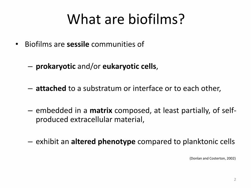
What are biofilms?
• Biofilms are sessile communities of
– prokaryotic and/or eukaryotic cells,
– attached to a substratum or interface or to each other,
– embedded in a matrix composed, at least partially, of self-produced extracellular material,
– exhibit an altered phenotype compared to planktonic cells (Donlan and Costerton, 2002)
2
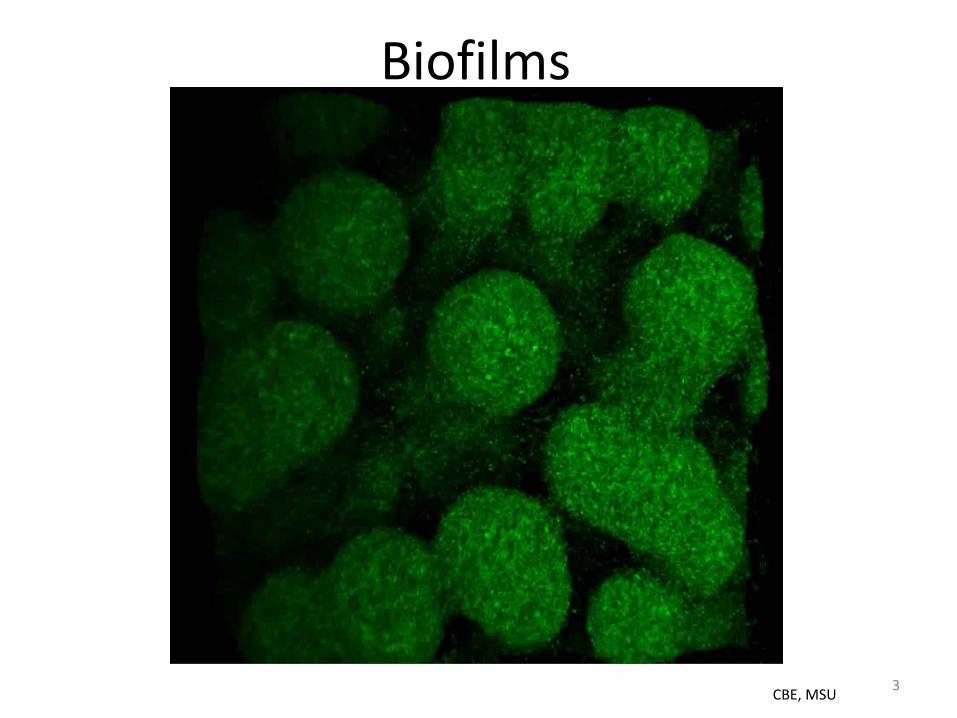
Biofilms
CBE, MSU 3
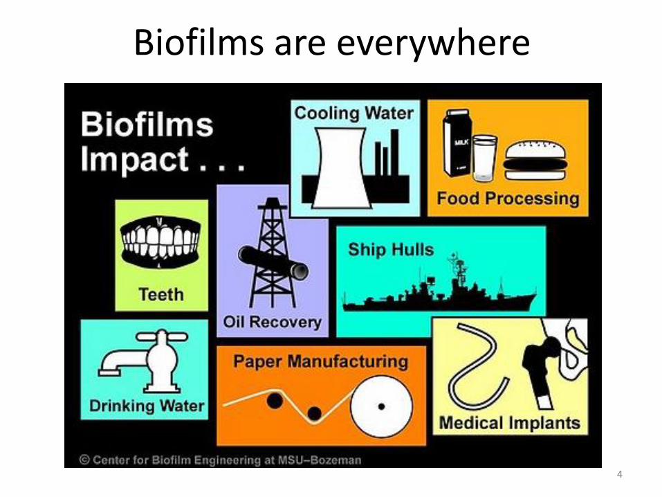
Biofilms are everywhere
4

Biofilm models
Closed Batch Static
Open Flow
Dynamic
In vitro
In vivo Invertebrate Non-mammalian
vertebrate mammalian
Ex vivo
Microcosm
Microcosm
5

Batch systems vs flow systems • Closed system
• Mixing is optional
• Cont. flow stirred tank reactor (CFSTR) • Perfect mixing • Steady state
• Plug flow reactor (PFR)
• Mixing only in radial direction
Coenye and Nelis, 2010 J. Microbiol Methods
Co
nce
ntr
atio
n
Co
nce
ntr
atio
n
Co
nce
ntr
atio
n
6
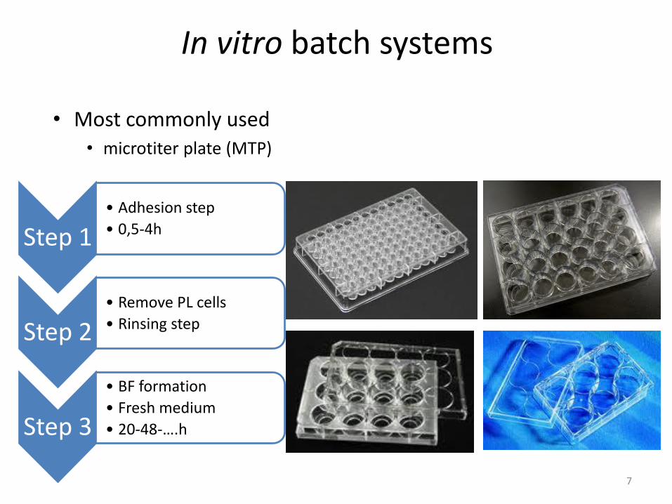
In vitro batch systems
• Most commonly used • microtiter plate (MTP)
Step 1 • Adhesion step
• 0,5-4h
Step 2 • Remove PL cells
• Rinsing step
Step 3
• BF formation
• Fresh medium
• 20-48-….h
7
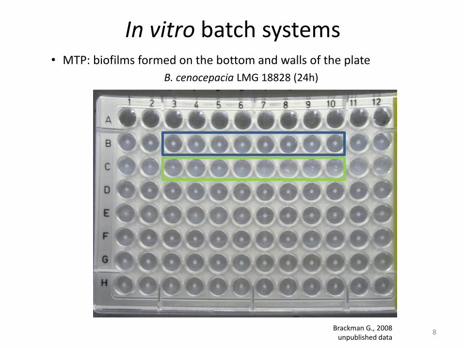
In vitro batch systems • MTP: biofilms formed on the bottom and walls of the plate
Brackman G., 2008 unpublished data
B. cenocepacia LMG 18828 (24h)
8

In vitro batch systems • MTP: biofilms formed on glass bottom
Udine et al., 2013, PloS ONE
B. cenocepacia LMG 16656 (WT and QS mutants) (24h; Live/Dead staining)
9
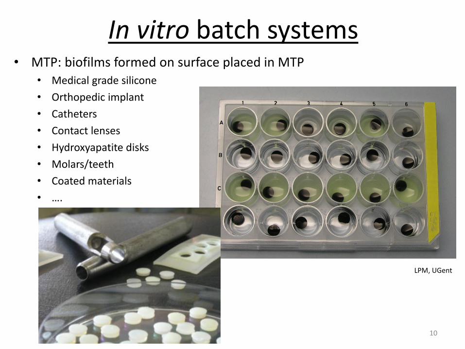
In vitro batch systems • MTP: biofilms formed on surface placed in MTP
• Medical grade silicone
• Orthopedic implant
• Catheters
• Contact lenses
• Hydroxyapatite disks
• Molars/teeth
• Coated materials
• ….
LPM, UGent
10
In vitro batch systems
• Minimum biofilm eradication concentration assay:
• The Calgary device (Ceri et al, 1999)
11
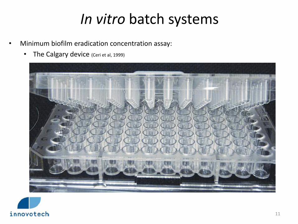
In vitro batch systems
• Minimum biofilm eradication concentration assay:
• The Calgary device (Ceri et al, 1999)
12
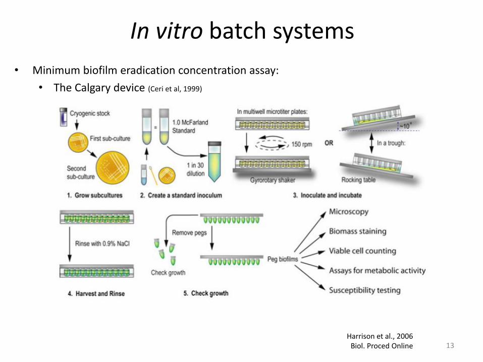
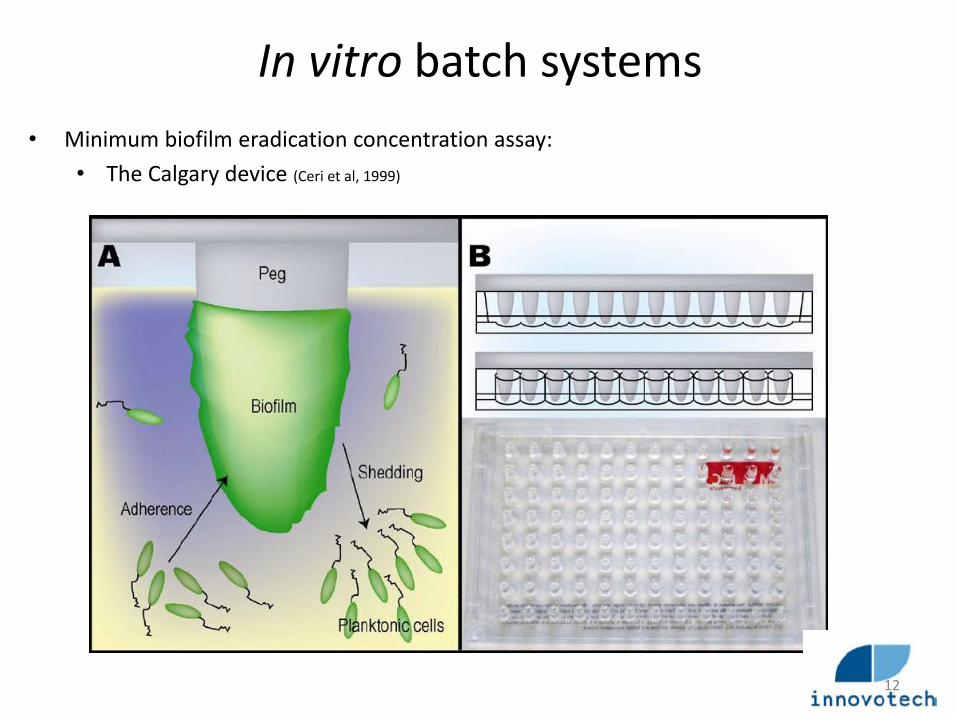
In vitro batch systems
• Minimum biofilm eradication concentration assay:
• The Calgary device (Ceri et al, 1999)
13 Harrison et al., 2006
Biol. Proced Online
14
In vitro Flow systems

In vitro flow systems: CFSTR
• Centers for disease control (CDC) biofilm reactor
Polyethylene Lid
“Water jacket”
1L vessel 8 Polypropylene coupon holders
Coupons
Magnetic stir
Media “IN” Air-vents
Media “OUT”
15

In vitro flow systems: CFSTR
• CDC biofilm reactor
set-up of the CDC biofilm reactor LPM, UGent. 16
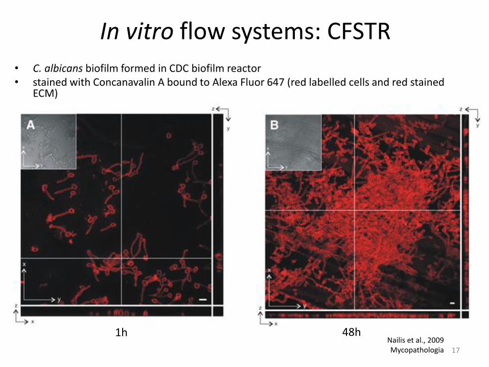
In vitro flow systems: CFSTR
• C. albicans biofilm formed in CDC biofilm reactor • stained with Concanavalin A bound to Alexa Fluor 647 (red labelled cells and red stained
ECM)
Nailis et al., 2009 Mycopathologia
1h 48h
17

In vitro flow systems: CFSTR
• CDC biofilm reactor
LPM, UGent
18

In vitro flow systems: CFSTR
• Other CFSTR systems
Rotating disk reactor Constant depth film
fermentor
Pratten, J. 2007 Curr. Protoc. Microbiol.
Biofilm annular reactor
19
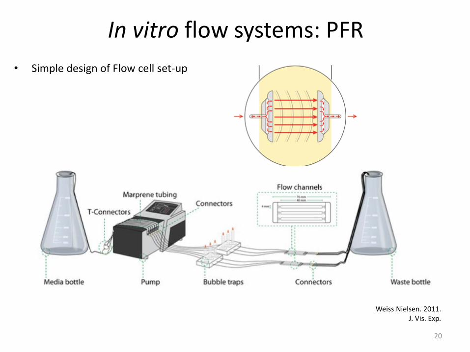
In vitro flow systems: PFR
• Simple design of Flow cell set-up
Weiss Nielsen. 2011. J. Vis. Exp.
20
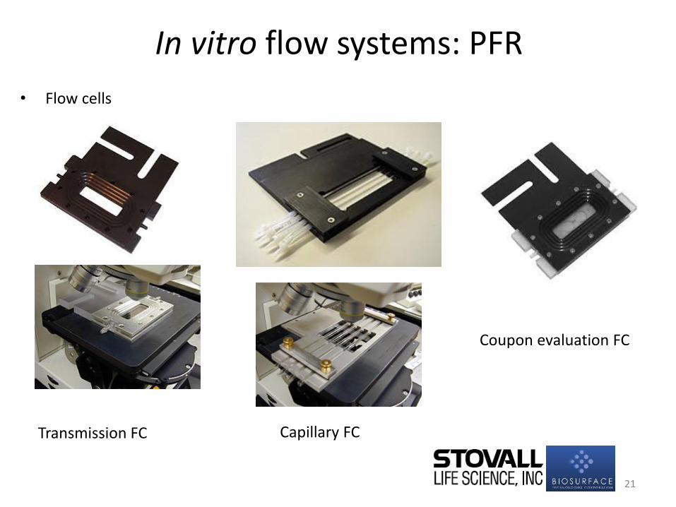
In vitro flow systems: PFR
• Flow cells
Transmission FC
Coupon evaluation FC
Capillary FC
21
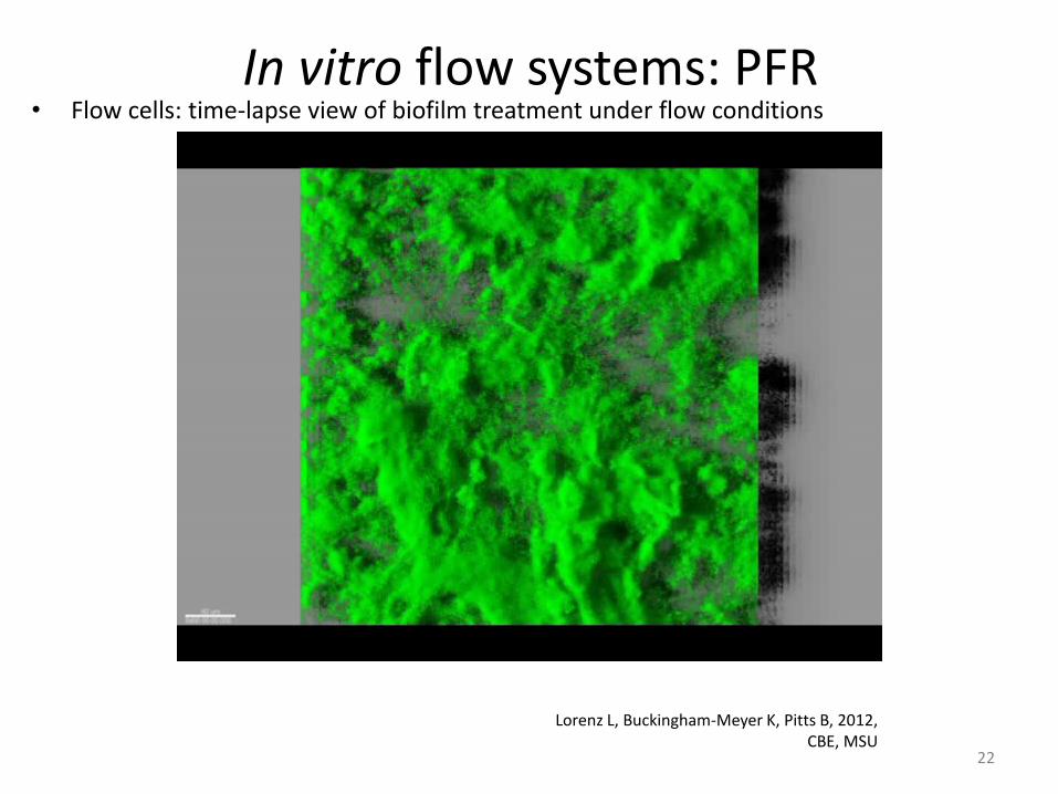
In vitro flow systems: PFR • Flow cells: time-lapse view of biofilm treatment under flow conditions
Lorenz L, Buckingham-Meyer K, Pitts B, 2012, CBE, MSU
22

In vitro flow systems: PFR • Microfluidic devices
Benoit et al., 2010 Appl. Environ. Microbiol. 23
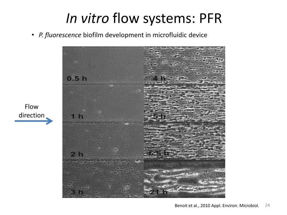
In vitro flow systems: PFR • P. fluorescence biofilm development in microfluidic device
Benoit et al., 2010 Appl. Environ. Microbiol.
Flow direction
24

In vitro flow systems: PFR
• Modified Robbins device (MRD)
McBain A.J., 2009 Adv. Appl. MIcrobiol
25
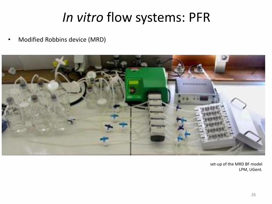
In vitro flow systems: PFR
• Modified Robbins device (MRD)
set-up of the MRD BF model LPM, UGent.
26

In vitro flow systems: PFR
• Modified Robbins device (MRD)
LPM, UGent. 27

In vitro flow systems: PFR
• Modifications on the MRD design
Garcia et al., 2010. J. Microbiol. Meth. 28

In vitro flow systems: PFR
• Implemented in industrial environments:
29

In vitro flow systems: PFR
• (Colony-)Drip flow reactor (DFR)
Goeres et al., 2009, Nature Protocols McBain A.J., 2009. Adv. Appl Microbiol.
Method E2647-08 Annual Book of ASTM Standards
30

In vitro flow systems: PFR
• (Colony-)Drip flow reactor
Goeres et al., 2009, Nature Protocols Method E2647-08 Annual Book of ASTM Standards 31

In vitro flow systems: PFR
• Polymicrobial wound biofilm in a C-DFR
Woods et al, 2012. J. Appl. Microbiol. 32

Simple in vitro flow systems
Uppuluri and Lopez-Ribot, 2010. Virulence.
Biofilms Hypertextbook P. Stoodley and J. Lennox
33
34
Ex vivo/Microcosms: Specific Biofilm models

• Lubbock chronic wound pathogenic biofilm model (LCWPB)
• Rapid (24h)
• Multispecies (Sa, Pa, Ef)
• Macro/Microscopically resembles in vivo wound biofilms
Sun et al., 2008. Wound Repair Regen.
Microcosms: Wound Biofilm model
35
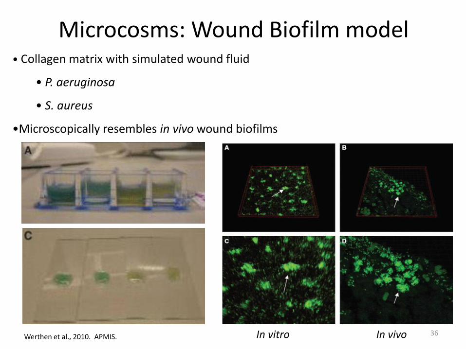
• Collagen matrix with simulated wound fluid
• P. aeruginosa
• S. aureus
•Microscopically resembles in vivo wound biofilms
Werthen et al., 2010. APMIS.
Microcosms: Wound Biofilm model
In vitro In vivo 36

Ex vivo biofilm models
• Reconstituted Human Epithelia (RHE)
• Human sinosal epithelial, skin, molars,…
• Microvascular endothelial cells (HMEC-1 Cells)
• HeLa cells
• CF-derived bronchial cells
• U2OS osteosarcoma cells
•…..
37

Overview
MTP/Batch: Flow systems:
Less labor intensive More labor intensive
No specialized equipment Require specialized
equipment/skills
Relatively cheap more expensive
multiplexing Limitation/run
≠ in vivo situation? Fluid Flow ≠ in vivo situation?
Microcosm/cell-culture-based model 38

39
In vivo (non-)mammalian Biofilm models

In vivo non-mammalian biofilm models
Lebeaux et al, 2013 Pathogens
40
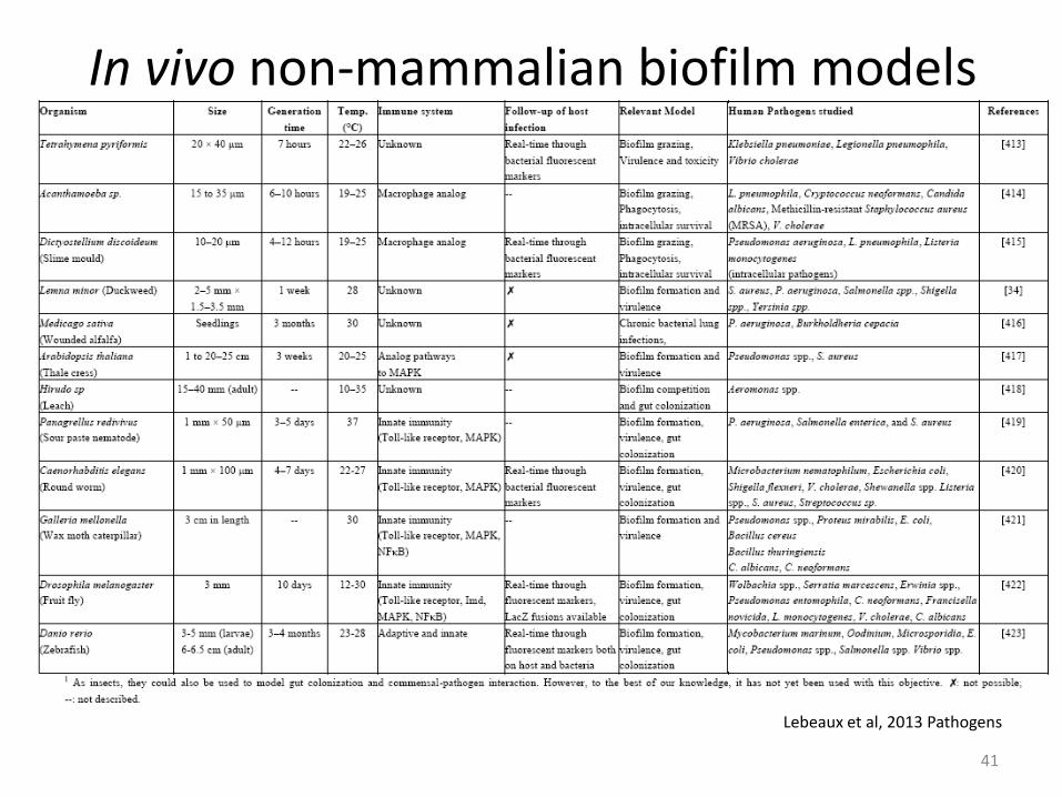
In vivo non-mammalian biofilm models
Lebeaux et al, 2013 Pathogens
41

In vivo non-mammalian biofilm models
Brackman G., 2013, unpublished data
42
Caenorhabditis elegans

Caenorhabditis elegans
Atkinson et al. 2011 PLoS Path.
In vivo non-mammalian biofilm models
43

In vivo vertebrate biofilm models
Brackman G., 2013, Unpublished data 44
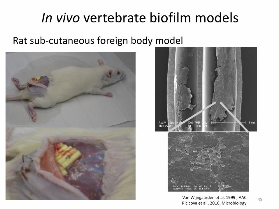
In vivo vertebrate biofilm models
Rat sub-cutaneous foreign body model
Van Wijngaarden et al. 1999 , AAC Ricicova et al., 2010, Microbiology
45
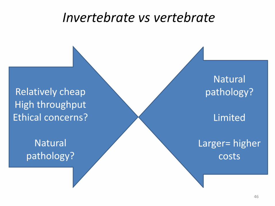
Invertebrate vs vertebrate
Relatively cheap High throughput Ethical concerns?
Natural
pathology?
Natural pathology?
Limited
Larger= higher
costs
46

Why is it important?
Smith et al. 2013 Infection and Immunity.
Based on Nailis et al. 2010 BMC Microbiology.
Susceptibility (Sa) Gene expression (Ca)
Importance of selecting a model that closely fits the research question in a certain research frame
47

Quantification tools
Direct quantification
Plating
Counting the colonies
Indirect quantification
Staining Labelling
Intensity ≈ # cells
48

Direct quantification
• Absolute cell numbers • Mixed BF: Possible to use selective
conditions • Isolates are available for further
research
• Time consuming & labor-intensive • Time to results is organism dependent
(>18h) • VBNC are not accounted for • Artificial situation might hinder growth
49

Great plate count anomaly
Lewis et al. 2010 ASM Microbeblog Staley and Konopka, 1985 Ann. Rev. Microbiol
• Often observed in biofilms (although
systematic studies are lacking!) • If you plate pieces of the biofilm:
• no recovery
• If you first disperse the biofilm: • recovery is much better
• Importance of dispersing the biofilm
• vortexing and sonication vs scraping
50

Indirect quantification Staining
• Crystal violet (living & dead cells, part of the matrix)
• DMMB (matrix in some organisms; particularly useful for staphylococci)
• Calcofluor white (EPS stain, N-acetylglucosamine)
• SYTO9 (living & dead cells, DNA in the matrix)
• FDA, XTT/MTT/…, CellTiter Blue/Resazurin/Alamar Blue (metabolically active
cells)
… and many more!
51

Resazurin staining
52
O
N
H
OHOH
hydroresorufin
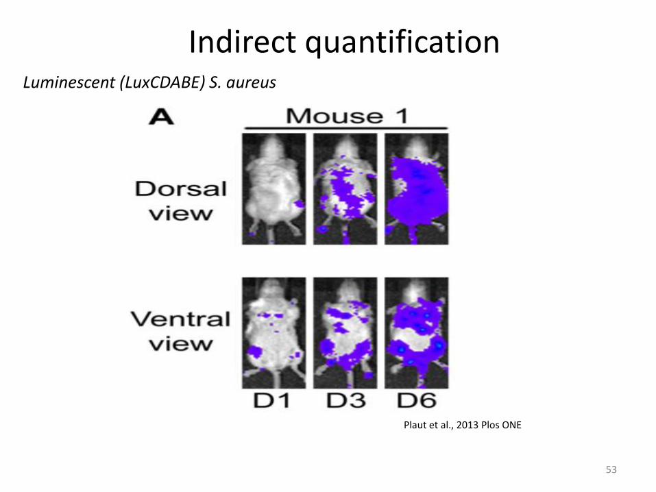
Indirect quantification Luminescent (LuxCDABE) S. aureus
Plaut et al., 2013 Plos ONE
53

Indirect quantification
Brackman et al., 2013, Unpublished data.
• Fast, high-throughput • Equipment • What are you measuring?
CTB fluorescence signal of a S. aureus Mu50 Biofilm after treatment
54
• Optimization is required
0.00E+00
5.00E+04
1.00E+05
1.50E+05
2.00E+05
2.50E+05
3.00E+05
3.50E+05
4.00E+05
untreated CTRL AB(1) AB(2)
flu
ore
sce
nce
(cp
m)
0.5h
1h
95 %
60 %
6 %
84 %

Indirect quantification
Brackman et al., 2013, J. Appl Microbiol.
Correlation between S. aureus pMV158gfp fluorescence signal and total CFU harvested from the biofilms.
• Detection limit for quantification
55
• Mixed biofilms?
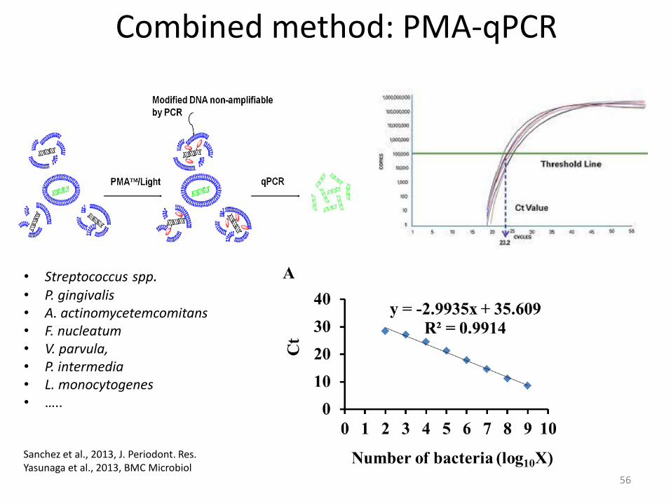
Combined method: PMA-qPCR
• Streptococcus spp. • P. gingivalis • A. actinomycetemcomitans • F. nucleatum • V. parvula, • P. intermedia • L. monocytogenes • …..
Sanchez et al., 2013, J. Periodont. Res. Yasunaga et al., 2013, BMC Microbiol
56

Take home messages
• Model systems are essential to study microbial biofilms • In combination with various quantification approaches they allow to mimic
in vivo situations in vitro • Importance of selecting a Biofilm model/quantification method that
closely fits the research question in a certain research frame • Selection based on
• Research question, Resources, level of the research, Preferences,…
• Caution extrapolating results from one model/method to another
57

Related Documents