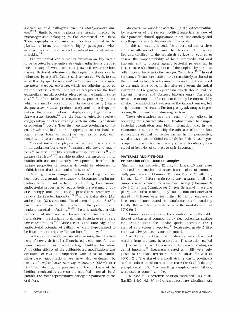
Biofilm formation on titanium implants counteracted by grafting gallium and silver ions Andrea Cochis, 1,2 Barbara Azzimonti, 1,2 Cinzia Della Valle, 2,3 Roberto Chiesa, 2,3 Carla Renata Arciola, 4,5 Lia Rimondini 1,2 1 Department of Health Sciences, University of Piemonte Orientale “A. Avogadro”, Novara, Italy 2 National Interuniversity Consortium for Materials Science and Technology (INSTM), Florence, Italy 3 Department of Chemistry, Materials Sciences and Chemical Engineering, Politecnico di Milano, Milan, Italy 4 Research Unit on Implant Infections, Rizzoli Orthopedic Institute, Bologna, Italy 5 Department of Experimental, Diagnostic and Specialty Medicine (DIMES), University of Bologna, Bologna, Italy Received 22 February 2014; revised 15 June 2014; accepted 30 June 2014 Published online 00 Month 2014 in Wiley Online Library (wileyonlinelibrary.com). DOI: 10.1002/jbm.a.35270 Abstract: Biofilm-associated infections remain the leading cause of implant failure. Thanks to its established biocompat- ibility and biomechanical properties, titanium has become one of the most widely used materials for bone implants. Engineered surface modifications of titanium able to thwart biofilm formation while endowing a safe anchorage to eukaryotic cells are being progressively developed. Here surfaces of disks of commercial grade 2 titanium for bone implant were grafted with gallium and silver ions by anodic spark deposition. Scanning electron microscopy of the sur- face morphology and energy dispersive X-ray spectroscopy were used for characterization. Gallium-grafted titanium was evaluated in comparison with silver-grafted titanium for both in vivo and in vitro antibiofilm properties and for in vitro compatibility with human primary gingival fibroblasts. Surface-modified materials showed: (i) homogeneous porous morphology, with pores of micrometric size; (ii) absence of cytotoxic effects; (iii) ability to support in vitro the adhesion and spreading of gingival fibroblasts; and (iv) antibiofilm properties. Although both silver and gallium exhibited in vitro strong antibacterial properties, in vivo gallium was significantly more effective than silver in reducing number and viability of biofilm bacteria colonies. Gallium-based treat- ments represent promising titanium antibiofilm coatings to develop new bone implantable devices for oral, maxillofacial, and orthopedic applications. V C 2014 Wiley Periodicals, Inc. J Biomed Mater Res Part A: 00A:000–000, 2014. Key Words: biofilm, biomaterials, implant infections, anodic spark deposition How to cite this article: Cochis A, Azzimonti B, Della Valle C, Chiesa R, Arciola CR, Rimondini L. 2014. Biofilm formation on titanium implants counteracted by grafting gallium and silver ions. J Biomed Mater Res Part A 2014:00A:000–000. INTRODUCTION Oral and orthopedic implants may be colonized by biofilm- forming bacteria that cling to the prosthesis surfaces and lead to periprosthesis infections. Then bacteria, protected, as they are, by the robust covering of the biofilm, become impervious to the host immune defenses and antibiotic therapies. 1–3 Biofilm-associated infection is still one of the most serious complications of the clinical application of biomedical devi- ces. Both in oral and in orthopedic implantology, implant- related infections are devastating and expensive events in terms of clinical sequelae and financial costs. In particular, a single episode of arthroplasty infection costs more than $50,000 dollars, which can further increase when repeated surgeries are required involving implant replacement and subsequent prolongation of systemic antibiotic therapy, pain, and disability. 4 Biomaterials are vulnerable to bacterial con- tamination because the bacteria of the skin and mucosae can come easily in contact with the implant surface during sur- gery, thus having a good chance of adhering to the prosthesis and to cause a deep bone tissue infection. 5,6 For instance, the occurrence of chronic osteomyelitis after insertion of an external prosthesis is as high as approximately 4%, 7 while oral peri-implantitis occurs in 5–8% of cases within selected implant systems. 7–9 Infections related to medical devices depend on several factors: chemical–physical properties of the biomaterial, design of the device, anatomical site in which it will be inserted, size of the surface involved, and duration of the surgical procedure. 10 Worthy of note are the host systemic and local health conditions, as they can counteract or favor bacterial colonization and infection. 11 Bacteria, fungi, and protozoa can all be enrolled in biomaterial contamination, although the most frequently recovered and documented microorganisms in orthopedic biomaterial infections are host endogenous commensal bacteria, such as Staphylococ- cus epidermidis and other coagulase-negative staphylococcal Correspondence to: C. R. Arciola; e-mail: [email protected] or L. Rimondini; e-mail: [email protected] Contract grant sponsors: INSTM and “5 per mille” grants for Health Research to the Rizzoli Orthopedic Institute of Bologna V C 2014 WILEY PERIODICALS, INC. 1

Welcome message from author
This document is posted to help you gain knowledge. Please leave a comment to let me know what you think about it! Share it to your friends and learn new things together.
Transcript
Biofilm formation on titanium implants counteracted by graftinggallium and silver ions
Andrea Cochis,1,2 Barbara Azzimonti,1,2 Cinzia Della Valle,2,3 Roberto Chiesa,2,3
Carla Renata Arciola,4,5 Lia Rimondini1,2
1Department of Health Sciences, University of Piemonte Orientale “A. Avogadro”, Novara, Italy2National Interuniversity Consortium for Materials Science and Technology (INSTM), Florence, Italy3Department of Chemistry, Materials Sciences and Chemical Engineering, Politecnico di Milano, Milan, Italy4Research Unit on Implant Infections, Rizzoli Orthopedic Institute, Bologna, Italy5Department of Experimental, Diagnostic and Specialty Medicine (DIMES), University of Bologna, Bologna, Italy
Received 22 February 2014; revised 15 June 2014; accepted 30 June 2014
Published online 00 Month 2014 in Wiley Online Library (wileyonlinelibrary.com). DOI: 10.1002/jbm.a.35270
Abstract: Biofilm-associated infections remain the leading
cause of implant failure. Thanks to its established biocompat-
ibility and biomechanical properties, titanium has become
one of the most widely used materials for bone implants.
Engineered surface modifications of titanium able to thwart
biofilm formation while endowing a safe anchorage to
eukaryotic cells are being progressively developed. Here
surfaces of disks of commercial grade 2 titanium for bone
implant were grafted with gallium and silver ions by anodic
spark deposition. Scanning electron microscopy of the sur-
face morphology and energy dispersive X-ray spectroscopy
were used for characterization. Gallium-grafted titanium was
evaluated in comparison with silver-grafted titanium for both
in vivo and in vitro antibiofilm properties and for in vitro
compatibility with human primary gingival fibroblasts.
Surface-modified materials showed: (i) homogeneous porous
morphology, with pores of micrometric size; (ii) absence of
cytotoxic effects; (iii) ability to support in vitro the adhesion
and spreading of gingival fibroblasts; and (iv) antibiofilm
properties. Although both silver and gallium exhibited in
vitro strong antibacterial properties, in vivo gallium was
significantly more effective than silver in reducing number
and viability of biofilm bacteria colonies. Gallium-based treat-
ments represent promising titanium antibiofilm coatings to
develop new bone implantable devices for oral, maxillofacial,
and orthopedic applications. VC 2014 Wiley Periodicals, Inc.
J Biomed Mater Res Part A: 00A:000–000, 2014.
Key Words: biofilm, biomaterials, implant infections, anodic
spark deposition
How to cite this article: Cochis A, Azzimonti B, Della Valle C, Chiesa R, Arciola CR, Rimondini L. 2014. Biofilm formation ontitanium implants counteracted by grafting gallium and silver ions. J Biomed Mater Res Part A 2014:00A:000–000.
INTRODUCTION
Oral and orthopedic implants may be colonized by biofilm-forming bacteria that cling to the prosthesis surfaces and leadto periprosthesis infections. Then bacteria, protected, as theyare, by the robust covering of the biofilm, become imperviousto the host immune defenses and antibiotic therapies.1–3
Biofilm-associated infection is still one of the most seriouscomplications of the clinical application of biomedical devi-ces. Both in oral and in orthopedic implantology, implant-related infections are devastating and expensive events interms of clinical sequelae and financial costs. In particular, asingle episode of arthroplasty infection costs more than$50,000 dollars, which can further increase when repeatedsurgeries are required involving implant replacement andsubsequent prolongation of systemic antibiotic therapy, pain,and disability.4 Biomaterials are vulnerable to bacterial con-tamination because the bacteria of the skin and mucosae cancome easily in contact with the implant surface during sur-
gery, thus having a good chance of adhering to the prosthesisand to cause a deep bone tissue infection.5,6 For instance, theoccurrence of chronic osteomyelitis after insertion of anexternal prosthesis is as high as approximately 4%,7 whileoral peri-implantitis occurs in 5–8% of cases within selectedimplant systems.7–9
Infections related to medical devices depend on severalfactors: chemical–physical properties of the biomaterial,design of the device, anatomical site in which it will beinserted, size of the surface involved, and duration of thesurgical procedure.10 Worthy of note are the host systemicand local health conditions, as they can counteract or favorbacterial colonization and infection.11 Bacteria, fungi, andprotozoa can all be enrolled in biomaterial contamination,although the most frequently recovered and documentedmicroorganisms in orthopedic biomaterial infections arehost endogenous commensal bacteria, such as Staphylococ-cus epidermidis and other coagulase-negative staphylococcal
Correspondence to: C. R. Arciola; e-mail: [email protected] or L. Rimondini; e-mail: [email protected] grant sponsors: INSTM and “5 per mille” grants for Health Research to the Rizzoli Orthopedic Institute of Bologna
VC 2014 WILEY PERIODICALS, INC. 1
species, or mild pathogens, such as Staphylococcus aur-eus.12,13 Similarly, oral implants are usually infected bymicroorganisms belonging to the commensal oral flora.These saprophytes are nonvirulent or less virulent in theplanktonic form, but become highly pathogenic whenarranged in a biofilm or when the natural microbial balanceis lacking.14
The events that lead to biofilm formation are key factorsto be targeted by preventive strategies. Adhesion is the firstinfection step allowing bacteria to grow and invade the hosttissues. Bacterial adhesion on the implant surfaces can beinfluenced by aspecific factors, such as van der Waals forces,as well as by specific microbial surface component recogniz-ing adhesive matrix molecules, which are adhesins harboredby the bacterial cell-wall and act as receptors for the hostextracellular matrix proteins adsorbed on the implant surfa-ces.7,15–17 After surface colonization by pioneering strains,which are mainly cocci spp. both in the oral cavity (whereStreptococcus mutans predominates) and in orthopedics(where the above-mentioned staphylococci, together withEnterococcus faecalis,18 are the leading etiologic species),coaggregation of other residing bacteria, either planktonicor adhering,19 occurs. They then replicate producing conflu-ent growth and biofilm. This happens on natural hard tis-sues (either bone or tooth) as well as on polymeric,metallic, and ceramic materials.20–23
Material surface too plays a role in these early phases.In particular, surface energy,24 micromorphology and rough-ness,25 material stability, crystallographic properties,21 andsurface chemistry22,26 are able to affect the susceptibility tobiofilm adhesion and its early development. Therefore, thesurface properties of biomaterials could be addressed toinhibit bacterial adhesion and colonization.27
Recently, several inorganic antimicrobial agents havebeen used as a promising strategy to discourage biofilm for-mation onto implant surfaces by exploiting their knownantibacterial properties to reduce both the systemic antibi-otic therapy and the surgical procedures necessary toremove the infected implants.3,27,28 In particular silver (Ag)and gallium (Ga), a semimetallic element in group 13 (31),have been shown to be effective in the prevention ofimplant surgical infections.29–35 Bacteriostatic/bactericideproperties of silver are well known and are mainly due toits multiform mechanisms to damage bacteria even at verylow concentrations.34,35 More recent is the knowledge of anantibacterial potential of gallium, which is hypothesized tobe based on an intriguing “Trojan horse” strategy.32
In the present work, we aim at examining the effective-ness of newly designed gallium-based treatments for tita-nium surfaces in counteracting biofilm formation.Antibiofilm efficacy of the gallium-based modifications wasevaluated in vivo in comparison with those of parallelsilver-based modifications. We have also evaluated, bymeans of confocal laser scanning microscopy (CLSM) afterLive/Dead staining, the presence and the thickness of thebiofilms produced in vitro on the modified materials by S.mutans, the most representative cariogenic pathogen of theoral flora.
Moreover, we aimed at ascertaining the cytocompatibil-ity properties of the surface-modified materials, in view oftheir potential clinical applications in oral implantology andin orthopedics as infection-resistant materials.
In this connection, it could be underlined that a closeand firm adhesion of the connective tissues (both noncalci-fied and calcified) to the prosthesis surface is required toensure the proper stability of bone orthopedic and oralimplants and to protect against bacterial penetration. Infact, a successful biointegration of the implant by the hostcells opposes bacteria in the race for the surface.36,37 In oralimplants a fibrous connective tissue tenaciously anchored tothe implant surface, besides nourishing and supplying bloodto the underlying bone, is also able to prevent the apicalmigration of the gingival epithelium, which should seal theimplant interface and obstruct bacteria entry. Therefore,resistance to implant infection can certainly be improved byan effective antibiofilm treatment of the implant surface, buta right connective tissue adhesion greatly advantages in pre-serving the implant from storming bacteria.
These observations are the reason of our efforts insearching for a surface titanium treatment able to hamperbacterial colonization and biofilm formation and, in themeantime, to support valuably the adhesion of the implantsurrounding stromal connective tissues. In this perspective,we also tested the modified-materials for their in vitro cyto-compatibility with human primary gingival fibroblasts, as amodel of behavior of connective cells in contact.
MATERIALS AND METHODS
Preparation of the titanium samplesTitanium disks (diameter: 12 mm; thickness: 0.5 mm) wereobtained by a mechanical cutter from a plate of commer-cially pure grade 2 titanium (Torresin Titanio Metalli S.r.l.,Limena, Italy). Before undergoing any treatment, all thesamples were cleaned by ultrasonic rinsing (Elmasonic S60/H, Elma Hans Schmidbauer, Singen, Germany) in acetone(RPE; Carlo Erba, Rodano, Italy) for 10 min and afterwardrinsed in Millipore water for further 10 min to remove sur-face contaminants related to manufacturing and handling.Finally, the samples were dried in a thermostatic oven at37�C for 2 h.
Titanium specimens were then modified with the addi-tion of antibacterial compounds by electrochemical surfacemodification using the anodic spark deposition (ASD)method as previously reported.38 Nontreated grade 2 tita-nium was always used as further control.
The different antibacterial treatments were developedstarting from the same base solution. This solution (calledSiB) is currently used to produce a biomimetic coating ondental implants.39 Specimens treated with SiB were sub-jected to an alkali treatment in 5 M NaOH for 2 h at60�C6 2�C. The aim of this alkali etching was to produce asurface sodium enrichment and increase the Ca/P (calcium/phosphorous) ratio. The resulting samples, called SiB-Na,were used as control samples.
The base SiB electrolytic solution contained 0.03 M di-Na2SiO3�2H2O, 0.1 M di-b-glycerophosphate disodium salt
2 COCHIS ET AL. Ga AND Ag COUNTERACT BIOFILM GROWTH
pentahydrate, 0.3 M calcium acetate�H2O, and 0.036 MNaOH (all provided by Sigma-Aldrich, St. Louis, MO). After-ward, antibacterial species and the chelating compoundswere added to the SiB solution to produce the final antibac-terial treatment.
The antibacterial agents used were silver nanoparticles,silver nitrate, silver acetate, and gallium nitrate (all pro-vided by Sigma) appropriately mixed with L-cysteine andoxalic acid dehydrate (all provided by Sigma-Aldrich) aschelating agents. These latter were used to avoid the mas-sive precipitation of silver and gallium salts in the electro-lytic solution, thus improving their migration toward theanode. Table I reports the complete scheme of the testedantibacterial agents, the concentrations of chelating agents,and the electrochemical parameters applied during the ASDtreatment.
Morphological and chemical analysisScanning electron microscopy (SEM) was used to investigatethe surface morphology of the samples. Briefly, after fixingon aluminum stubs using a conductive carbon tape, sampleswere observed with a StereoScan 360 SEM (CambridgeInstruments, Somerville, MA) at 10 kV with various magnifi-cations, using secondary electrons.39
The chemical qualitative analysis was made by anEnergy Dispersive Spectrometer (Inca energy 200; OxfordInstruments, Abingdon, UK) to identify the chemical speciespresent on the surfaces. The analysis was carried out on5003 acquired images.
Primary human gingival fibroblasts isolationPrimary human gingival fibroblasts (HGFs) were isolatedfrom a fresh gingival biopsy collected from tissues excisedfrom healthy teeth obtained from orthodontic procedures.The entire tissue was minced with a surgical blade anddigested for 45 min at 37�C with a solution of 1% type Icollagenase I (Worthington Biochemical Corporation, Lake-wood, NJ), 0.1% dispase I (Worthington) and 25% trypsin(Sigma-Aldrich) in a serum-free minimal essential mediumalpha modification (a-MEM; Sigma-Aldrich). Afterward, thedigested solution was filtered with a 0.45 lm pore size inorder to remove undigested debris and centrifuged 10 minat 800 rpm. The cellular pellet was then resuspended in
a-MEM supplemented with 10% fetal bovine serum (FBS;Sigma-Aldrich), 1% antibiotics/antimycotics (penicillin/streptomycin/gentamycin, Sigma-Aldrich) and the cells wereseeded into new polystyrene Petri plates (Sigma-Aldrich)containing fresh medium. Cells were grown up to a maxi-mum of about 80% confluence and detached with trypsin/EDTA before use; cells from passages 1 to 3 were used forexperiments.
Direct cytocompatibility evaluationTwelve millimeter samples were placed into 24-multiwellplates; 23104 cells/sample were seeded onto the surfacesof each sample in a low volume (200 lL) and allowed toadhere for 4 h. Afterward, each well was filled with 1 mL offresh medium and cell viability was evaluated with the(3-(4,5-dimethylthiazol-2-yl)-2,5-diphenyltetrazolium bro-mide) colorimetric assay (MTT; Sigma-Aldrich) at 24 h, 48h, and 72 h. Briefly, 100 lL of MTT solution (3 mg/mL inPBS) were added to each sample and incubated 4 h in thedark at 37�C; the composition of PBS was 137 mM NaCl,2.7 mM KCl, 8.1 mM Na2HPO4�2H2O, and 1.76 mM KH2PO4
(pH 7.4). Afterward, formazan crystals were solved with100 lL of dimethyl sulfoxyde (DMSO; Sigma-Aldrich) and50 lL were collected and centrifuged to remove any debris.
Supernatant optical density (OD) was evaluated at570 nm with a spectrophotometer (Spectra Count, PackardBell, Meriden, CT). Noncoated titanium OD was used as con-trol and considered as 100% cell viability while viability ofthe coated samples was calculated as follows: (sample OD/titanium control OD) 3 100. Experiments were performedsix times for controls and each different coating.36
Furthermore, immunofluorescence staining was per-formed in order to investigate cell morphology after 72 h ofadhesion onto the biomaterial surface. Cells were fixed 20min with 4% paraformaldehyde at room temperature (RT)and then washed three times with PBS. Phalloidin (rhoda-mine B tetramethyl isothiocynate, 1/2000 in PBS; AbCam,Cambridge, UK) solution was added for 45 min at RT inorder to investigate the cell cytoskeleton; afterward, sam-ples were washed three times with PBS and costained with40,6-diamidino-2-phenylindole (DAPI; Sigma-Aldrich). Cellmorphology was optically evaluated using fluorescence
TABLE I. Experimental Matrix of the Treatments
TreatmentAntibacterial
AgentAntibacterial
ConcentrationChelating
Agent
ChelatingAgent
Concentration(M)
Current(mA/cm2) Potential (V)
Time ofTreatment (s)
SiB-Na – – – 10 295 776 6 120AT^ 7200
AgCis AgNo3 0.004 M L-Cysteine 0.002 10 295 690 6 30GaCis Ga(NO3)3 0.004 M L-Cysteine 0.006 10 295 700 6 12GaOss Ga(NO3)3 0.004 M oxalic acid 0.306 10 325 606 6 66AgNPs Silver nanopar-
ticles (50 nm)3 g/L – – 10 295 656 6 49
Abbreviations: SiB-Na, control treatment; AT^, alkali etching treatment; AgCis, AgNO3 and L-cysteine treatment; GaCis, Ga(NO3)3 and L-cyste-
ine treatment; GaOss, Ga(NO3)3 and oxalic acid treatment; AgNPs, silver nanoparticles.
ORIGINAL ARTICLE
JOURNAL OF BIOMEDICAL MATERIALS RESEARCH A | MONTH 2014 VOL 00A, ISSUE 00 3
microscopy (Leica AF 6500; Leica Microsystems, Basel,Switzerland).
Indirect cytotoxicity evaluationSerum free a-MEM was incubated without cells for 1 week at37�C, 5% CO2 in direct contact with controls or coated sam-ples in a ratio of 1 mL/sample for a total of 10 mL/specimen.Afterward, eluates were collected, supplemented with 10%FBS and used to cultivate HGFs. Cells were seeded in a defi-nite number (2 3 104/well) into 24-well plates (Cell Star;PBI International, Milan, Italy) and cultivated for 1 week at37�C, 5% CO2. Afterward, cell viability was evaluated by theMTT colorimetric assay as described for the direct cytocom-patibility assay.40 Furthermore, cell morphology was visuallyinvestigated after 1 week of culture by light microscopy(Leica AF 6500; Leica Microsystems).
In vivo antibacterial activity assessmentThis study was an observational-blind, randomized, intrain-dividual comparative, and single-center clinical study. Thedesign of the study was in keeping with the ICH note forguidance on Good Clinical Practice (CPMP/ICH/135/95;1997) and the Declaration of Helsinki (RecommendationsGuiding Physicians in Biomedical Research Involving HumanSubjects, Helsinki; 1964) and subsequent amendments(1975). Nulla osta was received from the Local Ethics Com-mittee (Novara).
Seven volunteers (four males and three females; age 20–27 years, mean age 24 years) gave the informed consent toparticipate in the study and strictly complied with the pre-scriptions. Inclusion criteria were: good oral hygiene (pla-que index<10),41 good health conditions, normal salivary
secretion rate (stimulated salivary flow: 2.51 1.24 mL/min), normal buffer capacity (mean pH value: 6.31 1.72),and absence of marginal gingivitis and active carious lesion;the mean DMFS (Decayed, Missing due to caries, FilledTeeth/Surface)42 was 0.6 (range 0–4). Subjects who usedantibiotic or chlorhexidine during the 3 months before thestudy were not included as well as pregnant women.
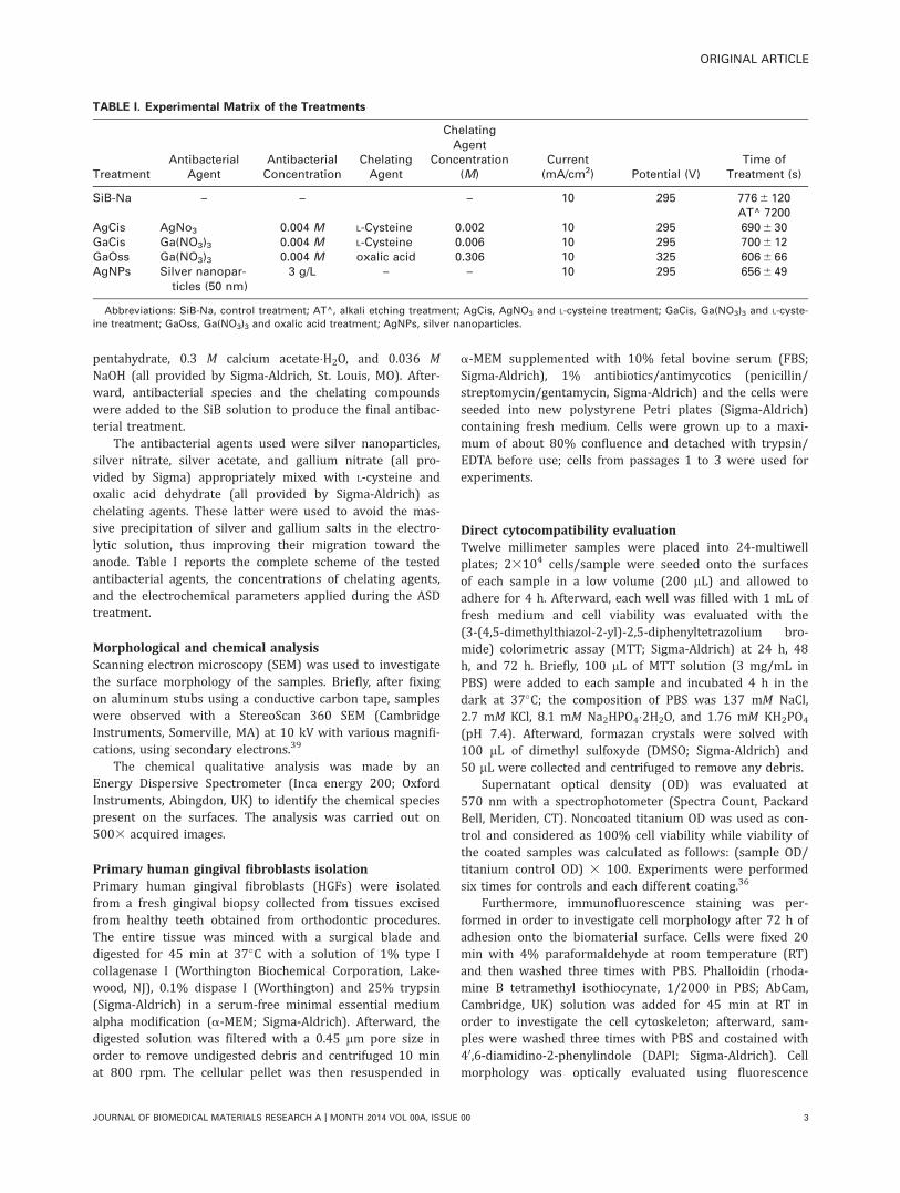
The volunteers were asked to wear oral appliances for24 h, shown in Figure 1. These were prepared with polyvi-nylchloride, containing six specimens (1 mm diameter) andtreated as previously described. The different samples werefixed to each appliance in random order as described inTable II.
Volunteers were asked to avoid the use of toothpasteand mouth rinse during the 24 h and to wear the appliancesconsecutively with the only exception at meals, when theappliances were stored in saline at RT.
After 24 h, the samples were collected and three sam-ples for each type of coating were used for the colorimetricviability assay (XTT; Sigma-Aldrich) and an additional threefor the number counts of the colony forming units (CFU).Briefly, the colorimetric assay 2,3-bis (2-methoxy-4-nitro-5-sulfophenyl)-5-((phenyl amino) carbonyl)-2H-tetrazoliumhydroxide (XTT; Sigma-Aldrich) was used to determinedehydrogenase activity as an indicator of the metabolic stateof the biofilm cells. Disks were transferred into new 24-wellculture tissue plates containing 1 mL PBS/well. Fifty mL XTTsolution (1 mg/mL in PBS) and 4 mL menadione (Sigma-Aldrich) solution (1 mM in acetone) were added to eachwell. Plates were incubated for 5 h in the dark at 37�C. Theentire content of each well was transferred into new 1.5 mLEppendorf tubes and centrifuged (5 min at 13,000 rpm) to
FIGURE 1. Appliances preparation process and chalk footprints (A); polyvinyl chloride (PVC) molds (B); and specimen insertion (C, red arrows).
[Color figure can be viewed in the online issue, which is available at wileyonlinelibrary.com.]
TABLE II. Schematic Representation of Specimen Application onto the Appliance (Sample and Position)
Topographic Site For Specimens Insertion
Subject 4.7 – 4.6 4.6 – 4.5 4.3 – 4.2 3.2 – 3.3 4.5 – 4.6 4.6 – 4.7
1 Ti ctrl AgNPs AgCis GaCis GaOss Ti ctrl2 SiB-Na Ti ctrl AgNPs AgCis Ti ctrl GaCis3 GaOss SiB-Na Ti ctrl Ti ctrl AgNPs AgCis4 GaCis Ti ctrl GaOss Ti ctrl SiB-Na AgNPs5 Ti ctrl AgCis GaCis Ti ctrl GaOss SiB-Na6 AgNPs AgCis Ti ctrl GaCis Ti ctrl GaOss7 SiB-Na AgNPs Ti ctrl AgCis GaCis Ti ctrl
Abbreviation: Ti crtl, untreated titanium.
Each appliance contains one single tested specimen in a random order. The points of insertion are indicated according to the World Dental
Federation Numbering System, published as ISO 3950 notation.65
4 COCHIS ET AL. Ga AND Ag COUNTERACT BIOFILM GROWTH
remove residual cells. One hundred microliters were trans-ferred from each tube to a new 96-well plate and the XTTformazan in the supernatant was determined spectrophoto-metrically at 690 nm.43
For the CFU counts, disks were resuspended in 1 mL ofPBS, vortexed, and sonicated at 60 Hz (Aquasonic 250HT;VWR International, Radnor, PA) for 30 s, five times for eachsample, to allow the detachment of biofilm from the surfaceof the disks. One hundred microliter suspensions weretransferred to a 96-well plate and used to perform six 10-fold dilutions by mixing 20 mL of biofilm suspension with180 mL of PBS. Twenty microliters of each dilution werespotted on nutrient agar plates and incubated for 72 h at37�C. Finally, the number of CFUs/disk was counted, in ablinded manner, using the following formula: (number ofcolonies) 3 10 3 (reverse of dilution value).44
In vitro antibiofilm activity assessmentIn vitro biofilm growth. Specimens were sterilized in 70%ethanol in water for 20 min and washed extensively withsterile deionized water. A single colony of the S. mutans ref-erence strain ATCC 25175 from an overnight culture onagar plate was resuspended in 9 mL of Tryptose Broth (TB,Biolife, Milan, Italy) and incubated at 37�C for 18 h. After
incubation, a new fresh TB tube diluted 1:10 was prepared.The new tube was incubated at 37�C for 3 h in order toachieve the logarithmic growth phase. At the end of theincubation period the bacterial suspension was read by aNephelometer Hach 2100 AN (Hach Lange, Manchester, Eng-land). The bacterial suspension was adjusted to obtain afinal turbidity of 10 nephelometric turbidity units (NTU).Aliquots of the dilute bacterial suspension were dispensedinto 24-well Microplates (Costar, Corning, N Y), each wellcontaining a material specimen (Ti Ctrl, SiB-Na, AgCis,AgNPs, GaCis, and GaOss) and then incubated for 24 h and72 h at 37�C. During the 72-h incubation, half culture brothof each well was substituted with fresh TB every day.
CLSM observations. CLSM observations aimed at detectingviability of bacteria grown for 24 h and 72 h at 37�C on thesix different materials. To determine the viability of bacteriaa Live/Dead BacLight bacterial viability kit (MolecularProbes, Life Technologies Italia, Monza, Italy) was used. Thekit includes two fluorescent nucleic acid stains: SYTO9 andpropidium iodide. SYTO9 penetrates and stains both viableand nonviable bacteria, while propidium iodide enters onlydamaged/dead cells and quenches SYTO9 fluorescence.Dead cells, which take up propidium iodide, fluoresce red,
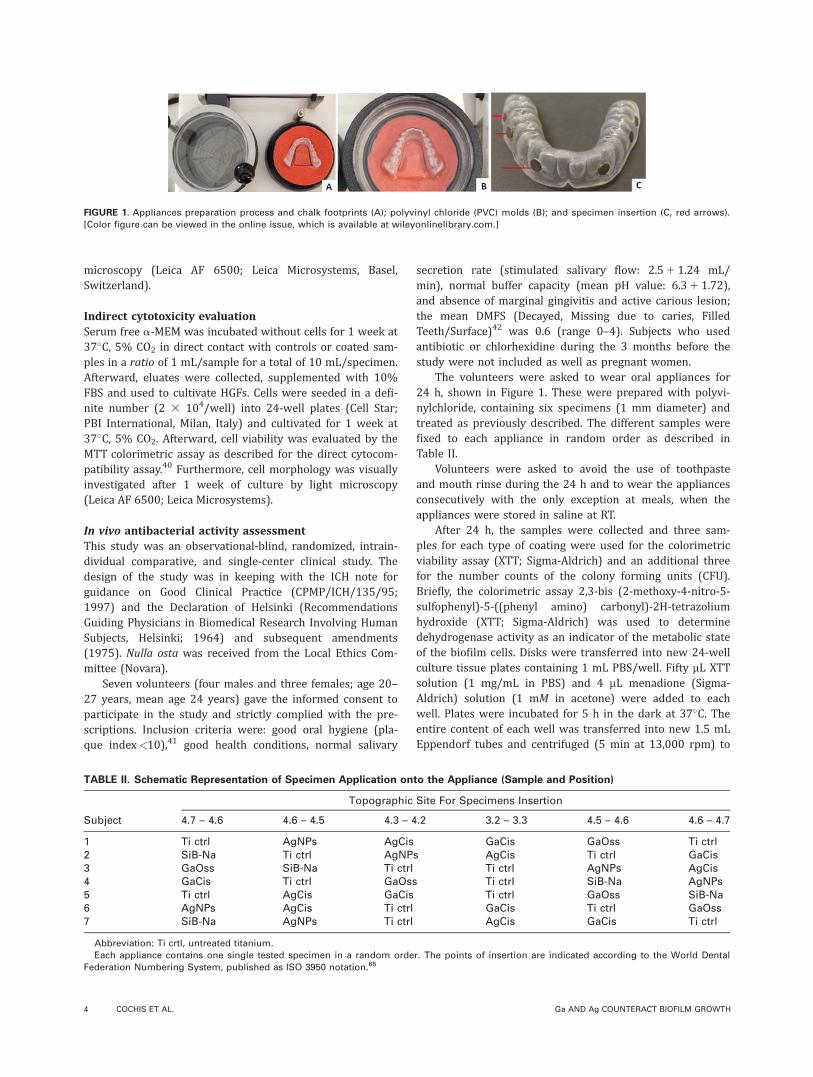
FIGURE 2. SEM images of the different specimens surface at the final stage of preparation. After fixing on aluminum stubs using a conductive
carbon tape, samples were observed with a StereoScan 360 SEM at 10 kV, using secondary electrons. Magnification 5 15003, bar scale 5 20 mm.
In AgNPs specimen small silver nanoparticle aggregates are present as white spots.
ORIGINAL ARTICLE
JOURNAL OF BIOMEDICAL MATERIALS RESEARCH A | MONTH 2014 VOL 00A, ISSUE 00 5
green cells fluoresce green. For assessing viability, 1 mL ofstock solution of each stain was added to 3 mL of PBS and,after mixing, the solution was distributed into the platescontaining the materials specimens and incubated at RT for15 min in the dark. Stained biofilms were examined by theConfocal Laser Scanning Microscopy Nikon Eclipse Ti invertedmicroscope equipped with an AR1 confocal unit, using a100x oil immersion objective. The excitation and emissionwavelengths used for detecting SYTO9 were 488 nm and525 nm, respectively. Propidium iodide was excited at520 nm, and its emission was monitored at 620 nm. Opticalsections of 1.0 mm were collected from the entire thicknessof the biofilm. For each sample, images from three randomlyselected positions were acquired. The resulting stacks ofimages were analyzed using NIS-elements AR 3.2 softwareconfocal software.
Statistical analysisStatistical analyses were performed using Statistical Packagefor Social Sciences (SPSS v20.0, IBM Co. Armonk, NY). Theassumptions of homogeneity of variances and normal distri-
bution of errors were checked for all the variables consid-ered using Levene’s and Kolmogorov–Smirnov tests,respectively. Since the assumptions were satisfied, analysisof variance one-way and post hoc Sheffe’s test were used.The significance level was set at 5%.
RESULTS
Morphological and chemical evaluationSEM images are reported in Figure 2. All the different coat-ings proved to be very stable and homogeneously distrib-uted on the sample surfaces. The untreated titanium (Tictrl) surface did not appear to be particularly smooth and itwas characterized by a raw “flake” morphology. In general,the ASD-treated surfaces showed a very similar morphology,with micrometric pore size and characterized by homogene-ous pore distribution. A different morphology is displayedby the GaOss treatment, in which the pore diameter issmaller than the other ASD coatings and it is characterizedby an irregular pore size distribution (Fig. 2). It is interest-ing to observe that some microcracks were detectable espe-cially on AgNPs, AgCis, and GaCis treatments, which are
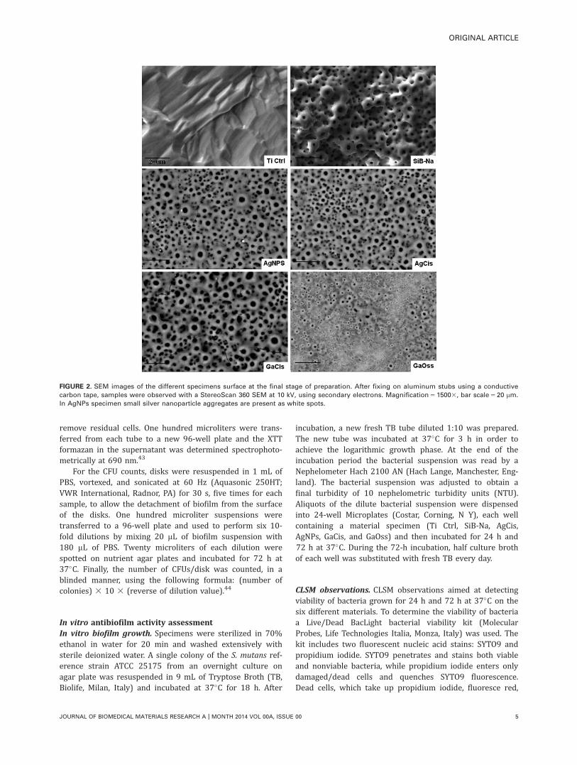
FIGURE 3. Qualitative chemical surface composition spectra (EDS). The EDS spectra highlighted the presence of calcium, phosphorous, and sili-
con for the SiB-Na control and for all the ASD coatings except for GaOss, for which the calcium signal was undetectable, although the maxi-
mum silicon peak was present. [Color figure can be viewed in the online issue, which is available at wileyonlinelibrary.com.]
6 COCHIS ET AL. Ga AND Ag COUNTERACT BIOFILM GROWTH
commonly present on the ASD treatments, but not represen-tative of the coating’s poor adherence or susceptibility todelamination. No cracks were visible on SiB-Na (control)and on GaOss treatment. AgNPs specimen clearly showedthe presence of small silver nanoparticle aggregates identifi-able by the presence of white spots (Fig. 2).
Chemical qualitative analysis [energy dispersive X-rayspectroscopy (EDS)] results are reported in Figure 3. The EDSspectra highlighted the presence of the chemical elementsshowing on the SiB-Na control such as calcium, phosphorous,and silicon for all the ASD coatings except for GaOss, forwhich the calcium signal was undetectable, although the max-imum silicon peak was present. The presence of calcium,phosphorous, and above all of silicon played a fundamentalrole in enhancing the mineralization process of the ASD coat-ing.39 In particular, the SiB-Na surface also showed the pres-ence of sodium, ascribed to the etching treatment performedafter the ASD treatments. For the EDS spectra of the ASD coat-ing, performed in the electrolytic solutions enriched with sil-ver (AgNPs and AgCis), there was no detectable silverpresence. Only by performing a careful EDS analysis whereSEM images showed a white spot, it was possible to confirmthe integration of silver within AgNPs surface. GaCis andGaOss spectra revealed gallium presence on surfaces; the gal-lium peak on GaOss, in particular, was higher than thoseobserved in the GaCis samples.
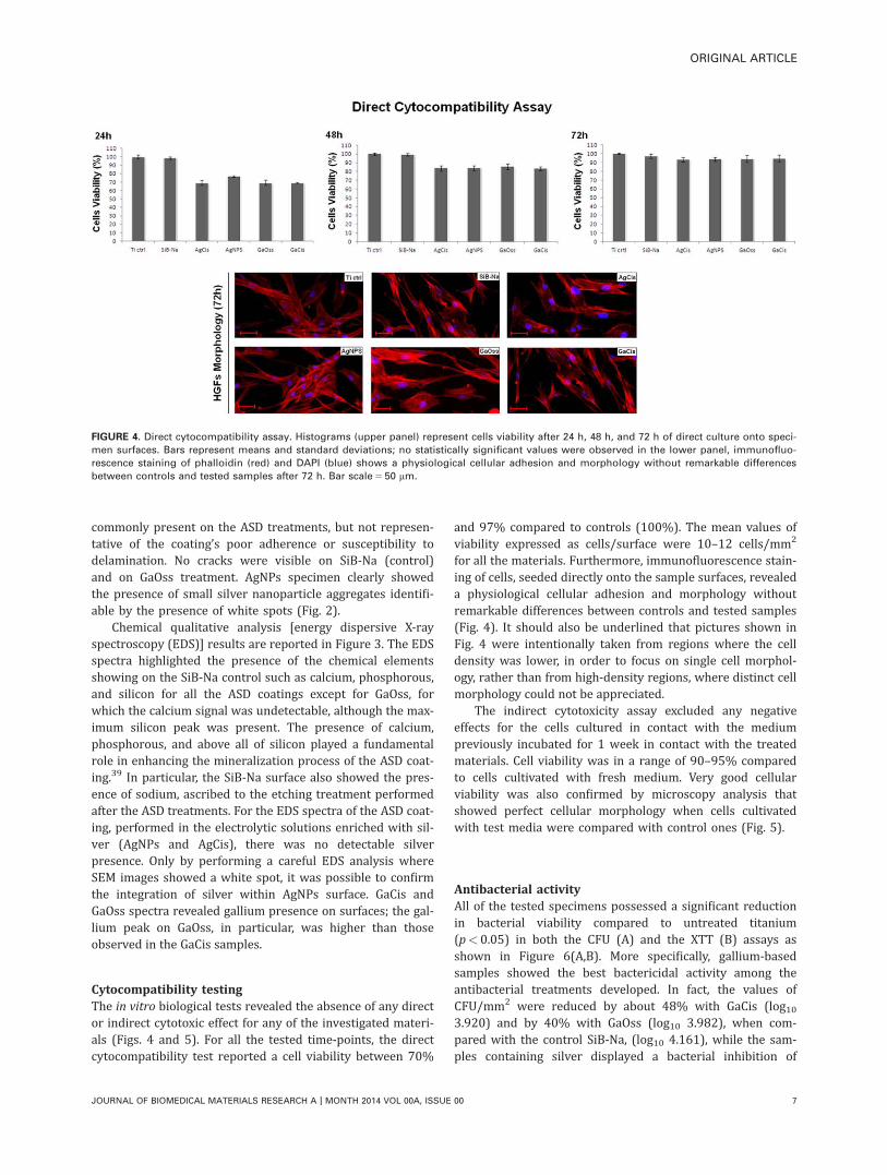
Cytocompatibility testingThe in vitro biological tests revealed the absence of any director indirect cytotoxic effect for any of the investigated materi-als (Figs. 4 and 5). For all the tested time-points, the directcytocompatibility test reported a cell viability between 70%
and 97% compared to controls (100%). The mean values ofviability expressed as cells/surface were 10–12 cells/mm2
for all the materials. Furthermore, immunofluorescence stain-ing of cells, seeded directly onto the sample surfaces, revealeda physiological cellular adhesion and morphology withoutremarkable differences between controls and tested samples(Fig. 4). It should also be underlined that pictures shown inFig. 4 were intentionally taken from regions where the celldensity was lower, in order to focus on single cell morphol-ogy, rather than from high-density regions, where distinct cellmorphology could not be appreciated.
The indirect cytotoxicity assay excluded any negativeeffects for the cells cultured in contact with the mediumpreviously incubated for 1 week in contact with the treatedmaterials. Cell viability was in a range of 90–95% comparedto cells cultivated with fresh medium. Very good cellularviability was also confirmed by microscopy analysis thatshowed perfect cellular morphology when cells cultivatedwith test media were compared with control ones (Fig. 5).
Antibacterial activityAll of the tested specimens possessed a significant reductionin bacterial viability compared to untreated titanium(p<0.05) in both the CFU (A) and the XTT (B) assays asshown in Figure 6(A,B). More specifically, gallium-basedsamples showed the best bactericidal activity among theantibacterial treatments developed. In fact, the values ofCFU/mm2 were reduced by about 48% with GaCis (log103.920) and by 40% with GaOss (log10 3.982), when com-pared with the control SiB-Na, (log10 4.161), while the sam-ples containing silver displayed a bacterial inhibition of
FIGURE 4. Direct cytocompatibility assay. Histograms (upper panel) represent cells viability after 24 h, 48 h, and 72 h of direct culture onto speci-
men surfaces. Bars represent means and standard deviations; no statistically significant values were observed in the lower panel, immunofluo-
rescence staining of phalloidin (red) and DAPI (blue) shows a physiological cellular adhesion and morphology without remarkable differences
between controls and tested samples after 72 h. Bar scale 5 50 mm.
ORIGINAL ARTICLE
JOURNAL OF BIOMEDICAL MATERIALS RESEARCH A | MONTH 2014 VOL 00A, ISSUE 00 7
about 30% (AgCis log10 4.047) to 34% [AgNPs log10 4021;Fig. 6(A)].
CFU counts were confirmed by the XTT viability assay:bacteria viability on gallium-treated samples as measuredby the inhibition ratio was in a range between 27% (GaOss)and 35% (GaCis) compared to controls. Silver samples con-firmed CFU results, but with a slightly lower inhibition ratiothan gallium [Fig. 6(B)].
In vitro antibiofilm activityPresence and thickness of the biofilms on the six materialswere evaluated by CLSM after Live/Dead staining. Figure 7
shows that after a 24-h culture S. mutans was growing inits typical twisted fine chains only on Ti ctrl and SiB-Naspecimens, while no bacterial colonies could be observed onthe other specimens.
Figure 8 shows that, after a 72-h culture, an abundantbiofilm was produced on Ti Ctrl and on SiB-Na disks. Thepercentages of live and dead cells were calculated by theNIS-elements AR 3.2 confocal software, measuring the fluo-rescence intensities of green and red channels. On the TiCtrl disk, 55% of cells appeared live and 45% dead. On theSiB-Na disk the number of cells was slightly lower and,from the intensities of the red and green channels, 45% of
FIGURE 5. Indirect cytotoxicity assay. After 1 week of culture with eluates, cells viability results comparable with controls. Bars represent means
and standard deviations. In the lower panel, microscopy observation shows cell morphology; no statistical differences were found between con-
trols and test samples. Bar scale 5 100 mm.
FIGURE 6. Antibacterial activity of the specimens. The number of CFUs was significantly reduced by GaCis (A). Also bacterial viability was signif-
icantly reduced by gallium samples (GaOss and GaCis) as reported by the XTT assay (B). Asterisks represent statistically significant values
(p< 0.005).
8 COCHIS ET AL. Ga AND Ag COUNTERACT BIOFILM GROWTH
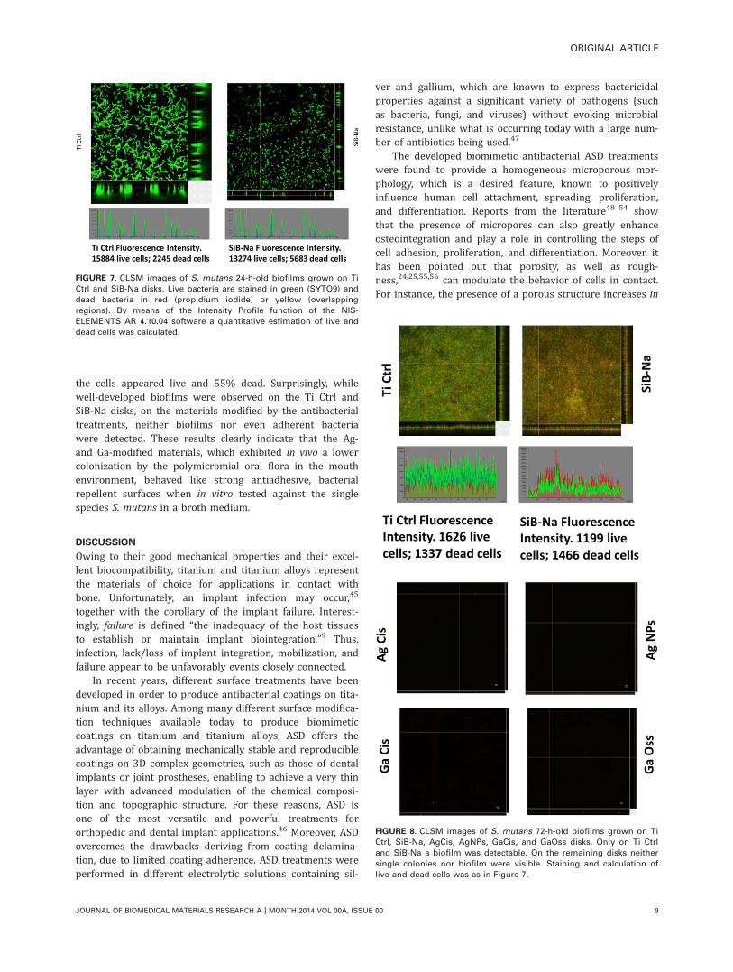
the cells appeared live and 55% dead. Surprisingly, whilewell-developed biofilms were observed on the Ti Ctrl andSiB-Na disks, on the materials modified by the antibacterialtreatments, neither biofilms nor even adherent bacteriawere detected. These results clearly indicate that the Ag-and Ga-modified materials, which exhibited in vivo a lowercolonization by the polymicromial oral flora in the mouthenvironment, behaved like strong antiadhesive, bacterialrepellent surfaces when in vitro tested against the singlespecies S. mutans in a broth medium.
DISCUSSION
Owing to their good mechanical properties and their excel-lent biocompatibility, titanium and titanium alloys representthe materials of choice for applications in contact withbone. Unfortunately, an implant infection may occur,45
together with the corollary of the implant failure. Interest-ingly, failure is defined “the inadequacy of the host tissuesto establish or maintain implant biointegration.”9 Thus,infection, lack/loss of implant integration, mobilization, andfailure appear to be unfavorably events closely connected.
In recent years, different surface treatments have beendeveloped in order to produce antibacterial coatings on tita-nium and its alloys. Among many different surface modifica-tion techniques available today to produce biomimeticcoatings on titanium and titanium alloys, ASD offers theadvantage of obtaining mechanically stable and reproduciblecoatings on 3D complex geometries, such as those of dentalimplants or joint prostheses, enabling to achieve a very thinlayer with advanced modulation of the chemical composi-tion and topographic structure. For these reasons, ASD isone of the most versatile and powerful treatments fororthopedic and dental implant applications.46 Moreover, ASDovercomes the drawbacks deriving from coating delamina-tion, due to limited coating adherence. ASD treatments wereperformed in different electrolytic solutions containing sil-
ver and gallium, which are known to express bactericidalproperties against a significant variety of pathogens (suchas bacteria, fungi, and viruses) without evoking microbialresistance, unlike what is occurring today with a large num-ber of antibiotics being used.47
The developed biomimetic antibacterial ASD treatmentswere found to provide a homogeneous microporous mor-phology, which is a desired feature, known to positivelyinfluence human cell attachment, spreading, proliferation,and differentiation. Reports from the literature48–54 showthat the presence of micropores can also greatly enhanceosteointegration and play a role in controlling the steps ofcell adhesion, proliferation, and differentiation. Moreover, ithas been pointed out that porosity, as well as rough-ness,24,25,55,56 can modulate the behavior of cells in contact.For instance, the presence of a porous structure increases in
FIGURE 7. CLSM images of S. mutans 24-h-old biofilms grown on Ti
Ctrl and SiB-Na disks. Live bacteria are stained in green (SYTO9) and
dead bacteria in red (propidium iodide) or yellow (overlapping
regions). By means of the Intensity Profile function of the NIS-
ELEMENTS AR 4.10.04 software a quantitative estimation of live and
dead cells was calculated.
FIGURE 8. CLSM images of S. mutans 72-h-old biofilms grown on Ti
Ctrl, SiB-Na, AgCis, AgNPs, GaCis, and GaOss disks. Only on Ti Ctrl
and SiB-Na a biofilm was detectable. On the remaining disks neither
single colonies nor biofilm were visible. Staining and calculation of
live and dead cells was as in Figure 7.
ORIGINAL ARTICLE
JOURNAL OF BIOMEDICAL MATERIALS RESEARCH A | MONTH 2014 VOL 00A, ISSUE 00 9
vivo bone-to-implant contact, improving the mechanicalinterlocking of the bone bonding. Noticeably, the GaOsstreatment revealed different pore diameters which aresmaller than those of the other studied ASD coatings, prob-ably due to the different chelating compound used (com-pared to GaCis treatment) and to the different voltagereached by the coating during the ASD process (325 vs.295 V).
Cytocompatibility testing revealed that the differenttreatments were compatible with the cells. In both directand indirect assays, results suggested that the modificationby the ASD treatment did not introduce toxic elements intothe specimens at the released concentrations. Small, non-significant, differences of viability values between ASD-materials and controls were observed in the MTT directcytocompatibility assays at 24. The viability values of ASD-materials were higher at 48 h and completely comparableto those of controls at 72 h, when gingival fibroblasts fullyadhered and spread on modified materials.
It should be remarked that two different types of cyto-toxicity assays were performed. The results of the indirectcytotoxicity assay after 1 week of cultivation with eluates(Fig. 5) shows that results were comparable with controls:no statistical differences were found between controls andtest samples. In the lower panel of Figure 5, microscopyobservations, showing the cell morphology, support the sta-tistical result of an absence of remarkable differences.
Results suggest that the viable and adherent gingivalfibroblasts observed in the in vitro model can be expectedto be able to form in vivo a connective tissue healthily sus-taining the gingival epithelium. Therefore, epithelium willact as an effective barrier against the colonization by theoral flora. In a different way, when epithelium is not prop-erly supported by the connective tissue, a gingival recessioncan occur that leads to the formation of a tissue pocketprone to the microbial contamination. Then bone resorptionand mobilization of the implant intervene.57
The results of in vivo inhibition of biofilm bacteria cellsare very promising. In general, CFU counts showed a reduc-tion in bacterial colonies on the treated samples comparedwith the controls. Silver-coated samples resulted in 30–34%decrease in bacterial colonies compared to the controls.These results confirm the antibacterial properties of silver,which is known to be able to damage bacterial cells mem-branes, interfere in ion transport, denaturate enzymes, andinhibit DNA transcription and cellular respiration. More spe-cifically, Ag1 ions interact with thiol groups in proteins andinhibit respiratory enzymes, resulting in the production ofreactive oxygen species.34,35
The study was performed in vivo since it was addressedto the effect of gallium against the human oral microbiome,which comprises over 500 bacterial species.58 An in vitrostudy would have necessarily limited the use of bacteria toone or a few biofilm forming species, such as S. mutans59
and Aggregatibacter actinomycetemcomitans,60 which wouldnot have been able to reproduce in vitro the biofilm formedin vivo by the several hundred species of bacteria of thehuman oral polymicrobial flora.61
Recently, the effect of gallium on the growth of the bio-film was studied in vitro by CFU counting of biofilm viablePseudomonas aeruginosa cells detached from disks ofgallium-carboxymethyl cellulose after different incubationtimes. Gallium-carboxymethyl cellulose was found to reduceby 15% the biofilm growth of P. aeruginosa at 24 h inrespect to sodium-carboxymethyl cellulose.62 In our in vivostudy, biofilm was evaluated by a CFU counting method sim-ilar to that used in Ref. [62. After 24 h-appliance to GaCisdisks, a 48% biofilm reduction was found, greater than thatobserved by Valappil et al.62
More interesting results were obtained with gallium-coated specimens, which showed higher values of inhibition(40% and 48%) compared to the controls. The antibacterialproperties of gallium have been recently highlighted. At pHvalues close to neutral, the free hydrated Ga31 ion hydro-lyzes forming highly insoluble amorphous Ga species (OH)3.The Ga31 is a hard acid that strongly binds in solutionLewis bases, particularly OH2 groups. Furthermore, galliumtends to generate chelates by binding oxygen and also nitro-gen atoms.29
From a metabolic point of view, Ga31 is very similar toFe31, and acts as an iron substitute in many biological path-ways.30,31 It is precisely thanks to the chemical similarity ofGa31 with Fe31, in terms of charge, ionic radius, electronicconfiguration, and coordination number, that gallium cansubstitute iron in the biological systems, thus carrying outits antibacterial effect. Indeed, since Ga31 cannot be reducedunder the same conditions as Fe31, sequential redox reac-tions critical for the biological functions of Fe31 areimpaired by the iron substitution with gallium. Therefore,gallium inhibits Fe31 biological functions by a “Trojanhorse” strategy.30–32 This strategy can be exploited to inter-fere with or disrupt the iron metabolism in a large numberof bacterial strains, including P. aeruginosa,30 Mycobacte-ria,32 Clostridium difficile, and the methicillin-resistant S.aureus.33
This general trend was also confirmed by XTT analysis.A reduction in the metabolic activity was observed in gal-lium- and silver-treated specimens, with the best inhibitionratio in the gallium specimens (27–35%). These values areparticularly significant since volunteers did not use anytoothpaste or mouth rinse during the trial. Thus, the inhibi-tion can be related only to the silver and galliumtreatments.
In two very recent in vitro studies, dealing with thedetachment of bacterial biofilms from disks, either by fluidfor oral rinses or by solutions of N-chlorotaurine for surgi-cal irrigations, the antimicrobial/antibiofilm effects wereevaluated by the XTT assay for bacterial metabolic activity,by live/dead staining, as well as by CFU counting.63,64 Live/dead staining did not apply to our in vivo study, since after24-h of oral application of the disks, besides bacteria, alsoepithelial cells exfoliating from the mouth mucosae couldhave adhered onto the disks, thus altering the results.
Although the specimens were worn only for a relativelyshort time (24 h), it should be remarked that none of thevolunteers presented signs of adverse effect toward the
10 COCHIS ET AL. Ga AND Ag COUNTERACT BIOFILM GROWTH

tested materials. Seen on the whole, these findings suggestthat gallium may represent a valid antibacterial agent forthe prevention of implant contamination.
The results of in vitro biofilm production by S. mutanshave shown that even after 72-h culture the Ag- and Ga-modified disks still exhibited antiadhesive properties in manytimes repeated observations. While on the control and on bio-mimetic titanium disks S. mutans produced biofilms withappreciable thickness, on the Ag- and Ga-modified materialsneither biofilms nor bacterial colonies were observed.
CONCLUSION
This work demonstrates the possibility of successfully usingthe ASD method to develop biomimetic treatments on tita-nium substrates, in order to obtain a promising materialwith antimicrobials properties and with a good tissue-integrative potential.
ACKNOWLEDGMENTS
The authors would like to thank Massimo Cavallo and SimoneStroppa for the excellent technical support. Special thanks toEurocoating S.p.A. (Trento, Italy) for their precious coopera-tion. Many thanks to Doctor Stefano Ravaioli, PhD, postdoc fel-low at the Rizzoli Orthopaedic Institute, for his skillful andrefined support for the CLSM analysis. The authors expresstheir greatest thanks to Professor Lucio Montanaro (RizzoliOrthopaedic Institute) for his masterly suggestions and forcritical reading the article.
REFERENCES1. Zimmerli W, Trampuz A, Ochsner PE. Prosthetic-joint infections.
N Engl J Med 2004;351:1645–1654.
2. Darouiche RO. Treatment of infections associated with surgical
implants. N Engl J Med 2004;350:1422–1429.
3. Arciola CR, Campoccia D, Speziale P, Montanaro L, Costerton JW.
Biofilm formation in Staphylococcus implant infections. A review
of molecular mechanisms and implications for biofilm-resistant
materials. Biomaterials 2012;33:5967–5982.
4. Berbari EF, Hanssen AD, Duffy MC, Steckelberg JM, Ilstrup DM,
Harmsen WS, Osmon DR. Risk factors for prosthetic joint infec-
tion: Case–control study. Clin Infect Dis 1998;27:1247–1254.
5. Zhao L, Chu PK, Zhang Y, Wu Z. Antibacterial coatings on tita-
nium implants. J Biomed Mater Res B Appl Biomater 2009;91:
470–480.
6. Zlowodzki M, Prakash JS, Aggarwal NK. External fixation of com-
plex femoral shaft fractures. Int Orthop 2007;31:409–413.
7. Berglundh T, Persson L, Klinge B. A systematic review of the inci-
dence of biological and technical complications in implant den-
tistry reported in prospective longitudinal studies of at least 5
years. J Clin Periodontol 2002;29:197–212.
8. Quirynen M, De Soete M, van Steenberghe D. Infectious risks for
oral implants: A review of the literature. Clin Oral Implants Res
2002;13:1–19.
9. Pye AD, Lockhart DEA, Dawson MP, Murray CA, Smith AJ. A
review of dental implants and infection. J Hosp Infect 2009;72:
104–110.
10. Campoccia D, Montanaro L, Arciola CR. A review of the clinical
implications of anti-infective biomaterials and infection-resistant
surfaces. Biomaterials 2013;34:8018–8029.
11. Seneviratne CJ, Jin L, Samaranayake LP. Biofilm lifestyle of Can-
dida: A mini review. Oral Dis 2008;14:582–590.
12. Montanaro L, Speziale P, Campoccia D, Ravaioli S, Cangini I,
Pietrocola G, Giannini S, Arciola CR. Scenery of Staphylococcus
implant infections in orthopedics. Future Microbiol 2011;6:1329–
1349. doi: 10.2217/fmb.11.117.
13. von Eiff C, Arciola CR, Montanaro L, Becker K, Campoccia D.
Emerging Staphylococcus species as new pathogens in implant
infections. Int J Artif Organs 2006;29:360–367.
14. Chen L, Wen YM. The role of bacterial biofilm in persistent infec-
tions and control strategies. Int J Oral Sci 2011;3:66–73.
15. Speziale P, Pietrocola G, Rindi S, Provenzano M, Provenza G, Di
Poto A, Visai L, Arciola CR. Structural and functional role of
Staphylococcus aureus surface components recognizing adhesive
matrix molecules of the host. Future Microbiol 2009;4:1337–1352.
16. Yang L, Liu Y, Wu H, H�oiby N, Molin S, Song ZJ. Current under-
standing of multi-species biofilms. Int J Oral Sci 2011;3:74–81.
17. Foster TJ, Geoghegan JA, Ganesh VK, H€o€ok M. Adhesion, inva-
sion and evasion: The many functions of the surface proteins of
Staphylococcus aureus. Nat Rev Microbiol 2014;12:49–62.
18. Arciola CR, Baldassarri L, Campoccia D, Creti R, Pirini V, Huebner
J, Montanaro L. Strong biofilm production, antibiotic multi-
resistance and high gelE expression in epidemic clones of Entero-
coccus faecalis from orthopaedic implant infections. Biomaterials
2008;29:580–586.
19. Wessel SW, Chen Y, Maitra A, van den Heuvel ER, Slomp AM,
Busscher HJ, van der Mei HC. Adhesion forces and composition
of planktonic and adhering oral microbiomes. J Dent Res 2014;93:
84–88.
20. Brambilla E, Ionescu A, Gagliani M, Cochis A, Arciola CR,
Rimondini L. Biofilm formation on composite resins for dental
restorations: An in situ study on the effect of chlorhexidine
mouthrinses. Int J Artif Organs 2012;35:792–799.
21. Giordano C, Saino E, Rimondini L, Pedeferri MP, Visai L, Cigada
A, Chiesa R. Electrochemically induced anatase inhibits bacterial
colonization on titanium grade 2 and Ti6Al4V alloy for dental and
orthopedic devices. Colloids Surf B Biointerfaces 2011;88:648–655.
22. Visai L, Rimondini L, Giordano C, Del Curto B, Sbarra MS,
Franchini R, Della Valle C, Chiesa R. Electrochemical surface mod-
ification of titanium for implant abutments can affect oral bacteria
contamination. J Appl Biomater Biomech 2008;6:170–177.
23. Rimondini L, Cerroni L, Carrassi A, Torricelli P. Bacterial coloniza-
tion of zirconia ceramic surfaces: An in vitro and in vivo study. Int
J Oral Maxillofac Implants 2002;17:793–798.
24. Rimondini L, Fare S, Chiesa R, Pedeferri MP, Carrassi A. The
effect of composition, wettability and roughness of the substrate
on in vivo early bacterial colonization of titanium. J Appl Bio-
mater Biomech 2003;1:131–138.
25. Rimondini L, Fare S, Brambilla E, Felloni A, Consonni C, Brossa F,
Carrassi A. The effect of surface roughness on early in vivo pla-
que colonization on titanium. J Periodontol 1997;68:556–562.
26. Petrini P, Arciola CR, Pezzali I, Bozzini S, Montanaro L, Tanzi MC,
Speziale P, Visai L. Antibacterial activity of zinc modified titanium
oxide surface. Int J Artif Organs 2006;29:434–442.
27. Campoccia D, Montanaro L, Arciola CR. A review of the biomate-
rials technologies for infection-resistant surfaces. Biomaterials
2013;34:8533–8554.
28. Arciola CR, Montanaro L, Costerton JW. New trends in diagnosis
and control strategies for implant infections. Int J Artif Organs
2011;34:727–736.
29. Bernstein LR. Mechanisms of therapeutic activity for Gallium.
Pharmacol Rev 1998;50:665–682.
30. Kaneko Y, Thoendel M, Olakanmi O, Britigan BE, Singh PK. The
transition metal gallium disrupts Pseudomonas aeruginosa iron
metabolism and has antimicrobial and antibiofilm activity. J Clin
Invest 2007;117:877–888.
31. Kelson AB, Carnevali M, Truong-Le V. Gallium-based anti-infec-
tives: Targeting microbial iron-uptake mechanisms. Curr Opin
Pharmacol 2013;13:707–716.
32. Olakanmi O, Britigan BE, Schlesinger LS. Gallium disrupts iron
metabolism of mycobacteria residing within human macro-
phages. Infect Immun 2000;68:5619–5627.
33. Pickup DM, Valappil SP, Moss RM, Twyman HL, Guerry P, Smith
ME, Wilson M, Knowles JC, Newport RJ. Preparation, structural
characterization and antibacterial properties of Ga-doped sol-gel
phosphate-based glass. J Mater Sci 2009;44:1858–1867.
34. Furno F, Morley KS, Wong B, Sharp BL, Arnold PL, Howdle SM,
Bayston R, Brown PD, Winship PD, Reid HJ. Silver nanoparticles
ORIGINAL ARTICLE
JOURNAL OF BIOMEDICAL MATERIALS RESEARCH A | MONTH 2014 VOL 00A, ISSUE 00 11
and polymeric medical devices: A new approach to prevention of
infection? J Antimicrob Chemother 2004;54:1019–1024.
35. Taglietti A, Arciola CR, D’Agostino A, Dacarro G, Montanaro L,
Campoccia D, Cucca L, Vercellino M, Poggi A, Pallavicini P, Visai
L. Antibiofilm activity of a monolayer of silver nanoparticles anch-
ored to an amino-silanized glass surface. Biomaterials 2014;35:
1779–1788.
36. Gristina AG, Naylor P, Myrvik Q. Infections from biomaterials and
implants: A race for the surface. Med Prog Technol 1988–1989;14:
205–224.
37. Busscher HJ, van der Mei HC, Subbiahdoss G, Jutte PC, van den
Dungen JJ, Zaat SA, Schultz MJ, Grainger DW. Biomaterial-asso-
ciated infection: Locating the finish line in the race for the surface.
Sci Transl Med 2012;4:153rv10.
38. Della Valle C, Visai L, Santin M, Cigada A, Candiani G, Pezzoli D,
Arciola CR, Imbriani M, Chiesa R. A novel antibacterial modifica-
tion treatment of titanium capable to improve osseointegration.
Int J Artif Organs 2012;35:864–875.
39. Della Valle C, Rondelli G, Cigada A, Bianchi AE, Chiesa R. A novel
silicon-based electrochemical treatment to improve osteointegra-
tion of titanium implants. J Appl Biomater Funct Mater 2013;11:
106–116.
40. Le VQ, Cochis A, Rimondini L, Pourroy G, Stanic V, Palkowski H,
Carrado A. Biomimetic calcium-phosphates produced by an auto-
catalytic route on stainless steel 316L and bio-inert polyolefin.
RSC Adv 2013;3:11255–11262.
41. Fischman SL. Current status of indices of plaque. J Clin Periodon-
tol 1986;13:371–374, 379–380.
42. Johnston L, Vieira AR. Caries experience and overall health sta-
tus. Oral Health Prev Dent 2014. doi: 10.3290/j.ohpd.a31670.
43. Hofmann CM, Bednar KJ, Anderson JM, Marchant RE. Disruption
of Staphylococcus epidermidis biofilm formation using a targeted
cationic peptide. J Biomed Mater Res A 2012;100:1061–1067.
44. Cochis A, Fracchia L, Martinotti MG, Rimondini L. Biosurfactants
prevent in vitro Candida albicans biofilm formation on resins and
silicon materials for prosthetic devices. Oral Surg Oral Med Oral
Pathol Oral Radiol 2012;113:755–761.
45. Esposito M, Hirsch JM, Lekholm U, Thomsen P. Biological factors
contributing to failures of osseointegrated implants (I). Success
criteria and epidemiology. Eur J Oral Sci 1998;106:527–551.
46. Calvert KL, Desai T, Webster TJ. Advances in calcium phosphate
coatings-anodic spark deposition: A review. Front Biosci (Land-
mark Ed) 2014;19:475–489.
47. Campoccia D, Montanaro L, Speziale P, Arciola CR. Antibiotic-
loaded biomaterials and the risks for the spread of antibiotic
resistance following their prophylactic and therapeutic clinical
use. Biomaterials 2010;31:6363–6377.
48. Giavaresi G, Fini M, Cigada A, Chiesa R, Rondelli G, Rimondini L,
Aldini NN, Martini L, Giardino R. Histomorphometric and micro-
hardness assessments of sheep cortical bone surrounding tita-
nium implants with different surface treatments. J Biomed Mater
Res A 2003;67:112–120.
49. Giavaresi G, Fini M, Cigada A, Chiesa R, Rondelli G, Rimondini L,
Torricelli P, Aldini NN, Giardino R. Mechanical and histomorpho-
metric evaluations of titanium implants with different surface
treatments inserted in sheep cortical bone. Biomaterials 2003;24:
1583–1594.
50. Sawase T, Jimbo R, Wennerberg A, Suketa N, Tanaka Y, Atsuta
M. A novel characteristic of porous titanium oxide implants. Clin
Oral Implants Res 2007;18:680–685.
51. Conserva E, Lanuti A, Menini M. Cell behavior related to implant
surfaces with different microstructure and chemical composition:
An in vitro analysis. Int J Oral Maxillofac Implants 2010;25:1099–
1107.
52. Singh RG. Evaluation of the bioactivity of titanium after varied
surface treatments using human osteosarcoma osteoblast cells:
An in vitro study. Int J Oral Maxillofac Implants 2011;26:998–1003.
53. Shim IK, Chung HJ, Jung MR, Nam SY, Lee SY, Lee H, Heo SJ,
Lee SJ. Biofunctional porous anodized titanium implants for
enhanced bone regeneration. J Biomed Mater Res A 2013. doi:
10.1002/jbm.a.35026.
54. Braem A, Van Mellaert L, Mattheys T, Hofmans D, De Waelheyns
E, Geris L, Ann�e J, Schrooten J, Vleugels J. Staphylococcal bio-
film growth on smooth and porous titanium coatings for biomed-
ical applications. J Biomed Mater Res A 2014;102A:215–224.
55. Campoccia D, Arciola CR, Cervellati M, Maltarello MC, Montanaro
L. In vitro behaviour of bone marrow-derived mesenchymal cells
cultured on fluorohydroxyapatite-coated substrata with different
roughness. Biomaterials 2003;24:587–596.
56. Arciola CR, Montanaro L, Moroni A, Giordano M, Pizzoferrato A,
Donati ME. Hydroxyapatite-coated orthopaedic screws as infec-
tion resistant materials: In vitro study. Biomaterials 1999;20:323–
327.
57. Baltriukiene D, Sabaliauskas V, Balciunas E, Melninkaitis A,
Liutkevicius E, Bukelskiene V, Rutkunas V. The effect of laser-
treated titanium surface on human gingival fibroblast behavior. J
Biomed Mater Res A 2014;102A:713–720.
58. Dewhirst FE, Chen T, Izard J, Paster BJ, Tanner AC, Yu WH,
Lakshmanan A, Wade WG. The human oral microbiome. J Bacter-
iol 2010;192:5002–5017.
59. Montanaro L, Campoccia D, Rizzi S, Donati ME, Breschi L, Prati C,
Arciola CR. Evaluation of bacterial adhesion of Streptococcus
mutans on dental restorative materials. Biomaterials 2004;25:
4457–4463.
60. Oettinger-Barak O, Dashper SG, Catmull DV, Adams GG, Sela
MN, Machtei EE, Reynolds EC. Antibiotic susceptibility of Aggre-
gatibacter actinomycetemcomitans JP2 in a biofilm. J Oral Micro-
biol 2013;5.
61. Wang Z, Shen Y, Haapasalo M. Dental materials with antibiofilm
properties. Dent Mater 2014;30:e1–e16.
62. Valappil SP, Yiu HH, Bouffier L, Hope CK, Evans G, Claridge JB,
Higham SM, Rosseinsky MJ. Effect of novel antibacterial gallium-
carboxymethyl cellulose on Pseudomonas aeruginosa. Dalton
Trans 2013;42:1778–1786.
63. Babu JP, Garcia-Godoy F. In vitro comparison of commercial oral
rinses on bacterial adhesion and their detachment from biofilm
formed on hydroxyapatite disks. Oral Health Prev Dent 2014.
64. Coraca-Huber DC, Ammann CG, Fille M, Hausdorfer J, Nogler M,
Nagl M. Bactericidal activity of N-chlorotaurine against biofilm-
forming bacteria grown on metal disks. Antimicrob Agents Che-
mother 2014;58:2235–2239.
65. ISO 3950:2009 Dentistry—Designation system for teeth and areas
of the oral cavity, published 05/01/2009 by International Organiza-
tion for Standardization.
12 COCHIS ET AL. Ga AND Ag COUNTERACT BIOFILM GROWTH
Related Documents