178 www.ecmjournal.org European Cells and Materials Vol. 20 2010 (pages 178-196) DOI: 10.22203/eCM.v020a15 ISSN 1473-2262 Abstract This study investigated the in vitro effects of bioactive titanium surfaces on osteoblast differentiation. Three titanium substrates were tested: a commercially pure titanium (Cp Ti), an alkali- and heat-treated titanium (AH Ti), and an apatite-formed titanium (Ap Ti) generated by soaking AH Ti in a simulated body fluid. Chemical evaluation of the surface reactivity was analysed at nanometre scale by X-ray photoelectron spectroscopy (XPS), and at micrometre scale by energy dispersive X- ray microanalysis (EDX). It showed that the estimated proportion of the surface covered by adsorbed serum proteins differed between the three substrates and confirmed the bioactivity of AH Ti, illustrated by surface calcium and phosphate deposition when immersed in biological fluids. Mouse calvaria osteoblasts were cultured on the substrates for 15 days with no sign of cytotoxicity. Enzyme immunoassay and Real-Time RT-PCR were used to follow osteoblast differentiation through the production of osteocalcin (OC) and expression of several bone markers. At day 15, a significant up-regulation of Runx2, Osx, Dlx5, ALP, BSP, OC and DMP1 mRNA levels associated with an increase of OC production were observed on AH Ti and Ap Ti when compared to Cp Ti. These results suggest that bioengineered titanium has a great potential for dental applications in enhancing osseointegration. Keywords: Biomaterials, biomimetics, surfaces, in vitro, cell/protein-material interactions, osteoblasts, differentiation. *Address for correspondence: Juliane Isaac Laboratoire de Physiopathologie Orale Moléculaire Centre de Recherche des Cordeliers, INSERM, UMRS 872, Equipe 5, Esc. E – 2ème étage, 15-21 rue de l’Ecole de Médecine, F-75006 Paris, France. Telephone Number: 33 (0)1. 43.26.94.96 FAX Number: 33 (0)1. 44.07.14.21 E-mail: [email protected] Introduction Treatment of partial and total edentulism with dental implants has become a reliable procedure for the majority of patients and is expected to play a major role in oral rehabilitation in the future. Studies of the long-term outcomes suggest that many of the implant techniques now consistently lead to their successful integration and therapeutic restoration (Adell et al., 1981; Buser et al., 1997). Titanium (Ti) and its alloys are used extensively for medical devices in dental, orthopaedic and cardiovascular fields due to their relatively low elastic modulus, high corrosion resistance and excellent biocompatibility (Pohler, 2000). Nevertheless, although surface topography of titanium implants has been shown to modulate the percentage of bone-implant contact and the mechanical strength of bone-implant attachment (Pearce et al., 2008; Hayes et al., 2009), titanium and its alloys are bioinert materials. Indeed, titanium implants do not chemically bond to the bone; rather, they are simply incorporated into a bone contact (Yan et al., 1997). To provide the metal with bone-bonding ability and thus improve dental implant osseointegration, alternative methods are under investigation. For example, the coating of metallic implants with bioactive materials, such as hydroxyapatite (HA), has become a well-established method. Many techniques including ion beam sputtering, dipping, and electrophoretic or electrochemical deposition are currently being studied for the application of HA coatings to metal substrates; however plasma spray is the most commonly used technique for clinical applications (Sun et al., 2001). However, while many studies report good short-term clinical results with enhanced performance of plasma-sprayed HA-coated implants, clinical studies show that long-term implantation is associated with bone loss and inflammation around many of the HA-coated implants (Albrektsson, 1998). These problems are thought to arise from degradation of the coating, a poor adhesion of the HA coating to the metal surface, the low crystallinity of the apatite and the non- uniformity of the thickness of the deposited apatite layer (Wang et al., 1995; Munting, 1996; Martini et al., 2003). To overcome these limitations, a biomimetic approach conferring bone-bonding ability to metallic materials has BIOENGINEERED TITANIUM SURFACES AFFECT THE GENE-EXPRESSION AND PHENOTYPIC RESPONSE OF OSTEOPROGENITOR CELLS DERIVED FROM MOUSE CALVARIAL BONES Juliane Isaac 1,2,3, *, Anouk Galtayries 5 ,Takashi Kizuki 6 , Tadashi Kokubo 6 , Ariane Berdal 1,2,3,4 , and Jean-Michel Sautier 1,2,3,4 1 INSERM, U872, Eq.5, Laboratoire de Physiopathologie Orale Moléculaire, F-75006 Paris, France 2 Centre de Recherche des Cordeliers, Université Pierre et Marie Curie, UMR S 872, F-75006 Paris, France 3 Université Paris Descartes, UMR S 872, F-75006 Paris, France 4 Université Paris7, UFR d’Odontologie, F-75006 Paris, France 5 Laboratoire de Physico-Chimie des Surfaces, CNRS ENSCP (UMR 7045), Ecole Nationale Supérieure de Chimie de Paris (Chimie ParisTech), F-75005 Paris, France 6 Department of Biomedical Sciences, College of Life and Health Sciences, Chubu University, Kasugai, Aichi 487- 8501, Japan.

Welcome message from author
This document is posted to help you gain knowledge. Please leave a comment to let me know what you think about it! Share it to your friends and learn new things together.
Transcript
178 www.ecmjournal.org
J Isaac et al. Biomimetic Ti-surfaces affect bone cell behaviourEuropean Cells and Materials Vol. 20 2010 (pages 178-196) DOI: 10.22203/eCM.v020a15 ISSN 1473-2262
Abstract
This study investigated the in vitro effects of bioactivetitanium surfaces on osteoblast differentiation. Threetitanium substrates were tested: a commercially puretitanium (Cp Ti), an alkali- and heat-treated titanium (AHTi), and an apatite-formed titanium (Ap Ti) generated bysoaking AH Ti in a simulated body fluid. Chemicalevaluation of the surface reactivity was analysed atnanometre scale by X-ray photoelectron spectroscopy(XPS), and at micrometre scale by energy dispersive X-ray microanalysis (EDX). It showed that the estimatedproportion of the surface covered by adsorbed serumproteins differed between the three substrates and confirmedthe bioactivity of AH Ti, illustrated by surface calcium andphosphate deposition when immersed in biological fluids.Mouse calvaria osteoblasts were cultured on the substratesfor 15 days with no sign of cytotoxicity. Enzymeimmunoassay and Real-Time RT-PCR were used to followosteoblast differentiation through the production ofosteocalcin (OC) and expression of several bone markers.At day 15, a significant up-regulation of Runx2, Osx, Dlx5,ALP, BSP, OC and DMP1 mRNA levels associated withan increase of OC production were observed on AH Ti andAp Ti when compared to Cp Ti. These results suggest thatbioengineered titanium has a great potential for dentalapplications in enhancing osseointegration.
Keywords: Biomaterials, biomimetics, surfaces, in vitro,cell/protein-material interactions, osteoblasts,differentiation.
*Address for correspondence:Juliane IsaacLaboratoire de Physiopathologie Orale MoléculaireCentre de Recherche des Cordeliers, INSERM, UMRS 872,Equipe 5, Esc. E – 2ème étage, 15-21 rue de l’Ecole deMédecine,F-75006 Paris, France.
Telephone Number: 33 (0)1. 43.26.94.96FAX Number: 33 (0)1. 44.07.14.21
E-mail: [email protected]
Introduction
Treatment of partial and total edentulism with dentalimplants has become a reliable procedure for the majorityof patients and is expected to play a major role in oralrehabilitation in the future. Studies of the long-termoutcomes suggest that many of the implant techniquesnow consistently lead to their successful integration andtherapeutic restoration (Adell et al., 1981; Buser et al.,1997).
Titanium (Ti) and its alloys are used extensively formedical devices in dental, orthopaedic and cardiovascularfields due to their relatively low elastic modulus, highcorrosion resistance and excellent biocompatibility(Pohler, 2000). Nevertheless, although surface topographyof titanium implants has been shown to modulate thepercentage of bone-implant contact and the mechanicalstrength of bone-implant attachment (Pearce et al., 2008;Hayes et al., 2009), titanium and its alloys are bioinertmaterials. Indeed, titanium implants do not chemicallybond to the bone; rather, they are simply incorporatedinto a bone contact (Yan et al., 1997).
To provide the metal with bone-bonding ability andthus improve dental implant osseointegration, alternativemethods are under investigation. For example, the coatingof metallic implants with bioactive materials, such ashydroxyapatite (HA), has become a well-establishedmethod. Many techniques including ion beam sputtering,dipping, and electrophoretic or electrochemical depositionare currently being studied for the application of HAcoatings to metal substrates; however plasma spray is themost commonly used technique for clinical applications(Sun et al., 2001). However, while many studies reportgood short-term clinical results with enhancedperformance of plasma-sprayed HA-coated implants,clinical studies show that long-term implantation isassociated with bone loss and inflammation around manyof the HA-coated implants (Albrektsson, 1998). Theseproblems are thought to arise from degradation of thecoating, a poor adhesion of the HA coating to the metalsurface, the low crystallinity of the apatite and the non-uniformity of the thickness of the deposited apatite layer(Wang et al., 1995; Munting, 1996; Martini et al., 2003).
To overcome these limitations, a biomimetic approachconferring bone-bonding ability to metallic materials has
BIOENGINEERED TITANIUM SURFACES AFFECT THE GENE-EXPRESSION ANDPHENOTYPIC RESPONSE OF OSTEOPROGENITOR CELLS DERIVED FROM
MOUSE CALVARIAL BONESJuliane Isaac1,2,3,*, Anouk Galtayries5,Takashi Kizuki6, Tadashi Kokubo6, Ariane Berdal1,2,3,4, and Jean-Michel
Sautier1,2,3,4
1 INSERM, U872, Eq.5, Laboratoire de Physiopathologie Orale Moléculaire, F-75006 Paris, France2 Centre de Recherche des Cordeliers, Université Pierre et Marie Curie, UMR S 872, F-75006 Paris, France
3 Université Paris Descartes, UMR S 872, F-75006 Paris, France4 Université Paris7, UFR d’Odontologie, F-75006 Paris, France
5 Laboratoire de Physico-Chimie des Surfaces, CNRS ENSCP (UMR 7045), Ecole Nationale Supérieure de Chimiede Paris (Chimie ParisTech), F-75005 Paris, France
6 Department of Biomedical Sciences, College of Life and Health Sciences, Chubu University, Kasugai, Aichi 487-8501, Japan.

179 www.ecmjournal.org
J Isaac et al. Biomimetic Ti-surfaces affect bone cell behaviour
been developed (Kim et al., 1996). The surface of theimplant is modified by a thermochemical treatment (i.e.,alkali- and heat-treatment) to create a layer of functionalgroups, which induce effective bone-like apatite growthafter in vivo implantation of the treated material (Kokuboet al., 2004; Yan et al., 1997). Interestingly, the in vivoapatite formation process can be reproduced in vitro usingalkali- and heat-treated titanium soaked in a simulated bodyfluid (SBF) with ion concentrations nearly equal to humanblood plasma (Kokubo and Takadama, 2006).
The use of bone explants and bone cell culture canprovide powerful in vitro tools for in-depth studies of thebiological mechanisms underlying osteoconduction andde novo bone formation on an endosseous implant surface(Davies, 1996; Cooper et al., 1998). We previouslyproposed and characterised an in vitro model using foetalcalvaria bone explants that mimic in vivo bone formationon endosseous dental implants (Isaac et al., 2009). Usingthis model of osteoconduction, we showed that alkali-andheat-treated titanium soaked in SBF creates a favourabletemplate for osteoblast migration, differentiation and bonenodule formation. However, only qualitative data can beobtained using this model due to the variable numbers ofmigrating cells between different explants. Acomplementary approach is therefore needed to providequantitative data.
The aim of the present study was to investigate, in aquantitative manner, the in vitro effects of “biomimetic”treatments on titanium surface reactivity and on biologicalevents associated with osteoblast viability anddifferentiation. Using X-ray photoelectron spectroscopy(XPS) and energy dispersive X-ray microanalysis (EDX),we firstly performed the chemical surface characterisationand then carried out an in vitro study of thephysicochemical surface reactivity of alkali- and heat-treated titanium (AH Ti) and AH Ti soaked in SBF (ApTi), with respect to commercially pure titanium (Cp Ti).Secondly, we studied bone cell behaviour on tested surfacesusing a primary cell culture system from foetal mousecalvaria that closely mimics the in vivo intramembranousbone formation process, from the stage of osteoprogenitorcell proliferation through to the stages of matrix formationand mineralization.
Materials and methods
Preparation of the titanium samplesMaterialsStandard medical grade commercially pure (Ti>99.5%)titanium (Cp Ti) samples (Nilaco Co., Tokyo, Japan) 10x10mm2 with a height of 1 mm in size were polished with N°400 diamond plate, and then washed in acetone, 2-propanol, and ultra-pure water using an ultrasonic cleanerfor 30 minutes each. To produce alkali- and heat-treatedtitanium (AH Ti), the Cp Ti was soaked in 5 ml of a 5 MNaOH solution at 60°C for 24 hours and subsequently heat-treated at 600°C for one hour. Apatite-formed titanium (ApTi) was generated by soaking the AH Ti for two days at36.5 °C in 24 ml of acellular simulated body fluid (SBF)with ion concentrations nearly equal to human bloodplasma (142.0 mM Na+, 5.0 mM K+, 1.5 mM Mg2+, 2.5
mM Ca2+, 147.8 mM Cl-, 4.2 mM HCO3-, 1.0 mM HPO4
2-
and 0.5 mM SO42-). SBF was prepared by dissolving
reagent grade chemicals – NaCl, NaHCO3, KCl,K2PO4.3H2O, MgCl2.6H2O, CaCl2, and Na2SO4 – intodistilled water, buffered at pH 7.40 withtrishydroxymethylaminomethane and 1.0 M HCl, at36.5°C. Prior to each experiment, the titanium sampleswere sterilized by dry heat at 180°C for 3 h in a furnace.
Surface CharacterisationXPSTi-based samples were treated with phosphate bufferedsaline (PBS), pH 7.4 (Invitrogen, San Diego, CA, USA)by covering the surface with a drop (100 μl) of PBS, for 1h at 37°C, in the same atmosphere as used for cell culture(see “Cell culture and analyses” part). Samples were thenrinsed three times in PBS for 10 min and dried in ambientair before X-ray photoelectron spectroscopy (XPS)analysis. XPS analysis was performed with the ThermoESCALAB 250 X-ray photoelectron spectrometer, withmonochromatised, focused Al Kα source (hν=1486.6 eV).Base pressure during analysis was about 10-9 mbar. Survey(wide-scan) spectra were recorded at a pass energy of 100eV (low energy resolution spectra). C 1s, Ca 2p, Cl 2p, N1s, Na 1s, O 1s, P 2p and Ti 2p core level spectra wererecorded at a pass energy of 20 eV (high energy resolutionspectra). All analyses were carried out with a take-off angleof 90°. Each sample was characterised by two different500 μm-diameter spot analyses. The spectrometer wascalibrated against the reference binding energies (BEs) ofclean Cu, Ag and Au samples. When necessary, a floodgun was used at an energy level of 2 eV. To take intoaccount the effects of surface charging, high resolutionspectra core level BEs were referenced by setting the lowestBE component of the resolved C 1s peak (correspondingto carbon in a hydrocarbon environment) to 285.0 eV. Dataprocessing was carried out using the commercial Avantagesoftware, provided by Thermo Electron Corporation(Thermo Scientific, Waltham, MA, USA), using a Shirley-type background and Gaussian/Lorentzian peak shapes.
In vitro chemical evaluation of surface reactivityXPSTi-based samples were treated with foetal calf serum (FCS,Invitrogen), by covering the surface with a drop (100 μl)10% FCS solution, diluted in PBS, for 1 h at 37°C, in thesame atmosphere as used for cell culture (see “Cell cultureand analyses” part). Samples were then rinsed three timesin PBS for 10 min and dried in ambient air. XPS analysiswas performed as described above.
EDXTitanium samples were soaked in cell-free culture medium(see “Cell culture and analyses” part) for 0, 3, 7 and 15days. The samples were then washed with PBS, pH 7.4(Invitrogen), dried at 37°C, mounted on specimen stubsand finally sputter-coated with carbon using a Polaron unit(Polaron Equipment, Watford, U.K). Chemical analysesof the samples were performed using a JEOL JSM 6100scanning electron microscope (SEM) (Jeol France,Croissy-Sur-Seine, France) with a 15 kV accelerating

180 www.ecmjournal.org
J Isaac et al. Biomimetic Ti-surfaces affect bone cell behaviour
voltage connected to an energy dispersive X-ray (EDX)microanalyser (EDX II; Link Analytical, OxfordInstruments, Oxford, U.K.).
Results of surface reactivity analyses are presented inthe supporting information.
Cell culture and analysesPrimary cell culturesOsteoblastic cells were enzymatically isolated fromcalvaria of Swiss OF1 mouse foetuses on embryonic day18. All procedures for the care and use of animals wereconducted after approval from the regional animalexperimentation ethics committee (p3/2008/043) and theFrench Ministry for the Environment and Research (no.5195). Briefly, frontal and parietal bones of calvaria wereaseptically dissected and bone fragments were digestedwith 0.25% collagenase type I (Sigma-Aldrich, St. Louis,MO, USA) in PBS for 2 hours at 37°C. Cells dissociatedfrom bone fragments were then directly plated onto Ti-based samples at a density of 2 x 10⁴ cells/cm2, and grownin osteogenic medium composed of DMEM (Invitrogen)supplemented with 10% FCS (Invitrogen), 50 μg/mLascorbic acid (Sigma), 10 mM β-glycerophosphate (Sigma-Aldrich), and 50 IU/mL penicillin-streptomycin(Invitrogen). The cells were maintained at 37 °C in a fullyhumidified atmosphere in air with 5% CO2. Culturemedium was changed at 48-h intervals. For eachexperiment eight pregnant mice were sacrificed. Averagesof eight foetuses were obtained from each animal and thenall foetuses were pooled before dissections of the calvaria.
Cell morphology-scanning electron microscopy (SEM)After 24 h of culture on the various titanium samples,calvarial bone cells were fixed for 1 h in Karnovskysolution (4% paraformaldehyde, 1% glutaraldehyde) andrinsed three times with 0.2 M sodium cacodylate buffer atpH 7.4. Samples were then dehydrated through a gradedseries of ethanol and amyl acetate solutions before criticalpoint drying in CO2. Finally, the samples were mountedon specimen stubs and sputter-coated with gold palladium.Samples were examined on a JEOL JSM 6100 scanningelectron microscope (Jeol France) with a 15 kVaccelerating voltage.
Cell viability-MTS testCell viability at 24h, 72h and day 7 was measured usingThe Cell Titer 96 Aqueous One Solution Cell ProliferationAssay (Promega, Madison, WI, USA). This assay measuresformazan production in living cells that are capable ofreducing the MTS tetrazolium compound ((3-(4,5-dimethylthiazol-2-yl)-5-(3-carboxymethoxyphenyl)-2-(4-sulfophenyl)-2H-tetrazolium), to coloured formazanderivatives. This intracellular bioreduction mostly occursin the mitochondria. The amount of coloured formazanderivatives excreted into the culture medium can then bemeasured with a microplate reader. The optical density(OD) of the cell supernatant was measured with aspectrophotometer (Precision Microplate Reader,Molecular Devices, Sunnyvale, CA, USA) at 490 nm. Cellsgrown on Thermanox Plastic coverslips (NUNC,Rochester, NY, USA) served as a control.
Evaluation of gene expression – real-time reversetranscription-polymerase chain reaction (RT-PCR)Calvarial bone cells grown on the three different titaniumsquares were recovered on days 3, 7 and 15. Total RNAwas extracted using the TriReagent kit (Euromedex,Souffelweyersheim, France), following the supplier’sinstructions. Briefly, total RNA was precipitated withisopropanol and centrifuged at 12000g at 4°C. The RNApellet was then washed with 75% ethanol and resuspendedin RNase-free water. The concentration and purity of totalRNA in each sample were determined by light absorbanceat 260 nm and by calculating the A260/A280 ratio,respectively. The integrity of RNA was confirmed byelectrophoresis on an agarose ethidium bromide gel. Foreach sample, 1 μg of total RNA was reverse transcribedinto cDNA using 200 units of Superscript II (Invitrogen)and 250 ng of random primers, according to themanufacturer’s instructions. Real-time PCR wasperformed using a MiniOpticon Real-Time PCR DetectionSystem (Bio-Rad Laboratory, Hercules, CA, USA). Asrecommended by the manufacturer, we used a reactionvolume of 15 μl containing 7.5 μl of IQ SYBR GreenSupermix (Bio-Rad), 50 ng of cDNA as template and 0.3μM of the appropriate primer-pairs (see Table 1)(Eurogentec, Liège, Belgium). The final mixture wasincubated in the MiniOpticon under the followingconditions: 98°C for 10 sec (denaturation), followed by40 amplification cycles of 95°C for 10 sec, 60°C for 20sec, and 72°C for 20 sec. We checked for specificity byensuring that each transcript tested was represented by asingle peak in the melting curve.
Each target cDNA amplification was run in triplicateon a single 48-well plate. Samples representative of alltreatments were included, as were no-template controlsand five dilutions (1/10–1/810) of a control cDNA sample.The amplification curve of the target gene was createdusing these various dilutions to estimate gene expressionof each sample. Gene expression levels were normalisedto the expression of the housekeeping gene GAPDH.Results were expressed as fold changes relative to theexpression levels in cells grown on commercially puretitanium samples (Cp Ti) at day 3.
Osteocalcin protein productionOsteocalcin (OC) protein levels in cell culture supernatantswere determined by an enzyme immunoassay (EIA) kitfor intact mouse OC (Biomedical Technologies, Stoughton,MA, USA). Cells were cultured on the various titaniumsamples for 3, 7 and 15 days. Culture medium was replacedby serum-free medium 48h prior to assaying. Culturemedium was replaced by serum-free medium 48h beforeassaying. Supernatants were collected and assayedaccording to the manufacturer’s instructions and OCprotein levels were normalised to total cell number.
Statistical analysisAll experiments were repeated three times usingindependent primary cell cultures, with the measurementsfor each biomarker at each time point performed intriplicate. Data from these three independent experimentshave been pooled for statistical computations and
181 www.ecmjournal.org
J Isaac et al. Biomimetic Ti-surfaces affect bone cell behaviour
expressed as the mean ± standard error of the mean (M ±SEM) for graph representations. Means for biomarkerswere compared within the three groups of Ti-basedsamples, except for the MTS test, in which control datawere also included. All statistical computations wereperformed with the Mann-Whitney test using Prism 4statistical software (GraphPad Software Inc., San Diego,CA, USA). A p value of less than 0.05 was consideredstatistically significant.
Results
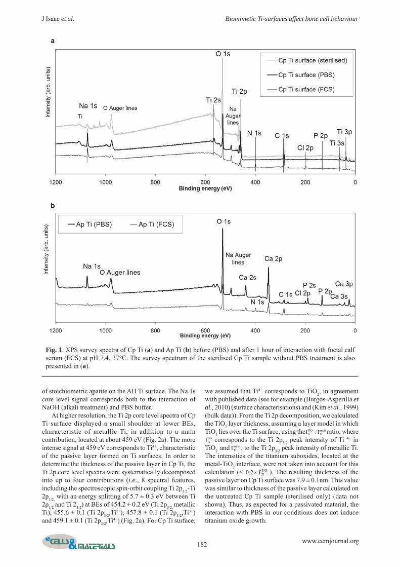
Surface chemical characterisationFig.1a shows the XPS survey spectra for Cp Ti. The corelevels of Ti (Ti 2s, Ti 2p, Ti 3s, Ti 3p), as well as the O 1score level (characteristic of the presence of oxides), and
the core level peaks of surface contaminants, such as C1s, P 2p and P 2s, Cl 2p and Cl 2s, K 2p and K 2s, and Na1s, were observed. Note that there was no signalcorresponding to the presence of Ca on the surface (alsochecked at higher resolution). P, Cl, K and Na were mostprobably contaminants from PBS buffer. As element ofcomparison, the survey spectrum of the untreated Cp Tisurface (sterilised sample only) showed the absence of P,Cl, K and Na as well as less attenuated O 1s and Ti 2p corelevel signals (Fig. 1a). The survey spectra for AH Tisamples showed the same core level peaks (data notshown), with the exception of a low intensity signalcorresponding to Ca, most probably associated withcontamination from prior alkali treatment. The Ca/P atomicratio, calculated from the Ca 2p and P 2p core levelintensities, recorded at high energy resolution (pass energyof 20 eV), was 0.16 ± 0.03, which excludes the presence
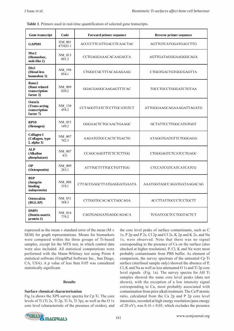
Gene transcript Code Forward primer sequence Reverse primer sequence
GAPDH XM_001 473623.1 ACCCCTTCATTGACCTCAACTAC AGTTGTCATGGATGACCTTG
Msx2 (Homeobox, msh-like 2)
NM_013 601.2 CCTGAGGAAACACAAGACCA AGTTGATAGGGAAGGGCAGA
Dlx5 (Distal-less homeobox 5)
NM_198 854.1 CTGGCCGCTTTACAGAGAAG CTGGTGACTGTGGCGAGTTA
Runx2 (Runt related transcription factor 2)
NM_009 820.2 GGACGAGGCAAGAGTTTCAC TGCCTGCCTGGGATCTGTAA
Osterix (Trans-acting transcription factor 7)
NM_130 458.2 CCTAGGTTATCTCCTTGCATGTCT ATTGGGAAGCAGAAAGATTAGATG
RP59 (Hemogen)
NM_053 149.2 GGGAACTCTGCAACTGAAGC GCTATTCCTTGGCATGTGGT
Collagen I (Collagen, type I, alpha 1)
NM_007 742.3 AAGATGTGCCACTCTGACTG ATAGGTGATGTTCTGGGAGG
ALP (Alkaline phosphatase)
NM_007 431 CCAGCAGGTTTCTCTCTTGG CTGGGAGTCTCATCCTGAGC
OP (Osteopontin)
NM_009 263.1 ATTTGCTTTTGCCTGTTTGG CTCCATCGTCATCATCATCG
BSP (Integrin binding sialoprotein)
NM_008 318.1 CTTACCGAGCTTATGAGGATGAATA AAATGGTAGCCAGATGATAAGACAG
Osteocalcin (BGLAP)
NM_031 368.3 CTTGGTGCACACCTAGCAGA ACCTTATTGCCCTCCTGCTT
DMP1 (Dentin matrix protein 1)
NM_016 779.2 CAGTGAGGATGAGGCAGACA TCGATCGCTCCTGGTACTCT
Table 1. Primers used in real-time quantification of selected gene transcripts.
182 www.ecmjournal.org
J Isaac et al. Biomimetic Ti-surfaces affect bone cell behaviour
of stoichiometric apatite on the AH Ti surface. The Na 1score level signal corresponds both to the interaction ofNaOH (alkali treatment) and PBS buffer.
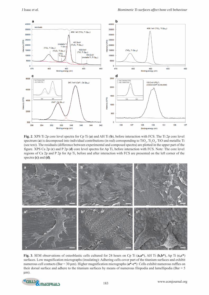
At higher resolution, the Ti 2p core level spectra of CpTi surface displayed a small shoulder at lower BEs,characteristic of metallic Ti, in addition to a maincontribution, located at about 459 eV (Fig. 2a). The moreintense signal at 459 eV corresponds to Ti4+, characteristicof the passive layer formed on Ti surfaces. In order todetermine the thickness of the passive layer in Cp Ti, theTi 2p core level spectra were systematically decomposedinto up to four contributions (i.e., 8 spectral features,including the spectroscopic spin-orbit coupling Ti 2p3/2-Ti2p1/2, with an energy splitting of 5.7 ± 0.3 eV between Ti2p1/2 and Ti 23/2) at BEs of 454.2 ± 0.2 eV (Ti 2p3/2, metallicTi), 455.6 ± 0.1 (Ti 2p3/2,Ti2+), 457.8 ± 0.1 (Ti 2p3/2,Ti3+)and 459.1 ± 0.1 (Ti 2p3/2,Ti4+) (Fig. 2a). For Cp Ti surface,
we assumed that Ti4+ corresponds to TiO2, in agreementwith published data (see for example (Burgos-Asperilla etal., 2010) (surface characterisations) and (Kim et al., 1999)(bulk data)). From the Ti 2p decomposition, we calculatedthe TiO2 layer thickness, assuming a layer model in whichTiO2 lies over the Ti surface, using the ratio, where
corresponds to the Ti 2p3/2 peak intensity of Ti 4+ inTiO2 and , to the Ti 2p3/2 peak intensity of metallic Ti.The intensities of the titanium suboxides, located at themetal-TiO2 interface, were not taken into account for thiscalculation (< ). The resulting thickness of thepassive layer on Cp Ti surface was 7.9 ± 0.1nm. This valuewas similar to thickness of the passive layer calculated onthe untreated Cp Ti sample (sterilised only) (data notshown). Thus, as expected for a passivated material, theinteraction with PBS in our conditions does not inducetitanium oxide growth.
Fig. 1. XPS survey spectra of Cp Ti (a) and Ap Ti (b) before (PBS) and after 1 hour of interaction with foetal calfserum (FCS) at pH 7.4, 37°C. The survey spectrum of the sterilised Cp Ti sample without PBS treatment is alsopresented in (a).
2TiO metalTi TiI / I
2TiOTiI
metalTiI
22.0 TiOTiI×
183 www.ecmjournal.org
J Isaac et al. Biomimetic Ti-surfaces affect bone cell behaviour
Fig. 2. XPS Ti 2p core level spectra for Cp Ti (a) and AH Ti (b), before interaction with FCS. The Ti 2p core levelspectrum (a) is decomposed into individual contributions (in red) corresponding to TiO2, Ti2O3, TiO and metallic Ti(see text). The residuals (difference between experimental and composed spectra) are plotted in the upper part of thefigure. XPS Ca 2p (c) and P 2p (d) core level spectra for Ap Ti, before interaction with FCS. Note: The core levelregions of Ca 2p and P 2p for Ap Ti, before and after interaction with FCS are presented on the left corner of thespectra (c) and (d).
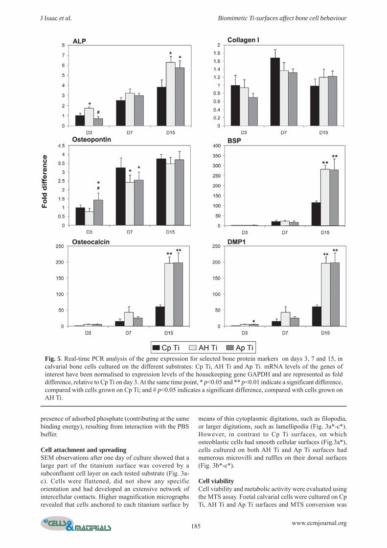
Fig. 3. SEM observations of osteoblastic cells cultured for 24 hours on Cp Ti (a,a*), AH Ti (b,b*), Ap Ti (c,c*)surfaces. Low magnification micrographs (insulating): Adhering cells cover part of the titanium surfaces and exhibitnumerous cell contacts (Bar = 30 μm). Higher magnification micrographs (a*-c*): Cells exhibit numerous ruffles ontheir dorsal surface and adhere to the titanium surfaces by means of numerous filopodia and lamellipodia (Bar = 5μm).

184 www.ecmjournal.org
J Isaac et al. Biomimetic Ti-surfaces affect bone cell behaviour
Fig. 2b shows the Ti 2p core level spectra of AH Tisurface. AH Ti spectra displayed one main peak, at about459 eV, characteristic of Ti4+. The absence of metalliccontribution in AH Ti surface indicated that the thicknessof the Ti oxide layer was greater than 10 nm (which is thelimit of XPS probing depth). This result was in agreementwith previous data obtained by Auger ElectronSpectroscopy (AES) (Kim et al., 1999). Indeed, Kim etal. (1999) reported that the untreated sample (Cp Ti) hadan ultra-thin TiO2 layer and AH samples were covered bya crystalline mixed sodium titanate and TiO2 layer, thickerthan 500 nm. Thus, we assumed that, in the case of AH Ti,the Ti 2p core level signal corresponds to Ti4+ both fromTiO2 and from sodium titanate, identified as Na2Ti5O11 byKim et al. (Kim et al., 1999) or as Na2TiO3 by Chu et al.(Chu et al., 2005)). The Na/Ti ratio for AH Ti surface,calculated from the Na 1s and Ti 2p (Ti4+) core levelintensities, was 0.90 ± 0.15. Unfortunately, it wasimpossible to propose a sodium titanate formula, becausethis ratio included the Ti4+ contribution from TiO2
and theNa contamination from PBS (as an illustration, the Na/Tiratio for Cp Ti surface was 0.60 ± 0.10). Thus, from achemical point of view, AH Ti and Cp Ti presented manysimilarities: a Ti4+ and Na+ -rich surface, the latter beingembedded in a crystalline titanate compound for AH Tiand being only randomly adsorbed in a two-dimensionalstructure for Cp Ti.
Fig. 1b shows the XPS survey spectra of Ap Ti. Themost striking difference between Ap Ti and both Cp Ti(Fig. 1a) and AH Ti (not shown) survey spectra was theabsence of any signal characteristic of Ti, indicating thatthe Ca-P inorganic layer is thicker than 10 nm on the Ap
Ti surface. Indeed, Kim et al. showed by AES depthprofiles that the Ap Ti surfaces were covered by more than1 μm of crystalline apatite (Kim et al., 1999). The expectedcore levels of Ca (Ca 2s, Ca 2p, Ca 3s, Ca 3p) and P (P 2s,P 2p), as well as those of O 1s (characteristic of the presenceof oxides) and the characteristic core level peaks of surfacecontaminants, such as C 1s, Cl 2p and Cl 2s, and Na 1s,were identified on Ap Ti surfaces. Cl and Na were mostprobably contaminants from the PBS buffer, with Pprobably coming from both the PBS buffer and the SBFtreatment forming the apatite layer on Ap Ti surface (see“Preparation of the titanium samples”).
At higher resolution, the Ca 2p core level spectra ofAp Ti surface showed a spectroscopic doublet (Ca 2p3/2-Ca 2p1/2) at 347.1 ± 0.2 eV (Ca 2p3/2), characteristic ofCa2+ (Burgos-Asperilla et al., 2010; Ferraz et al., 1999)(Fig. 2c). The P 2p core level peaks had an unresolveddoublet at 133.2 ± 0.2 eV (Fig. 2d), characteristic ofadsorbed PO4
3- (Burgos-Asperilla et al., 2010). The Ca/Psurface atomic ratio for Ap Ti was determined from theCa 2p and P 2p core level intensities, giving a ratio of 1.45± 0.25. This value is in very good agreement with theatomic ratio obtained from EDX (1.50 ± 0.30, Table S1).Taken together these results suggest that the compositionof the Ap Ti outermost 10 nm (XPS), as well as in its deeperlayers (EDX), could be compatible with stoichiometricapatite (Ca5(PO4)3OH) Ca/P theoretical value of 1.67. Aslightly lower experimental value has already been reported(Lakstein et al., 2009; Lu et al., 2000); in these cases, XPSCa/P atomic ratios of different calcium phosphate phaseswere consistently lower than the theoretical values. In ourcase, the small discrepancy could also be attributed to the
Fig. 4. MTS assay of osteoblastic cells grown on the different substrates at 24 hours, 48 hours and day 7. Measurementsfor controls (Thermanox Plastic coverslip) were set at 100% at each time point. Data are expressed in percentages,relative to controls. Statistical analyses were performed using the Mann-Whitney test (& p<0.05 indicates a significantdifference, compared with cells grown on the control surfaces; * p<0.05 indicates a significant difference, comparedwith Cp Ti; and # p<0.05 indicates a significant difference, compared with AH Ti. NS, no significant difference).
185 www.ecmjournal.org
J Isaac et al. Biomimetic Ti-surfaces affect bone cell behaviour
presence of adsorbed phosphate (contributing at the samebinding energy), resulting from interaction with the PBSbuffer.
Cell attachment and spreadingSEM observations after one day of culture showed that alarge part of the titanium surface was covered by asubconfluent cell layer on each tested substrate (Fig. 3a-c). Cells were flattened, did not show any specificorientation and had developed an extensive network ofintercellular contacts. Higher magnification micrographsrevealed that cells anchored to each titanium surface by
means of thin cytoplasmic digitations, such as filopodia,or larger digitations, such as lamellipodia (Fig. 3a*-c*).However, in contrast to Cp Ti surfaces, on whichosteoblastic cells had smooth cellular surfaces (Fig.3a*),cells cultured on both AH Ti and Ap Ti surfaces hadnumerous microvilli and ruffles on their dorsal surfaces(Fig. 3b*-c*).
Cell viabilityCell viability and metabolic activity were evaluated usingthe MTS assay. Foetal calvarial cells were cultured on CpTi, AH Ti and Ap Ti surfaces and MTS conversion was
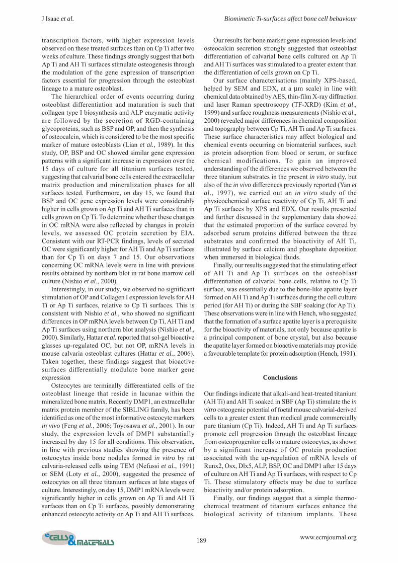
Fig. 5. Real-time PCR analysis of the gene expression for selected bone protein markers on days 3, 7 and 15, incalvarial bone cells cultured on the different substrates: Cp Ti, AH Ti and Ap Ti. mRNA levels of the genes ofinterest have been normalised to expression levels of the housekeeping gene GAPDH and are represented as folddifference, relative to Cp Ti on day 3. At the same time point, * p<0.05 and ** p<0.01 indicate a significant difference,compared with cells grown on Cp Ti; and # p<0.05 indicates a significant difference, compared with cells grown onAH Ti.

186 www.ecmjournal.org
J Isaac et al. Biomimetic Ti-surfaces affect bone cell behaviour
assessed after 24 hours, 48 hours and 7 days of culture(Fig. 4). Thermanox plastic coverslips were used as non-cytotoxic control surfaces. After 24 hours of culture, nosignificant differences in cell viability were observedbetween any of the three titanium surfaces tested andcontrols. After 48h and on day 7, cell viability on alltitanium surfaces – Cp Ti, AH Ti and Ap Ti – wassignificantly lower than that on control surfaces. After 48hours, cells that had grown on AH Ti surfaces showed thelowest MTS activity levels, reaching 61.4% of controlvalues, whereas cell viability on Ap Ti and Cp Ti surfaceswas higher, reaching 79% and 90.2% of control values,respectively. However, after 7 days, cell viability wassimilar for all three surfaces, reaching approximately 75%of control values.
Osteoblast differentiationTo investigate the effects of various titanium surfaces onosteoblastic differentiation of calvarial bone cells, geneexpression levels for selected osteoblast markers, includingbone protein markers (Fig. 5), osteoblastic transcriptionfactors (Fig. 6) and the protein RP59 (Fig. 7), weredetermined using real-time RT-PCR on days 3, 7 and 15.All selected genes were expressed throughout the cultureperiod but showed different expression patterns over timefor the different surfaces.
The alkaline phosphatase (ALP) gene expressionprofile showed a gradual and significant increase inexpression levels over the culture period for all titaniumsurfaces tested (p<0.01) (Fig. 5). However, cells culturedon the AH Ti and Ap Ti surfaces showed a significantlyhigher ALP mRNA levels on day 15 than cells on Cp Tisurfaces.
Collagen, type I, alpha 1 (collagen I) mRNA levels forboth Cp Ti and Ap Ti surfaces significantly increased upto day 7 (p<0.05) and then decreased at day 15 (p<0.05)(Fig. 5). However, for AH Ti surfaces, collagen I mRNAlevels remained stable throughout the culture period (NS).Moreover, we did not find any significant differencesbetween the three different surfaces at each time point.
Osteopontin (OP) gene expression levels showed agradual and significant increase over time, up to day 15,for all three surfaces (p<0.01) (Fig. 5). On day 7, OP mRNAlevels were significantly lower for both AH Ti and Ap Tisurfaces than for Cp Ti. However, we did not find anysignificant differences between the three different surfaceson day 15.
The extracellular bone matrix protein-related genesencoding integrin binding sialoprotein (BSP), osteocalcin(OC) and dentin matrix protein 1 (DMP1) showed similarexpression patterns for all surfaces, with low geneexpression levels observed on day 3, followed by a
Fig. 6. Real-time PCR analysis of the expression of genes encoding selected osteoblastic transcription factors ondays 3, 7 and 15 in foetal calvarial cells cultured on different substrates: Cp Ti, AH Ti and Ap Ti. mRNA levels of thegenes of interest have been normalised to expression levels of the housekeeping gene GAPDH and are representedas fold difference relative to Cp Ti on day 3. At the same time point, * p<0.05 and ** p<0.01 indicate a significantdifference, compared with cells grown on Cp Ti; and # p<0.05 indicates a significant difference, compared withcells grown on AH Ti.

187 www.ecmjournal.org
J Isaac et al. Biomimetic Ti-surfaces affect bone cell behaviour
substantial increase in expression over the following daysof culture (p<0.01) (Fig. 5). However, the magnitude ofthis observed increase was dependent on the titaniumsurface being investigated. Thus, on day 15, cells grownon the AH Ti and Ap Ti surfaces showed significantlyhigher expression levels of these genes than those grownon Cp Ti surfaces; thus, BSP, OC and DMP1 mRNA levelswere around three times higher on AH Ti and Ap Ti surfacesthan on Cp Ti.
To further investigate the effects of titanium surfaceson osteoblastic differentiation of calvarial bone cells, westudied the mRNA levels of selected transcription factors.Msx2 mRNA levels were detected throughout the cultureperiod, with a strong and stable expression on days 3 and7, followed by a significant decrease in expression levelson day 15, for all treated surfaces (p<0.05) (Fig. 6). Ondays 3 and 7, Msx2 mRNA levels were significantly lowerfor both AH Ti and Ap Ti surfaces than for Cp Ti. However,on day 15, we did not find any significant differencesbetween the three different surfaces. Overall, Runx2(Cbfa1) and Dlx5 showed similar expression profiles forall tested surfaces, showing a gradual and significantincrease up to day 7 (p<0.05); their expression levels thenremained stable up to day 15 (NS) (Fig. 6). Osterix (Osx)mRNA levels increased gradually and significantlythroughout the 15 days of culture for all surfaces tested
(p<0.01) (Fig. 6). Cells grown on both AH Ti and Ap Tisurfaces showed significantly higher Runx2 and OsxmRNA levels than those grown on Cp Ti surfaces, at alltime points. Dlx5 gene expression levels for AH Ti andAp Ti surfaces were also higher than Cp Ti surfaces onday 15.
We also analysed the transcription profile for theprotein RP 59. RP59 mRNA levels decreased considerablyover the culture period for all titanium surfaces (p<0.01),with levels 17 times (Cp Ti surfaces), 11 times (AH Tisurfaces) and 12 times (Ap Ti surfaces) lower on day 7than on day 3 (Fig. 7). On day 3, both AH Ti and Ap Tisurfaces showed lower levels of RP59 gene expressionthan Cp Ti surfaces. On days 7 and 15, however, we didnot find any significant differences in RP59 geneexpression levels between the three titanium surfaces.
Finally, osteocalcin protein production was assayed byEIA. OC protein levels were not detectable on day 3 butsignificantly increased over the observation period for alltitanium surfaces tested (p<0.05) (Fig. 8). Furthermore,whereas no significant differences were observed betweenAH Ti and Ap Ti surfaces, we found that secreted OC levelswere significantly higher for the two treated surfaces thanfor Cp Ti on days 7 and 15, with a three-fold differencebetween the two treated surfaces (AH Ti and Ap Ti) andCp Ti surfaces observed on day 15.
Fig. 7. Real-time PCR analysis of the expression of RP59on days 3, 7 and 15 in foetal calvarial cells cultured ondifferent substrates: Cp Ti, AH Ti and Ap Ti. mRNA levelsof the genes of interest have been normalised to expressionlevels of the housekeeping gene GAPDH and arerepresented as fold difference relative to Cp Ti on day 3.At the same time point, * p<0.05 and ** p<0.01 indicate asignificant difference, compared with cells grown on CpTi; and # p<0.05 indicates a significant difference,compared with cells grown on AH Ti.
Fig. 8. Osteocalcin (OC) protein production byosteoblastic cells grown on Cp Ti, AH Ti and Ap Tisurfaces on days 3, 7 and 15. Data are expressed as OC(ng/mL) secreted during 48h in the serum-free medium,and normalised to the total cell count. At the same timepoint, * p<0.05, ** p<0.01 and ***p<0.001 indicate asignificant difference, compared with cells grown on CpTi; and # p<0.05 indicates a significant difference,compared with cells grown on AH Ti.

188 www.ecmjournal.org
J Isaac et al. Biomimetic Ti-surfaces affect bone cell behaviour
Discussion
Over ten years ago, Kim et al. developed a thermo-chemical treatment resulting in the formation of an alkalititanate layer on titanium surface (Kim et al., 1996). Whenimplanted in bony sites, this modified titanium shows bone-binding ability due to the bone-like apatite layer formedon its surface (Kim et al., 1997). This bioengineeredtitanium was thus classified as a bioactive metal. It hassince been widely characterised and successfully tested inclinical trials for orthopaedic use; however, its mechanismof action on peri-implant bone cells is not yet fullyunderstood.
The aim of this study was to investigate the effects ofthermo-chemical treated surfaces on phenotypic changesassociated with the process of osteoblast differentiation.
As the use of cell lines often leads to incompletepatterns of osteoblastic differentiation (Cooper et al., 1998)with cells exhibiting various mechanisms of attachment(Kilpadi et al., 2004), in this study we used a primary cellculture model of foetal mouse osteoblasts isolated fromcalvaria. Indeed, the culture of calvarial bone cells in anosteogenic medium promotes the proliferation andcommitment of osteoprogenitor cells to mature osteoblasts(Nefussi et al., 1985; Owen et al., 1990).
Osteoblast differentiation and subsequent boneformation is a gradual, well orchestrated process associatedwith changes in gene expression over time andcharacterised in vitro by three developmental stages:proliferation, extracellular matrix production andmaturation, and matrix mineralization (Owen et al., 1990).Using this modelling of osteoblast differentiation, wemonitored by real-time RT-PCR the bone gene expressionpatterns of calvarial bone cells cultured on alkali- and heat-treated titanium (AH Ti), AH Ti soaked in SBF (Ap Ti)and commercially pure titanium (Cp Ti).
Firstly, we investigated the gene expression of earlymarkers associated with proliferative osteoprogenitors:Msx2 and RP59. Msx2, a member of the Msx (MuscleSegment homeobox) family, is a homeobox-containingtranscription factor. Several lines of evidence suggestedthat Msx2 gene expression could be associated withosteoblast proliferation and act as a repressor ofosteoblastic differentiation. Indeed, in vivo, Msx2 isexpressed in the proliferating mesenchyme of the cranialsuture and then decreased in the cells flanking theosteogenic front (De Coster et al., 2007) and, in osteoblastcell cultures, Msx2 is mostly expressed in proliferatingcells, with a gradual decrease in activity observed duringosteoblast differentiation (Hassan et al., 2004). Moreover,overexpression of the Msx2-encoding gene promotes theproliferation of osteoprogenitor cells, at the same timepreventing osteoblastic differentiation (Dodig et al., 1999;Liu et al., 1999) by repressing the activity of osteoblastmarkers, such as Runx2 (Shirakabe et al., 2001), ALP (Kimet al., 2004), collagen I (Dodig et al., 1999) and osteocalcin(Hassan et al., 2004). These results are in line with a recentstudy of Marijanovic et al. where the authors concludethat calvarial osteoblasts derived from Msx2 deficient miceshowed a lower rate of proliferation and an increased
osteoblastic differentiation when compared to osteoblastsderived from wild type mice (Marijanovic et al., 2009).Taken together, these results suggest that Msx2 could actin vivo to maintain the committed osteoblasts of osteogenicfront in undifferentiated stage as well as stimulating theirproliferation, then enabling the growth of cranial bones.In our study, Msx2 gene expression levels weresignificantly lower on day 15 than at the earlier time pointsfor all surfaces tested and were lower for both Ap Ti andAH Ti surfaces than for Cp Ti on days 3 and 7. Interestingly,RP59 mRNA levels were also significantly lower on day3 for both Ap Ti and AH Ti surfaces than for Cp Ti andthen RP59 mRNA decreased sharply after day 3, for allsurfaces tested. The RP59 protein has previously beenidentified as a marker for progenitor cells with the capacityto differentiate into osteoblasts (Wurtz et al., 2001). Indeed,in vivo, RP59 is present at variable levels in bone marrowcells and newly recruited osteoblasts during bone formation(Wurtz and Berdal, 2003) and fracture healing (Chen etal., 2004), with lower levels found in mature osteoblasts.Overall, the gene expression profiles for both Msx2 andRP59 suggested that the proliferation phase is mainlyassociated with the first days of culture for all threesurfaces. Additionally, because our SEM analysis and MTSassay results did not show any cytotoxic effect of the threetested surfaces on calvarial bone cells, we hypothesizedthat the reduced Msx2 and RP59 mRNA levels observedon Ap Ti and AH Ti surfaces at earlier times of culturemay be due to the fact that bioengineered surfaces drivethe commitment of osteoprogenitor cells to the osteoblasticphenotype more quickly than Cp Ti surfaces, where thecells could be longer maintained in a proliferating stage.
To investigate this further, we studied the changes inthe main phenotypic bone markers. We showed that ALPmRNA levels increased significantly over the cultureperiod for all surfaces but were significantly higher forboth AH Ti and Ap Ti than for Cp Ti on day 15. In linewith our findings, Nishio et al. previously showed thatALP enzymatic activity levels in rat bone marrow cellsgrown on Ap Ti and AH Ti surfaces were higher than inthose grown on Cp Ti surfaces on day 14 (Nishio et al.,2000). To further investigate osteoblast differentiation, wethen analysed gene expression levels for three transcriptionfactors with major roles in skeletal formation and bonehealing: Runx2, Osterix and Dlx5 (Ferguson et al., 1999;Wohl et al., 2009). Runx2 (cbfa1) and Osterix (Osx) arethe major osteoblast transcription factors involved inosteoblast differentiation and bone formation. Thus, Runx2or Osx gene ablation in mice results in a complete absenceof bone skeleton (Komori et al., 1997; Nakashima et al.,2002), while forced expression of these genes stimulatetranscription of both early and late bone marker genes(Ducy et al., 1997; Tu et al., 2006). Dlx5, a member of theDlx (Distal-less homeobox) gene family, is considered tobe an important regulator of osteoblast differentiation. Dlx5acts upstream of Runx2 and Osx (Ryoo et al., 2006) andits expression levels gradually increase during osteoblastdifferentiation (Hassan et al., 2004). In this study, we foundthat both Ap Ti and AH Ti surfaces significantly promotedthe gene expression of these three specific osteoblast
189 www.ecmjournal.org
J Isaac et al. Biomimetic Ti-surfaces affect bone cell behaviour
transcription factors, with higher expression levelsobserved on these treated surfaces than on Cp Ti after twoweeks of culture. These findings strongly suggest that bothAp Ti and AH Ti surfaces stimulate osteogenesis throughthe modulation of the gene expression of transcriptionfactors essential for progression through the osteoblastlineage to a mature osteoblast.
The hierarchical order of events occurring duringosteoblast differentiation and maturation is such thatcollagen type I biosynthesis and ALP enzymatic activityare followed by the secretion of RGD-containingglycoproteins, such as BSP and OP, and then the synthesisof osteocalcin, which is considered to be the most specificmarker of mature osteoblasts (Lian et al., 1989). In thisstudy, OP, BSP and OC showed similar gene expressionpatterns with a significant increase in expression over the15 days of culture for all titanium surfaces tested,suggesting that calvarial bone cells entered the extracellularmatrix production and mineralization phases for allsurfaces tested. Furthermore, on day 15, we found thatBSP and OC gene expression levels were considerablyhigher in cells grown on Ap Ti and AH Ti surfaces than incells grown on Cp Ti. To determine whether these changesin OC mRNA were also reflected by changes in proteinlevels, we assessed OC protein secretion by EIA.Consistent with our RT-PCR findings, levels of secretedOC were significantly higher for AH Ti and Ap Ti surfacesthan for Cp Ti on days 7 and 15. Our observationsconcerning OC mRNA levels were in line with previousresults obtained by northern blot in rat bone marrow cellculture (Nishio et al., 2000).
Interestingly, in our study, we observed no significantstimulation of OP and Collagen I expression levels for AHTi or Ap Ti surfaces, relative to Cp Ti surfaces. This isconsistent with Nishio et al., who showed no significantdifferences in OP mRNA levels between Cp Ti, AH Ti andAp Ti surfaces using northern blot analysis (Nishio et al.,2000). Similarly, Hattar et al. reported that sol-gel bioactiveglasses up-regulated OC, but not OP, mRNA levels inmouse calvaria osteoblast cultures (Hattar et al., 2006).Taken together, these findings suggest that bioactivesurfaces differentially modulate bone marker geneexpression
Osteocytes are terminally differentiated cells of theosteoblast lineage that reside in lacunae within themineralized bone matrix. Recently DMP1, an extracellularmatrix protein member of the SIBLING family, has beenidentified as one of the most informative osteocyte markersin vivo (Feng et al., 2006; Toyosawa et al., 2001). In ourstudy, the expression levels of DMP1 substantiallyincreased by day 15 for all conditions. This observation,in line with previous studies showing the presence ofosteocytes inside bone nodules formed in vitro by ratcalvaria-released cells using TEM (Nefussi et al., 1991)or SEM (Loty et al., 2000), suggested the presence ofosteocytes on all three titanium surfaces at late stages ofculture. Interestingly, on day 15, DMP1 mRNA levels weresignificantly higher in cells grown on Ap Ti and AH Tisurfaces than on Cp Ti surfaces, possibly demonstratingenhanced osteocyte activity on Ap Ti and AH Ti surfaces.
Our results for bone marker gene expression levels andosteocalcin secretion strongly suggested that osteoblastdifferentiation of calvarial bone cells cultured on Ap Tiand AH Ti surfaces was stimulated to a greater extent thanthe differentiation of cells grown on Cp Ti.
Our surface characterisations (mainly XPS-based,helped by SEM and EDX, at a μm scale) in line withchemical data obtained by AES, thin-film X-ray diffractionand laser Raman spectroscopy (TF-XRD) (Kim et al.,1999) and surface roughness measurements (Nishio et al.,2000) revealed major differences in chemical compositionand topography between Cp Ti, AH Ti and Ap Ti surfaces.These surface characteristics may affect biological andchemical events occurring on biomaterial surfaces, suchas protein adsorption from blood or serum, or surfacechemical modifications. To gain an improvedunderstanding of the differences we observed between thethree titanium substrates in the present in vitro study, butalso of the in vivo differences previously reported (Yan etal., 1997), we carried out an in vitro study of thephysicochemical surface reactivity of Cp Ti, AH Ti andAp Ti surfaces by XPS and EDX. Our results presentedand further discussed in the supplementary data showedthat the estimated proportion of the surface covered byadsorbed serum proteins differed between the threesubstrates and confirmed the bioactivity of AH Ti,illustrated by surface calcium and phosphate depositionwhen immersed in biological fluids.
Finally, our results suggested that the stimulating effectof AH Ti and Ap Ti surfaces on the osteoblastdifferentiation of calvarial bone cells, relative to Cp Tisurface, was essentially due to the bone-like apatite layerformed on AH Ti and Ap Ti surfaces during the cell cultureperiod (for AH Ti) or during the SBF soaking (for Ap Ti).These observations were in line with Hench, who suggestedthat the formation of a surface apatite layer is a prerequisitefor the bioactivity of materials, not only because apatite isa principal component of bone crystal, but also becausethe apatite layer formed on bioactive materials may providea favourable template for protein adsorption (Hench, 1991).
Conclusions
Our findings indicate that alkali-and heat-treated titanium(AH Ti) and AH Ti soaked in SBF (Ap Ti) stimulate the invitro osteogenic potential of foetal mouse calvarial-derivedcells to a greater extent than medical grade commerciallypure titanium (Cp Ti). Indeed, AH Ti and Ap Ti surfacespromote cell progression through the osteoblast lineagefrom osteoprogenitor cells to mature osteocytes, as shownby a significant increase of OC protein productionassociated with the up-regulation of mRNA levels ofRunx2, Osx, Dlx5, ALP, BSP, OC and DMP1 after 15 daysof culture on AH Ti and Ap Ti surfaces, with respect to CpTi. These stimulatory effects may be due to surfacebioactivity and/or protein adsorption.
Finally, our findings suggest that a simple thermo-chemical treatment of titanium surfaces enhance thebiological activity of titanium implants. These

190 www.ecmjournal.org
J Isaac et al. Biomimetic Ti-surfaces affect bone cell behaviour
bioengineered titanium surfaces may have a great potentialfor future dental applications as a template for enhancedosseointegration.
Acknowledgments
This work was supported by the Avenir Foundation (Projectn°ET7-462). The authors thank David Montero and AudreyAsselin for technical assistance. This study represents partof the dissertation of Juliane Isaac’s PhD degree at theUniversity Paris Descartes, France.
Supplementary Data
In vitro chemical evaluation of substrate surfacereactivityTwo approaches were used to study the in vitro chemicalreactivity of Cp Ti, AH Ti and Ap Ti surfaces: weinvestigated the adsorption of serum proteins on thesetitanium surfaces by XPS, and the surface chemicalmodifications in cell-free culture medium by EDX.
The reactivity of the substrates towards the proteins ofthe FCS was detected by the presence of the N 1s corelevel peak signal (XPS survey spectra of Fig. 1a and Fig.1b). At higher energy resolution, the N 1s core levelpresented a symmetrical peak centred at 400.1 ± 0.1 eV,characteristic of nitrogen in amide (mainly) and/or aminegroups (Fig.S1a). This signal can be used as a marker ofadsorbed proteins. The C 1s XPS core level spectraobtained after serum adsorption (Fig.S1b) showed threecontributions (see details in (Ithurbide et al., 2007; Payetet al., 2008)), corresponding to the well defined carbonbonds present in the proteins. These observations wereconsistent with previous data from Burgos-Asperilla et al.(Burgos-Asperilla et al., 2010). Spectral contributionsobserved at 286.4 ± 0.2 eV, labelled C2 and correspondingto C-N and C-O single bonds, and at 288.3 ± 0.2 eV,labelled C3 and corresponding to N-C=O bonds, were of
particular interest. The N 1s and C 1s core level peakswere better resolved for Cp Ti samples (smaller full widthsat half maximum) than for AH Ti and Ap Ti samples, whichare thicker, rougher, and more insulating samples. N/C2+C3 atomic ratios were systematically calculated forCp Ti surfaces. The obtained value of 0.49 ± 0.08corresponded to the theoretical ratio calculated fromamino-acid compositions, for example of albumin orfibronectin molecules (Lhoest et al., 2001)). This resultthus suggested that the interaction of Cp Ti surface withserum resulted in the chemical adsorption of the proteinspresent in the serum (according to the provider, albuminis the major protein in the FCS). Moreover, as regards theTi 2p, Ca 2p and P 2p core levels, the treatment with theserum did not affect the surface chemical composition ofCp Ti, AH Ti, Ap Ti surfaces, in our conditions. Anequivalent thickness of adsorbed protein was thendetermined, assuming a layer model where all protein(taken as albumin in the calculations) is adsorbed in acontinuous homogeneous layer on titanium oxide (takenas TiO2) or apatite. By using the attenuation of the XPSsignals, i. e.
for both Cp Ti and AH Ti, and
or
for Ap Ti, where is the intensity of Ti in TiO2 fromthe Ti 2p3/2 core level peak, is the intensity of Nfrom the N 1s core level peak, is the intensity of Cafrom the Ca 2p core level peak, and is the intensityof P from the P 2p core level peak, the adsorbed proteinequivalent layer was estimated at 4.2 ± 0.2 nm for both CpTi and AH Ti surfaces, and 3.3 ± 0.3 nm for Ap Ti surfaces(average value from the calculations with Ca 2p and P 2pcore levels). Protein adsorption was also quantitativelyevaluated using a model of islands of adsorbed proteinson the surface for XPS calculations (Payet et al., 2010). Inthis model, the height of the islands was considered as“infinite” (>10 nm, as defined by the XPS probed depth)
Atomic % Day P Ca Na Mg Cl O C Ti Ca/P
Cp Ti D3 0.18 0.08 3.22 0.09 1.16 8.66 29.27 57.35 0.44
Cp Ti D15 0.18 0.09 2.11 0.16 0.78 9.39 32.89 54.40 0.50
AH Ti D3 0.37 0.41 2.69 0.16 0.53 41.13 28.44 26.27 1.11
AH Ti D7 0.65 0.84 2.90 0.19 0.63 43.69 24.97 26.13 1.29
AH Ti D15 1.18 1.51 2.23 - 0.32 40.37 32.34 22.06 1.28
Ap Ti D3 8.85 13.34 1.17 0.75 0.59 42.82 31.64 0.83 1.50
Table S1. EDX microanalysis on Ti samples soaked in cell-free culture medium.
Semi-quantitative chemical analysis assessed by energy dispersive X-ray (EDX) microanalysis ontitanium samples soaked in cell-free culture medium for different durations (3, 7, 15 days). Atomicpercentages of phosphorus, calcium, sodium, magnesium, chlorine, oxygen, carbon and titanium andthe atomic calcium/phosphorus ratio are reported.
2
TiOTi
FCSadsorbedN
II
apatiteCa
FCSadsorbedN
II
apatiteP
N
II FCS adsorbed
2TiOTiI
FCSadsorbedNI
apatiteCaI
apatitePI

191 www.ecmjournal.org
J Isaac et al. Biomimetic Ti-surfaces affect bone cell behaviour
and the ratio of the intensity signals from the substratebefore ( or )
and after ( or )
treatment with the serum solution provides an estimationof the surface coverage by adsorbed proteins (Aeimbhu et
al., 2005). The
intensity ratio tended to 0.4, corresponding to a fractionof 0.6 of the surface covered by the adsorbed protein onCp Ti and AH Ti samples. The
intensity ratio tended to 0.7, corresponding to a fraction
Fig. S1. XPS N 1s (a) and C 1s (b) core level spectra for Cp Ti after treatment with FCS, at 37°C, pH 7.4, for 1 hour.The C 1s core level spectrum is decomposed into three components (see text for details).
Fig. S2. Spectral analysis of chemical composition, assessed by energy dispersive X-ray microanalysis (EDX) ontitanium samples soaked in cell-free culture medium for 3, 7 and 15 days). Detected atoms were phosphorus (P),calcium (Ca), sodium (Na), magnesium (Mg), chlorine (Cl), oxygen (O), carbon (C) and titanium (Ti).
2TiO[PBS] Ti,I apatite
[PBS] Ca,I2TiO
[FCS] Ti,I apatite[FCS] Ca,I
2
2
TiO[PBS] Ti,
TiO[FCS] Ti,
II
apatite[PBS] Ca,
apatite[FCS] Ca,
II
192 www.ecmjournal.org
J Isaac et al. Biomimetic Ti-surfaces affect bone cell behaviour
of 0.3 of the surface covered by the adsorbed protein onAp Ti samples. Thus, our XPS results showed that theequivalent thickness of a continuous (and homogeneous)adsorbed protein layer remained quite similar between thethree tested surfaces, with an adsorbed protein layerthickness of nearly 4 nm. However, using the model ofislands that we considered as more realistic in the case ofprotein adsorption, the estimated percentage of the surfacecovered by proteins from the serum significantly differedbetween the surfaces. Indeed, with a much lower surfacecoverage (30%), Ap Ti presented a different behaviour thanCp Ti and AH Ti (surface coverage of 60%).
At a larger time scale, the Ti-based substrates wereimmersed in cell-free culture medium up to 15 days andsurface topography and chemical modifications wereanalysed by SEM observations and EDX microanalysis.No differences in surface topography were observed forTi-based samples soaked in cell-free culture medium for3, 7 and 15 days, using SEM (data not shown). EDXmicroanalysis showed that the initial chemical compositionof Cp Ti and Ap Ti samples remained unchanged duringthe investigation period (Fig. S2 and Table S1). Thus, CpTi samples were mostly composed of titanium, whereasAp Ti first μm were mainly composed of Ca and P.However, EDX revealed changes in the chemicalcomposition of AH Ti over time (Fig.S2 and Table S1).Indeed, EDX spectra obtained for AH Ti showed that bothCa and P were present and that the atomic percentages ofthese elements increased with soaking time from day 3 today 15. This mineral layer was richer in calcium than inphosphorus with a Ca/P atomic ratio of 1.30 ± 0.30 after 7days of incubation (Table S1). Taken into account the EDXexperimental uncertainty, this atomic ratio is in reasonableagreement with the theoretical value of 1.67 expected forstoichiometric apatite. Note that all tested surfaces wereassociated with small amounts of sodium, magnesium andchlorine that were most probably contaminants from theculture medium and PBS (and SBF for Ap Ti samples),except for AH Ti surface where the Na signal shouldcorrespond both to the sodium titanate layer andcontamination from PBS and culture medium. Thesefindings illustrated the reactivity of AH Ti surface. Indeed,our EDX analyses showed that, from the first three daysof immersion in cell-free culture medium, calcium andphosphorus deposition occurred on AH Ti surfaces (TableS1). These observations were consistent with previous invivo and in vitro studies (Kokubo et al., 2004), reportingthat, after alkali- and heat-treatments, titanium implantscan initiate apatite nucleation on its surface through ionsexchange with biological fluids. We also showed that theatomic percentages of Ca and P elements graduallyincreased from day 3 to day 15, suggesting that the Ca-Player on the AH Ti surface grew with soaking time in thecell-free culture medium.
Finally, our observations may explain why the AH Tiand Ap Ti surfaces exhibited similar in vitro osteostimulatorpotential at later times of culture. Indeed, taking intoaccount the quite similar chemical composition of AH Tiand Cp Ti (even if the structural bulk composition differs),protein adsorption from serum on substrate surfaces may
be directly correlated to the surface chemical composition.Thus, given that the surface of AH Ti rapidly changed inthe chemical composition to form an apatite-like surfaceover time immersed in a biological fluid, we hypothesizedthat AH Ti surface should display a similar proteinadsorption profile to the Ap Ti surface at later times ofculture and implantation – in terms of distribution ofsurface coverage by the adsorbed protein.
References
Adell R, Lekholm U, Rockler B, Brånemark PI (1981)A 15-year study of osseointegrated implants in thetreatment of the edentulous jaw. Int J Oral Surg 10: 387-416.
Aeimbhu A, Castle JE, Singjai P (2005) Accountingfor the size of molecules in determination of adsorptionisotherms by XPS; exemplified by adsorption of chickenegg albumin on titanium. Surface Interface Anal 37: 1127-1136.
Albrektsson T (1998) Hydroxyapatite-coated implants:a case against their use. J Oral Maxillofac Surg, 56: 1312-1326.
Burgos-Asperilla L, Garcia-Alonso MC, Escudero ML,Alonso C (2010) Study of the interaction of inorganic andorganic compounds of cell culture medium with a Tisurface. Acta Biomater 6: 652-661.
Buser D, Mericske-Stern R, Bernard JP, Behneke A,Behneke N, Hirt HP, Belser UC, Lang NP (1997) Long-term evaluation of non-submerged ITI implants. Part 1: 8-year life table analysis of a prospective multi-center studywith 2359 implants. Clin Oral Impl Res 8: 161-172.
Chen YJ, Wurtz T, Wang CJ, Kuo YR, Yang KD, HuangHC, Wang FS (2004) Recruitment of mesenchymal stemcells and expression of TGF-beta 1 and VEGF in the earlystage of shock wave-promoted bone regeneration ofsegmental defect in rats. J Orthop Res 22: 526-534.
Chu CL, Chung CY, Zhou J, Pu YP, Lin PH (2005)Fabrication and characteristics of bioactive sodium titanate/titania graded film on NiTi shape memory alloy. J BiomedMater Res 75: 595-602.
Cooper LF, Masuda T, Yliheikkila PK, Felton DA(1998) Generalizations regarding the process andphenomenon of osseointegration. Part II. In vitro studies.IntJ Oral Maxillofac Impl 13: 163-174.
Davies JE (1996) In vitro modeling of the bone/implantinterface. Anat Rec 245: 426-445.
De Coster PJ, Mortier G, Marks LA, Martens LC (2007)Cranial suture biology and dental development: geneticand clinical perspectives. J Oral Pathol Med 36:447-455.
Dodig M, Tadic T, Kronenberg MS, Dacic S, Liu YH,Maxson R, Rowe DW, Lichtler AC (1999) Ectopic Msx2overexpression inhibits and Msx2 antisense stimulatescalvarial osteoblast differentiation. Dev Biol 209: 298-307.
Ducy P, Zhang R, Geoffroy V, Ridall AL, Karsenty G(1997) Osf2/Cbfa1: a transcriptional activator of osteoblastdifferentiation. Cell 89: 747-754.
Feng JQ, Ward LM, Liu S, Lu Y, Xie Y, Yuan B, Yu X,Rauch F, Davis SI, Zhang S, Rios H, Drezner MK, Quarles
193 www.ecmjournal.org
J Isaac et al. Biomimetic Ti-surfaces affect bone cell behaviour
LD, Bonewald LF, White KE (2006) Loss of DMP1 causesrickets and osteomalacia and identifies a role for osteocytesin mineral metabolism. Nat Genet 38: 1310-1315.
Ferguson C, Alpern E, Miclau T, Helms JA (1999) Doesadult fracture repair recapitulate embryonic skeletalformation? Mech Dev 87: 57-66.
Ferraz MP, Monteiro FJ, Santos JD (1999) CaO-P2O5glass hydroxyapatite double-layer plasma-sprayed coating:in vitro bioactivity evaluation. J Biomed Mater Res 45:376-383.
Hassan MQ, Javed A, Morasso MI, Karlin J, MontecinoM, van Wijnen AJ, Stein GS, Stein JL, Lian JB (2004)Dlx3 transcriptional regulation of osteoblastdifferentiation: temporal recruitment of Msx2, Dlx3, andDlx5 homeodomain proteins to chromatin of theosteocalcin gene. Mol Cell Biol 24: 9248-9261.
Hattar S, Loty S, Gaisser D, Berdal A, Sautier JM(2006) Effects of 58S sol-gel glasses on the temporalexpression of bone markers during mouse osteoblasticdifferentiation. J Biomed Mater Res 76: 811-819.
Hayes JS, Vos DI, Hahn J, Pearce SG, Richards RG(2009) An in vivo evaluation of surface polishing of TANintermedullary nails for ease of removal. Eur Cell Mater18: 15-26.
Hench LL (1991) Surface reaction kinetics andadsorption of biological moieties : a mechanistic approachto tissue attachment. In: The Bone-Biomaterials Interface(Davies JE, ed), University of Toronto, pp 33-48.
Isaac J, Loty S, Hamdan A, Kokubo T, Kim HM, BerdalA, Sautier JM (2009) Bone-like tissue formation on abiomimetic titanium surface in an explant model ofosteoconduction. J Biomed Mater Res A 89: 585-593.
Ithurbide A, Frateur I, Galtayries A, Marcus P (2007)XPS and flow-cell EQCM study of albumin adsorptionon passivated chromium surfaces: Influence of potentialand pH Electrochim Acta 53: 1336-1345.
Kilpadi KL, Sawyer AA, Prince CW, Chang PL, BellisSL (2004) Primary human marrow stromal cells and Saos-2 osteosarcoma cells use different mechanisms to adhereto hydroxylapatite. J Biomed Mater Res 68: 273-285.
Kim HM, Miyaji F, Kokubo T, Nakamura T (1996)Preparation of bioactive Ti and its alloys via simplechemical surface treatment. J Biomed Mater Res 32: 409-417.
Kim HM, Miyaji F, Kokubo T, Nakamura T (1997)Bonding strength of bonelike apatite layer to Ti metalsubstrate. J Biomed Mater Res 38: 121-127.
Kim HM, Miyaji F, Kokubo T, Nishiguchi S, NakamuraT (1999) Graded surface structure of bioactive titaniumprepared by chemical treatment. J Biomed Mater Res 45:100-107.
Kim YJ, Lee MH, Wozney JM, Cho JY, Ryoo HM(2004) Bone morphogenetic protein-2-induced alkalinephosphatase expression is stimulated by Dlx5 andrepressed by Msx2. J Biol Chem 279: 50773-50780.
Kokubo T, Kim HM, Kawashita M, Nakamura T (2004)Bioactive metals: preparation and properties. J Mater SciMater Med 15: 99-107.
Kokubo T, Takadama H (2006) How useful is SBF inpredicting in vivo bone bioactivity? Biomaterials 27: 2907-2915.
Komori T, Yagi H, Nomura S, Yamaguchi A, SasakiK, Deguchi K, Shimizu Y, Bronson RT, Gao YH, InadaM, Sato M, Okamoto R, Kitamura Y, Yoshiki S, KishimotoT (1997) Targeted disruption of Cbfa1 results in a completelack of bone formation owing to maturational arrest ofosteoblasts. Cell 89: 755-764.
Lakstein D, Kopelovitch W, Barkay Z, Bahaa M,Hendel D, Eliaz N (2009) Enhanced osseointegration ofgrit-blasted, NaOH-treated and electrochemicallyhydroxyapatite-coated Ti-6Al-4V implants in rabbits. ActaBiomater 5: 2258-2269.
Lhoest JB, Wagner MS, Tidwell CD, Castner DG(2001) Characterization of adsorbed protein films by timeof flight secondary ion mass spectrometry. J Biomed MaterRes 57: 432-440.
Lian JB, Stein GS, Stewart C, Puchacz E, MackowiakS, Aronow M, Von Deck M, Shalhoub V (1989)Osteocalcin: characterization and regulated expression ofthe rat gene. Connect Tissue Res 21: 61-68.
Liu YH, Tang Z, Kundu RK, Wu L, Luo W, Zhu D,Sangiorgi F, Snead ML, Maxson RE (1999) Msx2 genedosage influences the number of proliferative osteogeniccells in growth centers of the developing murine skull: apossible mechanism for MSX2-mediated craniosynostosisin humans. Dev Biol 205: 260-274.
Loty C, Sautier JM, Boulekbache H, Kokubo T, KimHM, Forest N (2000) In vitro bone formation on a bone-like apatite layer prepared by a biomimetic process on abioactive glass-ceramic. J Biomed Mater Res 49: 423-434.
Lu HB, Campbell CT, Graham DJ, Ratner BD (2000)Surface characterization of hydroxyapatite and relatedcalcium phosphates by XPS and TOF-SIMS. Anal Chem72: 2886-2894.
Marijanovic I, Kronenberg MS, Erceg Ivkosic I,Lichtler AC (2009) Comparison of proliferation anddifferentiation of calvarial osteoblast cultures derived fromMsx2 deficient and wild type mice. Coll Antropol 33: 919-924.
Martini D, Fini M, Franchi M, Pasquale VD, BacchelliB, Gamberini M, Tinti A, Taddei P, Giavaresi G, Ottani V,Raspanti M, Guizzardi S, Ruggeri A (2003) Detachmentof titanium and fluorohydroxyapatite particles in unloadedendosseous implants. Biomaterials 24: 1309-1316.
Munting E (1996) The contributions and limitationsof hydroxyapatite coatings to implant fixation: Ahistomorphometric study of load bearing implants in dogs.Int Orthop 20: 1-6.
Nakashima K, Zhou X, Kunkel G, Zhang Z, Deng JM,Behringer RR, de Crombrugghe B (2002) The novel zincfinger-containing transcription factor osterix is requiredfor osteoblast differentiation and bone formation. Cell 108:17-29.
Nefussi JR, Boy-Lefevre ML, Boulekbache H, ForestN (1985) Mineralization in vitro of matrix formed byosteoblasts isolated by collagenase digestion.Differentiation 29: 160-168.
Nefussi JR, Sautier JM, Nicolas V, Forest N (1991)How osteoblasts become osteocytes: a decreasing matrixforming process. J Biol Buccale 19: 75-82.
Nishio K, Neo M, Akiyama H, Nishiguchi S, Kim HM,Kokubo T, Nakamura T (2000) The effect of alkali- and

194 www.ecmjournal.org
J Isaac et al. Biomimetic Ti-surfaces affect bone cell behaviour
heat-treated titanium and apatite-formed titanium onosteoblastic differentiation of bone marrow cells. . JBiomed Mater Res 52: 652-661.
Owen TA, Aronow M, Shalhoub V, Shalhoub V, BaroneLM, Wilming L, Tassinari MS, Kennedy MB, PockwinseS, Lian JB, Stein GS (1990) Progressive development ofthe rat osteoblast phenotype in vitro: reciprocalrelationships in expression of genes associated withosteoblast proliferation and differentiation duringformation of the bone extracellular matrix. J Cell Physiol143: 420-430.
Payet V, Brunner S, Galtayries A, Frateur I, Marcus P(2008) Cleaning of albumin-contaminated Ti and Crsurfaces : an XPS and QCM study. Surf Interface Anal40: 215-219.
Payet V, Dini T, Brunner S, Galtayries A, Frateur I,Marcus P (2010) Pre-treatment of titanium surfaces byfibronectin: in situ adsorption and effect of concentration:an XPS and QCM study. Surf Interface Anal DOI:10.1002/sia.3298.
Pearce AI, Pearce SG, Schwieger K, Milz S, SchneiderE, Archer CW, Richards RG (2008) Effect of surfacetopography on removal of cortical bone screws in a novelsheep model. J Orthop Res 26: 1377-1383.
Pohler OE (2000) Unalloyed titanium for implants inbone surgery. Injury 31 Suppl 4: 7-13.
Ryoo HM, Lee MH, Kim YJ (2006) Critical molecularswitches involved in BMP-2-induced osteogenicdifferentiation of mesenchymal cells. Gene 366: 51-57.
Shirakabe K, Terasawa K, Miyama K, Shibuya H,Nishida E (2001) Regulation of the activity of thetranscription factor Runx2 by two homeobox proteins,Msx2 and Dlx5. Genes Cells 6: 851-856.
Sun L, Berndt CC, Gross KA, Kucuk A (2001) Materialfundamentals and clinical performance of plasma-sprayedhydroxyapatite coatings: a review. . J Biomed Mater Res58: 570-592.
Toyosawa S, Shintani S, Fujiwara T, Ooshima T, SatoA, Ijuhin N, Komori T (2001) Dentin matrix protein 1 ispredominantly expressed in chicken and rat osteocytes butnot in osteoblasts. J Bone Miner Res 16: 2017-2026.
Tu Q, Valverde P, Chen J (2006) Osterix enhancesproliferation and osteogenic potential of bone marrowstromal cells. Biochem Biophys Res Commun 341: 1257-1265.
Wang BC, Chang E, Lee TM, Yang CY (1995) Changesin phases and crystallinity of plasma-sprayedhydroxyapatite coatings under heat treatment: aquantitative study. J Biomed Mater Res 29: 1483-1492.
Wohl GR, Towler DA, Silva MJ (2009) Stress fracturehealing: fatigue loading of the rat ulna induces upregulationin expression of osteogenic and angiogenic genes thatmimic the intramembranous portion of fracture repair.Bone 44: 320-330.
Wurtz T, Berdal A (2003) Osteoblast precursors atdifferent anatomic sites. Crit Rev Eukar Gene Expr 13:147-161.
Wurtz T, Kruger A, Christersson C, Lundmark C (2001)A new protein expressed in bone marrow cells andosteoblasts with implication in osteoblast recruitment. ExpCell Res 263: 236-242.
Yan WQ, Nakamura T, Kobayashi M, Kim HM, MiyajiF, Kokubo T (1997) Bonding of chemically treated titaniumimplants to bone. J Biomed Mater Res 37: 267-275.
Discussion with Reviewer
Reviewer I: The peak increase in OC and DMP1expression appears around D15 for the treated samples.Did the authors culture for longer time periods to see ifthis peak occurred also for Cp Ti at later stages or was theexpression observed at D15 representative of peakexpression for Cp Ti also?Authors: The expression of Osteocalcin and DMP1 in cellscultured on Cp Ti samples significantly increased over the15 days of culture. Indeed, OC and DMP1 gene expressionswere 61 times and 12 times higher at D15 as compared toD3, respectively. To check that the peak expression of lateosteoblast markers (BSP, OC, DMP1 and Dlx5) observedat D15 was representative for the investigated surfaces,calvarial cells were cultured on the three titanium samplesduring 21 days and gene expression pattern of the 11osteoblast markers investigated in our study weremonitored by real-time RT-PCR at D17 and D21.
Our results show that mRNA levels of the early andlate osteoblast markers decreased or remained stable afterD15, independently of the investigated surface and gene(data not shown). Moreover, SEM microscopicobservations performed after D15 showed an importantcell death associated with a degradation of the extracellularmatrix on the three titanium surfaces, as well as on theThermanox plastic coverslips. These results suggest that,in our conditions of culture (i.e. osteogenic medium, celldensity), primary calvarial cells begin to die by necrosisand/or apoptosis after 2 weeks of culture, making the RT-PCR results inconsistent. These observations were in linewith previous studies showing that the process of in vitroosteoblast differentiation is followed by a phase ofapoptosis, characterized by the overexpression of apoptosismarkers, such as Bcl2, p53 and c-fox (Stein et al., 2004).Thus, our many years of practice with primary calvarialcell cultures led us to focus our gene expression analysison three time points that reflect the three key stages ofosteoblastic differentiation on plastic dishes. These timepoints (D3, D7 and D15) respectively correspond to 1)the decrease of the proliferating phase and the onset ofdifferentiation, 2) the matrix secretion and maturation, and3) the extracellular matrix mineralization with the peakexpression of the specific markers of mature osteoblastsand osteocytes, such as Osteocalcin and DMP1.
Reviewer I: Can the authors comment on the early peakof both OC and DMP-1 after just 15 days culturing? Howdid this compare to cells cultured on plastic?Authors: Before we started experiments with titaniumsamples, we first genetically characterized the primarycalvarial cell culture model. For this purpose, calvarial cellswere cultured on Thermanox plastic coverslips with asimilar cell density and the same osteogenic medium thatwe used for titanium experimentations. Real-time RT-PCRresults on cells cultured on Thermanox plastic coverslips

195 www.ecmjournal.org
J Isaac et al. Biomimetic Ti-surfaces affect bone cell behaviour
showed that OC and DMP1 exhibit a similar expressionpattern as compared to cells cultured on Cp Ti, AH Ti andAp Ti surfaces, with a low gene expression at D3 and D7,followed by a substantial increase in expression at D15(data not shown). However, since the aim of our studywas to determine the effects of different titanium surfaceson osteoblast differentiation, we decided to leave out thegene expression profile of the cells grown on Thermanoxplastic coverslips (that are specifically treated for cellularadherence) in order to make the manuscript clearer.
Reviewer I: ALP and BSP normally indicate initiation ofmineralization and decrease prior to terminaldifferentiation. However the peak for both these markersare at day 15, which happens to be the time when OC isalso peaked - can the authors consolidate this observation?Authors: The specific function of the alkaline phosphataseenzyme is not yet fully understood. However, as underlinedby the reviewer, the ALP enzyme is assumed to play a rolein the process of matrix mineralization and morespecifically in its initiation (Bellows et al., 1991; Kay andRobison, 1924). In line with this hypothesis, a humanmutation of the ALP gene is associated withhypophosphatasia (OMIM 241500) (for review (Mornet,2007)), and deletion of ALP in mice induces mineralizationdisorders (Wennberg et al., 2000).
In vitro, ALP is widely used as an early marker ofosteoblast differentiation, with peak level occurring in post-proliferative cells during osteoblast maturation (Garcia etal., 2002; Lian and Stein, 1992).
Surprisingly, in our study, RT-PCR results showed asignificant increase of ALP mRNA levels over the 15 daysof culture for all the investigated surfaces (includingThermanox plastic coverslips (data not shown)).Unfortunately, the ALP enzymatic activity was notmonitored because of the size of the titanium samples(1cm2) that did not allow us to obtain sufficient organicmaterial (even by pooling triplicate samples). However,ALP gene expression was monitored by real-time RT-PCRon three independent experiments and all data showed asimilar expression pattern. Although this profile couldappear in contradiction with the expected in vitroexpression of ALP, previous studies using primary bonecells cultured in osteogenic media on various surfaces alsoreported an increasing or a stable expression of the ALPmRNA level and/or enzymatic activity after 2 or 3 weeksof cultures (Hattar et al., 2006; Isaac et al., 2008; Nishioet al., 2000). Thus, these in vitro observations showed thatsustained gene expression of ALP does not prevent thepeak expression of mature osteoblast markers such as OC,BSP and DMP1. This suggests that ALP expression doesnot inhibit extracellular matrix production andmineralization in vitro. However, further studies are neededto conclude on the specific role of ALP during osteoblastterminal differentiation.
Reviewer I: Both Runx2 and Osx are known to be involvedwith osteoblast determination and early differentiation.Their expression peaks during early differentiation and thenreturns to basal levels. Furthermore, prolonged Runx2 has
been observed to have a negative impact on osteoblastterminal differentiation. Can the authors therefore commenton the fact that both these markers have maintainedincreases in expression over time while still observing peakexpression of OC, BSP, and ALP at D15?Authors: Runx2 is an essential transcription factor for thecommitment of pluripotent mesenchymal stem cells towardthe osteoblast lineage (for review (Stein et al., 2004)).However, the role of Runx2 in post-natal bonedevelopment and maintenance remains to be clarified. Invivo Runx2 is strongly expressed in preosteoblasts and isweakly detected in many osteocalcin-positive matureosteoblasts and its expression decreases during mouseaging (Maruyama et al., 2007). As underlined by thereviewer, previous studies also suggest a negative impactof Runx2 on osteoblast terminal differentiation. Indeed,in vivo overexpression of Runx2 in mature osteoblastsincreases bone resorption and blocks terminal osteoblastdifferentiation (Geoffroy et al., 2002; Liu et al., 2001).
In our study, real-time RT-PCR analyses showed thatboth Runx2 and Osterix mRNA levels increase and remainto a high level up to D15 for each surface tested. Theseprofiles are in line with many in vitro studies using primarybone cells in the field of biomaterials (see for examples(Hattar et al., 2006; Tsigkou et al., 2009)).
These observations led us to hypothesize that mRNAlevel of these two transcription factors was maintained and/or increased over the culture period because of theheterogeneity of our primary cell culture model. Indeed,previous works studying the sequential expression of bonematrix proteins during osteoblast differentiation of ratcalvarial cells have highlighted the simultaneous presenceof bone cells at various differentiation stages (e.g.osteoprogenitors, preosteoblasts and mature osteoblasts)(Nefussi et al., 1997).
Further analyses using flow cytometry could beperformed to obtain a homogeneous cell population.However, as the peri-implant bone-healing environmentis composed of a heterogeneous cell population in vivo,we believe that the calvarial bone cell model is relevant toinvestigate the in vitro effects of biomaterials on osteoblastdifferentiation.
Additional References
Bellows CG, Aubin JE, Heersche JN (1991) Initiationand progression of mineralization of bone nodules formedin vitro: the role of alkaline phosphatase and organicphosphate. Bone Miner 14: 27-40.
Garcia T, Roman-Roman S, Jackson A, Theilhaber J,Connolly T, Spinella-Jaegle S, Kawai S, Courtois B,Bushnell S, Auberval M, Call K, Baron R (2002) Behaviorof osteoblast, adipocyte, and myoblast markers in genome-wide expression analysis of mouse calvaria primaryosteoblasts in vitro. Bone 31: 205-211.
Geoffroy V, Kneissel M, Fournier B, Boyde A, MatthiasP (2002) High bone resorption in adult aging transgenicmice overexpressing cbfa1/runx2 in cells of theosteoblastic lineage. Mol Cell Biol 22: 6222-6233.
196 www.ecmjournal.org
J Isaac et al. Biomimetic Ti-surfaces affect bone cell behaviour
Isaac J, Hornez JC, Jian D, Descamps M, Hardouin P,Magne D (2008) beta-TCP microporosity decreases theviability and osteoblast differentiation of human bonemarrow stromal cells. J Biomed Mater Res 86: 386-393.
Kay HD, Robison R (1924) The Possible Significanceof Hexosephosphoric Esters in Ossification. . Biochem J18: 740-769.
Lian JB, Stein GS (1992) Concepts of osteoblast growthand differentiation: basis for modulation of bone celldevelopment and tissue formation. Crit Rev Oral Biol Med3: 269-305.
Liu W, Toyosawa S, Furuichi T, Kanatani N, YoshidaC, Liu Y, Himeno M, Narai S, Yamaguchi A, Komori T(2001) Overexpression of Cbfa1 in osteoblasts inhibitsosteoblast maturation and causes osteopenia with multiplefractures. J Cell Biol 155: 157-166.
Maruyama Z, Yoshida CA, Furuichi T, Amizuka N, ItoM, Fukuyama R, Miyazaki T, Kitaura H, Nakamura K,Fujita T, Kanatani N, Moriishi T, Yamana K, Liu W,Kawaguchi H, Nakamura K, Komori T (2007) Runx2determines bone maturity and turnover rate in postnatalbone development and is involved in bone loss in estrogendeficiency. Dev Dyn 236: 1876-1890.
Mornet E (2007) Hypophosphatasia. Orphanet J RareDis 2: 40.
Nefussi JR, Brami G, Modrowski D, Oboeuf M, ForestN (1997) Sequential expression of bone matrix proteinsduring rat calvaria osteoblast differentiation and bonenodule formation in vitro. J Histochem Cytochem 45: 493-503.
Stein GS, Lian JB, van Wijnen AJ, Stein JL, MontecinoM, Javed A, Zaidi SK, Young DW, Choi JY, PockwinseSM (2004) Runx2 control of organization, assembly andactivity of the regulatory machinery for skeletal geneexpression. Oncogene 23: 4315-4329.
Tsigkou O, Jones JR, Polak JM, Stevens MM (2009)Differentiation of fetal osteoblasts and formation ofmineralized bone nodules by 45S5 Bioglass conditionedmedium in the absence of osteogenic supplements.Biomaterials 30: 3542-3550.
Wennberg C, Hessle L, Lundberg P, Mauro S, NarisawaS, Lerner UH, Millán JL (2000) Functional characterizationof osteoblasts and osteoclasts from alkaline phosphataseknockout mice. J Bone Miner Res 15: 1879-1888.
Related Documents