Bioelectric Source Model and Brain Imaging Dezhong Yao School of Life Sci & Tech,UESTC

Bioelectric Source Model and Brain Imaging Dezhong Yao School of Life Sci & Tech,UESTC.
Dec 18, 2015
Welcome message from author
This document is posted to help you gain knowledge. Please leave a comment to let me know what you think about it! Share it to your friends and learn new things together.
Transcript

Bioelectric Source Model and Brain
Imaging
Dezhong Yao
School of Life Sci & Tech,UESTC

Special Thanks to
Prof Chen
for giving the chance of the talk!

1. Bioelectromagnetic Source models
2. 2D Imaging of brain activities
3. 3D Imaging of brain activities
4. EEG reference problem
CONTENT
(2) the Extracellular Current contributes directly to the scalp (EEG)
Bioelectric Source
(3) the Extracellular Current (EEG) is due to the intracellular current (source)
(1)For a live neuron, there are two currents
Conclusion: the source of EEG is the intracellular current

(1) MEG is generated by intracellular current
Biomagnetic Source
(2) The source of both MEG and EEG is the intracellular current
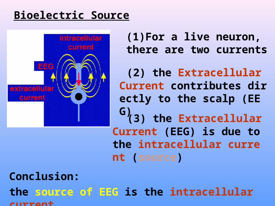
(1) The bioelectromagnetic source is the intracellular current
1.Bioelectromagnetic Source models
What is the bridge from current to charge or dipole model?
(2) The conventional source model is such as charge 、 dipole 、 quadruple...

Bridge 1: the physics
current is due to charge moving dipole is consisted of charges …
This bridge is complex, we do not need to take care of it.
1.Bioelectromagnetic Source models
1.Bioelectric Source models
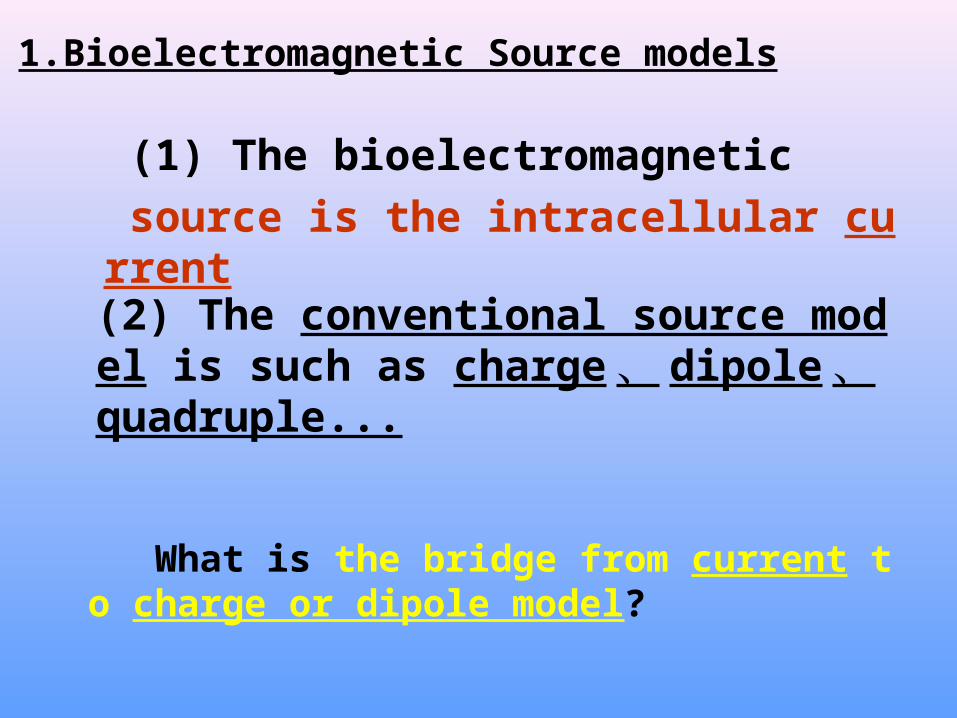
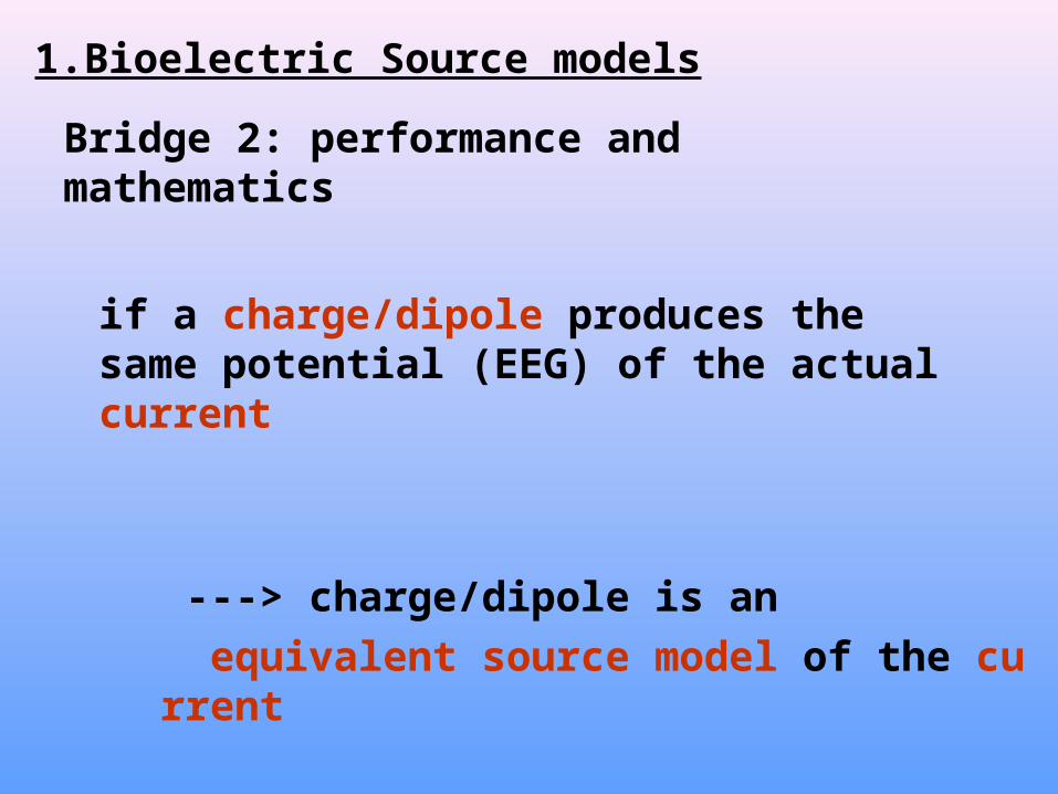
Bridge 2: performance and mathematics
if a charge/dipole produces the same potential (EEG) of the actual current
---> charge/dipole is an equivalent source model of the current
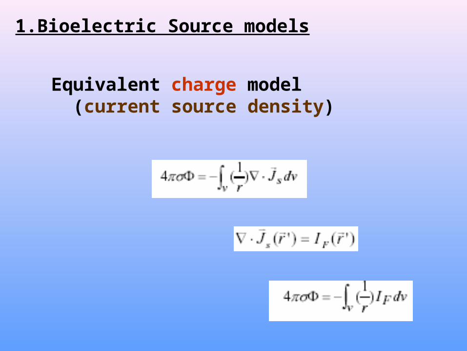
1.Bioelectric Source models
Equivalent charge model (current source density)
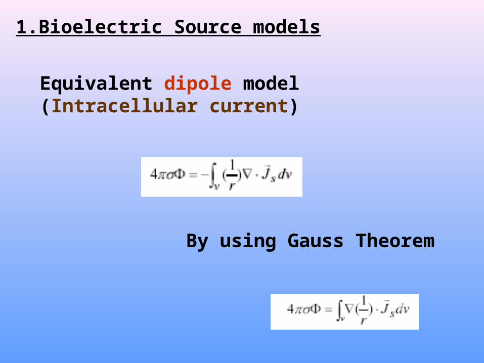
1.Bioelectric Source models
Equivalent dipole model(Intracellular current)
By using Gauss Theorem

1.Bioelectric Source models
Equivalent “potential” model
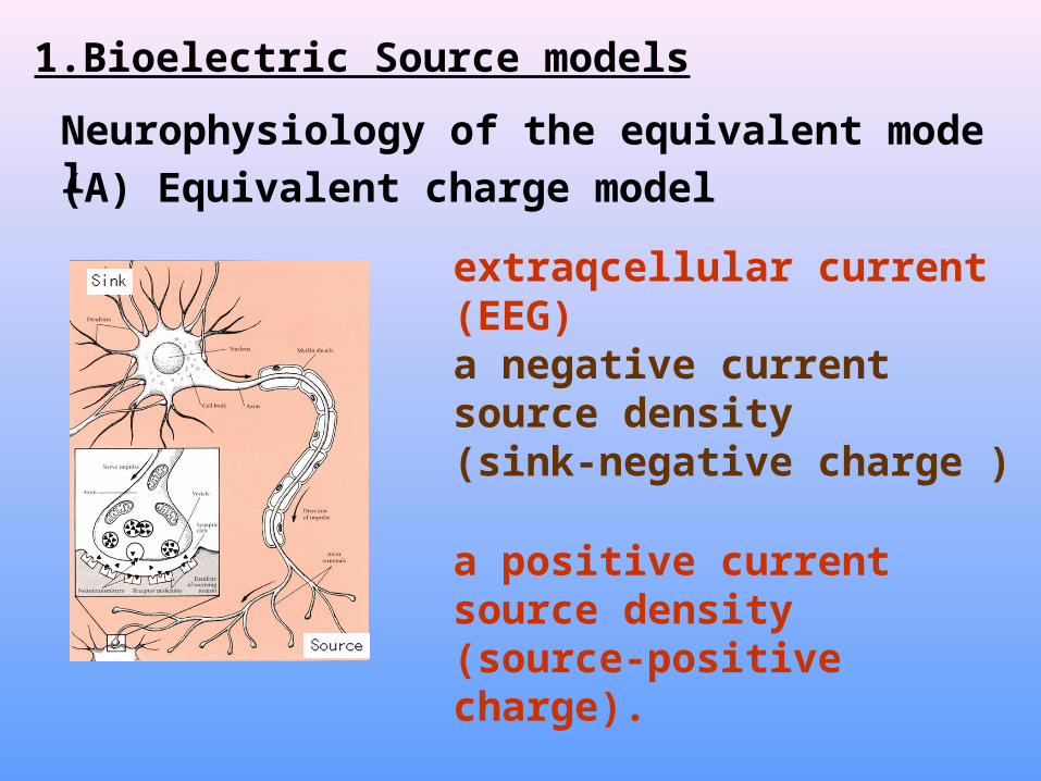
1.Bioelectric Source models
(A) Equivalent charge model
extraqcellular current (EEG)
Neurophysiology of the equivalent model
a negative current source density (sink-negative charge )
a positive current source density(source-positive charge).
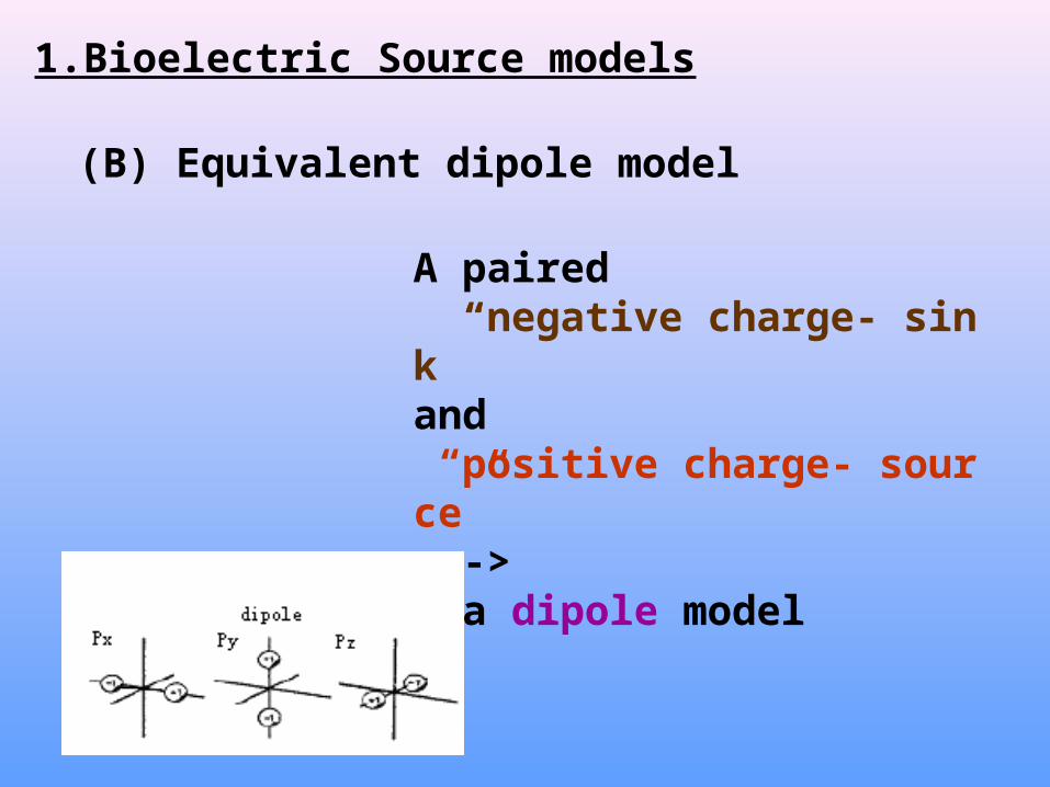
1.Bioelectric Source models
(B) Equivalent dipole model
A paired “negative charge- sink” and “positive charge- source”---> a dipole model
1.Bioelectric Source models
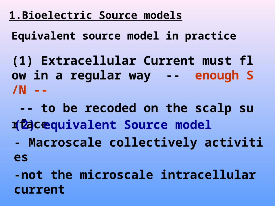
(1) Extracellular Current must flow in a regular way -- enough S/N -- -- to be recoded on the scalp surface
(2) equivalent Source model - Macroscale collectively activities-not the microscale intracellular current
Equivalent source model in practice

Comparison of the three models
Dipole Charge Potential Introduced year inEEG imaging
M Scherg ? 1985 D Yao ? 1996 Menendez etal 2000
parameter 3 1 1Current density Current source density Drive of current
Scalar or vector vector scalar scalar
Summary of Source models
three kinds of Source models each of them is an equivalent representation of the actual neuron “assembly”
2. 2D Imaging of brain activities
1) Image processing - Laplacian (deblurring the skull smearing effect)
2) Electric field analysis Cortical potential reconstruction Layer stripping(Equivalent dipole layer) Layer replacing(Equivalent charge layer)
Two approaches

2.2D Imaging of brain activities
h-radius of scalp, c-radius of the head
current source density(CSD) For a spherical head model (Yao, 2002)
Laplacian --- try to find the current emerge or disappear in the scalp layer
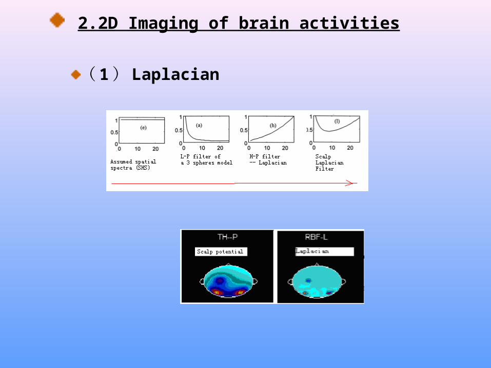
2.2D Imaging of brain activities
( 1 ) Laplacian
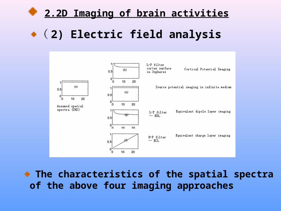
( 2) Electric field analysis
2.2D Imaging of brain activities
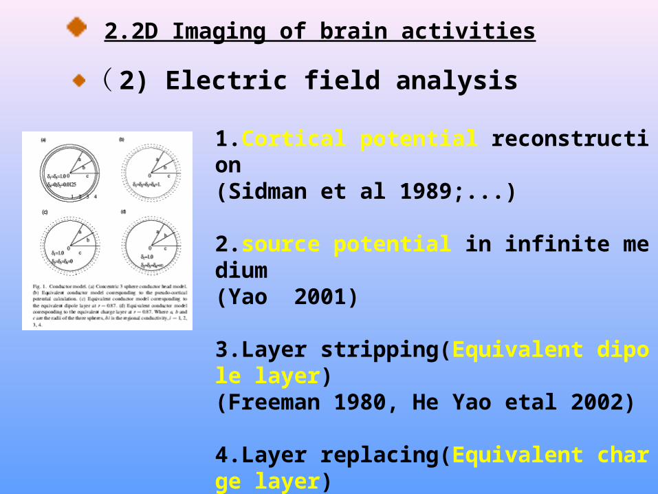
1.Cortical potential reconstruction (Sidman et al 1989;...)
2.source potential in infinite medium(Yao 2001)
3.Layer stripping(Equivalent dipole layer) (Freeman 1980, He Yao etal 2002)
4.Layer replacing(Equivalent charge layer)(Yao 2003)
( 2) Electric field analysis
2.2D Imaging of brain activities
The characteristics of the spatial spectra of the above four imaging approaches
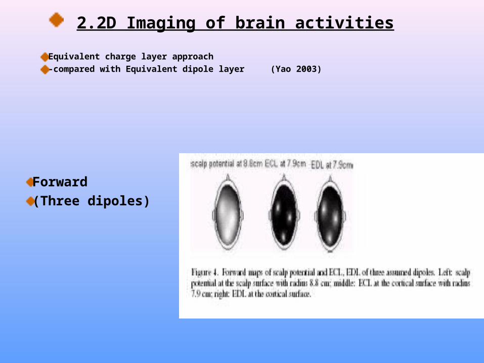
Equivalent charge layer approach -compared with Equivalent dipole layer (Yao 2003)
2.2D Imaging of brain activities
Forward(Three dipoles)
2.2D Imaging of brain activities
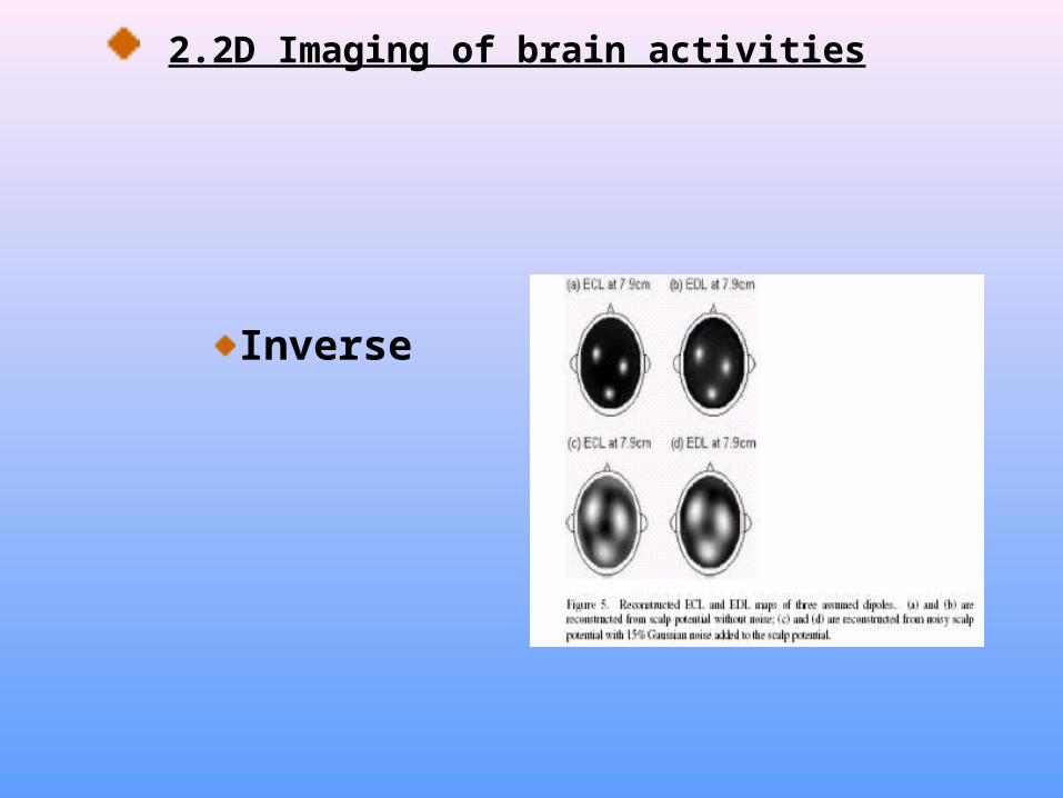
Inverse

2.2D Imaging of brain activities
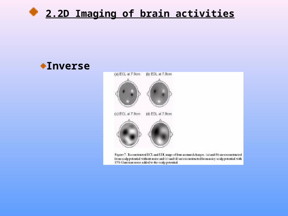
Forward (four charges)
2.2D Imaging of brain activities
Inverse

2.2D Imaging of brain activities
Application

The source models may be:
Dipole -- Potential -- charge
3.3D Imaging of brain activities
VEPsVEPs
ECEC
ED AED A
ED XED X
ED YED Y
ED ZED Z
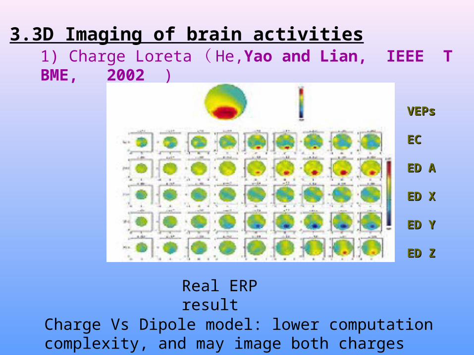
Real ERP result
1) Charge Loreta ( He,Yao and Lian, IEEE TBME, 2002 )
Charge Vs Dipole model: lower computation complexity, and may image both charges and dipoles
3.3D Imaging of brain activities
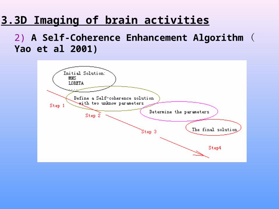
2) A Self-Coherence Enhancement Algorithm ( Yao et al 2001)
3.3D Imaging of brain activities
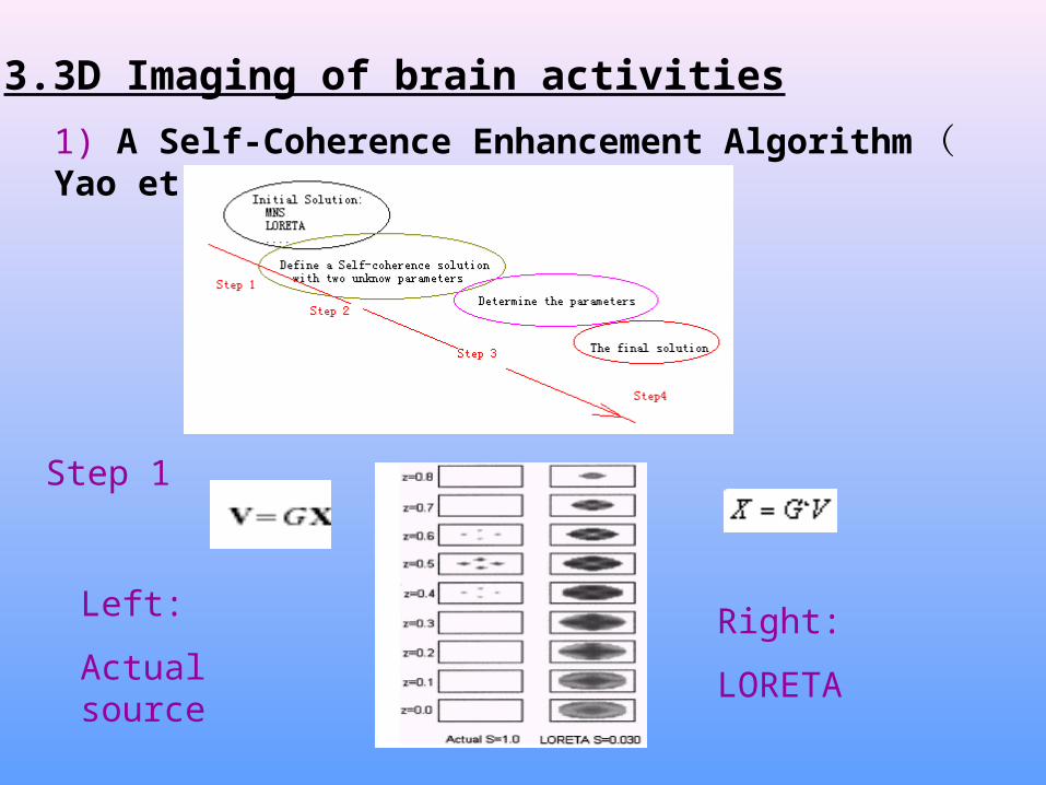
1) A Self-Coherence Enhancement Algorithm ( Yao et al 2001)
3.3D Imaging of brain activities
Step 1
Left:
Actual sourceRight:
LORETA

1) A Self-Coherence Enhancement Algorithm ( Yao et al 2001)
3.3D Imaging of brain activities
Two unknown parameters: K and alfa
Step 2 V G X

1) A Self-Coherence Enhancement Algorithm ( Yao et al 2001)
3.3D Imaging of brain activities
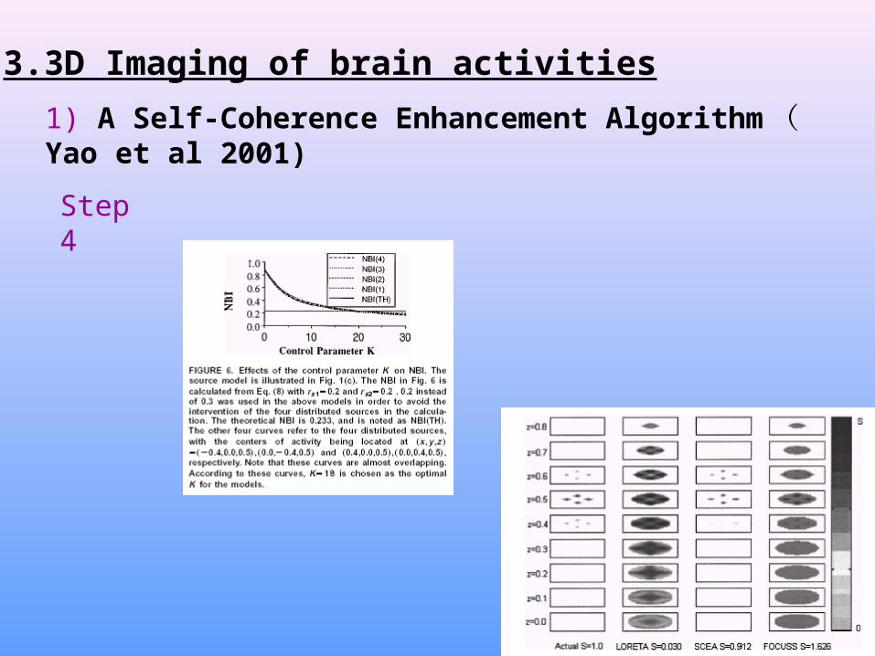
Comparing the NBIs of the solution and the actual source to chose a proper K
Actual neuronal source distribution is of neurophysiological smoothness. By defining a NBI (normalized blurring index )
Step 3
Determine alfa
Determine K
1) A Self-Coherence Enhancement Algorithm ( Yao et al 2001)
3.3D Imaging of brain activities
Step 4

Reference is the oldest problem of EEG
There is not a point that its potential is zero all the time (Geselowitz, 1998 )
A unitary reference is the best and ideal case
4. EEG Reference problem
EEG recordings
4. EEG Reference problem
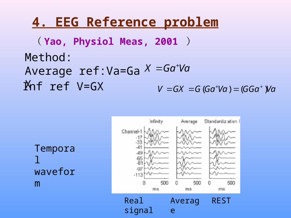
( Yao, Physiol Meas, 2001 )
Temporal waveform
Real signal Average REST
Method:Average ref:Va=GaX VaGaX
Inf ref V=GX VaGGaVaGaGGXV )()(
4. EEG Reference problem
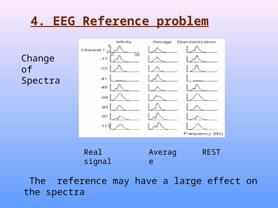
Change of Spectra
Real signal Average REST
The reference may have a large effect on the spectra
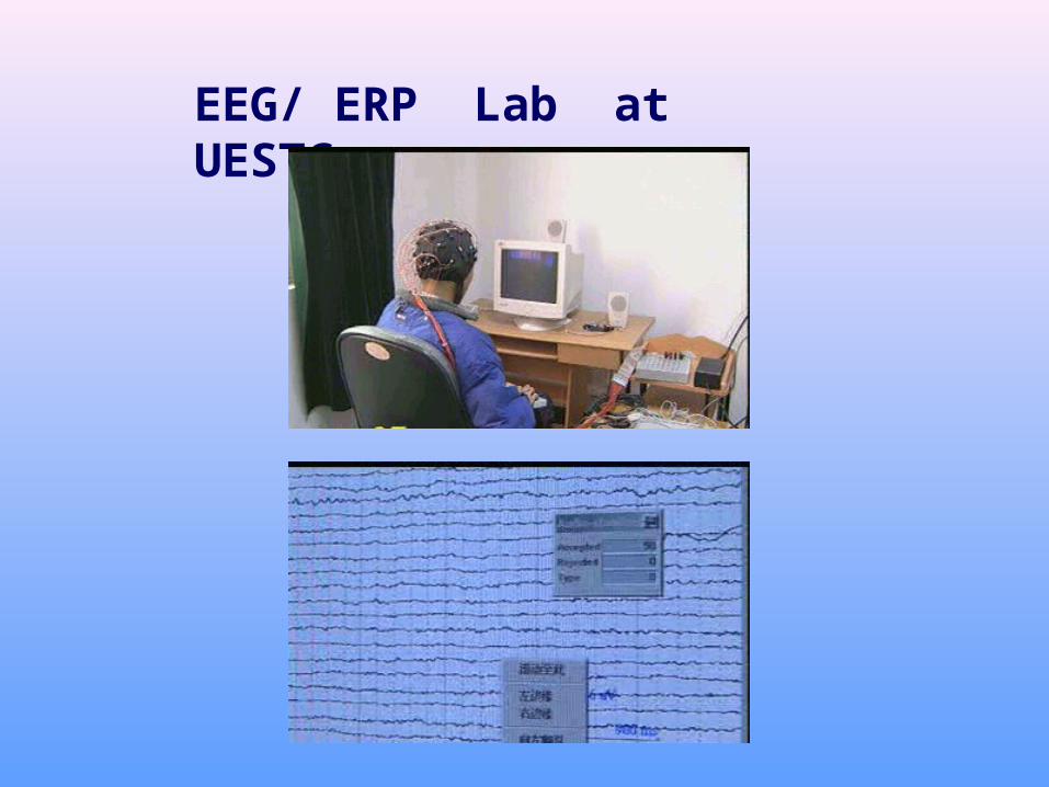
EEG/ ERP Lab at UESTC

Thanks
Related Documents