Journal of Industrial Microbiology (1996)17, 328-345 1996 Society for Industrial Microbiology 0169-4146/96/$12,00 Biodiversity of freshwater fungi TK Goh and KD Hyde Department of Ecology and Biodiversity, The University of Hong Kong, Pokfulam Road, Hong Kong There are more than 600 species of freshwater fungi with more known from temperate, as compared to tropical regions. These include ca 340 ascomycetes, 300 deuteromycetes, and a number of lower fungi which are not dis- cussed here. Aniptodera, Annulatascus, Massarina, Ophioceras and Pseudohalonectria are common freshwater ascomycetes, which appear to be well adapted for this lifestyle either in their ascospore types or their competitive- degradative characters. The most common genera of wood-inhabiting deuteromycetes include Cancellidium, Dacty- /aria, Dictyosporium and Helicomyces. They are categorized into four groups depending on their form and life style: the Ingoldian hyphomycetes; the aero-aquatic hyphomycetes; the terrestrial-aquatic hyphomycetes; and the sub- merged-aquatic hyphomycetes. The adaptations of aquatic fungi for their dispersal and subsequent attachment to new substrates are discussed. Keywords: hyphomycetes; ascomycetes; taxonomy; ecology; stream biology Introduction A broad definition of 'freshwater fungi' includes any spec- ies which, for the whole or part of their life cycle, rely on free freshwater [184]. These include any species growing on substrates that are predominantly aquatic or semi- aquatic. In other words, their habitats may be clearly of an aquatic nature or those that colonize submerged plant parts in freshwater environments. Hence, freshwater fungi are an ubiquitous and diverse group of organisms. These include representatives from different groups of fungi namely the zoosporic fungi [37], many hyphomycetes [39,80,116-133, 135-145,147-153], ascomycetes [65,86,175], basidio- mycetes [134,146,154-156,170], coelomycetes [27,51], zygomycetes [30], and trichomycetes [111]. Among these fungi, the freshwater hyphomycetes (Moniliales, Deuteromycotina) are well documented and the knowledge of their ecology and diversity is more comprehensive than that of other fungal groups in the aquatic environments. Role of fungi in freshwater ecosystems The main role of higher fungi in freshwater ecosystems is in the degradation of dead plant material, such as Juncus, Phragmites, Scirpus and Typha and dead leaves and woody material that find their way into the water. They may also be involved in the degradation of animal parts such as insect exoskeletons, fish scales and hair. Another less sig- nificant group are pathogens of both plants and animals, while a third group, the endophytes may colonize the living tissue of aquatic plants. The decay of dead plant tissue is a result of the ability of the fungi to degrade lignocellulose. Freshwater fungi occurring on submerged woody tissue have been relatively well studied. It appears that their suc- cess in colonizing submerged woody material lies in their ability to form soft-rot cavities [175] and to be antagonistic Correspondence: Dr TK Goh, Department of Ecology and Biodiversity, The University of Hong Kong, Pokfulam Rd, Hong Kong Received 20 August 1996; accepted 1 November1996 against other fungi [173]. Their ability to soften wood may also play an important role in increasing the palatability of wood to stream invertebrates. For a detailed discussion of the role of freshwater fungi the reader should refer to Shearer [175]. Biodiversity of freshwater fungi In this review of freshwater fungi we concentrate on the higher fungi, ie the Ascomycota and the mitosporic fungi. There is little available information on the lower fungi in freshwater and therefore these are not included. Freshwater ascomycetes In a review of the freshwater ascomycetes Shearer [175] listed 288 species. This included a wide variety of ascomy- ceres, encompassing discomycetes, plectomycetes, pyreno- mycetes and loculoascomycetes. When Shearer published her list in 1993 only 11 of the records were from tropical locations. Several tropical freshwater ascomycetes have since been described and a total of 50 species is now known to occur in the tropics, with 16 from water cooling towers, two from high altitude ponds and 32 from tropical streams [57,65]. Many of these data have resulted from studies on the tropical freshwater ascomycetes of north Queensland or cooling towers in India [48-50,52-57,66]. Three new freshwater ascomycetes have also been described recently from temperate regions [eg 166] and the number of asco- mycetes known to occur in freshwater habitats now stands at ca 340. These fungi are an unique group with both physiological and morphological adaptations for an aquatic lifestyle. There are many more freshwater ascomycetes awaiting discovery, both in temperate waters (Shearer, per- sonal communication) and particularly in tropical streams, rivers and other bodies of freshwater (Ho and Hyde, per- sonal observation). Shearer has now placed her list of fresh- water ascomycetes on the World Wide Web and it can be accessed using http://www.life.uiuc.edu/plantbio/fungi. Downloaded from https://academic.oup.com/jimb/article/17/5-6/328/5988978 by guest on 09 March 2022

Welcome message from author
This document is posted to help you gain knowledge. Please leave a comment to let me know what you think about it! Share it to your friends and learn new things together.
Transcript

Journal of Industrial Microbiology (1996) 17, 328-345 �9 1996 Society for Industrial Microbiology 0169-4146/96/$12,00
Biodiversity of freshwater fungi TK Goh and KD Hyde
Department of Ecology and Biodiversity, The University of Hong Kong, Pokfulam Road, Hong Kong
There are more than 600 species of freshwater fungi with more known from temperate, as compared to tropical regions. These include ca 340 ascomycetes, 300 deuteromycetes, and a number of lower fungi which are not dis- cussed here. Aniptodera, Annulatascus, Massarina, Ophioceras and Pseudohalonectria are common freshwater ascomycetes, which appear to be well adapted for this lifestyle either in their ascospore types or their competitive- degradative characters. The most common genera of wood-inhabiting deuteromycetes include Cancellidium, Dacty- /aria, Dictyosporium and Helicomyces. They are categorized into four groups depending on their form and life style: the Ingoldian hyphomycetes; the aero-aquatic hyphomycetes; the terrestrial-aquatic hyphomycetes; and the sub- merged-aquatic hyphomycetes. The adaptations of aquatic fungi for their dispersal and subsequent attachment to new substrates are discussed.
Keywords: hyphomycetes; ascomycetes; taxonomy; ecology; stream biology
Introduction
A broad definition of 'freshwater fungi' includes any spec- ies which, for the whole or part of their life cycle, rely on free freshwater [184]. These include any species growing on substrates that are predominantly aquatic or semi- aquatic. In other words, their habitats may be clearly of an aquatic nature or those that colonize submerged plant parts in freshwater environments. Hence, freshwater fungi are an ubiquitous and diverse group of organisms. These include representatives from different groups of fungi namely the zoosporic fungi [37], many hyphomycetes [39,80,116-133, 135-145,147-153], ascomycetes [65,86,175], basidio- mycetes [134,146,154-156,170], coelomycetes [27,51], zygomycetes [30], and trichomycetes [111]. Among these fungi, the freshwater hyphomycetes (Moniliales, Deuteromycotina) are well documented and the knowledge of their ecology and diversity is more comprehensive than that of other fungal groups in the aquatic environments.
Role of fungi in freshwater ecosystems
The main role of higher fungi in freshwater ecosystems is in the degradation of dead plant material, such as Juncus, Phragmites, Scirpus and Typha and dead leaves and woody material that find their way into the water. They may also be involved in the degradation of animal parts such as insect exoskeletons, fish scales and hair. Another less sig- nificant group are pathogens of both plants and animals, while a third group, the endophytes may colonize the living tissue of aquatic plants. The decay of dead plant tissue is a result of the ability of the fungi to degrade lignocellulose. Freshwater fungi occurring on submerged woody tissue have been relatively well studied. It appears that their suc- cess in colonizing submerged woody material lies in their ability to form soft-rot cavities [175] and to be antagonistic
Correspondence: Dr TK Goh, Department of Ecology and Biodiversity, The University of Hong Kong, Pokfulam Rd, Hong Kong Received 20 August 1996; accepted 1 November 1996
against other fungi [173]. Their ability to soften wood may also play an important role in increasing the palatability of wood to stream invertebrates. For a detailed discussion of the role of freshwater fungi the reader should refer to Shearer [175].
Biodiversity of freshwater fungi
In this review of freshwater fungi we concentrate on the higher fungi, ie the Ascomycota and the mitosporic fungi. There is little available information on the lower fungi in freshwater and therefore these are not included.
Freshwater ascomycetes In a review of the freshwater ascomycetes Shearer [175] listed 288 species. This included a wide variety of ascomy- ceres, encompassing discomycetes, plectomycetes, pyreno- mycetes and loculoascomycetes. When Shearer published her list in 1993 only 11 of the records were from tropical locations. Several tropical freshwater ascomycetes have since been described and a total of 50 species is now known to occur in the tropics, with 16 from water cooling towers, two from high altitude ponds and 32 from tropical streams [57,65]. Many of these data have resulted from studies on the tropical freshwater ascomycetes of north Queensland or cooling towers in India [48-50,52-57,66]. Three new freshwater ascomycetes have also been described recently from temperate regions [eg 166] and the number of asco- mycetes known to occur in freshwater habitats now stands at ca 340. These fungi are an unique group with both physiological and morphological adaptations for an aquatic lifestyle. There are many more freshwater ascomycetes awaiting discovery, both in temperate waters (Shearer, per- sonal communication) and particularly in tropical streams, rivers and other bodies of freshwater (Ho and Hyde, per- sonal observation). Shearer has now placed her list of fresh- water ascomycetes on the World Wide Web and it can be accessed using http://www.life.uiuc.edu/plantbio/fungi.
Dow
nloaded from https://academ
ic.oup.com/jim
b/article/17/5-6/328/5988978 by guest on 09 March 2022

Biodiversity of freshwater fungi TK Goh and KD Hyde
Taxonomic aspects Freshwater ascomycetes are not a monophyletic group and include examples from several orders in a range of classes and families. The largest group are the discomycetes with 112 representatives. The most specious family is the Helio- tales with 43 genera and 103 species. Remarkably, nearly all of the records of discomycetes are from temperate regions. One hundred and twelve discomycetes are given as occurring in freshwater habitats [175], but only two (ie Hymenoscyphus malawensis PJ Fisher and Spooner, Pezol- oma rhodocarpa PJ Fisher and Spooner) of these records are from tropical locations. The discomycetes H. malawensis, P. rhodocarpa and Saccobolus beckii Hei- meri are listed for the tropics [65] and none of these were recorded in authentic tropical situations. Among the notice- able absentees from tropical freshwater habitats, are there- fore, the discomycetes. In six years of collecting in tropical streams, KDH has picked up only two single discomycetes. We have no suggestions as to why there are so few tropical stream discomycetes, but the absence is most remarkable.
Another large group are the pyrenomycetes represented by 95 species. The most specious orders are the Halospha- eriales with five genera and 18 species, the Sordariales with 18 genera and 47 species and the Diaporthales with six genera and eight species. Of" the 93 pyrenomycetes listed [175], only eight of these were from tropical locations. We now know that these families with unitunicate asci are also commonplace in the tropics [65] (Table 1). The loculoasco- mycetes (ascomycetes with bitunicate asci) also form a large group, represented by 85 species [175]. The most spe- cious order is the Pleosporales, represented by 62 species. Of the eight loculoascomycetes listed [175] only two were tropical records. Again recent collections in tropical streams have shown that this group is well represented, although less well represented than the unitunicate ascomy- cetes (see Table 1).
In this section we address the question of 'how many more freshwater ascomycetes await to be discovered'. We presently know ca 340 species from freshwater, but if we examine the data on biogeography it is obvious that fresh- water habitats have only been examined in any detail in four countries: Australia (north Queensland only), Austria (a single lake [112]), USA (Illinois) and UK (Lake District and Devon) [175]. Most other data are the result of brief studies, single collections or species descriptions [eg 66, 176]. Evidence from recent studies indicates that many new
species await discovery in both temperate (Shearer, per- sonal communication) and tropical regions [65]. Most of the tropics awaits investigation and there are also few rec- ords of ascomycetes in most temperate countries.
A comparison of the ascomycetes found on submerged wood in streams in three tropical countries can give an idea of how many new fungi may be found when a new geo- graphical region is investigated in the tropics. KDH has recently examined 100 submerged wood samples from streams in Australia (Cape Tribulation), the Philippines (Mt Makiling) and Mauritius (Black River National Park). The fungi on these samples have been identified to genus and/or species where possible, and so it is possible to compare the numbers of ascomycetes and the numbers of identified and undescribed species at each site (Table 1).
The numbers of ascomycete species identified in each site are 26, 22 and 27 respectively. Only in Australia are half of the fungi identified to species level. In the Philip- pines and Mauritius only 41% and 30%, respectively, of the ascomycetes can be given species names, and probably most of these unnamed species are new to science. Only two of the unnamed species in the Philippines were also found in Mauritius. The reason for the higher numbers identified to species level in Australia is because KDH has worked on these fungi since 1989 and has described many new species. However, the most striking inference that can be made from these results is that in any new site chosen for investigation, one can presently expect that half of the fungi found will be new species. Unfortunately, there are no good modern keys to the genera of ascomycetes and in particular, no keys to the freshwater ascomycetes. Further- more, many of the fungi collected may have been described earlier in terrestrial genera or be terrestrial species occur- ring in freshwater and therefore the task of identifying these unknown species is daunting. The results, do however, indi- cate that we are far from knowing all the freshwater fungi and presently probably only know a small fraction of the species adapted for living in freshwater habitats. The results above are from small streams in rainforests or parks and there is little to no information on the ascomycetes inhabiting other bodies of water in the tropics, such as ponds and lakes.
Common genera of freshwater ascomycetes The most specious genera in temperate regions appear to differ from the specious genera in the tropics. In [175]
329
Table 1 Comparison of ascomycetes collected on submerged wood in streams at Cow Bay, Cape Tribulation, Australia, Mt Makiling, Los Bafios, the Philippines and Black River, Mauritius
Cow Bay Mt Makiling Black River
Discomycetes 0 0 0 Plectomycctes 0 0 0 Pyrenomycetes 17 14 18 Loculoascomycetes 9 7 8 No. genera 19 14 19 No. species 26 22 27 Identified species 13 9 8 Unidentified species 13 13 19 Overlapping unidentified species 0 2 2
Dow
nloaded from https://academ
ic.oup.com/jim
b/article/17/5-6/328/5988978 by guest on 09 March 2022

330
Biodiversity of freshwater fungi TK Gob and KD Hyde
where most species listed are temperate, the author records the most specious freshwater genera as Aniptodera/ Halosarpheia (8), Chaetomium (8), Hymenoscyphus (10), Mollisia (13), Ophioceras/Pseudohalonectria (11), Phaeo- sphaeria (15) and Vibrissia (15). There is little information on which are the most commonly encountered species. In tropical regions the most specious genera are Aniptodera/Halosarpheia (10), Annulatascus (10) and Savoryella (7). However, the most frequently recorded gen- era in 250 collections made in Australia, Brunei, Ecuador, Malaysia and the Philippines were Annulatascus, Massar- ina, Ophioceras, Savoryella and Aniptodera [65]. The trop- ical ascomycetes Aniptodera sp, Annulatascus spp, Ascag- ilus bipolaris, Bertia sp, Caryospora sp, Ophioceras dolichostomum, Savoryella verrucosa and Submersis- phaeria aquatica are discussed and illustrated below.
Annulatascus Hyde [48] has immersed to superficial, coriaceous, dark-walled ascoma (Figure 7), unitunicate cyl- indrical asci with a relatively massive refractive apical apparatus (Figures 6, 8, 11, 12) and unicellular or septate ascospores with various types of appendages or sheaths (Figures 9, 10, 13, 14). Three species (A. velatispora KD Hyde (Figures 6-10), A. bipolaris KD Hyde and A. biatriisporus KD Hyde (Figures 11-14)) are presently known and many more await publication (Hyde, unpublished).
In Aniptodera species ascomata are immersed, becoming erumpent, with a central small papilla. Asci are unitunicate with an apical thickening and pore (Figures 1, 2), whilst ascospores are ellispoidal or fusiform, some with polar appendages (Figures 3-5). The ascomata of Ascagilis bipolaris are unusual in that they comprise a peridium of large thin-walled cells (Figures 19, 20). Asci readily release their ascospores in water mounts (Figure 23) and asco- spores are brown, and bicellular with polar mucilaginous pads (Figures 21,22). In Bertia the ascomata are superficial and covered in warts (Figure 15). The asci have very long pedicels (Figure 16) and are released in the ascomal venter at maturity. The apex of the ascus has an apical thickening and pore (Figure 17) and the ascospores are J-shaped (Figure 18).
The ascomata of Caryospora are relatively large, black, superficial and carbonaceous (Figure 24). The asci are dark- brown, bicelled and surrounded by a mucilaginous sheath (Figure 25). In Ophioceras dolichostomum ascoma are immersed with long necks (Figure 36) and these can usually be seen as hair-like structures on the wood surface with the unaided eye. Asci are unitunicate and broadly cylindrical, characteristically lacking a pedicel and with a relatively small cylindrical refractive apical ring (Figures 37, 38). Ascospores are filiform with three septa and rounded ends (Figures 39, 40). In Savoryella verrucosa the ascomata are dark-coloured, immersed or superficial, and contain cylin- drical asci with thickened apical rings with a pore (Figures 26, 27). The ascospores are four-celled, with two brown central ceils and smaller hyaline end cells and a distinctly verrucose wall (Figures 28, 29). The ascomata of Submersi- sphaeria aquatica are immersed (Figure 30) and the asci (Figures 31, 34) resemble those of Annulatascus. The taxa, however differ, in that the ascospores in Submersisphaeria are brown and have apical germ pores (Figures 32-35).
Freshwater hyphomycetes Active research on the aquatic hyphomycetes in freshwater streams and lakes began more than 50 years ago when Ingold published the first of his many papers on these fungi from England [67]. In the early 1940s it was realized [66- 72] that this unique group of fungi occurs regularly on sub- merged decaying leaves, twigs and wood of dicotyledonous trees and shrubs. These fungi appear to be entirely absent from the needles of conifers. During the past 30 years, the majority of freshwater hyphomycetes were reported from cold and temperate regions. However, one would expect that in tropical countries, which have a rich fungal diver- sity, there should certainly be an equally rich diversity of freshwater hyphomycetes. It is now evident that the fresh- water hyphomycetes have a worldwide distribution [3- 6,16,17,22,26,31-35,39,46,47,67-72,74,76-81,92,110,113, 131,157,158,161-164,181-183,186,187]. Examples of species that are worldwide in distribution include Anguillo- spora crassa Ingold, Campylospora chaetocladia Ranzoni, Flagellospora penicillioides Ingold, Jaculispora submersa Hudson and Ingold, Lunulospora curvula Ingold, Tetra- chaetum elegans Ingold, Tetracladium setigerum (Grove) Ingold, Tricladium angulatum Ingold, Triscelophorus monosporus Ingold and Varicosporium elodeae Kegel [200]. Nevertheless, some species appear to be localized in a given climatic zone. Alatospora acuminata Ingold, Flag- ellospora curvula Ingold, Heliscella lugdunensis Sacc and Thdrry, and Lemonniera aquatica De Wild are some of the dominant species in the high North, whereas Angulospora aquatica S Nilsson, Brachiosphaera tropicalis Nawawi, Campylospora .filicladia Nawawi, Ingoldiella hamata Shaw, Isthmotricladia gombakiensis Nawawi, Lunulospora c3,mbiformis Miura, Phalangispora constricta Nawawi and Webster, Pyramidospora casuarinae Nilsson, Speiropsis pedatospora Tubaki, Tricladiomyces malaysianum (Nawawi) Nawawi, Triscelophorus acuminatus Nawawi are examples of tropical species.
To date, more than 300 species of freshwater hyphomy- cetes are known and the number is increasing at a rapid rate. Many new taxa have been discovered. For example, new genera published during the past few years include Canalisporium Nawawi and Kuthubutbeen [147], Quad- ricladium Nawawi and Kuthubutheen [148], Crucella Mar- vanov~ and Suberkropp [115], Nidulispora Nawawi and Kuthubutheen [150], Obeliospora Nawawi and Kuthubu- theen [153], Candelosynnema KD Hyde and Seifert [63], Isthmophragmospora Kuthubutheen and Nawawi [104], and Paracryptophiale Kuthubutheen and Nawawi [106]. Certainly, however, there are many more aquatic fungi awaiting isolation and identification.
Traditionally, the freshwater hyphomycetes are dis- tinguishable by their biological behaviour into two groups: the Ingoldian fungi and the aero-aquatic fungi. With the broadened concept of 'aquatic fungi' given by Thomas [ 184], however, two additional groups of freshwater hypho- mycetes can be categorized: the terrestrial-aquatic hypho- mycetes and the submerged-aquatic hyphomycetes (or 'fac- ultative-aquatic hyphomycetes'). It should be emphasized that these are 'biological groups', and, do not represent 'natural' groups in fungal systematics. Therefore, it is dif- ficult to give them precise definition. However, these four
Dow
nloaded from https://academ
ic.oup.com/jim
b/article/17/5-6/328/5988978 by guest on 09 March 2022

Biodiversity of freshwater fungi TK Goh and KD Hyde
331
Figures 1-18 Light micrographs of various freshwater ascomycetes. (1 5) Aniptodera sp. (1, 2) Asci. Note the apical thickening and pore in 1, which has ruptured in 2. (3-5) Ascospores with fine unfurling appendages (arrowed). (6 10) Annulatascus velatispora. (6, 8) Asci. Note the large apical ring. (7) Necks of ascomata. (9, 10) Ascospores with mucilaginous sheath. (11-14) Annulatascus biatriisporus. (11, 12) Asci. Note the large thickened apical ring. (] 3, 14) Ascospores. (15-18) Bertia convolutispora. (15) Ascoma. (16, 17) Asci. Note the apical thickening. (18) J-shaped ascospores. Bars: 7, 15 = 100/xm; 1-6, 8 14, 16-18= 10/~m.
Dow
nloaded from https://academ
ic.oup.com/jim
b/article/17/5-6/328/5988978 by guest on 09 March 2022

Biodiversity of freshwater fungi TK Goh and KD Hyde
332
Figures 1%40 Light micrographs of various freshwater ascomycetes. (19-23) Ascagilis bipolaris. (19, 20) Ascoma, illustrating large cells of peridium. (21, 22) Ascospores with bipolar mucilaginous pads. (23) Ascus. (24, 25) Caryospora sp. (24) Superficial ascoma. (25) Ascospore. (26-29) Savoryella verrucosa. (26, 27) Asci. Note the large thickened apical ring. (28, 29) Ascospores. Note the wall ornamentation and hyaline end cells. (30-35) Submersis- phaeria aquatica. (30) Section of immersed ascoma. (31, 34) Asci. Note the large apical ring. (32, 33, 35) Ascospores. Note the polar germ pores. (36- 40) Ophioceras dolichostomum. (36) Section of ascoma with long neck. (37, 38) Asci. Note the small apical ring. (39, 40) Filiform ascospores. Bars: 19, 24, 30, 36 = 100 p~m; 20-23, 25-29, 31-35, 37-40 = 10 ~m.
Dow
nloaded from https://academ
ic.oup.com/jim
b/article/17/5-6/328/5988978 by guest on 09 March 2022

Biodiversity of freshwater fungi TK Goh and KD Hyde
groups of freshwater hyphomycetes can be discerned as fol- lows:
(1) The Ingoldian fungi (Figures 71-82) abound in fast- flowing tree-lined streams, babbling brooks, and well- aerated lakes, growing on submerged leaves and twigs, but are relatively more sparse on woody substrates [80,178,199]. They form conidia which are released in water and are readily trapped in foam [82,83]. They predominantly have two basic shapes of conidia: branched or sigmoid. In the great majority of these branched conidia (Figures 71-75), they are tetra-radi- ate, ie usually consist essentially of four long arms diverging from a common point. Examples of this spore type are found in genera such as Alatospora, Actino- spora, Articulospora, Campylospora, Clavariopsis, Jaculi~pora, Lemonniera, Tetrachaetum, Tetracladium, Tricladium (Figures 67-69) and Triscelophorus. There are quite a number of branched conidia, however, that are not tetra-radiate, as in the genera Dendrospora, Polycladium, and Varicosporium (Figure 76). There exist also a great number of species that produce sig- moid conidia, ie long and worm-like, usually with a curvature in more than one plane. These sigmoid con- idia (Figures 77-79) are seen in genera such as Anguil- lospora, Flagellospora, Lunulospora and Mycocentro- spora. Besides these two basic shapes of conidia, other conidial shapes are occasionally found, eg variously coiled shrimp-like or flywheeled-shaped conidia (Figure 80) are represented by species of Gyoerffyella, and spherical or ovoid conidia (Figures 81-82) are seen in Margaritispora, Dactyllela, and Dimorphospora. Nevertheless, it is significant to note that nearly all the Ingoldian fungi have conidiophores and conidia that are hyaline and thin-walled. The Ingoldian fungi are exten- sively studied worldwide and for their monographic treatments, see [80,131,158].
(2) The aero-aquatic hyphomycetes (Figures 83-86), termed by Beverwijk [18-21], are more usually found in stagnant ponds, ditches, or slow-running streams and are capable of vegetative growth on submerged leaves or woody substrates under semi-anaerobic conditions. Sporulation in this biological group is unique, which does not occur below water. In contrast to the Ingoldian fungi in which the whole cycle of conidium production, liberation and dispersal normally takes place below water, the aero-aquatics sporulate only when the sub- strate is exposed to air, when they form buoyant propa- gules capable of dispersal when the substrate is sub- merged again. The conidia, or forms of propagules, are mostly coloured. They are often tightly helicoid in more than one plane (Figures 85, 86), or equipped with a special flotation device in the form of an intricate hyphal system (Figures 45-49, 83-84). Examples of these aero-aquatic genera are Arbuscula, Aegerita, Beverwykella, Cancellidium, Candelabrum, Clathro- sphaerina, Clathrosporium, Cristulariella, Fusticeps, Helicodendron, Helicoon, Helicomyces (Figure 70). Mycoenterolobium, Nidulispora and Spirosphaera [1,24,36,38,41,44,45,185,192].
(3) The terrestrial-aquatic hyphomycetes (Figures 87-90),
(4)
termed by Ando [7], are represented by a number of conidial fungi isolated from rain drops associated with intact terrestrial plant parts, such as the leaf-surfaces [10] or rainwater draining from intact tree trunks [11]. A number of species are described from such isolations with establishments of many new genera such as Ala- tosessilispora, Arborispora, Curuci~pora, Microstella, Ordus, Tricladiella, and Trifurcospora [7-14]. A major characteristic of this group of hyphomycetes is that they produce staurosporous conidia similar in shape to those of the Ingoldian group, but lacking conspicuous conidiophores (ie micronematous). These conidia are mostly hyaline and thin-walled, however, some demati- aceous species were also isolated (eg Ceratosporium cornutum Matshushima, Tetraploa aristata Berk and Broome, and Tripospermum infalcatum Ando and Tubaki). The submerged-aquatic hyphomycetes (Figures 91-95), first addressed by Ingold [80], represent a hetero- geneous assemblage of fungi growing on submerged decaying plant materials. Most of the species are found on wood litter blocked by rocks in fast-flowing streams or babbling brooks. These lignicolous, or to a lesser extent foliicolous, hyphomycetes are nearly all demati- aceous and produced relatively thick-walled conidio- phores and/or conidia. The conidiophores are distinctly macronematous, frequently in the form of long stipes, however, they may be solitary or synnematous. The conidiogenous loci may be denticulate, cicatrized, tretic or phialidic. Although some species may sporulate under submerged conditions, a vast number sporulate when the substrates are no longer under water. Incu- bation of such woody substrates in moist chambers yields a great number of different species. Their conidia are capable of air dispersal or dispersed by some other mechanisms. These hyphomycetes may be regarded as 'facultative-aquatic', as compared to the aquatic Ingoldian group. The conidia of submerged-aquatic hyphomycetes are basically regular in shape, ie ovoid (Figures 58-61, 91), cylindric (Figures 51-53, 92), obclavate (Figures 41-44, 93), pyriform (Figure 94), or fusiform (Figure 95). However, branched conidia are not uncommon and may also be found in foam samples (eg conidia of Casaresia sphagnorum, Pleiochaeta setosa and Tetraploa aristata). Systematic studies of these submerged-aquatic hyphomycetes began about 10 years ago, mostly in Malaysia [88-109A 41,142,147], and recently by Goh and Hyde with samples from Aus- tralia, Philippines, and South Africa [40-43,64]. Rep- resentative genera are Bactrodesmium (Figures 41-44), Brachydesmiella, Brachysporiella, Camposporidium, Canalisporium, Cryptophiale, Cryptophialoidea, Dactylaria, Dendryphiosphaera, Dictyochaeta, Exserticlava (Figures 58-61), Kionochaeta, Monotos- poriella, Nawawia (Figures 62-66), Phaeoisaria, Spad- icoides, Sporidesmiella, Sporidesmium, Sporoschisma, Sporoschismopsis, Trichocladium and Xylomyces. A few taxa, ie Diplocladiella appendiculata Nawawi [136], D. scalaroides Arnaud [136], D. tricladioides Nawawi [133], Setosynnema isthmosporum Shaw and Sutton [ 171], and Triscelosporium verrucosum Nawawi
333
Dow
nloaded from https://academ
ic.oup.com/jim
b/article/17/5-6/328/5988978 by guest on 09 March 2022

334
Biodiversity of freshwater fungi TK Goh and KD Hyde
Figures 41-57 Light micrographs of various freshwater hyphomycetes. (41-45) Bactrodesmium longisporum. (41) Habit on submerged wood. (42, 43) Conidia. Note the mucilaginous pads at the apex. (44) Synnema bearing conidia. (45--49) Cancellidium applanatum. (45) Habit on submerged wood. (46, 48) Air-trapping propagules. (47) Monilioid ceils of the propagules. (49) SEM of propagule. Note the monilioid cells inside. (50-53) Candelosynnema ranunculosporum. (50) Synemmata. (51 53) Conidia with appendages. (54-57) Delortia palmicola. (54, 55) Conidiophores with developing conidia. (56, 57) Mature conidia surrounded by mucilage. Bars: 41 = 100/~m; 44 = 50/Lm; 45 = 200/zm; 46 = 10/zm; 42, 43, 46-49, 51-57 = 10/zm; 50 - 500 tzm.
Dow
nloaded from https://academ
ic.oup.com/jim
b/article/17/5-6/328/5988978 by guest on 09 March 2022

Biodiversity of freshwater fungi TK Gob and KD Hyde ~
335
Figures 58-70 Light micrographs of various freshwater hyphomycetes. (58-61) Exserticlava vasifbrmis. (58) Conidiophore and conidia. (59) Conidio- phore showing the characteristic extension of the hyaline inner wall layer into a multiseptate, subulate structure. (60, 61) Conidia. (62-66) Nawawia dendroides. (62) Habit. (63) Synnema bearing conidia. (64-66) Conidia. (67-69) Tricladium sp. (67) Habit. (68, 69) Branched conidia. (70) Conidium of Helicomyces sp. Bars: 62, 67 = 1 ram; 63 = 100 ~m; 58-61, 64-66, 68-70 = 10/xm.
Dow
nloaded from https://academ
ic.oup.com/jim
b/article/17/5-6/328/5988978 by guest on 09 March 2022

336
Biodiversity of freshwater fungi TK Goh and KD Hyde
"U 73
75
76
'li9 '
Figures 71-82 Conidial forms of some lngoldian fungi (drawn at various scales). (71) Articulospora grandis. (72) Clavariopsis aquatica. (73) Lemonni- era aquatica. (74) Tricladium splendens. (75) Triscelophorus magnificus. (76) Varicosporium helicosporum. (77) Anguillospora crassa. (78) Flagellospora penicillioides. (79) Mycocentrospora acerina. (80) Gyoerffyella speciosa. (81) Margaritispora aquatica. (82) Dimorphospora foliicola.
and Kuthubutheen [138], have been treated as examples of aero-aquatic species, but should be regarded as sub- merged-aquatic (facultative-aquatic) hyphomycetes. This is because the conidia of these species, though formed when the substrate is no longer submerged, are either filiform or branched and do not possess a flo- tation device.
Although the vast majority of conidia seen in foam samples almost certainly have an aquatic or stream-side ori- gin [80], some species must come from the air spora. Indeed, it is not uncommon to see occasional conidia of such well-known terrestrial hyphomycetes as Alternaria, Beltrania, Cladosporium, Drechslera, Epicoccum or to encounter teliospores of smuts and uredospores of rusts.
Certain hyphomycetes have been found in aquatic habi- tats other than streams, lakes or ponds. Fungal species such as Geotrichum candidum, Fusarium aquaeductuum, Asper- gillus spp and Penicillium spp have been recorded world- wide in water treatment plants. These records, however, are referring to those species existing in fungal films of trick- ling or percolating filters, in sewage, and in activated sludge. For more discussion of hyphomycetes in such unusual aquatic environments, see [25,85,182].
Anamorph-teleomorph connections of freshwater hyphomycetes The majority of freshwater hyphomycetes are known only from their anamorphic states. Most of the known teleo- morphs, however, have been shown to be associated with ascomycetes [2,190,191]. However, studies showed that many of these connections are heterogeneous, ie the asco- mycetous teleomorphs include taxa of diverse relationships [ 191,194,195]. For example, Flagellospora penicillioides, which produces sigmoid conidia, and Heliscus lug- duenensis, which produces branched conidia, both have teleomorphic connections with Nectria spp (Pyrenomycetes). Anguillospora rosea and A. longissima have their teleomorphic connection with Orbilia sp (Discomycetes) and Massarina sp (Loculoascomycetes), respectively. Studies of freshwater hyphomycetes in the tropics [196,197,198] demonstrated that Tricladium indi- cure Sati and Tiwari [169], isolated from a South African river, has a teleomorphic connection with the ascomycete (Leotiales) Cudoniella indica Webster, kicker and Spooner [ 196]. These discoveries of the connections between fresh- water hyphomycetes and ascomycetes or the basidio- mycetes provide further evidence of the artificial nature in the taxonomy of these anamorphic genera [ 190,191,198].
Dow
nloaded from https://academ
ic.oup.com/jim
b/article/17/5-6/328/5988978 by guest on 09 March 2022

Biodiversity of freshwater fungi TK Goh and KD Hyde
87
Figures 83-90 (83-86) Propagules of some aero-aquatic fungi (drawn at various scales). (83) Cancellidium applanatum. (84) Spirosphaera floriformis. (85) Helicoon gigantisporum. (86) Helicodendron tubulosum. (87-80) Micronematous conidiophores and conidial forms of some terrestrial-aquatic hyphomycetes (drawn at various scales). (87) Arborispora palma. (88) Curucispora ombrogena. (89) Microstella pluvioriens. (90) Tricladiella pluvialis.
337
Freshwater basidiomycetes, coelomycetes and zygomycetes In addition to a great variety of freshwater hyphomycetes, there are also many other conidial fungi that are saprotrophs or parasites on submerged parts of many reed-swamp plants (eg Carex, Cladium, Eleocharis, Juncus, Phragmites, Sagit- taria, Schoenoplectus, and Typha). These hosts also provide favourable substrates for many ascomycetes and coelomy- cetes.
Foam surveys are extremely useful for estimation of the mycoflora of the freshwater communities at any one time, yielding longer lists than any other method known to date. Despite the rich specific variety of freshwater hyphomy- cetes, there do exist some other anamorphic propagules of other fungal groups, eg some basidiomycetes, coelomy- ceres, a few zygomycetes.
Species such as Crucella subtilis Marvanovfi and Suberk- ropp, Ingoldiella hamata Shaw, Naiadella fluitans Marv- anovfi and Bandoni, Taeniospora gracilis Marvanovit and Stalpers and T. gracilis var gracilis Marvanovfi and
T. gracilis var enecta Marvanovfi have been proved to be water-borne basidiomycetes. Evidence for this is the pres- ence of clamp connections on mycelial or conidial septa or on conidial germ tubes indicating dikaryotic propagules. Their holomorphic connections have been established in culture, and the teleomorphs are Camptobasidium hydro- philum (Atractiellales), Sistotrema hamaturn (Corticiaceae), Fibulomyces crucelliger (Corticiaceae), Leptosporomyces galzinii (Bourd) ]iJlich (Corticiaceae), and Fibulomyces sp (Corticiaceae), respectively [114,115,118,132,154- 156,170]. With the aid of electron microscopy and the nuclear stain ammoniacal Congo Red, two freshwater fungi have been shown to have binucleate hyphal cells with doli- phore septa [134]. The basidiomycetous nature of Varico- sporium splendens Nawawi [117] and Tricladium malaysi- anum Nawawi [121] have been confirmed with erection of new taxa, namely Dendrosporomyces splendens (Nawawi) Nawawi [154] and Tricladiomyces malaysianum (Nawawi) Nawawi [134], respectively.
Coelomycetes found in freshwater environments are not
Dow
nloaded from https://academ
ic.oup.com/jim
b/article/17/5-6/328/5988978 by guest on 09 March 2022

Biodiversity of freshwater fungi TK Goh and KD Hyde
338 91
0o 0
Q 3
e 9 4
92
9 3
95
Figures 91-95 Conidiophores and conidia of some submerged-aquatic fungi (drawn at various scales). (9I) Dictyochaeta subfuscospora. (92) Sporo- schisma saccardoi. (93) Subulispora malaysiana. (94) Spadicoides cordanoides. (95) Kionochaeta nanophora.
well-documented. These fungi occur on stream-side plants such as Eleocharis, Phragmitis and Villarsia, or on sub- merged wood litter [23,27-29,51,159,184]. Examples of these fungi are Ascochyta arundis. Fairm and F Lain, Chae- tospermum carneum Tassi, C. chaetosporium Smith and Ramsb, Clohesyomyces aquaticus KD Hyde, Hendersonia phragmitis Desm, H. scerpicola Cooke and Harkn, Melan- conium sphaerospermum (Pets) Link, Robillarda phrag- mitis Cunnell, Septoria limnanthemi Thfim, and Tiatvspora paludosa (Sacc and Fiori) H6hn.
The worldwide occurrence of freshwater zygomycetes has been poorly documented. The presence of water-borne species is based on the detection of conidia along with those of aquatic hyphomycetes in foam samples. A few zygomy- cetes, however, from freshwater habitats have been ident- ified, mostly belonging to the genus Erynia (Entomophthoraceae), which are parasitic on aquatic insects [30-31]. Erynia conica (Nowakowski) Remauadibre and Keller, E. plecopteri Descals and Webster, and E. rhizospora Thaxter are the three species frequently encountered in foam samples [30,31,181,184]. From avail-
able records, Entomophthora thaxteri Brumpt (Entomophthoraceae) and Acaulopage tetraceros Drechsler (Zoopagaceae) have been reported to parasitise aquatic insects (Diptera) and amoebae, respectively [80]. Undoubt- edly, more studies should be carried out to establish the diversity and ecology of this group of fungi in freshwater environments.
Adaptations of freshwater fungi Fungi, which exist as a unique kingdom amongst all living things in the universe, are cosmopolitan and fascinatingly occupy various ecological niches. A general discussion of fungal adaptations to freshwater existence is given by Thomas [184]. On a continental scale, freshwater environ- ments are very heterogeneous. These include water falls, lakes, dams, ditches, swamps, ponds, rivers, streams and creeks. Most fungi found in freshwater, however, must be able to cope with drought. There are different ways in which water-borne fungi survive during droughts, eg the zoosporic fungi form encysted spores in muds [37], the
Dow
nloaded from https://academ
ic.oup.com/jim
b/article/17/5-6/328/5988978 by guest on 09 March 2022
Biodiversity of freshwater fungi TK Goh and KD Hyde
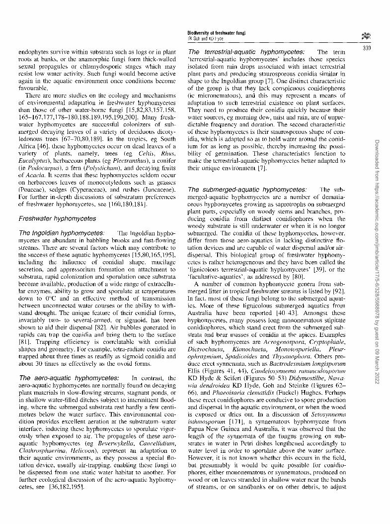
endophytes survive within substrata such as logs or in plant roots at banks, or the anamorphic fungi form thick-walled sexual propagules or chlamydosporic stages which may resist low water activity. Such fungi would become active again in the aquatic environment once conditions become favourable.
There are more studies on the ecology and mechanisms of environmental adaptation in freshwater hyphomycetes than those of other water-borne fungi [15,82,83,157,158, 165-167,177,178-180,188,189,195,199,200]. Many fresh- water hyphomycetes are successful colonizers of sub- merged decaying leaves of a variety of deciduous dicoty- ledonous trees [67-70,80,189]. In the tropics, eg South Africa [46], these hyphomycetes occur on dead leaves of a variety of plants, namely, trees (eg Celtis, Rhus, Eucalyptus), herbaceous plants (eg Plectranthus), a conifer (ie Podocarpus), a fern (Polystichum), and decaying fruits of Acacia. It seems that these hyphomycetes seldom occur on herbaceous leaves of monocotyledons such as grasses (Poaceae), sedges (Cyperaceae), and rushes (Juncaceae). For further in-depth discussions of substratum preferences of freshwater hyphomycetes, see [ 160,180,181 ].
Freshwater hyphomycetes
The Ingoldian hyphomycetes: The Ingoldian hypho- mycetes are abundant in babbling brooks and fast-flowing streams. There are several factors which may contribute to the success of these aquatic hyphomycetes [15,80,165,195], including the influence of conidial shape, mucilage secretion, and appressorium formation on attachment to substrata, rapid colonization and sporulation once substrata become available, production of a wide range of extracellu- lar enzymes, ability to grow and sporulate at temperatures down to 0~ and an effective method of transmission between unconnected water courses or the ability to with- stand drought. The unique feature of their conidial forms, invariably two- to several-armed, or sigmoid, has been shown to aid their dispersal [82]. Air bubbles generated in rapids can trap the conidia and bring them to the surface [81]. Trapping efficiency is correlatable with conidial shapes and geometry. For example, tetra-radiate conidia are trapped about three times as readily as sigmoid conidia and about 30 times as effectively as the ovoid forms.
The aero-aquatic hyphomycetes: In contrast, the aero-aquatic hyphomycetes are normally found on decaying plant materials in slow-flowing streams, stagnant ponds, or in shallow water-filled ditches subject to intermittent flood- ing, where the submerged substrata rest hardly a few centi- metres below the water surface. This environmental con- dition provides excellent aeration at the substratum-water interface, inducing these hyphomycetes to sporulate vigor- ously when exposed to air. The propagules of these aero- aquatic hyphomycetes (eg Beverwykella, Cancellidium, Clathrosphaerina, Helicoon), represent an adaptation to their aquatic environments, as they possess a special flo- tation device, usually air-trapping, enabling these fungi to be dispersed from one static water habitat to another. For further ecological discussion of the aero-aquatic hyphomy- cetes, see [36,182,195].
The terrestrial-aquatic hyphomycetes: The term 'terrestrial-aquatic hyphomycetes' includes those species isolated from rain drops associated with intact terrestrial plant parts and producing staurosporous conidia similar in shape to the Ingoldian group [7]. One distinct characteristic of the group is that they lack conspicuous conidiophores (ie micronematous), and this may represent a means of adaptation to such terrestrial existence on plant surfaces. They need to produce their conidia quickly because their water sources, eg morning dew, mist and rain, are of unpre- dictable frequency and duration. The second characteristic of these hyphomycetes is their staurosporous shape of con- idia, which is adapted so as to hold water around the conid- ium for as long as possible, thereby increasing the possi- bility of germination. These characteristics function to make the terrestrial-aquatic hyphomycetes better adapted to their unique environment [7].
The submerged-aquatic hyphomycetes: The sub- merged-aquatic hyphomycetes are a number of dematia- ceous hyphomycetes growing as saprotrophs on submerged plant parts, especially on woody stems and branches, pro- ducing conidia from distinct conidiophores when the woody substrate is still underwater or when it is no longer submerged. The conidia of these hyphomycetes, however, differ from those aero-aquatics in lacking distinctive flo- tation devices and are capable of water dispersal and/or air- dispersal. This biological group of freshwater hyphomy- cetes is rather heterogeneous and they have been called the 'lignicolous terrestrial-aquatic hyphomycetes' [39], or the 'facultative-aquatics', as addressed by [80].
A number of common hyphomycete genera from sub- merged litter in tropical freshwater streams is listed by [92]. In fact, most of these fungi belong to the submerged aquat- ics. More of these lignicolous submerged aquatics from Australia have been reported [40-43]. Amongst these hyphomycetes, many possess long mononematous stipitate conidiophores, which stand erect from the submerged sub- strata and bear masses of conidia at the apices. Examples of such hyphomycetes are Acrogenospora, Cryptophiale, Dictyochaeta, Kionochaeta, Monotosporiella, Pleur- ophragmium, Spadicoides and Thysanophora. Others pro- duce erect synnemata, such as Bactrodesmium longisporum Ellis (Figures 41, 44), Candelosynnema ranunculosporum KD Hyde & Seifert (Figures 50-53) Didymostilbe, Nawa- wia dendroidea KD Hyde, Goh and Steinke (Figures 62- 66), and Phaeoisaria clematidis (Fuckel) Hughes. Perhaps these erect conidiophores are conducive to spore production and dispersal in the aquatic environment, or when the wood is exposed or dries out. In a discussion of Setosynnema isthmosporum [171], a synnematous hyphomycete from Papua New Guinea and Australia, it was observed that the length of the synnemata of the fungus growing on sub- strates in water in Petri dishes lengthened accordingly to water level in order to sporulate above the water surface. However, it is not known whether this occurs in the field, but presumably it would be quite possible for conidio- phores, either mononematous or synnematous, produced on wood or on leaves stranded in shallow water near the bands of streams, or on sandbanks or on other debris, to adjust
339
Dow
nloaded from https://academ
ic.oup.com/jim
b/article/17/5-6/328/5988978 by guest on 09 March 2022

340
Biodiversity of freshwater fungi TK Gob and KD Hyde
their length according to the water depth in order to sporu- late above the surface.
Some of these lignicolous submerged aquatics, however, produce conidia with modified appendages, setulae, or arms and are functionally comparable to those Ingoldian hypho- mycetes in the aquatic habitats. Examples for these hypho- mycetes are Dictyochaeta, Nawawia (Figures 64-66), and Obeliospora which produce setulate conidia; Iyengarina and Sporidesmiella cornuta Kuthubutheen and Nawawi which produce conidia with arms; Dactylaria tunicata Gob and KD Hyde, and Delortia palmicola Pat (Figures 54-57), which produce conidia surrounded by a hyaline mucilagin- ous sheath. The occurrence of these freshwater hyphomy- cetes with these specialized conidial forms may be adap- tations to local dispersion in the tropical rainforest habitat with its constant elevated relative humidity and persistent fihns of water on surfaces of the submerged substrata. For thorough discussion of aquatic hyphomycetes as successful colonizer on wood, see [164,174,178,199].
Notes on Beltrania rhombica It is interesting that Beltrania rhombica Penz, normally regarded as a 'terrestrial' hyphomycete, is commonly encountered in foam samples in the tropics [60,130,131,180,183,184]. It is highly possible that this conceptually 'non-aquatic' hyphomycete can adapt to a submerged aquatic existence. Perhaps this 'terrestrial' fungus has a stream-side origin [80] in which its conidia are washed into the stream after heavy rain when water rises above parts of the bank where it is normally not sub- merged. Tan and Kok [183] speculated that the abundance of B. rhombica in the foam could not have been attributed solely to a terrestrial source because it was consistently the most or second-most abundant species of foam mycoflora observed during both dry and wet months through the per- iod of their studies in Singapore. In India, incubation of submerged leaves of various plants yielded conidia of B. rhombica which showed high percentage of leaf coloniz- ation [180]. From these observations, B. rhombica may indeed be a 'resident' species of submerged stream litter in the tropics, and the abundance of its conidia in foam need not always imply a terrestrial origin [183].
Freshwater ascomycetes Characteristics of freshwater fungi that are advantageous to their dispersal and subsequent attachment of new substrata, will either have been acquired from terrestrial lineages or morphologies that are preadaptive to the aquatic 'life style' or will have specifically evolved in the aquatic environ- ment. In most freshwater ascomycetes the asci are provided with ejection mechanisms as in many terrestrial fungi (Table 2). The asci are either bitunicate with fissitunicate dehiscence (eg Massarina spp, Ascagilis spp (Figure 23)) or unitunicate with relatively massive (eg Annulatascus spp (Figures 6, 8, 11, 12)) or smaller (eg Ophioceras spp (Figures 37, 38), Bertia spp (Figures 16, 17)) apical rings. These fungi actively eject their ascospores in air [57], but it has not been shown conclusively that freshwater ascomy- cetes can eject their ascospores under water. In squash mounts the ascospores are often readily ejected, but this is hardly natural. In submerged culture, both asci and asco-
spores were discharged simultaneously in species of Ophio- ceras and Pseudohalonectria [175]. Hyde (unpublished) placed ascomata of Pseudohalonectria growing on a wood sliver under water in a Petri dish overnight. The following morning numerous ascospores were lying on the base of the dish around the sample, indicating that active dispersal does occur underwater. It appears that active spore ejection is an important dispersal strategy of ascomycetes in tropical freshwater streams. On the other hand, it may be that the freshwater ascomycetes have not become totally adapted to submerged conditions (or that substrates in streams are more likely to be exposed, or subjected to periods of des- iccation, when ejection can occur).
In only a handful of freshwater species (eg Halosarpheia aquatica KD Hyde) do the asci deliquesce early to release the ascospores passively (Table 2). In only three genera, Aniptodera Shearer and Miller, Halosarpheia Kohlm and E Kohlm and Nais Kohlm are the spores passively released and form a cirrhus at the tip of the neck.
Floating is the next problem faced by the freshwater pro- pagule. The lipid globules found in the ascospores of most freshwater ascomycetes (Figures 3, 21, 25), probably act as flotation devices as well as food reserves as in the mar- ine ascomycetes.
The final obstacle faced by the propagule is entrapment and attachment and colonization of new substrates. The ascomycetes in tropical streams are mostly provided with appendaged ascospores or are sigmoid in shape. The appendages include typical to elaborate mucilaginous sheaths (ie Massarina spp, Pleospora scirpicola), polar pads (Ascagilis bipolaris), or uncoiling filamentous strands (Annulatascus bipolaris, Halosarpheia aquatica). An account of the various appendage types is given below. Appendaged spores do not appear to be so commonplace in temperate freshwater ascomycetes.
Five appendage types in freshwater ascomycetes are recognised (Table 3): 1) The release of a drop of mucilage (eg Ophioceras spp); 2) hamate or cap-like appendages which uncoil to form viscous threads (eg Aniptodera (Figures 3-5) and Halosarpheia spp). These separate from the spore and uncoil in water to form long viscous polar threads [48,50,172]. Besides mucilaginous sheaths, fila- mentous appendages appear to be most commonly found in ascospores of tropical freshwater ascomycetes; 3) muci- laginous sheaths (eg A. velatispora (Figures 9, 10), Kirchs- teiniothelia elaterascus Shearer, Fluviatispora reticulata KD Hyde, Massarina ingoldia Shearer and KD Hyde), which may or may not have extensions; 4) pad-like attach- ment (eg Ascagilis bipolaris KD Hyde (Figures 21, 22), Mamillisphaeria dimorphospora KD Hyde et al); 5) irregu- lar amorphous appendages without a fibrillar component (eg Ceriospora caudaesuis Ingold); and 6) adhesive spore wall (eg Savoryella verrucosa (Figures 28, 29)).
These appendage types are thought to be involved in dis- persal and attachment of these freshwater propagules in a similar way to marine ascomycetes [58,59,61,62]. The mucilaginous drops at the ends of the spores of Ophioceras spp, probably attached the spores at one end, which then twist in the water current and lie parallel to the flow of water. These spores then stand a better chance of remaining attached as in the sigmoid hyphomycete spores [193]. The
Dow
nloaded from https://academ
ic.oup.com/jim
b/article/17/5-6/328/5988978 by guest on 09 March 2022

Biodiversity of freshwater fungi TK Goh and KD Hyde
Table 2 Ascus dehiscence and ascospore appendage types in the ten most common genera of tropical freshwater ascomycetes
Genus Ascus dehiscence Ascospore appendages
341
Aniptodera Passive Polar filaments Annulatascus Active Sheath or various Ascagilis Active Polar pads or sticky walls Ceratosphaeria Active Wall ornamentations Halosarpheia Passive Polar filaments Kirschsteiniothelia Active Sticky walls Mamillisphaeria Active Polar pads Massarina Active Sheath/with modifications Ophioceras Active Sigmoid Pseudohalonectria Active None Savoryella Active Sticky walls
TabLe 3 Spore adhesion types observed in tropical freshwater ascomycetes
Description Species Reference
1. Sticky mucilaginous sheaths Annulatascus velatispora [49, 54, 63] Massarina spp Vaginatispora aquatica
Ascagilis bipolaris Mamillisphaeria dimorphaspora
Annulatascus spp Aniptodera spp Halosarpheia spp
Savoryella spp
Loramyces
2. Polar pads [48, 50, 66]
3. Polar filaments
4. Adhesive spore wall
5. Other types or combinations of the above types
[48, 50, 54] Wong, Hyde and Jones, pers. obs.
Ho and Hyde, pers. obs.
Hyde, pers. obs.
unravelling threads of Aniptodera and Halosarpheia prob- ably serve to catch debris in the water, thus anchoring the spores to new substrates. The role of the sheath surrounding various propagules is probably two-fold. SEM micrographs illustrate that the sheaths are sticky and one function is likely to be adhesion [59]. A second function may be to hold the spore at the air water interface, perhaps allowing it to float on the surface and be dispersed in this way.
The sheath in Massarina ingoldiana initially envelops the ascospore, but once released in water it swells to form massive arms which appear to be highly effective in attach- ment. A similar massive sheath is found in Pleospora scir- picola (DC) Karsten [75]. In Velatispora aquatica KD Hyde an internal collar-like structure with an unknown function encircles the ascospore and is part of the larger sheath structure [56].
The pad-like mucilaginous appendages in Ascagilis bipolaris (Figures 21, 22) and Mamillisphaeria dimorphos- pora are probably sticky and function to adhere the asco- spores in much the same ways as the marine species Cerio- sporopsis circumvestita and Ondiniella torquata [84,87,168]. The amorphous coiled appendages at each end in Ceriospora caudae-suis [73] are persistent and do not appear to be the same type as those found in the genera Aniptodera and Halosarpheia. They probably, however, have the same function in trapping and sticking propagules to debris. Spore adhesion can be achieved with sticky spore walls, such as an adhesive coating or a slightly more elabor-
ate verrucose wall. In Savoryella verrucosa the spore wall (Figures 28, 29) is highly verrucose. The ascospores of sev- eral other freshwater ascomycetes are provided with verru- cose wall ornamentation and these probably serve to stick the spores to debris.
Active versus passive dispersal Why do most marine unitunicate ascomycetes release their spores passively, while many tropical freshwater ascomy- ceres release their spores actively? Perhaps evolution has played a key role in this outcome, most marine fungi having evolved from ancestors of the Microascales (Spatafora, per- sonal communication) and most Annulatascus-like species having evolved from other terrestrial ancestors.
Why is active spore release more common in freshwater ascomycetes? Assuming active spore release readily occurs under water then what advantage does it have over passive spore release, if any? One explanation may involve the boundary layer, which was also used to explain the attach- ment of sigmoid ascospores [193]. If spores are released passively they may never leave the boundary layer and become attached to the adjacent substrate. Ejected asco- spores, on the other hand, may be expelled beyond the boundary layer and then dispersed in the turbulent current. The long neck found in Ophioceras species (Figure 36) might also achieve the same effect, but this may be fraught with inherent habitat problems (eg aquatic insects).
A simple explanation may also account for the predomi-
Dow
nloaded from https://academ
ic.oup.com/jim
b/article/17/5-6/328/5988978 by guest on 09 March 2022

342
Biodiversity of freshwater fungi TK Goh and KD Hyde
nance of freshwater species that actively release their spores. Streams and even rivers are subjected to periodic drying and it may be during these periods that spores are actively released. Alternatively, during heavy rains or typhoons, flash flooding may occur and it may be that dur- ing these spates the substrates are washed to the edges of the streams and exposed as the waters subside. Active spore dispersal may then occur in air.
Loculoascomycetes with fissitunicate dihiscence asci are also common in freshwater ecosystems. These asci also 'shoot' their ascospores, but whether they do this under submerged conditions or only in air is speculative. Notable spore release occurs in Boerlagiomyces species and Kirchs- teiniothelia elaterascus. In Boerlagiomyces Butzin the bitu- nicate asci contain ascospores which are actively released in pairs, enveloped in a gelatinous sheath. In K. elaterascus the base of the ascus contains a coiled endoascus. Shearer [175] reported that it 'may be that when ascomata of K. elaterascus are submerged, entire, unfurled endoasci are discharged and become entangled with the substrate'. Asci of a similar type have also been recorded in an unrelated Macroventuria-like species from freshwater [175].
R e f e r e n c e s
1 Abdullah SK and J Webster. 1980. Occurrence of aero-aquatic fungi in soil. Trans Br Mycol Soc 75:511 514.
2 Abdullah SK, E Descals and J Webster. 1981. Teleomorphs of three aquatic hyphomycetes. Trans Br Mycol Soc 77: 475-483.
3 Alasoadura SO. 1968. Some aquatic hyphomycetes from Nigeria. Trans Br Mycol Soc 51:535 540.
4 Alasoadura SO. 1968. Flabellospora crassa n gen n sp aquatic hyphomycete from Nigeria. Nova Hedwigia 15: 415-418.
5 Alasoadura SO. 1968. Flabellospora verticillata a new species of aquatic hyphomycete from Nigeria. Nova Hedwigia 15: 419M21.
6 Anastasiou CJ. 1964. Some aquatic fungi imperfecti from Hawaii. Pac Sci 18:202 222.
7 Ando K. 1992. A study of terrestrial aquatic hyphomycetes. Trans Mycol Soc Japan 33: 415-425.
8 Ando K and 1 Kawamoto. 1986. Arborispora, a new genus of hypho- mycetes. Trans Mycol Soc Japan 27: 119-128.
9 Ando K and l Kawamoto. 1986. Materials for the fungus flora of Japan (41). Trans Mycol Soc Japan 27: 321-326.
10 Ando K and K Tubaki. 1984. Some undescribed hyphomycetes in the rain drops from intact leaf-surface. Trans Mycol Soc Japan 25: 21-37.
11 Ando K and K Tubaki. 1984. Some undescribed hyphomycetes in rainwater draining from intact trees. Trans Mycoi Soc Japan 25: 39-47.
I2 Ando K and K Tubaki. 1984. Materials for the fungus flora of Japan (37). Trans Mycol Soc Japan 25: 395-398.
13 Ando K and K Tubaki. 1985. Three new hyphomycetes from Japan: Anthopsis microspora, Scutisporus brunneus and Titaeella capno- phila. Trans Mycol Soc Japan 26: 151-160.
14 Ando K, K Tubaki and M Arai. 1987. Trifurco,spora, a new generic name for Flabellospora irregularis. Trans Mycol Soc Japan 28: 469-473.
15 Au DWT. 1993. Enzymatic studies of conidial attachment and lectin- gold histochemical investigation of the extracellular mucilages of Lemonniera aquatica De Wild and Mycocentrospora filifi)rmis (Petersen) Iqbal. PhD Thesis, University of Hong Kong, 326 pp.
16 Betancourt C, J Cruz, J Garcia and L Galarza. 1986. Estudio prelimi- nat de los hifomicetos acuaticos (deuteromicotina) de la Republica Dominicaua. Carib J Sci 22: 49-51.
17 Betancourt C, J Cruz and J Garcia. 1987. Los hifomicetos acuaticos de la Quebrada Dona Juana en al Bosque Estatal de Toro Negro, Villalba, Puerto Rico. Carib J Sci 23:278 284.
18 Beverwijk van AL. 1951. Zalewski's Clathrosphaera spiriJera'. Trans Br Mycol Soc 34: 280-290.
19 Beverwijk van AL. 1951. Candelabrum spinulosum, a new fungus species. Antonie van Leeuwenhoek 17:278 284.
20 Beverwijk van AL. 1953. Helicosporous hyphomycetes. I. Trans Br Mycol Soc 36:111 124.
21 Beverwijk van AL. 1954. Three new fungi: Helicoon pluriseptatum, Papulaspora pulmonaria and Tricellulu inaequalis. Antonie van Leeuwenhoek 20:1 16.
22 Bhat DJ and C-Y Chien. 1990. Water-borne hyphomycetes found in Ethiopia. Trans Mycol Soc Japan 31: 147-157.
23 Brittlebank CC. 1940. Reference index of Australian Fungi. Limited distribution manuscript, Victorian Department of Agriculture.
24 Carmichael JW, WB Kendrick, IL Conners and L Sigler. 1980. Gen- era of Hyphomycetes. University of Alberta Press, Edmonton, 386 pp.
25 Cooke WB. 1970. Our Mouldy World, a Study in the Fungi of Our Environment with Emphasis on Water. US Department of the Interior, Federal Water Pollution Control and Administration, Cincin- nati, pp 1 533.
26 Crane JL and KP Dumont. 1975. Hyphomycetes from the West Indies and Venezuela. Can J Bot 53:843 851.
27 Cribb AB and JW Cribb. 1993. Fungal spores in freshwater foam from Cape York Peninsula. Proc R Soc Queensl 103: 65-73.
28 Cunnell GJ. 1958. On Robillarda phragmitis sp nov. Trans Br Mycol Soc 41: 405-412.
29 De Fonseka RN. 1960. The morphology of Chaetospermum chaetos- porium. Trans Br Mycol Soc 43: 631-636.
30 Descals C and J Webster. 1984. Branched aquatic conidia in Erynia and Entomophtora sensu lato. Trans Br Mycol Soc 83:669 682.
31 Descals C, F Pelfiez and L L6pez. 1995. Fungal spora of stream foam from central Spain I. Conidia identifiable to species. Nova Hedwigia 60: 533-550.
32 Descals C, F Pelfiez and L Ltpez. 1995. Fungal spora of stream foam from central Spain II. Chorology, spore frequency and unknown forms. Nova Hedwigia 60: 551-569.
33 Dix NJ and J Webster. 1994. Aquatic fungi. In: Fungal Ecology, pp 225-265, Chapman and Hall, London, UK.
34 Dixon PA. 1959. Stream spora in Ghana. Trans Br Mycol Soc 42: 174 176.
35 Dudka IO. 1974. Ukrainian aquatic Hyphomycetes in Ukrainian. Academy of Science, Ukrainian RSRMG. Holodny Botanical Insti- tute, Naukova Dumka, Kiev, pp 1-240.
36 Fisher PJ. 1977. New methods of detecting and studying the sapro- phytic behavior of aero-aquafic hyphomycetes from stagnant water. Trans Br Mycol Soc 68:407411.
37 Fuller MS and A Jaworski. 1987. Zoosporic Fungi in Teaching and Research. Southeastern Publishing Corporation, Athens, Georgia, 303 pp.
38 Glen-Bott JI. 1951. Helicodendron giganteum n sp and other aerial sporing hyphomycetes of submerged leaves. Trans Br Mycol Soc 34: 275-279.
39 Goh T-K. 1996. Tropical freshwater hyphomycetes. In: Biodiversity of Tropical Microfungi (Hyde KD, ed), pp 187-223, Hong Kong Univ Press, Hong Kong.
40 Goh TK and KD Hyde. 1996. Brachydesmiella anthostomelloidea, a new species of dematiaceous hyphomycete fi'om Australia. Mycol Res 100: 1364-1366.
41 Goh TK and KD Hyde. 1996. Helicoon gigantisporum sp nov from Australia and an amended key to the genus. Mycol Res 100: 1485-1488.
42 Goh TK and KD Hyde. 1996. Co,ptophiale multiseptata sp nov from submerged wood in Australia, and keys to the genus. Mycol Res 100: 999-1004.
43 Goh TK and KD Hyde. The genus Delortia, with a description of D. palmicola and two new species from submerged wood in a fresh- water stream in Australia. Mycol Res 101: (in press).
44 Goos RD, SK Abdullah, PJ Fisher and J Webster. 1985. The ana- morph genus Helicodendron. Trans Br Mycol Soc 84: 423-435.
45 Goos RD, SK Abdullah, PJ Fisher and J Webster. 1986. The ana- morph genus Helicoon. Trans Br Mycol Soc 87:115-122.
46 Greathead SK. 1961. Some aquatic hyphomycetes in South Africa. JSA Bot 27: 195-228.
47 Hudson JH and CT lngold. 1960. Aquatic hypbomycetes from Jamaica. Trans Br Mycol Soc 43: 469-478.
48 Hyde KD. 1992. Tropical Australian freshwater fungi. I. Some asco- mycetes. Aust Syst Bot 5: 109-116.
Dow
nloaded from https://academ
ic.oup.com/jim
b/article/17/5-6/328/5988978 by guest on 09 March 2022

49 Hyde KD. 1992. Tropical Australian freshwater fungi, lI. Annulat- ascus velatispora gen et sp nov, A. bipolaris sp nov and Ophioceras dolichostomum Ascomycetes. Aust Syst Bot 5 :117 124.
50 Hyde KD. 1992. Tropical Australian freshwater fungi. IV. Halosar- pheia aquatica sp nov, Garett~jonesia lacunosispora gen et sp nov and Ophioceras dolichostomum Ascomycetes. Aust Syst Bot 5: 407-4 14.
51 Hyde KD. 1993. Tropical Australian freshwater fungi. VI. Tiarospor- ella paludosa and Clohesyomyces aquaticus gen et sp nov Coelomy- cetes. Aust Syst Bot 5: 169-173,
52 Hyde KD. 1993. Tropical Australian freshwater fungi. V. Bombardia sp, Jahnula australiensis sp nov, Savoo'ella aquatica sp nov and S. lignicola sp nov. Aust Syst Bot 6:161 167.
53 Hyde KD. 1994. Aquatic fungi on rachids of Livistona in the Western Province of Papua New Guinea. Mycol Res 98: 719-725.
54 Hyde KD. 1995. Tropical Australian freshwater thngi. VII. New gen- era and species of ascomycetes. Nova Hedwigia 6i: 119-140.
55 Hyde KD. 1995. Tropical Australian freshwater fungi. VIII. Bertia convoluti~pora sp nov. Nova Hedwigia 61: 141-146.
56 Hyde KD. 1995. Tropical Australian freshwater fungi. IX. Vaginatis- pora aquatica gen et sp nov. Nova Hedwigia 6l: 233-241.
57 Hyde KD. 1996. Tropical Australian Freshwater Fungi. X. Submersi- sphaeria aquatica gen et sp nov. Nova Hedwigia 62: 171-175.
58 Hyde KD, R Greenwood and EBG Jones. 1993. Spore attachment in marine fungi. Mycol Res 97 :7 14.
59 Hyde KD and EBG Jones. 1989. Observations on ascospore mor- phology in marine fungi and their attachment to surfaces. Bot Mar 32: 205-218.
60 Hyde KD and JS Stanley. 1994. Larger fungi. In: Aquatic Crypto- grams of Australia. A Guide to the Larger Fungi, Lichens, Macroal- gae and Mosses of Australian Inland Waters (Entwisle T, ed), pp 4 - 16, Australian Society for Limnology, Special Publication No. 10, Abbotsford, Victoria 3067, Australia.
61 Hyde KD, EBG Jones and ST Moss. 1986. How do fnngal spores attach to surfaces? In: Biodeterioration 6 (Barry S, DR Houghton, GC Llewelyn and CE O'Rea, eds), pp584-589, Commonwealth Agricultural Bureaux and the Biodeterioration Society, London.
62 Hyde KD, ST Moss and EBG Jones. 1989. Attachment studies in marine fungi. Biotbuling l: 287-298.
63 Hyde KD and KA Seifert. 1992. Tropical Australian freshwater fungi. IIl. Candelosynnema ranunculosporuma, a new genus and species of synnematous Hyphomycetes. Aust Syst Bot 5: 401-405.
64 Hyde KD, TK Goh and T Steinke. 1996. Nawawia dendroidea, a new synnematous hyphomycetes from submerged Phragmites in South Africa. Mycol Res 100: 810-814.
65 Hyde KD, WS Wong and EBG Jones. 1996. Biodiversity of aquatic ascomycetes. KD Hyde. In: Biodiversity of Tropical Microfnngi (Hyde KD, ed), Hong Kong Univ Press, Hong Kong.
66 Hyde KD, SW Wong and EBG Jones. 1996. Tropical Australian Freshwater Fungi. XI. Mamillisphaeria dimorpho,spora gen et sp nov and notes on freshwater ascomycetes with dimorphic ascospores. Nova Hedwigia 62: 171-175.
67 Ingold CT. 1942. Aquatic hyphomycetes of decaying alder leaves. Trans Br Mycol Soc 25: 339-417.
68 Ingold CT. 1943. Further observations on aquatic hyphomycetes. Trans Br Mycol Soc 26:104-115.
69 Ingold CT. 1943. Triscelophorus monosporus n g e n n sp an aquatic hyphomycete. Trans Br Mycol Soc 26: 148-152.
70 lngold CT. 1943. On the distribution of aquatic hyphomycetes sapro- phytic on submerged decaying leaves. New Phytol 42:139 143.
71 Ingold CT. 1944. Some new aquatic hyphomycetes. Trans Br Mycol Soc 28: 35~43.
72 Ingold CT. 1949. Aquatic hyphomycetes from Switzerland. Trans Br Mycol Soc 32: 341-345.
73 Ingold CT. 1951. Aquatic ascomycetes: Ceriospora caudae-suis n sp and Ophiobolus typhae. Trans Br Mycol Soc 34: 210-215.
74 ingoid CT. i952. Actinospora megalospora n sp, an aquatic hypho- mycete. Trans Br Mycol Soc 35: 66-70.
75 Ingold CT. 1955. Aquatic ascomycetes: further species from the English Lake district. Trans Br Mycol Soc 38: 157-168.
76 Ingold CT. 1956. Stream spora in Nigeria. Trans Br Mycol Soc 39: 106-110.
77 Ingold CT. 1958. Aquatic hyphomycetes from Uganda and Rhodesia. Trans Br Mycol Soc 41:109-114.
Biodiversity of freshwater fungi TK Goh and KD Hyde
78 lngold CT 1959. Aquatic spora of Omo forest, Nigeria. Trans Br Mycol Soc 42: 479-485.
79 Ingold CT. 1960. Aquatic hyphomycetes in southern Rhodesia. Proc Rhodesian Sci Assoc 43: 49-53.
80 Ingold CT. 1975. An Illustrated Guide to Aquatic and Water-borne Hyphomycetes Fungi Imperfecti with Notes on their Biology. Fresh- water Biol Assoc Sci Publ 30: 1-96.
81 Ingold CT and J Webster. 1973. Some aquatic hypholnycetes from India. Kavaka l: 4-9.
82 Iqbal SH. 1995. Further studies on efficiency of artificial foam in trapping conidia of Ingoldian fungi. Can J Bot 73:1176-I185.
83 lqbal SH and J Webster. 1973. The trapping of aquatic hyphomycete spores by air bubbles. Trans Br Mycol Soc 60: 37-48.
84 Johnson RG, EBG Jones and ST Moss. 1987, Taxonomic studies of the Halosphaeriaceae: Cerios;ooropsis, Haligena, and appendichord- ella gen nov. Can J Bot 65: 931-942.
85 Jones EBG (ed). 1976. Recent Advances in Aquatic Mycology. Elek Science, London.
86 Jones EBG. 1976. Aquatic fungi: freshwater and marine. In: Recent Advances in Aquatic Mycology (Jones EBG, ed), pp 337-376, Elek Science, London.
87 Jones EBG. 1995. Fungal adhesion. Mycol Res 98:961 981. 88 Kuthubutheen AJ. 1987. Another new species of Cryptophiale from
Malaysia. Trans Br Mycol Soc 89: 274-278. 89 Kuthubntheen AJ. 1987. Two new species of Dictyochaeta from
Malaysia. Trans Br Mycol Soc 89:353 358. 90 Kuthubutheen AJ. 1987. A new synnematous Dictyochaeta from
Malaysia. Trans Br Mycol 89: 411414 . 91 Kuthubutheen AJ. 1987. A new species of Phalangiapora and further
observations on P. constricta from Malaysia. Trans Br Mycol Soc 89: 414-420.
92 Kuthubutheen AJ. 1993. Tropical hyphomycetes from submerged lit- ter in freshwater streams. In: Aspects of Tropical Mycology (Isaac S, JC Frankland R, Watling and AJS Whalley, eds), pp 296-297, Cambridge University Press.
93 Kuthubutheen AJ and A Nawawi. 1987. Cr>7)tophialoidea gen nov on decaying leaves from Malaysia. Trans Br Mycol Soc 89:581 583.
94 Kuthubutheen AJ and A Nawawi. 1987. A new species of Speiropsis from Malaysia. Trans Br Mycol Soc 89: 584-587.
95 Kuthubutheen AJ and A Nawawi. 1988. Two new species of Kiono- chaeta Hyphomycetes and K. ramifera from Malaysia. Trans Br Mycol Soc 90: 437-444.
96 Kuthubutheen AJ and A Nawawi. 1988. A new species of Wiesneri- omyces Hyphomycetes from submerged decaying leaves. Trans Br Mycol Soc 90: 619-625.
97 Kuthubutheen AJ and A Nawawi. 1988. A new species of Selenospo- rella Hyphomycetes from Malaysia. Trans Br Mycol Soc 91: 33i 334.
98 Kuthubutheen AJ and A Nawawi. 1990. Dictyochaeta hamata and D. pahangensis, two new species with lateral phialides. Mycol Res 94: 840-846.
99 Kuthubutfieen AJ and A Nawawi. 1991. Dictyochaeta macrospora sp nov: a litter-inhabiting hyphomycete from Malaysia. Mycol Res 95: 248-250.
100 Kuthubutheen AJ and A Nawawi. 1991. Three new species of Dictyochaeta with non-setose conidiophores and non-septate setulate conidia from Malaysia. Mycol Res 95: 104-107.
101 Kuthubutheen AJ and A Nawawi. 1991. Eight new species of Dictyochaeta Hyphomycetes from Malaysia. Mycol Res 95: 1211- 1219.
102 Kuthubutheen AJ and A Nawawi. 1991. Dictyochaeta guadalcana- lensis comb nov and several new records of the genus in Malaysia. Mycol Res 95: 1220-1223.
103 Kuthubutheen AJ and A Nawawi. 1991. Key to Dictyochaeta and Codinaea species. Mycol Res 95: 1224-1229.
104 Kuthubutheen AJ and A Nawawi. 1992, New litter-inhabiting hypho- mycetes from Malaysia: Isthmophragmospora verruculosa, lyengar- ina asymmetrica, and Iyengarina Jurcata. Can J Bot 70: 101-106.
105 Kuthubutheen AJ and A Nawawi. 1993. Three new and several inter- esting species of Sporidesmiella from submerged litter in Malaysia. Mycol Res 97: 1305-1314.
106 Kuthubutheen AJ and A Nawawi. 1994. Paracryptophiale kamarud- dinii gen et sp nov from submerged litter in Malaysia. Mycol Res 98: 125-126.
343
Dow
nloaded from https://academ
ic.oup.com/jim
b/article/17/5-6/328/5988978 by guest on 09 March 2022

~ Biodiversity of freshwater fungi TK Goh and KD Hyde
344 107 Kuthubutheen AJ and A Nawawi. 1994. Henicospora longissima sp nov Obeliospora triappendiculata sp nov, Paraulocladium )dbispo- rum sp nov and other hyphomycetes from Malaysia. Mycol Res 98: 677-685.
108 Kuthubutheen AJ and A Nawawi. 1994. CryptophialoideaJ)isciculata sp nov and C manifesta comb nov from Malaysia. Mycol Res 98: 686-688.
109 Kuthubutheen AJ, GM Liew and A Nawawi. 1992. Nawawia nitida sp nov Hyphomycetes and further records of Nawawiafiliformis from Malaysia. Can J Bot 70: 96-100.
110 Le'John HB. 1965. Sierra Leone freshwater hyphomycetes. Trans Br Mycol Soc 48: 261-264.
111 Lichtwardt RW and MC Williams. 1992. Tasmanian Trichomycete gut fungi in aquatic insect larvae. Mycologia 84: 384-391.
112 Magnes M and J Hafellner. 1991. Ascomyceten auf Get~isspflanzen an Ufern von Gebirgsseen in den Ostalpen. Biblioth Mycol 139: 1 182.
113 Marvanovfi L and P Marvan. 1969. Aquatic hyphomycetes in Cuba. Cesk~, MyKol 23: 135-140.
114 Marvanovfi L and JA Stalpers. 1987. The genus Taeniospora and its teleomorphs. Trans Br Mycol Soc 89: 489-498.
115 Marvanovfi L and K Suberkropp. 1990. Camptobasidium hydrophi- lure and its anamorph Crucella subtilis: a new heterobasidiomycete from streams. Mycologia 82: 208-217.
116 Nawawi A. 1973. Clavatospora fihlfbrmis sp nov, an aquatic hypho- mycete from Malaysia. Trans Br Mycol Soc 61:390 393.
117 Nawawi A. 1973. A new species of Varico,sporium from Malaysia. Nova Hedwigia 24: 3943.
118 Nawawi A. 1973. Two clamp-bearing aquatic fungi from Malaysia. Trans Br Mycol Soc 61: 521-528.
119 Nawawi A. 1973. A new species of Flabellospora from Malaysia. Malay J Sci 2: 55-58.
120 Nawawi A. 1974. Two new Varicosporium species. Trans Br Mycol Soc 63:27 31.
121 Nawawi A. 1974. Two new Tricladium species. Trans Br Mycol Soc 63:267 272.
122 Nawawi A. 1974. A new CampylosToora. Trans Br Mycol Soc 63: 603-606.
123 Nawawi A. 1975. Another hyphomycete with branched conidia. Trans Br Mycol Soc 64: 243-246.
124 Nawawi A. 1975. Triscelophorus acuminatus sp nov. Trans Br Mycol Soc 64:345 348.
125 Nawawi A. 1976. A new genus of hyphomycetes. Trans Br Mycol Soc 66: 344-347.
126 Nawawi A. 1976. Condylospora gen nov a hyphomycete from a foam sample. Trans Br Mycol Soc 66: 363-365.
127 Nawawi A. 1976. Another new Flabellospora. Trans Br Mycol Soc 66: 543-547.
128 Nawawi A. 1976. Filosporella gen nov, an aquatic hyphomycete. Trans Br Mycol Soc 67:173 176.
129 Nawawi A. 1982. Phalangiospora constricta gen et sp nov, a sporo- dochial hyphomycete with branched conidia. Trans Br Mycol Soc 79: 65-68.
130 Nawawi A. 1985. Another aquatic hyphomycete genus from foam. Trans Br Mycol Soc 85: 174-177.
131 Nawawi A. 1985. Aquatic hyphomycetes and other water-borne fungi from Malaysia. Malay Nat J 39: 75-134.
132 Nawawi A. 1985. More Tricladium species from Malaysia. Trans Br Mycol Soc 85: 177-182.
133 Nawawi A. 1985. Some interesting hyphomycetes from water. Myco- taxon 24: 217-226.
134 Nawawi A. 1985. Basidiomycetes with branched water-borne condia. Bot J Linn Soc 91: 51-60.
135 Nawawi A. 1987. Clavariopsis axlanii sp nov a new aquati hyphomy- cete from Malaysia. Trans Br Mycol Soc 88: 428432.
136 Nawawi A. 1987. Diplocladiella appendicula sp nov a new aero- aquatic hyphomycete from Malaysia. Mycotaxon 28: 297-303.
137 Nawawi A and AJ Kuthubutheen. 1987. Clathrosporiurn intricatum gen et sp nov an aero-aquatic hyphomyeete. Trans Br Mycol Soc 89: 407-411.
138 Nawawi A and AJ Kuthubutheen. 1987. Triscelosporium verrucosum gen et sp nov, a dematiaceous aero-aquatic hyphomycete with tetrara- diate conidia. Mycotaxon 29: 285-290.
139 Nawawi A and AJ Kuthubutheen. 1987. Tetrabrachium elegans gen
and sp nov, a hyphomycete with tetraradiate conidia. Mycotaxon 29: 291-297.
140 Nawawi A and AJ Kuthubutheen. 1988. Another new hyphomycete from leaf litter. Mycotaxon 31:339 343.
141 Nawawi A and AJ Kuthubutheen. 1988. Camposporidium, a new phragmoconidial genus of hyphomycetes. Mycotaxon 32: 161-167.
142 Nawawi A and AJ Kuthubutheen. 1988. A new species of the genus Dendryphiosphaera. Mycotaxon 32: 461466.
143 Nawawi A and AJ Kuthubutheen. 1988. Additions to Condylospora Hyphomycetes from Malaysia. Mycotaxon 33: 329-338.
144 Nawawi A and AJ Kuthubutheen. 1988. Tricladiospora, a new genus of dematiaceous Hyphomycetes with staurosporous conidia from sub- merged decaying leaves. Trans Br Mycol Soc 90: 482-487.
145 Nawawi A and AJ Kuthubutheen. 1988. Beverwykella cerebriJbrmis sp nov, an aero-aquatic hyphomycete from Malaysia. Trans Br Mycol Soc 90: 487-491.
146 Nawawi A and AJ Kuthubutheen. 1988. Tricladiomyces geniculatus sp nov, a conidial basidiomycete. Trans Br Mycol Soc 90: 670-673.
147 Nawawi A and AJ Kuthubutheen. 1989. Canalisporium, a new genus of lignicolous hyphomycetes from Malaysia. Mycotaxon 34: 475- 487.
148 Nawawi A and AJ Kuthubutheen. 1989. Quadricladium Aquaticum gen et sp nov, an aquatic hypbomycete with tetraradiate conidia. Mycotaxon 34: 489-495.
149 Nawawi A and AJ Kuthubutheen. 1989. A new taxon in Colispora Hyphomycetes from Malaysia. Mycotaxon 34: 497-501.
150 Nawawi A and AJ Kuthubutbeen. 1990. Nidulispora gen nov, a hyphomycete genus with crateriform conidia. Mycotaxon 36: 329- 336.
151 Nawawi A and AJ Kuthubutheen. 1990. New species and combi- nations in Vermiculariopsiella Hyphomycetes. Mycotaxon 37: 173- 182.
152 Nawawi A and AJ Kuflmbutheen. 1990. Addition to Subulispora from Malaysia. Mycotaxon 37: 389-393.
153 Nawawi A and AJ Kuthubutheen. 1990. Obeliospora, a new genus of setose, phialosporous hyphomycetes with appendaged conidia. Mycotaxon 37: 395-400.
154 Nawawi A and J Webster. 1982. Sistotrema hamatum sp, the teleo- morph of Ingoldiella harnatu. Trans Br Mycol Soc 78: 287-291.
155 Nawawi A, J Webster and RA Davey. 1977. Lepto,sporomyces glazi- nii, the basidial state of a clamped branched conidium from fresh- water. Trans Br Mycol Soc 68: 31-36.
156 Nawawi A, J Webster and RA Davey. 1977. Dendro,sporomyces pro- lifer gen et sp nov, a basidiomycete with branched condidia. Trans Br Mycol Soc 68:59 63.
157 Nayo SG. 1975. The ecology and distribution of aquatic hyphomy- ceres around Bogor. Biotrop, SEAMEO Regional Centre for Tropical Biology, TFRS 75/151, Bogor, Indonesia, pp 1-61.
158 Nilsson S. 1964. Freshwater hyphomycetes: taxonomy, morphology and ecology. Symbolae Botanicae Upssalienses 182: 1-130.
159 Osborn TGB. 1915. Some new records of fungi for South Australia. Trans Roy Soc South Aust 39: 352--356.
160 Padgett D. 1976. Leaf decomposition by fungi in a tropical rainforest stream. Biotropica 8: 166-178.
161 Petersen RH. 1962. Aquatic hyphomycetes from North America. I. Aleuriosporae Part 1, and key to the genera. Mycologia 54: 117-151.
162 Petersen RH. 1963. Aquatic hyphomycetes fi'om North America. 11. Aleuriosporae Part 2 and Blastosporae. Mycologia 55: 18-29.
163 Petersen RH. 1963. Aquatic hyphomycetes from North America. III. Phialosporae and miscellaneous species. Mycologia 55: 570-581.
164 Ranzoni FV. 1979. Aquatic hyphomycetes from Hawaii. Mycologia 71: 786-795.
165 Read SJ, ST Moss and EBG Jones. 1991. Attachment studies of aquatic Hyphomycetes. Phil Trails R Soc Lond B 334: 449-457.
166 Read SJ, ST Moss and EBG Jones. 1992. Attachment and germi- nation of conidia. In: The Ecology of Aquatic Hyphomycetes (Biirlocher F, ed), pp 135 151, Springer-Verlag, Germany.
167 Rees G. 1980. Factors affecting the sedimentation rate of marine fun- gal spores. Bot Mar 23:375 385.
168 Rees G and EBG Jones. 1984. Observations on the attachment of spores of marine fungi. Bot Mar 27: 145-160.
169 Sati SC and N Tiwari. 1992. A new species of Tricladium from Kumaun Himalaya, India. Mycol Res 96: 229-232.
170 Shaw D. 1972. lngoldiella humata gen et sep nov a fungus with
Dow
nloaded from https://academ
ic.oup.com/jim
b/article/17/5-6/328/5988978 by guest on 09 March 2022

clamp connexions from a stream in North Queensland. Trans Br Mycol Soc 59: 255-259.
171 Shaw D and BC Sutton. 1985. A new aero-aquatic hyphomycete from Papua New Guinea and Australia. Bot J Linn Soc 91: 25-36.
172 Shearer CA. 1989. Aniptodera Halosphaericeae from wood in fresh- water habitats. Mycologia 81: 139-146.
173 Shearer CA. 1989. Pseudohalonectria Lasiosphaeriaceae, an antagon- istic genus from wood in freshwater. Can J Bot 67: 1944-1955,
174 Shearer CA. 1992. The role of woody debris. In: The Ecology of Aquatic Hyphomycetes (B~irlocher F, ed), pp 77 78, Springer-Ver- lag, Germany.
175 Shearer CA. 1993. The freshwater ascomycetes. Nova Hedwigia 56: 1-33.
176 Shearer CA. 1993. A new species of Kirschsteiniothelia Pleosporales with an unusual fissitunicate ascus. Mycologia 85: 963-969.
177 Shearer CA and LC Lane. 1983. Comparison of three techniques for the study of aquatic hyphomycete communities. Mycologia 75: 498-508.
178 Shearer CA and J Webster. 1991. Aquatic hyphomycetes in the River Teign IV. Twig colonization. Mycol Res 95: 413420.
179 Singh N and TM Musa. 1977. Terrestrial occurrence and the effect of temperature on growth, sporulation and spore germination of some tropical aquatic hyphomycetes. Trans Br Mycol Soc 68: 103-105.
180 Sridhar KR and KM Kaveriappa. 1989, Colonization of leaves by water-borne hyphomycetes in a tropical stream. Mycol Res 92: 392-396.
181 Sridhar KR, KR Chandrashekar and KM Kaveriappa. 1992. Research on the Indian subcontinent. In: The Ecology of Aquatic Hyphomy- cetes (B~irlocher F, ed), pp 182-211, Springer-Verlag, Germany.
182 Snbramanian CV. 1983. Hyphomycetes in the freshwater habitat. In: Hyphomycetes: Taxonomy and Biology. pp 270-282, Academic Press, London.
183 Tan TK and LL Koh. 1995. Various in foam spora in a lowland tropical forest stream. Nova Hedwigia 60: 519-526.
184 Thomas K. 1996. Fresh water fungi. In: Introductory Volume to the Fungi. Part 2 C. (Grgurinovic, ed), pp 1-37, Fungi of Australia Vol IB, ABRS, Canberra.
185 Tubaki, K. 1975. Notes on the Japanese hyphomycetes. VII. Cancelli- dium, a new hyphomycete genus. Trans Mycol Soc Japan 16: 357- 360.
186 Tubaki K, TK Tan and Y Ogawa. 1993. Water-borne hyphomycetes in Singapore, with a description of an undescribed species of Tricho- cladium. Mycotaxon 46: 437-444.
Biodiversity of freshwater fungi TK Goh and KD Hyde
187 Tubaki K, K Watanabe and L Manoch. 1983. Aquatic hyphomycetes from Thailand. Trans Mycol Soc Japan 6:11-16.
188 Webster J. 1959. Experiments with spores of aquatic Hyphomycetes. 1. Sedimentation, and impaction on smooth surfaces. Ann Bot New Ser 23: 595-611.
189 Webster J. 1981. Biology and ecology of aquatic hyphomycetes. In: The Fungal Community, its Organisation and Role in the Ecosystem (Wicklow DT and GC Carroll, eds), pp 681-691, Marcel Dekker, New York.
190 Webster J. 1992. AnamoFph-teleomorph relationships. In: The Ecol- ogy of Aquatic Hyphomycetes (B~irlocher F, ed), pp99-117, Springer-Verlag, Berlin, Heidelberg.
191 Webster J. 1993. Nectria curta sp nov, Ascomycetes, Hypocreales an aquatic fungus and its Flagellospora anamorph. Nova Hedwigia 56: 455-464.
192 Webster J and RA Davey. 1980. Two aero-aquatic Hyphomycetes from Malaysia. Trans Br Mycol Soc 75: 341-345.
193 Webster J and RA Davey. 1984, Sigmoid conidial shape in aquatic fungi. Trans Br Mycol Soc 83: 43-52.
194 Webster J and E Descals. 1979. The teleomorphs of water-borne hyphomycetes from fresh water. In: The Whole Fungus (Kendrick WB, ed), pp419-451, National Museum of Natural Sciences, Ottawa, Canada.
195 Webster J ~ind E Descals. 1981. Morphology, distribution and ecology of conidial fungi in freshwater habitats. In: Biology of Conidial Fungi (Kendrick B and GT Cole, eds), pp 295-355, Academic Press, New York.
196 Webster J, A Eicker and BM Spooner. 1995. Cudoniella indica sp nov, Ascomycetes, Leotiales, the teleomorph of Tricladium indicum, an aquatic fungus isolated from a South African river. Nova Hedwigia 60: 493-498.
197 Webster J, L Marvanovfi and A Eicker. 1994. Spores from foam from South African rivers. Nova Hedwigia 59: 379-398.
198 Webster J, CA Shearer and BM Spooner. 1993. Mollisia casaresiae Ascomycetes, the teleomorph of Casaresia sphagnorum, an aquatic fungus. Nova Hedwigia 57: 483~487.
199 Willough GL and JF Archer. 1973. The fungal spora of a freshwater stream and its colonization on wood, Freshwater Biol 3: 219-239.
200 Wood-Eggenschwiler S and F B~irlocher. 1985. Geographical distri- bution of Ingoldian fungi. Verhandlungen Internationaler vereinigung fuer Theoretische und Angewanot d Liminologie 22: 2780-2785.
345
Dow
nloaded from https://academ
ic.oup.com/jim
b/article/17/5-6/328/5988978 by guest on 09 March 2022
Related Documents