Biodiversity of cactophilic microorganisms in western Argentina: community structure and species composition in the necroses of two sympatric cactus hosts Nicol as MONGIARDINO KOCH a, *, Ignacio M. SOTO a,b , Miguel GALVAGNO c,d , Esteban HASSON a,b , Leopoldo IANNONE d,e a Departamento de Ecolog ıa, Gen etica y Evoluci on, Facultad de Ciencias Exactas y Naturales, Universidad de Buenos Aires, Intendente G€ uiraldes 2160, Ciudad Universitaria, C1428EGA, Cap. Fed., Argentina b Instituto de Ecolog ıa, Gen etica y Evoluci on de Buenos Aires, IEGEBA (CONICET), Intendente G€ uiraldes 2160, Ciudad Universitaria, C1428EGA, Cap. Fed., Argentina c Departamento de Ingenier ıa Qu ımica, Facultad de Ingenier ıa, Universidad de Buenos Aires, Intendente G€ uiraldes 2160, Ciudad Universitaria, C1428EGA, Cap. Fed., Argentina d IIB-INTECH (CONICET), Av. 25 de Mayo y Francia, 1650, San Mart ın, Buenos Aires, Argentina e PROPLAME-PRHIDEB-Facultad de Ciencias Exactas y Naturales, Universidad de Buenos Aires (CONICET), Intendente G€ uiraldes 2160, Ciudad Universitaria, C1428EGA, Cap. Fed., Argentina article info Article history: Received 6 December 2013 Revision received 1 August 2014 Accepted 29 September 2014 Available online Corresponding editor: Anne Pringle Keywords: Cactus Cactophilic yeasts Community structure Drosophila Microbial ecology Saprophytic microfungi abstract The cactus-yeast-Drosophila system is a model system in evolutionary biology, and the participating saprotrophic microorganisms represent one of the most thoroughly studied microbial communities. However, much of the cactus-dominated regions of South Amer- ica, home to endemic versions of this classical system, remain understudied. A combined morpho-physiological and molecular approach was employed to identify the fungal members of the cactus-yeast-Drosophila system in western Argentina. We identified twenty one species of saprotrophic organisms in the necroses of Opuntia sulphurea and Trichocereus terscheckii in a region of sympatry, where both cacti are exploited by cactophilic Drosophila. After excluding opportunistic isolates, we determined that the saprobe community of O. sulphurea was composed of eight species (including the first consideration of filamentous fungi as community members), whereas the community of T. terscheckii represented a subgroup of the former. We explain this nested pattern by considering the physiological and ecological attributes of both hosts and vectors involved. ª 2014 Elsevier Ltd and The British Mycological Society. All rights reserved. * Corresponding author. Tel.: þ54 11 4576 3300. E-mail address: [email protected] (N. Mongiardino Koch). available at www.sciencedirect.com ScienceDirect journal homepage: www.elsevier.com/locate/funeco http://dx.doi.org/10.1016/j.funeco.2014.10.001 1754-5048/ª 2014 Elsevier Ltd and The British Mycological Society. All rights reserved. fungal ecology 13 (2015) 167 e180

Welcome message from author
This document is posted to help you gain knowledge. Please leave a comment to let me know what you think about it! Share it to your friends and learn new things together.
Transcript
.sciencedirect.com
f u n g a l e c o l o g y 1 3 ( 2 0 1 5 ) 1 6 7e1 8 0
available at www
ScienceDirect
journal homepage: www.elsevier .com/locate/ funeco
Biodiversity of cactophilic microorganisms inwestern Argentina: community structure andspecies composition in the necroses of twosympatric cactus hosts
Nicol�as MONGIARDINO KOCHa,*, Ignacio M. SOTOa,b, MiguelGALVAGNOc,d, Esteban HASSONa,b, Leopoldo IANNONEd,e
aDepartamento de Ecolog�ıa, Gen�etica y Evoluci�on, Facultad de Ciencias Exactas y Naturales, Universidad de Buenos
Aires, Intendente G€uiraldes 2160, Ciudad Universitaria, C1428EGA, Cap. Fed., ArgentinabInstituto de Ecolog�ıa, Gen�etica y Evoluci�on de Buenos Aires, IEGEBA (CONICET), Intendente G€uiraldes 2160, Ciudad
Universitaria, C1428EGA, Cap. Fed., ArgentinacDepartamento de Ingenier�ıa Qu�ımica, Facultad de Ingenier�ıa, Universidad de Buenos Aires, Intendente G€uiraldes
2160, Ciudad Universitaria, C1428EGA, Cap. Fed., ArgentinadIIB-INTECH (CONICET), Av. 25 de Mayo y Francia, 1650, San Mart�ın, Buenos Aires, ArgentinaePROPLAME-PRHIDEB-Facultad de Ciencias Exactas y Naturales, Universidad de Buenos Aires (CONICET),
Intendente G€uiraldes 2160, Ciudad Universitaria, C1428EGA, Cap. Fed., Argentina
a r t i c l e i n f o
Article history:
Received 6 December 2013
Revision received 1 August 2014
Accepted 29 September 2014
Available online
Corresponding editor:
Anne Pringle
Keywords:
Cactus
Cactophilic yeasts
Community structure
Drosophila
Microbial ecology
Saprophytic microfungi
* Corresponding author. Tel.: þ54 11 4576 33E-mail address: [email protected]
http://dx.doi.org/10.1016/j.funeco.2014.10.0011754-5048/ª 2014 Elsevier Ltd and The Britis
a b s t r a c t
The cactus-yeast-Drosophila system is a model system in evolutionary biology, and the
participating saprotrophic microorganisms represent one of the most thoroughly studied
microbial communities. However, much of the cactus-dominated regions of South Amer-
ica, home to endemic versions of this classical system, remain understudied. A combined
morpho-physiological and molecular approach was employed to identify the fungal
members of the cactus-yeast-Drosophila system in western Argentina. We identified twenty
one species of saprotrophic organisms in the necroses of Opuntia sulphurea and Trichocereus
terscheckii in a region of sympatry, where both cacti are exploited by cactophilic Drosophila.
After excluding opportunistic isolates, we determined that the saprobe community of O.
sulphurea was composed of eight species (including the first consideration of filamentous
fungi as community members), whereas the community of T. terscheckii represented a
subgroup of the former. We explain this nested pattern by considering the physiological
and ecological attributes of both hosts and vectors involved.
ª 2014 Elsevier Ltd and The British Mycological Society. All rights reserved.
00.r (N. Mongiardino Koch).
h Mycological Society. All rights reserved.
168 N. Mongiardino Koch et al.
Introduction supporting the existence of the cactophilic niche. Within this
Cacti are a central physiognomic element of most arid and
semi-arid regions of the New World, dominating the plant
communities of desert environments (Anderson, 2001). Their
large population sizes, along with their capacity to store
massive amounts of water and maintain high energy pro-
duction efficiencies in arid climates (Ehleringer and Monson,
1993) has led to their establishment as key players in the
ecological dynamics of neotropical and nearctic deserts
(Fleming and Valiente-Banuet, 2002; Wolf & Mart�ınez del Rio,
2003). In particular, cactus necroses represent extremely rich
microhabitats in an otherwise hostile environment, hosting
outstandingly diverse arthropod (Olson, 2000; Castrezana and
Markov, 2001) and microbial communities (Foster and
Fogleman, 1993; Lachance et al., 1988). Among this bio-
diversity that depends on the exploitation of decaying cactus
tissues, rests the cactus-yeast-Drosophila system, a model for
studies in evolutionary biology (Barker and Starmer, 1982),
microbial ecology (Ganter, 2011) and chemical ecology
(Fogleman and Danielson, 2001).
The system relies on three major interactors: (a) members
of the family Cactaceae, both prickly pears (Opuntia spp.) and
columnar cacti, whose necrotic cladodes and stems are used
as substrata (and are therefore referred to as hosts); (b) the
community of saprotrophic microorganisms that initiate and
participate in the decomposing process; and (c) a guild of
cactophilic species of the genus Drosophila, whose adult and
larval stages feed on the decaying cactus tissues, also known
as “rots”, and the microorganisms present (Barker and
Starmer, 1982; Starmer et al., 1991). The process is initiated
when cacti are damaged or senescent, leading to the initial
stages of decomposition which are thought to be dominated
by bacteria (Lachance et al., 1988; Fogleman and Foster, 1989).
The volatile compounds produced during the bacterial fer-
mentation of the tissues attract certain species of arthropods,
among which are cactophilic Drosophila. These feed and lay
their eggs on the necrosis, inoculating in the process certain
species of fungi (especially yeasts). These yeasts grow vigo-
rously, chemically and physically modifying the substratum
and serving as food for larvae while producing host specific
volatile profiles which in turn are used by the flies as cues for
suitable environments (Fogleman and Foster, 1989; Barker and
Starmer, 1999; Fogleman and Danielson, 2001). The inter-
action between cactophilic Drosophila and yeasts has been
described as a diffuse mutualism (Starmer et al., 1991), with
yeasts benefiting through their use of flies as vectors that
transport them to temporally bounded and spatially dispersed
resources (Ganter, 2011).
The study of the cactophilic yeast community was pivotal
in the early steps of microbial ecology as a discipline
(Lachance and Starmer, 1998), and is therefore one of themost
thoroughly studied microbial communities. The diversity of
yeasts found in cactus necroses is quite limited, with rela-
tively few endemic species dominating the habitat (Ganter,
2011). These species are nutritionally specialized (Lachance
et al., 1988), and make up a community that is temporally
persistent (Latham, 1998) and different from other sympatric
yeast communities (Ganter et al., 1986; Ganter, 2011),
community, some yeasts are considered to be generalists and
are widely distributed, others are restricted to certain host
species or geographic regions (Starmer et al., 1991). Both
community structure and species composition are qualities
that depend on a myriad of factors, of which vector ecology
and host chemistry are likely to be themost important (Ganter
et al., 1986; Heed et al., 1976; Starmer et al., 1991). It is,
therefore, expected that the biological attributes of both vec-
tors and hosts have an impact on the structure of the cacto-
philic microbial community at a local scale, raising the value
of research aiming at the characterization of regional versions
of the classical cactus-yeast-Drosophila system.
One of themost successful clades of the genus Drosophila is
the repleta species group (Throckmorton, 1975), which diver-
sified in the arid and semi-arid regions of the New World due
to their ability to colonize cactus necroses (Durando et al.,
2000; Oliveira et al., 2012). Within this group, the buzzatii
cluster is amonophyletic group of seven species foundmainly
in open, xerophytic regions of South America’s “diagonal of
dry formations” (Manfrin and Sene, 2006). Two of these spe-
cies, Drosophila buzzatii and D. koepferae, occur in the Andean
regions of western Argentina (Fontdevila et al., 1988; Ruiz and
Wasserman, 1993). D. buzzatii uses necrotic cladodes of
Opuntia spp. as primary hosts, whereas D. koepferae mainly
exploits columnar cacti of the genera Cereus and Trichocereus
(Fanara et al., 1999; Hasson et al., 2009; Soto et al., 2012).
Nonetheless, a certain degree of overlap in host exploitation
occurs in the vast areas where both species live in sympatry,
with the two species emerging from both resources, despite
maintaining their preference for their respective primary host
(Hasson et al., 1992, 2009). In some of these regions of sym-
patry, two dominant species, the prickly pear Opuntia sul-
phurea and the columnar cactus Trichocereus terscheckii
(card�on) are used as feeding and breeding substrata by both fly
species (Soto et al., 2012). Although this oligophagous habit is
not uncommon in the D. repleta group (Oliveira et al., 2012),
most Drosophila species only exploit a single host at a given
locality, as occurs for example in the cactus-yeast-Drosophila
system of the Sonoran desert (Fogleman and Abril, 1990;
Fogleman and Danielson, 2001). Since cactophilic yeasts dis-
perse exclusively through the use of flies as vectors, both their
biogeography and their realized niche are dependent on the
ecology of their vectors (Ganter, 2011). Consequently, systems
with highly specialized Drosophila species, that feed and breed
on a single cactus host, develop differentiated cactus-specific
yeast communities even in sympatry (Starmer and Fogleman,
1986; Ganter, 1988, 2011). Whether or not this pattern of iso-
lated communities will develop in systems where there is a
constant flow of vectors between different cactus species is
still an open question.
Once the requisite of dispersion has been fulfilled, the
second factor affecting yeast community structure is the
presence of toxic host chemicals (Starmer et al., 1991). Col-
umnar cacti are known to present more complex chemistries
than prickly pears, being rich in allelochemicals such as tri-
terpene glycosides, medium-chain fatty acids, alkaloids and
sterol diols (Fogleman and Danielson, 2001). Several cases in
which the cactus chemistry affects host use by both flies and
Cactophilic microorganisms in western Argentina 169
microorganisms have been reported (Starmer and Fogleman,
1986; Fogleman and Abril, 1990). With this respect, host-
vector and host-microbe chemical interactions in the arid
regions of South America may be analogous to those taking
place in the Sonoran desert. So far, alkaloids isolated from T.
terscheckii were shown to differentially affect fitness related
traits in D. buzzatii and D. koepferae (Corio et al., 2013; Padr�o
et al., submitted; Soto et al., 2014), and samples of T. terscheckii
and O. sulphurea have been shown to differ in their relative
concentrations of medium-chain fatty acids (Carreira et al.,
2014; Padr�o and Soto, 2013). These two types of compounds
are known to shape patterns of host use by cactophilic Dro-
sophila and yeasts in the Sonoran desert (Fogleman and
Danielson, 2001; Fogleman and Heed, 1989), however, their
impact in the structure of other similar systems has been
seldom considered.
The cactophilic yeast community has been studied in a
wide range of geographic locations (see for example Barker
et al., 1984; Starmer et al., 1987, 1990). However, many arid
regions of South America, home to endemic species of cac-
tophilic Drosophila, remain understudied. This is basically the
case for species of the cluster D. buzzatii, a model for evolu-
tionary studies (Manfrin and Sene, 2006), for which only pre-
liminary notes on associated yeasts have been published
(Moraes et al., 2005; Spencer et al., 1996). The aim of this study
is to characterize the fungal community present in the
decaying tissues of T. terscheckii and O. sulphurea in an area of
sympatric occurrence, where both plants are used as hosts by
D. buzzatii and D. koepferae. To this end, we characterize the
saprotrophic yeast-like and microfungal community of both
host species. Our goal was to establish the structure and
composition of the microbe community in the cactus-yeast-
Drosophila system in the arid regions of South America.
Materials and methods
The study was performed in the Valle F�ertil Natural Reserve
(30� 380 100 S, 67� 270 5900 W, San Juan province, Argentina). The
reserve is an area of high endemismsituatedwithin theMonte
phytogeographic region (Cabrera, 1971), with warm and dry
weather and 6e9 months of periodic droughts per year (Mirr�e,
1976). Vegetation is typical of a xeric environment, with scarce
arboreal presence, mainly represented by the genus Prosopis,
and a landscape otherwise dominated by shrubs of the family
Zygophyllaceae and cacti (Roig-Ju~nent et al., 2001). Among the
latter, the most frequent are columnar cacti of the genera
Trichocereus and Cereus and two species of the genus Opuntia,
O. sulphurea (the most abundant) and O. cordobensis.
Collections were made on Mar. 2012, Mar. 2013 and Apr.
2014. A total of 17 samples of decaying tissues of O. sulphurea
and 15 of T. terscheckiiwere collected by aseptically cutting one
sample of necrotic tissue per plant and placing it in individual
sterile vials that were subsequently transported and stored
under refrigerated conditions. Previous work in the area of
study has shown that these two cactus species are the only
ones to develop active necroses (Soto et al., 2012). All samples
were collected within the natural reserve in a transect
approximately 200 m wide and 2 km long in which both spe-
cies were evenly interspersed. The number of samples taken
per host meets the number empirically established by
Lachance and Starmer (1998) as guaranteeing “an accurate
reflection of community composition for cactus necroses”.
Upon arrival at the laboratory, a portion of approximately
1e2 g of tissue from each sample was weighed and suspended
in 20 ml of sterile saline solution. Suspensions were placed in
an orbital shaker at 160 rpm for 48 hr at 28 �C, after which a
fraction of the solution was serially diluted and plated in
triplicate on glucose-peptone-yeast extract agar (GPY agar)
with the addition of 100 mg l�1 of chloramphenicol (Lachance
and Starmer, 1998) to inhibit bacterial growth. Plates were
incubated at 28 �C for 72 hr, after which all colonies were
examined and classified into morphotypes according to colo-
nial and microscopical characteristics. For macroscopic
characterization color, aspect and radial size of the colony
were recorded. For microscopical characterization a piece of
the colony was mounted on a slide and stained with Floxine B
and calcofluor white M2R if necessary, and observed with a
Carl Zeiss Axioskop microscope under white or UV light. For
unicellular yeast-like and dimorphic isolates, budding pattern
and size and shape of vegetative cells, or the characteristics of
the hyphae, were recorded. For filamentous isolates, the
morphology of the conidogenous cells, conidial ontogeny and
conidia size and shape were recorded. Following the protocol
of Lachance and Starmer (1998), for each plate the number of
colonies of each morphotype was counted to estimate the
number of CFU per mg of necrotic tissue, and one or two
representative colonies per morphotype were then purified by
restreaking them twice in new plates with the same medium.
To accomplish an accurate identification, approximately
half the strains of each morphotype were randomly selected
and characterized using a combined physiological and
molecular approach. The D1/D2 domain of the large subunit
(26S) rDNA was chosen as molecular marker given its optimal
resolution at the level of species and the availability of a very
complete database (Kurtzman, 2006). Each isolate was cul-
tured in 25 ml of liquid GPY medium and placed in an orbital
shaker for 48 hr at 28 �C. In the case of unicellular organisms
(yeast and yeast-like taxa), 2e3 ml of the culture were cen-
trifuged to obtain 50e100 mg of cells. In the case of fila-
mentous fungi, a similar amount of mycelium was placed in
an Eppendorf tube and ground using a plastic pestle. After-
wards, total genomic DNA was extracted using the kit ZR
Fungal/Bacterial DNA MiniPrep� (Zymo Research, Irvine,
California, USA) following the manufacturer’s specifications.
DNA samples were quantified by fluorometry using the
Quant-iT DNA HS assay kit (Invitrogen, Carlsbad, California,
USA). The approximately 600 bp long D1/D2 domain of the LSU
rDNA was symmetrically amplified using the universal fungal
primers NL-1 and NL-4 (O’Donnell, 1993) in a 50 ml reaction
volume, containing: 5 ml of 10� PCR buffer (Applied Bio-
systems, Foster City, California, USA), 1.5 mM MgCl2, 125 mM
each of dATP, dCTP, dGTP, dTTP, 200 nM of each primer,
0.025 U/ml AmpliTaq Gold DNA polymerase (Applied Bio-
systems), and 10e20 ng of genomic DNA. PCR conditions,
modified after Kurtzman and Robnett (1997), included an ini-
tial denaturation step of 94 �C for 3 min, followed by 40 cycles
of denaturation at 94 �C for 45 s, annealing at 55 �C for 45 s, and
extension at 72 �C for 1 min. A final extension of 7 min at 72 �Ccompleted the reaction. PCR products were purified using the
170 N. Mongiardino Koch et al.
PureLink� Quick PCR Purification Kit (Invitrogen). Sequencing
reactions were performed using BigDye� Terminator v3.1
Cycle Sequencing Kit (Applied Biosystems) in both directions
using the same primer combinations. Sequences were
obtained on an ABI 3730xl DNA Analyzer, and subsequently
analyzed using the software Sequence Scanner (Applied Bio-
systems). Consensus sequences were computed from the
forward and reverse sequences using Vector NTI Advance 10
(Invitrogen) software package. Resulting gene sequences were
deposited in and GenBank, and accession numbers are shown
in Table 1.
Sequences were compared against databases using the
basic local alignment search tool of BLAST software program
from NCBI (BLAST, 2011). Isolates were assigned to a certain
species using a cut-off for intraspecific polymorphisms of 1 %
(i.e. approximately six non-contiguous differences with
respect to the nearest type strain in a pairwise alignment)
(Kurtzman and Robnett, 1998; Kurtzman, 2006). Highly similar
sequences (those with high bit score) were obtained from the
site and included in a phylogenetic analysis to confirm species
identification. When possible, two reference sequences of
each species were used in the analysis, including that of the
type strain. When only a single reference sequence was
available in the databases, sequences from closely related
taxa were also included. Sequences were aligned with the
multiple sequence alignment program ClustalW (Thompson
et al., 1994) using standard parameters. Phylogenetic analy-
sis was performed using maximum likelihood with software
MEGA version 6 (Tamura et al., 2013), and branch support was
calculated using 1 000 replicates of bootstrap (Felsenstein,
Table 1 e Molecular characterization of fungal isolates and the
Species GenBank accessionof sequenced str
Acremonium stromaticum KP017410
Candida sonorensis KP017409
Cryptococcus terrestris KP017375, KP017376
Dipodascus australiensis KP017377, KP017378, KP0173
KP017381, KP017382
FIESC KP017391
FSSC KP017414
Fusarium lunatum KP017368, KP017369, KP0173
KP017372, KP017373, KP0173
Fusarium oxysporum KP017397, KP017398, KP0173
Galactomyces candidum KP017413
Magnusiomyces capitatus KP017400
Magnusiomyces ingens KP017403
Magnusiomyces spicifer KP017393, KP017394, KP0173
Peyronellaea prosopidis KP017406
Phoma opuntiae KP017404, KP017405
Pichia cactophila KP017383, KP017384, KP0173
KP017387, KP017388, KP0173
Prototheca zopfii KP017367
Rhodospiridium fluviale KP017412
Rhodotorula sp. KP017402
Sporopachydermia cereana ‘australis’ KP017361, KP017362, KP0173
KP017365, KP017366
Tortispora phaffii KP017401
Yarrowia deformans KP017407, KP017408
FIESC ¼ Fusarium incarnatum-equiseti species complex. FSSC ¼ Fusarium s
1985). Initially, all sequences were included in a single phy-
logenetic analysis. However, correct alignment of deeply
divergent sequences could not be achieved, leading to the
misplacement of several taxa or the collapse of deep
branches. Therefore, and since this marker has been tradi-
tionally used for intraphylum phylogenies (Fell et al., 2000;
Kurtzman and Robnett, 1997, 1998), sequences belonging to
different phyla were aligned and analyzed separately, result-
ing in three different phylogenies. The nucleotide substitution
model was chosen separately for each of these partitions
using the model selection implementation of MEGA 6. In all
three cases, TN93 þ G was determined to be the model with
the greatest goodness-of-fit according to the Bayesian Infor-
mation Criterion (BIC).
Finally, the API C aux system (Biomerieux) was used to
physiologically characterize the sequenced strains, by testing
their ability to assimilate 19 different sources of carbon. In
case subsequent tests were needed, they were prepared using
standard methods (Lachance et al., 1988; Yarrow, 1998). This
allowed us to further confirm species identification in the few
cases where the rDNA sequence lacked the appropriate level
of resolution, as well as to compare the physiological profile of
our strains to those reported in bibliography for the respective
type strains.
Since cactus rots are very rich microhabitats, the proba-
bility of isolating opportunistic organisms that are not mem-
bers of the cactophilic microbial community as an ecological
entity is high. However, such cases of opportunistic infections
can be separated from endogenous cactophilic saprobes due
to their low frequency of isolation and low within-rot
ir comparison to corresponding type strains
numberains
Percentage of identity with type strain(n� of nucleotidic differences)
99 % (4e5)
99 % (1)
100 % (0)
79, KP017380, 99 % (1e4)
100 % (0)
100 % (0)
70, KP017371,
74
99 % (1e3)
99 99 % (0e1)
99 % (2)
99 % (1)
98 % (6)
95, KP017396 99 % (0e4)
100 % (0)
99 % (1e3)
85, KP017386,
89, KP017390
99 % (3)
99 % (2)
99 % (3)
97 % (13)
63, KP017364, 99 % (1e3)
99 % (3)
99 % (0e1)
olani species complex.
Cactophilic microorganisms in western Argentina 171
densities (Barker et al., 1987; Lachance et al., 1988; Starmer
et al., 1991, 2006; Ganter, 2011). Five criteria were therefore
defined in order to discriminate species that are autoch-
thonous to the rotting cactus as habitat: (1) Dominance (i.e. is
the species with the highest density for a given sample); (2)
High frequency of recovery (>10 % of total samples); (3) High
density (>30 UFC/mg of necrotic tissue); (4) Temporal persis-
tence (repeated isolation from samples of different years) and
(5) Association with cactus necroses in other areas of study.
Species satisfying all or most of these criteria were considered
as members of the cactophilic community.
Finally, species richness indices and rarefaction curves per
host species were calculated using the software EstimateS
(Colwell, 2013), using a sample-based incidence dataset. For
other statistical enquiry, the software Statistica 7 (StatSoft
Inc., 2001) was employed.
Results
An average of 242 and 289 colonies per plate were obtained for
Opuntia and Trichocereus samples, respectively. After mor-
phological inspection, 21 different morphotypes were estab-
lished and a total of 105 fungal strains were isolated and
purified. Approximately half of the representatives of each
morphotype, adding up to 55 strains, were then randomly
chosen for LSU rDNA sequencing and physiological profiling.
All of these were successfully assigned to already described
species using the rDNA sequence, except for an isolate from a
T. terscheckii sample that was assigned to the genus Rhodotor-
ula. This strain, here referred to as Rhodotorula sp., presents 13
nucleotide differences and a 97 % global similarity with the
type strain of Rhodotorula araucariae, and probably belongs to
an undescribed species (Table 1). Furthermore, only in two
cases did species identification rely on physiological capa-
bilities, given that LSU rDNA sequencing lacked the resolution
needed for correct species assignment. This was the case for
Magnusiomyces spicifer, which cannot be differentiated from
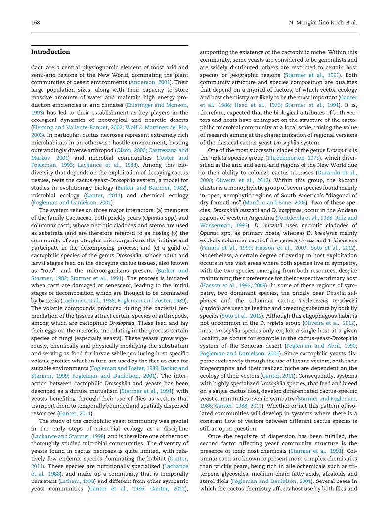
Saprochaete clavata (Fig 1A; see also Kurtzman and Robnett,
1995), and Rhodospiridium fluviale, which clustered with Spor-
idiobolus microsporus as well (Fig 1B). Strains were eventually
assigned to M. spicifer and R. fluviale due to their ability to
assimilate D-xylose (Smith & Poot, 1998) and maltose (Fell
et al., 1988, 1998), respectively.
From the 21 taxa identified (Fig 1), 20 were fungi and one
was a green alga of the genus Prototheca. Within the fungi, 17
isolateswere assigned to the phylumAscomycota and three to
the Basidiomycota, a pattern of biodiversity consistent with
previous surveys of the microbiota associated with rotting
cacti (Lachance et al., 1988). Despite isolating a relatively high
proportion of filamentous fungi, adding up to more than one
third of all species, most of these were taken to represent
opportunistic or accidental infections (see below for a detailed
discussion), and are therefore not likely to be members of the
cactophilic community. The only exception to this was Fusa-
rium lunatum, which was present in more than two thirds of
the sampled necroses of both O. sulphurea and T. terscheckii
(Table 2), always in high densities andmost of the times being
the dominant organism present.
Isolates that clustered within the group identified as F.
lunatum were the only ones to show a certain degree of phe-
notypic variability. These differed widely in the amount of red
pigment produced, as well as in the onset of its production.
Likewise, they differed in their ability to assimilate xylitol,
sorbitol, N-acetyl-glucosamine and raffinose. However, the
26S rDNA sequences of these strains were very similar, and
they all grouped with the sequence of the type strain of the
species (Fig 1A). Otherwise, the only isolated strain that dif-
fered from the physiological profile of the species to which it
was phylogenetically assigned was Prototheca zopfii. Despite
being clearly nested within the P. zopfii clade (Fig 1C), strains
recovered from cactus rots possessed the ability to grow on a
medium containing galactose as the sole carbon source, con-
trary to all described species and varieties of the clade (Ueno
et al., 2005). Due to this characteristic, these isolates would
be assigned to P. stagnora in most keys of the genus (Arnold
and Ahearn, 1972; Padhye et al., 1979; Pore, 1998, 1985; Ueno
et al., 2005).
Eleven species of saprobes did not fulfill any of the afore-
mentioned criteria designed to identify cactophilic micro-
organisms. These included Acremonium stromaticum, F.
oxysporum, Galactomyces candidum, Peyronellaea prosopidis,
Phoma opuntiae, M. ingens, M. capitatus, R. fluviale, Rhodotorula
sp. and amember each of the F. incarnatum-equiseti (FIESC) and
F. solani (FSSC) species complexes. All of these were only
recovered from 1 to 3 samples and always at very low den-
sities (Table 2), and were therefore not considered to be cac-
tophilic organisms. Similarly, two species of yeasts generally
considered as cactophilic, Tortispora phaffii and Candida sonor-
ensis (Lachance and Kurtzman, 2013; Ganter, 2011), were also
not considered to be active members of the community under
study, given that they were recovered from only one cactus
sample each.
The cactophilic community of Valle F�ertil was, therefore,
defined to consist of eight species of organisms: Cryptococcus
terrestris, Dipodascus australiensis, F. lunatum, M. spicifer, Pichia
cactophila, P. zopfii, Sporopachydermia cereana ‘australis’ and
Yarrowia deformans. These eight species added up to 78 % of all
isolates. Most of these organisms had already been described
as cactophilic species (Ganter, 2011), except for F. lunatum, Y.
deformans and C. terrestris, which represent novel members of
this community. From these eight cactophilic species, Pi. cac-
tophila, F. lunatum, P. zopfii, Y. deformans, M. spicifer and Di.
australiensis were recovered from both cactus hosts, although
the last two had much higher incidences in Opuntia samples.
The remaining species, S. cereana and C. terrestris, were iso-
lated exclusively from samples of O. sulphurea.
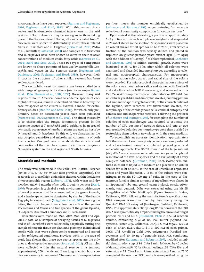
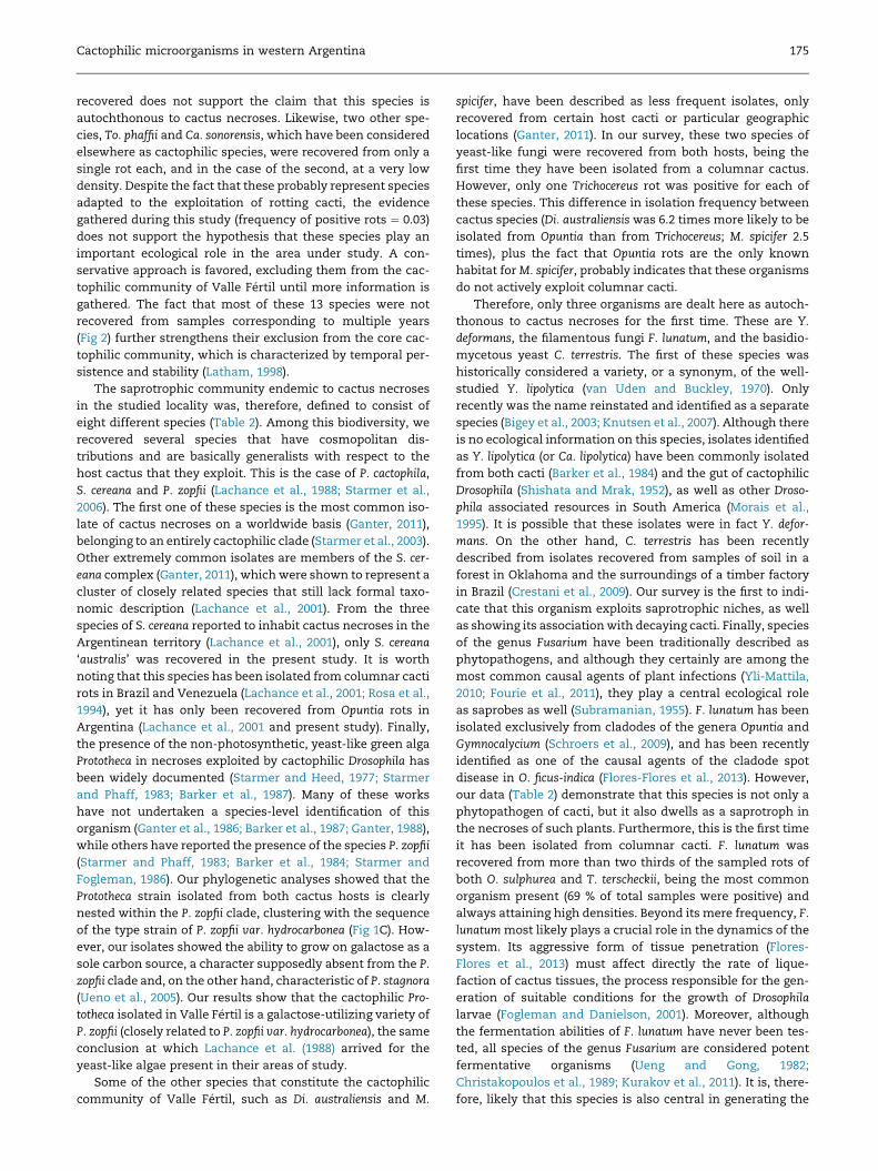
The presence of most cactophilic species was largely con-
stant through successive samplings, with similar frequencies
of isolation in samples from different years (Fig 2). The only
exceptions to this were the absence ofM. spicifer from samples
of 2012, and the exclusive presence of Y. deformans in samples
of 2014. On the contrary, almost all species considered
allochthonous to cactus necroses were only found in samples
corresponding to a particular year, reinforcing the hypothesis
that they represent fortuitous infections. The only non-
cactophilic species to be isolated from samples correspond-
ing to successive years were Ph. opuntiae, an Opuntia endo-
phyte, and F. oxysporum, an extremely common member of

172 N. Mongiardino Koch et al.
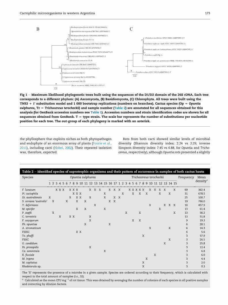
Fig 1 e Maximum likelihood phylogenetic trees built using the sequences of the D1/D2 domain of the 26S rDNA. Each tree
corresponds to a different phylum: (A) Ascomycota, (B) Basidiomycota, (C) Chlorophyta. All trees were built using the
TN93 D G substitution model and 1 000 bootstrap replications (numbers on branches). Cactus species (Op [ Opuntia
sulphurea, Tr [ Trichocereus terscheckii) and sample number (Table 2) are annotated for all sequences obtained for this
analysis (for GenBank accession numbers see Table 1). Accession numbers and strain identification codes are shown for all
sequences obtained from GenBank. T [ type strain. The scale bar represents the number of substitutions per nucleotide
position for each tree. The out-group of each phylogeny is marked with an asterisk.
Cactophilic microorganisms in western Argentina 173
the phyllosphere that exploits niches as both phytopathogen
and endophyte of an enormous array of plants (Fourie et al.,
2011), including cacti (Nobel, 2002). Their repeated isolation
was, therefore, expected.
Table 2 e Identified species of saprotrophic organisms and the
Species Opuntia sulphurea
1 2 3 4 5 6 7 8 9 10 11 12 13 14 15 16 17
F. lunatum X X X X X X X X X X X
Pi. cactophila X X X X
Di. australiensis X X X X X X X
S. cereana ‘australis’ X X X X X X
Y. deformans X
M. spicifer X X X
P. zopfii X
C. terrestris X X X X
F. oxysporum X
Ph. opuntiae X X
A. stromaticum
FIESC X X
To. phaffi
FSSC
G. candidum
Pe. prosopidis X
Ca. sonorensis X
R. fluviale
M. ingens
M. capitatus
Rhodotorula sp.
The ‘X’ represents the presence of a microbe in a given sample. Species
respect to the total amount of samples (i.e., 32).a Calculated as the mean CFUmg�1 of rot tissue. This was obtained by av
and correcting by dilution factors.
Rots from both cacti showed similar levels of microbial
diversity (Shannon diversity index: 2.24 vs 2.29; inverse
Simpson diversity index: 7.45 vs 6.88, for Opuntia and Tricho-
cereus, respectively), althoughOpuntia rots presented a slightly
ir pattern of occurrence in samples of both cactus hosts
Trichocereus terscheckii Frequency Meandensitya
1 2 3 4 5 6 7 8 9 10 11 12 13 14 15
X X X X X X X X X X X 69 362.4
X X X X X X 31 678.5
X 25 100.7
19 766.0
X X X X 16 457.9
X 13 61.4
X X X 13 56.2
13 51.8
X X 9 19.3
6 28.1
X X 6 14.3
6 5.6
X 3 57.9
X 3 26.5
X 3 25.8
3 12.4
3 6.8
X 3 6.0
X 3 4.4
X 3 2.0
X 3 0.3
are ordered according to their frequency, which is calculated with
eraging the number of colonies of each species in all positive samples
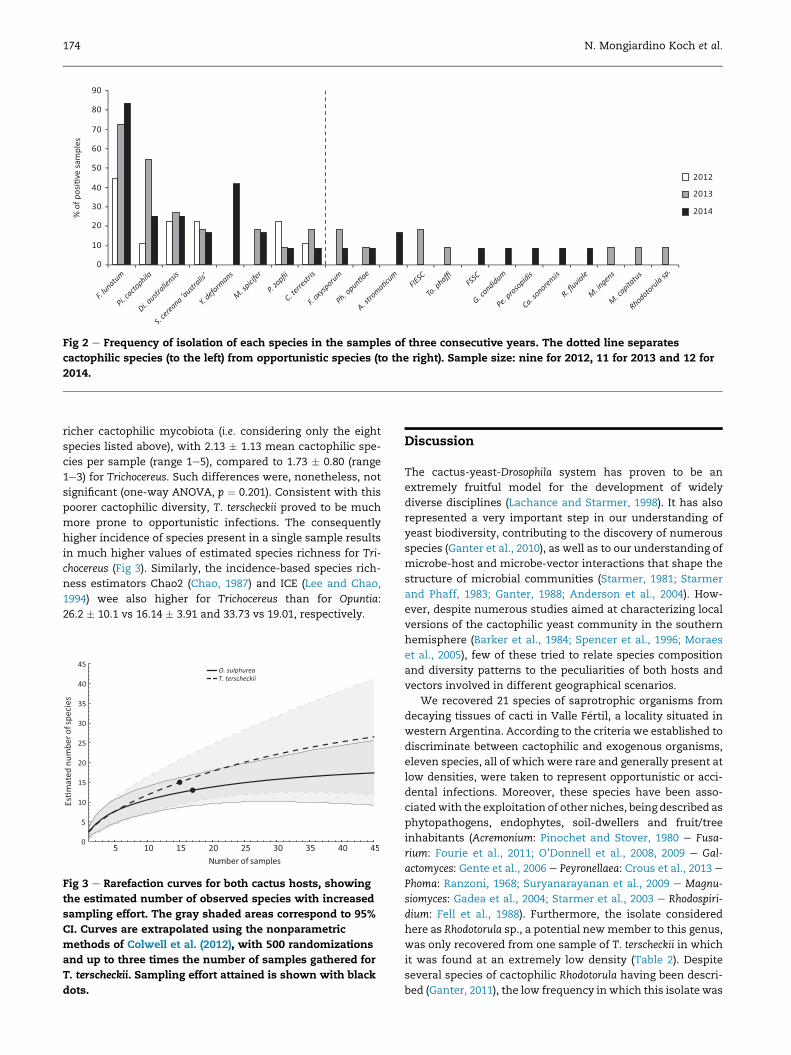
Fig 2 e Frequency of isolation of each species in the samples of three consecutive years. The dotted line separates
cactophilic species (to the left) from opportunistic species (to the right). Sample size: nine for 2012, 11 for 2013 and 12 for
2014.
174 N. Mongiardino Koch et al.
richer cactophilic mycobiota (i.e. considering only the eight
species listed above), with 2.13 � 1.13 mean cactophilic spe-
cies per sample (range 1e5), compared to 1.73 � 0.80 (range
1e3) for Trichocereus. Such differences were, nonetheless, not
significant (one-way ANOVA, p ¼ 0.201). Consistent with this
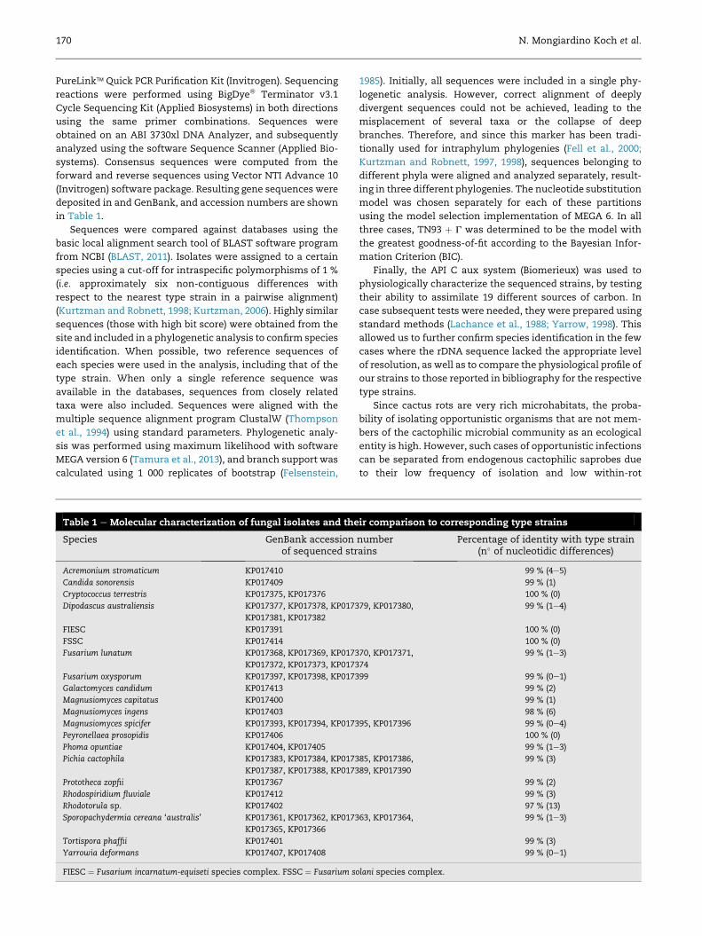
poorer cactophilic diversity, T. terscheckii proved to be much
more prone to opportunistic infections. The consequently
higher incidence of species present in a single sample results
in much higher values of estimated species richness for Tri-
chocereus (Fig 3). Similarly, the incidence-based species rich-
ness estimators Chao2 (Chao, 1987) and ICE (Lee and Chao,
1994) wee also higher for Trichocereus than for Opuntia:
26.2 � 10.1 vs 16.14 � 3.91 and 33.73 vs 19.01, respectively.
Fig 3 e Rarefaction curves for both cactus hosts, showing
the estimated number of observed species with increased
sampling effort. The gray shaded areas correspond to 95%
CI. Curves are extrapolated using the nonparametric
methods of Colwell et al. (2012), with 500 randomizations
and up to three times the number of samples gathered for
T. terscheckii. Sampling effort attained is shown with black
dots.
Discussion
The cactus-yeast-Drosophila system has proven to be an
extremely fruitful model for the development of widely
diverse disciplines (Lachance and Starmer, 1998). It has also
represented a very important step in our understanding of
yeast biodiversity, contributing to the discovery of numerous
species (Ganter et al., 2010), as well as to our understanding of
microbe-host and microbe-vector interactions that shape the
structure of microbial communities (Starmer, 1981; Starmer
and Phaff, 1983; Ganter, 1988; Anderson et al., 2004). How-
ever, despite numerous studies aimed at characterizing local
versions of the cactophilic yeast community in the southern
hemisphere (Barker et al., 1984; Spencer et al., 1996; Moraes
et al., 2005), few of these tried to relate species composition
and diversity patterns to the peculiarities of both hosts and
vectors involved in different geographical scenarios.
We recovered 21 species of saprotrophic organisms from
decaying tissues of cacti in Valle F�ertil, a locality situated in
western Argentina. According to the criteria we established to
discriminate between cactophilic and exogenous organisms,
eleven species, all of which were rare and generally present at
low densities, were taken to represent opportunistic or acci-
dental infections. Moreover, these species have been asso-
ciatedwith the exploitation of other niches, being described as
phytopathogens, endophytes, soil-dwellers and fruit/tree
inhabitants (Acremonium: Pinochet and Stover, 1980 e Fusa-
rium: Fourie et al., 2011; O’Donnell et al., 2008, 2009 e Gal-
actomyces: Gente et al., 2006 e Peyronellaea: Crous et al., 2013 e
Phoma: Ranzoni, 1968; Suryanarayanan et al., 2009 e Magnu-
siomyces: Gadea et al., 2004; Starmer et al., 2003 e Rhodospiri-
dium: Fell et al., 1988). Furthermore, the isolate considered
here as Rhodotorula sp., a potential newmember to this genus,
was only recovered from one sample of T. terscheckii in which
it was found at an extremely low density (Table 2). Despite
several species of cactophilic Rhodotorula having been descri-
bed (Ganter, 2011), the low frequency in which this isolate was
Cactophilic microorganisms in western Argentina 175
recovered does not support the claim that this species is
autochthonous to cactus necroses. Likewise, two other spe-
cies, To. phaffii and Ca. sonorensis, which have been considered
elsewhere as cactophilic species, were recovered from only a
single rot each, and in the case of the second, at a very low
density. Despite the fact that these probably represent species
adapted to the exploitation of rotting cacti, the evidence
gathered during this study (frequency of positive rots ¼ 0.03)
does not support the hypothesis that these species play an
important ecological role in the area under study. A con-
servative approach is favored, excluding them from the cac-
tophilic community of Valle F�ertil until more information is
gathered. The fact that most of these 13 species were not
recovered from samples corresponding to multiple years
(Fig 2) further strengthens their exclusion from the core cac-
tophilic community, which is characterized by temporal per-
sistence and stability (Latham, 1998).
The saprotrophic community endemic to cactus necroses
in the studied locality was, therefore, defined to consist of
eight different species (Table 2). Among this biodiversity, we
recovered several species that have cosmopolitan dis-
tributions and are basically generalists with respect to the
host cactus that they exploit. This is the case of P. cactophila,
S. cereana and P. zopfii (Lachance et al., 1988; Starmer et al.,
2006). The first one of these species is the most common iso-
late of cactus necroses on a worldwide basis (Ganter, 2011),
belonging to an entirely cactophilic clade (Starmer et al., 2003).
Other extremely common isolates are members of the S. cer-
eana complex (Ganter, 2011), which were shown to represent a
cluster of closely related species that still lack formal taxo-
nomic description (Lachance et al., 2001). From the three
species of S. cereana reported to inhabit cactus necroses in the
Argentinean territory (Lachance et al., 2001), only S. cereana
‘australis’ was recovered in the present study. It is worth
noting that this species has been isolated from columnar cacti
rots in Brazil and Venezuela (Lachance et al., 2001; Rosa et al.,
1994), yet it has only been recovered from Opuntia rots in
Argentina (Lachance et al., 2001 and present study). Finally,
the presence of the non-photosynthetic, yeast-like green alga
Prototheca in necroses exploited by cactophilic Drosophila has
been widely documented (Starmer and Heed, 1977; Starmer
and Phaff, 1983; Barker et al., 1987). Many of these works
have not undertaken a species-level identification of this
organism (Ganter et al., 1986; Barker et al., 1987; Ganter, 1988),
while others have reported the presence of the species P. zopfii
(Starmer and Phaff, 1983; Barker et al., 1984; Starmer and
Fogleman, 1986). Our phylogenetic analyses showed that the
Prototheca strain isolated from both cactus hosts is clearly
nested within the P. zopfii clade, clustering with the sequence
of the type strain of P. zopfii var. hydrocarbonea (Fig 1C). How-
ever, our isolates showed the ability to grow on galactose as a
sole carbon source, a character supposedly absent from the P.
zopfii clade and, on the other hand, characteristic of P. stagnora
(Ueno et al., 2005). Our results show that the cactophilic Pro-
totheca isolated in Valle F�ertil is a galactose-utilizing variety of
P. zopfii (closely related to P. zopfii var. hydrocarbonea), the same
conclusion at which Lachance et al. (1988) arrived for the
yeast-like algae present in their areas of study.
Some of the other species that constitute the cactophilic
community of Valle F�ertil, such as Di. australiensis and M.
spicifer, have been described as less frequent isolates, only
recovered from certain host cacti or particular geographic
locations (Ganter, 2011). In our survey, these two species of
yeast-like fungi were recovered from both hosts, being the
first time they have been isolated from a columnar cactus.
However, only one Trichocereus rot was positive for each of
these species. This difference in isolation frequency between
cactus species (Di. australiensis was 6.2 times more likely to be
isolated from Opuntia than from Trichocereus; M. spicifer 2.5
times), plus the fact that Opuntia rots are the only known
habitat forM. spicifer, probably indicates that these organisms
do not actively exploit columnar cacti.
Therefore, only three organisms are dealt here as autoch-
thonous to cactus necroses for the first time. These are Y.
deformans, the filamentous fungi F. lunatum, and the basidio-
mycetous yeast C. terrestris. The first of these species was
historically considered a variety, or a synonym, of the well-
studied Y. lipolytica (van Uden and Buckley, 1970). Only
recently was the name reinstated and identified as a separate
species (Bigey et al., 2003; Knutsen et al., 2007). Although there
is no ecological information on this species, isolates identified
as Y. lipolytica (or Ca. lipolytica) have been commonly isolated
from both cacti (Barker et al., 1984) and the gut of cactophilic
Drosophila (Shishata and Mrak, 1952), as well as other Droso-
phila associated resources in South America (Morais et al.,
1995). It is possible that these isolates were in fact Y. defor-
mans. On the other hand, C. terrestris has been recently
described from isolates recovered from samples of soil in a
forest in Oklahoma and the surroundings of a timber factory
in Brazil (Crestani et al., 2009). Our survey is the first to indi-
cate that this organism exploits saprotrophic niches, as well
as showing its associationwith decaying cacti. Finally, species
of the genus Fusarium have been traditionally described as
phytopathogens, and although they certainly are among the
most common causal agents of plant infections (Yli-Mattila,
2010; Fourie et al., 2011), they play a central ecological role
as saprobes as well (Subramanian, 1955). F. lunatum has been
isolated exclusively from cladodes of the genera Opuntia and
Gymnocalycium (Schroers et al., 2009), and has been recently
identified as one of the causal agents of the cladode spot
disease in O. ficus-indica (Flores-Flores et al., 2013). However,
our data (Table 2) demonstrate that this species is not only a
phytopathogen of cacti, but it also dwells as a saprotroph in
the necroses of such plants. Furthermore, this is the first time
it has been isolated from columnar cacti. F. lunatum was
recovered from more than two thirds of the sampled rots of
both O. sulphurea and T. terscheckii, being the most common
organism present (69 % of total samples were positive) and
always attaining high densities. Beyond its mere frequency, F.
lunatummost likely plays a crucial role in the dynamics of the
system. Its aggressive form of tissue penetration (Flores-
Flores et al., 2013) must affect directly the rate of lique-
faction of cactus tissues, the process responsible for the gen-
eration of suitable conditions for the growth of Drosophila
larvae (Fogleman and Danielson, 2001). Moreover, although
the fermentation abilities of F. lunatum have never been tes-
ted, all species of the genus Fusarium are considered potent
fermentative organisms (Ueng and Gong, 1982;
Christakopoulos et al., 1989; Kurakov et al., 2011). It is, there-
fore, likely that this species is also central in generating the
176 N. Mongiardino Koch et al.
volatile patterns that initially attract cactophilic Drosophila to
the necrosis. Finally, the vectoring of other Fusarium species
by Drosophila has also been demonstrated (Swart and Swart,
2003). Although no filamentous fungus has ever been
acknowledged as being native to the cactophilic habitat, there
is no good reason why yeast and yeast-like fungi should be
considered as the exclusive eukaryote inhabitants of this
particular environment. In a similar vein as has been
expressed above, Lachance et al. (1988) proposed the inclusion
of Geotrichum species to the cactophilic community mainly by
citing their frequency in necroses, their potential as phyto-
pathogens and their use of Drosophila as vectors.
Although diversity indexes rendered similar values for
both hosts, the mycobiota associated with Trichocereus had
much higher values of expected species richness, as shown by
the values of Chao2 and ICE estimators. This can be attributed
to the fact that these numbers (as well as the asymptotic
values for the curves in Fig 3) are largely based on the proba-
bility of isolating further infrequent species. As a whole, the
community of T. terscheckii was found to consist of fewer
cactophilic species, while at the same time having a higher
incidence of opportunistic and infrequent inhabitants. From
the 15 species isolated from T. terscheckii, 60 % were found in
only one sample; a value that drops to 38 % for samples of O.
sulphurea. The consequences that this depauperate and less
predictable microbiota can have for the biology of the asso-
ciatedDrosophila remains to be explored. However, despite not
attaining sampling efforts that guaranteed the saturation of
rarefaction curves, especially in the case of T. terscheckii, the
cactophilic community of both resources is likely to be cor-
rectly characterized, with unobserved species representing
further exogenous infections. This is validated by the repeat-
ability of isolation of cactophilic species (Fig 2), as well as the
meeting of the sampling criterion for cactus necroses estab-
lished by Lachance and Starmer (1998) (it should be noted as
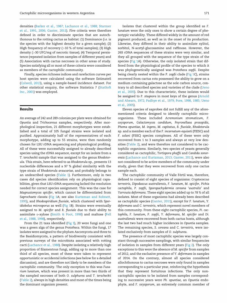
Fig 4 e Representation of the cactophilic saprotrophic
community of both host plants. The community of T.
terscheckii is completely nested within that of O. sulphurea.
The placement of M. spicifer and Di. australiensis as
exclusive inhabitants of T. terscheckii is highly probable yet
not conclusive, and they are therefore marked with an
asterisk.
well that the number of isolated species lies within the 95 % CI
at saturation values for both hosts, see the black dots in Fig 3).
As already discussed, analysis of the pattern of occurrence
of the eight cactophilic species in both cactus hosts under
study, revealed an interesting pattern. All of the species were
found in the decaying tissues of O. sulphurea, while only a
subset of these was present in T. terscheckii (Fig 4). Such a
pattern of nested biodiversity has not been described, to our
knowledge, in any study that characterized the cactophilic
communities associated with different cactus species living in
sympatry. Since the presence of a microbe in a given host
depends, in the first place, on the vectoring processes per-
formed by cactophilic Drosophila, and secondly on the chem-
istry of the host cactus (Starmer et al., 1991), we propose that
the particular biological characteristics of both hosts and
vectors that constitute the cactus-yeast-Drosophila system in
western Argentina are causing this peculiar pattern of nest-
edness. First of all, the shared use of hosts between both
species of cactophilic Drosophila in this region is quite
uncommon, and clearly differs from the characteristic spe-
cificity of other studied systems (Fellows andHeed, 1972; Heed
et al., 1976). This constant flow of flies between cactus species
most likely results in the homogenization of the associated
mycobiotas. Secondly, the more complex chemistry of T. ter-
scheckii might be involved in the selection of a limited subset
of organisms capable of tolerating that particular environ-
ment. For example, O. sulpurea and T. terscheckii differ in their
relative concentrations of medium-chain fatty acids (Carreira
et al., 2014; Padr�o and Soto, 2013), compounds that are known
to be inhibitors of yeast growth (Starmer, 1982; Starmer and
Fogleman, 1986). Although further empirical corroboration is
required, these two processes acting simultaneously may be
responsible for the nested structure of cactophilic microbial
diversity (Fig 4). The possibility that the absence (or low fre-
quency) of these four saprobe species from T. terscheckii rots is
spurious and a result of sampling bias is low, given the rela-
tively high frequency in which, for example, S. cereana ‘aus-
tralis’ and Di. australiensis were found in Opuntia rots. This
pattern is further validated by previous works, since S. cereana
‘australis’ has been exclusively isolated from Opuntia samples
in Argentina (Lachance et al., 2001), while Opuntia rots are the
only known natural habitat of M. spicifer (de Hoog et al., 1986),
as well as themost common forDi. australiensis (von Arx, 1997;
Ganter, 2011). Furthermore, the nested pattern is even robust
to whether or notM. spicifer andDi. australiensis are considered
to inhabit exclusively Opuntia rots.
Ganter (2011) argued that the yeast’s use of arthropod
vectors for dispersal is the reason theymanaged to escape the
‘everything is everywhere, the environment selects’ pattern of
microbial spatial distribution. The reason for this is that such
a relationship results in the microorganism’s biogeography
resembling that of its vector. It was, therefore, expected that a
system consisting of two vectors that have the ability to
exploit both available hosts will result in these hosts devel-
oping highly similar microbial communities. On the other
hand, several studies have pointed out that host chemistry
may be more important than the activity of the vector in
determining yeast community structure (Ganter et al., 1986;
Starmer et al., 1980), although the covariation between host
species and vector species may result in a confounding effect

Cactophilic microorganisms in western Argentina 177
on the relative importance of these two processes (Ganter
et al., 1986; Starmer, 1981). The lack of strict host specificity
in our site of study may effectively decouple both processes
and allow for a reevaluation of their importance for deter-
mining species composition in the associated microbial
communities. So far, both factors seem to play equally
essential roles.
Despite the intense study that this model system has
received, many of the cactus-dominated arid regions of the
NewWorld are still poorly studied. In those regions, analogous
communities to that present in the Sonoran desert may pro-
vide novel insights into the ecological and evolutionary
dynamics of microbe-vector and microbe-host interactions.
So far, the study of the system present in western Argentina
has resulted in the discovery of some unique qualities,
including an important role of filamentous fungi and a nested
pattern of microbial biodiversity in distinct sympatric hosts.
The identification of new species of cactophilic organismswas
also possible, and the necrotic tissues of cacti demonstrated
they still represent a source of unknown yeast species. The
characterization of the saprotrophic community of cactus
necroses exploited by members of the D. buzzatii cluster
effectively adds a new dimension to the ecology and evolu-
tionary history of these flies, considered to be model organ-
isms for studies in evolutionary biology.
Acknowledgments
The authors wish to thank J. Padr�o and P. Fontanarrosa for
help with the collection of samples, P. D. Mc Cargo for assis-
tance during molecular characterization of strains, and two
anonymous reviewers whose comments greatly improved the
present manuscript. This work was supported by grants to
IMS, MG and LI from ANPCyT (PICT 2011-1527 and 2013-1506)
and Universidad de Buenos Aires. NMK is a student fellow of
the Universidad de Buenos Aires. IMS, MG, EH and LI are
members of Carrera del Investigador Cient�ıfico (CONICET).
r e f e r e n c e s
Anderson, E.F., 2001. The Cactus Family. Timber Press, Portland,Oregon, USA.
Anderson, M.T., Lachance, M.-A., Starmer, W.T., 2004. Therelationship of phylogeny to community structure: the cactusyeast community. American Naturalist 164, 709e721.
Arnold, P., Ahearn, D.G., 1972. The systematics of the genusPrototheca with a description of a new species, P. filamenta.Mycologia 64, 265e276.
Barker, J.S.F., East, P.D., Phaff, H.J., Miranda, M., 1984. The ecologyof the yeast flora in necrotic Opuntia cacti and of associatedDrosophila in Australia. Microbiology Ecology 10, 379e399.
Barker, J.S.F., Starmer, W.T. (Eds.), 1982. The Cactus-yeast-Drosophila Model System. Academic Press, New York.
Barker, J.S.F., Starmer, W.T., 1999. Environmental effects and thegenetics of oviposition site preference for natural yeastsubstrates in Drosophila buzzatii. Hereditas 130, 145e175.
Barker, J.S.F., Starmer, W.T., Vacek, D.C., 1987. Analysis of spatialand temporal variation in the community structure of yeasts
associated with decaying Opuntia cactus. Microbial Ecology 14,261e276.
Bigey, F., Tuery, K., Bougard, D., Nicaud, J.M., Moulin, G., 2003.Identification of a triacylglycerol lipase gene family in Candidadeformans: molecular cloning and functional expression. Yeast20, 233e248.
BLAST, 2011. Basic Local Alignment Search Tool. InternetResource: http://blast.ncbi.nlm.nih.gov/Blast.cgi.
Cabrera, A.L., 1971. Fitogeograf�ıa de la Rep�ublica Argentina.Bulletin of the Botanical Society of Argentina 14, 1e42.
Carreira, V.P., Padr�o, J., Mongiardino Koch, N., Fontanarrosa, P.,Alonso, J.I., Soto, I.M., 2014. Nutritional composition of Opuntiasulphurea (G. Don in Loudon) cladodes. Haseltonia 19, 38e45.
Castrezana, S., Markov, T.A., 2001. Arthropod diversity in necrotictissue of three species of columnar cacti (Cactaceae). CanadianEntomologist 133, 301e309.
Chao, A., 1987. Estimating the population size for capture-recapture data with unequal catchability. Biometrics 43,783e791.
Christakopoulos, P., Macris, B.J., Kekos, D., 1989. Directfermentation of cellulose to ethanol by Fusarium oxysporum.Enzyme and Microbial Technology 11, 236e239.
Colwell, R.K., 2013. EstimateS: Statistical Estimation of SpeciesRichness and Shared Species from Samples. Version 9.Persistent URL: http://purl.oclc.org/estimates/.
Colwell, R.K., Chao, A., Gotelli, N.J., Lin, S.-Y., Mao, C.X.,Chazdon, R.L., Longino, J.T., 2012. Models and estimatorslinking individual-based and sample-based rarefaction,extrapolation, and comparison of assemblages. Journal of PlantEcology 5, 3e21.
Corio, C., Soto, I.M., Carreira, V., Padr�o, J., Betti, M.I.L., Hasson, E.,2013. An alkaloid fraction extracted from the cactusTrichocereus terscheckii affects fitness in the cactophilic flyDrosophila buzzatii (Diptera: Drosophilidae). Biological Journal ofthe Linnean Society 109 (2), 342e353.
Crestani, J., Landell, M.F., Faganello, J., Vainstein, M.H.,Vishniac, H.S., Valente, P., 2009. Cryptococcus terrestris sp. nov.,a tremellaceous, anamorphic yeast phylogenetically related toCryptococcus flavescens. International Journal of Systematic andEvolutionary Microbiology 59, 631e636.
Crous, P.W., Wingfield, M.J., Guarro, J., Cheewangkoon, R., van derBank, M., et al., 2013. Fungal planet description sheets:154e213. Persoonia 31, 188e296.
de Hoog, G.S., Smith, M.T., Gu�eho, E., 1986. A revision of the genusGeotrichum and its telomorphs. Studies in Mycology 29, 1e131.
Durando, C.M., Baker, R.H., Etges, W.J., Heed,W.B., Wasserman, M.,DeSalle, R., 2000. Phylogenetic analysis of the replete speciesgroup of the genus Drosophila using multiple sources ofcharacters. Molecular Phylogenetics and Evolution 16, 296e307.
Ehleringer, J.R., Monson, R.K., 1993. Evolutionary and ecologicalaspects of photosynthetic pathway variation. Annual Review ofEcology, Evolution, and Systematics 24, 411e439.
Fanara, J.J., Fontdevila, A., Hasson, E., 1999. Ovipositionpreference and life history traits in cactophilic Drosophilakoepferae and D. buzzatii in association with their naturalhosts. Ecology and Evolution 13, 173e190.
Fell, J.W., Blatt, G.M., Statzell-Tallman, A., 1998. Validation of thebasidiomycetous yeast, Sporidiobolus microsporus sp. nov.,based on phenotypic and molecular analyses. Antonie vanLeeuwenhoek 74, 265e270.
Fell, J.W., Boekhout, T., Fonseca, A., Scorzetti, G., Statzell-Tallman, A., 2000. Biodiversity and systematics ofbasidiomycetous yeasts as determined by large-subunit rDNAD1/D2 domain sequence analysis. International Journal ofSystematic and Evolutionary Microbiology 50, 1351e1371.
Fell, J.W., Kurtzman, C.P., Tallman, A.S., Buck, J.D., 1988.Rhodospiridium fluviale sp. nov., a homokaryotic red yeast froma subtropical brackish environment. Mycologia 80, 560e564.
178 N. Mongiardino Koch et al.
Fellows, D.P., Heed, W.B., 1972. Factors affecting host plantselection in desert-adapted cactiphilic Drosophila. Ecology 53,850e858.
Felsenstein, J., 1985. Confidence limits on phylogenies: anapproach using the bootstrap. Evolution 39, 783e791.
Fleming, T.H., Valiente-Banuet, A. (Eds.), 2002. Columnar Cactiand Their Mutualists: Evolution, Ecology, and Conservation.University of Arizona Press, Tucson.
Flores-Flores, R., Vel�azquez-del Valle, M.G., Le�on-Rodriguez, R.,Flores-Moctezuma, H.E., Hern�andez-Lauzardo, A.N., 2013.Identification of fungal species associated with cladode spot ofprickly pear and their sensitivity to chitosan. Journal ofPhytopathology 161, 544e552.
Fogleman, J.C., Abril, J.R., 1990. Ecological and evolutionaryimportance of host plant chemistry. In: Barker, J.S.F.,Starmer, W.T., MacIntyre, R.J. (Eds.), Ecological andEvolutionary Genetics of Drosophila. Plenum Press, New York,pp. 121e143.
Fogleman, J.C., Danielson, P.B., 2001. Chemical interactions in thecactus-microorganism-Drosophila model system of theSonoran Desert. American Zoologist 41, 877e889.
Fogleman, J.C., Heed, W.B., 1989. Columnar cacti and desertDrosophila: the chemistry of host plant specificity. In:Schmidt, J. (Ed.), Special Biotic Relationships in the AridSouthwest. University of New Mexico Press, Albuquerque,pp. 1e24.
Fogleman, J.C., Foster, J.L.M., 1989. Microbial colonization ofinjured cactus tissue (Stenocereus gummosus) and itsrelationship to the ecology of cactophilic Drosophila mojavensis.Applied and Environmental Microbiology 55, 100e105.
Fontdevila, A., Pla, C., Hasson, E., Wasserman, M., Sanchez, A.,Naveira, H., Ruiz, A., 1988. Drosophila koepferae: a new memberof the Drosophila serido (Diptera, Drosophilidae) superspecietaxon. Annals of the Entomological Society of America 81, 380e385.
Foster, J.L.M., Fogleman, J.C., 1993. Identification and ecology ofbacterial communities associated with necrosis of threecactus species. Applied and Environmental Microbiology 59, 1e6.
Fourie, G., Steenkamp, E.T., Ploetz, R.C., Gordon, T.R., Viljoen, A.,2011. Current status of the taxonomic position of Fusariumoxysporum formae specialis cubense within the Fusariumoxysporum complex. Infection, Genetics and Evolution 11, 533e542.
Gadea, I., Cuenca-Estrella, M., Prieto, E., Diaz-Guerra, T.M.,Garcia-Cia, J.I., Mellado, E., Tomas, J.F., Rodriguez-Tudela, J.L.,2004. Genotyping and antifungal susceptibility profile ofDipodascus capitatus isolates causing disseminated infection inseven hematological patients of a tertiary hospital. Journal ofClinical Microbiology 42, 1832e1836.
Ganter, P.F., 1988. The vectoring of cactophilic yeasts byDrosophila. Oecologia 75, 400e404.
Ganter, P.F., 2011. Everything is not everywhere: the distributionof cactophilic yeast. In: Fontaneto, D. (Ed.), Biogeography ofMicroscopic Organisms. Cambridge University Press,Cambridge, pp. 130e176.
Ganter, P.F., Cardinali, G., Boundy-Mills, K., 2010. Pichia insulanasp. nov., a novel cactophilic yeast from the Caribbean.International Journal of Systematic and Evolutionary Microbiology60, 1001e1007.
Ganter, P.F., Starmer, W.T., Lachance, M.-A., Phaff, H.J., 1986.Yeast communities from host plants and associated Drosophilain southern Arizona: new isolations and analysis of therelative importance of hosts and vectors on communitycomposition. Oecologia 70, 386e392.
Gente, S., Sohier, D., Coton, E., Duhamel, C., Gueguen, M., 2006.Identification of Geotrichum candidum at the species and strainlevel: proposal for a standardized protocol. Journal of IndustrialMicrobiology and Biotechnology 33, 1019e1031.
Hasson, E., Soto, I.M., Carreira, V.P., Corio, C., Soto, E.M.,Betti, M.I.L., 2009. Host plants, fitness and developmental
instability in a guild of cactophilic species of the genusDrosophila. In: Santos, E.B. (Ed.), EcotoxicologyResearch Developments. Nova Publishers, New York,pp. 89e109.
Hasson, E., Naveira, H., Fontdevila, A., 1992. The breeding sites ofthe Argentinian species of the Drosophila mulleri complex(subgenus Drosophila repleta group). Revista Chilena De HistoriaNatural 65, 319e326.
Heed, W.B., Starmer, W.T., Miranda, M., Miller, M.W., Phaff, H.J.,1976. An analysis of the yeast flora associated with cactiphilicDrosophila and their host plants in the Sonoran Desert and itsrelation to temperature and tropical associations. Ecology 57,151e160.
Knutsen, A.K., Robert, V., Poot, G.A., Epping, W., Figge, M., Holst-Jensen, A., Skaar, I., Smith, M.T., 2007. Polyphasicreexamination of Yarrowia lipolytica strains and thedescription of three new Candida species: C. oslonensis, C.alimentaria and C. hollandica. International journal of systematicand evolutionary microbiology 57, 2426e2435.
Kurakov, A.V., Khidirov, K.S., Sadykova, V.S., Zvyagintsev, D.G.,2011. Anaerobic growth ability and alcohol fermentationactivity of microscopic fungi. Applied Biochemistry andMicrobiology 47 (2), 169e175.
Kurtzman, C.P., 2006. Yeast species recognition from genesequence analyses and other molecular methods. Mycoscience74, 65e71.
Kurtzman, C.P., Robnett, C.J., 1995. Molecular relationshipsamong hyphal ascomycetous yeasts and yeastlike taxa.Canadian Journal of Botany 73, 824e830.
Kurtzman, C.P., Robnett, C.J., 1997. Identification of clinicallyimportant ascomycetous yeasts based on nucleotidedivergence in the 50 end of the large-subunit (26S) ribosomalDNA gene. Journal of Clinical Microbiology 35, 1216e1223.
Kurtzman, C.P., Robnett, C.J., 1998. Identification and phylogenyof ascomycetous yeasts from analysis of nuclear large subunit(26S) ribosomal DNA partial sequences. Antonie vanLeeuwenhoek 73, 331e371.
Lachance, M.-A., Kaden, J.E., Phaff, H.J., Starmer, W.T., 2001.Phylogenetic structure of the Sporopachydermia cereana speciescomplex. International Journal of Systematic and EvolutionaryMicrobiology 51, 237e247.
Lachance, M.-A., Kurtzman, C.P., 2013. The yeast genusTortispora gen. nov., description of Tortispora ganteri sp. nov.,Tortispora mauiana f.a., sp. nov., Tortispora agaves f.a., sp.nov., Tortispora sangerardonensis f.a., sp. nov., Tortisporacuajiniquilana f.a., sp. nov., Tortispora starmeri f.a., sp. nov.,and Tortispora phaffii f.a., sp. nov., reassignment of Candidacaseinolytica to Tortispora caseinolytica f.a., comb. nov.,emendation of Botryozyma, and assignment of Botryozyma,Tortispora gen. nov., and Trigonopsis to the familyTrigonopsidaceae fam. nov. International Journal of Systematicand Evolutionary Microbiology 63, 3104e3114.
Lachance, M.-A., Starmer, W.T., 1998. Ecology and yeasts. In:Kurtzman, C.P., Fell, J.W. (Eds.), The Yeasts: A TaxonomicStudy, fourth edn. Elsevier Science, Amsterdam, pp. 21e30.
Lachance, M.-A., Starmer, W.T., Phaff, H.J., 1988. Identification ofyeasts found in decaying cactus tissue. Canadian Journal ofMicrobiology 34, 1025e1036.
Latham, B.P., 1998. Yeast community persistence in a spatiallystructured environment. Microbial Ecology 36, 60e65.
Lee, S.-M., Chao, A., 1994. Estimating population size via samplecoverage for closed capture-recapture models. Biometrics 50,88e97.
Manfrin, M.H., Sene, F.M., 2006. Cactophilic Drosophila in SouthAmerica: a model for evolutionary studies. Genetica 126, 57e75.
Mirr�e, J.C., 1976. Descripci�on geol�ogica de la Hoja 19e, Valle F�ertil.Provincias de San Juan y La Rioja. Secretar�ıa de Estado deMiner�ıa, Buenos Aires. Bolet�ın N� 147.

Cactophilic microorganisms in western Argentina 179
Moraes, E.M., Rosa, C.A., Sene, F.M., 2005. Preliminary notes onyeasts associated with necrotic cactus stems from differentlocalities in Brazil. Brazilian Journal of Biology 65 (2), 299e304.
Morais, P.B., Martins, M.L., Klaczko, L.B., Mendonca-Hagler, L.C.,Hagler, A.N., 1995. Yeast succession in the amazon fruitParahancornia amapa as resource partitioning amongDrosophila spp. Applied and Environmental Microbiology 61,4251e4257.
Nobel, P.S. (Ed.), 2002. Cacti: Biology and Uses. University ofCalifornia Press, Berkley.
O’Donnell, K., 1993. Fusarium and its near relatives. In:Reynolds, D.R., Taylor, J.W. (Eds.), The Fungal Holomorph:Mitotic, Meiotic and Pleomorphic Speciation in FungalSystematic. CAB International, Wallingford, pp. 225e233.
O’Donnell, K., Sutton, D.A., Fothergill, A., McCarthy, D.,Rinaldi, M.G., Brandt, M.E., Zhang, N., Geiser, D.M., 2008.Molecular phylogenetic diversity, multilocus haplotypenomenclature, and in vitro antifungal resistance within theFusarium solani species complex. Journal of Clinical Microbiology46, 2477e2890.
O’Donnell, K., Sutton, D.A., Rinaldi, M.G., Gueidan, C., Crous, P.W.,Geiser, D.M., 2009. A novel multi-locus sequence typingscheme reveals high genetic diversity of human pathogenicmembers of the Fusarium incarnatum-F. equiseti and F.chlamydosporum species complexes within the U.S. Journal ofClinical Microbiology 47, 3851e3861.
Oliveira, D.C.S.G., Almeida, F.C., O’Grady, P.M., Armella, M.A.,DeSalle, R., Etges, W.J., 2012. Monophyly, divergence times,and evolution of host plant use inferred from a revisedphylogeny of the Drosophila repleta species group. MolecularPhylogenetics and Evolution 64, 533e544.
Olson, C.A., 2000. Insects and the saguaro. In: A Natural History ofthe Sonoran Desert. University of California Press, California,pp. 353e356. Arizona-Sonora Desert Museum.
Padhye, A., Baker, J., D’Amato, F., 1979. Rapid identification ofPrototheca species by the API 20C system. Journal of ClinicalMicrobiology 10, 579e582.
Padr�o, J., Soto, I.M., 2013. Exploration of the nutritional profile ofTrichocereus terscheckii (Parmentier) Britton & Rose stems.Journal of the Professional Association for Cactus Development 15,1e12.
Pinochet, J., Stover, R.H., 1980. Fung associated with nematodelessions on plantains in Honduras. Nematropica 10, 112e115.
Pore, R.S., 1985. Prototheca taxonomy. Mycopathologia 90, 129e139.Pore, R.S., 1998. Prototheca, a yeastlike alga. In: Kurtzman, C.P.,
Fell, J.W. (Eds.), The Yeasts: A Taxonomic Study, fourth edn.Elsevier Science, Amsterdam, pp. 883e888.
Ranzoni, F.V., 1968. Fungi isolated in culture from soils of theSonoran Desert. Mycologia 60, 356e371.
Roig-Ju~nent, S., Flores, G., Claver, S., Debandi, G., Marvaldi, A.,2001. Monte desert (Argentina): insect biodiversity and naturalareas. Journal of Arid Environment 47, 77e94.
Rosa, C.A., Morais, P.B., Hangler, A.N., Mendonca-Hangler, L.,Monteiro, R.F., 1994. Yeast communities of the cactusPilosocereus arrabidae and associated insects in the sandycoastal plains of southeastern Brazil. Antonie van Leeuwenhoek65, 55e62.
Ruiz, A., Wasserman, M., 1993. Evolutionary cytogenetics of theDrosophila buzzatii species complex. Heredity 70, 582e596.
Schroers, H.-J., O’Donnell, K., Lamprecht, S.C., Kammeyer, P.L.,Johnson, S., Sutton, D.A., Rinaldi, M.G., Geiser, D.M.,Summerbell, R.C., 2009. Taxonomy and phylogeny of theFusarium dimerum species group. Mycologia 101, 44e70.
Shishata, A.M.E.-T., Mrak, E.M., 1952. Intestinal yeast floras ofsuccessive populations of Drosophila. Evolution 6, 325e332.
Smith, M.T., Poot, G.A., 1998. Dipodascus capitatus, Dipodascusspicifer and Geotrichum clavatum: genomic characterization.Antonie van Leeuwenhoek 74, 229e235.
Soto, E., Goenaga, J., Hurtado, J., Hasson, E., 2012. Oviposition andperformance in natural hosts in cactophilic Drosophila. Ecologyand Evolution 26, 975e990.
Soto, I.M., Carreira, V.P., Corio, C., Padr�o, J., Soto, E.M., Hasson, E.,2014. Differences in tolerance to host cactus alkaloids inDrosophila koepferae and D. buzzatii. PLoS one 9, e88370.
Spencer, D.M., Spencer, J.F.T., de Figueroa, L.I., Garro, O.,Fengler, E., 1996. Yeasts associated with pods and exudates ofalgarrobo tres (Prosopis spp.) and species of columnar cacti innorthwest Argentina. Applied Microbiology and Biotechnology 44,736e739.
Starmer, W.T., 1981. A comparison of Drosophila habitatsaccording to the physiological attributes of the associatedyeast communities. Evolution 35, 38e52.
Starmer, W.T., 1982. Associations and interactions among yeasts,Drosophila and their habitats. In: Barker, J.S.F., Starmer, W.T.(Eds.), Ecological Genetics and Evolution: The Cactus-yeast-Drosophila Model System. Academic Press, Sydney,pp. 159e174.
Starmer, W.T., Aberdeen, V., Lachance, M.-A., 2006. Thebiogeographic diversity of cactophilic yeasts. In: P�eter, G.,Rosa, C.A. (Eds.), Biodiversity and Ecophysiology of Yeasts,The yeast handbook. Springer-Verlag, New York,pp. 485e499.
Starmer, W.T., Fogleman, J.C., 1986. Coadaptation of Drosophilaand yeasts in their natural habitat. Journal of Chemical Ecology12, 1037e1055.
Starmer, W.T., Fogleman, J.C., Lachance, M.-A., 1991. The yeastcommunity of cacti. In: Andrews, J.H., Hirano, S.S. (Eds.),Microbial Ecology of Leaves. Springer-Verlag, New York,pp. 158e178.
Starmer, W.T., Heed, W.B., 1977. The infection of Drosophilacultures by species of the genus Prototheca. DrosophilaInformation Service 52, 12.
Starmer, W.T., Kircher, H.W., Phaff, H.J., 1980. Evolution andspeciation of host plant specific yeasts. Evolution 34, 137e146.
Starmer, W.T., Lachance, M.-A., Phaff, H.J., 1987. A comparison ofyeast communities found in necrotic tissue of cladodes andfruits of Opuntia stricta on islands in the Caribbean Sea andwhere introduced into Australia. Microbial Ecology 14, 179e192.
Starmer, W.T., Lachance, M.-A., Phaff, H.J., Heed, W.B., 1990. Thebiogeography of yeasts associated with cactus tissue in NorthAmerica, the Caribbean and northern Venezuela. EvolutionaryBiology 24, 253e296.
Starmer, W.T., Phaff, H.J., 1983. Analysis of the communitystructure of yeasts associated with the decaying stems ofcactus. II. Opuntia species. Microbial Ecology 9, 247e259.
Starmer, W.T., Schmedicke, R.A., Lachance, M.-A., 2003. Theorigin of the cactus-yeast community. FEMS Yeast Research 3,441e448.
StatSoft, Inc, 2001. STATISTICA (Data Analysis Software System)version 6. Available at: www.statsoft.com.
Subramanian, C.V., 1955. The ecological and the taxonomicproblems in fusaria. Proceedings of the National Academy ofSciences B 41 (3), 102e109.
Suryanarayanan, T.S., Thirunavukkarasu, N.,Govindarajulu, M.B., Sasse, F., Jansen, R., Murali, T.S., 2009.Fungal endophytes and bioprospecting. Fungal Biology Review23, 9e19.
Swart, W.J., Swart, V.R., 2003. An overview of research on diseasesof cactus pear in South Africa. Journal of the ProfessionalAssociation for Cactus 5, 115e120.
Tamura, K., Stecher, G., Peterson, D., Filipski, A., Kumar, S., 2013.MEGA6: molecular evolutionary geneticsanalysis version 6.0.Molecular Biology and Evolution 30, 2725e2729.
Thompson, J.D., Higgins, D.G., Gibson, D.J., 1994. CLUSTAL W:improving the sensitivity of progressive multiple sequencealignment. Computer Applications in the Biosciences 8, 189e191.

180 N. Mongiardino Koch et al.
Throckmorton, L.H., 1975. The phylogeny, ecology, and geographyof Drosophila. In: King, R.C. (Ed.), Handbook of Genetics.Plenum Press, New York, pp. 421e469.
Ueng, P.P., Gong, C.S., 1982. Ethanol production from penthosesand sugar-cane bagasse hemicelluloses hydrolysate by Mucorand Fusarium species. Enzyme and Microbial Technology 4 (3),169e171.
Ueno, R., Hanagata, N., Urano, N., Suzuki, M., 2005. Molecularphylogeny and phenotypic variation in the heterotrophicgreen algal genus Prototheca. Journal of Phycology 41,1268e1280.
van Uden, N., Buckley, H.R., 1970. Candida Berkhout. In: Lodder, J.(Ed.), The Yeasts: A Taxonomic Study, second edn. ElsevierScience, Amsterdam, pp. 77e100.
von Arx, J.A., 1977. Notes on Dipodascus, Endomyces and Geotrichumwith the description of two new species. Antonie vanLeeuwenhoek 43, 333e340.
Wolf, B.O., Mart�ınez del Rio, C., 2003. How important arecolumnar cacti as sources of water and nutrients for desertconsumers? A review. Isotopes in Environmental and HealthStudies 39, 53e67.
Yarrow, D., 1998. Methods for the isolation, maintenance andidentification of yeasts. In: Kurtzman, C.P., Fell, J.W. (Eds.), TheYeasts: A Taxonomic Study, fourth edn. Elsevier Science,Amsterdam, pp. 77e100.
Yli-Mattila, T., 2010. Ecology and evolution of toxigenic Fusariumspecies in cereals in northern Europe and Asia. Journal of PlantPathology 92, 7e18.
Related Documents