606 J. S. CHARNOCK, L. J. OPIT AND B. S. HETZEL 1962 3. Liver mitochondria isolated after suspension in 5 mM-sodium salicylate in 0-44m-sucrose carried out oxidative phosphorylation nearly as efficiently as the controls. 4. Salicylate was taken up by mitochondria at 00, but the binding was extremely labile. From 78 to 95 % of the salicylate taken up was removed by one rapid wash in sucrose medium. 5. The intracellular fluid of the livers of sail- cylate-poisoned rats contained concentrations of salicylate from 0-8 to 4 0 mm. These concentrations approximate to the concentrations of salicylate in the plasma of these animals. 6. The presence of salicylate in the intracellular fluid of the livers of rats poisoned with salicylate in vivo is compatible with a physiological un- coupling effect of salicylate on mitochondria in vivo. Uncoupling cannot be demonstrated in mitochondria isolated from intoxicated rats be- cause the salicylate is washed out during the isola- tion procedure. This work was aided by a grant from the National Health and Medical Research Council of Australia. Our thanks are due to Dr E. S. Holdsworth for many helpful discussions and to Miss R. Lockett for her skilled technical assistance and the care of the animals. REFERENCES Adams, S. S. & Cobb, R. (1958). Nature, Lond., 181, 773. Amoore, J. E. (1958). Biochem. J. 70, 718. Brody, T. M. (1956). J. Pharmacol. 117, 39. Charnock, J. S. & Opit, L. J. (1962). Biochem. J. 83, 596. Charnock, J. S., Opit, L. J. & Hetzel, B. S. (1961). Meta- boli8m, 10, 874. Fiske, C. H. & Subbarow, Y. (1925). J. biol. Chem. 66, 375. Hetzel, B. S., Charnock, J. S. & Lander, H. (1959). Meta- boli8m, 8,205. Hogeboom, G. H., Schneider, W. C. & Pallade, G. E. (1948). J. biol. Chem. 172, 619. Jeffery, S. W. & Smith, M. J. H. (1959). Biochem. J. 72,462. Meade, B. W. (1954). Ann. rheum. Di8. 13, 60. Penniall, R. (1958). Biochim. biophy8. Acta, 30, 247. Reid, J. (1958). Brit. med. J. ii, 724. Schales, 0. & Schales, S. S. (1941). J. biol. Chem. 140, 879. Smith, M. J. H. (1955). Biochem. J. 59, 52. Smith, P. K. (1949). Pharmacol. Rev. 1, 353. Sproull, D. H. (1954). Brit. J. Pharmacol. 9, 121. Taussky, H. A. & Shorr, E. (1953). J. biol. Chem. 202, 675. Trinder, P. (1954). Biochem. J. 57, 301. Weinbach, E. C. & Garbus, J. (1956). Nature, Lond., 178, 1225. Weinbach, E. C. & Garbus, J. (1959). J. biol. Chem. 234, 412. White, C. (1952). Biometrics, 8, 1. Widdowson, E. M. & Southgate, D. A. (1959). Biochem. J. 72, 200. Biochem. J. (1962) 83, 606 Ubiquinone and Vitamin K in Bacteria BY D. H. L. BISHOP, K. P. PANDYA AND H. K. KING Department of Biochemistry, University of Liverpool (Received 4 December 1961) The term 'ubiquinone' covers a family of 5,6- dimethoxytoluquinones with polyisoprenoid side chains; the member most commonly found in animals has aC50 side chain, i.e. with 10 isoprene units (Morton, 1961). The ubiquinones recall the naphtha- quinones of the vitamin K series; vitamin K1, found in plants, has a phytyl (C20) side chain with only one double bond. The vitamins of the K2 group are often found in micro-organisms and have side chains with different numbers of isoprene units but always have one double bond per C5 unit. The length of the side chain may be indicated by a figure after the name, e.g. 'ubiquinone-50' and 'vitamin K2-40' indi- cating C50 and C40 side chains, composed of 10 and 8 isoprene units respectively. The ubiquinones are also known as the 'coenzymes Q', the side chain being designated by the number of isoprene units incorporated; thus 'Q-10' is synonymous with 'ubiquinone-50'. Ubiquinone is widely distributed in animal tissues, being found' mainly, if not exclusively, in the mitochondria, where it occurs at about 1 ,umole/ g. dry wt. It is believed to play a part in electron transport, but its precise role is still unknown. Vitamin K occurs in animal tissues at a much lower concentration. It has not so far been isolated from animal tissues, though isotope experiments have recently shown that vitamin K2-20 can be formed in the liver from other K vitamins present in the diet (Martius & Esser, 1958; Martius, 1961). The amount present is too small for chemical or spectroscopic determination but the sensitive bio- assay (blood coagulation in chicks) indicates that the richest tissue, liver, contains about 0 01 ,tmole/ g. (dry wt.); 0 04,mole/g. is present in the mito- chondria (Green, Sondergaard & Dam, 1956). Like ubiquinone, vitamin K is also believed to be a catalyst in the energy metabolism of the cell. Its

Welcome message from author
This document is posted to help you gain knowledge. Please leave a comment to let me know what you think about it! Share it to your friends and learn new things together.
Transcript
606 J. S. CHARNOCK, L. J. OPIT AND B. S. HETZEL 19623. Liver mitochondria isolated after suspension
in 5 mM-sodium salicylate in 0-44m-sucrose carriedout oxidative phosphorylation nearly as efficientlyas the controls.
4. Salicylate was taken up by mitochondria at00, but the binding was extremely labile. From 78to 95 % of the salicylate taken up was removed byone rapid wash in sucrose medium.
5. The intracellular fluid of the livers of sail-cylate-poisoned rats contained concentrations ofsalicylate from 0-8 to 4 0 mm. These concentrationsapproximate to the concentrations of salicylate inthe plasma of these animals.
6. The presence of salicylate in the intracellularfluid of the livers of rats poisoned with salicylatein vivo is compatible with a physiological un-coupling effect of salicylate on mitochondria invivo. Uncoupling cannot be demonstrated inmitochondria isolated from intoxicated rats be-cause the salicylate is washed out during the isola-tion procedure.
This work was aided by a grant from the National Healthand Medical Research Council of Australia. Our thanks aredue to Dr E. S. Holdsworth for many helpful discussionsand to Miss R. Lockett for her skilled technical assistanceand the care of the animals.
REFERENCES
Adams, S. S. & Cobb, R. (1958). Nature, Lond., 181, 773.Amoore, J. E. (1958). Biochem. J. 70, 718.Brody, T. M. (1956). J. Pharmacol. 117, 39.Charnock, J. S. & Opit, L. J. (1962). Biochem. J. 83, 596.Charnock, J. S., Opit, L. J. & Hetzel, B. S. (1961). Meta-
boli8m, 10, 874.Fiske, C. H. & Subbarow, Y. (1925). J. biol. Chem. 66, 375.Hetzel, B. S., Charnock, J. S. & Lander, H. (1959). Meta-
boli8m, 8,205.Hogeboom, G. H., Schneider, W. C. & Pallade, G. E. (1948).
J. biol. Chem. 172, 619.Jeffery, S. W. & Smith, M. J. H. (1959). Biochem. J. 72,462.Meade, B. W. (1954). Ann. rheum. Di8. 13, 60.Penniall, R. (1958). Biochim. biophy8. Acta, 30, 247.Reid, J. (1958). Brit. med. J. ii, 724.Schales, 0. & Schales, S. S. (1941). J. biol. Chem. 140, 879.Smith, M. J. H. (1955). Biochem. J. 59, 52.Smith, P. K. (1949). Pharmacol. Rev. 1, 353.Sproull, D. H. (1954). Brit. J. Pharmacol. 9, 121.Taussky, H. A. & Shorr, E. (1953). J. biol. Chem. 202, 675.Trinder, P. (1954). Biochem. J. 57, 301.Weinbach, E. C. & Garbus, J. (1956). Nature, Lond., 178,
1225.Weinbach, E. C. & Garbus, J. (1959). J. biol. Chem. 234, 412.White, C. (1952). Biometrics, 8, 1.Widdowson, E. M. & Southgate, D. A. (1959). Biochem. J.
72, 200.
Biochem. J. (1962) 83, 606
Ubiquinone and Vitamin K in Bacteria
BY D. H. L. BISHOP, K. P. PANDYA AND H. K. KINGDepartment of Biochemistry, University of Liverpool
(Received 4 December 1961)
The term 'ubiquinone' covers a family of 5,6-dimethoxytoluquinones with polyisoprenoid sidechains; the member most commonly found inanimals has aC50 side chain, i.e. with 10 isoprene units(Morton, 1961). The ubiquinones recall the naphtha-quinones of the vitamin K series; vitamin K1, foundin plants, has a phytyl (C20) side chain with only onedouble bond. The vitamins of the K2 group are oftenfound in micro-organisms and have side chains withdifferent numbers of isoprene units but always haveone double bond per C5 unit. The length of the sidechain may be indicated by a figure after the name,e.g. 'ubiquinone-50' and 'vitamin K2-40' indi-cating C50 and C40 side chains, composed of 10 and8 isoprene units respectively. The ubiquinones arealso known as the 'coenzymes Q', the side chainbeing designated by the number of isoprene unitsincorporated; thus 'Q-10' is synonymous with'ubiquinone-50'.
Ubiquinone is widely distributed in animaltissues, being found' mainly, if not exclusively, inthe mitochondria, where it occurs at about 1 ,umole/g. dry wt. It is believed to play a part in electrontransport, but its precise role is still unknown.Vitamin K occurs in animal tissues at a muchlower concentration. It has not so far been isolatedfrom animal tissues, though isotope experimentshave recently shown that vitamin K2-20 can beformed in the liver from otherK vitamins present inthe diet (Martius & Esser, 1958; Martius, 1961).The amount present is too small for chemical orspectroscopic determination but the sensitive bio-assay (blood coagulation in chicks) indicates thatthe richest tissue, liver, contains about 0 01 ,tmole/g. (dry wt.); 0 04,mole/g. is present in the mito-chondria (Green, Sondergaard & Dam, 1956).
Like ubiquinone, vitamin K is also believed to bea catalyst in the energy metabolism of the cell. Its
UBIQUINONE AND VITAMIN K IN BACTERIAfunction is not yet clarified but is believed to be inthe phosphorylation rather than the oxidationmechanism (Martius & Nitz-Litzow, 1953, 1954;Brodie & Ballantine, 1960).
Different types of micro-organisms show morevariety in their pathways of oxidative metabolismthan do higher animals. One approach to thefunctions of ubiquinone and vitamin K is to studytheir distribution in bacterial species. This papergives the results of such a survey. Some data arealready available (cf. Lester & Crane, 1959;Jacobsen & Dam, 1960; Page et al. 1960).
METHODS
Materials. Samples of synthetically prepared ubiqui-none-30, -35, -40, -45 and -50 and vitamins K2-30, -35, -40and -45 were provided by Hofmann-La Roche and Co. Ltd.,Basle, Switzerland.
Organisms listed in Table 3 with National Collection ofType Cultures (N.C.T.C.) or National Collection of In-dustrial Bacteria (N.C.I.B.) numbers were obtained fromthese sources. Others were stock laboratory strains.Corynebacterium diphtheriae (Park-Williams 8 strain) wasgrown in bulk at the Wellcome Physiological Laboratoriesunder the supervision of Dr D. C. Edwards, to whom weexpress our thanks. One batch, recorded as 'low-Fe', wasgrown on a medium of minimal iron content as used forcommercial toxin production; the cells contained 13 pg. ofFe/g. dry wt. A second, 'high-Fe', batch was grown on thesame medium with addition of iron salts to give optimumgrowth; these cells contained 350,ug. of Fe/g. dry wt.(analysis by Dr Edwards).
All organisms were maintained by periodic subculture onnutrient-agar slopes.
Media. Lipid analysis called for at least 25 g. dry wt. oforganisms if constituents present in small amount were tobe detected. This required the culture of one or more 151.batches for each species. The media used were mostlybased on a compromise between the simplest media per-mitting growth and richer media giving better yields oforganisms. Simple media had the advantage of reducingthe risk ofintroducing extraneous lipid material. The follow-ing glucose medium was used in most cases: 01 % ofcasein hydrolysate (Oxo Ltd., London), 2% (w/v) ofNaH2PO4 and 0 1% of (NH4)2SO4, in tap water adjusted topH 7-4 (NaOH). Glucose (2%, w/v; autoclaved at pH 4)was added as a concentrated solution after sterilization ofthe other components.
Sarcina lutea and Staphylococcus albus (Micrococcuspyogenes var. albus) were grown on nutrient broth. Granulesof Nutrient Broth no. 2 (Oxo Ltd., London) were dissolvedin tap water in accordance with the manufacturer's in-structions. The medium thus prepared contained meatextract (1 %), peptone (1 %) and NaCl (0.5%) at pH 7-4.The Bacillus species are known to give good growth on
glutamate medium (Shah & King, 1959) containing 0-1% ofcasein hydrolysate (Oxo Ltd., London), 2% (w/v) ofNaH2PO4, 0-1% of (NH4)2SO4 and 0-5% of L-glutamic acidin tap water adjusted (NaOH) topH 7-4. Clostridium sporo-genes was grown in thioglycollate broth, i.e. nutrient brothwith addition of 0 1% of thioglycollic acid (Brewer, 1940).
Lactobacillus casei was grown in whey broth (Mackie &McCartney, 1948). For Azotobacter chroococcum mannitolmedium containing 0-02% of KH2PO4 and 2% (w/v) ofmannitol in tap water was used (Wilson & Miles, 1957).
Samples of the constituents of nutrient broth and glut-amate media were extracted with 2,2,4-trimethylpentane('octane') and the extracts did not show appreciable ab-sorption between 220 and 450 mM4. The whey-broth mediummight have been regarded as a possible source of extraneouslipid, but no ubiquinone or vitamin K was obtained fromthe organism grown on this medium.
Culture conditions. Pyrex bottles (20 1.) were completelyfilled for anaerobic growth; for aerobic growth each bottlecontained 15 1. and was fitted with tubes for forced aeration.Neoprene bungs and tubing were used to avoid the possiblecontamination with isoprenoid materials that might beencountered with rubber. Sterilization was by autoclavingat 25 lb./in.2 for 45 min. Aeration, at 5 1./min., was main-tained with air sterilized by passage through a cotton-woolfilter. The outlet from the vessel led through a trap contain-ing dilute Lysol; the exit was guarded with cotton wool.Antifoaming agents were required only with the whey-broth medium; 10 drops of Silicone M.S. Antifoam Emul-sion RD (Hopkin and Williams Ltd.) was added. This gaverise to difficulties as the silicone adhered to the bacteriaand was difficult to separate from the bacterial lipids.However, only one organism (L. casei) was grown on thewhey medium, and this contained neither vitamin K norubiquinone. This was confirmed in a run in which foamingwas controlled by restricted aeration without recourse toantifoam agents.For anaerobic culture four techniques (methods 1-4,
below) were tried. Methods (1), (2) and (4) were used withEscherichia coli and yielded concordant results. Proteusvulgaris likewise gave close agreement between culturesobtained by methods (1) and (4). Pseudomonas aeruginosaand Staphylococcus aureus were grown under condition (1).Results recorded in Table 3 for these organisms apply tocultures grown by method (1).Method (1). The surface of the medium was sealed with a
i in. layer of medicinal liquid paraffin, and sterile N2 (02-free) was pasbed through for 30 min. before inoculation.However, the paraffin tended to adhere to the organismsand to contaminate the lipid extracts.Method (2). A slow stream of sterile H2 was passed
through the medium, which was covered with a i in. layerof medicinal liquid paraffin.Method (3). Clostridium sporogenes grew satisfactorily on
the thioglycollate medium without special precautions.Method (4). No special precautions were taken beyond
filling the vessel to the neck. This proved satisfactory forthe growth of the facultative anaerobes. As soon as growthcommenced anaerobiosis was maintained by the organismsthemselves. This was demonstrated by addition of methyl-ene blue to the medium. The dye was fully reduced, save fora thin layer at the small area of surface exposed at the neckof the vessel. The batches to which the dye was added wereused only for demonstrating anaerobiosis and were dis-carded after incubation.
Culture vessels were inoculated by adding 300 ml. of anaerobic broth culture (for Cl. 8porogenes, 300 ml. of thio-glycollate-broth culture). The whole apparatus (includingthe air-compressor) was at 370 (except for Azotobacterchroococcum and Sarcina lutea, at 300; Pasteurella pseudo-
Vol. 83 607
D. H. L. BISHOP, K. P. PANDYA AND H. K. KINGtuberculosis and Achromobacter hartlebii, at 250). In at leastone run with each organism, samples were withdrawnaseptically at intervals and growth was estimated turbidi-metrically; this enabled harvesting to be timed for the endof the logarithmic growth phase. In all cultures a samplewas taken at harvesting and examined microscopically andby plate-culture for the presence of contaminating or-
ganisms. Batches containing contaminants were dis-carded. Aeration was not sufficiently vigorous to permitgrowth rates as rapid as those attained in thoroughly shakencultures, though final yields were nearly as high. In testruns with E. coli and Proteus vulgaris methylene blue addedto the cultures remained in the oxidized state as long as
aeration was maintained but became decolorized in lessthan a minute when aeration was stopped.The organisms were harvested in a Sharples centrifuge,
sporing organisms being killed by autoclaving before centri-fuging. The packed mass of the organism was scraped fromthe centrifuge bowl and washed by resuspending in 1-21. ofwater and centrifuging again, twice.The spores of Bacillus s8ubtilis were obtained by growing
the organism on the surface of nutrient agar (Oxo Ltd.,London) for 12-15 days at 370, and harvested by washingand detaching the spores with a rubber-tipped rod. Thesuspension was centrifuged and washed twice on the centri-fuge. Microscopic examination revealed a pure spore
preparation, and manometry indicated negligible respira-tion; the rate of glucose oxidation was less than 1% of thatobtained with the vegetative cells of this organism.Fifteen Roux bottles yielded 40-50 g. (wet wt.) of spores.
Rate of respirationRespiratory activity was determined by conventional
manometry immediately after harvesting. A sample of theorganism was suspended in 01 M-phosphate buffer, pH 7,and adjusted to give a reading of 60 in the EEL PortableColorimeter without a filter; this corresponds to approx.
2 mg./ml. dry wt. for most organisms and this figure wasused for calculating the Q02 values in Table 3. The maincompartment of the manometers contained 1 ml. of thecell suspension and 1-3 ml. of buffer. The side arms con-
tained 0 5 ml. of 0O05M-glucose or of 0O05M-DL-lactate.A portion (0-2 ml.) of NaOH (40%) was placed in thecentre well. Determinations were performed in duplicateand blanks without substrate were run; Q02 values re-
corded in Table 3 are not corrected for endogenous respira-tion.
Extraction of lipidsExtraction of non-saponifiable materials from animal
tissues is normally preceded by removal of the bulk of theglycerides and phosphatides by saponification. In mostbacteria there is insufficient lipid material for preliminarysaponification to be necessary. Elimination of this step isalso advantageous in view of the susceptibility of certainnon-saponifiable materials (including vitamin K and ubi-quinone) to destruction by strong alkali. On the other hand,some lipids may be bound to the cell structure in a manner
that prevents their extraction by solvents unless the tissuehas been subjected to drastic hydrolytic procedures (Salton,1953). We therefore employed a variety of extractionprocedures involving both simple solvent extraction, and
also extraction after hydrolysis with acid or alkali.Octane extraction of dry material (method 1). The or-
ganisms were freeze-dried and 15 g. of the dry material
was thrice extracted for 4-8 hr. in a Soxhlet apparatus with200 ml. of 2,2,4-trimethylpentane ('octane').
Methanol extraction (method 2). A portion (100 g.) of thewet bacterial mass was refluxed with 300 ml. of methanolfor 2 hr. The solid residue was filtered off and the methanolextract (250 ml.) diluted with 3 vol. of water. The lipid-soluble material was extracted with 3 x 400 ml. of ether.The cell residues were then refluxed with two further lots of300 ml. of methanol, which were also diluted and extractedwith ether. Considerable difficulty was encountered be-cause of formation of emulsions at the ether-extractionstage: these were usually resolved on addition of NaCl.The combined ether extracts were dried over anhydrous
Na2SO4, filtered, reduced to small volume by distillationand the remaining solvent was evaporated off at 300 in astream of N2.
Saponification (method 3). A portion (100 g. wet wt.) ofbacteria was refluxed for 45 min. with a mixture of 200 ml.of methanol, 50 ml. of KOH (60%, w/v) and 2 g. of pyro-gallol (to prevent destruction of ubiquinone; Mervyn &Morton, 1959). The alkaline extract was diluted with 3 vol.of water and extracted with 3 x 400 ml. of ether. The etherextract was washed with water until the washings were nolonger alkaline (phenolphthalein), dried over anhydrousNa2SO4 and the solvent was removed first by distillationand finally by evaporation at 300 in a stream of N2.
Acid hydrolysis (method 4) (Salton, 1953). A portion(100 g. wet wt.) of bacteria was heated to 1000 for 2 hr.with 200 ml. of 6N-HCl. Water (1 vol.) was then added andthe material extracted with 3 x 400 ml. of ether. The etherextracts were combined and washed with water until freeof chloride, then dried over anhydrous Na2SO4 and thesolvent was removed first by distillation and finally byevaporation at 300 in a stream of N2.
Octane plus methanol (method 5). A portion (15 g.) offreeze-dried bacteria was extracted first with octane andthen with methanol. The solvents were removed by distil-lation and evaporation as described above.
Fractionation of lipidsThe quantity of lipid varied according to the organism
and the mode of extraction (Tables 2 and 3). Normally,each 151. batch of culture provided at least 200 mg. oflipid for fractionation on alumina columns. The procedureadopted was similar to that described by Heaton, Lowe &Morton (1957) for examination of ubiquinone in animaltissues. Alumina (Type 0; Peter Spence and Sons Ltd.,Widnes, Lancs.) was allowed to stand overnight withN-HCI, then washed free of acid with water, dried at 1200,activated by heating to 5000 for 45 min. and ground withsufficient water (6-8 ml./100 g. of alumina) to dilute toBrockmann grade 3. The amount of water required wasdetermined for each batch of alumina by standardizationwith dyes (Brockmann & Schodder, 1941).A column (7.0 cm. x 1-3 cm.) containing about 10 g. of
alumina was suitable for fractionating 100 mg. of lipid,which was dissolved in the minimum amount of lightpetroleum, b.p. 40-60° (dried over sodium and redistilled),and poured on to the top of the column. Elution was com-menced with 100 ml. of the same solvent ('light petroleumfraction'). Elution was continued with successive 100 ml.volumes of light petroleum containing 2, 4, 6, 8 and 20%(v/v) of ether, and finally with pure ether. These eluates arereferred to as the '2% ether', '4% ether fractions' etc. In
1962608
UBIQUINONE AND VITAMlIN K IN BACTERIAsome cases a further elution with methylal was performedand minor variations of the standard procedure wereadopted at times.Each chromatographic fraction was reduced to small
bulk by distillation and then to dryness by heating at 300in a stream of N2. Fractions were weighed, dissolved incyclohexane and the ultraviolet-absorption spectrum wasexamined over the range 200-400 m,u in the 1 cm. cell of aSP. 500 or SP. 700 spectrophotometer (Unicam Ltd.,Cambridge).
Infrared-absorption spectra (Infracord spectrophoto-meter, Perkin-ElmerLtd.,Slough, Bucks.) were determinedfor some fractions, usually by placing a drop of the oilymaterial without solvent between a pair of rock-salt disks.
Determination of vitamin K and ubiquinoneVitamin K. This was determined by measuring the
absorption of the '2% ether fraction' at 249 mj andaccepting a molecular-extinction coefficient e 19 000 (Isler& Wiss, 1959) at that wavelength. This applies to purevitamins K2 but allowance may be made for other materialpresent and absorbing at this wavelength. In practice,however, we either found the characteristic four-bandedspectrum well defined in the '2% ether fraction' (cf. Fig. 1)or else it was entirely absent and absorption was low in thisfraction. The problem was thus simplified by the fact thateither the bacteria examined contained substantial amountsof vitamin K or else the vitamin was absent within thelimits of spectroscopic detection. The form of the absorp-tion spectrum itself, with a peak at 270 m, falling veryabruptly to a low minimum at 285 mp, also facilitates thedecision whether irrelevant absorption will affect theresults in any given case. Furthermore, Jacobsen & Dam(1960) have reported good agreement between vitamin Kdeterminations by spectroscopy and bioassay in somebacteria.Only one organism, Sarcina lutsea, gave an anomalous
absorption in the vitamin K region; this problem is dis-cussed below.
Ubiquinone. This material has a single peak at 272 m,i incyclohexane (Fig. 2) and can likewise be determined bymeasurement of the extinction at this wavelength, in the'6% ether fraction'. Like vitamin K, it was present eitherin substantial amount or in amount below the limit ofdetection. The direct spectroscopic determination of ubi-quinone has been supplemented by a more specific assaybased on the diminution of extinctions at 275 m,i when thefraction was reduced with sodium tetrahydroborate inethanol (Lester & Crane, 1959). The estimations by directspectroscopy and by tetrahydroborate reduction are re-corded in Tables 2 and 3 as 'maximum' and 'minimumsubiquinone values; in no case do these results divergewidely.Paper chromatography. The molecular weight, and hence
the side-chain length, of a vitamin K or a ubiquinone canbe deduced from the spectroscopic data if a pure sample isavailable, since the ultraviolet molar-absorption coefficientsare the same for each member of the series. However, thematerials were not obtained at the requisite degree ofpurity and identification was more safely made by paper-chromatography comparisons with samples of known side-chain length (cf. Lester & Ramasarma, 1959). Whatmanno. 1 paper was treated with white soft paraffin, B.P. (5 %,w/v, in light petroleum, b.p. 60-80'). The solvent was
39
allowed to evaporate and 10-20 4. of a 0-1% solution ofthe standard or test material in light petroleum wasapplied. The chromatograms were developed as follows;ubiquinones in the ascending mobile phase of water-NN-dimethylformamide (2-5:97-5, v/v); vitamin K, in thedescending mobile phase of butan-l-ol-butyl acetate-acetic acid-water (59:4-5:12:24-5, by vol.).Chromatograms were run until the solvent front reached
the end of the paper (30 cm. in about 18 hr.). Solvents wereremoved by heating in a current of air at 40-500. Thequinones were revealed as dark spots when the papers wereexamined under a mercury-vapour lamp emitting predomi-nantly in the 254 mp* line (Chromatolite, Hanovia Ltd.,Slough, Bucks.).
RESULTS
Fractionation of lipidsThe results obtained for Protews vulgari8 are
described in detail. This organinm is chosen becauseit produces both ubiquinone and vitamin K.
Glucose medium, 151. with 0-01 % of nicotin-amide (a growth factor for thin organism), wasinoculated with 300 ml. of a 16 hr. broth cultureand incubated at 370 with forced aeration. After24 hr. the pH of the medium had fallen to pH 5-5and was restored to the original value ofpH 7-4 byaseptic addition of sodium hydroxide solution.Incubation was then continued for a total of 72 hr.The cells were harvested on the Sharples centrifugeand washed by suspending in 21. of water andcentrifuging again. The packed mass of cellsweighed 128-5 g. The organisms were freeze-driedand the 27-5 g. of dry material was extracted twicewith octane (200 ml., 4 hr.). Evaporation of thesolvent left 1-40 g. of lipid (5-1 % of the dry wt. ofthe cells). The lipid was dissolved in 10 ml. of lightpetroleum and poured on to a 12 cm. x 3-8 cm.column containing 100 g. of alumina, acid-washed,diluted to Brockmann grade 3. Elution was per-formed with light petroleum, ether-light petroleummixtures, and ether, and the eluates were evapor-ated to dryness (Table 1).Of the total lipid 6-5% was either not adsorbed
or was bound so lightly that it was washed throughwith light petroleum. This fraction showed nospecific ultraviolet or visible absorption. Its lowaffinity for alumina suggested that it was relativelynon-polar material and its infrared spectrum wasconsistent with the presence only of saturatedhydrocarbons. In some organisms this fractioncontained also triglycerides.
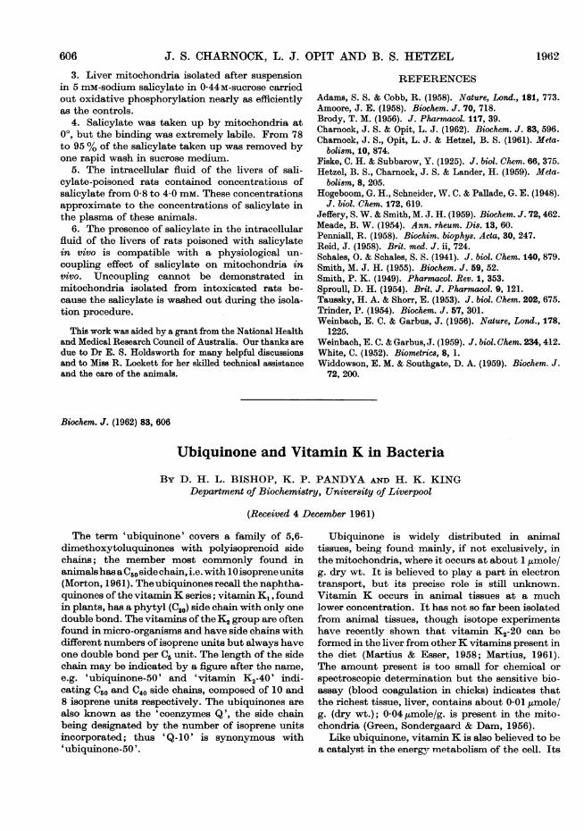
Fraction II showed the characteristic vitamin Kspectrum (Fig. 1, continuous line); comparisonwith an authentic specimen (Fig. 1, broken line)suggested that it was virtually free of impuritiesabsorbing selectively in this region. At A.249 mix, El%' was 177. Paper chromatographyshowed that only the vitamin K2-40 was present, forwhich El% was 264 (Isler & Wiss, 1959). This
Bioch. 1962, 83
Vol. 83 609

D. H. L. BISHOP, K. P. PANDYA AND H. K. KING
Table 1. Fractionation of octane extract of Proteus vulgarisLipid (1-40 g.) was placed on the column. The total eluted was 131 mg.
EluentA
Lightpetroleum
(%)
100989694800
Vol. ofeluent(ml.)10005005001000500500
Wt. offraction(mg.)92190-1181.4
10
RemarksHydrocarbons?Vitamin K (see Fig. 1)Ubiquinone (see Fig. 2)No defined u.v. spectrumOnly slight u.v. absorption
02[
1-a
I I I it tI I / klM l
I I'
IIE
IOI
I
I I
, \%
280 320Wavelength (m,u)
Fig. 1. Absorption spectra of fraction II (Table 1) fromProteus vtdgaris (-) and authentic vitamin R-40 (---);both were at concentrations of 38 mg./l. in cyclohexane.
021
0240 280 320
Wavelength (m,)360
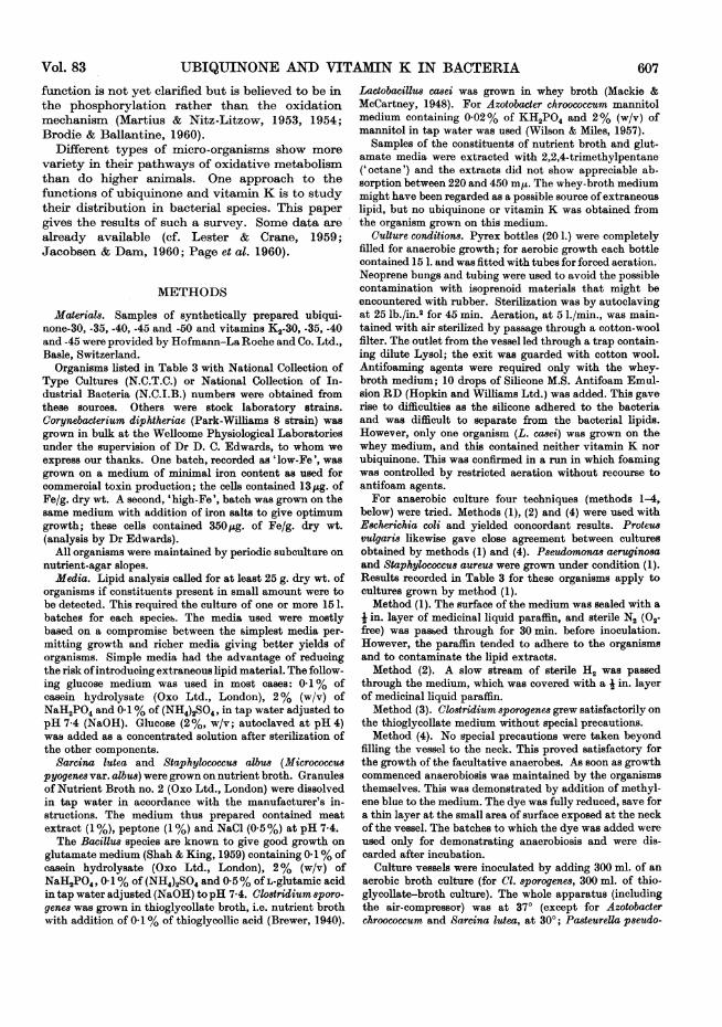
Fig. 2. Absorption spectrum of fraction IV (Table 1) fromProteu vulgaris before (-) and after (--- ) reduction withNaBH4. Both substances were at concentrations of36 mg./ml. in ethanol.
fraction must therefore contain 12-6 mg. ofvitamin K2.
Elution with 4% of ether in light petroleumremoved no significant amount of material (frac-tion III), but the '6% ether fraction' (fraction IV)had the characteristic ultraviolet absorption ofubiquinone and showed the same changes on treat-ment with sodium tetrahydroborate (Fig. 2, con-
tinuous line) (Lester & Crane, 1959). Paper chro-matography indicated ubiquinone-40. The fractioncontained 18 mg. of material; estimation of ubi-quinone gave a 'maximum' value of 13-1 mg. (fromE at 272 m,u) or a 'minimum of 12-6mg. (bytetrahydroborate reduction) (Fig. 2, broken line).Very little material was removed with 20 %
ether (fraction V) and the fraction eluted with
100% ether also contained no material withcharacteristic ultraviolet absorption.The fractions eluted totalled 131 mg. or 9-4% of
the total octane extract. Most of the extractedlipids remained firmly attached to the alumina andwere thus probably highly polar phospholipids.Indeed, the total lipid in certain micro-organismshas been reported to consist mostly of phospholipid(Macfarlane, 1961), and this would be expected toremain on the column under our conditions.
Five runs were carried through with Proteusvulgaris and results are summarized in Table 2. Thethree different extraction methods showed littledivergence in respect of the values for total lipid,ubiquinone or vitamin K. The E1%. given for
Fractionno.
IIIIIIIV
vVI
Ether(%)024620100
610 1962
B
041
--A-

UBIQUINONE AND VITAMIN K IN BACTERIA
Table 2. Ubiquinone and vitamin K in Proteus vulgaris (aerobic culture)Five batches of P. vulgaris were grown on glucose medium with addition of 0.01% of nicotinamide (see text)
and extracted by the method indicated in col. 2. The total lipid was fractionated by alumina chromatography.Col. 4 gives the yield of vitamin K (from E at 249 m,i) and col. 5 El%m for this fraction, the value for purevitamin K2-40 being 264. Col. 6 gives the ubiquinone content, estimated by E at 272 m,u, and col. 7 the valueobtained from diminution of E at 275 m, on tetrahydroborate reduction in ethanol.
TotalExtraction lipidmethod (%, dry wt.)
bane 5-1bane 4-3bane 5-3thanol 4-9bane + methanol 5-4
Vitamin K
(pmoleg.) El%,0-72 1770-65 191Not determined0-60 2180-71 226
U biquinone(ILmole/g.)
Max. Mi.0-66 0-630x69 0-60
0-650*67
0-610*59
vitamin K indicate the degree of purity obtained inthis fraction; they should be compared with thevalue of 264 for the pure compound.
Experience showed with Proteus and others thatthe yields of vitamin K and ubiquinone were rela-tively independent of the extraction procedure(except for the expected losses of vitamin K inextraction method 3, which involved saponifica-tion). The octane-extraction method (1) was adoptedas best for general use, as it involved fewer tech-nical difficulties. The spores of B. subtilis releasedvery little lipid on octane extraction, however, andacid hydrolysis (method 4) was used. Some Gram-positive organisms also yielded more lipid, but notmore vitamin K, after hydrolysis. They did notcontain ubiquinone. Micrococcus lysodeikticus hasalso proved a difficult organism for lipid extractionby octane although the vitamin K of this organismis completely extracted by methanol.
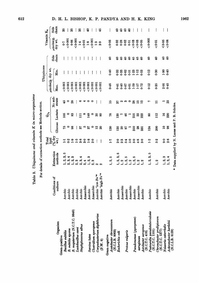
Table 3 summarizes the results obtained with allorganisms investigated. The first colunm lists theextraction procedures applied. The total lipid ofTable 3 is the amount given by the extractionmethod giving the highest yield; the acid- andalkaline-hydrolysis methods (4 and 3) are excluded.The maximal and minimal values for ubiquinonehave been explained above: they are the averages
from the various extraction methods used. Thevitamin K figures are also averages but results fromthe saponification method (3) have been excluded.
It is thought that vitamin K2 rather than K1 isalways present because the former is the typehitherto reported in bacteria; moreover, the be-haviour of our samples on paper chromatograms isconsistent with the K2 series and clearly eliminatedordinary vitamin K1-20, the only K1 homologue so
far proved to occur in Nature. The K1 and K2 seriesmay also be differentiated by differences in thefine structure of the infrared spectra; although wewere unable to exploit this method critically, our
infrared data for E. coli, P. vulgaris and Coryne-bacterium diphtheriae were consistent with thepresence of vitamins belonging to the K2 series.
Ubiquinone or vitamin K, when detected, werepresent in amounts much greater than the mini-mum required for tentative identification; therewere thus no 'doubtful' results. Sarcina lutea,however, was anomalous in that the '2% etherfraction', which normally contains vitamin K2when the latter is present, did not show the typicalfour-banded spectrum but gave a single main peakat 249 ms (with a lower peak at 325 m,u), andinflexions at 243, 256 and 272 m,u; these are theapproximate positions of the typical vitamin Kpeaks. Either vitamin K was present and itsspectrum was being obscured by a chromogenabsorbing in the same region or an 'abnormal'vitamin K was present. Further investigation willbe needed to decide this point; but three separateruns with Sarcina lutea gave essentially similarresults; the form of the spectrum did not change onrechromatography, and paper chromatogramsshowed only a single spot running in the positionof vitamin K2-40.
Examination for long-chain alcoholsAlcohols with long polyisoprenoid chains have
beenreported recently, e.g. solanesol, C45H73OH, fromtobacco leaves (Rowland, Latimer & Giles, 1956) anddolichol, C0ooH1610H, from animal tissues (Pennock,Hemming & Morton, 1960). The role of these isnot known but they may be related to the supplyof the side chain of vitamin K or ubiquinone. Threeorganisms were examined for their presence; thesaponification procedure (extraction method 3) wasapplied as it is necessary to remove triglycerides.
Pasteurella pseudotuberculosis (107 g. dry wt.)yielded 461 mg. of non-saponifiable material, whichwas fractionated on alumina as described above.After elution of the ubiquinone in the '6 % etherfraction', elution with ether-light petroleum(10:90, v/v) yielded 37 mg. of material; this frac-tion would be expected to contain the long-chainalcohols, if present (Pennock et al. 1960). The ultra-violet spectrum showed no specific absorption savefor that due to a trace of ubiquinone; the infrared
39-2
Expt.no.1 Oct2 Oct3 Oct4 Mel5 Oct
Vol. 83 611
D. H. L. BISHOP, K. P. PANDYA AND H. K. KING
O CIt tIo I I
o-COOo 0E0CO0 0 0 CO6
. . *-0 0*
- CO
V V V V
4-1
0 o o o o o o o o
u ooV6VV 6VV 6V V
10 CO w °- ° I0
E'o 0o C
O 00
_ e CDea )o
4
O mi_>
100 0io
0 CO0Ci 01o0o0
V V VV
o o Co 0o o
r4 COCO 10C)U
- o o o o N oq q r-cC eso :
u: r cs ceoa:u
CO C0 00 0
o l00:0o roXC mcOC OC) cqe
- _ CO CO
10 Ci 10 10 o- r-----
oT ci cq ci c a aie*c"4 _ z _z Iz Iz _z "4
6V
P- t 00O 0 06 66V V V
0 0 0 0
Ci O O O
O
C)i 14 ~oio- CO O
CD0
O Oq Co O
C)O O
O -
e:1 Ci
ea > o 4
~0
ci cSi C IC N- 4 4
*
0
~~~~~~~~~~C)~~~~~~~~~~~~~~~~~C°
C) C) C) C) )C) C)
- - o 0.oooo21i; 1i; s; $4O sO0 0 0 0 0 ¢0 ¢¢0 ¢ ¢ 0 ¢0¢0¢¢
CO~~ ~ ~ ~ ~ ~ ~ C
L~o
~~ ~ ~ ~2
co
:
.-
].QGo
.".
~6r-
-
,
612
*g 0.
7I C
-1e
1962
C1)0
._n1.
0S
SI
C)s
eWt$
O .4*ZP C.)Wffl
*O
0
41
C)_C)
0
C)
OD
Go
0
41
.4
0
C)w
'4-
0
m
Ca
4)
0
0
tQ
II
CA)
0
~0.i
cli
(E)

Vl8UBIQUINONE AND VITAMIN K IN BACTERIAWave no. (cm.-1)
30004000o 1500
2000900 700
1 000 800
3500 2500 1500 1000 900 800
Wave no. (cm.-')
0-210=3 E
107I^ IA I 0
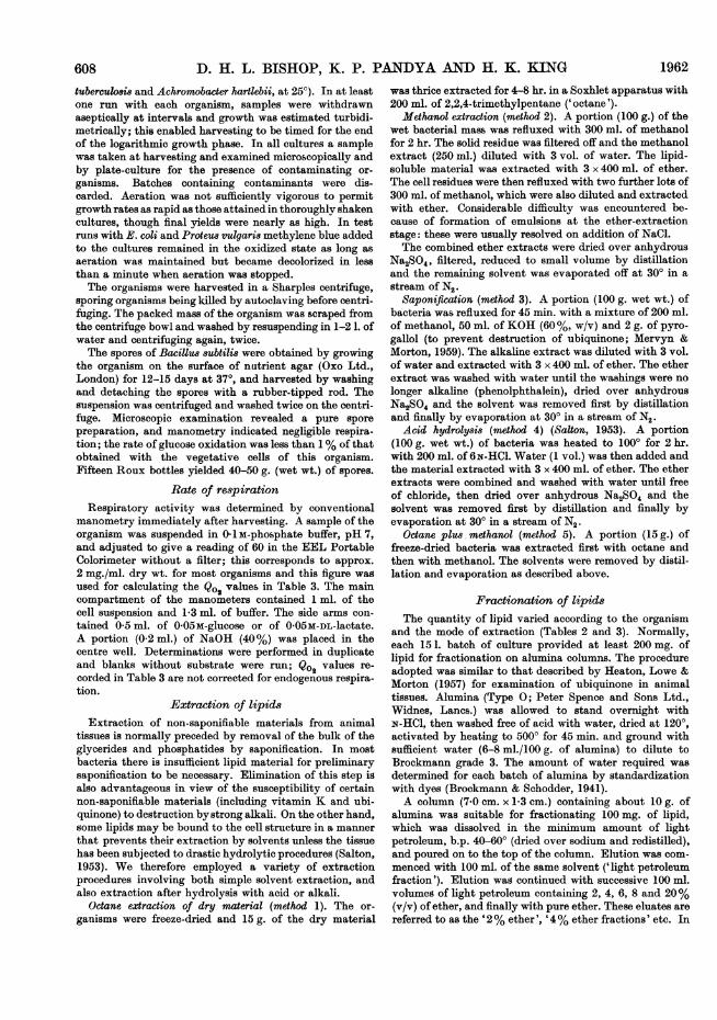
Fig. 3. Infrared-absorption spectrum of long-chain alcoholfrom Pasteurella p8eudotuberculosis.
spectrum suggested a long-chain alcohol but thecharacteristic isoprenoid frequencies were notobserved. The material was now purified by acetyl-ation. The fraction was dissolved in 1 ml. of an-
hydrous pyridine and 15 ml. of acetic anhydrideadded at 00. The mixture was left overnight at roomtemperature, then poured into hydrochloric acid(100 ml., 2N) at 0°. The product was extracted withlight petroleum, the extract washed with hydro-chloric acid (2 N) to remove pyridine, then water (toremove hydrochloric acid), dried over anhydroussodium sulphate and evaporated to dryness. Thematerial was then dissolved in light petroleum andapplied to a column containing 5 g. of aluminadiluted with 3 % of water. Most of the materialwas eluted from the column with light petroleum;it was transparent in the ultraviolet region and theinfrared absorption was consistent with the acetateof a long-chain saturated alcohol. The alcohol was
recovered by saponification of the ester and itsinfrared-absorption spectrum is shown in Fig. 3.Well-marked peaks attributable to the OH groupare seen at 3400 and 1065 cm.-'. The lack ofabsorption around 1660 cm.-' suggests that no
double bonds are present. The weak CH3 group bandat 1380 cm.-', in comparison with the strongCH2 group band at 1490 cm.-', also suggests a longunbranched chain.A value for the molecular weight was obtained by
forming the azobenzoate, and determining theintensity of the chromophore in the recrystallizedcompound (Pennock et al. 1960). The value of 290-320 suggested a chain length of C20-C22. No furthercharacterization was attempted.
Proteus vulgaris and E. coli were similarly ex-
amined for long-chain isoprenoid alcohols, withnegative results. Such compounds, if present, musthave been less than 5 mg./100 g. dry wt. of or-
ganism.
DISCUSSION
The results incorporated in Table 3 show thatorganisms with a well-developed oxidative meta-bolism contain either ubiquinone or vitamin K (or,
in two cases, both) in substantial amount, i.e. atconcentrations comparable with those of ubi-quinone found in the mitochondria of animaltissues and also in the spadix of Arum (Pumphrey& Redfearn, 1960). These values, of the order of1 ,umole/g. dry wt., represent roughly the amount ofan oxidation-reduction substance required forparticipation in the main electron-transport chain(Redfearn, 1961). In the three organisms studiedthat are known to be lacking in a cytochromesystem, neither quinone could be detected; and itseems certain that, if present, their concentrationmust have been much lower. There is a hint herethat ubiquinone and vitamin K may be alter-natives for some function connected with oxidativeprocesses. Clearly, however, the situation is notas simple as this. Vitamin K, by nutritional criteria,plays a vital part in animal metabolism, but attissue concentrations too low for easy detection. Thebacteria that we have reported as lacking vitaminK might still contain it in trace amounts. Amongthe aerobic organisms recorded in Table 3, there isno quantitative relation between the amounts ofthe quinones and the Q°2 values. The latter, how-ever, were determined on intact cells and factorsother than the electron-transport mechanism mayhave been governing oxygen uptake.The behaviour of the facultative anaerobes is in-
teresting. Four organisms were grown, both withforced aeration and also under conditions proved tobe anaerobic by the criteria of methylene-bluereduction and also the production of cells withdrastically reduced oxidative capacity. Yet inthree out of the four organisms no appreciable fallin amounts of quinone was found. Lester & Crane(1959), however, reported a cessation of ubiquinoneformation when E. coli was grown anaerobically,and Kashket & Brodie (1960) record a reduction inthe formation of both quinones in this organism.Although we examined a strain of this organismdifferent from that employed by either of thesegroups, we consider that there is an unexplainedconflict in these findings.A noteworthy feature of these results has been
the finding that, in the aerobic organisms, ubi-quinone is found in the Gram-negative types,vitamin K in the Gram-positive types, with bothquinones present in E. coli and Proteus vulgaria.This adds another item to the list of characteristicsthat separate these organisms, but its significanceis not known.
SUMMARY
1. Sixteen species of bacteria have been ex-amined for their content of vitamin K and ubi-quinone.
2. E8cherichia coli and Proteu8 vulgari8 containboth ubiquinone (-40) and vitamin K2 (-40).
Vol. 83 613
700

614 D. H. L. BISHOP, K. P. PANDYA AND H. K. KING 19623. Seven other Gram-negative bacteria contain
ubiquinone but not vitamin K.4. None of the Gram-positive organisms ex-
amined contains ubiquinone. Vitamin K2 wasfound in five types, but was not in Lactobacilluscasei, Clostridium sporogenes, anaerobically cul-tured Staphylococcu albus or in spores of Bacillus8ubtilis.
5. No long-chain polyisoprenoid alcohols weredetected in Escherichia coli, Proteus vulgaris orPasteurella pseudotubercu0osis. The last-named,however, yielded a compound tentatively identifiedas a long-chain saturated alcohol.
We are very grateful to Professor R. A. Morton, F.R.S.,for his interest in this work, to Dr D. C. Edwards and theWellcome Research Laboratories, and to Dr 0. Isler andHofmann-La Roche and Co. Ltd. for materials; and to theAgricultural Research Council for the award of a Scholar-ship to D.H.L.B.
REFERENCES
Brewer, J. H. (1940). J. Bact. 39, 10.Brockmann, H. & Schodder, H. (1941). Ber. dtsch. chem.
Ge8. 74, 73.Brodie, A. F. & Ballantine, J. (1960). J. biol. Chem. 235,
226.Green, J. P., Sondergaard, E. & Dam, H. (1956). Biochim.
biophy8. Acta, 19, 182.Heaton, F. W., Lowe, J. S. & Morton, R. A. (1957).
Biochem. J. 67, 208.Isler, 0. & Wiss, 0. (1959). Vitam. & Horm. 17, 53.Jacobsen, B. K. & Dam, H. (1960). Biochim. biophys. Acta,
40, 211.Kashket, E. & Brodie, A. F. (1960). Bact. Proc. p. 61.
Lester, R. L. & Crane, F. L. (1959). J. biol. Chem. 234,2169.
Lester, R. L. & Ramasarma. J. (1959). J. biol. Chem. 234,673.
Macfarlane, M. G. (1961). Biochem. J. 79, 4P.Mackie, J. T. & McCartney, J. E. (1948). Handbook of
Practical Bacteriology, p. 186, 8th ed. Edinburgh: E. andS. Livingstone Ltd.
Martius, C. (1961). In Ciba Foundation Symp., Quinones inElectron Transport, p. 312. Ed. by Wolstenholme,G. E. W. & O'Connor, C. M. London: J. and A.Churchill Ltd.
Martius, C. & Esser, H. 0. (1958). Biochem. Z. 331, 1.Martius, C. & Nitz-Litzow, D. (1953). Biochim. biophys.
Acta, 12, 134.Martius, C. & Nitz-Litzow, D. (1954). Biochim. biophys.
Acta, 13, 152.Mervyn, L. & Morton, R. A. (1959). Biochem. J. 72, 106.Morton, R. A. (1961). Vitam. & Horm. 19, 1.Page, A. C., jun., Gale, P., Wallick, H., Walton, R. B.,
McDaniel, L. E., Woodroff, H. B. & Folkers, K. (1960).Arch. Biochem. Biophys. 89, 318.
Pennock, J. F., Hemming, F. W. & Morton, R. A. (1960).Nature, Lond., 186, 470.
Pumphrey, A. M. & Redfearn, E. R. (1960). Biochem. J.76, 61.
Redfearn, E. R. (1961). In Ciba Foundation Symp.,Quinones in Electron Transport, p. 346. Ed. by Wolsten-holme, G. E. W. & O'Connor, C. M. London: J. and A.Churchill Ltd.
Rowland, R. L., Latimer, P. H. & Giles, J. A. (1956).J. Amer. chem. Soc. 78, 4680.
Salton, M. R. J. (1953). Biochim. biophys. Acta, 10, 512.Shah, P. C. & King, H. K. (1959). J. gen. Microbiol. 20, 78.Wilson, G. S. & Miles, A. A. (Ed.) (1957). Topley and Wil-
son's Principles of Bacteriology and Immunity. vol. 1,p. 587. 4th ed. Baltimore: The Williams and Wilkins Co.
Biochem. J. (1962) 83, 614
The Localization of Cerebral Phosphoprotein Phosphatase
BY S. P. R. ROSE*Department of Biochemistry, Maudsley Institute of Psychiatry (British Post-Graduate Medical Federation),
Denmark Hill, London, S.E. 5
(Received 4 December 1961)
The partial purification of a phosphoproteinphosphatase from ox brain has been reported byRose & Heald (1960, 1961). The enzyme was labile,magnesium-activated, and extremely specific, act-ing only on certain phosphoproteins, e.g. casein,phosvitin and brain phosphoprotein, and partiallyhydrolysed phosphopeptides prepared from a-casein. The enzyme differed from other mammalianphosphoprotein phosphatases (Hofman, 1958;
Glomset & Porath, 1960; Revel & Racker, 1960) inthat it liberated only a small portion of the phos-phoprotein phosphorus before ceasing to act. Thisreleased phosphorus included all the radioactivityincorporated into phosvitin during phosphorylationwith radioactive phosphate in a mitochondrial pre-paration (Burnett & Kennedy, 1954; Rabinowitz& Lipmann, 1960).Such distinctive properties made desirable a
knowledge of the distribution and intracellularlocalization of the enzyme. In this paper some data
* Present address: Department of Biochemistry, SouthParks Road, Oxford.
Related Documents