Biochemistry of Extracellular Matrix Jana Novotná

Biochemistry of Extracellular Matrix Jana Novotná.
Dec 17, 2015
Welcome message from author
This document is posted to help you gain knowledge. Please leave a comment to let me know what you think about it! Share it to your friends and learn new things together.
Transcript

Biochemistry of Extracellular Matrix
Jana Novotná
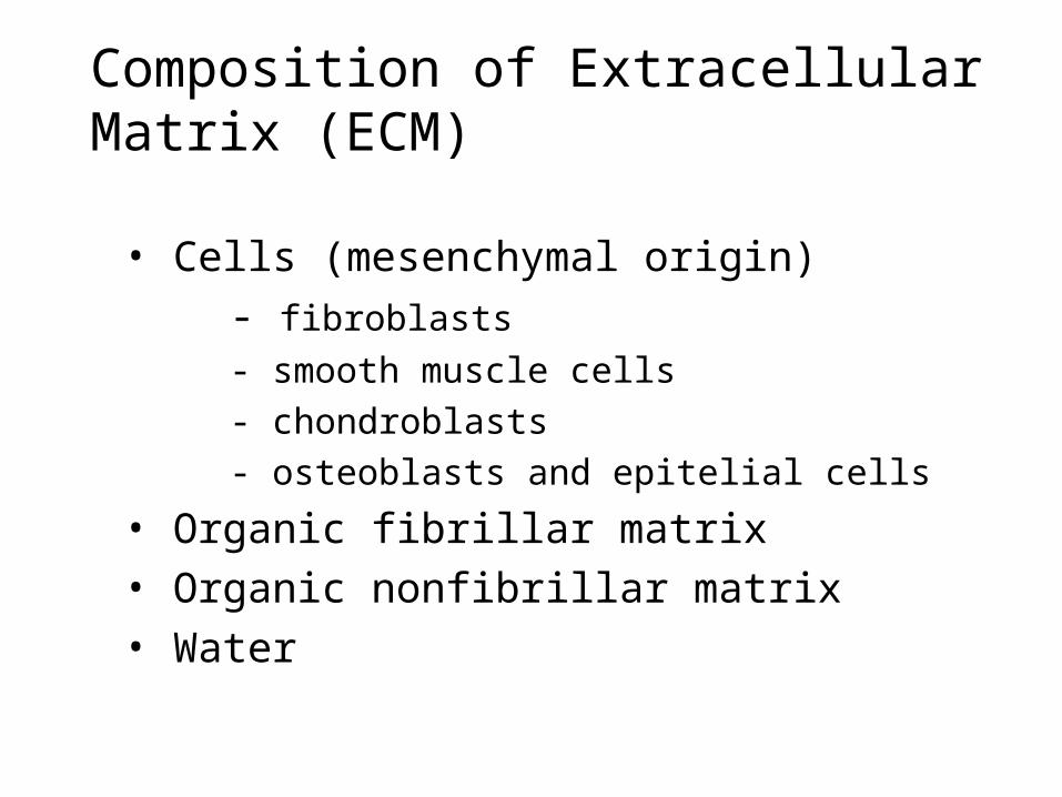
• Cells (mesenchymal origin)
- fibroblasts
- smooth muscle cells
- chondroblasts
- osteoblasts and epitelial cells
• Organic fibrillar matrix• Organic nonfibrillar matrix• Water
Composition of Extracellular Matrix (ECM)

Function of ECM
• Provides support and anchorage for cells.• Regulates and determine cells dynamic behaviour :
- polarity of cells- cell differentiation- adhesion- migration
• Provides mechanical support for tissues and organ architecture
- growth- regenerative and healing processes - determination and maintenance of the structure
• Place for active exchange of different metabolites, ions, water.
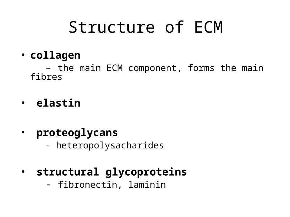
Structure of ECM
• collagen – the main ECM component, forms the main fibres
• elastin
• proteoglycans - heteropolysacharides
• structural glycoproteins - fibronectin, laminin
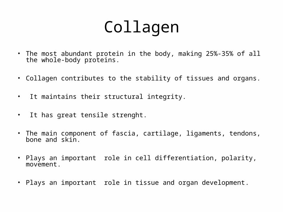
Collagen
• The most abundant protein in the body, making 25%-35% of all the whole-body proteins.
• Collagen contributes to the stability of tissues and organs.
• It maintains their structural integrity.
• It has great tensile strenght.
• The main component of fascia, cartilage, ligaments, tendons, bone and skin.
• Plays an important role in cell differentiation, polarity, movement.
• Plays an important role in tissue and organ development.
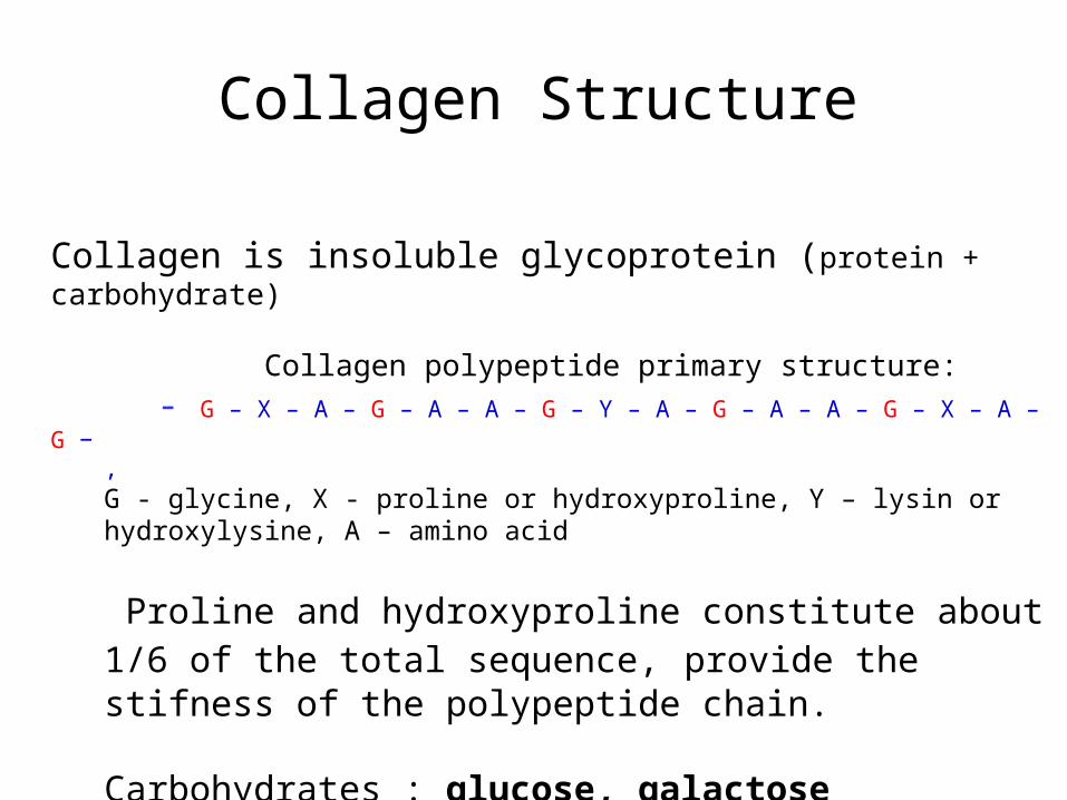
Collagen is insoluble glycoprotein (protein + carbohydrate)
Collagen polypeptide primary structure:
- G – X – A – G – A – A – G – Y – A – G – A – A – G – X – A – G −,G - glycine, X - proline or hydroxyproline, Y – lysin or hydroxylysine, A – amino acid
Proline and hydroxyproline constitute about 1/6 of the total sequence, provide the stifness of the polypeptide chain.
Carbohydrates : glucose, galactose
Collagen Structure
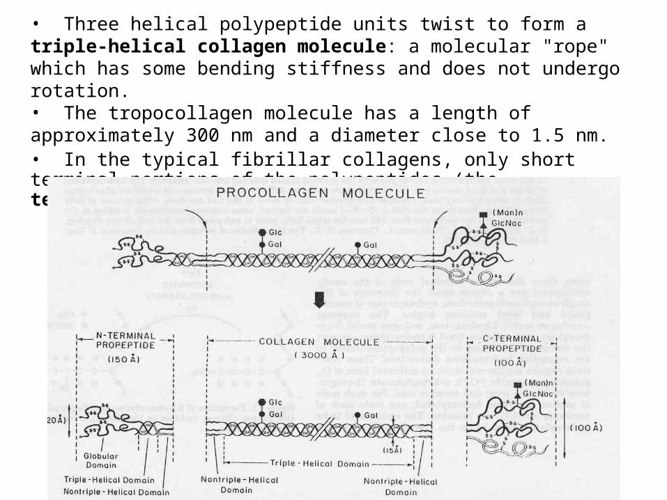
• Three helical polypeptide units twist to form a triple-helical collagen molecule: a molecular "rope" which has some bending stiffness and does not undergo rotation. • The tropocollagen molecule has a length of approximately 300 nm and a diameter close to 1.5 nm.• In the typical fibrillar collagens, only short terminal portions of the polypeptides (the telopeptides) are not triple helical.

1. Synthesis of a chains of pre-procollagen on ribosomes.
2. Hydroxylation of lysine and proline in rER/Golgi by lysyl-5-hydroxylase and prolyl-4-hydroxylase.
3. Glycosylation: addition of galactose and glucose to some hydroxylysine residues (galactosyl transferase and glycosyl transferase).
4. Assembly of -chains to form procollagen. Reaction needs the formation of disulphide bonds between registration peptides, at both ends of the prepro- collagen.
Synthesis

5. Secretion of procollagen molecules by exocytosis into the extracellular space.
6. Cleavage of registration peptides is catalysed by procollagen peptidases. The resulting molecule is called tropocollagen.
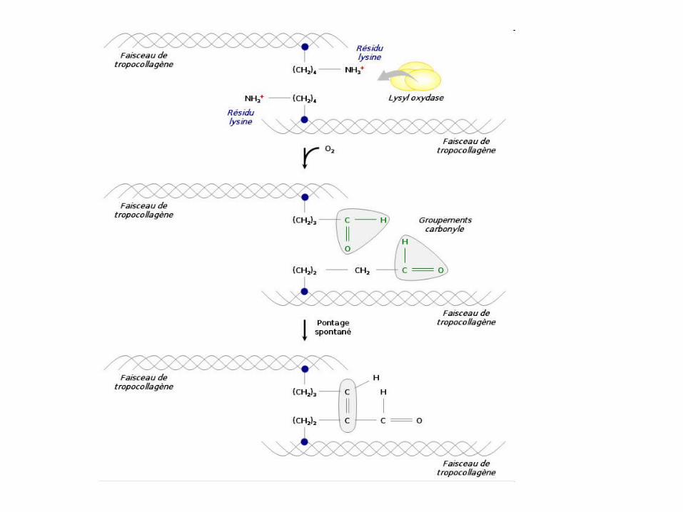
7. Oxidation – deamination of the hydroxylysine, the removal of (NH2) group has a net oxidative effect and the formation of covalent cross-links. Reaction is catalyzed by lysyl oxidase (or catalase).
8. Self-assembly or polymerization of tropocollagen molecules form collagen fibrils. Cross-linkage between adjacent tropocollagen molecules stabilizes the fibrils.
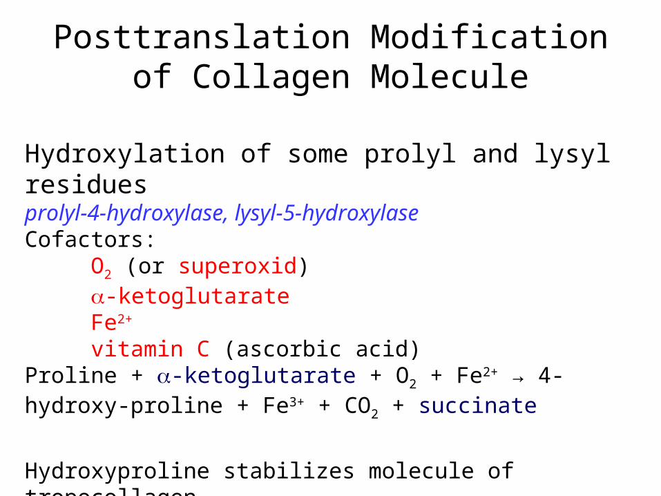
Hydroxylation of some prolyl and lysyl residuesprolyl-4-hydroxylase, lysyl-5-hydroxylaseCofactors:
O2 (or superoxid)-ketoglutarateFe2+
vitamin C (ascorbic acid)Proline + -ketoglutarate + O2 + Fe2+ → 4-hydroxy-proline + Fe3+ + CO2 + succinate
Hydroxyproline stabilizes molecule of tropocollagen.
Posttranslation Modification of Collagen Molecule

Crosslinking of tropocollagen - aldol condensation
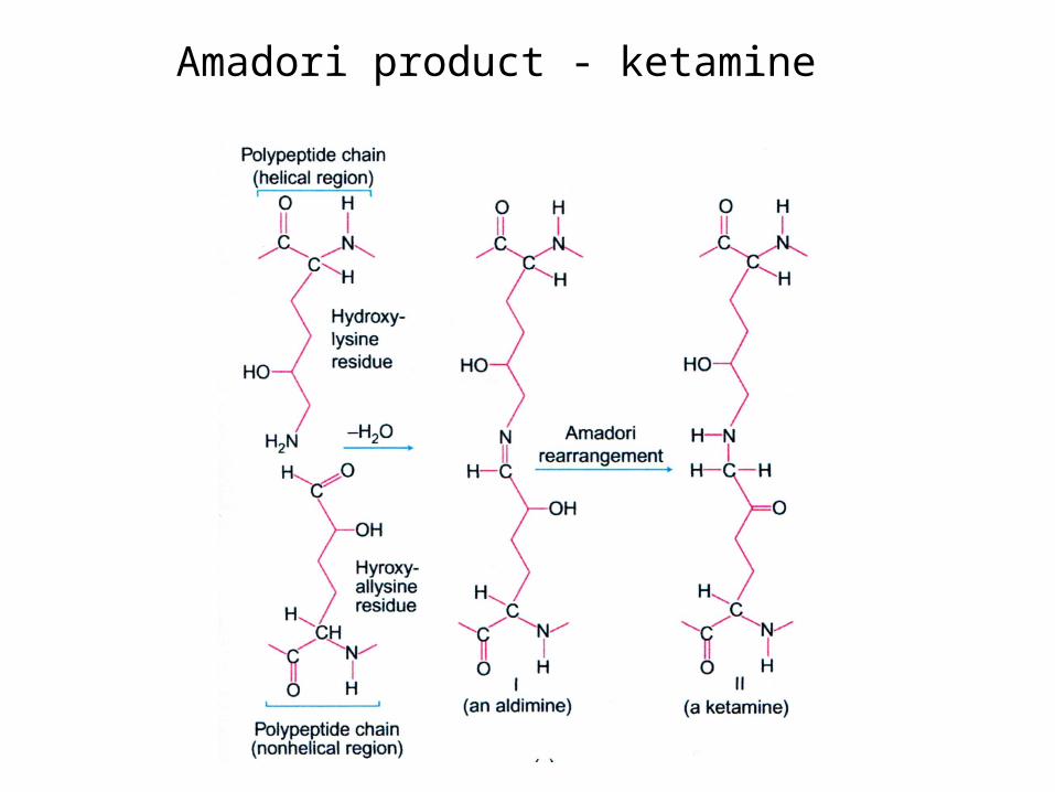
Amadori product - ketamine

Lysinonorleucine cross-link

The typical staggered array of tropocollagen molecules in the collagen fibril. The telopeptides participate in covalent crosslinking.
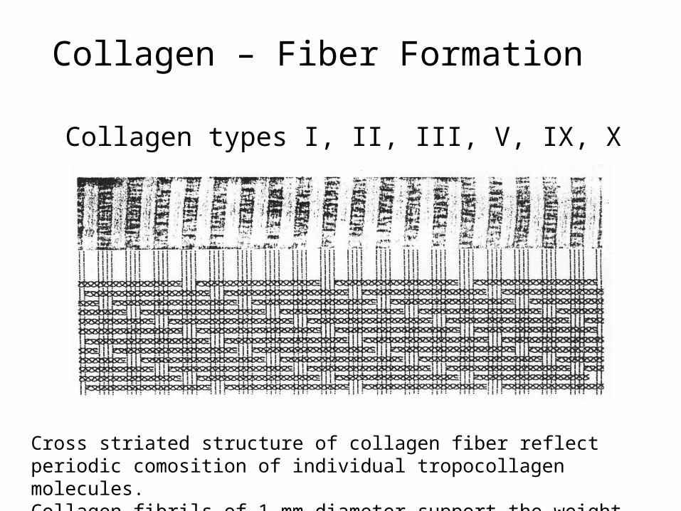
Collagen – Fiber Formation
Collagen types I, II, III, V, IX, X
Cross striated structure of collagen fiber reflect periodic comosition of individual tropocollagen molecules.Collagen fibrils of 1 mm diameter support the weight of 10 kg.
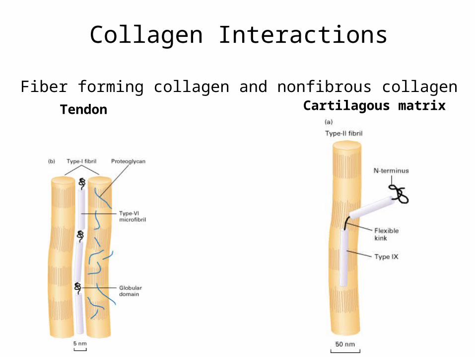
Collagen Interactions
Fiber forming collagen and nonfibrous collagenCartilagous matrixTendon

Collagens Classification
1. Fibril-forming collagens (I, II, III, V, X)
2. Fibril-associated collagens (FACIT)
3. Network-forming collagens
4. Anchoring fibrils collagens
5. Transmembrane collagens
6. Basement membrane collagens
7. Other collagens with unique function
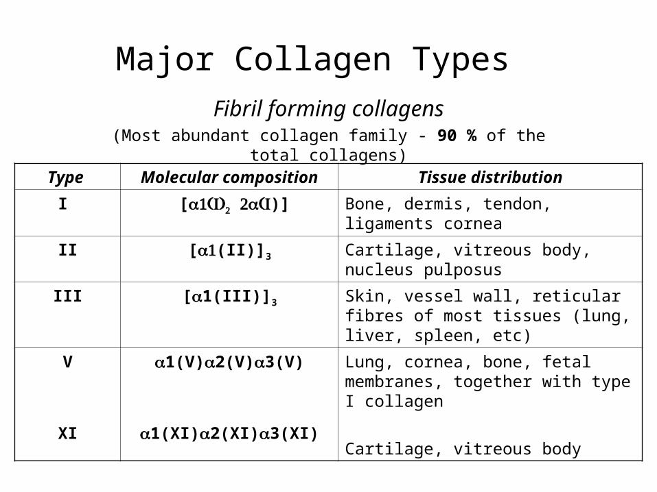
Major Collagen Types
Type Molecular composition
Tissue distribution
I [ )] Bone, dermis, tendon, ligaments cornea
II [(II)]3 Cartilage, vitreous body, nucleus pulposus
III [1(III)]3 Skin, vessel wall, reticular fibres of most tissues (lung, liver, spleen, etc)
V
XI
1(V)2(V)3(V)
1(XI)2(XI)3(XI)
Lung, cornea, bone, fetal membranes, together with type I collagen
Cartilage, vitreous body
Fibril forming collagens(Most abundant collagen family - 90 % of the total collagens)

Basement membrane collagens
IV 1(IV)2V)]; 1 – 6 Basement membrane
Short non-helical amino-terminal domain, a long Gly-X-Y repeat domain with numerous small interruptions, and a highly conserved carboxyl-terminal globular NC1 domain. It polymerizes into a disulfide-bonded polygonal network via tetramerization between amino-terminal domains and dimerization between NC1 domains.
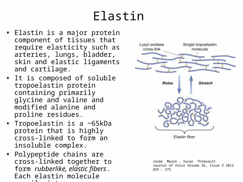
• Elastin is a major protein component of tissues that require elasticity such as arteries, lungs, bladder, skin and elastic ligaments and cartilage.
• It is composed of soluble tropoelastin protein containing primarily glycine and valine and modified alanine and proline residues.
• Tropoelastin is a ~65kDa protein that is highly cross-linked to form an insoluble complex.
• Polypeptide chains are cross-linked together to form rubberlike, elastic fibers. Each elastin molecule uncoils into a more extended conformation when the fiber is stretched and will recoil spontaneously as soon as the stretching force is relaxed.
Elastin
Jaime Moore , Susan ThibeaultJournal of Voice Volume 26, Issue 3 2012 269 - 275

• Desmosine (isodesmosine) - the most common interchain cross-link
• conversion of NH3 groups of lysine (hydroxylisine) to reactive aldehydes by lysyl oxidase.• desmosine cross-link is spontaneously formed.
Elastin
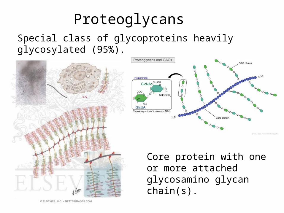
Proteoglycans Special class of glycoproteins heavily glycosylated (95%).
Core protein with one or more attached glycosamino glycan chain(s).
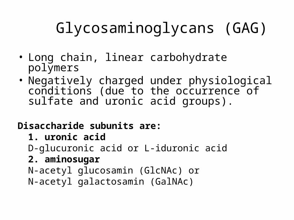
Glycosaminoglycans (GAG)
• Long chain, linear carbohydrate polymers • Negatively charged under physiological
conditions (due to the occurrence of sulfate and uronic acid groups).
Disaccharide subunits are:1. uronic acidD-glucuronic acid or L-iduronic acid2. aminosugarN-acetyl glucosamin (GlcNAc) orN-acetyl galactosamin (GalNAc)
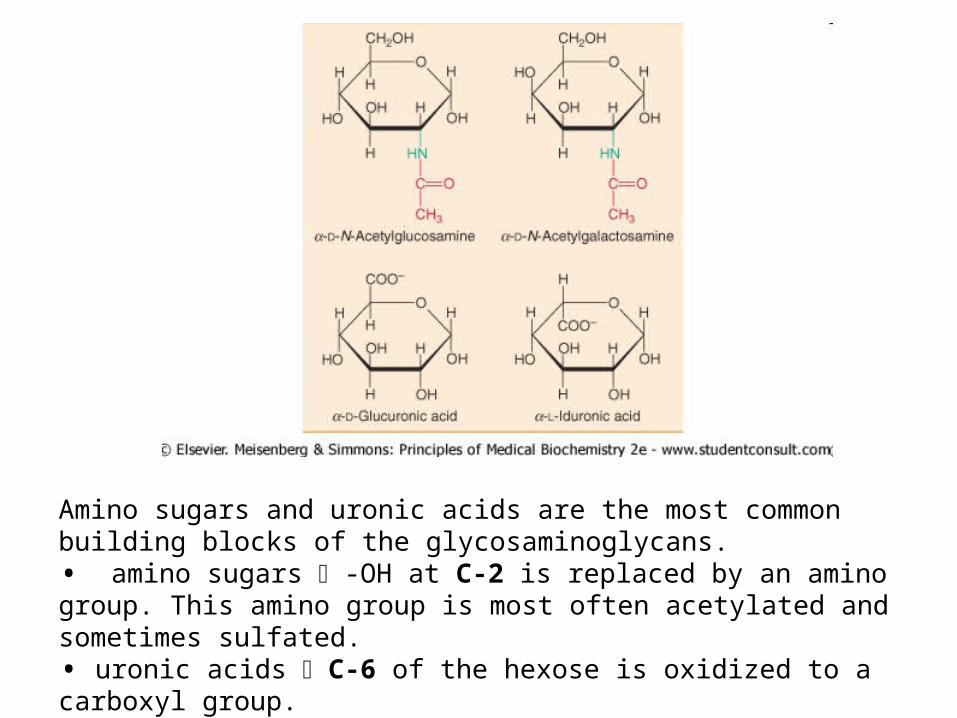
Amino sugars and uronic acids are the most common building blocks of the glycosaminoglycans. • amino sugars -OH at C-2 is replaced by an amino group. This amino group is most often acetylated and sometimes sulfated. • uronic acids C-6 of the hexose is oxidized to a carboxyl group.
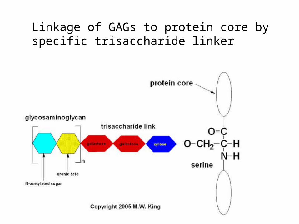
Linkage of GAGs to protein core by specific trisaccharide linker
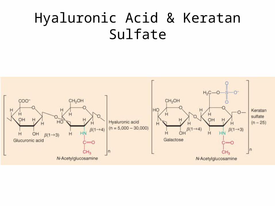
Hyaluronic Acid & Keratan Sulfate

Chondroitin 6-sulfate & Heparan Supfate
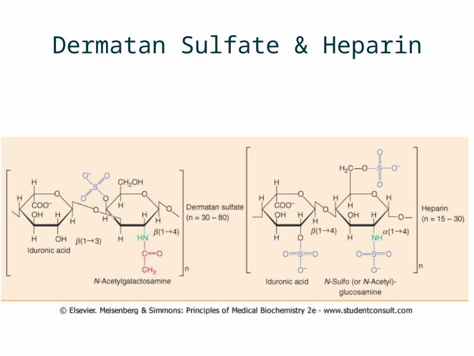
Dermatan Sulfate & Heparin

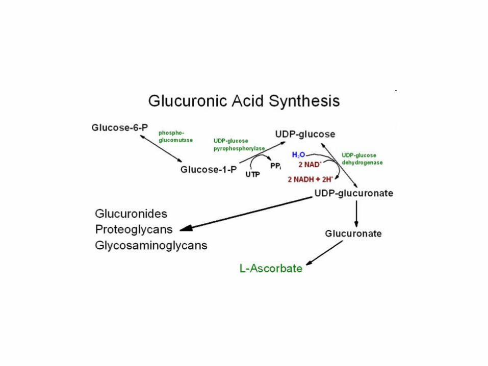
Biosynthesis
• The protein component is synthesized by ribosomes and transocated into the lumen of the RER.
• Glycosylation of the proteoglycan occurs in the Golgi apparatus in multiple enzymatic steps.
• First a special link tetrasaccharide is attached to a serine side chain on the core protein to serve as a primer for polysaccharide growth.

Biosynthesis
• Then sugars are added by glycosyltransferase.• Some glycosyltransferases catalyse sugar transfer to
tyrosine, serine and threonine to give O-linked glycoproteins, or to asparagine to give N-linked glycoproteins.
• Mannosyl groups may be transferred to tryptophan to generate C-manosyl tryptophan
• The completed proteoglycan is then exported in secretory vesicles to the extracellular matrix of the cell.

Biosynthesis of Heparan Sulfate proteoglycan

Degradation of Heparan Sulfate Proteoglycan

Glycosaminoglycan Occurence
Proteoglycans can be categorised depending upon the nature of their glycosaminoglycan chains.
• Hyaluronic acid (does not contain any sulfate)– non-covalent link complex with proteoglycans
• Chondroitin sulfate – cartilage, bone
• Dermatan sulfate – skin, blood vessels
• Heparan sulfate – basement membrane, component of cells surface
• Keratan sulfate– cornea, bone, cartilage, often aggregated with chondroitin
sulfate
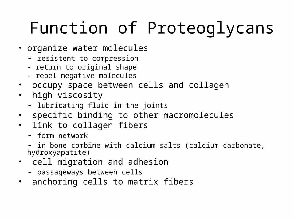
Function of Proteoglycans• organize water molecules
- resistent to compression- return to original shape- repel negative molecules
• occupy space between cells and collagen• high viscosity
- lubricating fluid in the joints• specific binding to other macromolecules• link to collagen fibers
- form network
- in bone combine with calcium salts (calcium carbonate, hydroxyapatite)• cell migration and adhesion
- passageways between cells• anchoring cells to matrix fibers
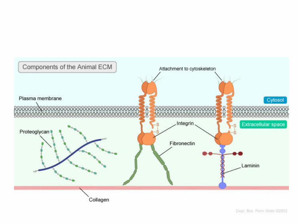
Structural Glycoproteins
• Direct linkage to collagen or proteoglycans– anchoring collagen fibers to cell membrane– covalent attachment to membrane lipid
• Major adhesive structural glycoproteins– fibronectin– laminin

Fibronectin
• High-molecular weight (~440kDa) glycoprotein • Attached to cell membrane by membrane-spanning
receptor – integrin.• Crosslinks and stabilizes other components of ECM • Enhances cell addhesion to extracellular matrix
components (collagen, fibrin and heparansulfate proteoglycans).
• Related to blood clotting - soluble FN crosslinks platelets together using membrane bound heparin
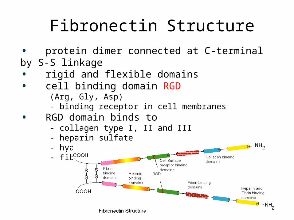
Fibronectin Structure
• protein dimer connected at C-terminal by S-S linkage• rigid and flexible domains• cell binding domain RGD
(Arg, Gly, Asp) - binding receptor in cell membranes
• RGD domain binds to - collagen type I, II and III - heparin sulfate - hyaluronic acid - fibrin
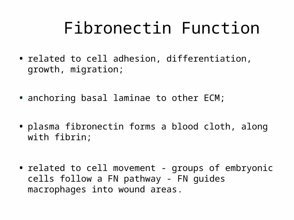
Fibronectin Function
• related to cell adhesion, differentiation, growth, migration;
• anchoring basal laminae to other ECM;
• plasma fibronectin forms a blood cloth, along with fibrin;
• related to cell movement - groups of embryonic cells follow a FN pathway - FN guides macrophages into wound areas.
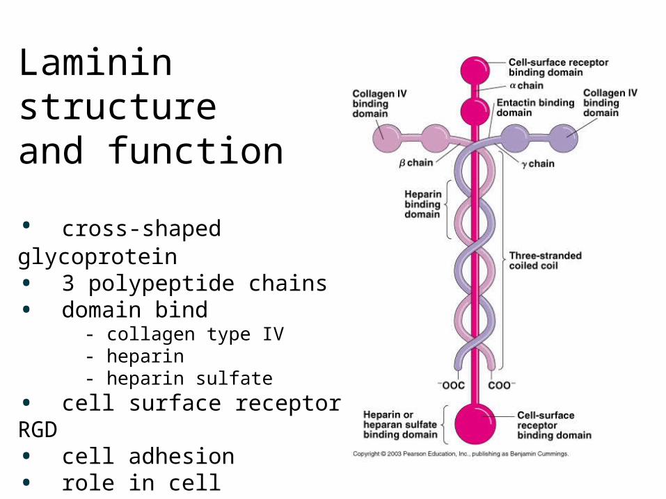
Laminin structure and function
• cross-shaped glycoprotein• 3 polypeptide chains• domain bind - collagen type IV - heparin - heparin sulfate
• cell surface receptor RGD• cell adhesion• role in cell differentiation• anchoring the glycoprotein to basal laminae
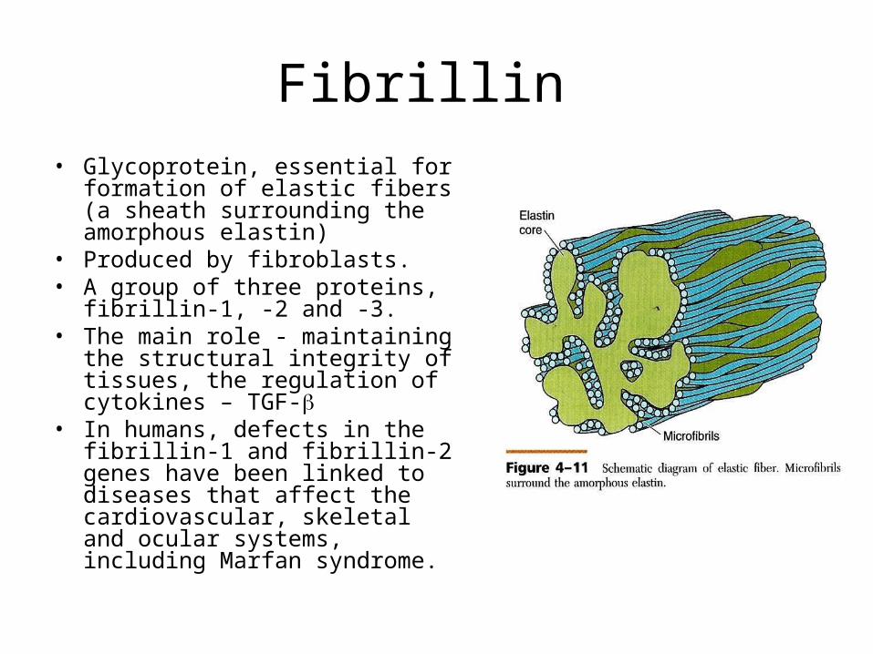
Fibrillin • Glycoprotein, essential for
formation of elastic fibers (a sheath surrounding the amorphous elastin)
• Produced by fibroblasts. • A group of three proteins, fibrillin-
1, -2 and -3. • The main role - maintaining the
structural integrity of tissues, the regulation of cytokines – TGF-
• In humans, defects in the fibrillin-1 and fibrillin-2 genes have been linked to diseases that affect the cardiovascular, skeletal and ocular systems, including Marfan syndrome.
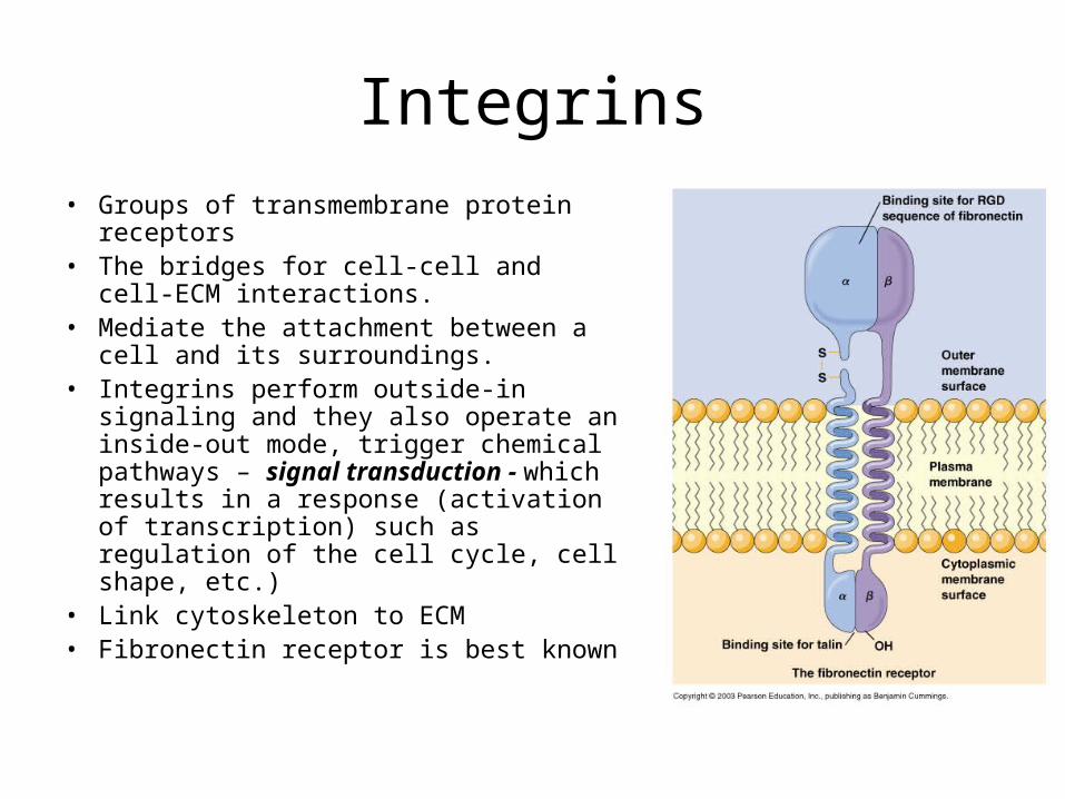
Integrins
• Groups of transmembrane protein receptors
• The bridges for cell-cell and cell-ECM interactions.
• Mediate the attachment between a cell and its surroundings.
• Integrins perform outside-in signaling and they also operate an inside-out mode, trigger chemical pathways – signal transduction - which results in a response (activation of transcription) such as regulation of the cell cycle, cell shape, etc.)
• Link cytoskeleton to ECM• Fibronectin receptor is best known

Tenascins
• Abundant in the extracellular matrix of developing vertebrate embryo.
• Tenascin-C contains an RGD motif and is recognized by diverse integrins. Mainly synthesized by the nervous system and connective tissues.
• Tenascin-R is found in the nervous system• Tenascin-X and tenascin-Y are found primarily in muscle
connective tissues.• Tenascin-W is found in kidney and developing bone.
Related Documents


![VORYiãNRODVWURMQLFNi2ORPRXF W OLVWRSDGX ... · Název: Sports Jméno autora: Mgr. Jana Novotná 3 HGP W DQJOLFNêM D]\N -D]\N DQJOLFNê þHVNê .OtþRYiVORYD sports, verbs ± go,](https://static.cupdf.com/doc/110x72/5f7d4a8bd5a919177a562e21/voryinrodvwurmqlfni2orprxf-w-olvwrsdgx-nzev-sports-jmno-autora-mgr.jpg)


