Biochemistry of Addison’s Disease

Biochemistry of Addison’s Disease. ANATOMICALLY: The adrenal gland is situated on the anteriosuperior aspect of the kidney and receives its blood supply.
Jan 01, 2016
Welcome message from author
This document is posted to help you gain knowledge. Please leave a comment to let me know what you think about it! Share it to your friends and learn new things together.
Transcript

Biochemistry of
Addison’s Disease

ANATOMICALLY:
• The adrenal gland is situated on the anteriosuperior aspect of the kidney and receives its blood supply from the adrenal arteries.
HISTOLOGICALLY:• The adrenal gland consists
of two distinct tissues of different embryological origin, the outer cortex and inner medulla.
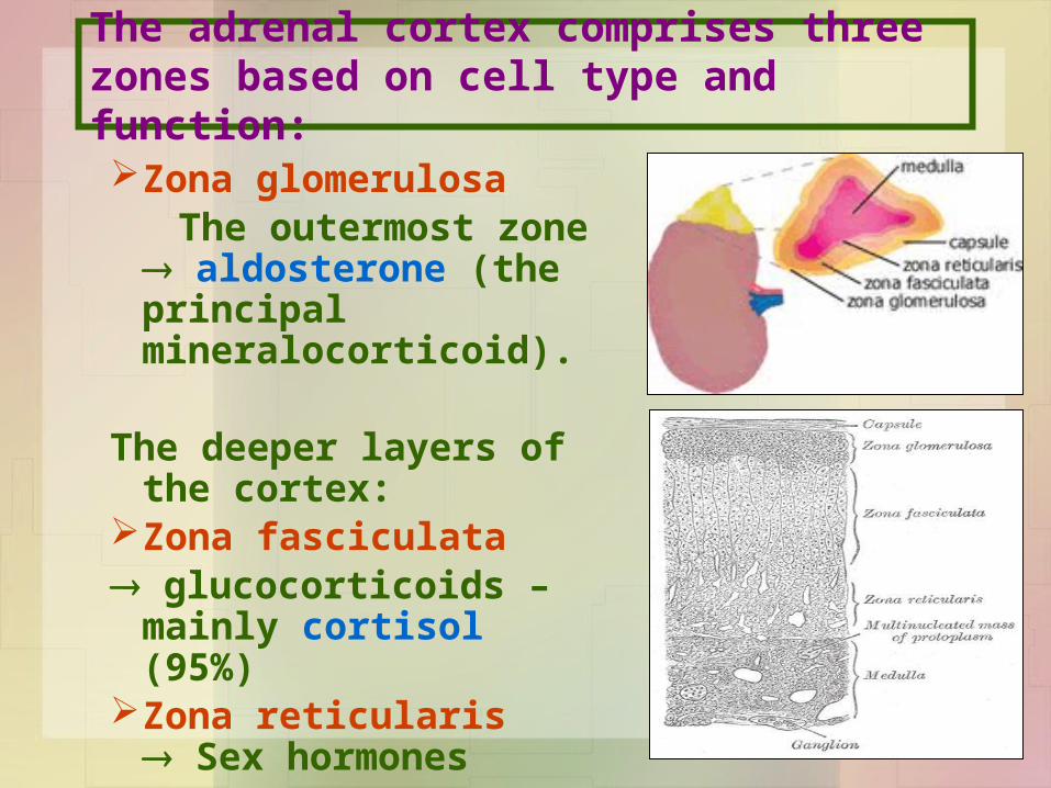
The adrenal cortex comprises three zones based on cell type and function: Zona glomerulosa The outermost zone
aldosterone (the principal mineralocorticoid).
The deeper layers of the cortex:
Zona fasciculata glucocorticoids – mainly
cortisol (95%)Zona reticularis
Sex hormones

Steroid Hormone Synthesis Steroid Hormone Synthesis
Cholesterol
Pregnenolone (C21)3-β-Hydroxysteroid dehydrogenase
Progesterone (C21)
17-α-Hydroxylase
17-α-Hydroxyprogesterone (C21)
Androstenedione (C19)
Testosterone (C19)
Estradiol (C18)
11-Deoxycortisol (C21)11-Deoxycorticosterone (C21)
Cortisol (C21)
21-α-Hydroxylase
11- β -Hydroxylase
Corticosterone
Aldosterone (C21)
Per
iph
eral
tis
sues

Aldosterone Hormone:• The principal physiological function of
aldosterone is to conserve Na+, mainly by facilitating Na+ reabsorption and reciprocal K+ or H+ secretion in the distal renal tubule.
• aldosterone is a major regulator of water and electrolyte balance, as well as blood pressure.

• Aldosterone, by acting on the distal convoluted tubule of kidney, leads to:
• potassium excretion • sodium and water reabsorption
• Renin-Angiotensin system is the most important regulatory mechanism for aldosterone secretion

The renin - angiotensin system:
• It is the most important system controlling aldosterone secretion.
• It is involved in B.P. regulation.
Renin:• a proteolytic enzyme produced by the juxtaglomerular
cells of the afferent renal arteriole.
• Sensitive to B.P. changes through baroreceptors
• released into the circulation in response to :– a fall in circulating blood volume. – a fall in renal perfusion pressure.– loss of Na+.

Renin
Angiotensin I
Angiotensin II
ACE
Vasoconstriction
B.P
• Aldosterone sec.• Renin release
Degraded
Angiotensin III
Angiotensinogen (α2-Globulin made in the liver)

Causes of adrenocortical hypofunction
A. Primary destruction of adrenal gland: AutoimmuneInfection, e.g., tuberculosisInfiltrative lesions, e.g., amylodosis
B. Secondary to pituitary disease:Pituitary tumorsVascular lesions TraumaHypothalmic diseasesIatrogenic (steroid therapy, surgery or radiotherapy)

Signs and symptoms of primary adrenal failure (Addison’s
disease)The symptoms are precipitated by trauma,
infection or surgery:Lethargy, weakness, nausea & weight loss.
Hypotension especially on standing (postural)
Hyperpigmentation (buccal mucosa, skin creases, scars)
Deficiency of both glucocorticoids and mineralocorticoids
Hypoglycemia, Na+, K+ and raised urea
Life threatening and need urgent care.

Hyperpigmentation in Addison’s disease
Hyperpigmentation occurs because melanocyte-stimulating hormone (MSH) and (ACTH) share the same precursor molecule, Pro-opiomelanocortin (POMC).
The anterior pituitary POMC is cleaved into ACTH, γ-MSH, and β-lipotropin.
The subunit ACTH undergoes further cleavage to produce α-MSH, the most important MSH for skin pigmentation.
In secondary adrenocortical insufficiency, skin darkening does not occur.

Investigation of Addison’s disease (AD)
• The patient should be hospitalized
• Basal measurement of:Serum urea, Na+, K+ & glucoseSerum cortisol and plasma ACTH
• Definitive diagnosis and confirmatory tests should be done later after crisis.
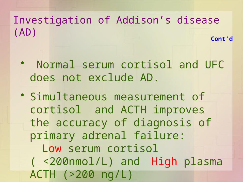
Investigation of Addison’s disease (AD)
• Normal serum cortisol and UFC does not exclude AD.
• Simultaneous measurement of cortisol and ACTH improves the accuracy of diagnosis of primary adrenal failure:
Low serum cortisol ( <200nmol/L) and High plasma ACTH (>200 ng/L)
Cont’d
Confirmatory Tests:1. Short tetracosactrin (Synacthen) test
(Short ACTH stimulation test)
• Measure basal S. cortisol• Stimulate with I.M. synthetic ACTH (0.25 mg)• Measure S. cortisol 30 min after I/M injection• Normal: of S. cortisol to >500 nmol/L • Failure of S. cortisol to respond to stimulation,
confirm AD.• Abnormal results:
– emotional stress– glucocorticoid therapy– estrogen contraceptives.

Confirmatory Tests:2. Adrenal antibodies
• Detection of adrenal antibodies in serum of patients with autoimmune Addison’s disease
3. Imaging (Ultrasound/CT)
Ultrasound or CT for adrenal glands for identifying the cause of primary adrenal failure

Investigation of Secondary AC Insufficiency
• Low serum cortisol with low plasma ACTH
• No response to short synacthen test: Adrenocortical cells fail to respond to short ACTH stimulation
• Depot Synacthen test (confirmatory test) 1. Measure basal S. cortisol
2. Stimulate with I.M. synthetic ACTH (1.0 mg) on each of three consecutive days
3. Measure S. cortisol at 5 hours after I.M. injection on each of the three days

Investigation of Secondary AC Insufficiency
Depot Synacthen test …. Cont’d
Interpretation of results:- Addison’s disease: No rise of S. cortisol >600 nmol/L
at 5 h after 3rd injection.
- Secondary AC: Stepwise increase in the S. cortisol after successive injections
- Limitations: Hypothyroidism: Thyroid deficiency must be
corrected before testing of adrenocortical functions
Prolonged steroid therapy

Investigation of Secondary AC Insufficiency …. Cont’dOther Investigations
• Insulin-induced hypoglycemia:
Adrenal failure secondary to pituitary causes
• MRI for pituitary gland

Screenin
g
• Basal plasma ACTH and basal serum cortisol, glucose, urea and electrolytes
• High ACTH and Low cortisol
Confirmati
on
• Short ACTH stimulation test: No response
Others
• Adrenal autoantibodies• Ultrasound/CT adrenal glands
Investigation for Addison’s disease

Screenin
g
• Low ACTH and Low cortisol
Confirmati
on
• Long ACTH stimulation test: Stepwiseincrease in S. cortisol
Others
• Insulin-induced hypoglycemia• MRI pituitary gland
Investigation for Secondary AC Insufficiency

Lecture Notes in Clinical Chemistry – 8th/9th edition
Related Documents