pH indicator BIOCHEMICAL TESTS FOR IDENTIFICATION OF BACTERIA 1) Carbohydrate Fermentation Aim To determine the ability of microorganisms to degrade and ferment carbohydrate with the production of acid and gas Principle Most microorganisms use carbohydrate differently depending on their enzymes components. In fermentation, substrate and alcohols undergo anaerobic dissimilation and produce an organic acid (For example lactic acid, formic acid or acetic acid). The pH indicator Phenol Red is used to detect the production of acid, which is red at a neutral pH 7 and changes to yellow at a slightly acidic pH of 6.8. This indicates a positive reaction. Glycolysis Carbohydrates + Bacteria Aldehyde or acid + CO2 + H2 (Colour Change) In some cases, acid production is accompanied by the evaluation of gas such as Hydrogen or Carbon dioxide. To detect the presence of gas produced or Durham’s tube (an inverted inner vial) is placed in the fermentation broth, in which the evaluation of gas will be visible as a bubble. Cultures that are not capable of fermenting any carbohydrate and not producing concomitant evolution of gas are noted. This is a negative reaction. Materials Required 8 ml Test Tube, Durham’s Tube, Phenol Red Indicator, Sugar (Glucose, Lactose, Sucrose) Procedure • Using sterile technique, culture was inoculated into its appropriately labeled medium by means of loop inoculation. • Care was taken during this step not to shake the fermentation tube. • 1 tube of each fermentation broth was kept uninoculated as a comparative control. • All the tubes were incubated at 37°C for 24 hours and the reaction was observed. Observation All carbohydrate broth cultures were observed for colour and presence or absence of gas bubble by comparing with the uninoculated tube (control).

Welcome message from author
This document is posted to help you gain knowledge. Please leave a comment to let me know what you think about it! Share it to your friends and learn new things together.
Transcript

pH indicator
BIOCHEMICAL TESTS FOR IDENTIFICATION OF BACTERIA
1) Carbohydrate Fermentation
Aim
To determine the ability of microorganisms to degrade and ferment carbohydrate with the production of
acid and gas
Principle
Most microorganisms use carbohydrate differently depending on their enzymes components. In fermentation,
substrate and alcohols undergo anaerobic dissimilation and produce an organic acid (For example lactic acid,
formic acid or acetic acid). The pH indicator Phenol Red is used to detect the production of acid, which is red
at a neutral pH 7 and changes to yellow at a slightly acidic pH of 6.8. This indicates a positive reaction.
Glycolysis
Carbohydrates + Bacteria Aldehyde or acid + CO2 + H2
(Colour Change)
In some cases, acid production is accompanied by the evaluation of gas such as Hydrogen or Carbon dioxide.
To detect the presence of gas produced or Durham’s tube (an inverted inner vial) is placed in the fermentation
broth, in which the evaluation of gas will be visible as a bubble.
Cultures that are not capable of fermenting any carbohydrate and not producing concomitant evolution of gas
are noted. This is a negative reaction.
Materials Required
8 ml Test Tube, Durham’s Tube, Phenol Red Indicator, Sugar (Glucose, Lactose, Sucrose)
Procedure
• Using sterile technique, culture was inoculated into its appropriately labeled medium by means of loop
inoculation.
• Care was taken during this step not to shake the fermentation tube.
• 1 tube of each fermentation broth was kept uninoculated as a comparative control.
• All the tubes were incubated at 37°C for 24 hours and the reaction was observed.
Observation
All carbohydrate broth cultures were observed for colour and presence or absence of gas bubble by comparing
with the uninoculated tube (control).
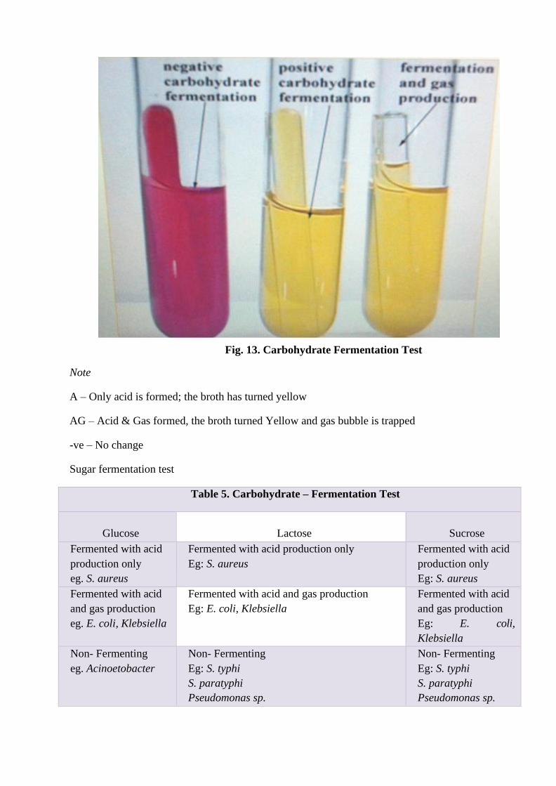
Fig. 13. Carbohydrate Fermentation Test
Note
A – Only acid is formed; the broth has turned yellow
AG – Acid & Gas formed, the broth turned Yellow and gas bubble is trapped
-ve – No change
Sugar fermentation test
Table 5. Carbohydrate – Fermentation Test
Glucose
Lactose
Sucrose
Fermented with acid
production only
eg. S. aureus
Fermented with acid production only
Eg: S. aureus
Fermented with acid
production only
Eg: S. aureus
Fermented with acid
and gas production
eg. E. coli, Klebsiella
Fermented with acid and gas production
Eg: E. coli, Klebsiella
Fermented with acid
and gas production
Eg: E. coli,
Klebsiella
Non- Fermenting
eg. Acinoetobacter
Non- Fermenting
Eg: S. typhi
S. paratyphi
Pseudomonas sp.
Non- Fermenting
Eg: S. typhi
S. paratyphi
Pseudomonas sp.

2) Oxidation – Fermentation Test
Aim
To determine the oxidation fermentation characteristics of microorganisms
Principle
This method depends upon the use of semisolid tube
medium containing the carbohydrate (Glucose) together
with a pH indicator. The acid is produced only at the surface
of medium where conditions are aerobic the attack on the
medium where conditions are aerobic the attack on the
sugar is oxidative. If acid is produced throughout the
medium including lower layers and where the conditions
are aerobic breakdown is fermentative.
Fermenting organism (Enterobacteriaceace, Vibrio)
produce an acidic reaction throughout the medium in the
covered (anaerobic) as well as open (aerobic) tube.
Oxidizing organisms (Pseudomonas) produce an acidic
reaction only in the open tube. Organisms that cannot
breakdown carbohydrate aerobically/anaerobically (alkali
genes faecalis) produce an alkaline reaction in the open tube
and no change in the covered tube. This medium may be
used for detecting gas production and motility.
Materials Required
Bacterial broth culture, D-F medium, liquid paraffinaol
Fig. 14. Oxidation Fermentation Test
Procedure
• Using sterile technique, two tubes of medium were inoculated by stabbing with sterile urine.
• Two inoculated tubes were used as control.
• Liquid paraffin was poured over the medium to form a layer about 1cm in depth into one of
the tube of each pair.
• The tubes were incubated at 37°C for 24-48hrs was observed.
Observation
The tubes were observed for the colour of the medium and the type of metabolism was recorded.
Table 6. Oxidation – Fermentation Test
OXIDATIVE FERMENTATIVE
Eg. Pseudomonas Eg. Klebsiella
S. typhi
S. paratyphi A
E. coli

3) Indole Production Test
Aim
To determine the ability of microorganisms to decompose the
amino acid tryptophan to indole
Principle
Tryptophan an essential amino acid oxidized by some bacteria by
the enzyme tryptophanase resulting in the formation of indole,
pyruvic acid and ammonia. In this experiment, the medium
contains the substrate tryptophan which is utilized by the
microorganisms.
Fig. 15. Indole Production Test
Enzymatic Degradation of Tryptophan
This ability to hydrolyse tryptophan with the production of indole is not a characteristic of all microorganisms
and therefore serves as a biochemical mask. The presence of indole is detected by adding Kovac’s reagent,
which produces a cherry red reagent layers. This colour is produced by the reagent which is composed of
Paradimethyl aminobenzaldehyde yielding the cherry red colour
Indole Reaction with Kovac’s Reagent
Culture producing a red reagent layers following addition of the Kovac’s reagent are indole positive. The
absence of red colouration demonstrates that the substrate tryptophan was not hydrolyzed and indicating
indole negative reaction.
Another reagent used is Ehrlisch’s reagent. It’s believed to be more sensitive than Kovac’s reagent and is
recommend for the detection of indole production by anaerobic and non-fermentative Gram negative
organism Kovac’s reagent was used usually initially to classify the members of Enterobacteriaceace family.
Materials Required
15 ml test tubes, bacterial culture, peptone water, Kovac’s reagent
Procedure
• The peptone water tubes were inoculated with bacterial broth culture using sterile needle technique.
• An uninoculated tube was kept as control.
• Both tubes were incubated at 37°C for 24-48 hours.
• After proper incubation, 1 ml of Kovac’s reagent was added to both tubes including the control.
• The tubes were shaken gently after an interval for 10 – 15 minutes.
Observation
The tubes were observed for the colour in the top reagent layer.
Note
Development of cherry red colour in the top layer of the tube is a positive test. Absence of red colouration is
indole negative.
Examples
Positive: E. coli, Proteus vulgaris
Negative: Klebsiella sp., Proteus mirabilis
4) Methyl Red Test
Aim
To determine the ability of microorganism to oxidize glucose with
the production and stabilization of high concentrations of acid end
products
Principle
All enteric organisms oxidize glucose for energy production and the
end products of this process will vary depending on the specific
enzymatic pathway present in the bacteria. In this test, the pH
indicator methyl red detects the presence of large concentrations of
acidic red detects the presence of large concentrations of acidic
products. The test can be used in differentiating Escherichia coli and
Enterobacter aerogenes (both coliform bacteria) that are used as
indicator of the sanitary quality of water, foods etc.
Both of these organisms initially produce organic acid end products
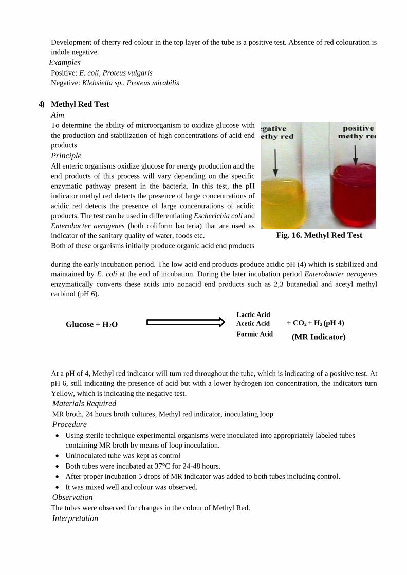
Fig. 16. Methyl Red Test
during the early incubation period. The low acid end products produce acidic pH (4) which is stabilized and
maintained by E. coli at the end of incubation. During the later incubation period Enterobacter aerogenes
enzymatically converts these acids into nonacid end products such as 2,3 butanedial and acetyl methyl
carbinol (pH 6).
Lactic Acid
Glucose + H2O Acetic Acid
Formic Acid
+ CO2 + H2 (pH 4)
(MR Indicator)
At a pH of 4, Methyl red indicator will turn red throughout the tube, which is indicating of a positive test. At
pH 6, still indicating the presence of acid but with a lower hydrogen ion concentration, the indicators turn
Yellow, which is indicating the negative test.
Materials Required
MR broth, 24 hours broth cultures, Methyl red indicator, inoculating loop
Procedure
• Using sterile technique experimental organisms were inoculated into appropriately labeled tubes
containing MR broth by means of loop inoculation.
• Uninoculated tube was kept as control
• Both tubes were incubated at 37°C for 24-48 hours.
• After proper incubation 5 drops of MR indicator was added to both tubes including control.
• It was mixed well and colour was observed.
Observation
The tubes were observed for changes in the colour of Methyl Red.
Interpretation

The colour of MR reagents remaining red is a positive test and the colour turning to yellow is negative.

Examples
MR positive – E. coli, Proteus sp; Salmonella sp.
MR negative – Klebsiella, Enterobacter sp.
5) Voges – Proskauer Test
Aim
To determine the ability of many microorganisms to produce acetone (acetyl methyl carbinol) during
fermentation of glucose
Principle
This determines the ability of many bacteria to ferment carbohydrates with the production of non- acidic /
neutral end products, acetyl methyl carbinol or its reduction product, acetyl methyl carbinol or its reduction
products, acetyl- methyl carbinol or its reduction product 2,3 Butylene glycol from the organic acids.
The reagent used in this test, Barrett’s reagent, consists of a mixture of alcoholic α- naphthol and 40%
potassium hydroxide solution. Detection of the acetyl methyl carbinol requires this end product to be oxidized
to a diacetyl compound. This reaction will occur in the presence of α- naphthol catalyst and a guanidine group
that is present in the peptone. At a result, a pink complex a guanidine group that is present in the peptone. As
a result, a pink complex is complex is formed imparting a rose colour to the medium.
Acetyl Methyl Carbinol reaction with Barrett’s reagent
Development of deep rose colour in culture with in a minute following the addition of Barrett’s reagent is
indicative of presence of the acetyl methyl carbinol and represents a positive result. The absence of rose
colouration is a negative result.
Procedure
• Using sterile technique, the experimental organism was
inoculated into VP broth by means of loop inoculation.
• One tube is kept uninoculated as control.
• The tube will be incubated at 37°C for 24-48 hours.
• After proper incubation, about 3 ml of Barrett’s reagent
A & 1 ml of Barrett’s reagent B was added into both
tubes including control.
• The tubes were shaken gently for 30 seconds with the
caps off to expose the media to oxygen.
• The reaction was allowed to complete in 15 – 30 minutes
and tubes were observed.
Observation
The tubes were observed for the development of crimson red
colour.
Note: the colour may be more intense at the surface.
Fig. 17. Voges Proskauer Test

Interpretation
Red colour formation indicates a positive test and colour change is negative.
eg. Positive – Klebsiella sp., Enterobacter
Negative – E. coli, Proteus sp.
6) Citrate Utilization Test
Aim
To determine the ability of a microorganism to utilize citrate as the sole source of carbon and as energy
source for the growth and ammonium salt as a sole source of nitrogen
Principle
Citrate test is used to differentiate among enteric bacteria on the basis of their ability to utilize / ferment citrate
as the sole carbon source. In the absence of glucose or lactose some microorganisms utilize citrate as a carbon
source. This ability depends on the presence of citrase enzyme that facilitates the transport of citrate in the
cell. Citrate, the first major intermediate in Krebs’s cycle is produced by the condensation of active acetyl
CoA with oxalo acetic acid and acetate. These products are then enzymatically converted to pyruvic acid and
carbon dioxide. During this reaction the
medium becomes alkaline; CO2 combines with sodium and
water to form carbonate, an alkaline product. This changes the
bromothymol blue indicator in the medium from green to
Prussian blue.
Citrate test is preferred / performed by inoculating the
microorganisms in to an organic synthetic medium. Simmons
citrate agar (solid) or Koser’s citrate medium (liquid) in which
sodium citrate is the only source of carbon and energy.
Bromothymol blue is green when acidic (pH 6.8 and below).
When alkaline (pH 7.6 and above). Formation of blue colour
constitutes a positive test. Citrate negative culture will show no
growth and the medium will remain green.
Materials Required
Bacterial broth, Simmons Citrate Agar Slants, Inoculation
Loop
Procedure
Fig. 18. Citrate Utilization
• Using sterile technique Simmons citrate agar slant was inoculated with the test organism by means of a
stab and streak inoculation.
• An uninoculated tube was kept as control.
• Both tubes were incubated at 37°C for 24 – 48 hours & was observed
Observation
The tubes were observed for growth and colouration of the medium.
Interpretation
Colour of the medium if turned blue, a positive result is indicated. Colour of the medium remains as green,
indicates a negative result.

2 3
3 2 2
7) Nitrate Reduction Test
Aim
To determine the ability of bacteria to produce an enzyme nitrate reductase
Principle
The reduction of nitrate by some aerobic and facultative anaerobic microorganisms occur in the absence of
molecular oxygen an anaerobic process whereby the cell uses in organic substances such as nitrates or
sulphates to supply oxygen that is subsequently utilized as a final hydrogen acceptor during energy formation.
The biochemical transformation may be utilized as follows:
NO - + 2H+ + 2e- Nitrate reductase NO + H O
Some organisms possess the enzymatic capacity to act further on nitrates to reduce them to ammonia or
molecular nitrogen. These reactions may be described as follows:
NO - NH +
Nitrate reduction can be determined by cultivating organisms a nitrate broth medium. The medium is basically
a nutrient broth supplemented with 0.1% potassium nitrate (KNO3) as the nitrate substrate. In addition, the
medium is made into a semisolid by the additional of 0.1% agar. The semisolid impedes the diffusion of
oxygen in to the medium, there by favoring the anaerobic requirement necessary for nitrate reduction. An
organisms ability to reduce nitrate to nitrite is determined by the addition of two reagent solution A, which is
sulphanlic acid followed by solution B, which is α-napthylamine followed reduction, the addition of solution
A and B will produce an immediate cherry red colour.
NO3 –
Nitrate Reductase NO2
-
Cultures not producing a colour change suggest one of two possibilities
• Nitrates were not reduced by the organism
• The organism possessed such potent nitrate reductase enzymes that nitrate were rapidly reduced beyond
nitrates to ammonia or even molecular nitrogen.
This test determines the production of an enzyme called nitrate reductase, resulting in the reduction of nitrate
(NO3). With this enzyme, nitrate is reduced to nitrite (NO2). It then forms nitrous acid that reacts with the first
reagent sulphanlic acid, and that reacts with the other reagent α-napthylamine to form a red colour. The
development of red colour, therefore, verifies that nitrates were not reduced to nitrites by the organism. If
nitrites were reduced a negative nitrate reduction had occurred. If the addition of zinc does not produce colour
change, the nitrates in the medium were reduced beyond nitrites to ammonia or nitrogen gas. This is a positive
reaction or result. Reduction of nitrate is generally an anaerobic respiration in which an organism derives its
oxygen from nitrate.
Materials Required
Bacterial broth, Nitrate broth, Nitrate reagent and inoculation loop.
Procedure
• Using sterile technique the test organism was inoculated in to nitrate broth by means of loop inoculation.
• An uninoculated broth was kept as control.
• Both tubes were incubated at 37°C for 24-48 hours.

• After proper incubation equal amounts of
nitrate reagent (solution A & B) were added to
nitrate broth Cultures and to the control tube
and the reaction was observed
Observation
The tubes were observed to see a red colour has
been developed or not.
A minute quantity of zinc was added to cultures
in which no red colour was developed and it was
observed to see if red colour has been developed
or not.
Interpretation
Fig. 19. Nitrate Reduction Test
Development of red colour indicates nitrate positive and no colour change indicates a negative test.
Eg: Positive: all members of Enterobacteriaceace
Negative: Haemophilus duceryi.
8) Urease Test
Aim
To determine the ability of microorganism to degrade urea by
means of the enzyme urease
Principle
Urease is a hydrolytic enzyme that attacks nitrogen and carbon
bond in amide compounds such as urea and forms the alkaline
end products ammonia. The presence of urease is detectable
when the organisms are grown in a urea broth medium
containing the pH indicator phenol red. As the substrate urea
is split into its products, the phenol red to turn to a deep pink.
This is a positive reaction for the presence of urease. Failure
of deep pink colour to develop is evidence of negative
reaction.
Materials Required
Fig. 20. Urease Test
Bacterial broth cultures, Christener’s urea agar slant and the inoculation loop
Procedure

• Using sterile technique, the test organism was inoculated the media by means of loop of inoculation.

• An uninoculated tube was kept as control.
• The tubes were incubated at 37°C for 24-24 hours and the reaction was observed.
Observation
The tubes were observed to see if pink colour has developed or not
Interpretation
Development of pink colour indicator a positive test and no colour change shows a negative test,
Eg Urease Positive – Klebsiella sp., Proteus sp.
Urease Negative – E. coli, Salmonella sp.
9) Mannitol Motility Test
Aim
To detect whether the given organism is motile and also mannitol is fermenting or not
Principle
Mannitol motility test medium is an example of semisolid agar media; motile bacteria swarm and give a
diffused spreading growth that is easily recognized by the naked eye. The final sterile medium should be quite
clear and transparent. After incubating the stabbed culture, non-motile bacteria generally give growth that are
confined to stab line and have sharply defined margins leaving the surrounding medium clearly transparent.
Motile bacteria typically give diffused, hazy growth that spreads throughout the medium rendering it slightly
opaque. This test also helps to identify whether the microorganisms ferment Mannitol or not. It produces
acidic end products which in turn change the red colour of phenol red indicator to yellow.
Materials Required
Bacterial culture broth, mannitol fermentation media (semisolid) and inoculation loop
Procedure
• Using sterile technique the test organism was inoculated in to the medium using stab inoculation
method.
• An uninoculated tube was kept as control.
• Both tubes were incubated at 37°C for 24-48 hours and the reaction was observed.
Observation
The tubes were observed for motility and also for
colour changes from red to yellow.
Interpretation
Diffused growth – Motile bacteria eg:
Pseudomonas sp.
Growth at stab line only – Non-motile bacteria
eg: Staphylococcus aureus only
Red colour – Mannitol non-
fermenting eg: Bacillus cereus
Yellow colour - Mannitol fermenting
eg: E. coli
Fig. 21. Mannitol Motility Test

10) Triple Sugar Iron Agar Test
Aim
To identify the microorganisms based on the ability to ferment the carbohydrates (Glucose, Sucrose and
Lactose)
Principle
The triple sugar- iron agar test is designed to differentiate among the different groups or genera of the
Enterobacteriaceace, which are all Gram negative bacilli capable of fermenting glucose with the production
of acid and to distinguish them from other gram negative intestinal bacilli. This differentiation is based on the
differences in carbohydrate fermentation patterns and hydrogen sulfide production by the various groups of
intestinal organisms. Carbohydrate fermentation is indicated by the presence of gas and a visible colour
change of the pH indicator, phenol red. The production of hydrogen sulphide in the medium is indicated by
the formation of a black precipitate that will blacken the medium in the butt of the tube.
To facilitate the observation of carbohydrate utilization patterns, TSI Agar contains three fermentative sugars,
lactose and sucrose in 1% concentrations and glucose in 0.1% concentration. Due to the production of acid
during fermentation, the pH falls. The acid base indicator Phenol red is incorporated for detecting
carbohydrate fermentation that is indicated by the change in colour of the carbohydrate medium from orange
red to yellow in the presence of
acids. In case of oxidative decarboxylation
of peptone, alkaline products are produced
and the pH rises. This is indicated by the
change in colour of the medium from orange
red to deep red. Sodium thiosulfate and
ferrous ammonium sulfate present in the
medium detects the production of hydrogen
sulfide and is indicated by the black colour
in the butt of the tube.
Carbohydrate fermentation is indicated by
the production of gas and a change in the
colour of the pH indicator from red to
yellow. To facilitate the detection of
organisms that only ferment glucose, the
glucose concentration is one-tenth the
concentration of lactose or sucrose. The
amount of acid production in the slant of the
tube during glucose fermentation
Fig. 22.Triple Sugar Iron Agar Test
oxidizes rapidly, causing the medium to remain orange red or revert to an alkaline pH. In contrast, the acid
reaction (yellow) is maintained in the butt of the tube since it is under lower oxygen tension.
After depletion of the limited glucose, organisms able to do so will begin to utilize the lactose or sucrose. To
enhance the alkaline condition of the slant, free exchange of air must be permitted by closing the tube cap
loosely. If the tube is tightly closed, an acid reaction (caused solely by glucose fermentation) will also involve
the slant.
Materials Required
Bacterial broth cultures, TSI agar slants, Inoculation Loop.
Procedure
• Using sterile technique, the test organism was inoculated into the media by means of stab and streak
inoculation.
• An uninoculated tube was kept as control
• Both tubes were incubated at 37°C for 24 hours and the reaction was observed
Observation
The tubes were observed for the colour of both the butt and slant and also gas production by means of
cracks or bubble or blackness of butt.
Observation Interference Examples
A/A without gas and H2S
production
Acid Slant / Acid butt
without gas & H2S
production
Staphylococcus aureus
A/A with gas and without
H2S production
Acid Slant / Acid butt with
gas & without H2S
production
E. coli, Klebsiella
K/A with gas and without
H2S production
Alkaline slant / Acid butt
with gas & without H2S
production
Salmonella paratyphi A
K/K without gas and H2S
production
Alkaline slant / Acid butt
without gas & H2S
production
Pseudomonas sp.
K/A with H2S production Alkaline slant / Acid butt
with H2S production
Salmonella typhi
Interpretation
• A/A: ferments glucose and either sucrose, lactose, or both.
• K/A: does not ferment lactose or sucrose; does ferment glucose.
• K/K: a non-fermenter.
• Black precipitate in stab: produces H2S (and ferments glucose).
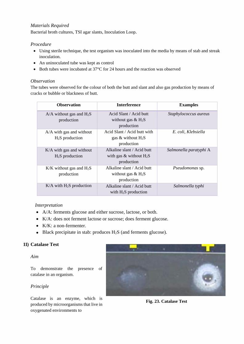
11) Catalase Test
Aim
To demonstrate the presence of
catalase in an organism.
Principle
Catalase is an enzyme, which is
produced by microorganisms that live in
oxygenated environments to
Fig. 23. Catalase Test

neutralize toxic forms of oxygen metabolites and H2O2. The catalase enzyme neutralizes the bactericidal
effects of hydrogen peroxide and protects them. Anaerobes generally lack the catalase enzyme.
Catalase mediates the breakdown of hydrogen peroxide H2O2 into oxygen and water. To find out if a particular
bacterial isolate is able to produce catalase enzyme, small inoculums of bacterial isolate is mixed into
hydrogen peroxide solution (3%) and the rapid elaboration of oxygen bubbles occurs. The lack of catalase is
evident by a lack of or weak bubble production.
Catalase-positive bacteria include strict aerobes as well as facultative anaerobes. They all have the ability to
respire using oxygen as a terminal electron acceptor.
Catalase-negative bacteria may be anaerobes, or they may be facultative anaerobes that only ferment and do
not respire using oxygen as a terminal electron acceptor (ie. Streptococci).
Uses
• The catalase test is primarily used to distinguish among Gram-positive cocci: Member of the genus
Staphylococcus is catalase-positive, and members of the genera Streptococcus and Enterococcus are
catalase-negative.
• Catalase test is used to differentiate aero tolerant strains of Clostridium, which are catalase negative,
from Bacillus species, which are positive.
• Semi quantitative catalase test is used for the identification of Mycobacterium tuberculosis
• Catalase test can be used as an aid to the identification of Enterobacteriaceace. Members of
Enterobacteriaceace family are Catalase positive.
Materials Required
24 hours old bacterial culture, glass slide, petridish, 3% H2O2, applicator sticks
Procedure
• Transfer a small amount of bacterial colony to a surface of clean, dry glass slide using a loop or sterile
wooden stick
• Place a drop of 3% H2O2 on to the slide and mix.
• A positive result is the rapid evolution of oxygen (within 5-10 s) as evidenced by bubbling.
• A negative result is no bubbles or only a few scattered bubbles.
• Dispose of your slide in the biohazard glass disposal container.
Precautions
• Do not use a metal loop or needle with H2O2; it will give a false positive and degrade the metal.
• If using colonies from a blood agar plate, be very careful not to scrape up any of the blood agar as
blood cells are catalase positive and any contaminating agar could give a false positive.
Observation
The release of bubbles was observed and compared with control.

Interpretation
Bubble Formation : Catalase Positive No
Bubble Formation: Catalase Negative
Examples
Catalase Positive : Staphylococcus aureus
Catalase Negative: Streptococcus pyogenes
Note
Care must be taken while performing catalase test of growth from blood agar plate because blood (RBC)
contains RBC catalase.
12) Oxidase Test
Aim
To test the production of oxidase bacteria
Principle
The oxidase test is a key test to differentiate
between the families of Pseudomonadaceae
(ox +) and Enterobacteriaceace (ox-), and is
useful for speciation and identification of
many other bacteria those that have to use
oxygen as the final electron acceptor in
aerobic respiration. The enzyme cytochrome
oxidase is involved with the reduction of
oxygen at the end of the electron transport
chain.
There may be different types of oxidase
enzymes produced by bacteria. The colorless
redox reagent, tetra methyl-p-
Fig. 24. Oxidase Test
phenylenediamine dihydrochloride (or dimethyl) used in the test will detect the presence of the enzyme
oxidase and reacting with oxygen, turn a colour. The oxidase reagent contains a chromogenic reducing agent,
a compound that changes color when it becomes oxidized, so it acts as an artificial electron acceptor for the
enzyme oxidase. The oxidized reagent forms the coloured compound indophenol blue.
Materials Required
Oxidase disc, 24 hours old test organism, applicator stick or glass rod.
Procedure
• The test organisms was rubbed over the reagent impregnated, filter paper disc using sterile applicator
sticks or glass rod.

• Controls were also kept along with the test and the reaction was observed within 10 seconds.

Observation
The colour changes to purple were observed with the prescribed time.
Important
Acidity inhibits oxidase enzymes activity therefore the oxidase test must not be performed on colonies that
produce fermentation on carbohydrates containing media like Mac Conkey Agar.
Interpretation
Formation of purple colour indicates a positive test. No colour changes show a negative test.
eg. Oxidase Positive: Pseudomonas sp., Vibrio sp.
Oxidase Negative: E. coli, Klebsiella
Precautions
• The test reagent is to be freshly prepared
• Nichrome wire is not used to take bacterial growth
• Cultures should not be very cold
• Culture from selective media should not be used
• The colour changes should be observed within the prescribed time
13) Coagulase Test
Aim
To distinguish coagulase producing Staphylococcus aureus from other species of Staphylococcus
Principle
Staphylococcus aureus is known to produce coagulase,
which can clot plasma into gel in tube or agglutinate
cocci in slide. This test is useful in differentiating S.
aureus from other coagulase-negative staphylococci.
Most strains of S.aureus produce two types of
coagulase, free coagulase and bound coagulase. While
free coagulase is an enzyme that is secreted
extracellular, bound coagulase is a cell wall associated
protein. Free coagulase is detected in tube coagulase test
and bound coagulase is detected in slide coagulase test.
Slide coagulase test may be used to screen isolates of S.
aureus and tube coagulase may be used for
confirmation. While there are seven antigenic types of
free coagulase, only one antigenic type of bound
coagulase exists. Free coagulase is heat labile while
bound coagulase is heat stable. Fig. 25. Coagulase Test

Slide Coagulase Test: The bound coagulase is also known as clumping factor. It cross-links α and β chain
of fibrinogen in plasma to form fibrin clot that deposits on the cell wall. As a result, individual coccus sticks
to each other and clumping is observed.
Tube Coagulase Test: The free coagulases secreted by S. aureus act with coagulase reacting factor (CRF)
in plasma to form a complex, which is thrombin. This converts fibrinogen to fibrin resulting in clotting of
plasma.
Materials Required
EDTA anticoagulant human plasma, clean glass slide, test tubes, pipettes, distilled water and inoculation loop.
Procedure
Slide Coagulase Test: Dense suspensions of Staphylococci from culture are made on two ends of clean
glass slide. One should be labeled as “test” and the other as “control”. The control suspension serves to rule
out false positivity due to auto agglutination. The test suspension is treated with a drop of citrated plasma and
mixed well. Agglutination or clumping of cocci within 5-10 seconds is taken as positive. Some strains of S.
aureus may not produce bound coagulase, and such strains must be identified by tube coagulase test.
Observation
The slides were observed for clumping or not within prescribed time.
Interpretation
Clumping formation - Positive reaction
No clumping formation – Negative reaction
Tube Coagulase Test
Three test tubes are taken and labeled “test”, “negative control” and “positive control”. Each tube is filled
with 0.5 ml of 1 in 10 diluted rabbit plasma. To the tube labeled test, 0.1 ml of overnight broth culture of test
bacteria is added. To the tube labeled positive control, 0.1 ml of overnight broth culture of known [[S.
aureus is added and to the tube labeled negative control, 0.1 ml of sterile broth is added. All the tubes are
incubated at 37°C and observed up to four hours. Positive result is indicated by gelling of the plasma, which
remains in place even after inverting the tube. If the test remains negative until four hours at 37°C, the tube
is kept at room temperature for overnight incubation.
Observation
The tubes were observed for clotting in the prescribed time.
Interpretation
Clot formation - Positive reaction
No clot formation – Negative reaction
Examples
Coagulase Positive: Staphylococcus aureus
Coagulase Negative: E. coli

Related Documents