Copyright is owned by the Author of the thesis. Permission is given for a copy to be downloaded by an individual for the purpose of research and private study only. The thesis may not be reproduced elsewhere without the permission of the Author.

Welcome message from author
This document is posted to help you gain knowledge. Please leave a comment to let me know what you think about it! Share it to your friends and learn new things together.
Transcript

Copyright is owned by the Author of the thesis. Permission is given for a copy to be downloaded by an individual for the purpose of research and private study only. The thesis may not be reproduced elsewhere without the permission of the Author.

This thesis is dedicated to my darl ing mama and dada
BIOCHEMICAL STUDIES ON ANIMAL MODELS OF
CEROID-LIPOFUSCINO
By
RYAN DENNIS MARTINUS
A thesis presented In partial
fulfilment of the requirements for
the degree of
DOCTOR OF PHILOSOPHY IN VETERINARY PATHOLOGY
Massey University
1 990
11
ABSTRACT
The ceroid-lipofuscinoses are recessively inherited lysosomal storage
diseases of children and animals, characterised by brain and retinal
atrophy and the accumulation of lipopigment in a variety of cells. A
systematic study of isolated lipopigment from an ovine form of the
disease had shown the major stored components to be proteinaceous.
This thesis presents further characterisation and identification of the
stored ovine lipopigment proteins. Separation of the lipopigment proteins
by LDS-PAG E showed the presence of the 3 .5 kDa and 14.8 kDa
proteins noted in earlier studies, and an additional band at 24 kDa. The
14.8 and 24 kDa bands varied between preparations and from different
gels of the same isolate. Radioiodination of lipopigment and silver
staining of the proteins separated by LDS-PAGE indicated that the 3.5
kDa protein was the dominant protein component. As these proteins
were unable to be separated from each other, exploitation of the molar
dominance of the 3 . 5 kDa protein led to its identification by a non
traditional sequencing approach. The major stored protein was shown to
be the full proteolipid subunit c of the mitochondrial ATP synthase
complex. The 14.8 and 24 kDa proteins were shown to be stable
oligomers of subunit c. Quantitaion of the sequence data showed that
subunit c accounted for at least 50% of the lipopigment mass. No other
mitochondrial protein was detected. Analyses of isolated mitochondria
showed that they were functionally normal and did not contain excess
amounts of subunit c.
Subunit c is classified as a proteolipid, due to its lipid-like solubility in
chloroform/methanol mixtures . Its storage in lysosome derived
l i popig m en t bodies explained many of the described physical
characteristics of lipopigment in the ceroid-lipofuscinoses.
Application of the same methodology showed that a bovine, and two
distinct canine forms of the ceroid- lipofuscinoses were also subunit c
storage diseases.
lt is postulated that the lesions in the ceroid-lipofuscinoses involve
defects in the degradative pathway of subunit c at some point after its
incorporation into the inner mitochondrial membrane.
Ill
ACKNOWLEDGEMENTS
I would like to express my sincere thanks to my chief supervisor Prof. R.
D. Jolly for giving me the opportunity to undertake this study, and for
providing the resources that enabled its completion. His attention to
detail and encouragement during the thesis production, was greatly
appreciated. I am also indebted to my other supervisors Dr. D. N.
Palmer and Dr. G. G. Midwinter. Dr. Palmer advised and helped with the
critical assessment of the experimental work. Dr. Midwinter supervised
the sequencing studies and helped with various aspects of protein
chemistry.
I would like to acknowledge the assistance, and thank a number of
people within the Department of Veterinary Pathology and Public Health
and other departments at Massey University as well as other research
institutions for their help with various aspects of the work presented in
this thesis. In particular; Mr. J. Reid from the Department of Chemistry
and Biochemistry at Massey University for the numerous amino acid
analyses and sequence runs, Dr. J. G. Shaw from the Biotechnology
Division, Department of Scientific and Industrial Research, Palmerston
North for doing the mass spectral analyses, Dr. I. M. Fearnley, Dr. J. M.
Skehel and Dr. J. E. Walker at the M. R. C. Laboratory of Molecular
Biology, Hills Road, Cambridge, U. K. for sequencing the PVDF blots, Mr.
K.B. Kirkland and Ms. I. Dopfmer for doing the negative staining electron
microscopy and the post mortems, Prof. R. D. Jolly, Dr. S. Cooper and
Mr. F . Sharpe also helped with the post mortems. I would also like to
thank Dr. M. Saifuddin for his help and advice during setting up of the
kidney cell cultures, Ms. S. L. Bayliss for assisting with the oxidative
phosphorylation measurements and her general technical help in the
laboratory, Mrs. P. Slack and Mrs. P. Davey for the preparation of
material for electron microscopy, and to Mr. T. Law for the production of
the thesis photographs. A very special thank you to my wife Nic, for
proof reading the thesis and for enduring through it all.
The work was supported by the United States National Institute of
Neurological and Communicative Disorders and Stroke Grant NS 1 1 238.
lv
PUBLICATIONS
Palmer, D. N. , Martinus, R. D. , Barns, G., Reeves, R. D. and Jolly, R. D.
( 1 988) . Ovine ceroid-lipofuscinosis 1: Lipopigment composition is
indicative of a lysosomal proteinosis. Am. J. Med. Genet. , Supp/.5
1 41 - 1 58.
Palmer, D. N. , Martinus, R. D. , Cooper, S. C., Midwinter, G. G . , Reid, J.
C. and Jolly, R. D. ( 1 989 ) . Ovine ceroid-lipofuscinosis: The major
lipopigment protein and the lipid-binding subunit of mitochondrial
ATP synthase have the same NH2-terminal sequence. J. Bioi.
Chem. 264(1 0) 5736-5740
Jolly, R. D. , Martinus, R. D. , Shimada, A. , Fearnley, I. M. and Palmer, D.
N. ( 1 990) . Ovine ceroid-lipofuscinosis is a proteolipid proteinosis.
Can. J. Vet. Res. 54 1 5-2 1 .
Fearnley, I. M. , Walker, J. E. , Martinus, R. D., Shaw, G. J. , Kirkland, K.
B., Jolly, R. D. and Palmer, D. N. ( 1 990) . The sequence of the
major protein stored in ovine ceroid lipofuscinosis is identical with
that of the dicyclohexylcarbodi-imide-reactive proteolipid of
mitochondrial ATP synthase. Biochem. J. 268 751 -758.
D., Bayliss, S. L., Jolly, R. D. , Hall, N. A. , Lake, B. D. and Wolfe, L.
S�, :t�aQ) .-��_ap��- · .:_ttle.J)QC���Qlj); -·�9W- . _'d: . -:'. . . . . .. •. . ... .... • ..,. :'t
slibtrn1t"OMtr1fO'cho1m'Piar�TP'""Sytitl'ia'Se i'n""tmm-a'fi;:ifrct Ov1� ._er -lipofuscinoses. In Lipofuscin And Ceroid Pigments (Ed. Porta, E.
Jolly, R . D. , Martinus, R. D. and Palmer, D. N. ( 1 990) . The ovine and
other models of ceroid-lipofuscinosis: Their relevance to Batten's
disease. Am. J. Med. Genet. (In Press).
V
Marti nus, R. D. , Harper, P . A. W., Jol ly, R. D. , Bayliss, S . L. , Midwinte r,
G . G . , Shaw , G. J . and Pa lmer, D. N. ( 1 990) . Bovi ne cero id
l ipofusci nosis (Batten's disease ) : The major species stored is the
D C C D - react ive proteo l i p id , subun it c, of mitoc h o nd ri a l ATP
synthase. (Submitted) .
vi
TABLE OF CONTENTS
Page
ABSTRACT .•.........•..•.......•...•••......•••..•.•...••...•.•....•..••...............•........................•............. I I ACKNOWLEDGEMENTS •.••••••••••.•••••••••.••••••.••••...••••.........•.....•.........................•............ III PUBLICATIONS .......•............•............•..•....•..•..•........••.•....••........•........•..•...•..•...•.•.......... Iv
TABLE OF CONTENTS .......••.•.......•.••..•...••..•.•............•.....•.......•.........•.....••.••................ vl
LIST OF FIGURES ..•........••..•.•.•.....••••.•....•.••.••...........••..••.•......••.......•...•..••..•.....•.•........ xll
LIST OF TABLES ...•..•••....••...•........•...••.••.•.•..•••••.•.......•..•.........•........•.......................... xvl
ABBREVIATIONS ..••..•..•...••.......•....•...••.•••.....•••............•.•.........•...........•..•...•.....•........ xvlll
CHAPTER 1 : GENERAL INTRODUCTION ...................................................... 1
CHAPTER 2: G ENERAL MATERIALS AND METHODS ........................... 1 3
2.1 ANIMALS AND TISSUES .................•............•............•.............. 1 3
2.21SOLATION O F LIPOPIGMENT .........................•...•................•. 1 3
2.3 THIN SECTION ELECTRON MICROSCOPY ...........•...•............ 14
2.4 AMINO ACID ANALYSIS ............•••••.....•..•.....•.........•................. 1 4
2.5 LITHIUM DODECYL SULPHATE POLYACRYLAMIDE GEL
ELECTROPHORESIS (LDS-PAGE) .....•....•...•..........................• 15
2.6 SILVER STAINING OF POLYACRYLAMIDE GELS ................. 1 5
2.7 CHEMICALS ...•••..••.•••.••.•....•.•.•........•....•.......••..•..•...•.....•....•.....• 1 6
CHAPTER 3 : LD5-PAGE BEHAVIOUR AND 1 251
RADIOLABELLING OF PANCREATIC
LIPOPIGMENT PROTEINS .................................................... 17
3.1 INTRODUCTION ••..•..••••••••.•••..••••....•.....•..........•..•..•••••••••••......... 1 7
3.2 SPECIAL MATERIALS AND METHODS .•••.••.•......•.................. 1 8
VII
Page
3.2.1 1 251 radlolabelllng of pancreatic llpoplgment protelns ...... 1 8
3.2.2 Detection of the radlolabel ................................•................... 1 8
3.3 RESULTS .........................................................•........................ 1 9
3.3.1 LDS-PAGE behaviour of llpoplgment protelns ................... 1 9
1 25 3.3.2 Incorporation of I Into llpoplgment protelns ................. 20
3.4 DISCUSSION ...••••••.•................................................................. 23
CHAPTER 4 : IDENTIFICATION OF THE 3.5 kDa LIPOPIGMENT
BAND ............................................................................................ 25
4.1 INTRODUCTION ........................................................................ 25
4.2 SPECIAL MATERIALS AND METHODS .................................. 25
4.2.1 Size exclusion high pressure liquid
chromatography (HPLC) ....................................................... 25
4.2.2 Amino acid sequenclng ......................................................... 26
4.2.3 Repetitive yield and Initial yield calculatlons ...................... 26
4.3 RESUL TS ................................................................................... 26
4.3.1 Size exclusion HPLC of llpoplgment protelns .................... 26
4.3.2 Amino acid sequenclng of the molar dominant
llpoplgment proteln ............................................................... 29
4.4 DISCUSSION ............................................................................. 34
CHAPTER 5 : THE CARBOXYL-TERMINAL DETERMINATION
VIII
Page
OF THE MAJOR STORED PROTEIN AND
CHARACTERISATION OF THE OTHER LIPOPIGMENT
PROTEINS ................................................................................... 37
5.1 INTRODUCTION ................................... ..................................... 37
5.2 SPECIAL MATERIALS AND METHODS .................................. 37
5.2.1 Extraction of proteollplds from llpoplgment ....................... 37
5.2.2 Diffusion elution from polyacrylamide gels .............. ....... ... 38
5.2.3 Electro blotting of llpoplgment protelns .............................. 38
5.2.4 Cyanogen bromide (CNBr) digestion of llpoplgment
proteollplds ....................................... . .................................... 39
5.2.5 Mass spectroscopy analysis of CNBr dlgests .................... 39
5.3 RESUL TS .................................................................................. 40
5.3.1 Characterisation of the proteollplds extracted
from llpoplgment ................................................................... 40
5.3.2 Identification of the 1 4.8 and 24 kDa llpoplgment
proteins ................................................................................... 43
5.3.3 CNBr cleavage of llpoplgment proteollplds and analysis
of the digest fragments .......................................................... 44
5.4 DISCUSSION ...................................................... ....................... 46
CHAPTER 6 : STUDIES ON MITOCHONDRIA
ISOLATED FROM CONTROL AND AFFECTED
SHEEP ........................................................................................... 49
6.1 INTRODUCTION ............ ............................................................ 49
IX
Page
6.2 SPECIAL MATERIALS AND METHODS .................................. 49
6.2.1 Isolation of mHochondrla and Inner mitochondrial
membrane vesicles from affected and control sheep ........ 49
6.2.2 Negative staining electron mlcroscopy ............................... so 6.2.3 Respiratory measurements .....•.....................••...•................. so
6.3 RESUL TS ................................................................................... 51
6.3.1 Electron mlcroscopy and LDS-PAGE of llpoplgment and
mitochondrial preparatlons .................................................. 51
6.3.2 Respiratory measurements on Isolated mitochondrial
fractlons ...••...•...••••••.••.•.............•.......•...................••............... 53
6.4 DISCUSSION •..........•................................................................. 56
C HAPTER 7 : CELL CULTURE OF OVINE KIDNEY EPITHELIAL
CELLS ........................................................................................... sa
7.1 INTRODUCTION .......................•................................................ 58
7.2 SPECIAL MATERIALS AND METHODS .................................. 59
7.2.1 Composition of growth and maintenance medla ................ 59
7.2.2 Preparation of affected and control kidney epithelial
cells for primary cell culture ................................................. 59
7.2.3 Growth and maintenance of cell cuHures ....•...................... 59
7.2.4 Preparation of cells for light and thin section
electron mlcroscopy .........••.•................................................ 60 7.2.5 Radlolabelllng of cuHured cells ............................................ 60
7.3 RESUL TS ................•.................................................................. 62
X
Page
7.3.1 Morphology and growth characteristics of cultured
kidney cells ............................................................................ 62
7.3.2 Measuring the synthesis of subunlt c
In cultured kidney cells ........................................•................ 65
7.4 DISCUSSION ............................................................................. 68
C HAPTER 8 : ISOLATION AND CHARACTERISATION OF
LIPOPIGMENT FROM A CASE OF BOVINE CEROID-
LIPOFUSCINOSIS ..................................................................... 70
8.1 INTRODUCTION ••...•.....•............................................................ 70
8.2 SPECIAL MATERIALS AND METHODS .................................. 70
8.2.1 Bovine tlssue ..••..................................................................... 70
8.2.2 Isolation of bovine llpoplgment bodles .......•...•.................... 70
8.2.3 Cyanogen bromide digestion of Isolated llpoplgment. ....... 71
8.3 RESULTS .....................•............•.....•.•.....•........•...•...•.•.•............. 72
8.3.1 Characteristics of bovine llpoplgments ............................... 72
8.3.2 Amino acid composition and LDS-PAGE of
bovine llpoplgment protelns ...•..•.......................................... 74
8.3.3 Amino acid sequenclng of bovine
llpoplgment ..•.•....•.....•.........•••......••......................................... 77
8.3.4 Mass spectral analysis of CNBr digest of bovine
llpoplgment ............................................................................ 78
8.4 DISCUSSION ..................................................................••......... 79
CHAPTER 9 : CANINE CEROID-LIPOFUSCINOSIS ................................ 80
9.1 INTRODUCTION ........................................................................ 80
xl
Page
9.2 SPECIAL MATERIALS AND METHODS ....•....•..••.•..•........•...•.. 80
9 .2.1 Canine tlssues ..............................................•........................ BO
9.2.2 Isolation of canine llpoplgment bodles ................................ 81
9.3 RESUL TS ......••.••••••••..•...•........••••...•...................•..•........•........... 81
9.3.1 Characteristics of canine llpoplgment. ................................ 81
9.3.2 Amino acid sequenclng of canine llpoplgment ................... 82
9 .4 DISCUSSION •..•...••....•••...........•....••......•........•..••.•.•..•...•..........•. 83
CHAPTER 1 0 : G ENERAL DISCUSSION ........................................................ 84
REFERENCES ......................................................................................................... 95

Figure
3.1 A,B
3.2 A,B
3.3
4.1 A,B & C
4.2
4.3
4.4
5.1
XII
LIST OF FIGURES
Page
LDS-20% PAGE of pancreatic llpoplgment protein
from the same Isolate run on different gels (A)
and llpoplgment proteins separated by LDS-20% PAGE
In the presence and absence of 2-mercaptoethanol (B) ............. 1 9
Incorporation of 1251 at various lodogen concentrations (A)
and at various times (B) ••............................................................... 20
LDS-20% PAGE of 12511abelled pancreatic llpoplgment
protelns ........................................................................................... 21
HPLC profile of llpoplgment proteins eluted from a
TSK G2000 SW column and a TSK G2000 SW and G4000 SW
column connected In series (A & B). LDS-20% PAGE of
fractions eluted from the columns connected In series (C) ....... 27
LDS-20% PAGE of llpoplgment proteins after acetone
precipitation and size exclusion HPLC .........................•.............. 28
Cycles 1 , 2, 3 and 1 0, 1 1 and 1 2 obtained when
pancreatic llpoplgment protein was sequenced ......•................... 29
The PTH amino acid yields of the major sequence obtained from
pancreas (A), brain (B) and kidney (C) llpoplgment proteln ....... 31
LDS-20% PAGE of total pancreatic llpoplgment proteins (A)
nonextractable proteins (B) and extracted proteolipid (C) .......... 41
Figure
5.2
5.3
5.4
5.5
6.1
6.2
6.3
7.1
7.2
XIII
Page
LDS-1 5% PAGE of pancreatic llpoplgment proteins that were
diffusion eluted from an LD$-1 5% polyacrylamide gel ................ 43
Coomassle blue stained pancreatic llpoplgment proteollplds
elctro blotted onto a PVDF membrane .......................................... 44
Linear mass spectral scan over the molecular Ion region
of the carboxyl-terminal CNBr cleavage fragment of subunlt c
extracted from pancreatic llpoplgment ......................................... 45
Amino acid sequence of the major stored llpoplgment
proteln ........................................................................................ ..... 46
Thin section electron micrographs of pancreatic llpoplgment
and Isolated mitochondrial preparations (A & B) and negatively
stained llpoplgment and Inner mltochodrlal membrane vesicles
(C & 0) ...............•.••..•......••............•.....................•.............•.............. 51
LDS-PAGE of Inner mitochondrial membrane vesicle
proteins from control and affected sheep livers (A) and PAGE of
bovine ATP synthase (8) ............................................................... 52
Respiratory activity of Isolated mitochondrial preparations
from affected (A) and control (B) kidney tlssue ........................... 53
Thin section electron micrographs of affected kidney
epithelial cells In primary culture (A) and In third
passage culture (B) .....................................................•................. 62
The growth of affected (A), and control (B), kidney epithelial
primary cultures •....•....•.•....••...••.....•........•...................................... 63
Figure
7.3
7.4
7.5
7.6
8.1
8.2
8.3
8.4
8.5
9.1
xlv
Page
Light microscopic appearance of affected (A) and control (B)
primary cultured cells stained with H&E and luxol fast blue ..... 63
Thin section electron micrographs of membrane bound
cytoplasmic bodies from affected cultured cells (A & B) and
autophagic structures from control cells (C & 0) .....•.................. 64
% of 3H Incorporated Into TCA preclpltable protein
from labelled affected (A) and control (B) primary kidney
cell cultures .................................................................................... 65
3H distribution amongst the proteolipid fractions
from labelled affected and control cultured cells separated by
LOS-1 5°/o PAGE ............................................................................... 67
Thin section electron micrographs of llpoplgment Isolated
from bovine pancreas, liver, kidney and brain (A, B, C & 0) ....... 72
Isolated bovine llpoplgments on CsCI Isopycnlc gradlents ........ 74
LOS-1 5% PAGE of llpoplgment protein Isolated from ovlne
pancreas (A) and bovine pancreas, kidney and liver (B,C & 0) .. 76
The PTH amino acid yields of the first 25 resldues
of subunlt c sequenced from bovine pancreatic llpoplgment .... 77
Linear mass spectral scan over the molecular Ion region
of the carboxyl-terminal CNBr cleavage fragment of subunlt c
Isolated from bovine pancreatic llpoplgment. ••....•••.•....•.....•........ 78
Thin section electron micrographs of llpoplgment Isolated
from frozen Border Collie brain (A) and Tibetan Terrier brain
(B & C) •....•.•..•.... ••..•••.•••.••.•••.•••••.••.•••••.....•..........•..•••••••••••••.........•... 81
Figure
9.2
XV
Page
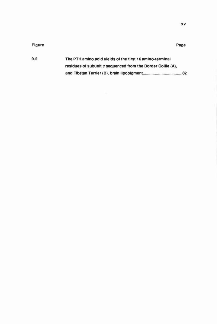
The PTH amino acid yields of the first 1 6 amino-terminal
resldues of subunlt c sequenced from the Border Collie (A),
and Tibetan Terrier (B), brain llpoplgment .................................... 82
Table
3.1
4.1
4.2
5.1
5.2
6.1
6.2
7.1
XVI
LIST OF TABLES
Page
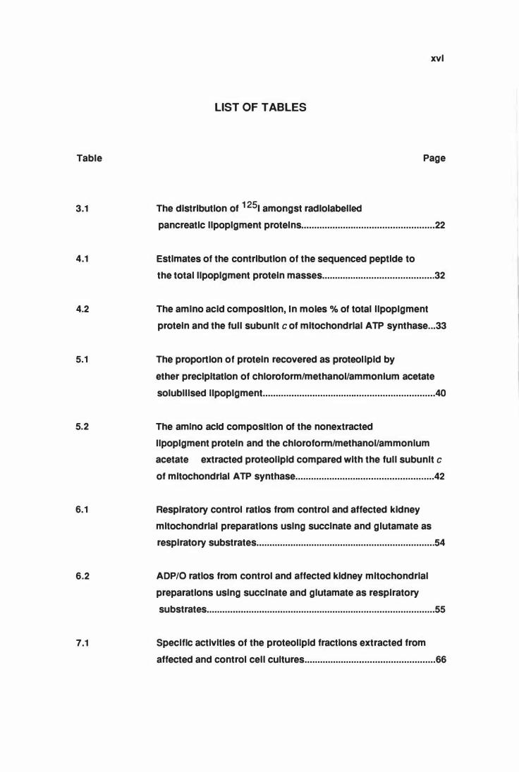
The distribution of 1251 amongst radlolabelled
pancreatic llpoplgment protelns ................................................... 22
Estimates of the contribution of the sequenced peptide to
the total llpoplgment protein masses ........................................... 32
The amino acid composition, In moles o/o of total llpoplgment
protein and the full subunlt cof mitochondrial ATP synthase ... 33
The proportion of protein recovered as proteolipid by
ether precipitation of chloroform/methanol/ammonium acetate
solubilised llpoplgment ...........•.......•.............................................. 40
The amino acid composition of the nonextracted
llpoplgment protein and the chloroform/methanol/ammonium
acetate extracted proteolipid compared with the full subunlt c
of mitochondrial ATP synthase ..........................•..•........•.............. 42
Respiratory control ratios from control and affected kidney
mitochondrial preparations using succinate and glutamate as
respiratory substrates .•...........•...................................................... 54
ADP/0 ratios from control and affected kidney mitochondrial
preparations using succinate and glutamate as respiratory
substrates ........•.••••.•....••.....•...•.•..........................•.......•.................. 55
Specific activities of the proteolipid fractions extracted from
affected and control cell cultures .....................••.....•..................... 66
Table
8.1
8.2
1 0.1
XVII
Page
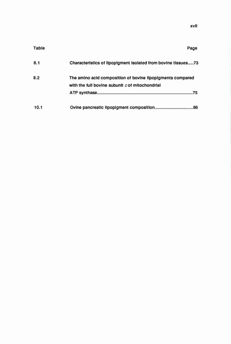
Characteristics of llpoplgment Isolated from bovine tlssues ..... 73
The amino acid composition of bovine llpoplgments compared
with the full bovine subunlt c of mitochondrial
ATP synthase ..........................•....................................................... 75
Ovlne pancreatic llpoptgment composttlon ................................. 86
ACR
ADP
ATP
ATV
Da
DCCD
E DTA
FBS
H&E
HPLC
kDa
LDS
LDS-PAGE
M E M
PAGE
PBS
PSK
PTH
PVDF
SOS
SOS-PAGE
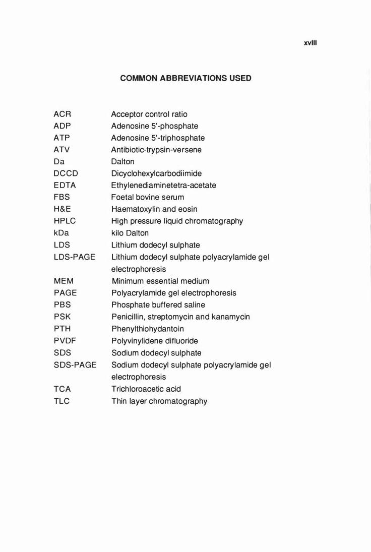
COMMON ABBREVIATIONS USED
Acceptor control ratio
Adenosine 5'-phosphate
Adenosine 5'-triphosphate
Antibiotic-trypsin-versene
Dalton
Dicyclohexylcarbodi imide
Ethylenediaminetetra-acetate
Foetal bovine serum
Haematoxyl in and eosin
High pressure l iquid chromatography
kilo Dalton
Lithium dodecyl sulphate
Lithium dodecyl sulphate polyacrylamide ge l
e lectrophoresis
Minimum essential medium
Polyacrylamide gel e lectrophoresis
Phosphate buffered sal ine
Penici l l in , streptomycin and kanamycin
Phenylthiohydantoin
Polyvinylidene difluoride
Sodium dodecyl sulphate
Sodium dodecyl sulphate polyacrylamide ge l
electrophoresis
TCA Trich loroacetic acid
TLC Thin layer chromatography
XVIII
1
C H A P T E R 1
GENERAL INTRODUCTION
The ceroid-l ipofusci noses are a g roup of recessive ly i n he rited lysosomal
storage d iseases of chi ldren and domestic animals. They are characterised
pathological ly by brain and retinal atrophy and the presence of fluorescent
l ipopigment bodies in neurones and a variety of other cel l types throughout
the body. Cl in ical features i nclude blindness, seizures, mental retardation
and dementia u ltimately leading to premature death.
The term ceroid-l i pofuscinosis was i ntroduced as a descri ptive name fo r
t h e se d i s eases by Zeman and Dyken ( 1 969 ) o n t h e bas is of s im i lar
h i stoche mical and fluorescent characteri stics of the sto red l ipopigment to
those of pigments ceroid and l ipofuscin . Prior to th is, these diseases were
g rouped with the gang liosidoses as forms of amaurotic fami lial idiocy. They
are prese ntly also known generical ly as Batten's disease. Lipofuscin (age
pig ment) and ceroid are two types of i ntracel lu lar cytosomes ( l ipopigments)
characte rised by a yel low-brown colour, fluorescence under u ltravio let l ight
and stai n i ng with l ip id stai ns . The former is regarded as a normal age
re lated phenomenon and the latter as a pathological p igment associated
with Vitamin E deficiency (Porta and Hartroff, 1 969) .
There are up to 1 0 subtypes of ceroid- lipofuscinosis which differ i n the age
of o nset and i n the p rog ress ion of c l in ical disease. H owever, the main
e nt i t i e s are t he i n fant i le ( H a lt i a-Santavu o ri ) , late i n fant i l e (Jansky
B i e lschowsky) , juven i le (Batten , Spie lmeyer-Sjogren) and an adult (Kufs)
fo rm (Lake, 1 984; Dyke n , 1 988; Berkovic, et al. , 1 988; Boustany, et al. ,
1 988; Wisniewski , et al. , 1 988) . Col lectively they are bel ieved to be the
m ost common type of lysosomal storage disease in humans. Prevalence
e st i mates as h igh as 1 i n 25 ,000 and 1 i n 1 2 ,500 l ive b i rths have been
reported (Zeman , 1 976 ; Rider and Rider, 1 988).
2
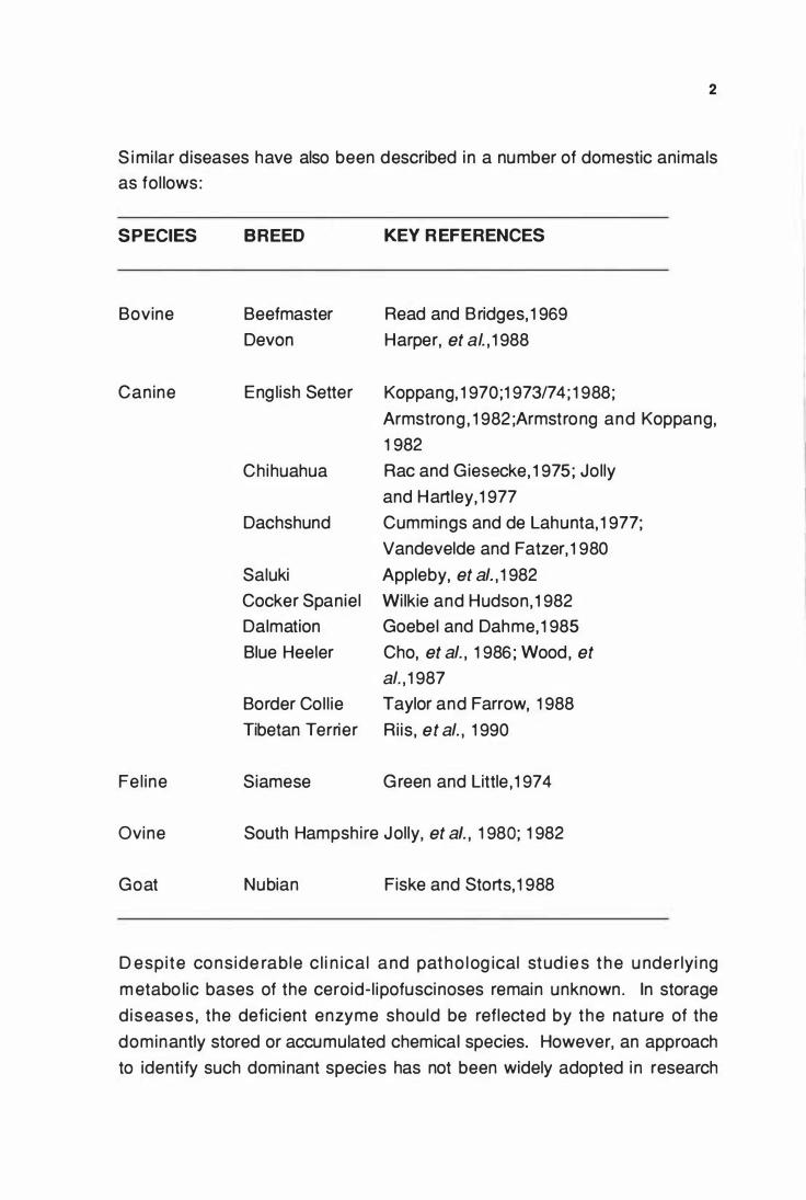
S i mi lar d iseases have also been described i n a number of domestic animals
as fol lows:
SPECIES BREED KEY R EFERENCES
Bovine Beefmaster Read and Bridges, 1 969
Devon Harper, et al. , 1 988
Canine Eng lish Setter Koppang, 1 970;1 973/74 ; 1 988;
Armstrong , 1 982 ;Armstrong and Koppang,
1 982
Chihuahua Rac and Giesecke, 1 975; Jolly
and Hartley, 1 977
Dachshund Cummings and de Lahunta, 1 977;
Vandevelde and Fatzer, 1 980
Saluki Apple by, et al. , 1 982
Cocker Spaniel Wi lkie and Hudson, 1 982
Dalmation Goebel and Dahme, 1 985
Blue Heeler Cho, et al. , 1 986 ; Wood, et
al. , 1 987
Border Col l ie Taylor and Farrow, 1 988
Tibetan Terrier Ri is , et al. , 1 990
Fe l ine Siamese Green and Little , 1 974
Ovine South Hampshire Jolly, et a/. , 1 980; 1 982
Goat Nubian Fiske and Storts, 1 988
D espi te cons ide rable c l i n ical and patho log ical stud i e s t h e u nderlyi ng
m etabo l ic bases of the ceroid- l ipofuscinoses remain unknown. In storage
d iseases , the deficient enzyme should be reflected by the nature of the
dominantly stored or accumulated chemical species. However, an approach
to identify such dominant species has not been widely adopted i n research
3
i nto the ceroid- l ipofusci noses. This is probably due, in part, to the l imited
a m o u nt of post mortem t i ssue avai lab le f ro m t h e h u m a n fo rms and
difficulties encountered in isolat ing and solubi l ising the l ipopigment.
T h e l i pi d stai n i ng and f luoresce nce properties of the sto red l ipopig ment ,
w hich had been l ikened to those of ceroid and l ipofuscin (Zeman and Dyken,
1 969) , i mplied that their accumulation reflected a simi lar pathogenesis. This
was perceived to be associated with peroxidation of l ipid and the formation
of Schiff base polymers from malonaldehyde and amino acids , produced
d u ring free radical peroxidat ion of po lyunsatu rated fatty acids (Chio and
Tappel , 1 969a,b).
A deficiency in leucocyte peroxidase in late infantile and juveni le forms of the
d i sease was reported by Armstrong , et al. , ( 1 973) . These fi ndings led
Z e man ( 1 974) to suggest that s i nce malonaldehyde cou ld be produced
during free radical peroxidation of polyunsaturated fatty acids, the formation
of f lu o resce nt l i pop igments i n the cero id- l i pofusc inoses cou ld i nvo lve
abnormal peroxidation of l ip ids . A nu mber of other reports conf irmed
deficiencies of leucocyte peroxidase i n the late i nfanti le (Armstrong , et al. ,
1 974a; Awasth i , et al. , 1 977 ; Jensen , et al. , 1 977), juven i le (Armstrong , et
al. , 1 974b ; Gadoth , 1 978) and adu lt forms of disease (Armstrong , et al. ,
1 974b; Bozdech , et al. , 1 980) . In contrast, no evidence for a peroxidase
d eficiency was found i n the i nfanti le (Anzi l , et al. , 1 975; Den Tandt and
Martin , 1 978; Seeker, et al. , 1 979) and juveni le forms of the disease (Haust,
et al. , 1 976 ; Den Tandt and Marti n , 1 978) . lsoe lectric focussi ng studies
revealed no qualitative or quantitative differences in these enzymes i n saliva
a nd parotid g land betwee n no rmal i ndividuals and pat ients with juven i le
neuronal ceroid- l ipofuscinosis (P i lz, et al., 1 976a,b; Pi lz and Goebel , 1 977).
A rm strong , ( 1 982) later reported that "pat ients with g e neral ised ceroid
l i pofusci nosis have relative ly normal levels of peroxidase if total enzyme is
m e asured , but differ i n the i nt race l lu lar d ist ributi on" , i . e . a decrease in
so lub le leucocyte peroxidase activity was accompanied by an increase in
i nsoluble or bound peroxidase activity. Much of the debate concerning the
apparent peroxidase deficiency in the ceroid- l ipofuscinoses has centred on
t h e m ethodo logy of measu rement and subce l lu lar d i st ri buti on of these
enzymes.
4
Spectrophotometric determi nation of peroxidase activity i nvo lved the use of
hydrogen peroxide as substrata and p-phenylenediamine as hydrogen donor
(Armstrong , et al. , 1 973). The use of this hydrogen donor was stressed by
Armstrong , et al. , ( 1 974b) as "when other donors are employed the enzyme
deficiency may not be demonstrated". Tsan , et al. , ( 1 978) , however found
the use of p-phenylenediamine to be unsuitable for the study of peroxidases.
T h e subcel luar distribut ion and enzymology of the leucocyte peroxidases
h ave not been wel l characterised.
G utteridge, et al. , ( 1 982) reported e levated leve ls of non-protein bound i ron
in the cerebrospinal fluid from patients affected with the i nfantile and juveni le
forms of cero id-lipofuscinosis. As iron is a catalyst of the l ipid peroxidation
process, an i ncrease in i ron levels was l inked to a decreased abi lity of the
cerebrospinal fluid to inhibit hydroxyl radical production , a necessary step in
l ip id peroxidation . A defect in i ron metabolism was subsequently suggested
as a possible mechanism i n the pathogenesis of the ceroid-l ipofusci noses
(Gutteridge , et al. , 1 983) . However, He iskala, et al. , ( 1 988) reported that
concentrat ions of loosely bound i ron and copper i n cerebrospinal f luid of
patients with i nfanti le , late i nfanti le and juven i le forms of d isease did not
correlate with cl in ical diagnosis, nor with the degenerative symptoms of the
d isease. They concluded that there was no support for a major ro le for i ron
toxicity in the development of neuronal degeneration.
The nature of the f luorophore responsible for the f luorescence of stored
l i popigment has also attracted much interest and speculation , as it has been
considered to be significant to the pathogenesis of the ceroid-l ipofuscinoses.
Studies of the l ipid component of l ipopigment showed the presence of a
f l u o re scent acid ic struct u re t h at co nce ntrated at t he o rig i n i n no rmal
p hospol ipid thin layer chromatography developing solvents (Siakotos, et al. ,
1 972 ; S iakotos and Koppang , 1 973 ; Zeman , 1 976). The so cal led "acidic
l ipid polymer" or "Schiff base polymer" was thought to be formed by reaction
of malonylaldehyde, a product of peroxidation of polyunsaturated fatty acids,
w it h am ino g roups. S im i larity of the fl uorescent and ch romatog raphic
p ropert ies of th i s mate ria l to t hose generated with in vitro peroxidation
stud ies (E I Ieder , 1 98 1 ) , l ater added support to t h is theory . Corrected
5
excitation-emission spectra by Katz , et al. , ( 1 988) showed that l ipopigment
fro m Batten's disease tissues emitted i n the yellow-orange reg ion (520-540
n m ) and not i n the blue reg ion (460-480 nm) expected of the "Shift base
polymer''. An i ncrease in the absorption spectra at 280 nm was also noted.
This was interpreted to be due to "non-fluorescent l ipids".
Another fluo rescent polymer found in the neutral l ipids of l ipopigment was
designated "polymalonaldehyde" again on the basis of the simi larities of its
f l u o re sce n t a n d c h ro m ato g ra p h i c p ro p e rt i e s to in vitro g e n e rated
"polymalonaldehyde" (Siakotos, et al. , 1 972 ; Zeman, 1 976 ; Gutteridge , et al. ,
1 977) .
A noth e r h ypot hes is l i nked to the putat ive l i p i d natu re of t h e sto red
l i po pig ment i mpl ied a pri mary defect in fatty acid metabol ism. This was
developed fol lowing observations of abnormal fatty acid profi les i n the major
phospholipids (phosphatidylchol ine, phosphatidylinosito l , phosphatidylserine
and phosphatidylethano lam i n e ) of b rai n fro m chi ldren affected with the
i nfant i le form of disease (Hagberg , et al. , 1 968; 1 974; Svennerholm, et al. ,
1 975 ; 1 987 ; Svennerholm, 1 976) . In particu lar, an i ncrease in 20:4(n-6), a
decrease in 22 :4(n-6) and 22:6(n-3) was reported. The name
" p o ly u n satu rated fatty acid l i p idos is" was subsequ e nt ly proposed to
distinguish this disease from the other forms in which the fatty acid changes
were not observed. In contrast, Pul larkat, et al. , ( 1 982) reported a decrease
i n t h e proport ions of 22 :6 (n-3) i n g rey matter phosphat idylseri ne in the
i nfanti le , late infant i le, adu lt and 'pigment variant' forms, but no changes in
the fatty acids of the other phospholipids.
The l ipid peroxidation hypothesis provided a rationale for the therapeutic use
of antioxidants such as vitamin E , butylated hydroxytoluene or iron-chelating
a g e n t s s u c h as des fe r ri o x a m i n e ( Ze m a n , 1 9 7 4 ; S antavuo ri and
Weste rmarck, 1 984 ; Santavuo ri , et al. , 1 988) . However, "ant ioxidant
therapy" has not produced any significant cl inical improvement attributed to
t h i s fo rm of t reatment i n pat i e nts affected w i th ce ro i d- l i pofuscinosis
( S a n tavu o r i , et al. , 1 9 8 8 ) , a nd t h e m e t h od o f t re at m e n t re m a i n s
controversial .
6
A f luorescent protein complex was left after mu lt ip le solvent extractions of
l i po pigment i so lated from brains of late i nfanti le form of disease (Wolfe , et
al., 1 977) . Spectral and chemical analysis of th is component led them to
s u g g e st t h at the f l uore scence was d u e to the p resence of a ret ino id
component , possibly complexed to a smal l peptide. As this component was
shown to account for 50% of the dry weight of the storage material , a defect
i n ret i noic acid metabo l ism was suggested as a possible mechanism of
pat h og e n e s i s i n t he ce ro i d - l i pofu sci noses . H oweve r , these spectral
assignments were ambiguous and have also been shown to be compatible
with a cholesterol and/or reti nol-cho lesterol complex (Nelson and Hal ley,
1 977) .
The presence of dolicho l , another i soprenoid, at "elevated" levels i n brain
t issue , isolated sto rage cytosomes and i n the uri nary sedi ment of patients
with i nfanti le, late infanti le and juveni le forms of disease has been reported
(Wolfe , et al., 1 977; Ng Ying Kin and Wolfe , 1 982 ; Ng Ying Kin , et al., 1 983) .
As a consequence defects i n dol ichol metabol ism or processing of golg i
de rived lysosomes and membranes have bee n suggested as possib le
causes of the disease. However, simi lar incorporation of
[3H]mevalonolactone and [1 4c]acetate i nto dolichol fractions in cu ltured skin
fib rob lasts from pat ients with neuro nal ceroid-l i pofusci nosis and control
f ibroblasts led Paton and Poulos, ( 1 984) to conclude that the disease did not
i nvo lve a defect i n do l icho l metabo l ism . I ncrease i n dol ichol i n u ri nary
sediment led to the suggestion that urine dol ichol measurements could be
used as a biochemical marker of the disease (Wolfe, et al., 1 986) . The low
s p ec i f icity of the test d u e to n u m e ro u s false posit ives fro m bacteri al
contamination , alcohol i ngestion and vigorous exercise prior to the test may
l im it its usefulness (Wolfe, et al., 1 988). lt is possible that the uri nary dolichol
l eve ls ref lect the excret ion of the sto rage l ipopig ment in slough ed renal
tubu lar ce l l cytoplasm ( R . D. Jol ly, pers . comm. ) . I ntrace l lu lar dolichol is
fou nd with in lysosomes and is general ly regarded as a lysosomal marker
(Wong , et al., 1 982) . I ncreased dol ichol levels i n the ce rebral cortex of
pat ients with Alzhei mer's disease and in aged ind ividuals (Wo lfe , et al.,
1 982 ; Pul larkat and Reha, 1 982; Ng Ying K in , et al., 1 983) , suggested that
i ncreased do l icho l leve ls i n b rains of ceroid- l i pofuscinosis pat ients may
reflect a secondary phenomenon. E levated uri nary dolicho l levels i n chronic
7
alcohol ics has also been reported (Pul larkat and Raguthu , 1 985).
Significantly h igher contents (up to 20 times) of phosphorylated dolichols (P
dol ichol) have been reported in whole tissue extracts of patients with ceroid
l i pofusci nosis relative to that i n age-matched cont ro ls (Hal l and Patrick,
1 985; Pu l larkat , et al. , 1 988 ; Danie l , 1 990). Qualitative analyses i ndicated
that the P-do l ichol that accumulated i n brains of late infanti le and juveni le
pat ients with cero id- l ipofuscinosis was largely l i nked to ol igosaccharides
rang ing in size from four to fourteen monosaccharide units (Hall and Patrick,
1 988). A defect i n the metabolism of dolichol-l inked ol igosaccharides (Do l
P P- O S ) i n vo lved i n t h e g lycosy lat i o n of p rote i n s was subsequent ly
suggested as a possible biochemical basis of the disease (Pul larkat, et al.,
1 988; Pul larkat, 1 990). However, no evidence has yet been reported for the
presence of any abnormal ol igosaccharides or defective g lycoproteins in the
ce ro id- l ipofusci noses. Wolfe , et al. , ( 1 988) suggested that an i ncrease in
do l ichyl phosphates may not be unique to the ceroid-l ipofuscinosis, as they
were also found to be i ncreased in brai ns of GM 1 -gang l iosidosis and Tay
Sachs disease patients. As the Do l-PP-OS isolated from l ipopigment was
estimated to account for 7% or less of the dry weight of l ipopigment, (Hal l , et
al. , 1 990) their presence in the disease was unl ike ly to represent the primary
biochemical defect.
I vy , et al. , ( 1 984) showed that i nject ions of leupept in (a th io l-prote inase
i nh ib itor) or ch loroquine (a general lysosome enzyme inh ibitor) i nto brains of
rats i nduced lysosome associated g ranu lar aggregates resembl i ng the
l i po p i g m e nt found i n pat i e nts w i t h n e u ro n a l cero id - l i pofusci noses .
Accumulat ion of l ipopigment i n these diseases by a s imi lar defect ive (or
absent) lysosomal prote i nase was suggested. These f i ndi ngs and the
observed concommitant increases i n dolichol levels in rat brains treated with
leupepti n and chloroquine led Wolfe, et al. , ( 1 987) to propose a relationship
between "dolichols and lysosomal organelle membrane turn over". A specific
defect in a cysteine proteinase, important to the recycl ing and exocytosis of
o rganel lar membrane protei ns was postulated as a possible cause for the
cero id-lipofuscinoses. As g lycosylation of thiol-endoproteases are regu lated
by the levels of dol icho l phosphates i n the golgi and endoplasmic reticu lm,
over-g lycosylation due to h ig h levels of dolicho l phosphates has also been
8
suggested as a possible mechanism leading to a putative protease defect
(Boustany and Kolodny, 1 989).
A variable decrease in cathepsin B activity in f ibroblasts from patients with
various forms of ce ro id- l ipofusci nos is has been reported (Dawson and
G laser , 1 987) . This was conside red to be a seco ndary e ffect due to
accumulation of abnormal peroxides, resulting from a deficiency of a specific
p h os p h o l i pase A2 ( Daws o n and G laser , 1 988 ) . Howeve r , i mpai red
lysosomal phospholipase A1 activity and normal activity of phosphol ipase A2 i n tissues from "some cases" of humans with neuronal ceroid-l ipofusci nosis
h as also been reported (Dawson, 1 990) . The significance of these results to
the d isease, if any, remains to be determined.
Eto, et al., (1 990) reported that cathepsins B, H , and L activities in cu ltured
ski n fi broblasts from patients with the juven i le form of the d isease were
s imi lar to those determined from control fibroblasts.
R educt ion i n fasting serum very low density l ipoprotein (VLDL) i n patients
with the juveni le form of the disease, suggested a metabolic defect in l ipid
t ransport (G i l l is , et al., 1 987; Bennett , et al., 1 988) . Another observation
i nvo lved a decrease in e ryth rocyte membrane f lu idity in patients with the
juveni le form of disease, possibly due to a decrease in docosahexenoic acid
( Koh lschutter, et al., 1 988) .
I mmunoreactivity of l ipopigments from the juveni le form of the disease was
n oted with monoclonal antibodies raised against fragments of amyloid 13-
p rotei n . Local isat ion of t h i s immunoreactivity to a 3 1 kDa protei n from
i so l ated l i po p i g m e nt l e d K i tag u c h i , et al., ( 1 9 90 ) to s ug g e st t h at
pathoge nes is might i nvo lve defect ive processi ng of amylo id precu rsor
p rotein from which 13-protein is derived. The 13-protein is a major component
of amyloid deposits i n agi ng and Alzheimer's disease and i s thought to be
deposited as a resu lt of a p roteo lyt ic processi ng defect of the precu rsor
protei n ( Dyrks, et al., 1 988). However, as the 31 kDa protein isolated from
l ipopigment has not yet been characterised, the significance of this result to
the ceroid-l ipofuscinoses remains to be determined .
9
Most of the above expe rimental observations and hypotheses have been
based on the l ipid stai n i ng and f luorescence characteristics of the stored
l i po pigment , and com pou nds found at "e levated leve ls" i n total t issue
extracts. None have led to the e lucidat ion of the underly ing biochemical
anomaly in the ceroid- l ipofuscinoses.
Ceroid-l i pofuscinosis has been recorded in a number of animal species as
noted ear l ie r. However, most of these reports are case studies and the
d iseases have not been fu l ly characterised. The disease in the Eng l ish
Setter dog though , is one that has been studied cl inically and pathologically
as a model of the juven i le human disease (Koppang , 1970; 1973/74; 1988;
Goebel , et al., 1982).
B i ochem ical i nvesti gat i ons i nto t h e can i ne d isease have cante red on
pe roxidase e n zymes and fatty acid abnorma l it i es (Pate l , et al., 1974;
S ia kotos , et al., 1978; A rm stro n g , et al., 1978a,b; Armstrong , 1982;
Armstrong and Koppang , 1982; Farnsworth, et a/., 1982; Keller, et a/., 1984;
Reddy, et al., 1985; Koppang , 1988). These studies have been l inked with
t h e putative l i p id pe rox idat ion theories of pathogenes is i n t he ceroid
l ipofuscinoses.
T h e d isease i n the South H amps h i re sheep h as also been extensive ly
studied as a model of the juveni le human disease (Jol ly , et al., 1980; 1982;
1988; 1989; Graydon and Jo l ly , 1984; Mayhew, et al., 1985). The use of
such an an ima l mode l h as g reat ly advanced t h e study of t h e cero id
l ipofusci noses. Not on ly i s experi mental mate rial readi ly avai lable but the
ab i l ity to euthanase an an i mal and commence iso lat ion of l i popigment
bodies with in mi nutes of death h as the advantage that artefactual post
mortem changes can be min imised.
The analogous d isease in sheep shows ret inal degeneration and severe
brain atrophy. The latter is a disti nctive feature of the ce roid-lipofuscinoses
re lative to other lysosomal storage diseases. Although the brai ns of new
born affected lambs were with in t he normal weight range for the fi rst 4
months, by terminal disease at 24-26 months of age , brai n weights were
50% of those of normal sheep at that age (Mayhew, et al., 1985; Jolly, et al.,
1 0
1 989) . Atrophy began with a laminar necrosis i n the parietal area of the
cerebral cortex becoming more diffuse and spreading to the occipital and
last ly the temporal lobes. lt was accompanied by an i ncreasingly severe
fib ri l lary astrocytosis and an i ncrease in l ipopigment-laden macrophages.
Although storage of physical ly simi lar l ipopigment occurs in a variety of other
lysosomal storage diseases and i n normal aging , neuronal necrosis is not
such a feature. Th is suggested that i n ceroid - l i pofusci nosis , neurona l
necrosis was associated with the metabo l ic les ion rather than storage of
l ipo pigment per se (Jol ly, et al., 1 989 ; 1 990).
Lipopigment stored in neurones stai ns with Sudan black, luxol fast blue and
pe r iodic acid-Sch iff (PAS) stai ns. They a lso have a yel low f luorescence
when i rradiated with 366nm uv l ight in both paraffi n and frozen sections.
I nt e ns ity of f l uoresce nce and sta i n i ng i ncrease s with age ref lect i n g
i nc reasi ng s ize and total amou nts o f t h e sto red l ipopigment . Storage
m ateria l i s also found i n cardiac muscle , hepatocytes and Kupffer ce l ls ,
kidney and pancreatic epithelial cel ls and a wide variety of other cells with in
the body (Jol ly , et al., 1 980; 1 982) . U ltrastructurally they appear as electron
dense, membrane-bound, i rregulari ly rounded bodies. They have a g ranular
textu re but also many lame l lar p rofi les which h ave been descri bed as
fingerprint, curvi l inear, crystal loid or multi lamel lar structures. The reason for
th is diversity of structures i s not yet understood. They have however, been
i nterpreted as complex, three dimensional matrices in which repeating un its
o f p rot e i n and l i p id form paracrysta l l i ne structu res as a resu lt of the
condensation of bi layer membranes (Jol ly, et al., 1 988; 1 989) . The i r freeze
f racture e lectron microscopic appearance and powder X-ray diffract ion
pattern is thought to support this interpretation (Jol ly, et al., 1 988) .
The primary role of peroxidation of polyunsaturated fatty acids i n l ipopigment
formatio n in ceroid-li pofuscinosis was questioned by Palmer, et al., ( 1 985).
Th is was on the basis of the similarity of the brain g rey matter phospholipid
fatty acid compositions of sheep affected with ovine ceroid- l ipofuscinosis to
those of control sheep. There were no signs of essential fatty acid deficiency
i n the affected sheep. Sheep as ruminants must conserve their restricted
fatty acids fo r structu ral functions , and as such , if there was a fatty acid
d e fect , it s h o u ld h ave been ref lected in t h e phospho l i p id fatty acid
compositions. l t was concluded that an abnormality in fatty acid metabol ism
1 1
was not primari ly i nvolved i n the pathogenesis of the disease.
A re lat ive ly s imp le m ethod of l i pop ig ment iso lat ion was deve loped by
Palmar, et al., (1 986a) . This enabled the systematic characterisation of the
sto red l i popigment. Li popigment i so lated from l iver, kidney, pancreas and
brai n of affected sheep was shown to contain 1 6-27% l ipid. Phospholipids
accounted fo r 50% of l iver l ipopig ment l ipids and contai ned the normal
mammalian membrane components phosphatidylchol ine,
phosphatidylethano lam ine , phosphatidyl i nosito l , phosphatidylseri ne and
b i s ( m o n oacy lg lyce ro ) p h osphate , a known lysosomal l i p id component
( B i e i ste i n , et al., 1 980) . The re m ai n i ng 50% of the l ip id fract ion was
accounted for by the neutral l ipids cho lestero l , dol ichol , dolichyl esters and
ubiquinone. These were thought to be i ndicative of a lysosomal orig in of the
l i po pig m ent . Do l icho l , i n the form of do l ic h o l py rophosphate l i nked
o l i gosaccharides , was a lso present accounti ng for 0 . 1 -2% of the total
l ipopigment mass (Hall , et al., 1 989) . The concentrations and distributions of
l i pop ig m ent metals accou nt i ng for 1 -2% (copper, i ron , calci u m , z i nc,
m a nganese , a l um i n u m , n icke l and ch rom i u m ) we re also cons ide red
consistent with the l ipopigment cytosomes functioning as lysosomes at some
stage i n their biogenesis (Palmer, et al., 1 988).
The majo r component of iso lated l ipopigment (65-75%) was found to be
p rotei naceous i n natu re . Th is mate rial was difficu lt to handle due to its
relative i nsolubi l ity. Of the large range of solvent systems tried , on ly SDS in
the presence of 5% 2-mercaptoethanol and formic acid were able to dissolve
t h e l ipop igment or de l i p idated p rote in obtai ned f rom it (Pa lmar , et al.,
1 986b) . Silver stain ing of l ipopigment proteins separated by so�ium dodecyl
su lphate polyacrylamide gel e lectrophoresis (SDS-PAGE) showed two major
protein bands, one that ran to the lower reg ion of the gel with an apparent
molecu lar weight of 3 .5 kDa and another at 1 4.8 kDa. These proteins were
relative ly insensitive to Coomassie b lue stai ns . The 3.5 kDa l ipopigment
band was present i n such amounts that its abnormal accumulation al lowed
the disti nction to be made between total control and affected whole tissue
homogenates, a classic criterio n for the identif ication of the abnormally
stored components in lysosomal storage diseases.
The analyses by Palmer, et a/, ( 1 986a,b) fai led to show any products of l ipid
1 2
peroxidation o r abnormality i n the stored l ipids, nor was there a depletion of
unsaturated fatty acids. No quantitatively dominant fluorophore was found i n
t h e l i p id fract ions . I n fact , the f luorescence of t h e bod ies was lost on
separatio n of the various components. There was no evidence to suggest
that disturbances i n metal metabo l ism led to l ipopigment format ion . The
smal l amounts of phosphorylated dol ichol (0 .2-2%) also i ndicated that a
defect i n metabol ism of do l ichol-l i nked o l igosaccharides was un l i ke ly to
ref lect t he pri mary cause of the ovi ne d isease (Ha l l , et al., 1 989) . The
p re sence of low mo lecu la r we i g ht prote i ns as the major l i pop igment
component led Palmer, et al., ( 1 986a,b) to conclude that the ovine disease
was not a l ipidosis but represented a lysosomal proteinosis.
As the ovine disease was considered to be a g ood model of the juveni le
hu man d isease , these fi ndings represented a considerable advance i n the
understanding of the ceroid-l ipofuscinoses.
A number of mechanisms that cou ld lead to a lysosomal proteinosis were
proposed by Jol ly , et al., ( 1 988) . These were a deficiency of a lysosomal
protease or its contro l , secondary perturbation of lysosomal function simi lar
to that i nduced by i at roge n ic drugs or NH4C I , t he presentat ion to t h e
lysosomal system o f post-translat ional ly modi fi ed protein that cannot be
catabo l ised, the presentat ion of prote in i n a complex that protected it from
p roteo lys is , o r a defect i n recycl i ng from lysoso mes of some specif ic
membrane domain .
This thesis describes the identification of the major stored ovine l ipopigment
protein , considered to be specific to the disease. The relative i nsolubi lity and
difficulty in separation of the l ipopigment protein resulted in a non-traditional
approach bei ng adopted i n its subsequent characterisation . Appl ication of
the methodology developed during studies on the ovine l ipopigment was
also used to define the major stored l ipopigment component from two canine
forms and a bovine case of ceroid-l ipofuscinosis.
C H A P T E R 2
G ENERAL MATERIALS AND METHODS
2.1 ANIMALS AND TISSU ES
1 3
Sheep with ceroid-l ipofuscinosis used in th is study were from an i nbred flock
of South Hampshi res, maintai ned by the mating of heterozygous ewes with
7 -8 mo nths o ld homozyg o u s ly affected ram s . These a n i ma ls we re
h usbanded at Massey U n ive rs ity under standard New Zealand pastu re
fa r m i ng co nd i t i o n s . D iag nos i s of affected lambs was establ i shed by
h istopathology of brai n b iops ies at 2.5-3 months of age ( Dickso n , et al. ,
1 989) . Tissues fo r expe ri mentati on were obtai ned at autopsy of affected
sheep 1 2-24 months old, and placed in ice-cold 0.0 1 M phosphate buffered
s a l i n e ( P B S ) , p H 7 . 2 w i t h i n m i nu tes of e u t h a n as i a by barb i tu rate
anaesthesia and exsangui nation . Confi rmation of the earl ier diagnosis of
ceroid-l ipofusci nosis was made from gross evidence of brain atrophy and/or
h istopathology. Control t issues were obtai ned from age matched normal
Southdown or New Zealand Romney sheep.
2.2 ISOLATION OF LIPOPIGMENT
Lipopigment was isolated from pancreas, kidney, brain and liver of affected
sheep by a combination of homogenizat ion , osmotic lysis, sonication and
centrifugation (Palmer, et al. , 1 988) .
From Pancreas, Liver and Brain
Pancreas, l iver o r brai n g rey matte r (2-40 g) , was ri nsed with PBS and
homogenized in 10 vol of ice-co ld 0.4 mM Tris-HCI, pH 7.4 for 1 min in a
Sorvall Omnimixer ( lvan Sorval l I nc. ) . The homogenate was fi ltered through
gauze , sonicated for 1 m i n , fi ltered through g lass wool , then pel leted in a
S o rva l l G LC- 1 ce nt ri fu g e fo r 30 m i n at 1, 400 g max · The pel let was
resuspended i n deion ized water and subjected to centrifugation at 12,000
gmax for 20 min . A white fluffy layer was gently washed off the pe l let which
was then resuspended i n deionized water, and pel leted by centrifugation at
1 2,000 gmax for 20 min. This procedure of washing and centrifugation was
14
repeated unt i l a uniform pel let was obtained. The fi nal pellet was suspended
i n deion i zed water or di sso lved in 1 % l i th ium dodecyl sulphate (LDS) and
sto red at -20°C unti l requ i red .
From Kidney
Lipopigment bodies from kidney cortical tissue were isolated using the same
m ethod as above , except that a soft brown pe l let was removed from the
son icated homogenate by sed imentat ion at 70 g max i n a So rval l GLC-1
centrifuge for 5 min.
2.3 THIN SECTION ELECTRON MICROSCOPY
Al iquots of isolated l i popig ment were fixed i n 2% g lutaraldehyde and 3%
parafo rmaldehyde i n 0 . 1 M phosphate buffe r at pH 7.2 , post f ixed in 1 %
osmium tetroxide and embedded i n epoxy resin . Thin sections were stained
w i th u rany l acetate and l ead c i t rate and e x a m i ned i n a P h i l l ips 200
transmission electron microscope.
2.4 AMINO ACID ANALYSIS
Samples were subjected to hyd ro lysis in vacuo in 6 M g lass dist i l led HCI
contai n i ng 1 % phenol fo r 24 h at 1 1 0°C. The hydrolysates were dried in
vacuo over NaOH pel lets and the amino acids analysed on a Beckman 1 1 9L
amino acid analyzer. Cystine content was determined in some samples by
analysis of cysteic acid after oxidat io n wit h pe rfo rmic acid (H i rs , 1 967) .
P rote i n co nce ntrat i ons were calcu lated from t h i s data and also by the
method of Lowry, et al. , ( 1 95 1 ) . The samples analysed were (a) proteins
p recipitated from solut ions of l ipopig ments disso lved in 1 % LDS with co ld
aceton e , (b) ether p rec ip itated p roteo l ip ids extracted f ro m l i popigment
dissolved i n chloroform/methano l (2 : 1 , v/v) contain ing 1 00 mM ammonium
acetate, (c) the nonextracted material that precipitated at the i nterface during
the m odif ied Folch extract ion , and (d) l ipopig ment bod ies suspended i n
deion ized water that were dried to constant we ight in vacuo over NaOH
pellets.
2.5 LITHIUM DODECYL SULPHATE POLYACRYLAMIDE G EL
ELECTROPHORESIS (LDS-PAGE)
1 5
LDS- 1 5% and 20% polyacrylamide ge ls, ( 1 .5 mm x 10 cm x 16 cm) , were
prepared i n a Protean Dual S lab Ce l l (B io- Rad , Rich mond, Ca) by the
method of Laemmli (1 970) , except that the bisacrylamide to acrylamide ratio
was 1 :29 w/w instead of 1 :37.5 w/w, and 1 0% w/w sucrose was added to the
main gel (Palmar, et al. , 1 986b) .
Fresh and frozen isolated l ipopigment proteins disso lved i n 1 % LDS at 2 mg
prote i n/m! were d i luted with de ion ized wate r and added to a solut ion of
boi led g l ycero l and b romopheno l blue to a fi na l conce ntrat ion of 1 2%
glyce ro l and 6 j.!g/m l of bromophenol blue. Lipopigment contain ing 1 5-25 llg
of protein in 50 Ill was loaded onto each lane of the gel . Samples contain ing
the l ipopigment proteins were not heated at any stage. E lectrophoresis was
carried out at a constant cu rrent, 8 mA, at 4°C, u nt i l the bromophenol blue
reached the bottom of the gel , i n approximate ly 1 7 h . Apparent mo lecu lar
weig hts of the prote i ns were determi ned by comparison of their mig ration
rates with those of mo lecu lar weight standards obtai ned from Sigma (Kit No.
MW-SOS 70L) .
2.6 SILVER STAINING OF POLYACRYLAMIDE G ELS
After e lectrophoresis the gels were fixed for 60 m in in 200 ml of 1 2% (w/v)
trichloroacetic acid. They were then washed for 60 min with 800 ml of 40%
methanol contain i ng 1 0% acetic acid , twice for 30 m in with 400 ml of 1 0%
eth ano l contai n i ng 5% acet ic acid and f i na l ly fo r 1 0 m i n with a 200 m l
so lut ion o f 3 .4 mM potass ium dichromate co ntai n ing 3 .2 m M n itr ic acid.
Afte r removal of a l l t h e oxid iz i ng agent from the ge ls by wash i n g with
deionized water, they were stai ned for 1 0 m in with 200 ml of 1 2 mM si lver
nitrate , rinsed for 2 m in with deion ized water and deve loped by successive
additions of port ions of a solut ion of 0 .28 M sodium carbonate contain ing
6.33 mM formaldehyde (0 .01 9%) . The fi rst development was for 1 m in , the
seco nd fo r 5 mi n and the th i rd was cont i n u ed unt i l the desi red stai n i ng
intensity was reached. Deve lopment was stopped by washi ng the ge ls for 5
m in with 400 m l of 5% (v/v) acetic acid. A l l steps afte r the addition of the
1 6
s i lver n i t rate so lut ion we re carri ed out u nder a photog raphic safe l ight
(Kodak, Wratten series fi lter OB). Gels were then destai ned for 2 min with
200 ml of a solut ion of 30 m M potassium ferricyanide and 65 mM sodium
th iosulphate (Farmer's reducer) . After complete removal of th is reagent by
wash i ng with deion ized water, the gels were stained again with 1 2 mM si lver
n itrate , and developed as before.
2.7 CHEMICALS
Lith ium dodecyl su lphate (LDS) , 2-mercaptoethanol , bovine seru m albumin
(BSA) , lodogen (1 ,3,4 ,6-tetrachloro 3a,6a-diphenylg lyco luri l ) , Coomassie
Bri l l iant B lue R-250, (3-[Cyclohexylamino]- 1 -propanesulfon ic acid) (CAPS)
cyanogen bromide and molecular weight standards (Kit No . MW-SOS 70 L)
were obtained from Sigma (St . Louis, MO. U .S .A.) . Al l chemicals required for
si lver stain i ng were obtained from Bio-Rad (Rich mond, · CA. U .S .A.) except
fo r sucrose and t richloroacetic acid (TCA) which were · obtained from BDH
(Poo le , E n g land) . The s i lver n itrate was obtai ned fro m Ajax chemicals
( N . S . W . , Aust ral ia ) . The 2 , 5-d ipheny loxazo le ( PPO) and 1 ,4-b is [2-(5-
phenyloxazo lyl)]benzene (POPOPlwere also obtai ned from BDH . Adenosi ne
5'- d iphosphate , disodium (ADP) cat No . 1 0490 was obtained from United
States Biochemical Corporation (Cleve land , Ohio, U .S.A. ) . Sephadex G-25
f i n e g rad e , was obta i n ed from P h armac ia F i n e c h e mica ls ( U ppsala,
Swede n ) . Carr i e r free N a 1 25 1 in N aO H (1 00 mC i/m l ) , N C S t i ssue
so lub i l iser and 3H-amino acid mixtu re (code TRK.440) ( 1 .0 mCi/ml) were
obtained from Amersham International P .L.C. (Amersham , Bucks, Eng land) .
Po lyvinyl idene difluoride membranes (PVDF) were obtained from Mi l l ipore
( M i l l i pore Corp. Bedfo rd , MA. U . S . A . ) . DC-P iasti kofo l i en ce l lu lose TLC
sheets were from Merck ( E . Me rck, Darmstadt , West Germany) . Eag les
min imum essential media (MEM) (Cat. No. 1 0-1 0 1 ) and MEM vitamins (Cat.
No. 1 6-0 1 4-49) were from Flow Laboratories, Inc. (West Germany). Trypsin ,
f oetal bovi ne serum (FBS) and t ryptose broth were obtai ned from Difco
Laboratories (Detroit, M l , U . S .A) . P lastic 25cm2 tissue cu ltu re flasks were
from Nu nclon , l nterMed (Denmark) and leig hton tubes were from Kimax
(U .S .A.) . All water was purified through a Mi l l i-Q Reagent water system and
a Mi l l istak GS filter from Mi l l ipore, so that it had a min imum resistance of 1 0
M ohms/cm. All other reagents were of an analytical g rade and all so lvents
used were double dist i l led.
1 7
C H A P T E R 3
LDS-PAGE BEHAVIOUR AND 1 251 RADIOLAB ELLING OF PANCREATIC
LIPOPIGMENT PROTEINS.
3.1 INTRODUCTION.
Analysis of l ipopig ment iso lated from pancreas, l iver, kidney and brain of
s h e e p affected w i t h ce ro id- l i pofusci nos is h as shown that 70% of t he
l ipopigment mass was protei n , most o f the remainder be ing neutral l ipids
and phosphol ip ids expected for the lysosome derived l ipopigment bodies.
Iso lated l ipopigment could be solubi l ised by sodium dodecyl sulphate on ly in
the prese nce of 5% 2-me rcaptoethano l . Subsequent separat io n of the
l ipopigment proteins by SOS-PAGE, showed a major band migrati ng with an
apparent molecu lar we ight of 3 .5 kDa at the l imit of reso lution of the gel ,
heterogeneous materia l between 5 .0 -9 .0 kDa and a band at 1 4 .8 kDa
(Palmer, et al. , 1 986b) . The PAGE behaviou r of these specific l ipopigment
prote i ns was furt he r i nvest igated . The presence of t he 3 .5 kDa band i n
affected but not i n control tissue homogenates indicated that i t was specific
to the disease . Its re lative si lver stai n ing i ntensity implied that it was a major
component of the iso lated l ipopigment. The si lver stain cou ld not however be
taken as a q u ant itative measu re of t hese p rote ins due to t he fact t hat
d iffe rent ia l se ns i t ivity of d iffe re nt prot e i n s to stai ns i s we l l documented
(Friedman, 1 982 ; Merri l , 1 986) .
The 1 4.8 and 3 .5 kDa lipopigment bands extracted from polyacrylamide gels
were shown to contai n s imi lar re lative amounts of tyrosi ne (S.M . Cooper,
pers . com m . ) . Radioi od i n at ion of pancre at ic l i popi g m e nt protei ns was
therefore i nvestigated as a means of measuri ng them quantitative ly and as a
method of obtain i ng a radiolabel led substrata.
3.2 SPECIAL MATERIALS AND METHODS
3.2.1 1 251 RADIOLABELLING OF PANCREATIC LIPOPIGMENT
PROTEINS
18
An lodoge n (1 ,3 ,4 ,6 -tetrach lo ro 3a, 6a-dipheny lg lyco lu ri l ) so lution was
prepared by the method of Markwel l and Fox (1 978) . lodogen was disso lved
i n dich loromethane, (40 Jlg/ml) , 1 00 , 200, 300 , 500, 700 J.!l and 1 ml of this
solut ion were added to g lass vials and evaporated to dryness under nitrogen
at 25°C. Removal of the dichloromethane produced a film of lodogen on the
g lass vials. A 1 0 111 solution of 0 .05 M ammon ium acetate , pH 7.4, was
added to the vials fol lowed by 25 J.!Ci of Na 1 251 . After the addition of 200 Jll
of pancreatic l ipopig ment disso lved i n 1 % LDS ( 1 . 5 mg/ml) , the reactants
were incubated at 25°C for 1 , 2, 5, 1 0 , 1 5, 20 and 30 min. Care was taken to
ensu re that the reactants were i n contact with the lodogen fi lm on the vials.
The iod i nat ions were terminated by transfer ing t he reactants to 500 Jll of
0 . 1 % LDS and leavi ng them for a further 1 0 m in to al low the un incorporated
i odous i o n s to retu rn to mo lec u l a r i odi n e . I nco rporat i o n of 1 25 1 i nto
l ipopigment was measu red by the method described below. The mixture was
app l i e d to a Sep hadex G -25 co l u m n (3 .8 x 2 0 0 m m ) t h at h ad bee n
pretreated by the e lution of 1 ml of bovine serum albumin 3% (w/v) i n 0 . 1 %
LDS to prevent the non-specific absorption of the radiolabel led protein . The
fi rst e luted radioactive peak was col lected and the protei n precipitated by
acetone (1 0 : 1 , v/v) . The precipitated protei n was re-so lubi l ised in 1 % LDS
and added to u n labe l l ed pancre at i c l i pop ig m e nt p rote i n fo r fu rt h e r
experimentation.
3.2.2 D ETECTION OF THE RADIOLABEL
Incorporation of 1 251 was determi ned by TLC. A 1 0 Jll al iquot of the label led
l ipopigment was spotted onto DC-Piastikfo l ien cel lu lose TLC strips (1 x 1 4
cm) and developed in trichloroacetic acid 1 2% (w/v). The dist ribution of the
rad ioact iv ity was determi ned by cutt i ng the strips i nto 1 cm pieces and
cou nt ing the gam ma emissions on a Nuclear Enterprise N E 1 600 gamma
coun te r. The p rote i n remai n ed at the o rig i n and the free iodine ( 1 25 1 )
chromatographed with the solvent front (Salaci nski , et al. , 1 981 ) . Proteins in
samples of iodinated lipopigment were also separated by LDS-PAGE. The
dist ribution of radioactivity in po lyacrylamide gels was determ ined by cutti ng
the gels i nto strips, counting the gamma emissions as above , and compari ng
the count distribution with si lver stained side strips.
19
3.3 RESULTS
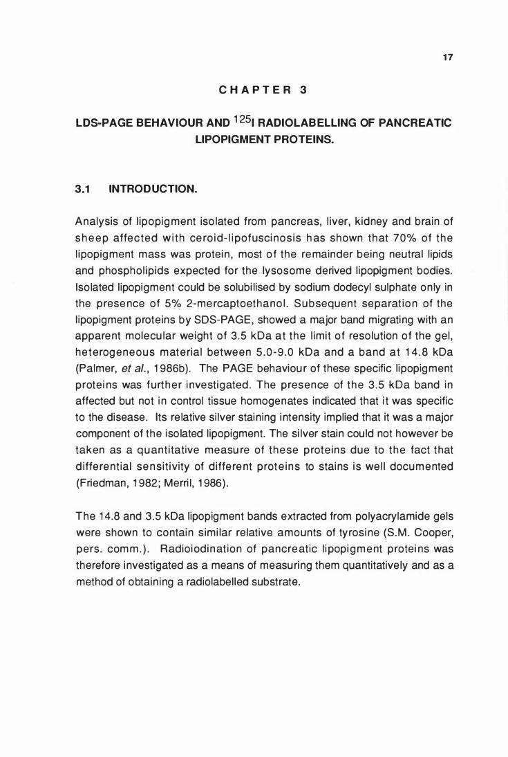
3.3.1 LDS-PAGE behaviour of l ipopigment proteins
So lub i l i ty of l i popig ments in LDS i n t h e abse nce of 2 -me rcaptoethanol
a l lowed t h e PAGE behaviour of the l i pop ig ment prote i ns to be fu rt he r
i nvest igated. Pancreatic l ipopigment protei n s , separated by LDS-PAGE ,
were characterised by a major band with a n apparent mo lecular weight of
3.5 kDa. Othe r bands at 1 4.8 and 24 kDa were also noted and considered
specific to the l ipopigment. However, the proportions of the latter two bands
varied between preparations and from different gels of the same iso late (Fig .
3. 1 ,A and Fig .4 .2) . The presence or absence of 5% 2-mecaptoethanol in the
sample had no i nf luence on the pattern of the major l ipopigment bands
obtai ned ( Fig . 3 . 1 , B).
(A) A
Fig 3.1 A, B
B A B
(B)
-24
1 4 . 8
-14.8 1 9-5
-3.5 I - 3 . 5
Si lver stained LDS-20% PAGE of (A) pancreatic l ipopigment prote in (lane A) and the same sample run under identical conditions on another ge l ( lane B) , ( 1 5 J..Lgllane ) . ( B) P ancreatic lipopigment protein in the presence ( lane A) and absence (lane B) , of 2-mercaptoethanol, (25 J..Lg/l ane) . The numbers o n t h e r ig ht hand side i nd icate apparent molecular weights calculated from the migration of molecular weight markers.
20
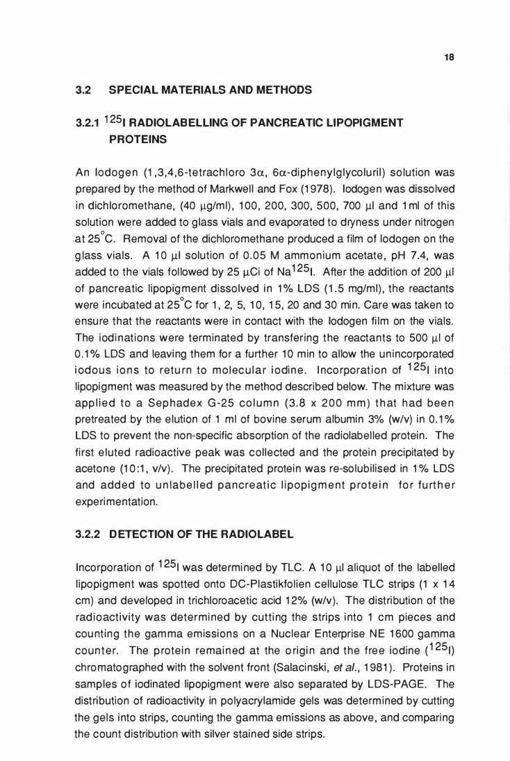
3.3.2 Incorporation of 1 251 into lipopigment proteins
I ncorporation of radioactive iodine into l ipopigment protei n was optimal at 1 2
J.19 o f l odogen/v ia l ( Fig . 3 . 2 A ) . At t h i s co ncentrat i on , a t ime cou rse
experiment showed that 1 0 min was the m in imum t ime requi red to obtain a
maximum incorporation of 70% of the radioactive iodine i nto the l ipopigment
proteins at 25°C (Fig . 3 .2 B) .
Fig.3.2 A,B
A 80 75 70
"0 65 CD 60 -as 55 ... 0 c. 50 ... 0 4 5 u 40 .E
.]) 35 C\J 30 ..- 25 � 0 20
1 5 1 0
5
8 8 0 75 70
"0 65 CD 60 -� 55 &. 50 0 45 u 40 .E 35 lli 30 C\J ..- 25 � 0 20
l 5 1 0
5
5 10 1 5
5 10
20 25
llQ lodogen/vial
1 5
Time (mln)
20
30 - 35 40
25 30
Incorporation of 1 25 1 at various lodogen concentrations, (A) and the incorporation of 1 251 at 1 2 Jlg lodogen/vial at various time intervals , (B). lodinations were carried out using total pancreatic lipopigment dissolved in 1 % LDS at ( 1 .5 mg/ml) and 25 Jl.Ci Na 1 25 1 , incorporat ion was determined by the dpm that remained at the o rigin of the TLC strips compared to the total dpm applied. Each point on the graphs represents individual estimations.
21
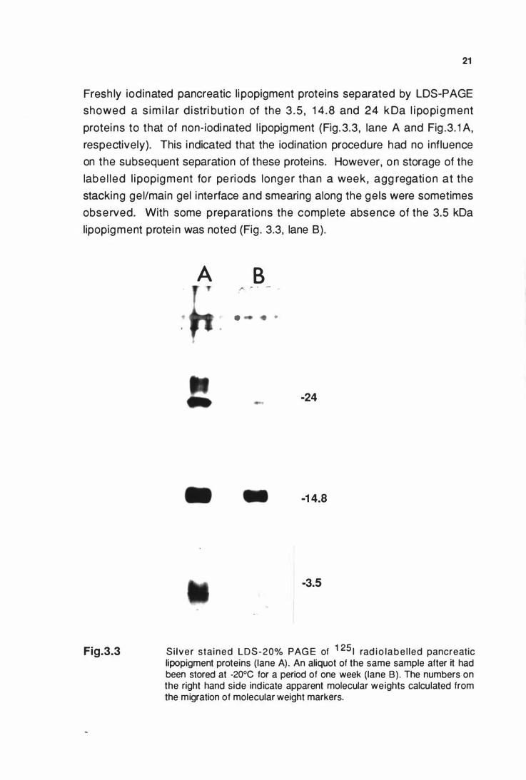
Fresh ly iodinated pancreatic l i popigment proteins separated by LDS-PAGE
showed a s im i lar d istri but io n of the 3 .5 , 14 .8 and 24 k Da l i popi g ment
protei ns to that of non-iodi nated l ipopigment (Fig. 3.3 , lane A and Fig .3 . 1 A,
respectively). This i ndicated that the iodination procedure had no influence
on the subsequent separation of these proteins. However, on storage of the
labe l led l i popigment for pe riods longer than a week , agg regati on at the
stacking ge l/main gel i nterface and smearing along the ge ls were sometimes
observed. With some preparat ions the complete absence of the 3.5 kDa
lipopigment protei n was noted (Fig . 3.3, lane B).
Fig.3.3
•
A B ,.. ... . -
• •
-24
-1 4.8
-3.5
Si lver stained LDS-20% PAG E of 1 25 1 rad i o la b e l led pancreatic lipopigment proteins (lane A) . An aliquot of the same sample after it had been stored at -20°C for a period of one week (lane B) . The numbers on the right hand side ind icate apparent molecular weights calculated from the migration of molecular weight markers.
22
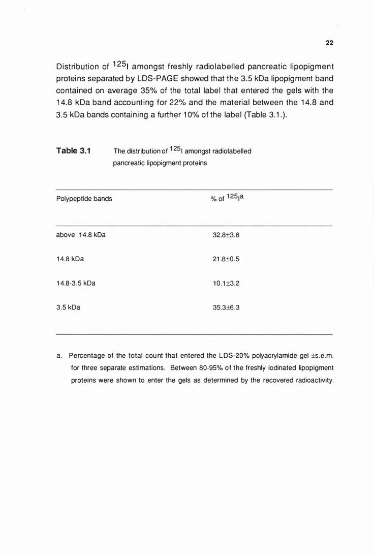
Dist ri bution of 1 25 1 amongst f resh ly radio label led pancreat ic l ipopigment
protei ns separated by LDS-PAGE showed that the 3 .5 kDa l ipopigment band
contai ned on ave rage 35% of the total labe l that entered the gels with the
1 4.8 kDa band account ing for 22% and the material between the 1 4.8 and
3.5 kDa bands contain ing a further 1 0% of the labe l (Table 3 . 1 . ) .
Table 3.1
Polypeptide bands
above 1 4.8 kDa
1 4.8 kDa
1 4.8-3 .5 kDa
3 .5 kDa
The distribution of 1 25 1 amongst radiolabelled
pancreatic lipopigment proteins
32.8±3.8
21 .8±0.5
1 0 . 1 ±3.2
35.3±6.3
a. Percentage of the total cou nt that entered the L DS-20% polyacrylamide gel ±s .e.m.
for three separate estimations. Between 80-95% of the freshly iodinated lipopigment
proteins were shown to enter the gels as determined by the recovered radioactivity.
23
3.4 DISCUSSION
Lipopigment bodies can be iso lated from pancreatic t issue re latively free
f rom c o nta m i nat i o n w i th o t h e r s u b ce l l u l a r o rg an e l l e s a n d d e b ri s .
L ipopig me nt prote i ns from th is t issue also g ive t h e best separation o n
po lyac ry l a m ide g e l e lect ro p h o re s i s . For t h e s e reaso n s pancre at ic
l ipopigment was chosen for the LDS-PAGE and radio label l ing studies.
The s imi lar e lectrophoretic patterns obtained when total l ipopigment prote in
d isso lved i n 1 % LDS i n the prese nce or absence of 2-mecaptoethanol
i ndicated that the 3.5 kDa and 1 4 . 8 kDa l ipopigment p rote ins were not
subu n i ts of a larg e r agg regate h e ld toget h e r by i nte rmo lecu lar cyst ine
disu lph ide bonds. However, the variabi l ity of the 24 and 1 4.8 kDa bands
betwee n different preparations and from different gels of the same iso late
suggested some form of aggregation that was resistant to dodecyl sulphate
and reduci ng conditions.
The rep lacement of aromatic hydrogen by e lectrophi l ic iodine in activated
aromat ic systems (tyrosi ne , h istid i ne , t ryptophan) i s widely used to labe l
prote ins . E lectroph i l ic iodine can be generated by a variety of oxidizi ng
agents : ch lo ramine-T (G reenwood and Hunte r, 1 963) , hydrogen peroxide
( H u b b a rd and Co h n , 1 9 72 ) , ch lo ri ne gas ( B utt , 1 9 7 2 ) , and sod i u m
hypochlorite (Redshaw and Lynch , 1 974). These form t h e basis of most
avai lable methods of radio-iodi nation . However, the use of oxidizing agents
that come into contact with protei ns i n so lution can lead to oxidative damage
to the prote ins . Lactoperoxidase and lactoperoxidase-catalyzed iodinations
using hydrogen peroxide alone or generated by g lucose oxidase systems
require the addition of extraneous protei n to the system (Marchalonis, 1 969).
This extraneous protei n which can itself become hig hly labe l led during the
i od inat i o n process , provides an addit ional compl ication . An a lternative
method e mploys conjugat ion labe l l i ng whe re N-succi n i midyl p ropionate
labe lled with iodine is attached covalently to lys ine residues or to the amino
terminus of the protein (Bo lton and Hunter, 1 973). Although this techn ique
offe rs s o m e advantag es ove r t h e c h l o ram i n e-T and l actoperoxidase
method s , i t is a ve ry tedi ous m ethod of iod i nation . The i nt roduction of
1 , 3 ,4 , 6-tetrach lo ro-3a,6a-dipheny lg lyco lur i l ( l odogen) as an i odi nat ing
24
reagent fo r so lub le and membrane protei ns provided a method of protein
i od i nat i o n u n d e r m i ld cond it i o n s , wi t h out sacri f ici ng ce l l u la r i nteg rity
(Markwel l , and Fox, 1 978). As the lodogen method was techn ical ly simpler
to use , and because it did not require the addition of extraneous protein or
st ro ng oxidants to i n it iate the react ion , nor a stro ng reduci ng reagent to
t e rmi nate i t , i t was chose n as the m e t h od fo r the rad i o i od i n at i on of
pancreatic l ipopigment proteins.
lt was reasoned that since LDS is a denatu ring detergent, 1 251 radio label l ing
of po lypept ides i n i t shou ld i nco rpo rate iod ine equal ly i nto a l l ty rosine
residues. Pre l im inary ami no acid analysis i ndicated that the 1 4.8 and 3.5
kDa l ipopig ment bands contained s imi lar mo lar ratios of tyrosine residues
( S . M . Coope r , pe rs . com m . ) . The refo re , t h e d ist ri but i o n of rad io labe l
amongst total pancreat ic l ipop igment p rote i ns separated by LDS-PAGE
should provide at least a semi-quantitative estimate of these polypeptides.
The 1 4.8 and 3 .5 kDa ovine l ipopigment prote in bands together accounted
fo r 67% of the radioactive label that entered the LDS-PAG E (Table 3. 1 ) ,
suggest ing that they were the dominant ly stored chemical species i n ovine
ceroid-lipofuscinosis. The resu lts also showed that si nce the 3.5 kDa protein
band is the major contributor to the l ipopigme nt protein mass , and is of low
molecu lar weight , it is present in total l ipopigment in a higher molar ratio than
any othe r co mponent . A va lue of 71 moles % was calcu lated from the
radioiodination data presented by assuming an average molecu lar weight of
50 kDa for the l ipopigment protei ns above 1 4.8 kDa and of 5 .0 kDa for those
between the 1 4.8 and 3.5 kDa l ipopigment bands. Exploitation of this molar
dominance enabled the identification of the 3 .5 kDa lipopigment protein by
quantitative amino acid sequence analysis, which is described i n the next
chapte r. The loss of the 3.5 kDa prote i n , which was considered to be the
major l ipopig ment prote i n , on sto rage of the labe l led l ipopigment (Fig.3.3,
lane B) suggested that the radioiodination of l ipopigment was not a suitable
method of obtain ing a label led substrata.
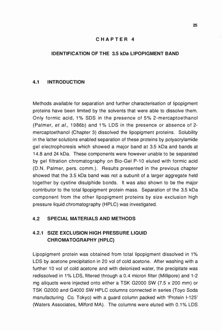
C H A P T E R 4
IDENTIFICATION OF THE 3.5 kDa LIPOPIGMENT BAND
4.1 I NTRODUCTION
25
Methods avai lable for separation and further characterisation of l ipopigment
proteins have been l imited by the solvents that were able to dissolve them.
O n ly fo rmic aci d , 1 % SOS in t h e p rese nce of 5% 2 - m e rcaptoethano l
( Pal m e r, et al. , 1 986b) and 1 % LDS i n the presence or absence of 2-
mercaptoethanol (Chapter 3) disso lved the l ipopigment protei ns. Solubi lity
i n the latter so lutions enabled separation of these proteins by polyacrylamide
ge l e lectrophoresis which showed a major band at 3.5 kDa and bands at
1 4.8 and 24 kDa. These components were however unable to be separated
by ge l fi ltrat ion chromatog raphy o n B io-Gel P- 1 0 e luted with formic acid
( D . N . Palmer, pers. comm. ) . Results presented i n the previous chapte r
showed that the 3.5 kDa band was not a subunit of a larger aggregate held
together by cysti ne disulph ide bonds. lt was also shown to be the major
contributor to the total l ipopigment protein mass. Separation of the 3.5 kDa
compo nent from the othe r l ipop igment p rote i ns by s ize exclusi o n h igh
pressu re l iquid chromatog raphy (HPLC) was investigated.
4.2 SPECIAL MATERIALS AND METHODS
4.2.1 SIZE EXCLUSION HIGH PRESSURE LIQUID
CHROMATOGRAPHY (H PLC)
Lipopigment prote in was obtai ned from total l ipopigment d isso lved i n 1 %
LDS by acetone precipitatio n in 20 vol of co ld acetone. After washing with a
further 1 0 vol of cold acetone and with deionized water, the precipitate was
redissolved i n 1 % LDS, fi lte red through a 0 .4 micron fi lter (Mi l l ipore) and 1 -2
mg al iquots were injected onto either a TSK G2000 SW (7.5 x 200 mm) or
TSK G2000 and G4000 SW HPLC columns connected in series (Toyo Soda
manufactu ring Co. Tokyo) with a guard co lumn packed with 'Protein 1- 1 25'
(Waters Associates, Mi lford MA). The co lumns were eluted with 0 . 1 % LDS
26
at a f low rate of 0 .6 ml/m in . Detection was at 280 nm us ing a Model 450
Variable Wavelength Detector (Waters Associates, Mi lford MA). The eluted
peaks were co l lected and freeze dri ed. After redissolving in deionized water
al iquots were analysed by LDS-PAGE as descri bed in section 2.5 and the
rest stored at -20°C unt i l requ ired.
4.2.2 AMINO ACID SEQUENCING
Fract ions e luted f rom the H P LC co lumns that contai ned t he l ipopigment
protei ns were precipitated with 20 vo l of cold acetone. After washing with a
fu rt h e r 1 0 vol of co ld aceto n e , samples were dissolved i n disti l led 98%
formic acid at a protein concentrat ion of 50 Jlg/m l , and al iquots , 50- 1 00 Jll ,
were loaded onto po lybrene treated g lass filter disks that had been through
three cycles of a standard ABI program 03RPRE (Appl ied Biosystems I nc. ,
Foster city CA. U .S.A) . Sequenci ng was done on an Applied Biosystems
470A gas phase sequence r coup led to a 1 20A PTH an alyzer us i ng a
standard ABI program 03RPTH . Aliquots of precipitated prote in were also
subjected to quantitative amino acid analysis as described in section 2 .4 to
al low an estimation of the total protei n loaded.
4.2.3 R EPETITIVE AND INITIAL YIELD CALCULATIONS
Yie lds of the phenylthiohydantoin (PTH) amino acids produced in the Edman
deg radat ion at each cycle were calcu lated from comparison with fresh ly
prepared PTH amino acid standards. Repetitive yields were calcu lated from
l inear reg ression best fit plots of the logarithms of the yie lds at each cycle.
In it ial yie lds were read from the intercept of the repetitive y ie ld slopes with
the y-axis .
4.3 R ESULTS
4.3.1 Size exclusion HPLC of l ipopigment proteins
Total l ipopigment e luted as a number of 280 nm absorb ing peaks on size
exclusion HPLC using a TSK G2000 SW column run in 0 . 1 % LDS (Fig. 4. 1
A). When pooled fractions from these peaks were exami ned by LDS-PAGE,
the peak and shoulder that e luted at 30 m in at the size exclusion l imit o f 50
k D a , was s h own to co nta i n a l l t h e spec i f ic l i popig m e n t prote i ns i n a
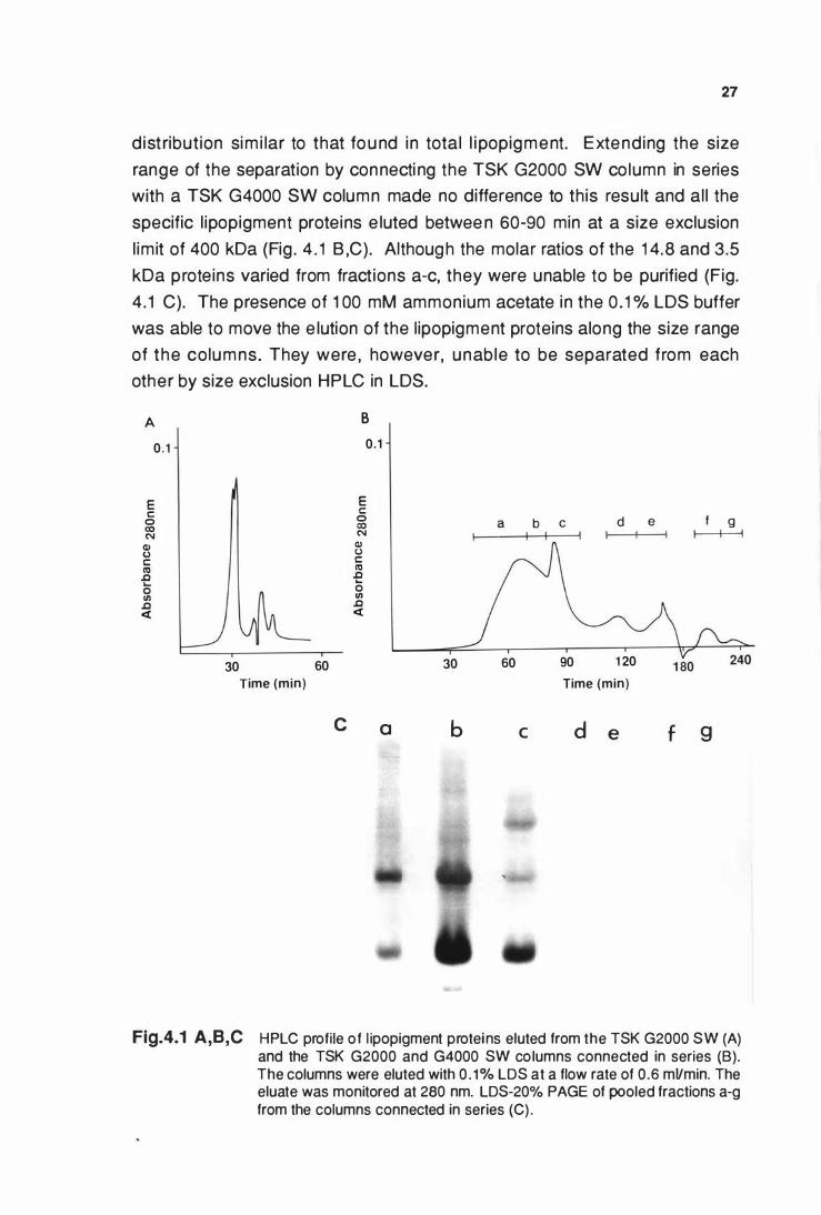
27
distribut ion sim i lar to that fou nd in total l i popigment. Extending the size
range of the separation by connecti ng the TSK G2000 SW co lumn in series
with a TSK G4000 SW column made no difference to this result and al l the
specific l ipopigment proteins e luted betwee n 60-90 min at a size exclusion
l im it of 400 kDa (Fig . 4 . 1 B ,C). Although the molar ratios of the 1 4.8 and 3.5
kDa proteins varied from fract ions a-c, they were unable to be purified (Fig.
4 .1 C). The presence of 1 00 mM ammonium acetate in the 0 . 1 % LDS buffer
was able to move the e lution of the l ipopigment proteins along the size range
of the columns. They were , however, unable to be separated from each
other by size exclusion HPLC in LDS.
A 0.1
E c:: 0 CO N Cll (,) c:: Cl) .0 ... 0 Cll .0 <
30 60 Time (min)
c
B 0.1
E c:: 0 CO N Cll (,) c:: Cl) .0 ... 0 Cll .0 <
a
30
b
a b c
60 90
d e
1 20 Time (min)
c d e
f g
f 9
I I
Fig.4.1 A,B,C HPLC profile of l ipopigment proteins eluted from the TSK G2000 SW (A) and the TSK G2000 and G4000 SW columns connected in series (B). The columns were eluted with 0 . 1% LDS at a flow rate of 0.6 mVmin. The eluate was monitored at 280 nm. LDS-20% PAGE of pooled fractions a-g from the columns connected in series (C) .
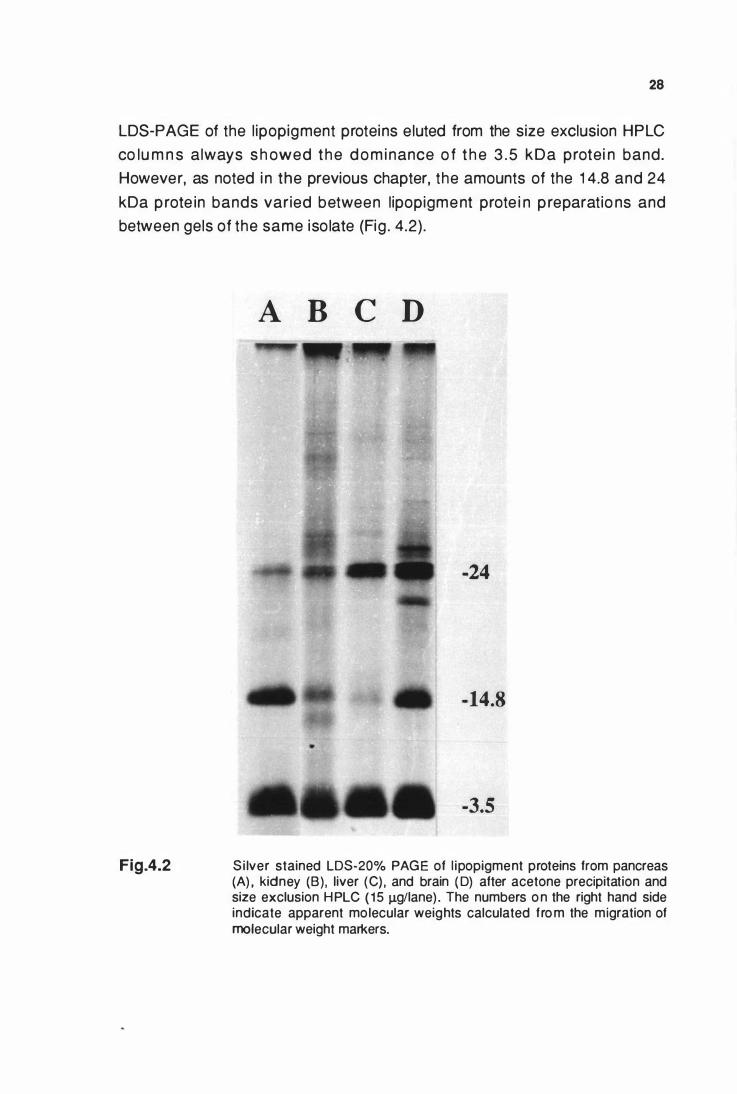
28
LDS-PAGE of the l ipopigment proteins eluted from the size exclusion HPLC
co lu m n s always s h owed the d o m i nance o f t h e 3 . 5 kDa protei n band.
However, as noted i n the previous chapter, the amounts of the 1 4.8 and 24
kDa p rote in bands varied between l ipopigment prote i n p reparatio ns and
between gels of the same iso late (Fig . 4.2) .
Fig.4.2
A B C D
-24
-14.8
•
-3.5
Si lver stained LDS-20% PAGE of l ipopigment proteins from pancreas (A) , kidney (B) , liver (C) , and brain ( D) after acetone precipitation and size exclusion HPLC ( 1 5 J.Lg/lane) . The numbers o n the right hand side indicate apparent molecular weights calculated fro m the migration of molecular weight markers.
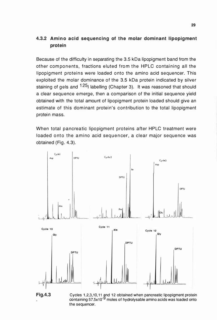
29
4.3.2 Ami n o ac id seq uen c i n g of t h e molar domi nant l ipopig ment
protein
Because of the difficu lty i n separating the 3.5 kDa l ipopigment band from the
othe r com po n e nts , f ract ions e luted f ro m t h e H P LC co ntai n i ng a l l the
l i pop ig ment p rote i n s we re loaded o nto the amino acid sequencer. Th is
exploited the molar dominance of the 3 .5 kDa protei n i ndicated by si lve r
stain ing of g e ls and 1 251 labell ing (Chapter 3). lt was reasoned that should
a clear sequ ence e merge, then a comparison of the in itial sequence yield
obtained with the total amount of l ipopigment protein loaded should give an
est imate of t h i s dom i nant prote i n 's contribut ion to the total l ipopigment
protei n mass.
When total pancreat ic l ipo pigment p rote i ns after HPLC treatment were
l oaded o nt o t h e a m i no ac id seq u e n cer , a c lear major seque nce was
obtained (Fig . 4.3).
Cyclel Asp
Ala
Cycle 1 0
Gly
Fig.4.3
DPTU Cycle 2 Cycle3
Asp
lie
DPTU
DPTU
4 ' Cycle 1 1
Ala Cycle 1 2
Gly
DPTU
DPTU
DPTU
V '
Cycles 1 ,2,3, 10, 1 1 and 1 2 obtained when pancreatic l ipopigment protein containing 57.5x1 o-9 moles of hydrolysable amino acids was loaded onto the sequencer.
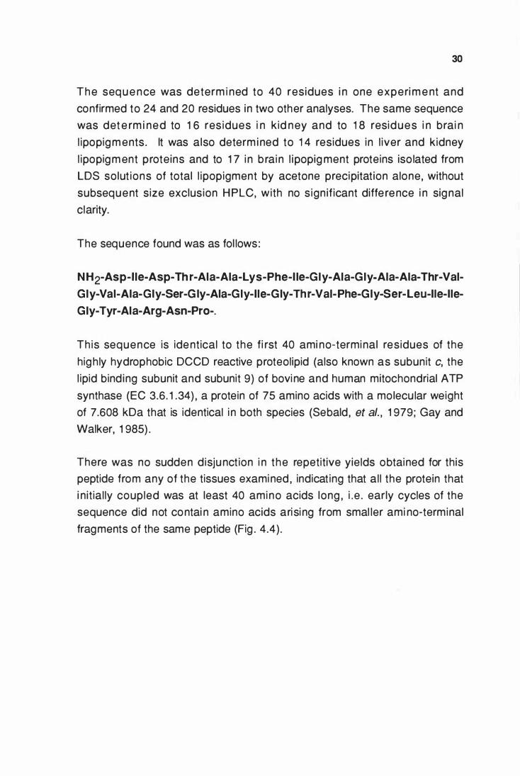
30
The seq u e nce was determ i ned to 40 residues i n one experi m e nt and
confirmed to 24 and 20 residues in two other analyses. The same sequence
was det e rm i ned to 1 6 res idues i n k id ney and to 1 8 residues i n brai n
l ipopigm ents . lt was also determ i ned to 1 4 residues in l iver and kidney
l ipopigm ent prote ins and to 1 7 i n b rai n l ipopig ment proteins iso lated from
LDS solut ions of total l ipopigment by acetone precipitat ion alone, without
subsequent s ize exclus ion H PLC, with no s ign if icant diffe rence i n signal
clarity.
The sequence found was as fol lows:
N H 2-As p-I le-Asp-Th r-Aia-Aia-Lys-Phe-l le-G ly-Ala-G ly-Ala-Aia-Thr-Val
Gly-Vai-Aia-Giy-Ser-G iy-Aia-Giy-lle-Giy-Th r-Val-Phe-Giy-Ser-Leu-lle-lle
Gly-Tyr-Aia-Arg-Asn-Pro-.
This sequence is identical to the fi rst 40 ami no-terminal residues of the
highly hydrophobic DCCD reactive proteolipid (also known as subunit c, the
l ipid bind ing subunit and subunit 9) of bovi ne and human mitochondrial ATP
synthase (EC 3.6. 1 .34) , a protein of 75 amino acids with a mo lecu lar weight
of 7.608 kDa that is identical i n both species (Sebald , et al. , 1 979; Gay and
Walker, 1 985) .
There was no sudden disjunct ion i n the repetitive yields obtained for this
peptide from any of the tissues examined , indicat ing that al l the protein that
i n it ial ly coupled was at least 40 amino acids long , i .e . early cycles of the
sequence did not contai n amino acids ari si ng from smal ler ami no-terminal
fragments of the same peptide (Fig . 4.4) .
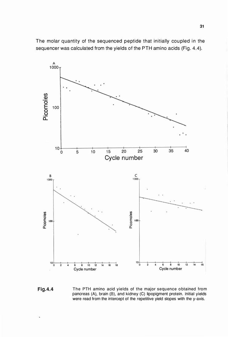
31
The molar q uantity of the sequenced peptide that in i t ia l ly cou pled i n the
sequencer was calculated from the yie lds of the PTH amino acids (Fig . 4 .4).
en Q) 0
A 1 000
E 1 oo 0
.5:2 a...
1 0+---�-----+----�----+---�r----+----�--�
"' Q) 0
B 1 000
E 100 8 a:
0 5 1 0 1 5 20 25 30 35 40
Cycle number
� 0
c 1 000
E 100 8 a::
1 0+---+---+-____,f----!---+--+____,f----!-� 0 2 4 6 8 10 12 14 16 18 10+----+--._._-+---+---!---+--+-----i 0 2 6 8 10 12 14 16
Fig.4.4
Cycle number Cycle number
Th e PTH amino acid y ie lds of the major sequ ence o btained f rom pancreas (A), brain (B) , and kidney (C) lipopigment protein. Initial yields were read from the intercept of the repetitive yield slopes with the y-axis.
32
E st i mates o f t h e co nt r i but i o n of t h e p e pt ide sequenced to t h e tota l
l i popigment p rotei n mass was made by compari ng the i n it ial y ie ld of the
sequenced peptide , with the total amount of l ipopigment protein loaded onto
t h e seque nce r . Fo r e xam p l e , w h e n pancreat i c l i pop igment p rote i ns
contai n ing 57.5x1 o-9moles of hydro lysab le amino acids were loaded onto
the sequencer, 0 .579x1 o-9moles of peptide coupled as the above sequence
(Fig . 4.4). As this peptide is at least 40 amino acids long , it must contribute
at least 40 X 0 .579x1 o-9moles Of ami no acids = 24.2X1 o-9moles, to the total
l ipopigment p rote in mass. Therefo re the sequenced peptide must account
for at least 24.2x1 o-9moles X 1 00 = 40.3% Of the total l ipopigment p rotein.
57.5x1 o-9moles
Simi lar estimates for the contribution of the sequenced peptide to the total
l ipopigment protei n mass from kidney and brain are shown (Table 4. 1 ) .
Table 4.1 Est i mates of the contribut ion of the sequenced pept ide to the total lipopigment protein masses
Source o f
l ipopigment
Pancreas
1 .
2 .
3 .
Kidneye
1 .
2 .
Brain
1 .
No . of
cycles
4 0
2 4
2 0
1 6
1 3
1 8
Repet it ive yielda
( % )
9 2 . 8
92 . 7
NDd
9 4 . 3
8 9 . 5
8 5 . 3
E s t i m a t e o f
the sequenced
p e p t i d e ' s
c o n t r i b u t i o n
t o t h e t o t a l
l i p o p i g m e n t
protein ma s s
( % ) b ( % ) c
4 0 . 3 7 5 . 6
3 6 . 5 6 8 . 4
4 0 . 7 7 6 . 3
4 1 . 0 7 6 . 9
3 8 . 7 7 2 . 6
2 6 . 4 4 9 . 5
a. The repetitive yields were calcu lated from l inear regression best fit plots of the logarithms of the yields at each cycle.
b. Calcu lated by mult iplying the initial yield by 40 (the length of the sequenced peptide) and expressed as a percentage of the total hydrolysable amino acids loaded onto the sequencer.
c. Calculated by multiplying the initial yield by 75 (the length of the full subunit c) . d. Pancreatic l ipopigment isolated from three affected sheep. e. Kidney lipopigment isolated from two affected sheep.
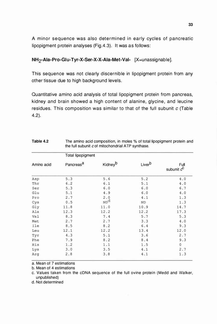
33
A m i n o r sequence was a lso dete rm i ned i n ear ly cycles of pancreat ic
l ipopigment p rotein analyses (Fig .4 .3) . l t was as fo l lows:
NH2-Aia-Pro-Giu-Tyr-X-Ser-X-X-Aia-Met-Val- [X=u nassig nab le].
This sequence was not clearly d iscern ib le in l ipopigment protei n from any
other tissue due to high background levels.
Quantitative amino acid analysis of total l ipopigment protein from pancreas,
kidney and brai n showed a h igh content of alan ine , g lyci ne , and leuci ne
residues. Th is composition was s im i la r to that of the fu l l subunit c (Table
4.2).
Table 4.2 The amino acid composition, in moles % of total lipopigment protein and the full subunit c ot mitochondrial ATP synthase.
Total lipopigment
Amino acid Pancreas a Kidneyb Live� Full subunit cf
Asp 5 . 3 5 . 6 5 . 2 4 . 0 Thr 6 . 2 6 . 1 5 . 1 4 . 0
Ser 5 . 3 6 . 0 6 . 0 6 . 7
Glu 5 . 1 4 . 9 6 . 0 4 . 0 Pro 2 . 7 2 . 0 4 . 1 1 . 3
Cys 0 . 5 NDd ND 1 . 3
Gly 1 1 . 8 1 1 . 0 1 0 . 9 1 4 . 7
Ala 1 2 . 3 1 2 . 2 1 2 . 2 1 7 . 3
Val 8 . 3 7 . 4 5 . 7 5 . 3 Met 2 . 7 2 . 7 3 . 3 4 . 0 I le 8 . 5 8 . 2 6 . 4 9 . 3 Leu 1 2 . 1 1 2 . 2 1 3 . 4 1 2 . 0
Tyr 4 . 3 5 . 1 3 . 6 2 . 7
Phe 7 . 9 8 . 2 8 . 4 9 . 3
His 1 . 2 1 . 1 1 . 5 0
Lys 3 . 0 3 . 5 4 . 1 2 . 7
Arg 2 . 8 3 . 8 4 . 1 1 . 3
a. Mean of 7 estimations b. Mean of 4 estimations c. Values taken from the cDNA sequence of the ful l ovine prote in (Medd and Walker,
unpublished) d. Not determined
4.4 DISCUSSION
34
Lipopig ment protei ns were unable to be separated by size exclusion HPLC
i n LDS . Su rpris i ng ly , the l ipop ig ment p rote i ns with apparent mo lecu lar
masses of 3.5, 1 4.8 and 24 kDa seen on PAG E eluted at size exclusion
l imits of up to 400 kDa (Fig .4. 1 ). This was possibly due to the l ipopigment
proteins migrating i n large dodecyl sulphate micel lar aggregates. Difficu lties
in separating and purifying the l ipopigment protei ns led to the non-tradit ional
approach of loadi ng the prote ins as a mixture o nto the sequencer. The
approach was based on the rationale that as the major low molecu lar weight
3 .5 kDa p rote i n was p resent i n h igher mo lar quantiti es than any other
component, the major sequence obtained should therefore be of this protein .
The sequence obtained was identical to the fi rst 40 amino-terminal residues
of a high ly hydrophobic protei n , subunit c of the mitochondrial ATP synthase.
This protei n's l ipid-l ike solubi lity in neutral chloroform/methanol mixtures has
led it to be classif ied as a proteol ip id (Folch and Lees, 1 951 ) . This is an
operational defin it ion that does not imply the presence of covalently bound
l ipids.
T h e seque nci ng strategy n ot o n ly a l lowed ide ntif icat ion of the major
l i po pigment p rote i n , but also enab led est imat ions to be made as to the
contribution of this peptide to the total l ipopigment protei n mass. This was
ach i eved by co m pari ng the i n i t ia l y ie ld i n t he seque ncer with the total
amount of p rote i n loaded. Values betwee n 26-4 1 % we re obtai ned as
estimates of the sequenced peptide to the total l ipopigment protein masses
fro m the ami n o -termi na l seque nces obtai n ed (Table 4 . 1 ). These are
min imum rathe r than actual values s ince the i n itial reactions of the Edman
chemistry used in the sequencer are not quantitative and can vary between
sam ples (Edman and Begg , 1 967) , and also because the length of the
sto red peptide remained to be established. The molecular weight of a 40
res idue pept ide i s c lose to the 3 . 5 kDa d e mo nst rated by LDS-PAG E .
However, as s im i lar proteol ip ids are known t o g ive anomalous mo lecular
weight estimations on dodecyl sulphate po lyacrylamide gel electrophoresis
( Farre l l , et al. , 1 988) , the resu lts obtai ned may not i nd icate the t rue
molecu lar weight , or the length of the sequenced peptide. There are two
possible explanations for such an anomaly. Fi rst a high detergent to protein
35
ratio i n the migrat ing mice l les, a consequence of the extreme hydrophobic
natu re of t he p rote i n maki ng it b i nd m o re than the normal amount of
detergent which resu lts i n a higher charge to mass ratio than normal (Lees,
et al., 1 979 ) . T h e seco nd re lates to i nco rrect assig n ment of t he low
molecu lar weight standards used to calibrate the apparent molecu lar weights
of this protein (Kratzin , et al., 1 989) .
Total l ipopigment p rote in contai ned high contents of g lyci ne , alanine and
leuci ne (Table 4 . 2 ) , consiste nt with a h ig h p ro po rtion of the sequenced
peptide bei ng derived from subunit c of mitochondrial ATP synthase. The
relative proportion s of the amino acids did not allow any conclusions to be
drawn as to the length of the stored peptide . The presence of h istid ine
i ndicated that other minor proteins were also p resent, as h istidine does not
occur i n subun it c of mitochondrial ATP synthase. No other mitochondrial
ATP synthase components were detected in the sequence studies.
The minor sequence detected is related to the amino-terminal sequence of a
1 6 kDa protei n isolated from mouse gap junction preparations (Walker, et
al., 1 986) and to residues 7- 1 7 of the p roteo l i pid iso lated from bovi ne
vacuolar (chromaffi n g ranule) ATPase (Walker, et al., 1 986 ; Mandel , et al.,
1 988 ; Dermietzel , et al., 1 989). The sign ificance of this minor sequence is
not yet known . Its presence in l ipopigment cou ld be due either to m inor
contamination by membranes contain ing gap junctions or to the presence of
a proteolipid fragment from a vacuolar ATPase.
Subunit c of mitochondrial ATP synthase has on ly previously been detected
i n mitochondria as part of the inner mitochondrial membrane F0 component
of the ol igomeric ATP synthase complex (Sebald and Hoppe, 1 981 ; Walker,
et al., 1 990) . Lysosomes and other subce l lu lar o rgane l les also contai n
o l igomeric p roton pumping p rotei ns with ATPase activity belong ing to a
class of vacuolar ATPases (Bowman, et al., 1 988a,b; Manolson , et al., 1 988;
Zimn iak, et al., 1 988). Lysosomal ATPase contains a subunit related , but
not ident ical to the mitochondrial subun it c proteo l ip id (Moriyama and
Nelso n , 1 989) . The protei n detected i n l ipopigment bodies is therefore not
part of a lysosomal ATPase.
36
The fu l l subun it c has properties s imi lar to the specifically stored 3.5 KDa
l ipo pigment prote in . Both share the properties of poor Coomassie blue dye
stai n ing and aggregation behaviour on dodecylsulphate polyacrylamide gels
(Palmer, et al. , 1 986b; Fi l l ingame, 1 976 ; Graf and Sebald , 1 978; Sebald and
Hoppe , 1 981 ). Physical properties described as being characteristic of the
l ipopigment stored i n the ce roid- l ipofusci noses, are also simi lar to those of
the full subunit c. These include a variable pH dependent
ch loroform/methano l extractab i l ity and s im i lar th in layer ch romatography
mig ration behaviour to that of the so called "Schiff base polymer" that was
thought to be formed as a result of l ipid peroxidation (Sierra and Tzago loff,
1 9 7 3 ; S i akot o s a n d K o p p a n g , 1 9 73) . T h e co m p l et e e xtract i o n of
l ipop igments from Batten's d isease in ch loroform/methano l as wel l as the
apparent extraction of autofluorescent material from histolog ical preparations
i n acid i fied ch lo roform/ethano l has been documented (Srivastava, et al. ,
1 982). However, the l ipid l ike behaviour of the proteol ipid, poor Coomassie
dye stai n i ng and a tendency to g ive inaccu rate protein estimations by the
Lowry method (Sebald and Hoppe, 1 981 ) may help to explai n why this type
of p rotein was not recognised as being associated with these d iseases in the
past.
The data presented in this chapter does not i ndicate how long a fragment of
the mitochondrial subunit c proteol ipid is present i n isolated l i popigment. If it
is l ittle more than 40 amino acids then it must account for at least 26-41 % of
the total l ipopigment protein mass. If it is the ful l protein (7.608 kDa) , then it
accounts for 50-77% (Table 4. 1 ) , and some of the highe r mo lecular weight
l ipopigment bands that are seen on LDS-PAGE are l ikely to be aggregates
of subunit c. Aggregation in dodecy lsulphate is a characte ristic property of
the fu l l p rote i n (Sebald and H o p pe , 1 981 ) , and cou ld acco unt fo r the
anomalous LDS-PAGE and HPLC behaviour of the l ipopigment proteins.
C H A P T E R 5
THE CARBOXYL-TERMINAL DETERMINATION OF THE MAJOR
STORED PROTEIN AND CHARACTERISATION OF THE OTHER
LIPOPIGMENT PROTEINS
5.1 INTRODUCTION
37
When total l i pop ig ment prote i n was loaded onto the sequencer , a c lear
sequence was identified. This sequence was shown to be identical with the
f i rst 40 a m i n o -te rm i na l res i d u e s of b o vi n e and h u man s u b u n i t c of
mitochondrial ATP synthase (Chapter 4) . A min imum estimate of 40% was
made fo r the contri bution of the sequenced protein to the total pancreatic
l i popigment protein mass on the basis that the fu l l stored protein was at least
40 residues lo ng . H owever, the sequenci ng stud ies described gave no
i ndication that the carboxyl-terminal had been reached. S imi larity between
the physical p ro pert ies of the fu l l subu n it c proteo l ip id , and l ipopigment
bodies stored i n the ceroid- l ipofusci noses , suggested that the ful l p rotei n
might be stored . This chapter describes the carboxyl-terminal determination
of the major sto red p rotein and further characterisation of the 1 4.8 and 24
kDa l ipopigment protein bands. Identification of subunit c, a proteol ipid , as
t h e majo r sto red l i pop ig m e nt p rote i n co m p o nent , e n abled its fu rthe r
p u r i f icat ion f ro m l i p o p ig m e nt sto rage b o d i e s by a specif ic extract ion
procedure .
5.2 SPECIAL MATERIALS AND METHODS
5.2.1 EXTRACTION OF PROTEOLIPIDS FROM LIPOPIGMENT
Iso lated l ipopigment bodies were solubi l ised by a modified Folch extraction
(Folch, et al. , 1 957). Lipopigment suspensions at 1 - 1 0 mg/ml were extracted
with 20 vol of chloroform/methanol (2 : 1 , v/v) containi ng 1 00 mM ammonium
acetate, pH 7.2. After addition of 0 .2 vol of deionized water the solution was
gently shaken and left to stand at 4°C, unti l the aqueous and organic phases
38
separated . The prote ins that precipitated at the i nterface were carefu l ly
withdrawn and retained. The lower organic phase was di luted with 1 vol of
ch lo roform and the vo lume reduced to one fifth by rotary evaporat ion at
35°C. Proteolipids were precipitated from this extract with 1 0 vol of diethyl
ether at -70°C for 1 h and harvested by centri fugation in a Sorval l GLC-1
centrifuge for 1 0 min at 1 ,400 g max at 4°C. This proteol ipid pel let and the
protei ns from the solvent interface were sequential ly washed with methano l
and deion ized wate r and then disso lved in 1 % LDS. At no stage were the
protei ns allowed to become dry.
5.2.2 DIFFUSION ELUTION FROM POLYACRYLAMIDE G ELS
Pancreatic l ipopigment containing 88 J..Lg of protei n was loaded into a 1 6-cm
wide we l l i n the stacki ng gel of a LDS- 1 5% po lyacry lamide ge l . After
e le ctrophoresi s , a s ide strip was cut from the gel and s i lver stai ned as
described in sections 2 .4-2 .5. The bands conta in ing the 3 .5 and 1 4.8 kDa
l ipopigment proteins were cut from the gel , crushed in 1 0 ml of 1 % LDS, and
shaken for 1 8 h at 25°C. The supernatant was freeze dried , redissolved in 1
m l of deionized water and subjected to size exclusion H PLC as described
ear l i e r (section 4 .2 . 1 ) . The e l uted protei n fract ions were col lected and
sequenced as described in section 4.2.2.
5.2.3 ELECTRO B LOTTING OF LIPOPIGMENT PROTEINS
Pancreatic l ipopigment protein contain ing 1 0-20 J..Lg of protei n per lane was
loaded onto LDS- 1 5% po lyacrylamide mini gels ( 1 .5 x 8 x 1 0 mm). 0
E lectrophoresis was carried out at 1 00 Volts for 2 h at 4 C , i n a " Mighty
S m a l l E l e ct ro p h o re s i s u n i t" ( H o efe r S c i e n t i f i c I nstru m e nts) . Afte r
electrophoresis the separated proteins were transfered onto polyvinylidene
d i f l uo ride membranes ( PVDF) accord ing to the method of Matsudai ra ,
( 1 987). The PVDF membranes were rinsed with 1 00% methanol and stored
i n t h e t ransfer bu ffe r . LDS- 1 5% po lyacry lam ide g e l s contai n i ng t h e
separated l ipopigment proteins were briefly soaked in transfer buffer, 1 OmM
[cyclohexylamino]- 1 -propanesulfonic acid (CAPS) , 1 0% methanol, pH 1 1 .0. 0
E lectroelution was performed at 0 .5 A in transfer buffer for 2 h at 4 C. After
e lectroelution the PVDF membranes were washed in deionized water for 2-3
39
min , stained with 0.2% Coomassie blue R-250 i n 50% methanol contai n i ng
1 0% acetic acid for 2-5 m in , and then destained i n 50% methanol contain i ng
1 0% acetic acid for 5- 1 0 m in at 25°C. Coo m assie b lue stai ned p rote i n
bands of interest were cut into small p ieces and placed i n the cartridge block
of a 470A gas phase sequencer, in the presence of only the Teflon seal , with
neither polybrene no r g lass fibre fi lter d iscs. The sequencing part of the
experiment was performed by I .M. Fearn ley and J. M. Skehel at the M.R .C.
Laboratory of Molecu lar Biology, H i l ls Road, Cambridge, U.K.
5.2.4 CYANOGEN B ROMIDE (CNBr) DIG ESTION OF LIPOPIGMENT
PROTEOLI PI DS
Lipopigment proteol ipids, (0.5- 1 mg} dissolved i n ch loroform/methanol (2/1 ,
v/v} contain ing 1 00 m M ammonium acetate, p H 7.2 , were precipitated with
diethyl ether at -70°C , and then d isso lved in d isti l led 98% formic acid ( 1 .0
ml } , contain ing CNBr ( 1 00 mg} . This solution was gassed with nitrogen and 0
left i n the dark at 25 C for 24 h . Solvents were removed by drying in vacuo
over NaOH pe l lets and the digest stored at -20°C unti l required.
5.2.5 MASS SPECTROSCOPY ANALYSIS OF CNBr DIGESTS
Mass spectra were obtained usi ng a VG70-250S double focussi ng magnetic
sector mass spectrometer (VG Analytical , Manchester, Eng land) fitted with a
VG liquid secondary mass spectrometry ion sou rce and associated caesium
ion g u n . The C N B r d igest of pancreat ic l i pop igment p roteo l ip ids were
dissolved in disti l led 98% formic acid . A 1 0 �I aliquot was suspended i n a
matrix of glycerol and loaded onto the stai n less steel target of the sample
i nsert ion probe . Samples were bombarded with 35 keV caesium ions ( ion
current, 1 -2 �A} . The secondary ion beam was accelerated from the source
at 8 keV and mass analysed at 5000 resolvi ng power (RP} . The magnet
was scanned l i nearly between 890 and 1 250 Da over 1 5 sec. Each analysis
consisted of 4-6 scans of caesium iodide dissolved in water (reference ions,
9 1 2 .3352 and 1 1 72. 1 451 } fol lowed by 4-6 scans of the sample.
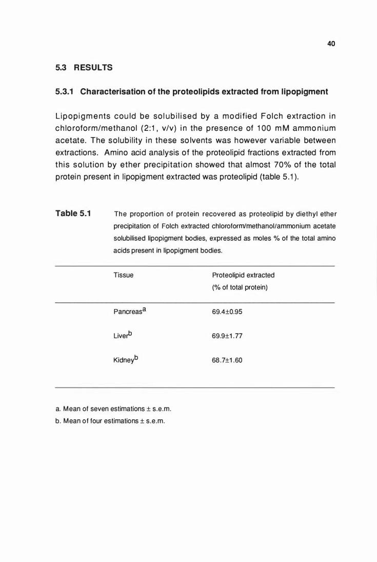
40
5.3 R ESULTS
5.3.1 Characterisation of the proteolipids extracted from l ipopigment
Li pop i g m e nts cou ld be so l ub i l i sed by a mod i f ied Fo lch extracti on i n
c h lo roform/methanol (2 : 1 , v/v) i n the p resence of 1 00 m M ammo n i u m
acetate. The so lub i l ity i n these so lvents was however variable between
extractions. Amino acid analysis of the proteolipid fractions extracted from
th i s so lut ion by ether precip i tat ion showed that almost 70% of the total
protei n present in l ipopigment extracted was proteolipid (table 5 . 1 ) .
Table 5.1 The proport ion of protein recovered as proteol ipid by diethyl ethe r
precipitation of Folch extracted chloroform'methanol/ammonium acetate
solubilised lipopigment bodies, expressed as moles % of the total amino
acids present in lipopigment bodies.
Tissue
Pancreasa
Liverb
Kidneyb
Proteolipid extracted
(% of total protein)
69.4±0.95
69.9±1 .77
68 .7±1 .60
a. Mean of seven estimations ± s.e.m.
b. Mean of four estimations ± s.e.m.
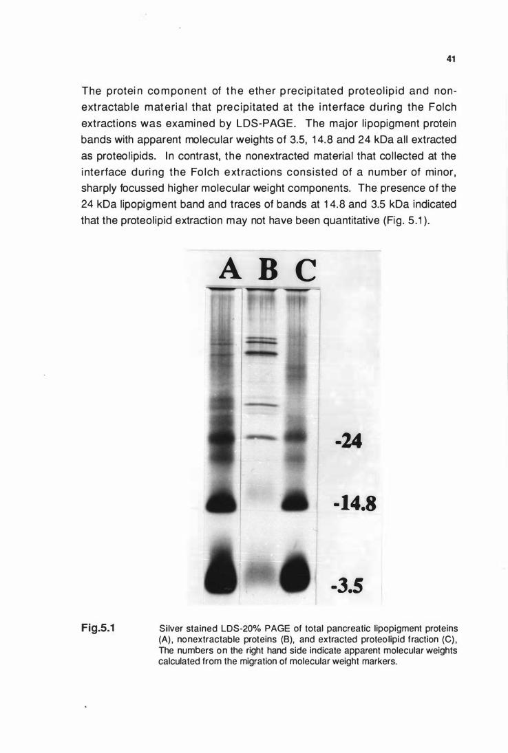
41
The protei n co m ponent of t h e eth er p rec ip i tated proteo l ip id and non
extractab le materia l that prec ip itated at the i nte rface d u ri ng the Folch
extractions was examined by LDS-PAGE. The major l ipopigment protein
bands with apparent molecu lar weights of 3.5, 1 4.8 and 24 kDa al l extracted
as proteo l ipids. I n contrast, the nonextracted material that col lected at the
i nte rface du ri ng the Fo lch ext ract ions cons isted of a number of mi nor,
sharply focussed higher molecular weight components. The presence of the
24 kDa lipopigment band and traces of bands at 1 4.8 and 3.5 kDa indicated
that the proteol ipid extraction may not have been quantitative (Fig . 5 . 1 ) .
Fig.5.1
A D C
-24
- 14.8
-3.5
Silver stained LDS-20% PAGE of total pancreatic lipopigment proteins (A) , nonextractable proteins (B), and extracted proteo lipid fraction (C) , The numbers on the right hand side indicate apparent molecular weights calculated from the migration of molecular weight markers.
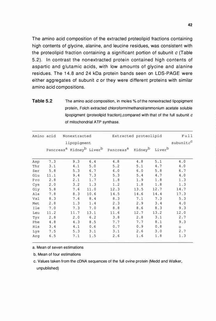
42
The amino acid composit ion of the extracted proteol ipid fractions containing
h igh contents of g lyci ne , alan ine , and leucine residues, was consistent with
the proteol ipid fract ion contai n ing a s ign ificant portion of subunit c (Table
5 . 2 ) . I n co ntrast t he non ext racted p rote i n conta ined h ig h co nte nts of
asparti c and g lutam ic acids , with low amou nts of g lyci ne and a lan i ne
residues. The 1 4. 8 and 24 kDa prote i n bands seen on LDS-PAG E were
either agg regates of subunit c o r they were d iffe rent protei ns with simi lar
amino acid compositions.
Table 5.2 The amino acid composition , in moles % of the nonextracted l ipopigment
prote in, Folch extracted chloroform/methanoVammonium acetate soluble
lipopigment (proteolipid fraction) ,compared with that of the full subunit c
of mitochondrial A TP synthase.
Amino acid Nonext racted Ext racted proteolipid F u l l
l ipopigment subunit cc
Pancreas a Kidneyb Liverb Pancre a s a Kidneyb Liverb
Asp 7 . 3 9 . 3 6 . 4 4 . 8 4 . 8 5 . 1 4 . 0
Thr 3 . 1 4 . 1 5 . 0 5 . 2 5 . 1 4 . 7 4 . 0
Ser 5 . 8 5 . 3 6 . 7 6 . 0 6 . 0 5 . 8 6 . 7
Glu 1 1 . 1 9 . 4 7 . 3 5 . 3 5 . 4 4 . 7 4 . 0
P ro 2 . 8 2 . 1 1 . 7 1 . 8 1 . 9 1 . 8 1 . 3
Cys 2 . 0 3 . 2 1 . 3 1 . 2 1 . 8 1 . 8 1 . 3
Gly 5 . 8 7 . 6 1 1 . 0 1 2 . 3 1 3 . 5 1 2 . 7 1 4 . 7
Ala 7 . 8 8 . 3 1 0 . 6 1 4 . 5 1 4 . 6 1 4 . 4 1 7 . 3
Val 8 . 3 7 . 6 8 . 4 8 . 3 7 . 1 7 . 3 5 . 3
Met 2 . 8 1 . 3 1 . 4 2 . 3 2 . 9 3 . 4 4 . 0
I le 7 . 0 7 . 3 7 . 0 8 . 8 8 . 6 8 . 3 9 . 3
Leu 1 1 . 2 1 1 . 7 1 3 . 1 1 1 . 6 12 . 7 1 3 . 2 1 2 . 0
Tyr 2 . 8 2 . 0 6 . 2 3 . 8 2 . 8 3 . 1 2 . 7
Phe 4 . 8 4 . 3 8 . 5 7 . 7 7 . 7 8 . 1 9 . 3
His 3 . 4 4 . 1 0 . 6 0 . 7 0 . 9 0 . 8 0
Lys 7 . 5 5 . 3 3 . 1 3 . 1 2 . 6 3 . 0 2 . 7
Arg 6 . 5 7 . 1 1 . 5 2 . 6 1 . 6 1 . 8 1 . 3
a. Mean of seven estimations
b. Mean of four estimations
c. Values taken from the cDNA sequences of the full ovine protein (Medd and Walker,
unpublished)
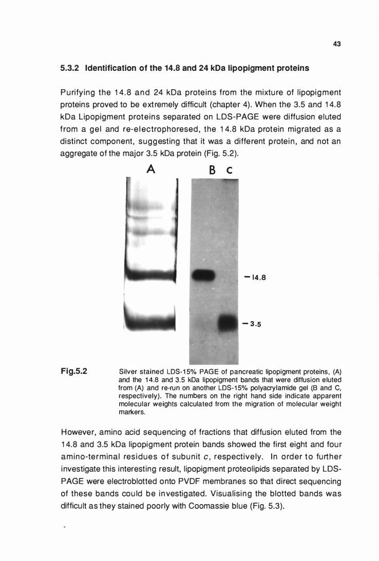
43
5.3.2 Identification of the 14.8 and 24 kDa lipopigment proteins
P u rify ing the 1 4 .8 and 24 kDa prote ins from the mixtu re of l ipopig ment
proteins proved to be ext remely difficult (chapter 4) . When the 3.5 and 1 4.8
kDa Lipopigment prote ins separated on LDS-PAGE were diffusion e luted
f rom a ge l and re-e l ectrophoresed , the 1 4.8 kDa protein mig rated as a
d isti nct component , suggesting that it was a diffe rent prote i n , and not an
aggregate of the major 3.5 kDa protein (Fig. 5 .2) .
Fig.5.2
A B c
- 14 . 8
- 3 .5
Silver stained LDS-1 5% PAG E of pancreatic lipopigment proteins , (A) and the 1 4.8 and 3.5 kDa lipopigment bands that were diffusion e luted from (A) and re-run on another LDS-1 5% polyacrylamide gel (B and C, respectively). The numbers on the right hand side indicate apparent molecular weights calculated from the migration of molecular weight markers.
However, amino acid sequencing of fractions that diffusion eluted from the
1 4.8 and 3.5 kDa l ipopigment protein bands showed the fi rst eight and fou r
am ino-te rmina l res idues o f subu n it c , respect ive ly. I n o rder t o further
i nvestigate this interest ing result, l ipopigment proteol ipids separated by LDS
PAGE were electroblotted onto PVDF membranes so that d i rect sequencing
of these bands cou ld be i nvestigated. Visual is i ng the blotted bands was
difficu lt as they stained poorly with Coomassie blue (Fig. 5.3).
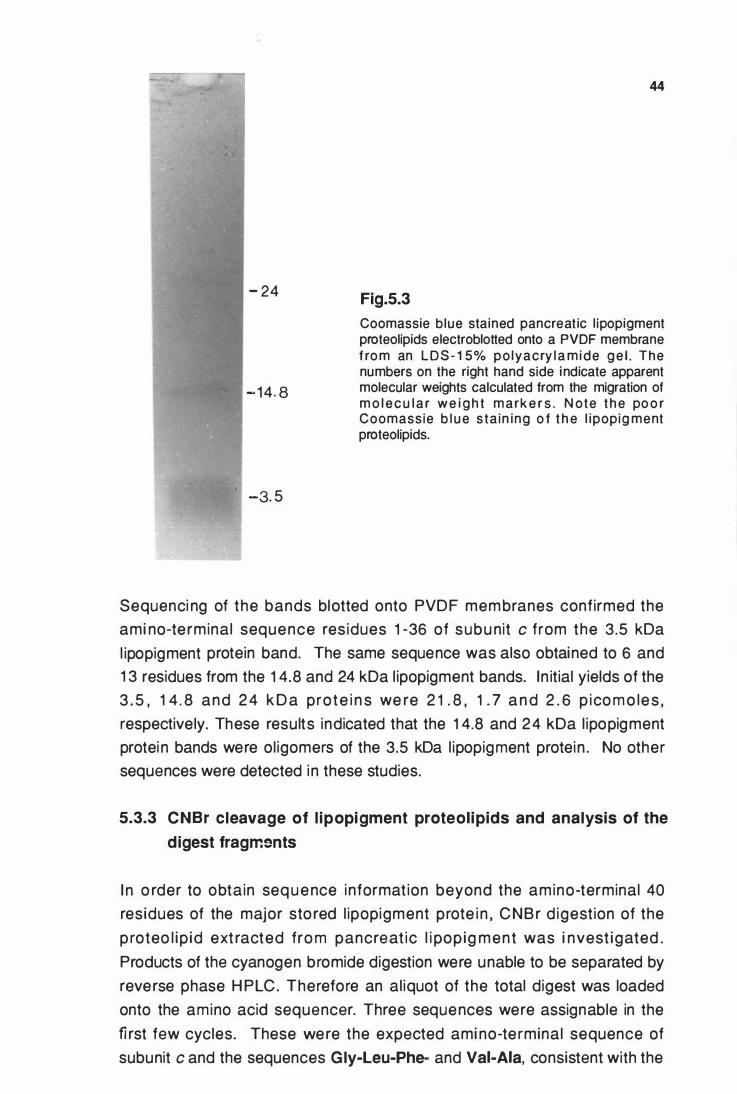
- 24
- 14 . 8
-3. 5
44
Fig.5.3
Coomassie b lue stained pancreat ic l ipopigment proteolipids electroblotted onto a PVDF membrane f ro m an L D S- 1 5% po lyacry l a m ide g e l . The numbers on the right hand side indicate apparent molecular weights calculated from the migration of m o l e c u l a r w e i g h t m a rk e r s . N ote t h e po o r Coomass ie b l u e s ta in ing o f t h e l ipopig ment proteolipids.
Sequenci ng of the bands blotted onto PVDF membranes confirmed the
ami no-te rminal sequence res idues 1 -36 of subu nit c from the 3.5 kDa
l ipopigment protein band. The same sequence was also obtained to 6 and
1 3 residues from the 1 4.8 and 24 kDa l ipopigment bands. In itial yields of the
3 . 5 , 1 4 . 8 and 2 4 k D a prote i n s we re 2 1 . 8 , 1 . 7 a n d 2 . 6 p ico m o le s ,
respectively. These results indicated that the 1 4.8 and 2 4 kDa l ipopigment
protei n bands were ol igomers of the 3.5 kDa l ipopigment protein . No other
sequences were detected i n these studies.
5.3.3 CNBr cleavage of l ipopigment proteolipids and analysis of the
digest fragm�mts
I n o rder to obtain seq u e nce i nformat ion beyond the amino-terminal 40
residues of the major sto red l ipopigment prote i n , CNBr d igestion of the
p roteo l ip id ext racted fro m pancreat ic l i pop ig ment was i nvest igated .
Products of the cyanogen bromide digestion were unable to be separated by
reverse phase H PLC. Therefore an al iquot of the total digest was loaded
onto the amino acid sequencer. Three sequences were assignable in the fi rst few cycles. These were the expected ami no-te rminal sequence of
subunit c and the sequences Gly-Leu-Phe- and Vai-Aia, consistent with the
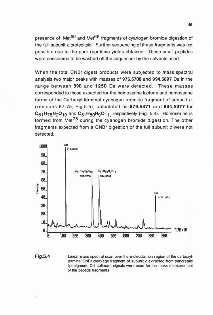
45
presence of Met60 and Met66 fragments of cyanogen bromide digestion of
the ful l subunit c proteol ipid. Further sequencing of these fragments was not
possib le due to the poor repetitive yields obtained. These small peptides
were considered to be washed off the sequencer by the solvents used.
W h e n t h e total CNBr d igest products were subjected to mass spectral
analysis two major peaks with masses of 976.5708 and 994.5897 Da in the
ra n g e b etwe e n 890 a n d 1 250 Da w e re d etecte d . T h e s e m asses
corresponded to those expected for the homoserine lactone and homoserine
fo rms of the Carboxyl-te rmi nal cyanogen b romide fragment of subun it c,
( r e s i d u e s 67-75 , F ig . 5 . 5 ) , ca lcu lated as 976.5871 a n d 994 .5977 for
Cs1 H7aN9o1 0 and C51 HaoN9o1 1 , respective ly (Fig. 5.4). Homoserine is
fo rmed from Met75 dur i ng the cyanogen brom ide d igest ion . The other
frag ments expected from a CNBr digestion of the fu l l subun it c were not
detected.
Fig.5.4
Csl 912.3352
C51 H7aN901 o C51 HaoN901 1 976.5708 994.5897
Csl 1 172.1451
100 200 300 400 500 600 700 800 900
Linear mass spectral scan over the molecular ion region of the carboxylterminal CNBr cleavage fragment of subunit c extracted from pancreatic lipopigment. Csl calibrant signals were used for the mass measurement of the peptide fragments.
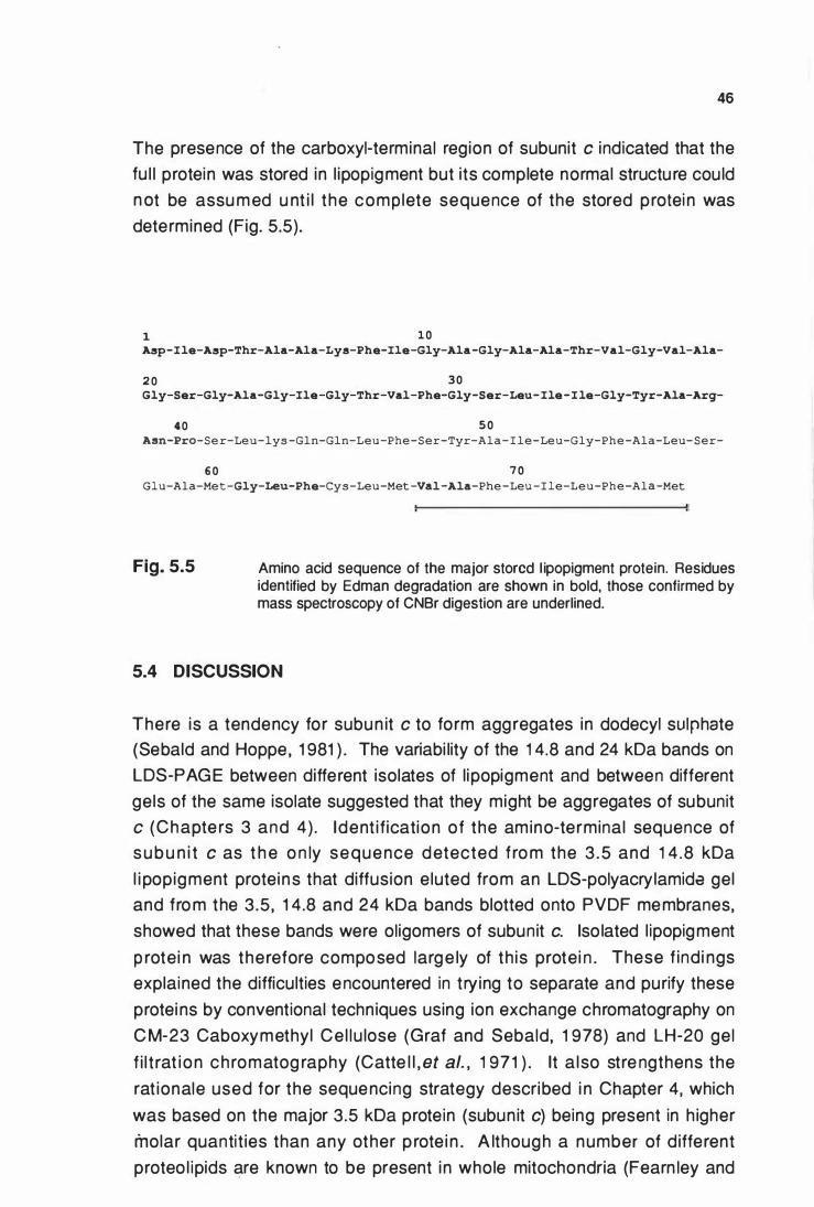
46
The presence of the carboxyl-terminal region of subunit c indicated that the
fu l l protein was stored in l ipopigment but its complete normal structu re cou ld
n ot be assu med u nt i l the complete sequence of the stored protei n was
determined (Fig. 5.5).
1 1 0 A5p-Ile-A5p-Thr-A1a-A1a-Lys-Phe-Ile-Gly-A1a-Gly-A1a-A1a-Thr-Val-Gly-Val-A1a-
2 0 3 0 Gly-Ser-Gly-Ala-Gly-Ile-Gly-Thr-Val-Phe-Gly-Ser-Leu- Ile-Ile-Gly-Tyr-Ala-Arg-
4 0 5 0 Asn-Pro-Se r-Leu-lys -Gln-Gln-Leu -Phe -Ser-Tyr-Ala - I le-Leu-Gly-Phe -Ala-Leu- Ser-
6 0 7 0 Glu-Ala-Me t -Gly-Leu-Phe-Cys-Leu-Met -Val -Ala-Phe -Leu - I le-Leu-Phe-Ala -Met
Fig. 5.5 Amino acid sequence of the major stored l ipopigment protein. Residues identified by Edman degradation are shown in bold, those confirmed by mass spectroscopy of CNBr digestion are underlined.
5.4 DISCUSSION
There is a tendency for subun it c to form aggregates in dodecyl sulphate
(Sebald and Hoppe, 1 981 ) . The variabi lity of the 1 4.8 and 24 kDa bands on
LDS-PAG E between different isolates of l ipopigment and between different
gels of the same isolate suggested that they might be aggregates of subunit
c (Chapte rs 3 and 4) . Ident i fication of the amino-terminal sequence of
s u b u n i t c as t h e o n ly sequence detected f ro m the 3 .5 and 1 4 .8 kDa
l i popigment proteins that diffusion e luted from an LDS-polyacry lamida gel
and fro m the 3.5, 1 4.8 and 24 kDa bands blotted onto PVDF membranes,
showed that these bands were oligomers of subunit c. Iso lated l ipopigment
p rotei n was therefore composed large ly of th is protei n . These f ind i ngs
explained the difficulties encountered in trying to separate and purify these
protei ns by conventional techniques using ion exchange chromatography on
C M-23 Caboxymethyl Ce l lu lose (Graf and Sebald, 1 978) and LH-20 gel
f i l t rat i on ch romatog raphy (Catte l l , et al. , 1 97 1 ) . lt a lso stre ngthens the
rat ionale used for the sequencing strategy described i n Chapter 4, which
was based on the major 3.5 kDa protein (subunit c) being present in h igher
molar quantit ies than any other p rote i n . A lthough a number of d ifferent
proteo l ipids are known to be present i n whole mitochondria (Fearn ley and
47
W a l k e r , 1 986 ) , n o oth e r m i toch o n d ri a l p rote i n was detected i n the
l ipopigment proteolipid fraction.
In conju nct ion with the p resent work, it has been establ ished from cDNA ·
studies that the normal ovi ne subunit c is identical to that of the bovi ne and
human protei n (Medd and Walker, unpublished). The fu l l bovine and human
subu n it c proteo lipids contai n th ree meth ion ine residues, at Met60 , Met66 ,
and at the carboxyl-terminus Met75. If the ful l subun it c is present i n ovine
l ipopigment then chemical cleavage with cyanogen bromide should give rise
to three fragments. The amino-terminal fragment was not detected as it had
a mass range beyond the capabi l ity of the inst rument used . A f rag ment
corresponding to cyanogen bromide cleavage at Met60 , was also not seen ,
possibly due to supression by other surface active components in the total
digest or by the low surface activity of the frag ment itse lf. The absence of
expected species due to these effects when mixtu res of digest products are
subjected to mass spectral analysis is we l l documented (Barber, et al. , 1 983 ;
B ieman n , 1 982 ; Clenc h , et al. , 1 985) . The carboxy l-te rm i nal cyanogen
bromide f rag ment , wh ich was identified by mass spectroscopy (Fig . 5.4) ,
i nd icated that the fu l l subun it c was present in li popigment. This was also
supported by the ami no acid com posit ion of the ext racted l ipopigment
proteolipid fraction , which was very simi lar to that of the ful l subunit c. The
contribution stored subun it c makes to total pancreatic l ipopigment protein
was therefore revised from a min imum of 40% to 73% (Table 4 . 1 ) . This
estimate is in accord with findi ngs recorded in this chapter, i .e . that 70 % of
the pancreatic l ipopigment extracted as proteolipid. These values are both
l i ke ly to be unde rest imates as the fi rst is based on the i n it ial y ie ld in the
amino-terminal sequencing which is not a quantitative reaction , and the
second on proteolipid extraction which was not complete (Fig . 5. 1 ) .
The complete amino acid sequence of the stored protein was determined by
co l laboration with researchers at the M.R.C. Molecu lar Biology Laboratory ,
Cambridge , U .K. This was achieved after covalently attachi ng the products
of t he react ions of t he l i pop igment p roteo l ip id f ract ion with cyanoge n
b romide and N-bromosucci n i mide to 3-aminopropyl g lass suppo rts. The
covalent coup l i ng to so l id supports was requ i red to prevent the digest
frag ments from bei ng washed off the sequencer i n the so lvents used ( I .M.
Fearnley, pers. comm.) . These sequencing studies showed that previously
unassigned residues 41 to 66 in the stored protei n were identical to those of
the fu l l subunit c (Fig . 5.5) .
48
Mass spectroscopy studies s howed that the re was no post-trans lat ional
m o d i f icat i ons to the carboxy l -t e rm i n u s ( M et66_Met75) no r was the re
e v i d e n ce to i n d i cate t h e p re s e n ce o f m od i f ied res i d u e s fro m both
sequenc ing and amino acid analyses. Th is however does not preclude the
possibi l ity of the presence of acid labi le modifications that wou ld not have
been detected by the studies described.
The subun it c proteo lipid in the inner mitochondrial membrane is not known
to be post-translationally modified (Sebald and Hoppe, 1 981 ). There was no
evidence for the presence in l ipopigment fragments of subun it c, tru ncated at
e ither amino or carboxyl terminals. lt is concluded that the complete subunit
c ot m itochondrial ATP synthase is stored i n ovine l ipopigment.
The fi nding that the 1 4.8 and 24 kDa bands were oligomers of subunit c was
a lso i mportant as it precluded any other major protei n com ponent being
s pecifical ly stored i n the d isease. As noted i n chapter 4 , this protein has
on ly previously been detected i n mitochondria as part of the F 0 component
of the ol igomeric ATP synthase complex, where it is estimated to account for
2 -4% of the i nner mitochondria l membrane prote i n (Sebald and Hoppe,
1 981 ) . As two th i rds of the l i popigment mass is protei n (Palmer, et al. ,
1 986b) , subunit c alone accounts for at least half the total l ipopigment mass.
I ts p rese nce i n lysosome derived l ipopig ment is therefo re most l i ke ly to
reflect the underlying biochemical lesion in ovine ceroid- l ipofuscinosis.
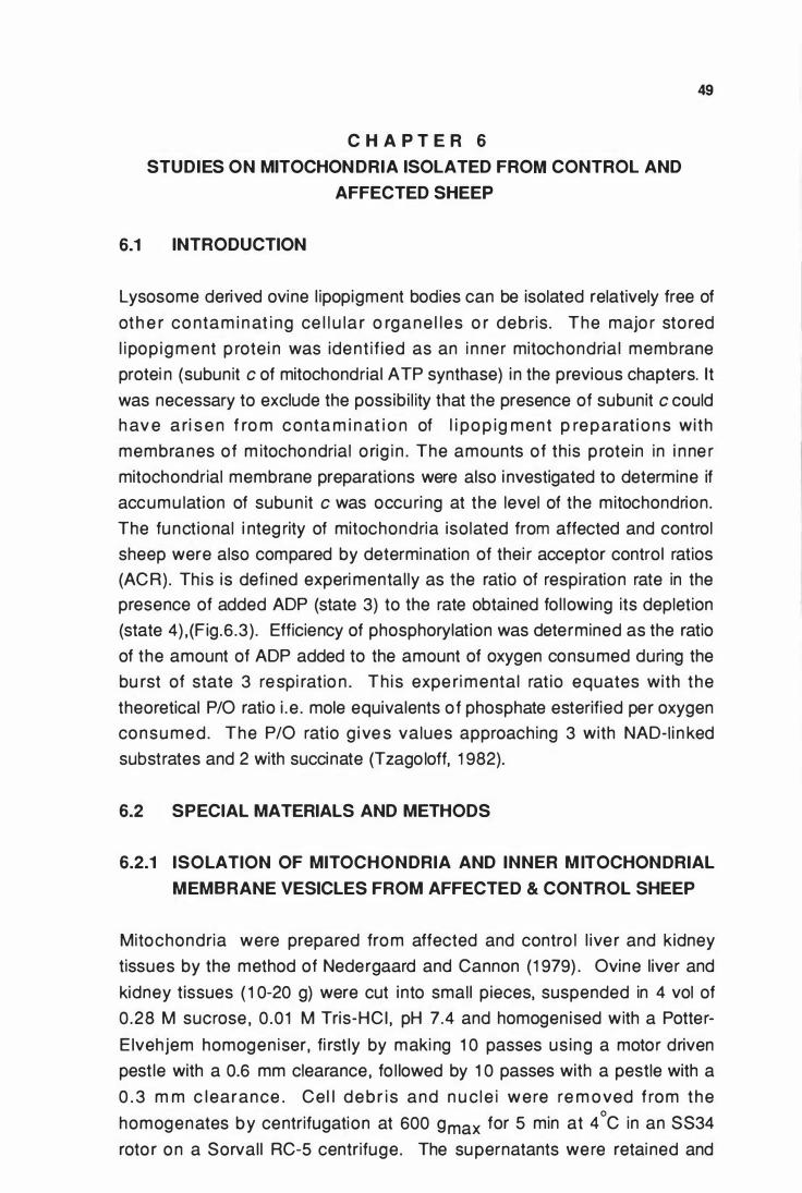
C H A P T E R 6
STUDIES ON MITOCHON DRIA ISOLATED FROM CONTROL AND
AFFECTED SHEEP
6.1 INTRODUCTION
49
Lysosome derived ovine l ipopigment bodies can be isolated relatively free of
oth e r co ntam i n at i ng ce l l u la r o rg a n e l les o r debris . The majo r sto red
l i pop ig ment p rote i n was ident i f ied as an i n ner mitochondria l membrane
protei n (subunit c of mitochondrial ATP synthase) in the previous chapters. lt
was necessary to exclude the possibi l ity that the presence of subunit c could
h av e a ri s e n f ro m conta m i n at i o n of l i po p ig ment p re p a rat i o n s with
membranes of m itochondrial o rig i n . The amounts of th is p rotein in i nne r
mitochondrial membrane preparations were also i nvestigated to determine if
accumu lat ion of subun it c was occu ri ng at the level of the mitochondrion.
The funct ional i nteg rity of mitochondria isolated from affected and control
sheep were also compared by determination of thei r acceptor control ratios
(ACR). Th is is defi ned experi mental ly as the ratio of respi ration rate in the
presence of added ADP (state 3) to the rate obtained fol lowing its depletion
(state 4) , (Fig .6.3) . Efficiency of phosphorylation was determined as the ratio
of the amount of ADP added to the amount of oxygen consumed during the
bu rst of state 3 respi ratio n . Th is expe ri mental rat io equates with the
theoretical P/0 ratio i .e . mole equivalents of phosphate esterified per oxygen
consu med . The P/0 rat io g i ve s values approaching 3 with NAD-I i n ked
substrates and 2 with succinate (Tzago loff, 1 982).
6.2 SPECIAL MATERIALS AND METHODS
6.2.1 ISOLATION OF MITOCHONDRIA AND INNER M ITOCHONDRIAL
MEMBRANE VESICLES FROM AFFECTED & CONTROL SHEEP
Mitochondria were prepared from affected and contro l l ive r and kidney
tissues by the method of Nedergaard and Cannon (1 979) . Ovine liver and
kidney tissues ( 1 0-20 g) were cut into small p ieces, suspended in 4 vol of
0 .28 M sucrose , 0 .01 M Tris-HCI , pH 7.4 and homogen ised with a Potter
E ivehjem homogen iser, fi rst ly by maki ng 1 0 passes us ing a motor driven
pest le with a 0.6 mm clearance, fo l lowed by 1 0 passes with a pestle with a
0 . 3 m m c learance . Ce l l d e b r i s and n u c l e i were re m oved f rom the
homogenates by centrifugation at 600 gmax for 5 min at 4°C in an SS34
roto r on a Sorval l RC-5 centrifuge. The supernatants were retai ned and
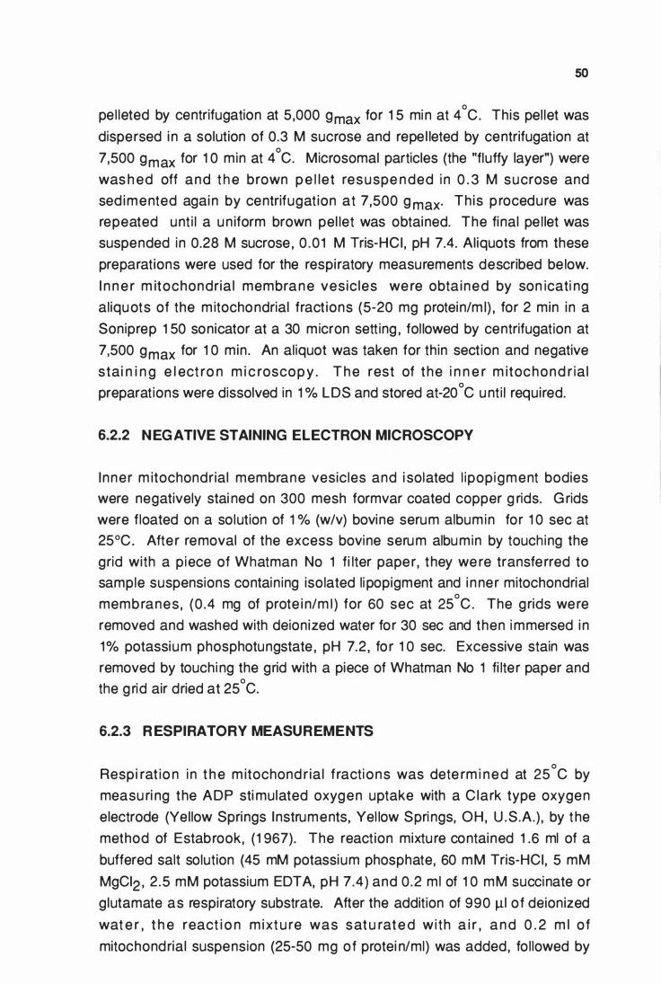
50
pel leted by centrifugation at 5 ,000 gmax for 1 5 mi n at 4°C. This pel let was
dispersed i n a solution of 0.3 M sucrose and repe l leted by centrifugation at
7,500 gmax for 1 0 min at 4°C. Microsomal particles (the "fluffy layer") were
was h ed off and t h e brown p e l let resuspe n ded i n 0 . 3 M sucrose and
sed imented agai n by centrifugat ion at 7,500 g max· Th is p rocedure was
repeated unt i l a u n iform brown pel let was obtained. The final pe l let was
suspended in 0.28 M sucrose , 0 .0 1 M Tris-HCI , pH 7.4. Al iquots from these
preparations were used for the respi ratory measurements described be low.
I n n e r m itochondria l membran e ves ic les we re obta ined by son icati ng
aliqu ots of the m itochondrial fract ions (5-20 mg protein/m !) , for 2 min in a
Soniprep 1 50 sonicator at a 30 micron sett ing , fol lowed by centrifugation at
7,500 gmax for 1 0 min . An aliquot was taken for thin section and negative
stai n i n g e l ect ro n m i c roscop y . T h e rest of t he i n n e r m itochond r ia l
preparations were dissolved in 1 % LDS and stored at-20°C u nt i l required.
6.2.2 N EG ATIVE STAINING ELECTRON MICROSCOPY
I nner m itochondrial membrane vesicles and i solated l ipopigment bodies
were negatively stai ned on 300 mesh formvar coated copper g rids. G rids
were floated on a solution of 1 % (w/v) bovine serum albumin for 1 0 sec at
25°C. Afte r removal of the excess bovine serum albumin by touching the
grid with a piece of Whatman No 1 fi lter paper, they we re transfe rred to
sample suspensions contain ing isolated li popigment and i nner mitochondrial
membranes , (0 .4 mg of prote i n/m ! ) for 60 sec at 25°C . The grids were
removed and washed with deionized water for 30 sec and then i mmersed i n
1% potassium phosphotungstate , pH 7.2 , fo r 1 0 sec. Excessive stain was
removed by touchi ng the grid with a piece of Whatman No 1 fi lter paper and
the g rid air dried at 25°C.
6.2.3 R ESPIRATORY MEASUREMENTS
Resp i rat io n i n t h e m itochond r ia l f racti ons was dete rm i ned at 25°C by
measu ri ng the ADP sti mulated oxygen uptake with a C lark type oxygen
electrode (Yellow Springs Instruments, Yel low Spri ngs, OH, U .S.A.) , by t he
method of Estabrook, (1 967). The react ion mixtu re contained 1 .6 ml of a
buffered salt solution (45 mM potassium phosphate, 60 mM Tris-HCI, 5 mM
MgCI2 , 2 .5 mM potassium EDTA, pH 7.4) and 0 .2 ml of 1 0 mM succinate or
glutamate as respiratory substrata. After the addition of 990 J.ll of deionized
wat e r , t h e react i o n m ixt u re was sat u rate d w i th a i r , a n d 0 . 2 m l of
mitochondrial suspension (25-50 mg of protei n/m!) was added, followed by
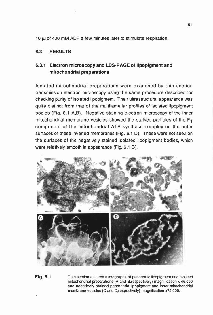
1 0 J.LI of 400 mM ADP a few minutes later to stimulate respiration .
6.3 RESULTS
6.3.1 Electron microscopy and LDS-PAGE of l ipopigment and
mitochondrial preparations
51
I s o l ated m itochondr ia l p re parat i o n s were exam i n ed by t h i n sect i o n
transmission e lectron microscopy using t h e same procedure described for
checking purity of i solated l ipopigment. Their u ltrastructural appearance was
quite dist inct from that of the mu lti lamel lar profi les of isolated l ipopigment
bodies (Fig . 6 . 1 A,B) . N egative stai n ing e lectron microscopy of the inner
m itochondrial membrane vesicles showed the stalked particles of the F 1 componen t of t h e mitocho ndr ia l ATP synthase co m plex o n the outer
surfaces of these inverted membranes (Fig . 6. 1 D) . These were not see.1 on
the surfaces of the negatively stai ned iso lated l ipopigment bodies, which
were re latively smooth in appearance (Fig. 6 . 1 C).
Fig. 6.1 Thin section electron micrographs of pancreatic lipopigment and isolated mitochondrial preparations (A and B,respectively) magnification x 46,000 and negat ively stained pancreatic lipopigment and inner mitochondrial membrane vesicles (C and D,respectively) magnification x72,000.
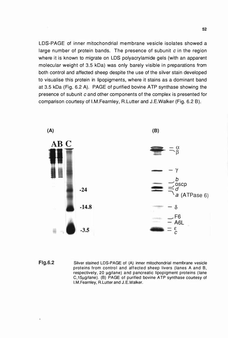
52
LDS-PAGE of i nner m itochondrial membrane vesicle iso lates showed a
large number of protein bands. The p resence of subun it c i n the reg ion
where i t is known to migrate on LDS polyacrylamide gels (with an apparent
molecu lar weight of 3 .5 kDa) was on ly barely visible i n preparations from
both control and affected sheep despite the use of the si lver stain developed
to visualise this protein in lipopigments, where it stains as a dominant band
at 3.5 kDa (Fig. 6.2 A) . PAGE of purified bovine ATP synthase showing the
presence of subunit c and other components of the complex is presented for
comparison courtesy of I .M .Fearnley, R .Lutter and J .E.Walker (Fig. 6 .2 B) .
(A)
AB C
Flg.6.2
(B)
-24
- 14.8
-3.5
- a --- p
- "( b =:; oscp
- d ' a (ATPase 6)
- o __. F6 - A6L - £ - c
Silver stained LDS-PAGE of (A) inner mitochondrial membrane vesicle prote ins from co ntro l and aff ected sheep l ivers ( lanes A and B , respectively, 20 J.LQ/Iane) a nd pancreatic l ipopigment prote ins ( lane C , 1 5J.Lg/lane) . (B) PAGE of purif ied bovine ATP synthase courtesy of I .M.Fearnley, R.Lutter and J .E.Walker.
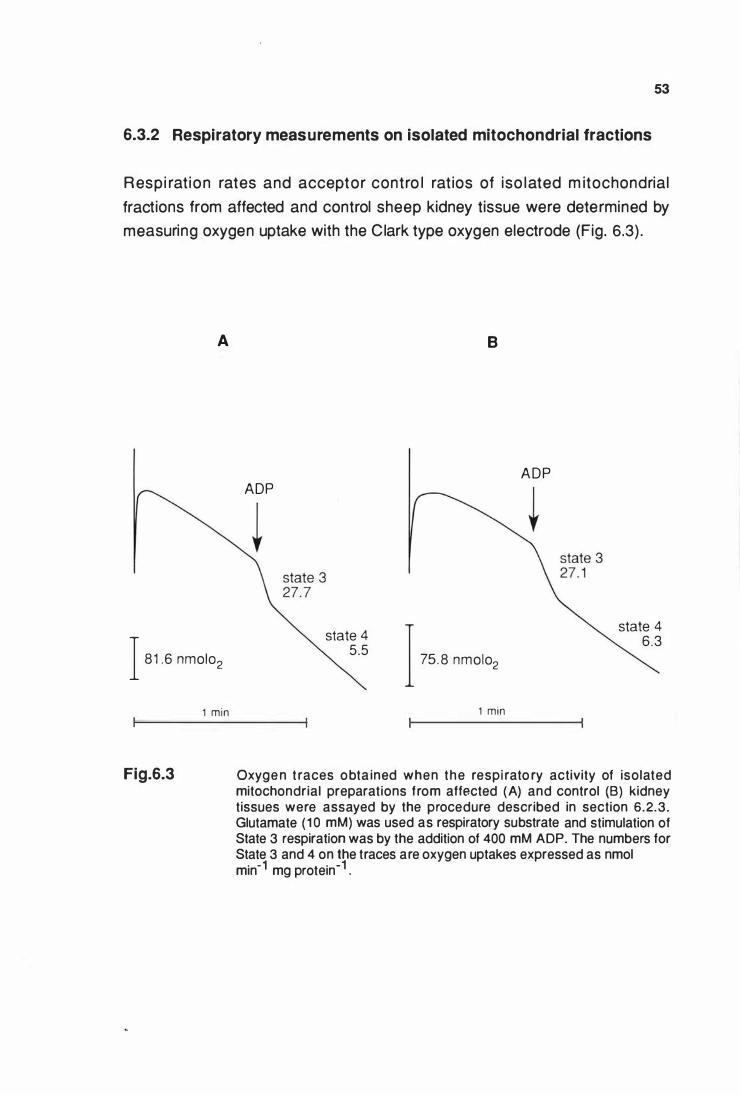
53
6.3.2 Respiratory measurements on isolated mitochondrial fractions
Respi rat ion rates and acceptor contro l rat ios of iso lated m itochondrial
fractions from affected and control sheep kidney tissue were determined by
measuring oxygen uptake with the Clark type oxygen electrode (Fig. 6.3) .
A
ADP
I 81 .6 nmolo2
state 4 5.5
B
I 75. 8 nmolo2
1 min 1 min
A D P
state 4 6 .3
Fig.6.3 Oxyge n t races obta ined when the respi rato ry activity of isolated mitochondrial preparations from affected (A) and control (B) kidney tissues were assayed by the procedure described in section 6.2.3 . Glutamate ( 1 0 mM) was used as respiratory substrate and stimulation of State 3 respiration was by the addition of 400 mM ADP. The numbers for State 3 and 4 on the traces are oxygen uptakes expressed as nmol min-1 mg protein-1 .
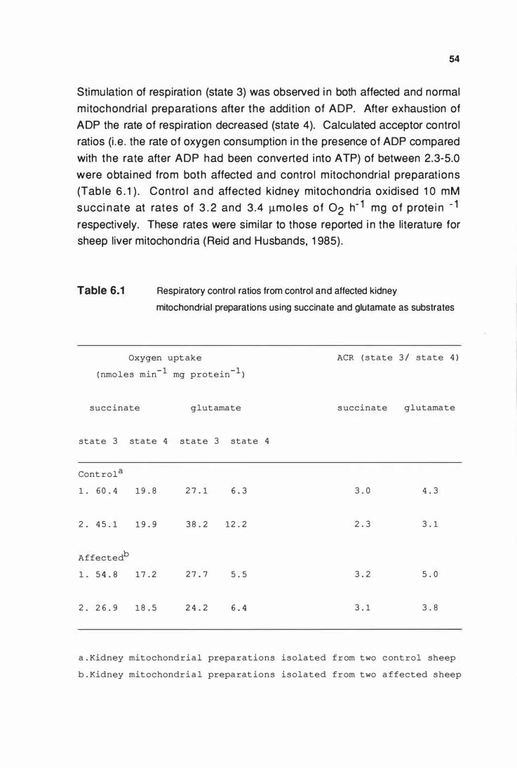
54
Stimu lation of respirat ion (state 3) was observed i n both affected and normal
m itochondrial preparat ions after the addit ion of ADP. After exhaustion of
ADP the rate of respi ration decreased (state 4). Calcu lated acceptor control
ratios (i . e . the rate of oxygen consumption i n the presence of ADP compared
with the rate after ADP had been converted i nto ATP) of between 2 .3-5.0
were obtai ned from both affected and contro l m itochondrial preparat ions
(Tab le 6 . 1 ) . Contro l and affected kidney mitochondria oxidised 1 0 mM
s ucci n ate at rates of 3 . 2 and 3 .4 Jlmo les of 02 h- 1 mg of prote i n - 1
respectively. These rates were simi lar to those reported i n the literature for
sheep liver mitochondria (Reid and Husbands, 1 985) .
Table 6.1 Respiratory control ratios from control and affected kidney
mitochondrial preparations using succinate and glutamate as substrates
Oxygen upt ake ACR ( state 3 / state 4 )
( nmoles min- 1 mg protein- 1 )
succ inate glutamate s uccinate glutamate
state 3 state 4 state 3 state 4
Cont rol a
1 . 6 0 . 4 1 9 . 8 2 7 . 1 6 . 3 3 . 0 4 . 3
2 . 4 5 . 1 1 9 . 9 3 8 . 2 12 . 2 2 . 3 3 . 1
Affectedb
1 . 5 4 . 8 1 7 . 2 2 7 . 7 5 . 5 3 . 2 5 . 0
2 . 2 6 . 9 1 8 . 5 2 4 . 2 6 . 4 3 . 1 3 . 8
a . Kidney mitochondrial preparations isolated f r om two cont rol sheep
b . Kidney mitochondri a l preparat ions isolated from two a ffected sheep
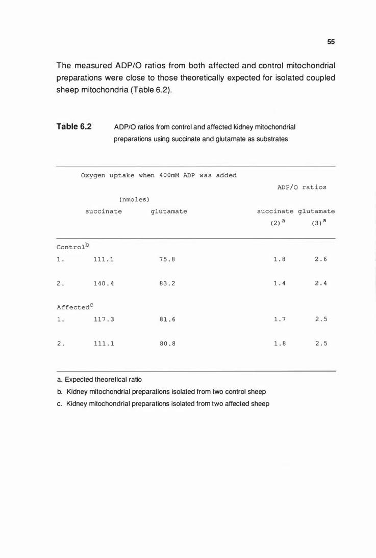
55
The measu red ADP/0 rat ios from both affected and control mitochondrial
preparations were close to those theoretically expected for iso lated coupled
sheep mitochondria (Table 6.2) .
Table 6.2 ADP/0 ratios from control and affected kidney mitochondrial
preparations using succinate and glutamate as substrates
Oxygen upt ake when 4 0 0mM ADP was added
AD P / 0
( nmo les )
succinate glutamate succinate
( 2 ) a
Controlb
1 . 1 1 1 . 1 7 5 . 8 1 . 8
2 0 1 4 0 . 4 8 3 . 2 1 . 4
Affectedc
1 . 1 1 7 . 3 8 1 . 6 1 . 7
2 0 1 1 1 . 1 8 0 . 8 1 . 8
a. Expected theoretical ratio
b. Kidney mitochondrial preparations isolated from two control sheep
c. Kidney mitochondrial preparations isolated from two affected sheep
rat ios
glutamate
( 3 ) a
2 0 6
2 . 4
2 . 5
2 . 5
6.4 DISCUSSION
56
The quantitative data presented i n Chapter 4 and 5 showed that subunit c
was stored i n l ipopigment isolated from sheep with ceroid- l i pofusci nosis.
Evidence that it cou ld not have ar isen from contaminat ion of l ipopigment
preparat ions with i n n e r m itochondri al me m b ranes was obtai ned by a
combination of electron m icroscopy and polyacrylamide gel e lectrophoresis.
Firstly, despite the common use of sonication and centrifugation steps in the
methods of isolation of l ipopigment and mitochondrial preparations, they had
qu ite separate and d isti nctive u ltrastructu ral appearances. I n negatively
stai ned preparations there was no i nd ication of the stalked particles of the
F1 component of the ATP synthase complex on the surfaces of l ipopigment
bodies, whereas these were clearly seen on the i nner mitochondrial vesicles
(Fig. 6 .2 C & D) . Secondly , the protei n composition of i nner m itochondrial
vesicles seen by LDS-PAGE was distinct from that of isolated l ipopigment,
w i t h s u b u n i t c b e i n g bare ly v i s i bi e i n t h e fo rmer . T h e refo re a n y
contami nation of l i popigment bodies by mitochondrial membranes wou ld
tend to diminish the concentration of subunit c i n the lipopigment, rather than
be a source of it. Further support for a lack of m itochondrial contamination
of iso lated l ipopigment bodies comes from the l ipid analysis of Palmer, et al.,
( 1 986a) , which showed that l ipopigment l ipids were those characteristic of
lysosomal membranes with no indication of the presence of the distinctive
mitochondrial phosphol ipid, cardiol ipin .
The quant itat ive d ata p resented in Chapte rs 4 & 5 and observat ions
recorded and discussed above, indicate that subunit c of mitochondrial ATP
synth ase i s specif ica l ly stored i n l i popi g m e nt f rom sheep with ce roid
l ipofuscinosis and its presence was not due to mitochondrial contamination
of i s o lates of l i po p i g m e nt s . S i m i lar amou nts of s u b u n it c i n i n ne r
mitochondrial membranes of affected and control preparations (Fig . 6.2 A)
i ndicated that accumu lation of this protein in the disease was not occuri ng i n
the mitochondrion.
Acceptor contro l rat ios of mitochondrial preparations from affected sheep
kidney tissue were comparable to those dete rmi ned from normal sheep
kidney tissue. They were also consistent with those reported in the l iterature
for sheep liver mitochondrial preparations (Reid and Husbands, 1 985) .
Measured respi ration rates and P/0 ratios were close to those theoretical ly

57
expected using succi nate or glutamate as substrates. This indicated that the
mitochondria l fract ions from affected sheep mai ntai ned a structu ral and
fu nctiona l i nteg rity enab l i ng acceptor contro l comparable to those from
control m itochondria l fractions. The les ion in the disease was therefore
un l ikely to be re lated to a mitochondrial respi ratory chai n o r an oxidative
phosphory lation defect related to the m itochondrial ATP synthase complex.
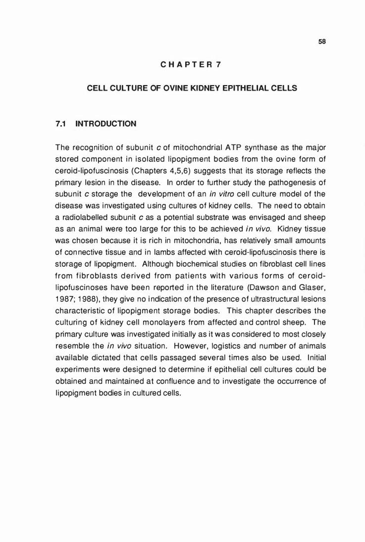
58
C H A P T E R 7
CELL CULTURE OF OVINE KIDNEY EPITHELIAL CELLS
7.1 INTRODUCTION
The recognition of subun it c of m itochondrial ATP synthase as the major
sto red component i n iso lated l ipopigment bodies from the ovi ne form of
ceroid-l ipofusci nosis (Chapters 4,5,6) suggests that its storage reflects the
primary lesion in the disease. In order to further study the pathogenesis of
subun it c storage the development of an in vitro ce l l cu lture model of the
disease was i nvestigated using cultu res of kid ney cel ls . The need to obtain
a radiolabel led subun it c as a potential substrata was envisaged and sheep
as an animal were too large for this to be ach ieved in vivo. Kidney tissue
was chosen because it is rich in mitochondria, has relatively small amounts
of con nective tissue and i n lambs affected with ceroid-lipofuscinosis there is
storage of l ipopigment . Although biochemical studies on fibroblast cell l i nes
f rom f i brob lasts d e rived from pat i ents wit h vari o u s forms of ce ro id
l i pofuscinoses have bee n reported i n the l ite ratu re (Dawso n and Glaser,
1 987; 1 988) , they g ive no i ndication of the presence of u ltrastructural lesions
characteristic of l ipopigment sto rage bodies. This chapter describes the
cu lturi ng of kidney ce l l mono layers from affected and control sheep. The
primary culture was i nvestigated in itial ly as it was considered to most closely
resemble the in vivo situation . However, log istics and number of animals
avai lable dictated that ce l l s passaged several t imes also be used. I n itial
experiments were designed to determine if epithe l ial cel l cultures could be
obtained and maintained at confluence and to i nvestigate the occurrence of
l ipopigment bodies i n cultured cel ls.
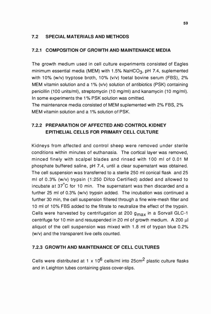
7.2 SPECIAL MATERIALS AND METHODS
7.2.1 COMPOSITION OF G ROWTH AND MAINTENANCE MEDIA
59
The growth medium used i n ce l l cu lture experi ments consisted of Eag les
min imum essential media (MEM) with 1 .5% NaHC03, pH 7.4, suplemented
with 1 0% (w/v) tryptose broth , 1 0% (v/v) foetal bovi ne seru m (FBS) , 2%
MEM vitamin solution and a 1 % (v/v) solution of antibiotics (PSK) contain ing
penici l l i n ( 1 00 units/ml) , streptomycin (1 0 mg/ml) and kanamycin ( 1 0 mg/ml) .
I n some experiments the 1 % PSK solution was omitted.
The maintenance media consisted of MEM suplemented with 2% FBS, 2%
MEM vitamin solution and a 1 % solution of PSK.
7.2.2 PREPARATION OF AFFECTED AND CONTROL KIDNEY
EPITHELIAL CELLS FOR PRIMARY CELL CULTU RE
Kid neys from affected and co ntro l sheep we re removed u nder steri l e
condit ions with in m inutes of euthanasia. The cortical layer was removed,
m i nced fi n e ly with sca lpe l b lades and ri n sed with 1 00 m l of 0 . 0 1 M
phosphate buffered saline , pH 7.4, unti l a clear supernatant was obtained.
The cel l suspension was transferred to a steri le 250 ml con ical flask and 25
m l of 0 . 3% (w/v) t ryps i n ( 1 :250 Di fco Cert i f ied) added and al lowed to
i ncubate at 37°C for 1 0 m in . The supernatant was then discarded and a
fu rther 25 ml of 0.3% (w/v) trypsin added. The i ncubation was continued a
further 30 min , the cel l suspension fi ltered through a fine wire-mesh fi lter and
1 0 ml of 1 0% FBS added to the fi ltrate to neutral ize the effect of the trypsin .
Ce l ls were harvested by centrifugat ion at 200 g max in a Sorvall GLC-1
centrifuge for 1 0 min and resuspended i n 20 ml of g rowth medium. A 200 �I
al iquot of the cel l suspension was m ixed with 1 .8 ml of trypan blue 0 .2%
(w/v) and the transparent live cells counted.
7.2.3 G ROWTH AND MAINTENANCE OF CELL CULTURES
Cel ls were d istributed at 1 x 1 o6 cel ls/m I into 25cm2 plastic cu lture flasks
and in Leighton tubes contain ing g lass cover-sl ips.
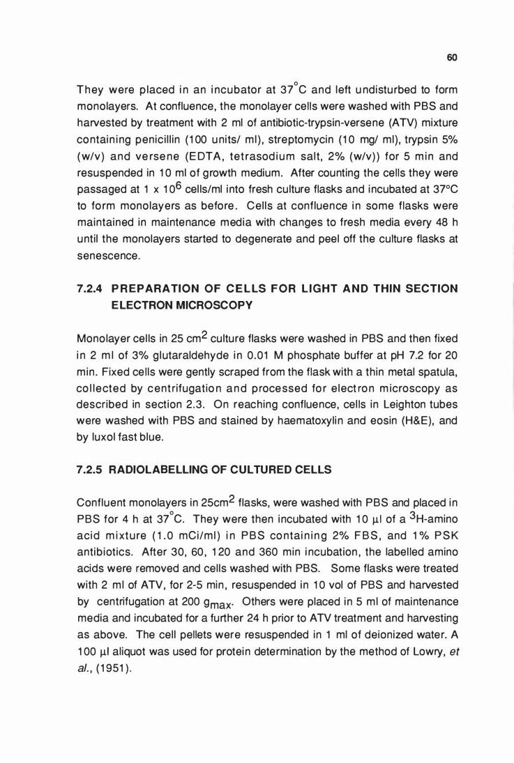
60
T h ey were p laced i n an i ncubator at 37°C and left u ndistu rbed to form
monolayers. At confluence , the monolayer cel ls were washed with PBS and
harvested by treatment with 2 ml of antibiotic-trypsin-versene (ATV) mixture
contai n i ng penic i l l in ( 1 00 u nits/ m l ) , streptomycin ( 1 0 mg/ ml} , trypsi n 5%
(w/v) and ve rse ne (E DTA , tet rasod i u m salt , 2% (w/v) ) for 5 m i n and
resuspended in 1 0 ml of g rowth medium. After counting the cells they were
passaged at 1 x 1 o6 cel ls/ml i nto fresh cu lture flasks and i ncubated at 37°C
to fo rm mono layers as before . Cel ls at confluence i n some flasks were
maintained in maintenance media with changes to fresh media every 48 h
unti l the monolayers started to degenerate and peel off the culture flasks at
senescence.
7.2.4 P R EPARATION OF C E L LS FOR L I G HT A N D THIN SECTION
E LECTRON MICROSCOPY
Monolayer cel ls in 25 cm2 cu lture flasks were washed in PBS and then fixed
i n 2 m l of 3% g lutaraldehyde i n 0 .01 M phosphate buffer at pH 7.2 for 20
min . Fixed ce l ls were gently scraped from the flask with a th in metal spatula,
co l lected by cent rifugatio n a n d p rocessed fo r e lect ro n m icroscopy as
described in sect ion 2 .3 . On reach ing confluence, cel ls in Leighton tubes
were washed with PBS and stained by haematoxyli n and eosin (H&E), and
by luxol fast blue.
7.2.5 RADIOLABELLING OF CULTURED CELLS
Confluent monolayers in 25cm2 flasks, were washed with PBS and placed i n
PBS for 4 h at 37°C. They were then i ncubated with 1 0 �I of a 3H-amino
acid m i xtu re ( 1 . 0 mCi/m l ) i n P B S co ntai n i n g 2% F B S , and 1 % P S K
antib iotics. After 30, 60 , 1 20 and 360 min i ncubation , the label led amino
acids were removed and cel ls washed with PBS. Some flasks were treated
with 2 m l of ATV, for 2-5 min , resuspended i n 1 0 vol of PBS and harvested
by centrifugation at 200 gmax· Others were placed i n 5 m l of maintenance
media and incubated for a further 24 h prior to ATV treatment and harvesting
as above. The cel l pellets were resuspended in 1 ml of deionized water. A
1 00 �I al iquot was used for protei n determination by the method of Lowry, et
al. , ( 1 951 ) .
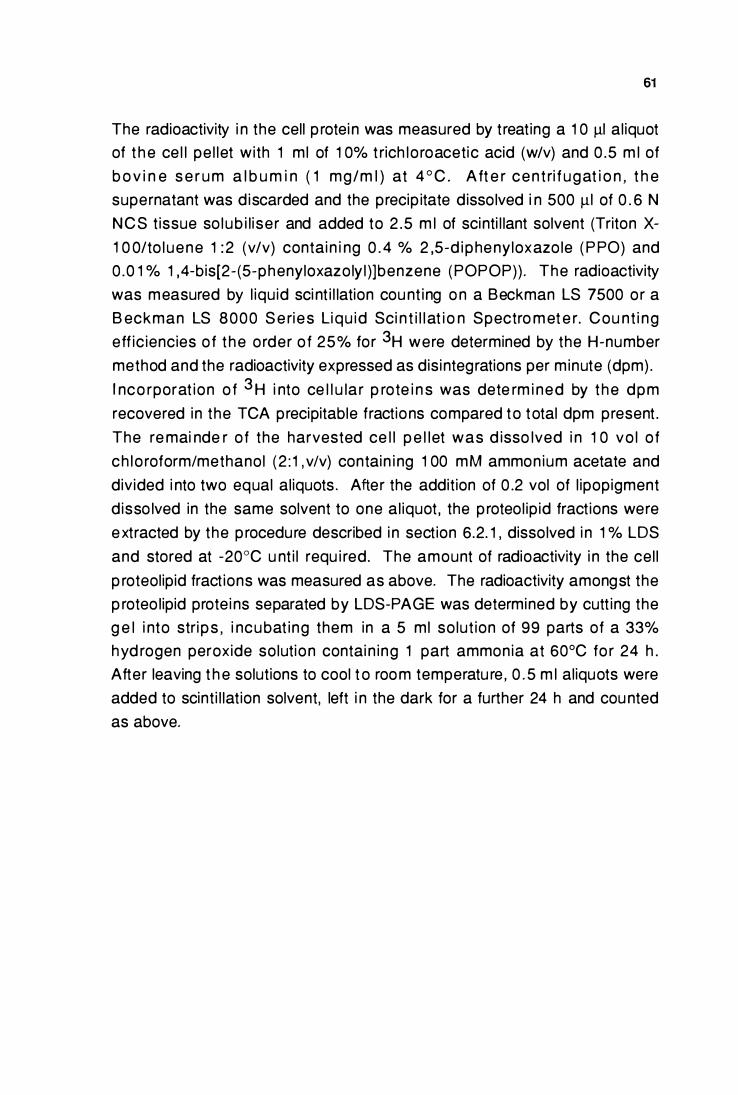
61
The radioactivity i n the cell p rotei n was measured by t reating a 1 0 !ll al iquot
of t h e ce l l pe l let with 1 ml of 1 0% t rich lo roacetic acid (w/v) and 0 .5 m l of
b o v i n e s e r u m a l b u m i n ( 1 mg/m l ) at 4 ° C . A ft e r cen t ri f ugat i o n , t h e
supernatant was d iscarded and the precip itate dissolved i n 500 !ll of 0 . 6 N
N C S t issue so lub i l i ser and added to 2 .5 m l of scinti l lant solvent (Triton X-1 0 0/to l uene 1 : 2 (v/v) contai n i ng 0 . 4 % 2 ,5-d i phenyloxazo le ( P PO) and
0 .0 1 % 1 ,4-bis[2- (5-phenyloxazoly l )]benzene (POPOP)) . The radioactivity
was measured by l iqu id sci nt i l lation cou nt ing o n a Beckman LS 7500 or a
B eckman LS 8000 S e ries Liqu id Sci n t i l l at i o n Spectro m et e r. Cou nt i ng
eff ic iencies o f t he o rder o f 25% for 3H were determined by the H-number
method and the radioactivity expressed as disintegrations per minute (dpm) .
I nco rporat ion o f 3 H i nto ce l l u la r p rote i n s was dete rm i ned by t he dpm
recovered in the TCA precipitable fractions compared to total dpm present .
The re mai nde r of the harvested ce l l p e l let was d isso lved i n 1 0 vo l of
ch lo roform/methanol (2 : 1 , v/v) contain i ng 1 00 mM ammon ium acetate and
divided i nto two equal aliquots . After the addition of 0 .2 vol of l ipopigment
d isso lved in the same solvent to one al iquot , the p roteol ip id fractions were
e xtracted by the p rocedure described i n section 6.2. 1 , dissolved i n 1 % LDS
and stored at -20°C u nt i l requ i red. The amount of radioactivity i n the cel l
p roteolipid fract ions was measured as above. The radioactivity amongst the
p roteo lipid protei ns separated by LDS-PAGE was determined by cutt ing the
g e l i nto stri ps , i ncubat ing them in a 5 ml so lut ion of 99 parts of a 33%
hyd rogen peroxide solution contai n ing 1 part ammonia at 60°C fo r 24 h .
After leaving t h e solutions to cool t o room temperatu re , 0 . 5 m l aliquots were
added to scinti l lat ion solvent, left i n the dark for a further 24 h and cou nted
as above.
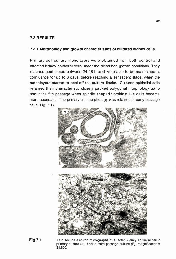
62
7.3 R ESULTS
7.3.1 Morphology and growth characteristics of cultured kidney cel ls
Pri mary ce l l c u lt u re mono layers we re o btai ned fro m both contro l and
affected kidney epithel ial ce l ls under the described growth conditions. They
reached conf luence between 24-48 h and were able to be maintai ned at
conf luence for u p to 6 days, before reach i ng a senescent stage , when the
mono layers started to peel off the cu lt u re flasks. Cu ltured epithe lial cells
retai n ed the i r characte ristic close ly packed po lygonal morphology up to
about the 5th passage when spi nd le shaped fibroblast- l i ke cel ls became
more abundant. The primary ce l l morpho logy was retained in early passage
ce l ls (Fig. 7. 1 ) .
Fig.7.1 Thin section e lectron micrographs of affected k idney epithelial cell in primary culture (A) , and in th i rd passage culture (B) , magnif ication x 31 ,800.
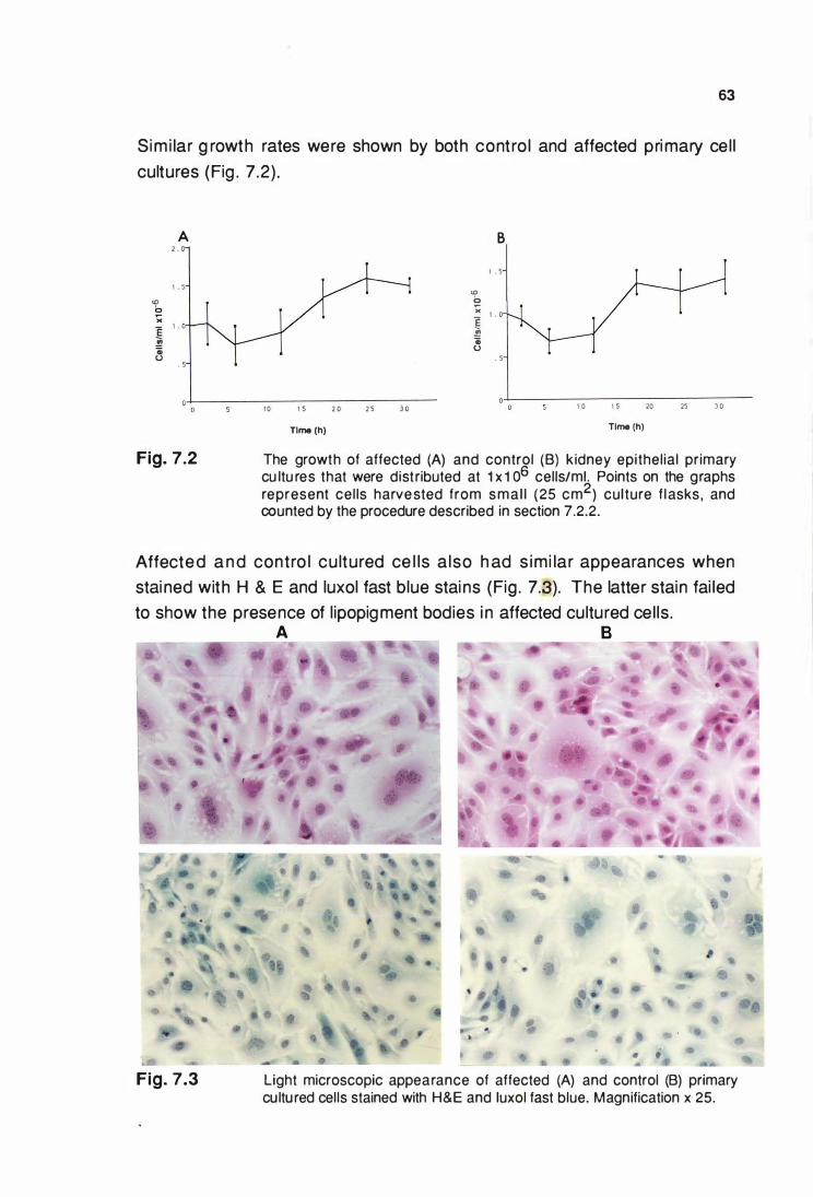
63
Simi lar g rowth rates were shown by both contro l and affected pri mary ce l l
cultures (Fig . 7 .2) .
A 2 . 0
1 . 5 <D b " e 1 .
!! Gi u
. 5
Fig. 7.2
1 0 1 s 2 0 2 5 3 0
Tlme (h)
B
1 . 5
<D b >< , � . Gi u
. 5
1 0 1 5 20 25 3 0
Tlme (h)
The growth of affected (A) and control (B) k idney epithel ial primary cu ltu res that were dist ributed at 1 x 1 o6 cells/m!. Points on the graphs represent cel ls harvested from sma l l ( 25 cm2) culture f l asks , and counted by the procedure described in section 7.2.2.
Affected a n d contro l cu ltured ce l ls a lso h ad s imi lar appearances when
stai ned with H & E and luxol fast blue stai ns (Fig . 7.�). The latter stain fai led
to show the presence of l ipopigment bodies i n affected cultured ce l ls.
Fig. 7.3
A B
• •
Light microscopic appearance of affected (A) and control (B) primary cu ltu red cells stained with H&E and luxol fast blue. Magnification x 25.
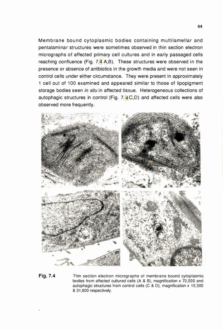
64
M e m b ra n e bo u nd cyto p las m i c b o d i es conta i n i ng m u lt i l ame l la r and
pentalami na r st ructures were somet imes observed in th in section electron
microg raphs of affected primary ce l l cu ltu res and in early passaged ce l ls
reaching confluence (Fig. 7.14 A,B). These structures were observed i n the
presence or absence of antibiotics in the growth media and were not seen i n
control ce l ls under either circumstance. They were present in approximate ly
1 ce l l out of 1 00 examined and appeared s imi lar to t hose of l ipopig ment
storage bodies seen in situ i n affected tissue. Heterogeneous collections of
autophag ic structures in contro l (Fig . 7. 4 C,D) and affected cel ls were also
observed more frequently.
Fig. 7.4 Thin sect ion e lectron micrographs of membran e bound cytoplasmic bodies fro m affected cultured cells (A & B) , magnification x 72,000 and autophagic structures from control cells (C & D), magnification x 1 5 ,300 & 31 ,800 respectively.
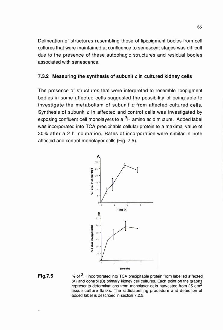
65
Del i neat ion of st ructu res resembl ing those of l ipopigment bodies from ce l l
cu ltures that were mai ntai ned at confluence to senescent stages was difficu lt
due to the presence of these autophagic st ructures and residual bod ies
associated with senescence.
7.3.2 Measuring the synthesis of subunit c in cultured kidney cells
The presence of structu res that were i nte rpreted to resemble l ipopigment
bod ies i n some affected ce l ls suggested the possib i l i ty of be ing able to
i nvest i g ate the metabo l i s m of subu n i t c f rom affected cu ltu red ce l l s .
Synthesis of subu n it c i n affected and contro l cel ls was i nvestigated by
exposing confluent cel l mono layers to a 3H amino acid mixture . Added label
was i ncorporated i nto TCA precipitable cellu lar protein to a maximal value of
30% afte r a 2 h i ncubat i o n . Rates of i nco rpo rat ion were s im i lar i n both
affected and control monolayer cells (Fig . 7.5).
Fig.7.5
A 30
i 25 ii � 20
8 .5 15
:z � 10
.,_
B 30
a: 25
ii � 20
� 15
) � 10
.,_
Tlme (h)
Tlme (h)
% of 3H incorporated into TCA precipitable protein f rom labelled affected (A) and control (B) primary kidney cell cultures. Each point on the graphs represents determinations from monolayer cells harvested from 25 cm2
tissue cu ltu re f lasks. The rad iolabel l ing procedure and detection of added label is described in section 7.2.5.
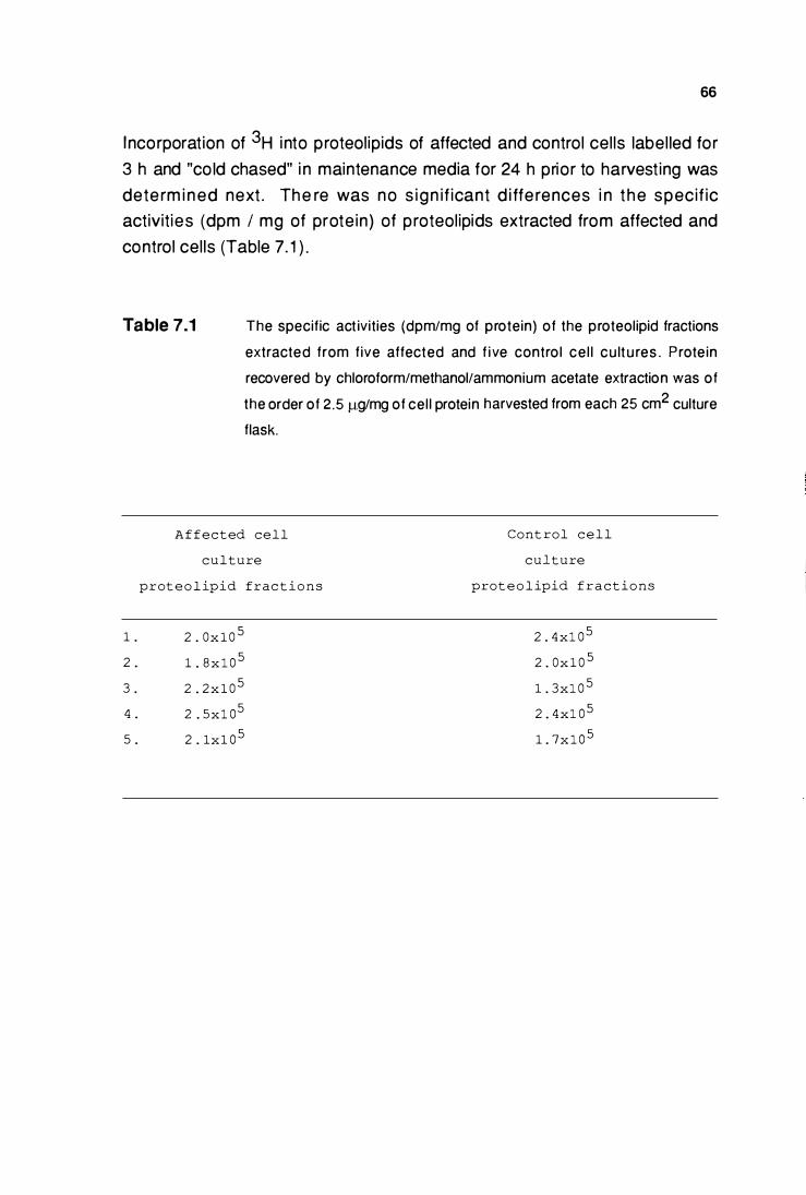
66
I ncorporation of 3H i nto proteolipids of affected and control cel ls label led for
3 h and "co ld chased" i n maintenance media for 24 h prior to harvest ing was
dete rm i n ed next . The re was no s ign if icant d i ffe rences i n t he specif ic
activities (dpm I mg of prote in) of proteolipids extracted from affected and
control cel ls (Table 7. 1 ) .
Table 7.1 The specific act ivities (dpm/mg of protein) of the proteolipid fractions
extracted from five affected and f ive contro l ce l l cu lt u res . Prote in
recovered by chloroform/methanol/ammonium acetate extraction was of
the order of 2.5 f.lg/mg of cel l protein harvested from each 25 cm2 culture
flask.
1 .
2 .
3 .
4 .
5 .
A f f ec t ed cell
c u l t u re
p r ot e o l ipid frac t i o n s
2 . 0 x 1 0 5
1 . 8 x l 0 5
2 . 2 x 1 0 5
2 . 5 x l 0 5
2 . 1x 1 0 5
Cont rol c e l l
c u l t u re
prot e o l ipid f r a c t i o n s
2 . 4 xl 0 5
2 . 0 x 1 0 5
1 . 3xl 0 5
2 . 4 xl 0 5
1 . 7 x l 0 5
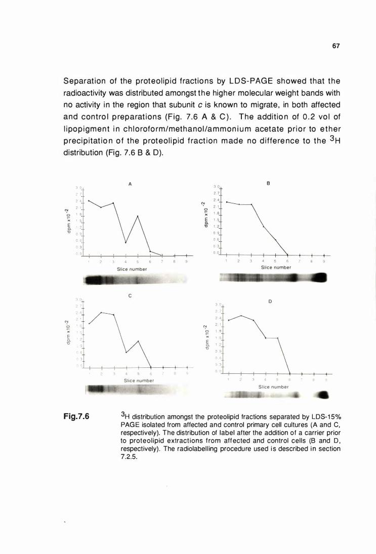
67
Separat ion of the proteo l ip id fract io ns by L D S- PAG E showed that the
radioactivity was distributed amongst the higher mo lecu lar weight bands with
no activity i n the region that subunit c is known to migrate , in both affected
and contro l p reparat ions (F ig . 7 .6 A & C ) . The addit io n of 0 . 2 vo l of
l i pop igment i n ch lo rofo rm/methano l /a m m on i u m acetate pr i o r to e the r
p reci p itat io n o f the p roteo l i p id f ract ion m ade no d i ffe re nce to the 3H distribution (Fig . 7.6 8 & D).
1 0
2 7
2 4
2 1 <:' 0 1 8 � )( 1 5 E
• 2 a. '0
0 9
' 6
3
"l 0
"! 0 � )( ::::, E a. u
Fig.7.6
A
Slice number
c
6
Slice number
<:' 0 � )( E a.
'0
"! 0 )( E a.
'0
B 3 0
2 7
2 4 2 1
1 8 1 5
1 2
0 9
0 6
0 3
0 0
2 3
Slice number
D 3 .. 2 -
2 4 2 1 I 8 ' �. 1 ?
0 g
Sl ice number
3H distribution amongst the proteolipid fractions separated by LDS-1 5% PAGE isolated from affected and control primary cell cultures (A and C, respectively). The distribution of label after the addition of a carrier prior to proteol ipid extract ions f rom affected and control cel ls (B and D , respectively) . The radiolabel l ing procedure used i s described i n section 7.2.5.
68
7.4 DISCUSSION
U ltrastruct u ra l feature s that we re i nte rp reted to resemble l ipopi g m e nt bodies
in t i ss u e secti o n s we re seen in so m e affected p ri mary cu ltu re d ce l ls and
e a rly passag e d c e l l s at co nfl u e n c e . T h e y were n ot s e e n i n co nt ro l ce l l
cu ltu res. The prese nce of these struct ures i n t h e presence o r absence of
a n t i b i o t i c s i n t h e m ed i a i n d i cate d t h at t hey we re u n l i ke ly to be lame l lar
bodies that can somet imes be i n duced in cu ltu red cel ls by amphi p h i l ic drugs
(Joshi , et al. , 1 989) . The smal l amounts p resent however, i n d icated that th is
les ion was m i n i mal ly expre ssed i n t issue cu ltu re s . Rap i d ce l l pro l i ferat ion
over c o m parat ively s h o rt t ime pe riods wou ld a lso have m i l itated agai n st
l eve ls o f sto rage t h at w o u l d n o rm a l ly h ave occu rred over a l o ng e r t i m e
pe ri od in vivo , ( i . e . rapid ce l l p ro l i fe rati on wo u ld resu lt i n d i v i s i o n o f t h e
stored m ateri a l qu icker than its storage) . The smal l n u mbers of l i popig m e nt
structure s m ay also explai n the fai l u re of the luxol fast blue to d etect these i n
affected cu ltu red cel ls .
P ri m a ry and p assag e d ce l l s reac h i ng co n f l u e n c e we re c h o s e n for t h e
label l ing exper iment a s they were considered t o most closely rese mble cel ls
u nder steady state conditions. Vari at ions in cel l n u m bers fro m e ach flask of
m o n o l aye r c e l l s c o u l d a lso be m i n i m i zed by t h e use o f c e l l s re ac h i n g
c o n f l u e n c e . Tot a l ce l l u la r p rote o l i p i d f ract i o n s we re e xt ract e d o n t h e
assumpt ion t h at if there was abnormal synthesis o f t h e proteol ipi d subun it c
i n affected c e l l s , t h e n it s h o u l d h ave be e n re f lected by a n i nc rease i n
i ncorporat ion of the added label i nto th is fract ion compared to that i n control
fract ions. Although n o d i fferences were detected u nder the condit ions of the
expe ri m e n t , no conc lus ions can be d rawn fro m t h i s res u lt as subsequent
separat i o n of the extracted proteol ip id fract ions by LDS-PAG E fai led to show
t h e presence of label led subu nit c, ( i . e there was no evidence that label led
subun it c was synth esized duri ng t h e label l ing peri od). The smal l amou nts of
p rotei n h a rvested f ro m each flask of conf luent monolaye r cel ls suggested
t h e p o s s i b i l i ty t h at f a i l u re to d et e ct su b u n it c may h ave b e e n d u e to
i nco m pl ete ether p recip itat i o n of the p roteo l i p i d . However, this poss ib i l ity
was e l i m i n ated as add i t ion of s u ffi c i e n t carrie r i n the form of l i po pi g m e nt
d i s s o l ve d i n c h l o roform/met h a n o l/a m mo n i u m acetate t o e n s u re et h e r
p re c i p it at i o n of p ro t e o l i p i d s , m ad e n o d i ffe re n ce t o t h e rad i o act i v e
distr ibution of proteo l i pi d prote ins separated b y LDS-PAGE (Fig . 7.6) .
69
The p resence of cytoplasmic st ructu res that were i nterpreted to resemble
l i pop ig m ent bodi es seen i n t issue sect i ons i n cu ltu red ep i the l ia l ce l ls ,
i ndicated that the kidney tissue cu lture system has potential t o b e developed
fu rther as an in vitro model of ovine ceroid- l ipofuscinosis.
Longer exposu re of conf luent ce l l mono layers to the amino acid deficient
med iu m and l o n g e r i ncubat ion with t h e added label i n the p resence of
agents such as i nsu l in and epidermal g rowth factor that selectively stimu late
p ro te i n sy n t h e s i s ( B a l l ard , 1 9 82} m i g ht i mp rove t h e l eve l s o f l abe l
i ncorporated i nto ce l lu lar p rote ins (F ig . 7.5) . Subcel lu la r f ract ion at ion to
i so l at e s u b u n it c i n i ts no rma l ce l l u l a r l ocat i o n ( i n n e r m i t oc h o n d ri a l
m e m brane) cou ld also b e i nvest igated to study t h e turnove r rates o f this
p rotei n . The u se of a l abe l with h i g h e r specif ic act i vi ty such as 35s methionine may also help with better detection .
Ext ract ion of subunit c from m itochondrial f ract ions isolated from kidney
ce l ls cu ltured i n the presence of radiolabel led amino acids could provide a
means of obtain ing a potential l abelled subst rata for further experimentation .
A major p rob lem e ncou ntered du ri ng t h is study was the re lative ly smal l
n u m be r of k idn ey epi the l ia l cel ls t h at were i n i t ial ly able to be cu ltured.
A lthough success ive passag i n g p rovided a way to ove rcom e th is , ce l ls
passaged a n um b e r of t imes wi l l u lt i m ate ly resu lt in the p ro l ife rati on of
fib ro blasts ce l ls i n which d isease specif ic lesions are not known to occur
(Graydon and Jol ly, pers. comm. ) . The deve lopment of a stable defined cel l
l i ne that shows d isease specific lesions is h igh ly desirable . A possibi l ity that
cou ld be considered wou ld be to i mmortalize the primary culture by nonvi ral
or viral mediated t ransformation simi lar to that achived with human epithel ial
cel ls (Stam pfe r and Bartley, 1 985; Chang , 1 986).
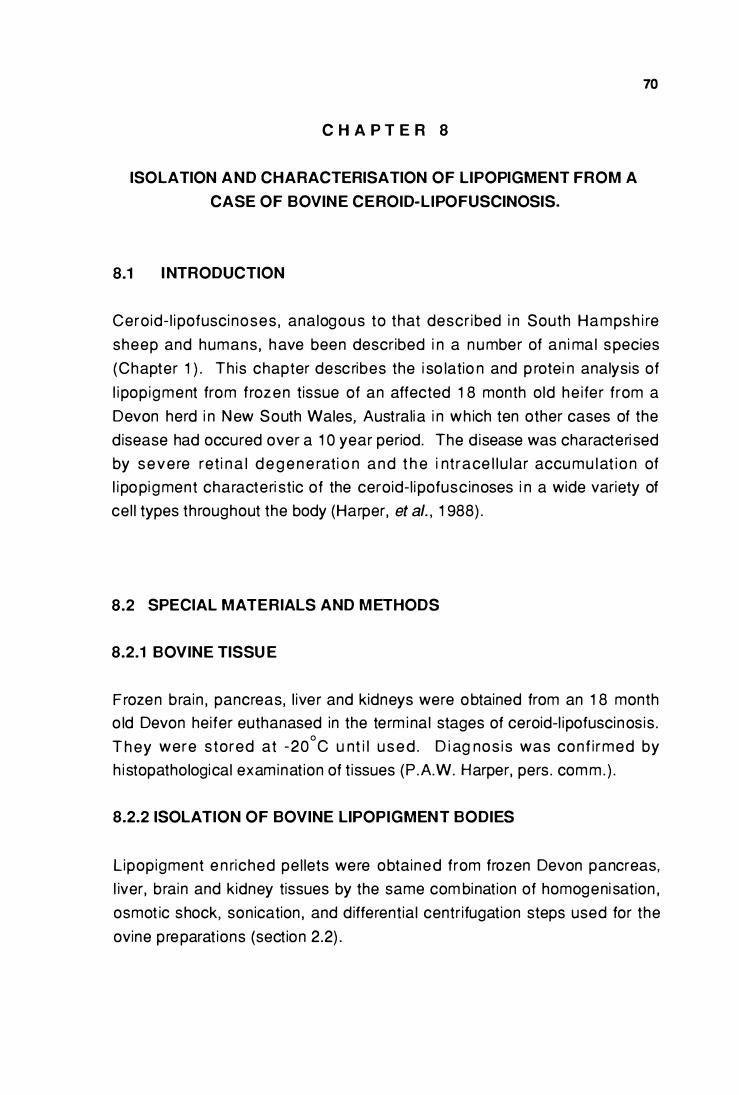
C H A P T E R 8
ISOLATION AND CHARACTERISATION OF LIPOPIGMENT FROM A
CASE OF BOVINE CEROID-LIPOFUSCINOSIS.
8.1 I NTRODUCTION
70
Cero id- l i pofusci noses , analogous to that descr ibed i n South Hamps h i re
sheep and humans , have been described i n a n u mber of an i ma l species
(Chapter 1 ) . Th is chapter desc ribes the i so latio n and p rotei n analysis of
l i pop igment from frozen tissue of an affected 1 8 month old he ifer from a
Devon herd i n New South Wales, Australi a i n wh ich ten other cases of the
d isease had occured over a 1 0 year period. The d isease was characteri sed
by seve re ret i n a l d e g e n e rat i o n and t h e i nt race l l u lar accu mu lat i o n of
l i po pi gment characteri st ic of the ceroid- l i pofusc inoses i n a wide variety of
cel l types throughout the body (Harper, et al. , 1 988) .
8.2 SPECIAL MATERIALS AND M ETHODS
8.2.1 BOVINE TISSU E
Frozen brain , pancreas, l iver and kidneys were obtained from an 1 8 month
o ld Devon heifer euthanased in the term i nal stages of ceroid-l ipofuscinos is .
T h ey were stored at -20°C u nt i l u s e d . D i ag nos i s was conf i rmed by
h i stopathological examinat ion of t issues (P .A.W. Harper, pers . com m . ) .
8.2.2 ISOLATION OF BOVINE LIPOPIGMENT BODIES
Lipopigment e n riched pel lets were obtained from frozen Devon pancreas,
l iver, brain and kidney tissues by the same com binat ion of homogeni sat ion ,
osmot ic shock, son ication , and d ifferent ial centr ifugat ion steps used for the
ovine preparat ions (sect ion 2.2) .
71
L i pop ig m e nts suspe n d e d i n d e i o n i zed wat e r w e re pu r i f i ed fu rthe r by
i sopycn ic centrifugation o n 1 5+ 1 5 m l l i near 0 .64-3.38 M caesium ch loride
(CsCI) density g radients at 1 41 ,000 gmax• 24 h at 4°C in a Beckman SW 28
swing out rotor on a Beckman LB-70 U ltracentrifuge. The l ipopigment bands
were identified by the i r f luorescence when i rradiated with 366 nm u ltraviolet
l i g ht . T h e f l u o resce nce was p h otographed t h ro u g h a y e l low f i lte r o n
E ktachrome 64 ASA fi lm . The l ipopigment bands were removed from the
g radients and their densit ies determined by measuri ng the refractive i ndex of
the CsCI solutions (Weast , et al. , 1 988). They were then washed twice with
deion ized water by resuspension and centrifugat ion at 48,000 g max for 1 0
m in . An aliquot was taken for th in section e lectron m icroscopy as described
i n sect ion 2 .3 . The rema in i ng pe l let was resuspended i n deionized water
and stored at -20 oc u nti l requ i red.
8.2.3 CYANOG EN B ROMIDE DIGESTION OF ISOLATED L IPOPIGMENT
Total pancreat ic l i pop ig m e nt d i ssolved in d i st i l led 98% formic acid was
t reated with cyanoge n bromide as described i n sect ion 5.2 .2 .
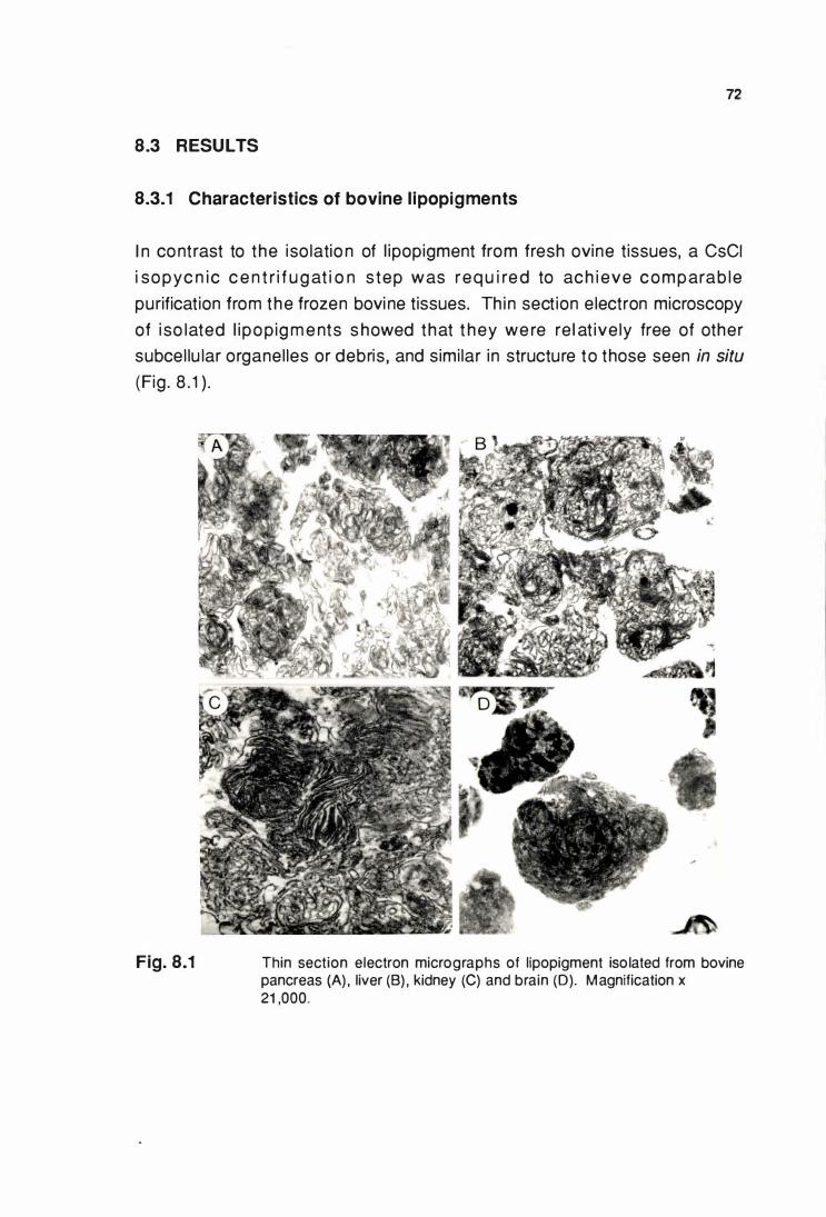
72
8.3 RESULTS
8.3.1 Characteristics of bovine l ipopigments
I n contrast to the iso latio n of l ipopigment from fresh ovine tissues, a CsCI
i sopyc n ic c e n t r i f u g at i o n s tep was req u i re d to ach i eve comparab le
purification from the frozen bovi ne tissues. Th in sect ion electron microscopy
of i so lated l i popig m e nts s howed that t hey we re re l at ive ly free of other
subcel lu lar organel les or debris , and similar in structure to those seen in situ
(Fig . 8 . 1 ) .
Fig. 8.1 Thin section electron micrographs of lipopigment iso lated from bovine pancreas (A) , liver (B) . kidney (C) and brain (0). Magnification x 21 ,000.
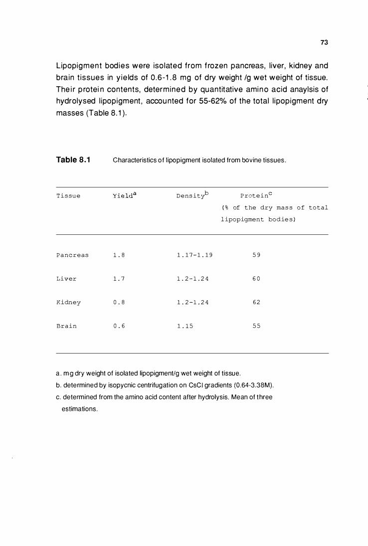
73
Lipopigment bod ies were iso lated from f rozen pancreas, l iver, kidney and
bra in t issues in y ie lds of 0 .6 - 1 . 8 mg of d ry weight /g wet weight of tissue.
The i r p rotei n contents, determ ined by quantitative ami n o acid anaylsis of
hydrolysed l ipopigment , accou nted for 55-62% of the total l ipopigment d ry
masses (Table 8 . 1 ) .
Table 8.1
T i s su e
P a nc r e a s
Liver
Kidn ey
B r a i n
Characteristics of lipopigment isolated from bovine tissues .
D e n s ityb
1 . 8 1 . 1 7 - 1 . 1 9
1 . 7 1 . 2 - 1 . 2 4
0 . 8 1 . 2 - 1 . 2 4
0 . 6 1 . 1 5
P rot e i nc
( % of t he dry ma s s o f t o t a l
l ipopigment bo d ie s )
5 9
6 0
6 2
5 5
a . m g dry weight of isolated lipopigment/g wet weight of tissue.
b . determined by isopycnic centrifugation on CsCI gradients (0.64-3.38M}.
c. determined from the amino acid content after hydrolysis. Mean of t hree
estimations.
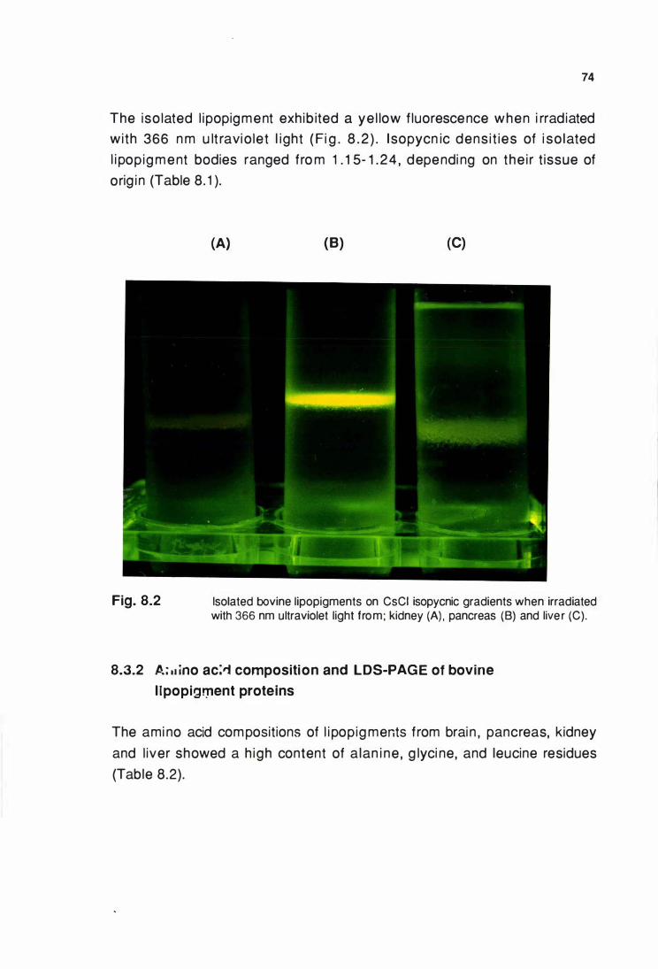
74
The iso lated l ipopigment exh ib ited a ye l low f luorescence when i rradiated
wi th 366 n m u lt ravio let l ight ( Fi g . 8 . 2 ) . l sopycn ic dens i t ies of i so lated
l i pop ig m e nt bodies ranged fro m 1 . 1 5- 1 . 2 4 , dependi ng on the i r t issue of
orig i n (Table 8. 1 ).
Fig. 8.2
(A) (B) (C)
Isolated bovine lipopigments on CsCI isopycnic gradients when irradiated with 366 nm ultraviolet light from; kidney (A) , pancreas (B) and live r (C).
8.3.2 A; • • ino ac:'i composition and LDS-PAGE of bovine
l lpopig!Jlent proteins
The ami no acid compositions of l i popig ments from brai n , pancreas, kidney
and l iver showed a h igh content of a lan i ne , g lyci ne, and leucine residues
(Table 8.2) .
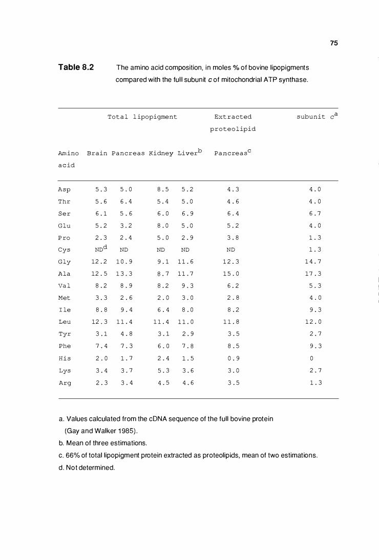
Table 8.2 The amino acid composition, i n moles % of bovine lipopigments
compared with the ful l subun it c of mitochondrial ATP synthase.
T o t a l l ipopigment Ext r a c ted s ubu n i t
p r o t e o l ipid
Amino Brain P a n c r e a s Kidney Liverb
P a n c re a sc
a c id
Asp 5 . 3 5 . 0 8 . 5 5 . 2 4 . 3 4 . 0
Thr 5 . 6 6 . 4 5 . 4 5 . 0 4 . 6 4 . 0
S e r 6 . 1 5 . 6 6 . 0 6 . 9 6 . 4 6 . 7
Glu 5 . 2 3 . 2 8 . 0 5 . 0 5 . 2 4 . 0
P ro 2 . 3 2 . 4 5 . 0 2 . 9 3 . 8 1 . 3
Cys NDd ND ND ND ND 1 . 3
Gly 1 2 . 2 1 0 . 9 9 . 1 1 1 . 6 1 2 . 3 1 4 . 7
Ala 1 2 . 5 1 3 . 3 8 . 7 1 1 . 7 1 5 . 0 1 7 . 3
Va l 8 . 2 8 . 9 8 . 2 9 . 3 6 . 2 5 . 3
Met 3 . 3 2 . 6 2 . 0 3 . 0 2 . 8 4 . 0
I le 8 . 8 9 . 4 6 . 4 8 . 0 8 . 2 9 . 3
Leu 1 2 . 3 1 1 . 4 1 1 . 4 1 1 . 0 1 1 . 8 1 2 . 0
Tyr 3 . 1 4 . 8 3 . 1 2 . 9 3 . 5 2 . 7
Phe 7 . 4 7 . 3 6 . 0 7 . 8 8 . 5 9 . 3
H i s 2 . 0 1 . 7 2 . 4 1 . 5 0 . 9 0
Lys 3 . 4 3 . 7 5 . 3 3 . 6 3 . 0 2 . 7
Arg 2 . 3 3 . 4 4 . 5 4 . 6 3 . 5 1 . 3
a. Values calculated from the cDNA sequence of the ful l bovine protein
(Gay and Walker 1 985) .
b. Mean of three estimations.
c. 66% of total lipopigment protein extracted as proteolipids, mean of two estimations .
d . Not determined.
75
ea
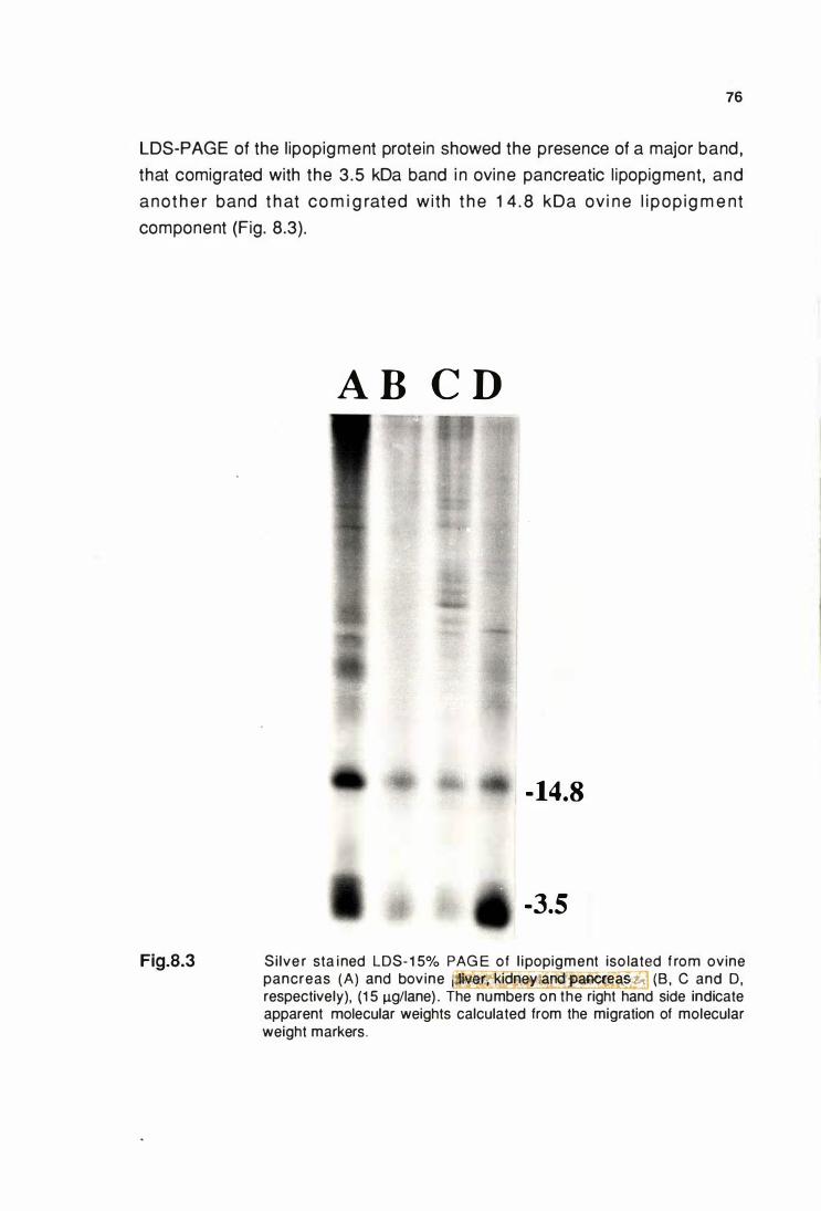
76
LDS-PAGE of the l ipopigment protein showed the presence of a major band ,
that comigrated with the 3 .5 kDa band i n ovine pancreatic l ipopigment, and
anot h e r band t h at co m i g rated wi th t h e 1 4 . 8 kDa ov i n e l i pop ig m e nt
component (Fig. 8.3).
Fig.8.3
A B C D
- 14.8
-3.5
Si lve r sta ined LDS- 1 5% PAG E of l ipopigment iso lated f rom ovine pancreas (A) and bovine ·wr� kidney aoo aJ;le«leas� (B , C and D, respectively), (1 5 J.Lg/lane) . The numbers on the right hand side indicate apparent molecular weights calculated from the migration of molecular weight markers.
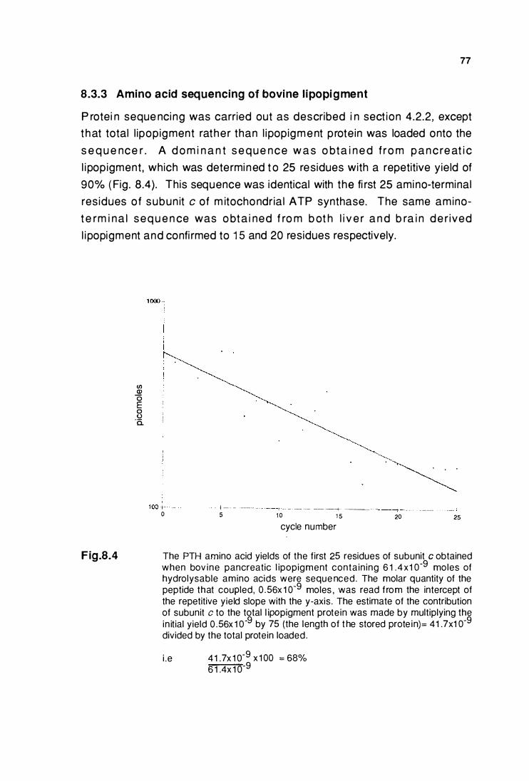
77
8.3.3 Amino acid sequencing of bovine l ipopigment
P rotei n seque nci ng was carried out as described i n sect ion 4.2.2, except
t h at total l ipopigment rathe r than l ipopigm e nt p rote in was loaded onto the
s e q u e nce r . A d o m i n a n t s e q u e n c e w a s o bta i n e d f ro m p a n c re at i c
l i popigment , which was determi ned t o 25 residues with a repetitive yield of
90% ( Fig. 8.4). This sequence was identical with the fi rst 25 amino-terminal
res idues of subun it c of m itochondr ia l ATP synthase. The same amino
t e rm i n a l seq u e n ce was o bt a i ned f ro m b ot h l i v e r a n d b ra i n d e r ived
l i popigment and confi rmed to 1 5 and 20 residues respectively.
Fig.8.4
(/) Q) 0 E 0 u
·a.
0 · ···-----t--·----f---- --+--5 1 0 1 5 cycle number
' ' '
---+-------20 25
The PTH amino acid yields of the first 25 residues of subunit c obtained when bov ine pancreatic l ipopigment contain ing 6 1 .4x1 o-9 moles of hydrolysable amino acids were seque nced. The molar quantity of the peptide that coupled, 0 .56x 1 0-9 moles , was read from the intercept of the repetitive yield slope with the y-axis. The estimate of the contribution of subunit c to the total l ipopigment protein was made by multiplying the
in itial yield 0 .56x 1 o-9
by 75 (the length of t he stored prote in)= 41 .7x1 o-9
divided by the total protein loaded.
i .e 41 .7x 1 o-9 x 1 00 = 68%
6 1 .4x 1 o-9
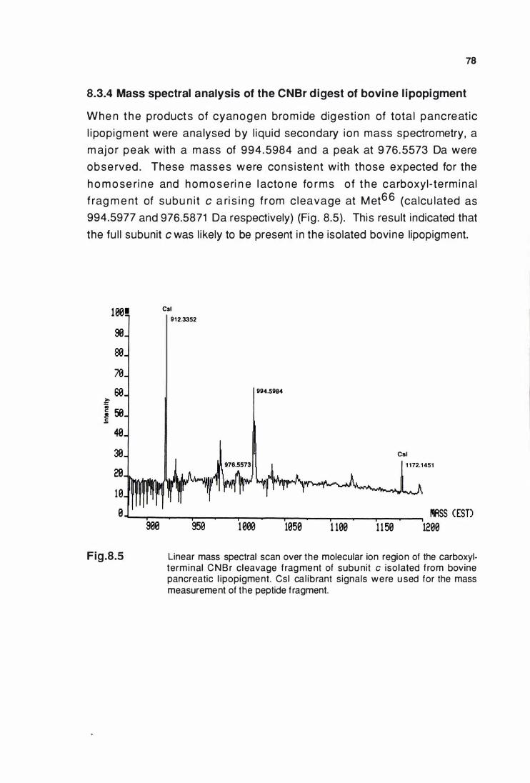
78
8.3.4 Mass spectral analysis of the CNBr digest of bovine l ipopigment
Whe n t h e p roducts of cyanogen bro m ide dig esti on of tota l pancreat ic
l i pop igment were analysed by l iqu id secondary i on mass spectrometry, a
m ajo r peak with a m ass of 994 .5984 and a peak at 9 76.5573 Da were
obse rved . These masses were cons i ste nt with those expected for the
h o m o se r i n e and h o m o s e ri n e l acto ne fo rms of t h e carboxyl-te rm i nal
f rag m e nt of subu n i t c a ri s i n g f rom c leavage at M et66 (calcu lated as
994.5977 and 976.5871 Da respectively) (Fig . 8.5). This result i ndicated that
the fu l l subunit c was l ikely to be present i n the isolated bovi ne l ipopigment.
H.�• se
se
70
se � .. � 58 .5
40
30
20
10
e
Fig.8.5
Csl 91 2.3352
994.5984
950 1 000 1050 1 100 1 1 50 IIIASS CESD
1200
Linear mass spectral scan over the molecular ion region of the carboxylterminal CNBr cleavage frag ment of subu nit c isolated f rom bovine pancreatic l ipopigment. Csl ca l ibrant signals were u sed for the mass measurement of the peptide f ragment.
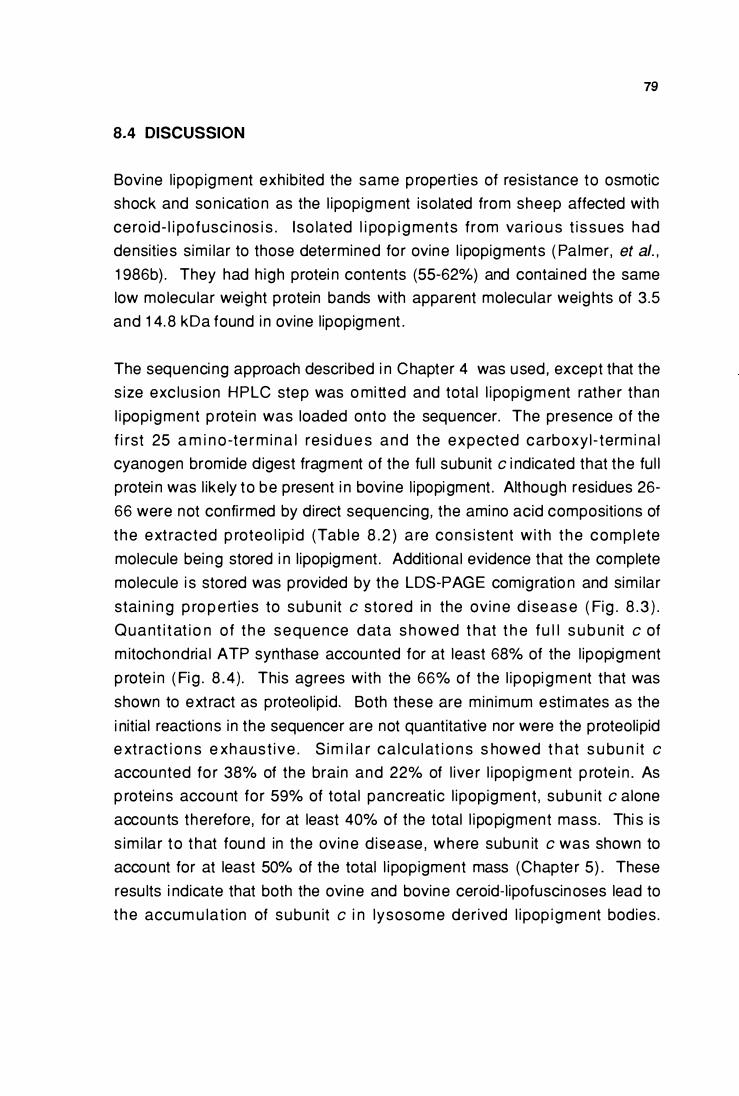
79
8.4 DISCUSSION
Bovi ne l ipopigment exhib ited the same p rope rties of res istance to osmotic
shock and son ication as the l ipopigment isolated from sheep affected with
ce ro id- l i pofusc i nos i s . I so lated l i po p i g ments from va ri o u s t i ssues had
densities sim i lar to those determined for ovine l ipopigments ( Pa lmer, et al. ,
1 986b) . They had h igh protei n contents (55-62%) and contai ned the same
low molecu lar weight p rotein bands with apparent molecular weights of 3 .5
and 1 4. 8 kDa found i n ovine l ipopigment .
The sequenci ng approach described i n Chapter 4 was used, except that the
s ize exc lus ion HPLC step was o mi tted and total l i popigm e nt rathe r than
l i popigment p rote i n was loaded onto the sequencer. The presence of the
f i rst 25 a m i n o -te r m i n a l resi d u e s and t he expected carboxy l - term i na l
cyanogen bromide d igest fragment of the fu l l subunit c i ndicated that the fu l l
p rotei n was l ikely to be present i n bovine l ipopigment . Although residues 26-
66 were not confi rmed by direct sequencing, the amino acid compositions of
t he ext racted p roteo l ip id (Tab le 8 . 2 ) a re cons i stent w i th t he complete
molecule being stored i n l ipopigment . Additional evidence that the complete
molecule i s stored was provided by the LDS-PAGE comigratio n and sim i lar
stai n i n g p ro p e rt ies to subun it c sto red in the ovi n e d i se as e ( Fig . 8 . 3 ) .
Quant i t at i o n of t h e sequence d ata s h owed t h at t h e fu l l s u b u n it c of
m itochondria l ATP synthase accounted for at least 68% of the l ipopigment
p rote i n ( Fi g . 8 .4) . This agrees with the 66% of the l ipopi g m e nt that was
shown to ext ract as proteol ipid. Both these are min imum est im ates as the
i nitial reactions in the sequencer are not quantitative nor were the proteolipid
e xt ract i o n s e xh aust ive . S i m i l a r ca lcu lat i o n s s howed t h at s u bu n it c
accounted fo r 38% of the brain and 22% of l ive r l i pop igm e nt p rote i n . As
p rotei ns accou nt for 59% of total pancreat ic l ipopigment , subun it c alone
accounts therefore, for at least 40% of the total l i popigment mass. Thi s is
s im i lar to t hat found in the ovin e d isease , where subun it c was shown to
account for at least 50% of the total l i popigment mass (Chapte r 5) . These
resu lts i ndicate that both the ovine and bovine ceroid-l ipofuscinoses lead to
t he accum u lat ion of subun it c i n lysosome der ived l i pop igment bodies .
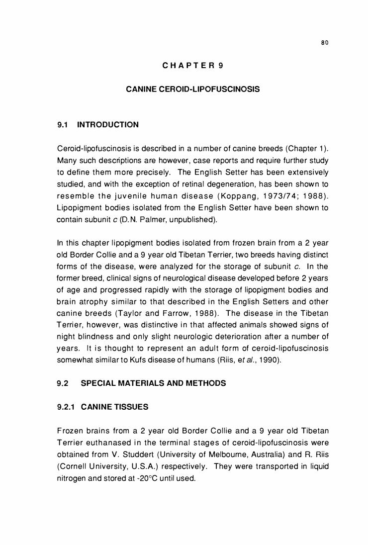
8 0
C H A P T E R 9
CANINE CEROID-LIPOFUSCINOSIS
9.1 INTRODUCTION
Ceroid- l ipofuscinosis is described in a number of canine breeds (Chapter 1 ) .
Many such descri pt ions are however , case reports and requ i re furthe r study
to defi ne them m o re precise ly. The Eng l i sh Setter has been extens ive ly
studied , and with the exception of retinal degeneration , has been shown to
res e m b l e t h e j uv e n i l e h u ma n d i s e a s e ( Ko p p a ng , 1 9 73/7 4 ; 1 98 8 ) .
L ipopigment bod ies iso lated from the E ng l i sh Sette r have been shown to
contain subun it c (D. N. Palmer, u npubl ished).
In th is chapter l i popigment bodies i so lated from frozen b rai n from a 2 year
o ld Border Col l ie and a 9 year old Tibetan Terrier, two breeds having dist inct
forms of the disease, were analyzed for t he sto rage of subun it c. I n the
former breed , c l in ical signs of neurological d isease developed before 2 years
of age and prog ressed rapid ly with the storage of l ipopigment bodies and
b ra i n at rophy s i m i lar to t hat descr ibed i n the E ng lish Setters and other
can i n e b reeds (Tay lo r and Farrow , 1 988) . The d isease i n the Tibetan
Terri e r, h owever, was d ist i nctive i n t hat affected animals showed signs of
n ight b l i ndness and o n ly s l ight neuro log ic dete rio ration after a number of
years. l t i s t hought to represent an adu l t fo rm of ce ro id- l ipofusci nos is
somewhat sim i lar to Kufs disease of humans (R i is , et al. , 1 990).
9.2 SPECIAL MATERIALS AND METHODS
9.2.1 CANINE TISSUES
F rozen b ra i n s from a 2 year old Borde r Co l l i e and a 9 year o ld Tibetan
Terr i e r eut h a n ased i n the te rmi na l stag e s of ceroid- l i pofusci n os is were
obtained from V. Studde rt ( U nive rsity of Melbourne , Austral ia} and R. Ri is
(Corne l l U n iversity, U .S .A. ) respect ive ly . They were t ransported in l iquid
n itrogen and stored at -20°C u nt i l used.
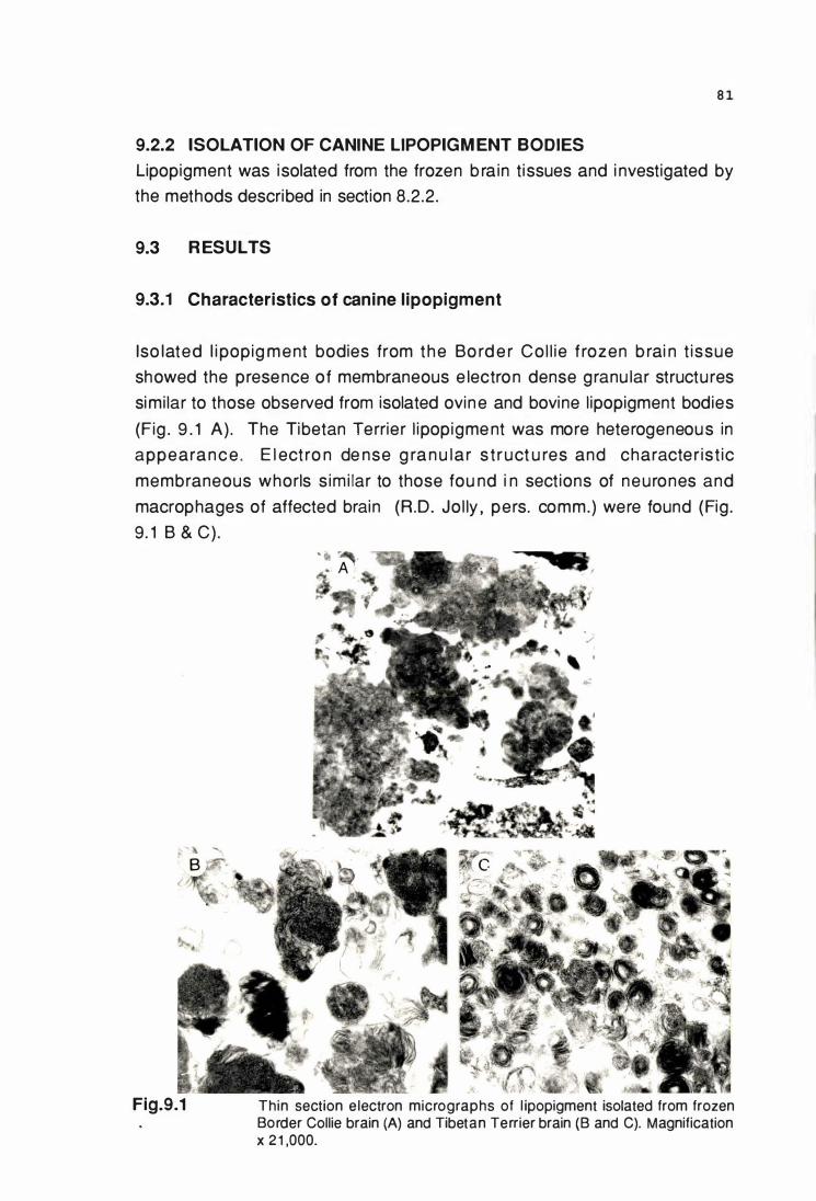
8 1
9.2.2 ISOLATION OF CANINE LIPOPIGM ENT BODIES
Lipopigment was isolated from the frozen b ra in t issues and i nvestigated by
the methods described in section 8.2.2.
9.3 R ESULTS
9.3.1 Characteristics of canine l ipopigment
Iso lated l i pop ig ment bodies from the Bo rd e r Co l l ie f rozen b rai n t issue
showed the presence of membraneous e lectron dense granu lar structu res
simi lar to those observed from isolated ovin e and bovine l ipopigment bodies
(Fig . 9 . 1 A) . The Tibetan Terrier l ipopigme nt was more heterogeneous in
appeara n c e . E l ect ro n de nse g ra n u la r s t ruct u res and characte r ist ic
membraneous whorls s im i lar to those fou nd i n sect ions of neu rones and
macrophages of affected brain (R .D. Jo l ly , pers. comm.) were found (Fig .
9. 1 B & C) .
Fig.9.1 Thin section e lectron micrographs of l ipopigment isolated from frozen Border Collie brain (A) and Tibetan Terrier brain (B and C). Magnification X 2 1 ,000.
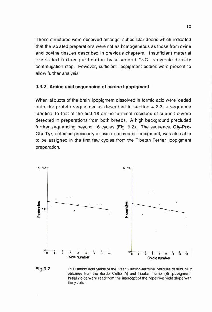
8 2
These structures were observed amongst subce l lu lar debris which i ndicated
that the iso lated preparations were not as homogeneous as those from ovine
and bovi ne t issues descri bed i n p revious chapte rs . I n suff icient mate rial
p r e c l u de d f u rt h e r pu r i f i cat i o n by a seco n d C s C I i s o pycn i c d e n s i ty
centrifugation step. However, sufficient l ipopigment bodies were present to
allow further analysis.
9.3.2 Amino acid sequencing of canine l ipopigment
When al iquots of the brai n l ipopigment d isso lved in formic acid were loaded
o nto the p rote i n seque ncer as desc ri bed i n sect ion 4 .2 . 2 , a seq u e nce
i dentical to that of the fi rst 1 6 amino-te rmi nal residues of subu n it c were
detected i n p reparat ions from both breeds. A high background precluded
fu rther sequ enci ng beyond 1 6 cycles (Fig . 9 .2 ) . The sequence, G ly-Pro
G iu-Tyr, detected previously in ovi ne pancreatic l ipopigment, was also able
to be assig ned in the fi rst few cycles from the Tibetan Terrie r l ipopigment
preparation.
A 1000
(/) Q) 0 E 1 00
8 a:
B 100
(/) Q) 0 E 8 a:
1 0+--+--+--+--+---+--+----+-------l 1 0+---+--t--+---+--+----i-+--� 0 2 4 6 8 1 0 1 2 14 1 6 0 2 4 6 8 10 1 2 14 16
Fig.9.2
Cycle number Cycle number
PTH amino acid yields of the first 1 6 amino-terminal residues of subu nit c obtained from the Border Collie (A) and Tibetan Terrier (B) lipopigment. Initial yields were read from the intercept of the repetitive yield slope with the y-ax is .
8 3
9.4 D ISCUSSION
The presence of the 1 6 amino-terminal residues of subunit c in l ipopigment
i solated from the two can i ne breeds suggested that they also be longed to
the forms of ceroid-l ipofuscinoses that specifically store subunit c. The molar
quant ity of subun it c t hat coup led was dete rm i ned as before , from the
repetitive yield graphs of the PTH amino acids obtained at each cycle (Fig .
9 .2) . If the fu l l 75 residue protei n was stored , estimates of 55% and 33%
were calcu lated for t he contribut ion of subun it c to the total l i popig ment
masses from the Border Col l ie and Tibetan Terri er, respectively. These
estimations are min imum values because of relative impurity of preparations
and the fact that the i n itia l reactions in the sequencer are not quantitative .
They are also based on the assumption that the ful l protei n is stored as i n
t h e ovi ne and bov i n e forms o f t he d isease (Chapters 5 & 8) . Fu rther
c haracterisation o f the stored protei n was not possible due to the l im ited
amount of material avai lable.
As the development of modern breeds with in a species is relatively recent it
i s l ikely that some forms of the disease with in the same species may reflect
the same mutation . I n th is regard, the diseases i n the Border Coll ie (Taylor
and Farrow, 1 988) and B lue Healer dog (Cho , et al., 1 986 ; Wood, et al.,
1 987) have been shown to be cl i n ical ly and patho log ical ly s im i lar to the
English Setter disease (Koppang , 1 973/74 ; 1 988) , and may reflect the same
m utation . The disease in Tibetan Terrier is however, disti nctively d ifferent
( Ri is, et al., 1 990).
The fi nd ing of two d ist inct diseases in dogs wh ich both store at least an
amino-terminal fragment of subunit c i n l ipopigment, i ndicates two distinct
m utations. Whether these affect the same gene or different genes may be
determ ined by cross breeding experiments. If the same gene i s mutated
t hen affected offspri ng should have a disease with characteristics between
t he pare nt types. I f no d i sease resu lts f ro m the cross breed i ng t h e n
i nvolvement of a second mutant gene is implied. The different human forms
of the d isease cou ld reflect simi lar mutations.
84
C H A P T E R 1 0
GENERAL DISCUSSION
I n born e rrors of m etabol ism are reflected by e levation of the deficient e n zy me's substrata or de rivatives of it , a p ri nciple fi rst e n u nciated by Garrod i n 1908. I n the case of storage diseases, characterisation of the domi nantly stored species has led in many instances to elucidation of the u nderlyi ng biochemical defect. The same p ri nciple should therefore be expected to hold for the ceroid-lipofusci noses. Palmer, et al. , (1986a,b) showed that in the ovi ne disease the major sto red components were low m o lecu lar we i g ht p rote i n s in lysoso me d e ri ved l i po pig m e nt bodi e s . C haracteri sat i o n o f t h ese prote i n s b y a co m bi n at ion of LDS-PAG E , quantitative amino acid sequenci ng and mass spectroscopy studies has l e d to the m ajor sto red compo n e nt be i ng i d e nt ified as the co m plete d i cyclohexylcarbodi i m ide ( DCCD) reactive p roteol ipid (subunit c of the
, · i,"o_cpq·�a��.,.;£ ATP synt h ase ) . T h i s i s a h i g h ly hydro p h o b i c p rote i n no rmal ly found i n the i nner mitocho ndrial membrane as part of the ATP synthase o ligome ric protei n co mplex. The ATP synthases (also known as proton translocating ATPase, H+-ATPase, or F1 F0-ATPase) are fou nd in the cytoplasmic membranes of eubacteria, the thylakoid membranes of ch loroplasts and in the i n ne r membranes of mitochondria. Their common fu nction is to catalyze the synthesis of ATP fro m ADP and phosphate by employing an e lectrochem ical pote ntial g radi e nt across the mem brane. T h e m i t o c h o n d ri a l A T P s y n t h a s e i s a c o m p l e x of 14 d i ffe re n t po lype ptides; six of th em (a, � . g , cr , e , OSC P and I ) constitute F1, the extri nsic membrane domai n of the complex. The remainde r (ATPase-6 , su bunit c, subu nit b, subunit d, A6L) make u p the membrane sector F0. Two additional components, subu nit e and F6 are also fou nd (Walker, et
al. , 1 9 90 ) . S u b u n it c, i s an e sse n t i a l e l e m e nt i nvolved i n t h e H + t ra n s locat i n g pro p e rt i e s o f Fo a n d acco u nts fo r 2-4% o f t h e i n n e r mitochondrial membrane protein (Sebald and Hoppe , 1 981 ).
So lubi l ity of the l ipopigment protei ns in LDS, in the presence o r absence of 2-mercaptoethanol was impo rtant as it showed that the aggregation
85
behaviour of these p rote ins i n dodecy l su lphate was not due to the
presence of covalent disulphide bonding between them. The difficu lty of
separat i ng the m aj o r 3 . 5 kDa l i pop igment p rote i n f ro m t h e oth e r
l i popig ment prote ins led to the non-traditional approach of load ing a n
apparent mixture of protei ns onto the amino acid sequencer (Chapter 4).
lt was reasoned that should a sequence emerge, then it would be of the
low molecular weight component shown in Chapter 3 to be present in
total l i po pi g ment p rote i n as t h e dom inant mo lar component . The
approach led to the detection and quantitation of subunit c i n l ipopigment.
Ident i ficat ion of the 1 4 . 8 and 24 kDa l ipop igm e nt protei ns as being
o l igo m e rs of the m ajor stored p rote i n (subun i t c) strengthened the
rat ionale used i n the earl ie r sequenci ng stud ies on tota l l i popigment
p rotei ns. This result explained the difficulties encountered in separati ng
the 3.5, 1 4.8 and 24 kDa proteins by conventional techniques. Subunit c
was therefore the major protein present i n l ipopigment bodies. No other
m itochondrial protein was detected during th is study.
The aggregation behaviour of the proteol ipid subunit c, particularly i n
dodecyl su lphate , i s wel l documented (Sebald and Hoppe, 1 98 1 ) . As
noted earl ier the agg regation behaviour of subunit c stored in l ipopigment
was not due to the presence of inte rmolecular disu lphide bonding. The
u nusual stabil ity of the ol igomers of subunit c in l ipopigment was shown
by the 1 4.8 kDa protei n that diffusion e luted from a polyacrylamide gel
behavi ng as a component distinct from the 3.5 kDa protein (Chapter 5).
The physico-chemical basis for this ol igomeric stabi l ity remains unknown .
The e lectrophoretic variabi l ity of the 1 4.8 and 24 kDa bands observed
between different iso lates and between different gels of the same isolate
could now be rationalized on the basis of variable aggregation of subunit
c present. The behaviour of the 1 25 todinated l ipopigment proteins on
storage described in Chapter 3 was also probably due to this agg regation
p roperty of subunit c. The radioiod ination of l ipopigment protei ns as a
means of obtai n ing a lab e l led substrata t h e refo re warrants fu rt h e r
i nvestigation.
The stored protein was shown to account for at least 50% of the isolated
ovine l i popigment mass, and as such was considered specif ic to the
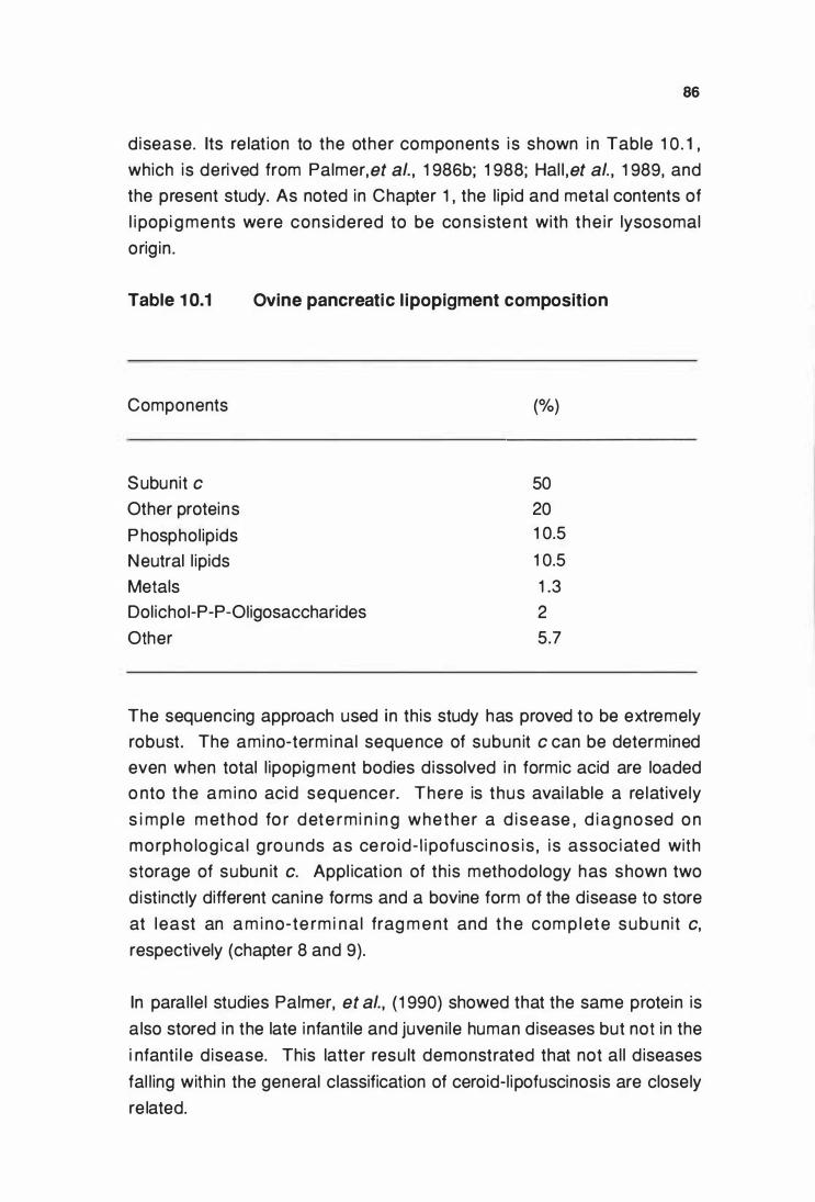
86
disease. Its re lat ion to the other components i s shown i n Table 1 0 . 1 ,
which is derived from Palmer,et al., 1 986b; 1 988; Hal l ,et al., 1 989, and
the present study. As noted in Chapter 1 , the l ipid and metal contents of
l i popi g ments were cons idered to be cons iste nt with the i r lysosomal
orig in .
Table 1 0.1 Ovine pancreatic l ipopigment composition
Components (%)
Subunit c 50
Other proteins 20
P hospholipids 1 0.5
Neutral l ipids 1 0.5
Metals
Dolichoi-P-P-Oiigosaccharides
Other
1 .3
2
5.7
The sequencing approach used in this study has proved to be extremely
robust. The amino-termi nal sequence of subunit c can be determined
even when total l ipopig ment bodies d issolved i n formic acid are loaded
o nto the a m i no acid sequencer. There is thus avai lable a re lative ly
s i mp le met h od fo r dete rm i n i n g whether a d i sease , d i agnosed o n
morpho log ica l g ro u nds as ce ro id- l i pofusci nos is , i s assoc iated with
storage of subun it c. Appl icat ion of th is methodo logy has shown two
d istinct ly different canine forms and a bovine form of the disease to store
at least an a m i no-te rm i nal frag m e nt and t h e com p l ete s u b u nit c,
respectively (chapter 8 and 9).
In paral lel studies Palmer, et al., ( 1 990) showed that the same protein is
a lso stored in the late i nfantile and juveni le human d iseases but not in the
i nfanti l e d isease. This latter resu lt demonstrated that not al l d iseases
fal l ing with in the general classification of ceroid-l ipofuscinosis are closely
re lated.
87
The described methodology could be used to determine the dominantly
stored components from the other human variants and animal forms of
the d isease. Once l i popigment is isolated from subcel l u lar organel les
and debris , only four al iquots of isolated l ipopigment are required ; one for
e l ect ro n m ic rosco p y t o dete rm i n e pu ri ty of preparat i o n , two fo r
quantitative amino acid analysis and amino-terminal sequencing and one
a l iquot fo r cyanoge n bro m ide c leavage to d ete rmi ne the carboxy l
terminus of subunit c. lt is l ike ly that a classification of subunit c storage
d i sease cou ld be made from the mere dem o n strat ion of the amino
te rm i n u s of subu n i t c by d i rect sequenci ng stud ies , when there i s
i nsufficient material for the other analyses. Such was demonstrated in the
the two canine diseases (Chapter 9).
The storage of subunit c rather than peroxidised l ipid/protein po lymers as
previously postu lated , al lows a major conceptual change in the approach
to cero i d - l i pofusci n o s i s research . H i stoc h e m i cal and f luo rescent
properties of the stored l ipopigment which were i nterpreted to be due to
the i r l i pid nature , l ed to a n umber of theories on pathogenesis , most
based on l ipid peroxidation (Chapter 1 ). None of these have stood the
test of time or led to the elucidation of the underlying biochemical defect
i n the ceroid- l ipofusci noses. An "acidic l ipid po lymer" extracted from
l ipopigments by l ipid solvents and named "cero id" was proposed as the
"stored substance" in the neuronal ceroid- l ipofuscinoses (Siakotos, et al. ,
1 972). I n retrospect , it seems l ike ly that the designated " acidic l ipid
polymer'' was the proteol ipid , subunit c.
The extreme hydrophobic nature of subunit c resu lts in it having unusual
physical properties, including the l ipid l ike solubi l ity i n
chloroform/methanol mixtures. lt i s this property which results i n it being
referred to as a proteol ipid. The definition is an operational one and does
not imply the presence of covalently bound lipids. Subunit c stains poorly
with Coomassie blue and g ives i naccurate protei n estimat ions by the
Lowry method (Sebald and Hoppe , 1 981 ) . These characteristics may
h e lp exp lai n why t h i s type of prote in was not recog n i zed as bei ng
associated with the ceroid-l ipofuscinoses in the past.
88
M a n y characte r ist i cs of t h e sto red l i pop i g m e nt bod ies and othe r
e nigmatic observations can now be explai ned by the knowledge that it is
subunit c that is stored. These i nclude the characteristic h istochemical
feature of staining with certain lipid stains. Lipid stains can be expected
to react with the neutra l l ipids and phospholipids present i n l ipopigment
bodies but may also react with the hydrophobic proteo l ipid subunit c.
Luxol fast blue , considered a stain for phospholipids and
phosphol ipid/protein complexes i n fixed t issues (Pearse, 1 985) , is also
used in neuropatho logy as a stai n for myel in . As mye l in contains the
p rototype proteol ipid protein (Folch and Lees, 1 95 1 ) it is l ikely that it is
t h is type of mo lecule that stains both mye l in and l ipopigment bodies in
paraffin block sections (Jol ly, et al. , 1 989). This is supported by the fact
t hat luxol fast blue stain ing is a feature of al l but the infanti le human form
of ceroid- l ipofusci nosis ( Lake , 1 984) i n which subun i t c is not stored
( Palmer, et al. , 1 990). The l uxol fast blue stai n ing may therefore have
some diagnostic importance.
Autofluorescence in the yel low-orange reg ion (520-540 nm) is another
characteristic feature of stored lipopigment. To date neither the nature of
t h e f lourophor nor its s ig n if icance , if any, has been dete rm i ned. A
n u m ber of mechan isms whe reby prote i ns can be modif ied to form
f luorophores have been proposed. These include peroxidation reactions
l eading to the formation of malonaldehyde, which can react with protein
to fo rm fluo rescent sch iff base products (Chic and Tappel , 1 969a) and
n o nenzym ic g lycat ion (g lycosy lat i on ) of lysi ne res idues producing
h eterocycl ic f luorescent compounds (Pongor, et al. , 1 984) . There has
however, been no evidence for t he presence of such compounds i n
l i popigment bodies iso lated from t h e cero id-l ipofuscinoses. Palmer, et
al. , ( 1 986a) suggested that fluorescence may be due to a property of the
i nteraction of the stored protein and its pecu liar l ipid environment. This
i de a i s s u pported by reco nst i t u t i o n of f l u o re scen t b o d i e s fro m
nonf luorescent pu rified subunit c and phospholipids (D. N . Palmer, pers.
comm.) .
Detect ion of S-methylated methionine i n "storage protein" i solated from
the late i nfantile form of the human disease has recently been reported
89
(Katz and Gerhardt, 1 990) . lt was suggested that such modified amino
acids might be responsib le for the autofluorescence of storage bodies
u nder uv i l l um inat ion . However, the amino acid compositions of the
protei n i so lated from l ipopig ment in the study differs substantially from
that of subunit c, identified as the major stored species i n the late i nfanti le
d isease (Palmer, et al. , 1 990) . The s ignificance of this observation i n
terms of a possible post-translational modification to subunit c stored i n
t he ceroid-l ipofuscinoses remains to be clarified. There was however, no
evidence in the present studies to suggest that the stored protein in the
ov i n e , bovi ne and can i n e fo rms of ce ro id - l i pofusci n osis was post
translationally methylated.
T h e pre se nce of la rg e amou nts of t h e co m p lete and appare nt ly
u n modif ied subun it c i n the ce ro id- l i pofusci noses i nd icates that the
biochemical basis of the lesion in these diseases is l ike ly to involve the
metabol ism of subunit c .
I n humans and catt le , subun it c has two expressed nuclear genes, P1
and P2. These encode precursor protei ns contain i ng the same mature
protei n , but with diffe rent positively charged amino-terminal extensions
(presequences) of 61 and 68 amino acids respectively (Gay and Walker,
1 985 ; Dyer, et al. , 1 989) . I n addition to the P1 and P2 genes, numerous
sp l iced and partly spl iced pseudogenes re lated to P1 or P2 have also
been discovered in human, bovine and ovine tissues (Dyer and Walker,
1 990 ; Medd, et al. ,unpublished) .
The presequences contain the necessary information to target the protei n
to m itochondria (van Loon , e t al. , 1 987; van Loon and Schatz, 1 987 ;
Hart l , et al. , 1 986 ; 1 987). Import receptors on the outer su rface of the
outer mitochondria l m e m brane are i nvo lved i n the i n it ial recog n it ion
process of these precursors (Zwizinski , et al. , 1 984 ; Pfal ler, et al. , 1 988;
Pfa l l e r a n d N e u pe rt , 1 987) . 1t i s a lso thou g ht that t h e cytoso l i c
p recurso rs o f m itochondria l protei n s have to m ai nta in an u nfo lded
conformation for membrane translocation (Hartl , et al. , 1 989) . This may
be achieved by the i nteraction with the heat shock proteins of relative
molecular mass 70 kDa and by the action of other factors in the cytosol
90
( Deshaies , et al. , 1 988; Pel h am , 1 989) . The transport of precursors
across the two mitochondrial membranes are thought to occur i n a single
ste p at contact sites betwee n the oute r and i n ne r membranes by a
process requi ring an electrochemical potential g radient across the i nner
m itochondrial membrane (Schleyer, et al. , 1 982 ; Sch leyer and Neupert,
1 985 ; Schwaiger , et al. , 1 987) . The amino-te rmina l extensions are
removed during , or short ly fo l lowing , the t ranslocat ion process by the
action of a specific matrix localised metal dependent processing enzyme
i n a two step process ( Pfan ner, et al. , 1 988; Schm idt, et al. , 1 984;
Hendrick, et al. , 1 989) .
The underlyi ng biochemical anomaly leading to the storage of subu nit c
i n lysosome derived l ipopig ment bodies remains to be defined. There
are a number of possib le mechanisms that cou ld lead to its abnormal
accumu lation due to defect(s) occu rring at several points in the normal
biosynthetic pathway of this protein.
The gene sequences coding for subunit c (P1 and P2) i n sheep affected
with ovine ceroid- l ipofuscinosis have been sequenced and shown to be
identical to those of contro l sheep. Simi lar amounts of the mRNA for P1
a n d P2 were fou nd i n n orma l and d iseased s h eep l ive r t issue by
Northern blot analysis (Medd , et al. , 1 990). The storage of subunit c i n
l i po p i g m e nt bod ies i s t h e refore not cau sed by a m utat i o n i n the
presequences of either P1 or P2 resu lting in misdirection o f precursors of
subu n it c d i rectly to lysosomes rather than to m itochondria , nor by a
m utation affecting the normal gene products. These genes are normal ly
reg u l ated at the t ranscri pti onal leve l . Over-express ion resu lt i ng i n
subunit c being synthesised i n g reater amounts than i s normally requi red
for assembly of the ATP synthase also seems unl ikely on the basis of the
Northern b lot resu lts. However, as the bovi ne P1 and P2 genes are
known to be expressed i n a tissue-specific manner (Gay and Walker,
1 985), the possibi l ity exists that the lesion i n ovi ne ceroid-l ipofuscinosis
cou ld resu lt from a defect in the tisSUe.JIE· '!!tiC expression of these genes.
L it t le is k n own about t h e mechan isms u nd e rl y i ng the fo ld ing and
assembly of imported prote ins i nto ol igomeric enzyme complexes with in
91
m itochondri a nor about the coordinat ion of assembly of m itochondrial
a n d n uclear gene products. A nuc lear encoded heat s hock protei n
( hsp60) found i n the mitochondri al matrix has recently been shown to be
requ i red for the correct assembly into o ligomeric complexes of protei ns
i mported i nto the mitochondrial matrix (Cheng , et al. , 1 989 ; Readi ng , et
al. , 1 989) . l t has been suggested that a fu nct ion of the hsp60 i s to
p revent the formation of misfo lded protei ns (Ostermann , et al. , 1 989) .
The lesion cou ld i nvolve a defect in a simi lar component required for the
co rrect fold i ng and o ri entat ion of subu n it c i nto the ATP synthase
complex.
S u bu n it c p lays a key ro le in the assembly and stab i l ity of the F0-
c o m po nent , wh ich is essent ia l to the fo rmat ion of a fu nct i onal ATP
synthase complex (linnane , et al. , 1 985; Hadikusumo, et al. , 1 988) . lt is
therefore un like ly that the genetic lesion i nvolves a component requi red
fo r co rrect assembly of subu nit c i nto the ATP synthase o l igomeric
complex. D isrupt ion of the ATP synthase wou ld lead to uncoupling of
oxidative phosporylation and subsequent death of the cel l . Mitochondria
i solated from diseased kidneys were also shown to maintain structural
and functional integrity comparable to those isolated from control animals
(Chapter 6) .
Storage of the ful l subunit c, identical to that of the mature mitochondrial
p rotei n , suggests that the ami no-term i nal preseque nces have been
cleaved by the specific protease located i n the mitochondrial matrix. This
i mpl ies a defect in its degradative pathway fol lowing correct incorporation
i nto mitochondrial i nner membranes. The normal amounts of this protein
shown to be present in affected liver i nner mitochondrial membranes by
LDS-PAGE and the normal function ing of affected kidney mitochondria
(Chapter 6) suggested that the accumu lation was not occurring i n the
mitochondrion.
Lysosomes catabo l i se po l ym e ri c mater ia l by the act i o n of many
e n zymes . There are a numbe r o f i n bo rn errors o f such catabo l ism
affect ing complex l ipids , po lysaccharides and mucopolyaccharides, but
none have been described i nvolvi ng proteins . There is no reason why
92
such errors should not occur by chance. Fai lure to recognize them may
be associated with the relative lack of specificity of lysosomal proteases.
The cleavage specificities of lysosomal endopeptidases (cathepsi ns) and
exopeptidases (Kirschke and Barrett, 1 987) are such that a deficiency of
o n e cou ld be theoretical ly masked and covered by the activity of the
rem ainder. Therefore a deficiency of a major cathepsin as a source of
subun it c sto rage seems u n l i kely . However, there could be a specific
u nknown lysosomal protease requi red to degrade subunit c or more l ike ly
a cofactor specifically associated with the in it ial degradative step for this
proteo lipid. A deficiency of such a cofactor is known and accounts for
o n e of the variants of GM2 Gangl iosidosis (Conzelmann and Sandhoff,
1 9 7 8 ) . l t i s a l so poss i b l e t h at the n o rma l d e g radat i ve e n zymes
associated w i th the catabo l i sm of subun it c are unaffected and that
s u b u n it c i s presented to t h e m i n a conformation that i s resistant to
p roteo lys is . Li popigment sto rage bodies have bee n i nterpreted as
co m p l e x parac rysta l l i n e st ructu res t h at are not degraded by t h e
lysosomal system (Jolly, et al. , 1 989). Analogous protei n i nclusion body
agg regates res istant to ce l lu lar proteases are found i n bacteria as a
resu lt of over-expression of cloned mammalian genes (Cheng , et al. ,
1 981 ; Kane and Hartley, 1 988)
M itochondr ia l p rote i n deg radat ion and prote i n turnove r are poorly
u nderstood. lt is generally assumed that mitochondria are encapsulated
i nto auto p h ag i c vacu o l e s and t h e n deg raded by a combi nat io n of
lysosomal and mitochondria l proteases (Luzikov, 1 985; Pfeifer, 1 987) .
Lipopigment bodies in ovine foetuses and neonates have been described
a s w h o r l s of b i layer m e m b ra n e s co n de n s i n g i n to m u lt i l a m e l l a r
paracrystal l ine structures (Jol ly, et al. , 1 989) . These were thought to be
the simplest and earliest lesions yet noted and suggested that autophagy
of m itochondria was not the source of subun it c found i n l ipopigment
bod ies . The fact that no oth e r mitochondria l ATP synthase prote in
components were shown to be stored during this study also suggest that
t h i s m ech a n i s m was u n l i ke l y to be t h e maj o r ro u t e l e ad i ng to
accumulation of subunit c i n lysosome derived l ipopigment bodies.
M itochondr ia l prote ins h ave been shown to have a heterogene ity of
93
measured half l ives ranging from 20 min to several days. Proteins with in
t h e s a m e m i toc h o n d ri a l com partmen t and eve n w it h i n the same
mu ltisubunit complex have been shown to differ widely i n their measured
tu rnover rates (li pskey and Pedersen , 1 981 ; Hare and H edges, 1 982) .
These observations suggest the presence of mitochondria l proteolytic
e n zymes capable of selectively contro l l ing the turnover of m itochondrial
proteins. Several mitochondrial membrane bound proteases have been
identified , i nc luding an ATP dependent , vanadate sensitive p rotease
l oca l ised i n the mitochondria l m atri x wh ich is capable of hydro lysing
p rote ins to amino acids ( Desautels and Goldberg , 1 982a,b) . An ATP
i ndependent protease that is capable of degrading oxidative ly denatured
p roteins in the mitochondrial matrix has also been reported (Marci l lat, et
al., 1 988) . 1 t i s not yet known whether any of these p roteases are
i nvolved i n the normal degradative pathway of subun it c. If the defect
i nvo lves a mitochondrial protease then it is l ikely to i nvolve an in itial step
i n the catabo lism of this mo lecu le, as the fu l l subunit c protei n is stored.
G enetical ly d isti nct human and an imal forms of the disease have been
shown to sto re subunit c. This i mplies that they are l ike ly to represent
d i ffe rent mutat ions of the same o r different genes associated with the
metabo l ism of subunit c. There are many instances in other lysosomal
storage diseases where a ser ies of m utat ions affect ing one or more
genes resu lts i n storage of the same substrata e .g . G M2 gangl ioside
( Neufeld, 1 989) . If two or more mutations affect the same gene, then
i ntermediate diseases associated with double heterozygosity can be
expected. Although this has not been demonstrated in any of the ceroid
l i pofu sci noses , t he larg e n u m ber of h u m a n vari a nts suggest th i s
possibi l ity. Linkage studies have reported l inkage of the juven i le form to
the haptog lobi n locus on chromosome 1 6 (Eiberg , et al., 1 989) . Further
l i nkage studies and gene mapping may help defi ne the defective gene or
genes. Inter breed genetic studies between clinically and pathological ly
d ist inct forms of the disease such as the Border Coll ie and the Tibetan
Terriers , both shown to store subunit c, could indicate whether more than
one gene is associated with defects of subun it c metabolism leading to
ceroid-l ipofuscinosis (Chapter 9).
94
C ha racterisat ion of the dom i nant ly sto red co mpo n e nt i n a storage
d i s e as e s h o u l d ref l e ct t h e u nd e rly i n g m et ab o l i c d efect . Th i s
characterisation has been achieved. Defin ing the metabolic process that
leads to the abnormal accumu lat ion of subunit c i n lysoso me derived
l ipopigment remains to be done. Its relationship to the neuronal necrosis
m ay b e a key to u nd e rst an d i ng t h e pat hoge nes i s of t h e ce ro id
l i pofusci noses. Storage of physical ly s im i lar l i popig ment occurs i n a
var iety of oth e r lysosomal sto rage d iseases and with age . Severe
neuronal necrosis is , however, unique to the ceroid-l ipofusci noses. This
suggested that in the cero id- l i pofusci noses, neurona l necros is was
associated with the metabol ic lesion rather than storage of l ipopigment
per se (Jol ly, et al. , 1 989 ; 1 990).
R esearch into the ceroid-lipofuscinoses is main ly driven by the need to
u nd e rstand the i r pathogeneses so that bette r d iag nostic , control or
therapeutic strategies can be deve loped. In l ight of the results presented
i n t h i s study , e l ucidation of the biochemical defect may also he lp to
fu rt h e r u nderstand the b io logy of the mitochondr ia l ATP synthase
enzyme complex.
95
REFERENCES
Anz i l , A .P . , B l i nz inger, K . , Harze r, K. , Reithe r, M. and Zimmerman, G .
( 1 975) . Cytosom e morp hology and d ist ri but ion of gene ral ized
ceroid- l ipofusci nos is i n a twenty-e ight month old boy with normal
myeloperoxidase activity. Neuropadiatrie 6 259-283.
A p p l e b y , E . C . , Lo ng staffe , J . A . a n d B e l l , F . R . ( 1 982 ) . C e ro id
l ipofuscinosis i n two Saluki dogs. J. Comp. Path. 92 375-380.
A rm st rong , D . , D im mit t , S . , G rider , L . and van Worme r , D . ( 1 9 73) .
Defici ent leukocyte peroxidase activity i n the Batten -Spielmeyer
Vogt syndrome. Am. Neurol. Assoc. Trans. 98 3-7.
A rmst rong , D. , Dim mitt, S. and Van Wormer, D .E . ( 1 974a). Studies i n
Batten disease. I . Pe roxidase deficiency i n granu locytes. Arch.
Neurol. 30 1 44-1 52.
Armstrong , D. , Dimmitt , S . , Boeh me , D .H . , Leonberg J r. , S .C . and Vogel ,
W . ( 1 974b). Leucocyte peroxidase deficiency in a fami ly with a
dominant form of Kuf's d isease. Science 1 86 1 55- 1 56.
Armstrong , D . , S iakotos , A . , Koppan g , N . and Conne le , E . ( 1 9 78a) .
Studi es on the reti na and the p igment ep i the li u m i n he reditary
can ine ceroid lipofuscinosis . Invest. Opthalmol. Vis. Sci. 1 7 608-
6 1 7.
A rm st rong , D . , Conno le , E . , Feeney , L . and Berman , E . R . ( 1 978b) .
P e rox idases i n t h e n e u ra l ret i na and p i g m e nt ep i t he l i u m . J.
Neurochem. 31 761 -769.
96
Armstrong, D. ( 1 982). Perox idase activity in human and canine ceroid
l i pofusci nos is , wit h s p ec ia l emphas is on the eye . In Ceroid
lipofuscinosis (Batten 's disease) (Eds. Armstrong , D . , Koppang, N .
a n d R i d e r , J . A . ) . p p . 2 4 7 - 2 7 0 . E l s e v i e r B i o m e d i ca l P res s ,
Amsterdam.
Armstrong, D. and Koppang , N. ( 1 982). H istochemical evidence of l ipid
pe roxidation in can ine ceroid l ipofusci nosis. In Ceroid-lipofuscinosis
(Batten 's disease) (Eds . Armstrong , D. , Koppang , N . and R ider,
J .A . ) . pp. 1 59- 1 65. Elsevie r Biomedical Press, Amsterdam.
Awast h i , Y . C . , M o rr i s , H . H . , Schoc h et J r . , S . S . , Powe l l , G . F . ,
Schma lst i eg , F . C a n d S rivastava, S . K. ( 1 9 7 7 ) . Stud ies i n
neuronal ceroid- l ipofuscinosis : Leukocyte peroxidase deficiency in
a patient with neurona l ceroid-l ipofusci nosis (Jansky-Bie lschowsky
type). J. Lab. Clin. Med. 89(4) 770-780.
Bal lard , F. J. ( 1 982) . Regu lat ion of protein accumulation in cu ltured cel ls .
Biochem. J. 208 275-287 .
Barbe r, M . , Bordol i , R. S. , E l l iott , G . J . , Sedgewick, R . D . and Tyler, A. N .
( 1 983) . Fast atom bombardment mass spectrometry (FABMS). A
study of su rface cove rage effects i n FABMS. J. Chem. Soc. ,
Faraday Trans. 1 79 1 249-1 255.
Seeker, K. , Goebe l , H . -H . , Svennerholm , L. , Wendel, U . and Bremer, H .
J . ( 1 979) . Cl in ical , morpho logical and biochemical i nvestigations on
a pat ient with an u nu s u al form of ceroid- l i pofusci nosis. Eur. J.
Pediatr. 1 32 1 97-206.
97
Bennett , M. J . , Hosking , G . P . , Gayton , R . , Thompson , G . , Gal loway, J .
H . a nd Cartwrig ht , I . J . ( 1 9 88) . T h e ra peut ic m od i f i cat i o n o f
m e m b ra n e l i p i d ab n o rm a l i t i e s i n j u ve n i le n e u ro n a l ce ro id
l ipofusci nosis (Batten disease) . Am. J. Med. Genet., Suppl. 5 275-
281 .
Be rkovic, S. F. , Andermann , F. , Andermann , E . , Carpenter, S . and Wolfe ,
L. ( 1 988) . Kufs d isease : Cl i n ical featu res and forms. Am. J. Med.
Genet. , Suppl. 5 1 05- 1 09.
B ieman n , K. (1 982). Sequencing of proteins. lnt. J. Mass Spectrom. Ion.
Phys. 45 1 83- 1 94.
B le iste in , J . , Heidrich , H. G. and Debuch , H. ( 1 980) . The phospholipids of
l ive r lysosomes from u ntreated rats. Hoppe-Seyler's Z. Physiol.
Chem. 351 595-597.
Bo lton , A. E. and Hunter, W. M. (1 973). The label l ing of p roteins to high
specific radioact ivities by conjugation to a 1 251-contai n ing acylating
agent. Biochem. J. 1 33 529-539.
B o u st a n y , R - M . N . , A l roy , J . a n d Ko l o d n y , E . H. ( 1 988 ) . C l i n i ca l
c lassificat ion of neuro nal ce roid- l i pofusci nosis subtypes. Am. J.
Med. Genet., Suppl. 5 47-58.
B oustany, R-M. N . and Kolodny, E . H . ( 1 989) . Neuro log ical prog ress.
The neuronal ceroid l ipofuscinoses: A review. Rev. Neural. (Paris).
1 45(2) 1 05- 1 1 0.
B owman , B. J . , Al ie n , R. , Wechse r, M. A. and Bowman , E . J . (1 988a) .
Iso lat ion of genes e n codi ng the Neurospora vacuo lar ATPase.
A n a l y s i s of vma-2 e n cod i n g t h e 5 7 - kDa p o l y p e pt i d e a n d
comparison to vma- 1. J. Bioi. Chem. 263(28) 1 4002-1 4007.
98
B ow m a n , E . J . , · Te n n ey , K . and Bowman, B . J . ( 1 988b) . I so lat ion of
g e n e s encod ing the Neurospora vacuo lar ATPase. Analysis of
vma- 1 e ncod i ng t he 67-kDa subu n it reveals homo logy to other
ATPases. J. Bioi. Chem. 263(28) 1 3994- 1 4001 .
B o zd ec h , M . J . , Ba i n to n , D . F . and M ustacc h i , P . ( 1 9 8 0 ) . Part ia l
peroxidase defici ency in neut roph i ls and eosi noph i ls associated
with neuro logic disease. Am. J. Clin. Path. 73(3) 409-4 1 6.
B utt , W. R . ( 1 972 ) . The i od i n at i o n of fo l l i c le- sti mulat i n g and oth e r
hormones for radioimmunoassay. J. Endocr. 55 453-454.
Catte l l , K. J . , Li ndor, C. R . , Knight, I. G. and Beechey, R. B. ( 1 971 ) . The
identification of the site of action of NN '-Dicyclohexylcarbodi- imide
as a proteolipid in mitochondrial membranes. Biochem. J. 1 25 1 69-
1 77 .
C hang , S . E . ( 1 986). I n vit ro t ransfo rmat ion of human epithel ia l ce l ls .
Biochim. Biophys. Acta 823 1 61 - 1 94.
C h e n g , M. Y. , Hart l , F-U . , M a rt i n , J . , Po l lock, R. A. , Ka lousek , F . ,
Neupert , W. , Hal lberg , E . M . , Hal lberg , R. L. and H a rwich , A. L.
( 1 989) . Mitocho n d ria l heat-shock protei n hsp60 i s essent ia l for
assembly of proteins i mported i nto yeast mitochondria. Nature 337
620-625.
C h e n g , Y-S. , E . , Kwoh , D . Y . , Kwo h , T. J . , Brook, C. and Zipser, D .
( 1 98 1 ) . Stabi lization o f a degradable p rotein by its ove rexpression
i n Escherichia coli Gene 1 4 1 21 - 1 30 .
Ch io , K .S . and Tappel , A .L. ( 1 969a). Synthesis and characterizat ion of
the fluo rescent p roducts derived from malonaldehyde and amino
acids. Biochemistry 8 282 1 -2827.
99
Chfo , K.S . and Tappel , A .L. ( 1 969b) . I nactivation of ribonuclease and
oth e r enzym es by pe rox id i z i n g l i p ids and by ma lona ldehyde .
Biochemistry 8 2827-2832.
C ho , D-Y. , Leipo ld , H .W. and Rudolph , R. ( 1 986). Neuronal ceroidosis
(ce roid- l ipofusci nosis) i n a B l ue Hee ler dog . Acta Neuropathol.
(Berl.) 69 1 6 1 - 1 64.
C le nch , M . R . , Garne r , G. V . , G o rdon , D . B. and Barber, M . ( 1 985) .
Su rface effects i n FAB mapping of p rotei ns and peptides. Biomed.
Mass Spectrom. 1 2(7) 355-357.
C o n z e l m a n n , E . and S a n d h off , K . ( 1 9 78 ) . AB varia n t of i n fant i le
GM2gang l iosidosis : Deficiency of a factor necessary for stimu lat ion
of hexosamin idase A-catalyzed deg radat ion of gang l ioside G M2 and glyco l ipid GA2. Proc. Natl. Acad. Sci. USA 75(8) 3979-3983.
C u m m i ngs , J . F . and de Lahu nta, A. ( 1 977) . An adu lt case of can i n e
neuronal ceroid- l ipofusci nosis. Acta Neuropath. (Berl.) 3 9 43-51 .
Dan ie l , P . F. ( 1 990) . Abnormal do l ichol metabo lism i n NCL. Abstract i n :
3rd International symposium on neuronal ceroid-lipofuscinoses.
lndianapolis, 1990.
Dawson , G . and G laser, P . ( 1 987) . Apparent cathepsin B deficiency i n
n e u rona l ce ro id l i po fu sc i nos i s can be exp lai ned b y perox ide
i nhibition . Biochem. Biophys. Res. Commun. 1 47(1 ) 267-274.
Dawson , G. and Glaser, P. T. ( 1 988) . Abnormal cathepsi n B activity i n
Batten disease. Am. J. Med. Genet. , Suppl. 5 209-220.
D awso n , G. ( 1 990) . Regu l at i o n of mam mal ian phospho l ipases and
re levance to Batte n d i s e a s e . A bs tract i n : 3rd International
symposium on neuronal ceroid-lipofuscinosis. lndianapolis, 1 990.
100
D e n Tandt , W. R . and Mart i n , J . J . ( 1 978 ) . Peroxidase i n cero id-
l ipofuscinosis. J. Neurol. Sci. 38 1 9 1 -1 93.
Dermietzel , R . , Vo lker, M . , Hwang, T-K. , Berzborn, R. J. and Meyer, H .
E . ( 1 989). A 1 6 kDa prote in eo- isolat ing with gap junctions from
brai n t issue belonging to the class of proteo lipids of the vacuolar
H+-ATPases. FEBS Left. 253(1 &2) 1 -5.
Desautels, M. and Goldberg , A. L. ( 1 982a) . Liver mitochondria contain an
ATP-dependent, vanadate-sensitive pathway for the deg radation of
prote ins. Pro c. Natl. A cad. Sci. USA 79 1 869- 1 873.
Desaute ls , M. and Go ldberg , A. L. ( 1 982b) . Demonstrat ion of an ATP
dependent , vanadate-sensitive endoprotease i n the mat rix of rat
l iver mitochondria. J. Bioi. Chem. 257(1 9) 1 1 673-1 1 679.
Deshaies, R . J . , Koch , B . D . , We rner-Washbu rne , M . , Craig , E. A. and
Schekman , R. ( 1 988) . A subfami ly of st ress prote i ns faci l i tates
t r a n s l o c at i o n o f s e c re t o ry a n d m i t o c h o n d r i a l p re c u rs o r
po lypeptides. Nature 332 800-81 0.
Dickson , L. R. , Dopfmer, 1. , Dalefie ld , R. R. , Graydon , R . J . and Jolly, R.
D. (1 989). A method of cerebro-cortical biopsy in lambs. N.Z. Vet.
J. 37 21 -22.
Dyer, M . R. , Gay, N. J. and Walker, J . E. ( 1 989). DNA sequences of a
bovi ne gene and of two re lated pseudogenes for the p roteo li pid
subunit of m itochondrial ATP synthase . Biochem. J. 260 249-258.
Dyer, M. R. and Walker, J. E. ( 1 990). Sequence analysis of members of
the human gene fami ly fo r the proteol ipid subun it of m itochondrial
ATP synthase. J. Mol. Bioi. ( I n Press).
101
Dyken , P. R. ( 1 988) . Reconsideration of the classification of the neuronal
ceroid- l ipofuscinoses. A m. J. Med. Genet. , Suppl. 5 69-84.
Dyrks , T. , Weidemann , A. , Multhaup , G . , Salbaum, J. M. , Lemaire , H-G . ,
Kang, J . , Mul le r-Hi l l , B . , Maste rs, C . L. and Beyreuther, K . ( 1 988).
Ide ntif icat ion , transmembrane o rientat ion and biogenesis of the
amyloid A4 p recu rsor of A lzhe imer's disease. EMBO J. 7(4) 949-
957.
Edman , P. and Begg, G. ( 1 967). A protein sequenator. Eur. J. Biochem.
1 80-91 .
E i be rg , H . , Gard i n e r , R . M . and M o h r, J . ( 1 989 ) . B atte n d i sease
(Spie lmeyer-Sjog ren disease) and the haptog lobins (HP) : i ndication
of l inkage and assignment to ch r. 1 6 . Clin. Genet. 36 2 1 7-2 1 8.
E l leder, M . ( 1 981 ) . Che mical characterizat ion of age pig ments. I n
Age Pigments (Ed . Sohal , R . S . ) . pp .203-241 . E lsevier North
Hol land Biomedical Press, Amsterdam.
Estabrook , R. W. ( 1 967 ) . M i toch o n d ria l resp i ratory contro l and t he
po la rog rap h i c m e asu re m e nt o f A D P :O rat ios . I n Methods in
Enzymology Volume X Oxidation and phospho rylation (Eds .
Estabrook, R . W. and Pu l lman, M . E . ) . pp. 41 -47. Academic Press,
New York.
Eto , Y. , Tsuda, T. , Harada, E . , Ohash i , T. and Tokoro, T. ( 1 990) . The role
of p roteases and l i pids in neu ronal ce ro id l i pofusci nosis (NCL) .
Abstract i n : 3rd International symposium on n euronal ceroid
lipofuscinosis. Jndianapolis, 1 990.
1 02
Farnsworth , C . , Armstrong, D. , Koppang, N . , Handelman , G . , Stone, W.
and D ratz , E. ( 1 982). Cani ne ce roid l i pofuscinos is : P lasma fatty
acid composit ion and g lutath ione peroxidase activity as a function
of ag e . I n Ceroid-lip o fuscin osis (Batten 's disease) (Eds .
Armstrong , D . , Koppang , N . and Rider, J .A. ) . pp.27 1 -282. E lsevier
Biomedical Press, Amsterdam.
Farre l l , L. B . , Geari ng , D. P. and Nagley , P. ( 1 988) . Reprogrammed
expression of subun it 9 of the mitochondrial ATPase complex of
Saccharomyces cerevisiae. Express ion in vitro from a chemically
synthesized gene and import i nto iso lated mitochondria . Eur. J.
Biochem. 1 73 1 31 - 1 37.
Fearn ley, I. M. and Walker J. E . ( 1 986). Two overlapping genes in bovine
m i to c h o nd ri a l D N A e n co de m e m b rane compo n e nts of AT P
synthase. EMBO J. 5(8) 2003-2008.
Fi l l i ngame , R. H. ( 1 976). Purification of the carbodi imide-reactive protei n
compo nent o f t he ATP ene rgy-transducing system o f Escherichia
coli. J. Bioi. Chem. 251 (21 ) 6630-6637.
F iske , R. A. and Sto rts , R.W. ( 1 988) . Neu ronal ceroid- l ipofuscinosis i n
Nubian goats. Vet. Pathol. 2 5 1 71 - 1 73.
Folc h , J. and Lees , M . ( 1 951 ) . P roteo l i p ides , a new type of t issue
l ipoproteins. Their isolat ion from brai n . J. Bioi. Chem. 1 91 807-81 7.
Folch , J . , Lees, M . and Sloane Stanley, G .H . ( 1 957) . A simple method
for the isolation and purification of total l ipides from animal tissues.
J. Bioi. Chem. 226 497-509.
Fr iedman , R . D . ( 1 982) . Comparison of fou r d i fferent s i lve r-stai n i ng
techn iques for salivary prote in detect ion i n alkali ne polyacrylamide
gels. Anal. Biochem. 1 26 346-349.
1 03
Gadoth , N . ( 1 978). Neuronal ce ro id- l ipofuscinosis (Batten's disease):
D iag nostic approach and resu lts of a the rapeutic tr ial . Metab.
Opthalmol. 2 1 93- 1 96.
Garrod , A. E . ( 1 908). lfH)orn e rro rs of metabo l ism. (Croonian lectures)
Lancet 2, i 73, 142, 214. cited by Stanbu ry et al. ( 1 9 78) . I n The
Met ab o I i c .. ' .$; ..... ? .. � :Jlti e: �:, � ;. . . '1!. ( E d s. S tan b u ry , J . B . ,
Wyngaarden , J . B. and Fredrickson, D. S.) . pp. 2-31 . McGraw-Hi l l
Book Co. , New York.
Gay, N . J . and Walker, J. E. ( 1 985) . Two genes encod ing the bovi ne
mitochondrial ATP synthase p roteo l ip id specify p recu rso rs with
different import sequences and a re expressed in a t issue-specific
manner. EMBO. J. 4(1 3A) 351 9-3524.
Gi l l is, W. S . , Bennett, M. J . , Gal loway, J . H . , Cartwright, I. J . , Hosking, G .
P . and Smit h , C . M . L . ( 1 987) . L ip id abnormalit ie s i n B atte n 's
disease . J. In her. Me tab. Dis. 1 0 Suppl. 2 329-332.
Goebe l , H . H . , Kohnecke , B . and Koppang , N . ( 1 982) . U lt rastructu ral
stu d i e s on t he ret i na i n h u m a n and can i ne n e u rona l ce roid
l i pofu sc i no ses and oth e r lyso soma l d isorders. Birth Defects:
Original Article Series 1 8(6) 241 -253.
Goebe l , H . H. and Dahme, E. ( 1 985). Retinal ultrastructu re of neuronal
cero id - l i pofusci nos is i n t he Da lmat ian dog . Acta Neuropathol.
(Berl.) 68 224-229.
G raf, T. and Sebald , W. (1 978) . The d icyclohexylcarbodi i mide-binding
p rote i n of the mitochondria l ATPase complex fro m beef heart.
Isolation and amino acid composition. FEBS Lett. 94(2) 2 1 8-222.
104
G raydon R . J . and Jo l ly , R . D . ( 1 984) . Ceroid- l ipofusci nosis (Batten's
disease) . Sequential e lect rophysio logic and patho log ic changes in
the reti na of the ovine model . Invest. Opthalmol. Vis. Sci. 25 294-
30 1 .
Green, P .D . and Litt le , P. B . ( 1 974) . Neuronal ceroid- l ipofusci n sto rage
in S iamese cats. Can. J. Comp. Med. 38 207-21 2.
G reenwood, F. C. and Hunter, W. M. ( 1 963) . The preparat ion of 1 31 1-
lab e l l ed h uman g rowth ho rmo n e of h i gh specific rad ioactivity .
Biochem. J. 89 1 1 4- 1 23.
G utte ri dg e , J . M. C . , Heys , A . D. and Lunec , J. (1 977) . F luorescent
m a l o n d i a l d e h yd e p o l y m e rs f r o m h yd ro lysed 1 , 1 , 3 , 3 ,
tetramethoxypropane. Anal. Chim. Acta 94 209-21 1 .
G utte ri dg e , J . M. C . , Rowley , D .A . , Ha l l iwel l , B. and Weste rmarck, T.
( 1 9 8 2 ) . I nc re ased n o n - p rot e i n - bo u nd i ron and d e c re ased
protect i on agai nst s u p e rox ide- radical damage i n cerebrospi nal
f l u ids fro m pat ients with n e u ro n al ce ro id l ipofusci noses. The
Lancet 459-460.
Gutteridge , J . M.C. , Westermarck, T. and Santavuori , P. ( 1 983). I ron and
oxyge n radicals in tissue damage : imp l ications fo r the neu ronal
ceroid l ipofusinoses. Acta Neurol. Scand. 68 365-370.
Hadikusumo , R. G . , Me ltzer, S . , C hoo , W. M. , Jean-Franco is , M . J . B. ,
L i n n a n e , A . W . a n d M a rz u k i , S . ( 1 9 8 8 ) . The d e f i n i t i o n o f
mitochondrial H+ ATPase assem bly defects i n mit - mutants of
Saccharomyces cerevisiae with monoclonal antibody to the enzyme
complex as an assembly p robe. Biochim. Biophys. Acta 933 21 2-
222.
1 05
Hagberg , B . , Sourander, P . and Svennerholm, L. ( 1 968). Late infant i le
prog ress ive encephalopathy with d istu rbed polyu nsatu rated fat
metabolism. Acta Paediat. Scand. 57 495-499.
Hagberg , B . , H a lt ia , M . , Sou rand e r, P . , Sve n n e rho l m , L . and Eeg
Oiufsson , 0. (1 974) . Polyunsatu rated fatty acid l ipidosis . I nfant i le
fo rm of so-cal led neuronal ceroid l ipofusci nosis. I . C l i n ical and
morpho log ical aspects. Acta Paediatr. Scand. 63 753-763.
Hal l , N .A . and Patrick , A .D . ( 1 9 85) . Do l icho l and phosph o ry lated
do licho l content of tissues in ceroid-l ipofuscinosis. J. lnher. Metab.
Dis. 8 1 78- 1 83.
Hal l , N . A. and Pat rick, A. D. ( 1 988 ) . Accumulation of do l icho l- l i nked
ol igosaccharides in ceroid- l ipofusci nosis (Batten d isease) . Am. J.
Med. Genet. , Suppl. 5 221 -232.
Hal l , N . A . , Jo l ly , R. D . , Palmer, D. N . , Lake , B . D. and Pat rick, A. D.
( 1 989 ) . Analys is of do l ichyl pyrophospho ry l o l igosaccharides in
pu r i f i ed s torage cytosomes f rom ovi n e cero id- l i pofusci nos is .
Biochim. Biophys. Acta 993 245-251
Hal l , N. A . , Lake , B. D. , Palmer, D. N . , Jo l ly, R. D. and Pat rick, A. D.
( 1 9 9 0 ) . G lycoconjugates i n sto rag e cytosomes fro m cero id
l ipofu sc inosis (Batte n 's d isease ) and i n l i pofusc in from o ld-age
brain . Am. J. Med. Genet. ( In Press) .
Hare , J . F . and Hedges, R. ( 1 982 ) . Tu rnove r of mitochondria l i nne r
membrane protei ns in hepatoma monolaye r cultures. J. Bioi. Chem.
257(7) 3575-3580 .
Harper, P . A. W. , Walker, K. H . , Healy, P .J . , Hartley, W. J . , G ibson , A . J .
and Smith , J . S . ( 1 988) . Neurovisceral ceroid-l ipofusci nosis i n b l ind
Devon catt le. Acta Neuropathol. (Berl.) 75 632-636.
1 06
Hart ! , F-U . , Schmidt, B . , Wachter, E . , Weiss, H . and Neupert, W. ( 1 986).
Transport i nto mitochondria and i nt ramitochondrial sorting of the
Fe/S protei n of ubiquinol-cytoch rome c reductase. Ce/1 47 939-951 .
H a rt ! , F- U . , Osterma n n , J . , G u ia rd , B . and N e u p e rt , W. ( 1 9 8 7) .
Successive translocat ion i nto and out of the mitochondrial matrix :
Targeti ng of prote i n s to the i ntermembrane space by a b ipartite
signal peptide . Ce// 51 1 027-1 037.
H art ! , F- U . , Pfan n e r, N . , N icholso n , D. W. and Neupert , W. ( 1 989) .
Mitochondrial protein import. Biochim. Biophys. Acta 988 1 -45.
Haust , M .D . , Gordon , B.A. and Hinton , G .G. ( 1 976). U ltrastructu re and
peroxidase of leucocytes in five patients with juven i le form of ceroid
l ipofusci nosis . I n Current Trends in Sphingolipidoses and Allied
Disorders (Eds. Vo lk, B .W. and Schneck, L. ) . pp.4 1 3-427. Plenum
Press , New York.
H eiskala, H . , G utteridg e , J . M . C. , Weste rmarck, T. , Alane n , T. and
S a n t a v u o r i , P . ( 1 9 8 8 ) . B l e o m y c i n - d e tec ta b l e i ro n a n d
phenanth ro l i ne-detectab le copper i n the cerebrospi na l f l u id of
patients with neurona l ce roid- l ipofusci noses. Am. J. Med. Genet.,
Suppl. 5 1 93-202.
Hendrick, J . P . , Hedges, P. E. and Rosenberg , L. E. (1 989) . Survey of
amino-terminal proteolytic cleavage sites in mitochondrial precursor
prote ins : Leader peptides cleaved by two matrix proteases share a
th ree-amino acid motif. Proc. Natl. Acad. Sci. USA 86 4056-4060.
H i rs , C. H . W. ( 1 967) . Determi natio n of cyst i ne as cyste ic acid. I n
Methods in Enzymology Volume XI Enzyme Structure (Ed. H i rs , C.
H. W.). pp. 59-62. Academic Press, New York.
1 07
Hubbard, A. L. and Cohn , Z. A. ( 1 972). The e nzymatic iodination of the
red ce l l membrane. J. Cell. Bioi. 55 390-405.
Ivy , G .O . , Schott ler, F. , Wenze l , J . , Baudry , M. and Lynch , G. ( 1 984).
Inh ibito rs of lysoso mal enzymes: Accumu lation of l i pofuscin- l i ke
dense bodies in the brain . Science 226 985-987.
Jensen, G . E . , Clausen, J . , Melchior, J .C. and Konat, G. ( 1 977). Cl inical,
soc ia l a n d b i och e m ica l st ud ies o n B attens syndro m e , a l i as
Spielmeyer-Vogt o r Stengel syndrome. Eur. Neurol. 15 203-2 1 1 .
Jo l ly , R. D. and Hart ley, W. J . ( 1 977 ) . Sto rage diseases of domestic
animals. Aust. Vet. J. 53 1 -8 .
Jo l ly , R .D. , Jan maat , A. , West, D .M . and Morrison, I . ( 1 980) . Ovi ne
ceroid- l ipofusci nosis: A model o f Batten 's disease. Neuropathol.
Appl. Neurobiol. 6 1 95-209.
Jo l l y , R . D . , J an maat , A . , G raydo n , R . J . and Clemett , R .S . ( 1 982 ) .
Ceroid- l ipofusci nosis : The ovi ne mode l . In Ceroid-lipofuscinosis
(Batten 's disease) (Eds. Armstrong , D . , Koppang , N . and Ride r,
J .A.) . pp .21 9-228. E lsevie r Biomedical Press, Amsterdam.
Jol ly, R. D. , Sh imada, A. , Craig , A. S. , Ki rkland , K. B. and Palmer, D. N .
( 1 9 8 8 ) . Ov i n e c e ro i d - l i po fusc i n o s i s 1 1 : Pat h o log ic changes
interpreted i n l ight o f biochemical observations. Am. J. Med Genet.,
Suppl. 5 1 59- 1 70.
Jo l ly , R. D. , Sh imada, A. , Dopfmer, 1. , S lack, P. M . , B i rtles, M. J. and
Palmer , D . N . ( 1 989) . Ceroid- l i pofusci nosis (Batten's d isease) :
Pathoge n es is and sequential neu ropatho logical changes i n the
ovine model . Neuropathol. Appl. Neurobiol. 15 371 -383.
1 08
Jo l ly , R . D. , Mart i nus , ·R. D . and Palmer , D . N . (1 990) . The ovine and
other models of ce roid- l ipofusci nosis : Their re levance to Batten's
disease. Am. J. Med. Genet. ( I n Press) .
Josh i , U . M. , Rao , P . , Kodavanti , S. , Lockard , V. G . and Mehendale , H .
M. (1 989) . Fluorescence studies o n binding of amphiphi lic drugs to
isolated lame l lar bod ies : re levance to phospho l ip idosis. Biochim.
Biophys. Acta 1 004 309-320.
Kane, J. F. and Hartley, D. L. ( 1 988). Formation of recombinant protein i n
i nclusion bodies i n Escherichia coli TIBTEC 6 95- 1 0 1 .
K atz , M . L. , E ld red , G . E . , S iakotos , A. N . and Koppang , N . ( 1 988) .
Characterization of disease-specific b rai n fluorophores in ceroid
l ipofuscinosis. Am. J. Med. Genet., Suppl. 5 253-264.
Katz , M. L. and Gerhardt , K . 0. (1 990) . Sto rage protei n i n he reditary
ce ro id- l ipofusc i nosis contai ns s-methylated meth ion ine . Mech.
Ageing Dev. 53 277-290 .
K e l l e r , R . K . , Armst rong , D . , C ru m , F . C . and Koppang , N . ( 1 984) .
Dolichol and do lichyl phosphate levels i n brain tissue from Engl ish
setters with ceroid l ipofuscinosis. J. Neurochem. 42 1 040- 1 04 7 .
K i rschke , H. and Barrett , A. J . ( 1 987). Chemistry of lysosomal proteases.
I n Lysosomes 7f�'[.;, · · - ,. · ·t� ·:�! - · · ·. j) � Eds. G laumann , H .
and Ballard, F . J . ). pp. 1 93-238. Academic Press, London.
K itaguch i , T. , Wisniewski , K. E . , Maslinski , S. , Maslinska, D. , Wisniewski ,
T. M. and Kim, K. S. ( 1 990). p-protein immunoreact ivity i n brains of
pat ie nts with neu ro n al ce roid l i pofusci nosis: u lt rast ructural and
biochemical demonstration. Neurosci. Lett. 1 1 2 1 55- 1 60 .
1 09
K o h l schu tte r , A . , H u b n e r, C . and G a rt n e r , J . ( 1 988 ) . Decre ased
me mbra n e f l u id ity o f lymphocytes f ro m patie nts wi th juven i l e
neuronal ce roid- l ipofusci nosis. Am. J. Med. Genet. , Suppl. 5 203-
207.
Koppang, N. ( 1 970) . N eu ro nal ceroid- l ipofuscinosis in Eng l ish Setters .
J. Small Ani m. Pract. 1 0 639-644.
Koppang , N. ( 1 973/74). Can ine ceroid- l ipofuscinosis- A model for human
neu ronal ceroid- l ipofuscinosis and agi ng. Mech. Ageing Dev. 2 421 -
445.
Koppang , N . ( 1 988) . The E ng l ish Sette r wit h ceroid- l ipofuscinos is : A
su itable mode l fo r t h e juven i l e type of ceroid- l ipofusci nos is i n
humans. Am. J. Med Genet. , Suppl. 5 1 1 7- 1 25.
K ratzi n , H . D . , Wi ltfang , J . , Karas, M . , Neuhoff. V. and H i lschmann , N .
(1 989) . Gas-phase sequencing after e lectroblotti ng po lyvi nylidene
d i f l u o r ide m e m b ra n e s ass ig n s co r rect molecu l a r we ig hts to
myog lobin mo lecu lar weight markers. Anal. Biochem. 1 83 1 -8.
Lae m m l i , U . K . ( 1 9 70 ) . C leavage of st ructu ral prote i n s d u ri ng t h e
assembly of the head o f bacteriophage T 4. Nature 227 680-685.
Lake , B. D . ( 1 984) . Lysosomal e nzyme defic iencies . I n Greenfields
Neuropathology. (Eds. Adams, J . H . , Corsel l is, J. A. W. , Duchen , L.
W.) . pp. 49 1 -572. Edward Arnold, London.
Lees, M. B . , Saku ra, J. D. , Sapi rstei n , V. S. and Curato lo , W. ( 1 979) .
St ructure and functi o n of proteo l ipids i n myel in and non-mye l i n
membranes. Biochim. Biophys. Acta. 559 209-230.
1 1 0
Li n nane, A . W. , Lukins , H . B . , Nagley, P . , Marzuki , S. , Hadikusumo, R .
G . , Jean-Franco is , M . J . B . , John , U . P . , Ooi , B . G . , Watki ns, L. ,
Wil lson , T. A. , Wright , J . and Meltzer, S . ( 1 985). Assembly of the
yeast mitochondrial H+-ATPase : Corre lative studies i nvolvi ng gene
s e q u e nc i n g a n d i m m u n oc h e m i ca l p ro bes of a s s e m b l y . I n
Achievements and Perspectives of Mitochondrial Research Volume
1 : Bioenergetics (Eds. Quag l iariel lo, E . , S later, E. C. , Palmieri , F. ,
Saccone , C. and Kroo n , A. M . ) . pp. 2 1 1 -222. E lsevie r/Nort h
Hol land Biomedical Press, Amsterdam .
Lipskey, N . G . and Pedersen , P . L. ( 1 981 ) . M itochondrial tu rnover i n
a n i m a l c e l i s . H a l f- l i ves o f m i to c h o n d r i a and m i tochondr ia l
subtractions of rat l iver based on [1 4c]bicarbonate incorporation. J.
Bioi. Chem. 256(1 6) 8652-8657.
pp. 1 32-1 53. Consu ltants Bureau , New York.
Low ry , O . H . , Rosebrough , N . J . , Farr , A. L . and Randal l , R .J . (1 951 ) .
P rote i n measurement with the Fo l i n p heno l reage nt. J. Bioi.
Chem. 1 93 265-275.
Mandel, M . , Moriyama, Y., Hu lmes, J. D . , Pan , Y-C. E . , Nelson , H. and
N e l s o n , N . ( 1 9 8 8 ) . c D N A s eq u e n ce e ncod i ng t h e 1 6 - k D a
proteol i pid of ch romaffi n g ranu les i mpl ies gene duplication i n the
evolut ion of H +-ATPases. Proc. Natl. Acad. Sci. USA 85 552 1 -
5524.
Manolson , M. F . , Ouel lette , B. F. F. , Fi l i on , M . and Poole , R. J. ( 1 988).
cDNA sequence and homologies of the "57-kDa" nucleotide-binding
subu nit of the vacuolar ATPase from Arabidopsis. J. Bioi. Chem.
263(34) 1 7987-1 7994.
1 1 1
Marchalonis, J . J . ( 1 969) . An enzymic method for the trace iodinat ion of
immunoglobul ins and other protei ns . Biochem. J. 1 1 3 299-305.
Marc i l lat , 0 . , Zhang , Y. , L i n , S. W. and Davi es , K . J . A. ( 1 9 88 ) .
Mitochondria contain a proteo lyt ic system which can recognize and
degrade oxidative ly-denatured proteins. Biochem. J. 254 677-683.
Markwel l , M . A. K. and Fox , C. F. ( 1 978) . Surface-specific iodination of
membrane p rote ins of vi ruses and eucaryotic ce lls using 1 ,3 ,4 ,6-
tetrach loro-3a,6a-di phenylg lyco luri l . Biochemistry 1 7(22) 4807-
481 7.
Matsudai ra, P. ( 1 987) . Sequence from picomole quantit ies of protei ns
electroblotted onto polyvinyl iden e dif luoride membranes. J. Bioi.
Chem. 262(21 ) 1 0035-1 0038.
Mayhew, I . G . , Jo l ly, R .D . , Pickett, B.T. and S lack, P.M. ( 1 985). Ceroid
l ipofuscinosis (Batten's disease) : Pathogenesis of b l indness i n the
ovine mode l . Neuropathol. Appl. Neurobiol. 1 1 273-290 .
Medd, S. M . , Walker, J . E . , Fearn ley, I . M . , Jo l ly, R. D. and Palmer, D. N .
( 1 990 ) . Lysosomal storage o f a mitochondrial prote in i n Batten 's
disease (Ceroid l ipofuscinosis) . I n Molecular basis of neurological
disorders and their treatment ( In Press).
Merri l , C. R. ( 1 986) . Deve lopment and mechanisms of si lver stai ns for
electrophoresis. Acta Histochem. Cytochem. 1 9(5) 655-667.
Mo riyama , Y. a nd N e lso n , N . ( 1 9 8 9 ) . Lysosomal H+-trans locat i ng
ATPase has a s imi lar subun it st ructure to chromaffi n g ranu le H +
ATPase complex. Biochim. Biophys. Acta 980 241 -247.
1 1 2
N ed e rgaard , J . and C a n n o n , B . ( 1 979 ) . Overview- Preparat ion and
propert ies of mitochondria from different sou rces. I n Methods in
Enzymology volume L V Biomembranes Part F: Bioenergetics
oxidative phosphorylation (Eds. Fleischer, S. and Packer, L. ) . pp. 3-
9. Academic Press , New York.
N elson , E. C. and Hal ley, B .A. ( 1 977) . Retinoyl complexes i n Batten
disease. Science 1 98 527-528.
N e u fe ld , E . F. ( 1 989) . Natu ra l h isto ry and i n h e rited d isorders of a
lysoso mal enzy m e , J3 -Hexosa m i n idase . J. Bioi. Chem. 264(1 9)
1 0927-1 0930.
Ng Ying K in , N .M .K. and Wo lfe , L .S . ( 1 982) . P resence of abnormal
amou nts of do l icho ls in the u ri na ry sedi ment of Battensdisease
patients. Pediatr. Res. 1 6 530-532.
Ng Ying Ki n , N .M .K. , Pale , J . , Haltia, M. and Wolfe, L.S. ( 1 983) . High
leve ls of b rai n do l i cho ls in neu rona l cero id- l ipofusc inosis and
senescence. J. Neurochem. 40(5) 1 465-1 4 73.
Oste rma n n , J . , Harwi c h , A. L. , Neupert , W. and Hart l , F- U . ( 1 989) .
Protei n foldi ng i n m itochondria requ i res complex fo rmat ion with
hsp60 and ATP hydrolysis. Nature 341 1 25-1 30.
Pa lmer, D. N . , Husbands, D . R. and Jol ly , R . D. ( 1 985) . P hospholipid
fatty acids in b rai n s of no rm a l sheep and sheep with cero id
lipofuscinosis. Biochim. Biophys. Acta 834 1 59-1 63.
Palmer, D. N . , Husbands, D. R. , Winter, P. J . , Blunt, J. W. and Jol ly, R .
D . ( 1 986a). Ceroid- l ipofuscinosis in sheep I . Bis(monoacylg lycero)
phosphate , do l i cho l , u biqu i none , phosphol ip ids, fatty acids and
fluorescence in l iver l ipopigment l ipids. J. Bioi. Chem. 261 (4) 1 766-
1 772.
1 1 3
Palmer , D . N . , Barns, G . , H usbands, D . R . and Jol ly , R . D . ( 1 986b) .
Cero id- l i pofusci nosis in sheep 11 . The major com ponent of the
l ipopig m e nt i n l ive r, k idney, pancreas, and brain i s low molecu lar
weight prote in . J. Bioi. Chem. 261 (4) 1 773-1 777.
Palmer, D. N . , Martinus, R. D . , Barns, G . , Reeves, R. D. and Jol ly, R. D.
( 1 988) . Ovi ne ce roid- l ipofuscinosis I : Lipopigment composition is
indicative of a lysosomal proteinosis. Am. J. Med. Genet. , Suppl. 5
1 41 - 1 58 .
Palmer, D. N . , Fearnley, I . M . , Medd , S. M . , Walker, J . E . , Mart inus, R.
D. , Bayl iss, S . L. , Hal l , N . A. , Lake , B . D. , Wolfe , L. S . and Jo lly, R.
D. ( 1 990) . Lysosomal storage of the DCCD reactive proteol ipid
subun it of m itochondrial ATP synthase i n human and ovine ceroid
lipofuscinoses. In Lipofuscin and Ceroid Pigments (Ed. Porta, E . A).
pp. 221 -233. Plenum Press, New York.
P at e l , V . , K o p pang , N . , Pate l , B . and Ze m a n , W . ( 1 9 74 ) . Q
Pheny len ed iam ine- med iated perox idase defic ie ncy i n Eng l i s h
Setters with neu ronal ceroid-l ipofusci nosis. Lab. Invest. 3 0 366-
368.
Paton, B.C. and Poulos, A. ( 1 984). Dolichol metabolism in cu ltu red ski n
fi brob lasts f rom pat ie nts with " neu ro nal" cero id l i pofusci nos is
(Batten's d isease). J. lnher. Metab. Dis. 7 1 1 2-1 1 6 .
Pearse, A. G . E . (1 985). Histochemistry, Theoretical and Applied. Vol 2.
Analytical Technology. 4th Edition pp . 6 8 6 -6 9 0 . C h u rc h i l l
Livi ngston , Edinburgh.
Pe l ham , H . R. B. ( 1 989) . Heat s hock and the sort ing of l u m i nal ER
protei ns . EMBO J. 8(1 1 ) 31 71 -31 76.
1 1 4
Pfaller, R . and Neupert, W . ( 1 987). High-affin ity binding sites involved in
the import of porin i nto mitochondria. EMBO J. 6(9) 2635-2642.
P fa l l e r, R . , Steger , H. F. , Rassow, J . , Pfanner, N. and N eupert, W.
( 1 988) . Import pathways of precu rsor p rotei ns i nto m itochondria:
M u lt ip le receptor s i tes a re fo l l owed by a com mo n me mbrane
i nsertion site. J. Cell Bioi. 1 07(6) 2483-2490.
Pfanne r, N . , Hart l , F-U. and Neupert , W. ( 1 988) . Impo rt of proteins i nto
mitochondria: a mu lti-step process. Eur. J. Biochem. 1 75 205-2 1 2.
Pfeifer, U . ( 1 987). Functional morpho logy of the lysosomal apparatus. In
Lysosomes: Their role in protein breakdown (Eds. G laumann , H .
and Bal lard, F. J . ) . pp. 3-59. Academic Press, London .
P i l z , H . , O 'B rien , J . S . and He i pert z , R . ( 1 976a) . Human leucocyte
peroxidase : Activity of a solub le and membrane-bound enzyme
fo rm i n n o rma l pe rs o n s and pat i en ts with n e u ro n a l ce ro id
l ipofuscinosis. Metabolism 25 56 1 -570.
P i l z , H . , O ' B ri e n , J . S . and He ipe rt z , R . ( 1 976b) . H u m a n sa l iva
peroxidase: Microanalytical isoelect ric fractionation and propert ies
in normal persons and i n cases with neuronal ceroid- l ipofusci nosis.
Clin. Biochem. 9 85-88.
P i l z , H. and Goebe l , H . H . ( 1 9 77). Peroxidase activity i n the parotid
g land i n j uve n i le n e u ro nal ce ro id- l i pofuscinosi s . Neuropathol.
Appl. Neurobiol. 3 93- 1 0 1 .
Pongor, S . , U l rich , P. C. , Bencsath , F. A. and Cerami , A. ( 1 984). Aging of
proteins: I so lation and identificat ion of a fluorescent chromophore
from the reaction of po lypeptides with g lucose . Proc. Natl. Acad.
Sci. USA 81 2684-2688.
1 1 5
Porta, E . A. and Hartroff, W.S. ( 1 969). Lipid pigments i n re lat ion to agi ng
and d ietary facto rs ( l i pofusci ns ) . I n Pigments in Pathology (Ed.
Wolfan , M.) . pp. 1 9 1 -235. Academic Press, New York.
Pu llarkat, R .K. and Reha, H . ( 1 982). Accumu lat ion of dolichols in brains
of e lderly. J. Bioi. Chem. 257 599 1 -5993.
Pu l l a rkat , R . K . , R e h a , H . , P ate l , V . K . a n d Goebe l , H . H . ( 1 9 8 2 ) .
Docosahexaenoic acid levels i n b rains of various forms of ceroid
l ipofuscinosis . I n Ceroid-lipofuscinosis (Batten's disease) (Eds.
Armstrong, D . , Koppang , N. and Rider, J .A. ) . pp.335-343. E lsevie r
Biomedical Press , Amsterdam.
Pul larkat , R .K. and Raguthu , S. ( 1 985) . E levated urinary dol ichol levels
in chronic alcoho lics . Alchol. Clin. Exp. Res. 9 28-30.
Pu l larkat , R. K . , K i m , K. S . , Sklowe r , S. L. and Pate I , V. K. ( 1 988) .
Ol igosaccharyl d iphosphodo l icho ls in the ceroid- l i pofusci noses.
Am. J. Med. Genet. , Suppl. 5 243-251 .
Pu l larkat , R . K. ( 1 990) . Biochem ical basis of storage of ol igosaccharyl
d iph osphod o l i c h o l i n c e ro i d - l i pofu sc i n o ses. Abst ract i n : 3rd
International symposium on neuro nal ceroid-lipofuscinoses.
lndianapolis, 1 990.
Rac, R . and G iesecke , P . R . ( 1 975) . Lysosomal storage d isease i n
Chi huahuas. Aust. Vet. J. 5 1 403-404.
Read , W. K . and B ridges , C . H . ( 1 969) . N e u ronal l i podystro p h y .
Occurrence i n an i nbred strai n of cattle. Path. Vet. 6 235-243.
Reading, D. S . , Hal lbe rg , R. L. and Myers, A. M. (1 989). Characterizat ion
of the yeast HSP60 g e n e cod i ng for a mitochondrial assemb ly
factor. Nature 337 655-659.
1 1 6
Reddy, T. S . , Armstrong , D. and Bazan, N . G. ( 1 985). Arach idonic acid
and othe r long-chai n fatty acids i n can ine ceroid l i pofusci nosis .
Dist ri but io n in g lyce ro l ipids , metabol ism , and pathophysiolog ical
correlations. Neurochem. Pathol. 3 83-97.
Redshaw, M. R. and Lynch , S. S. ( 1 974) . An i mproved method for the
preparation of iodinated antigens for radioimmunoassay. J. Endocr.
60 527-528.
Reid, J .C.W. and Husbands, D. R. ( 1 985) . Oxidative metabo lism of long
cha i n fatty ac ids i n m i toc h o n d r i a f ro m sheep a n d rat l ive r .
Evidence that sheep conserve l i no leate by l imit ing i ts oxidat ion .
Biochem. J. 225 233-237.
Rider, J. A. and Rider, D. L. ( 1 988). Batten Disease : Past, Present, and
Futu re. Am. J. Med. Genet. , Suppl. 5 2 1 -26 .
R i is , R. C . , Cummings , J . F. , Loew, E . and delahunta, A. ( 1 990) . The
Tibetan Terrier and Kuffs disease. Am. J. Med. Genet. ( I n Press) .
Salacinski , P . R. P . , Mclean , C . , Sykes, J . E. C. , Clement-Jones, V. V.
and Lowry, P. J. ( 1 981 ) . Iodi nat ion of proteins, g lycoproteins , and
peptides us ing a sol id-phase oxid is ing age nt , 1 ,3,4,6-tetrachloro-
3a,6a-diphenyl g lycolu ri l ( lodogen) . Anal. Biochem. 1 1 7 1 36-1 46
Santavuo ri , P . and Westermarck, T . ( 1 984) . Ant iox idant the rapy i n
neu ronal ceroid- l ipofusci nosis. Med. Bioi. 62 1 52-1 53.
Santavuo ri , P . , Heiskala, H . , Westermarck, T. , Sain io , K. and Moren, R.
( 1 9 88) . Experience over 1 7 years with antioxidant t reatment i n
Spie lmeye r-Sjogren disease . Am. J. Med. Genet. , Suppl. 5 265-
274.
1 1 7
Sch leyer, M . , Schmidt , B . and Neupert , w� ( 1 982). Requi rement o f a
membran e potential fo r the posttrans lational t ransfe r of prote i n s
i nto mitochondria. Eur. J. Biochem. 1 25 1 09-1 1 6.
Sch leyer , M . and N e u pe rt , W. ( 1 985 ) . Transport of p rote i n s i n to
mitochondria : Translocat ion i ntermediates spann ing contact sites
between outer and inner membranes. Ce/1 43 339-350.
S c h m id t , B . , Wac h t e r , E . , S e bald , W . a n d Neupe rt , W. ( 1 9 84 ) .
P rocess i ng pept idase of Neurospora m itocho nd r ia . Two-step
cleavage of imported ATPase subun it 9 . Eur. J. Biochem. 1 1 4 581 -
588.
Schwaige r, M. , Herzog , V. and Neupert , W. ( 1 987) . Characterization of
trans locat ion contact sites involved i n the import of mitochondrial
proteins. J. Cell Bioi. 1 05 235-246.
Sebald, W. , Hoppe, J . and Wachter, E. ( 1 979) . Amino acid sequence of
t he ATPase p roteo l i p id from m itoc h o ndria , ch l o rop lasts and
bacte r ia (Wi ld type and mutants) . I n Function and Molecular
Aspects o f Biomembrane Transport ( Eds. Q u a g l i a ri e l l o , E . ,
P a l m i e r i , F . , P a p a , S . and K l i n k e n b e rg , M . ) . p p . 6 3 - 74 .
Elsevier/North-Holland Biomedical Press, Amsterdam.
Sebald , W. and Hoppe, J . ( 1 981 ). On the st ructure and genetics of the
proteo l i p id s u b u n i t of t he ATP synthase co mp lex . Curr. Top.
Bioenerg. 1 2 1 -64.
S iakotos , A. N . , Goebe l , H . H . , Pate l , V . , Watanabe , I. and Zeman , W.
( 1 972) . The morphogenesis and biochemical characterist ics of
ceroid iso lated from cases of neu ro nal ceroid- l ipofusc i n os is . I n
Sphingolipids, Sphingolipodoses and Allied Disorders (Eds. Volk,
B.W. and Aronson, S . M . ) . pp.53-61 . Plenum Press, New York.
1 1 8
Siakotos, A. N . and Koppang , N , ( 1 973) . Procedures for the isolation of
l ipopig ments from b ra i n , heart and l iver and the i r propert ies : A
review. Mech. Ageing Dev. , 2 1 77-200.
S iakotos, A. N . , Armstro n g , D . , Koppang, N . and Connole , E. ( 1 978) .
Studi es on the ret i na and t he pig ment epi the l i um i n hereditary
can i ne ceroid- l i pofusci nos is . 1 1 . The s u bce l l u lar distribution of
lysosomal hydrolases and othe r enzymes. Invest. Opthalmol. Vis.
Sci. 1 7(7) 61 8-633.
S ierra , M. F. and Tzago loff , A. ( 1 973) . Assembly of the m itochondrial
membrane system. P u rif icat ion of a mitochond rial product of the
ATPase. Proc. Natl. Acad. Sci. USA 70(1 1 ) 31 55-31 59.
Srivastava, S. K. , Garg , H . S . and Awasthi , Y. C . ( 1 982) . Quantitaion of
ceroid and lipofusci n pigments i n the brai n tissue of normal subjects
and a patient with Batten-Spie lmeyer-Vogt syndrome. I n Ceroid
lipofuscinosis (Batten 's disease) (Eds. Armstrong , D. , Koppang , N .
a n d R i d e r , J . A . ) . pp . 1 85- 1 88 . E l s e v i e r B i o m edica l P ress ,
Amsterdam.
Stampfer, M. R. and Bartley , J . C. ( 1 985). Induction of transformation and
conti nuous ce l l l i nes f rom normal human m ammary epithelial cel ls
after exposu re to benzo[a]pyrene. Proc. Natl. Acad. Sci. USA 82
2394-2398.
Svennerholm, L. , Hagberg , B. , Haltia, M . , Sourander, P. and Vanier, M.-
T . ( 1 975) . P o l y u n satu rated fatty acid l i p idos i s . 1 1. Li p i d
biochemical studies. Acta Paediatr. Scand. 64 489-496.
Svennerholm , L. ( 1 976) . Polyu nsatu rated fatty acid l ip idosis : A new
nosological entity. In Current Trends in Spingolipidoses and Allied
Disorders (Eds. Yolk, B.W. and Schneck, L. ) . pp. 389-402. Plenum
Press, New York.
1 1 9
Svennerho lm , L. , Fredman, P . , Jungbjer, B. , Mansson, J-E . , Rynmark, B
M. , Bostrom, K. , Hagberg , B . , Noren , L. and Santavuori , P . ( 1 987) .
Larg e alte ratio n s i n gang l ios ide and neut ral g lycosph i ngo l i p id
patte rns i n b ra i n s f rom cases w i t h i n fant i le n e u ro n a l ce ro id
l ipofusci nosis/polyunsatu rated fatty acid l ipidosis . J. Neurochem.
49(6) 1 772- 1 783.
Taylor, R. M . and Farrow, B. R. H. ( 1 988). Ceroid-l ipofusci nosis in Border
Col l ie dogs. Acta Neuropathol. (Berl.) 75 627-631 .
Tsa n , M - F . , G a l e , A . N . , M u rphy , E . A . and Mc l nt rye , P . A . ( 1 978) .
Neu ro nal ceroid- l ipofuscinosis. Studies of granu locyte enzyme
activit ies. J. Neural. Sci. 36 1 3-24.
Tzag o l off , A. ( 1 982 ) . C h a pte r 6 : O x i d at i ve phos p h o ry l at i o n . I n
Mitochondria (Ed. Tzago loff, A . ) . pp. 1 - 1 56. Ple num P ress, New
York.
Vandeve lde , M. and Fatzer, R. ( 1 980) . Neu ronal ceroid-l ipofusci nosis i n
older Dachshunds. Vet. Pathol. 17 686-692.
van Loo n , A. P . G. M . , B randl i , A. W. , Pesold-Hurt, B . , B lank , D. and
Sch atz , G . ( 1 987 ) . Transport of p rote i ns to t he m i tochondr ia l
i nte rme m b rane space : the ' m at ri x -target i ng ' and the 'sort i ng '
domains i n the cytoch rome c1 presequence. EMBO J. 6(8) 2433-
2439.
van Loon, A. P . G . M. and Schatz, G . ( 1 987). Transport of proteins to the
mitochond rial i ntermembrane space : the 'sort ing' domai n of the
cytochrome c1 presequence is a stop-transfer sequence specific for
the m itochondrial i nner membrane. EMBO J. 6(8) 2441 -2448.
Walker, J. E . , Fearn ley, I . M. and Blows R. A. ( 1 986). A rapid so lid-phase
protein microsequencer. Biochem. J. 237 73-84.
1 20
Walker, J . E . , Fearn ley , I . M . , Lutter , R . , Todd, R. J. and Runswick, M. J .
(1 990). Structural aspects o f proton-pumping ATPases. Phi/. Trans.
R. Soc. Lond. B. 326 367-378.
Weast , R . C . , Ast l e , M. J . and Beye r , W. H . (Eds . ) . ( 1 988) . CRC
Handbook of Chemistry and Physics, 69th Edition. pp. 0225-0226.
CRC Press, I nc. Boca Ratan , Florida.
Wi lki e , J . S. N. and Hudson , E. B. ( 1 982) . Neuronal and general ized
ceroid-l ipofuscinosis in a Cocker Spaniel . Vet. Pathol. 1 9 623-628.
Wisn iewski , K. E . , Rapi n , I . and Heaney-Ki eras, J . ( 1 988 ) . C l i n ico
pat ha l o g i ca l var i a b i l i ty i n t h e eh i Id hood n e u ro na l ce r o i d
l ipofuscinoses and new observations o n g lycoprotei n abnormalities.
Am. J. Med. Genet. , Suppl. 5 27-46.
W o l fe , L . S . , Ng Y i ng Ki n , N . M . K . , Bake r , R . R . , Carpente r , S . and
Andermann , F . ( 1 977) . Identification of retinoyl complexes as the
autofluorescent component of the neu ronal sto rage mate rial i n
Batten disease. Science 1 95 1 360-1 362.
Wolfe , L .S. , Ng Ying Kin , N .M .K . , Palo, J. and Haltia, M. ( 1 982). Raised
levels of cerebral co rtex do l ichols in Alzheimer's d isease. The
Lancet 99.
Wolfe , L. S. , Palo , J . , Santavuo ri , P. , Ande rmann, F. , Ande rmann , E . ,
Jacob, J . C . and Ko lodny, E . (1 986). U rinary sediment dolichols i n
the diagnosis of neuro nal ceroid-lipofuscinosis. Ann. Neurol. 1 9(1 3)
270-274.
Wolfe , L. S. , Ivy, G. 0. and Witkop, C. J. ( 1 987). Ool ichols, lysosomal
membrane turnover and relationships to the accumulation of ceroid
and l ipofuscin in inherited diseases, Alzheimer's disease and ag ing.
Chemica Scripta 27 79-84.
1 21
Wolfe , L . S . , Gauth ie r, S . , Haltia , M . and Palo , J . (1 988). Do lichol and
dolichyl phosphate in the neuronal ceroid- l ipofuscinoses and othe r
diseases. Am. J. Med. Genet., Suppl. 5 233-242.
Wong , T. K . , Decke r, G . L. and Lennarz , W. J . ( 1 982) . Localization of
do lichol in the lysosomal fraction of rat l iver. J. Bioi. Chem. 257(1 1 )
661 4-66 1 8.
Wood, P . A. , Sisk, D. B . , Styer, E. and Baker, H . J . (1 987) . Animal model :
cero idosis (ce roid- l ipofusci nosis) i n Austral ian cattle dogs. Am. J.
Med. Genet. 26 891 -898.
Z e m an , W. and Dyke n , P. ( 1 9 6 9 ) . N e u ro n a l cero i d - l i pofusci nos is
( B atte n 's d i sease ) : Re lat i o n s h i p to a m au roti c fam i ly id iocy?
Pediatrics 44(4) 570-583.
Zeman , W. ( 1 974) . Studies i n the neuro n al ce roid- l i pofusci noses. J.
Neuropathol. Exp. Neurol. 33(1 ) 1 - 1 2.
Zeman , W. ( 1 976) . The neu ronal ce roid- l ipofusci noses. In Progress in
Neuropathology Vo l . I l l . ( Ed . Zi m merman , H . M . ) . pp. 203-223.
G rune and Stratton , New York.
Zim n iak, L. , Dittrich , P . , Gogarten , J . P . , K ibak, H . and Taiz, L. ( 1 988) .
The cDNA sequence of the 69-kDa subun it of the carrot vacuolar
H+-ATPase. Homo logy to the �-chai n of F0F1 -ATPases. J. Bioi.
Chem. 263(1 9) 9 1 02-91 1 2.
Zwiz i n ski , C. , Sch leye r, M . and Neupe rt , W. ( 1 984) . P rote i naceous
receptors for the import of m itochondrial p recursor proteins. J. Bioi.
Chem. 259(1 2) 7850-7856.
Related Documents