ORIGINAL PAPER Biochemical characterization of glyceraldehyde-3-phosphate dehydrogenase from Thermococcus kodakarensis KOD1 Baolei Jia • Le Thuy Linh • Sangmin Lee • Bang Phuong Pham • Jinliang Liu • Hongyu Pan • Shihong Zhang • Gang-Won Cheong Received: 1 November 2010 / Accepted: 28 February 2011 / Published online: 16 March 2011 Ó Springer 2011 Abstract Glyceraldehyde-3-phosphate dehydrogenase (GAPDH) plays an essential role in glycolysis by catalyzing the conversion of D-glyceraldehyde 3-phosphate (D-G3P) to 1,3-diphosphoglycerate using NAD ? as a cofactor. In this report, the GAPDH gene from the hyperthermophilic archa- eon Thermococcus kodakarensis KOD1 (GAPDH-tk) was cloned and the protein was purified to homogeneity. GAP- DH-tk exists as a homotetramer with a native molecular mass of 145 kDa; the subunit molecular mass was 37 kDa. GAP- DH-tk is a thermostable protein with a half-life of 5 h at 80–90°C. The apparent K m values for NAD ? and D-G3P were 77.8 ± 7.5 lM and 49.3 ± 3.0 lM, respectively, with V max values of 45.1 ± 0.8 U/mg and 59.6 ± 1.3 U/mg, respec- tively. Transmission electron microscopy (TEM) and image processing confirmed that GAPDH-tk has a tetrameric structure. Interestingly, GAPDH-tk migrates as high molecular mass forms (*232 kDa and *669 kDa) in response to oxidative stress. Keywords GAPDH Thermophilic protein Oxidative stress Protein aggregation TEM Introduction Glyceraldehyde-3-phosphate dehydrogenase (GAPDH) is an enzyme in the glycolytic pathway that catalyzes the phosphorylation of glyceraldehyde-3-phosphate to form 1,3-bisphosphoglycerate. The reaction catalyzed by GAP- DH is the sum of two processes: the oxidation of the aldehyde to a carboxylic acid by NAD ? and the joining of the carboxylic acid and an orthophosphate to form the acyl-phosphate product. This enzyme occurs universally throughout the bacterial, eukaryotic, and archaeal domains of life (Brunner et al. 1998). GAPDH has been studied in many organisms, including archaea. The archaeal enzymes share less identity with bacterial and eukaryotic GAPDH (16–20%) and display dual cofactor specificity for NAD ? and NADP ? (Little- child and Isupov 2001), in contrast to the bacterial and eukaryotic enzymes, which are usually specific for NAD ? (Brunner et al. 1998, 2001). The GAPDH from Methan- othermus fervidus reacts with both NAD ? and NADP ? and is not inhibited by pentalenolactone (Stefan et al. 1988). Sulfolobus solfataricus GAPDH is able to use both NAD ? and NADP ? , but exhibits a marked preference for NADP ? (Carol et al. 1995). In addition, many archaea, including Thermoproteus tenax, also harbor a structurally distinct non-phosphorylating glyceraldehyde-3-phosphate dehydrogenase, referred to as GAPN (Lorentzen et al. 2004). Communicated by T. Matsunaga. B. Jia and L. T. Linh contributed equally. Electronic supplementary material The online version of this article (doi:10.1007/s00792-011-0365-4) contains supplementary material, which is available to authorized users. B. Jia J. Liu H. Pan S. Zhang College of Plant Sciences, Jilin University, Changchun 130-062, China B. Jia L. T. Linh S. Lee B. P. Pham G.-W. Cheong (&) Division of Applied Life Sciences (BK21 Program), Gyeongsang National University, Jinju 660-701, Korea e-mail: [email protected] G.-W. Cheong Environmental Biotechnology National Core Research Center, Gyeongsang National University, Jinju 660-701, Korea 123 Extremophiles (2011) 15:337–346 DOI 10.1007/s00792-011-0365-4

Welcome message from author
This document is posted to help you gain knowledge. Please leave a comment to let me know what you think about it! Share it to your friends and learn new things together.
Transcript
ORIGINAL PAPER
Biochemical characterization of glyceraldehyde-3-phosphatedehydrogenase from Thermococcus kodakarensis KOD1
Baolei Jia • Le Thuy Linh • Sangmin Lee •
Bang Phuong Pham • Jinliang Liu •
Hongyu Pan • Shihong Zhang • Gang-Won Cheong
Received: 1 November 2010 / Accepted: 28 February 2011 / Published online: 16 March 2011
� Springer 2011
Abstract Glyceraldehyde-3-phosphate dehydrogenase
(GAPDH) plays an essential role in glycolysis by catalyzing
the conversion of D-glyceraldehyde 3-phosphate (D-G3P) to
1,3-diphosphoglycerate using NAD? as a cofactor. In this
report, the GAPDH gene from the hyperthermophilic archa-
eon Thermococcus kodakarensis KOD1 (GAPDH-tk) was
cloned and the protein was purified to homogeneity. GAP-
DH-tk exists as a homotetramer with a native molecular mass
of 145 kDa; the subunit molecular mass was 37 kDa. GAP-
DH-tk is a thermostable protein with a half-life of 5 h at
80–90�C. The apparent Km values for NAD? and D-G3P were
77.8 ± 7.5 lM and 49.3 ± 3.0 lM, respectively, with Vmax
values of 45.1 ± 0.8 U/mg and 59.6 ± 1.3 U/mg, respec-
tively. Transmission electron microscopy (TEM) and image
processing confirmed that GAPDH-tk has a tetrameric
structure. Interestingly, GAPDH-tk migrates as high
molecular mass forms (*232 kDa and *669 kDa) in
response to oxidative stress.
Keywords GAPDH � Thermophilic protein � Oxidative
stress � Protein aggregation � TEM
Introduction
Glyceraldehyde-3-phosphate dehydrogenase (GAPDH) is
an enzyme in the glycolytic pathway that catalyzes the
phosphorylation of glyceraldehyde-3-phosphate to form
1,3-bisphosphoglycerate. The reaction catalyzed by GAP-
DH is the sum of two processes: the oxidation of the
aldehyde to a carboxylic acid by NAD? and the joining of
the carboxylic acid and an orthophosphate to form the
acyl-phosphate product. This enzyme occurs universally
throughout the bacterial, eukaryotic, and archaeal domains
of life (Brunner et al. 1998).
GAPDH has been studied in many organisms, including
archaea. The archaeal enzymes share less identity with
bacterial and eukaryotic GAPDH (16–20%) and display
dual cofactor specificity for NAD? and NADP? (Little-
child and Isupov 2001), in contrast to the bacterial and
eukaryotic enzymes, which are usually specific for NAD?
(Brunner et al. 1998, 2001). The GAPDH from Methan-
othermus fervidus reacts with both NAD? and NADP?
and is not inhibited by pentalenolactone (Stefan et al.
1988). Sulfolobus solfataricus GAPDH is able to use both
NAD? and NADP?, but exhibits a marked preference for
NADP? (Carol et al. 1995). In addition, many archaea,
including Thermoproteus tenax, also harbor a structurally
distinct non-phosphorylating glyceraldehyde-3-phosphate
dehydrogenase, referred to as GAPN (Lorentzen et al.
2004).
Communicated by T. Matsunaga.
B. Jia and L. T. Linh contributed equally.
Electronic supplementary material The online version of thisarticle (doi:10.1007/s00792-011-0365-4) contains supplementarymaterial, which is available to authorized users.
B. Jia � J. Liu � H. Pan � S. Zhang
College of Plant Sciences, Jilin University,
Changchun 130-062, China
B. Jia � L. T. Linh � S. Lee � B. P. Pham � G.-W. Cheong (&)
Division of Applied Life Sciences (BK21 Program),
Gyeongsang National University, Jinju 660-701, Korea
e-mail: [email protected]
G.-W. Cheong
Environmental Biotechnology National Core Research Center,
Gyeongsang National University, Jinju 660-701, Korea
123
Extremophiles (2011) 15:337–346
DOI 10.1007/s00792-011-0365-4
However, recent evidence demonstrates that GAPDH is
a multifunctional protein (Tristan et al. 2010). Roles for
GAPDH in membrane transport and membrane fusion
(Tisdale 2001), microtubule assembly (Launay et al. 1989),
phosphotransferase activities (Engel et al. 1998), nuclear
RNA export, DNA replication, and DNA repair (Sirover
1996) have been documented. In addition to the various
functions of GAPDH described above, GAPDH is fre-
quently associated with oxidative stress. Nitric oxide (NO)
is one of the major cellular signaling molecules and is
known to mediate cell death. GAPDH has received par-
ticular attention as a major target of NO in cells, following
the discovery of NO-induced ADP ribosylation of GAPDH,
which inhibits its glycolytic activity (Hara et al. 2005; Hara
and Snyder 2006).
Although GAPDH has previously been examined in
thermophilic archaea, the non-glycolytic properties of
archaeal GAPDH have not been investigated. In this report,
we cloned and overexpressed GAPDH from Thermococcus
kodakarensis KOD1 (GAPDH-tk) (Atomi et al. 2004). We
have biochemically characterized the enzyme and used
mutants to analyze the catalytic mechanism. The quater-
nary structure of GAPDH was observed by TEM and image
processing. Lastly, we tested the effect of oxidative stress
on the native conformation of the protein.
Materials and methods
Cloning of GAPDH-tk from T. kodakarensis KOD1
Polymerase chain reaction (PCR) with T. kodakarensis
KOD1 genomic DNA as a template was performed to
isolate GAPDH-tk using the oligonucleotide primers: for-
ward, 50-GGA ATT CCA TAT GAA GGT GAA AGT
C-30; and reverse, 50-CCG CTC GAG TCA CTT CAG
AAT TCC AA-30). The PCR products were ligated into the
pET28(a) vector, transformed into Escherichia coli BL21
(DE3), and sequenced.
Expression and purification of GAPDH-tk
E. coli BL21(DE3) cells containing the pET28a-GAPDH-tk
plasmid were cultured in 2 l of LB broth with 30 lg/ml
kanamycin at 37�C for 3 h. When the OD600 reached 0.7,
isopropyl-b-D-thiogalactopyranoside (IPTG) was added to
induce protein expression. The cells were cultured in the
presence of IPTG for 4 h with shaking, harvested by cen-
trifugation at 6,000 rpm for 10 min, and then resuspended
in lysis buffer containing 50 mM Tris (pH 8.0), 300 mM
NaCl, 20 mM 2-mercaptoethanol, and 20 mM imidazole.
The cell suspension was sonicated and heated at 65�C for
1 h. The thermostable components in the supernatant were
collected following centrifugation and loaded on an
Ni–NTA column. After washing the column with lysis
buffer, GAPDH-tk was eluted using an imidazole gradient
(50–250 mM). The purified GAPDH-tk was visualized
after separation by 12.5% sodium dodecyl sulfate poly-
acrylamide gel electrophoresis (SDS-PAGE). The eluted
proteins were dialyzed against 50 mM Tris buffer (pH
8.0) containing 300 mM NaCl and 20 mM 2-mercap-
toethanol. Protein concentrations were estimated by the
method of Bradford using bovine serum albumin (BSA)
as a standard.
Gel filtration chromatography
Gel filtration chromatography was performed using a Super-
dex 200 10/30 column (GE Healthcare) on an FPLC system
(BIO-RAD). The column was equilibrated with 50 mM Tris,
300 mM NaCl, and 20 mM 2-mercaptoethanol, pH 8.0.
Protein standards included thyroglobulin (669 kDa), ferritin
(440 kDa), catalase (232 kDa), aldolase (158 kDa), albumin
(67 kDa), and ovalbumin (43 kDa). Collected protein sam-
ples were examined by SDS-PAGE and native PAGE.
Site-directed mutagenesis of GAPDH-tk
The primers used for the single cysteine to serine mutant
were as follows: forward, 50-GTC GTC TCC AGT AAC
ACG ACT -30; reverse, 50-AGT CGT GTT ACT GGA
GAC GAC-30. The pET28a-GAPDH-tk plasmid was used
as the DNA template. The PCR reaction was performed for
18 cycles (94�C for 30 s, 55�C for 1 min and 68�C for
12 min). After amplification, the PCR mixture was diges-
ted with DpnI and used to transform E. coli BL21(DE3).
The mutant was confirmed by DNA sequencing. The
GAPDH-C141S protein was purified by the same methods
as that of the wild-type protein, as described above.
Enzyme assays
GAPDH enzyme activity was measured by following the
increase in absorbance at 340 nm at 80�C when the enzyme
was incubated in a standard assay mixture (total volume
1.0 ml) that contained 50 mM Tris/HCl (pH 7.0), 20 mM
2-mercaptoethanol, 10 mM potassium arsenate, 10 mM
D-glyceraldehyde-3-phosphate (D-G3P), and 1 mM NAD?.
To analyze the effect of phosphate, 10 mM potassium
phosphate was added in the reaction system if necessary.
Glyceraldehyde-3-phosphate is thermolabile and was
therefore added immediately before the reaction was star-
ted by the addition of the enzyme. Unlike D-G3P, NAD? is
stable up to 70�C (Stefan et al. 1988).
The enzyme activity was determined from the initial
velocity of the reaction. One unit GAPDH-tk activity is
338 Extremophiles (2011) 15:337–346
123
defined as the amount of enzyme that catalyzes the for-
mation of 1 lmol NADH/min under the conditions of the
assay. The specific activity of the enzyme is determined by
the ratio of enzyme activity to the amount of protein in the
assay.
Although NADH is not stable at higher temperatures
and therefore the measurement of NADH-producing
enzymatic reactions will tend to underestimate the enzyme
activities, there is only a 0.4% difference between the
directly measured increase in absorption and the increase in
absorption corrected for NADH decay up to 70�C (Stefan
et al. 1988).
Kinetic study
For kinetic studies, the initial velocities of the enzymatic
reaction were examined by varying the concentration of
D-G3P (from 0.04 to 10 mM), phosphate (from 0.04 to
10 mM) or NAD? (from 0.02 to 2 mM). Values of the
Michaelis constants (Km), dissociation constants (kcat), and
maximal velocity (Vmax) were obtained by mathematical
calculations according to the method of Cleland (1963).
Effect of oxidative stress on GAPDH-tk
GAPDH-tk was treated with 1 mM H2O2 or 1 mM sodium
nitroprusside (SNP), a NO donor. The mixtures were
incubated at 80�C for 1 h. After treatment, the proteins
were analyzed by gel filtration chromatography, native
PAGE, and TEM.
Electron microscopy and image processing
Samples of purified GAPDH-tk in a buffer containing
50 mM Tris (pH 8.0), 300 mM NaCl, and 20 mM
2-mercaptoethanol were placed on glow-discharged car-
bon-coated copper grids. After allowing the protein to
adsorb for 1–2 min, the grids were rinsed with deionized
water and stained with 2% (w/v) uranyl acetate. Speci-
mens were examined in an FEI Technai-12 transmission
electron microscope at an acceleration voltage of 120 kV
using a low-dose unit (Park et al. 2006). Electron micro-
graphs were recorded on Kodak film (SO163) at a nominal
magnification of 967,000. Light-optical diffractograms
were used to select the micrographs, to examine the
defocus, and to verify that no drift or astigmatism was
present. Suitable areas were digitized as arrays of
1,024 9 1,024 pixels using Leaf Scan 45 at a pixel size of
20 lm corresponding to 0.30 nm at the specimen level.
For image processing, the SEMPER (Saxton et al. 1979)
and EM (Hegerl 1996) software packages were used.
From digitized micrographs, smaller frames of 32 9 32
pixels containing individual particles were extracted.
These images were aligned translationally and rotationally
using standard correlation methods (Kim et al. 2000). For
analyzing the rotational symmetry of top-on view images,
the aligned images were subjected to multivariate statis-
tical analysis (Van Heel and Frank 1981). The individual
images were aligned translationally but not rotationally as
described by Marco et al. (1994). The resulting eigen-
images represent all the important structural features of
the original data set. If the images have different rota-
tional symmetries in the original data set, the eigenimages
reveal the different symmetry axes. Moreover, these
images can be distinguished and, subsequently, separated
based on eigenimages. The rotationally aligned images
were classified based on eigenvector–eigenvalue data
analysis, and subsequent averaging was performed for
each class separately.
Results
Alignment of the GAPDH-tk sequence
with homologous sequences
The DNA sequence encoding GAPDH-tk (Tk0765) has an
open reading frame of 1,005 nucleotides, indicating that
GAPDH-tk is composed of 334 amino acids with a pre-
dicted molecular mass of *37 kDa. GAPDH homologs
were found using BLAST in the NCBI database. Sequences
were aligned using Clustal W. GAPDH-tk exhibited high
identity to previously characterized GAPDH from archaea,
such as the GAPDH from S. solfataricus (Sso0528) (46%)
(Littlechild and Isupov 2001) and M. fervidus (Mfe0276)
(54%) (Stefan et al. 1988), but much lower identity to
GAPDH from Bacillus stearothermophilus (16%) (Roitel
et al. 1999), E. coli (16%) (Z2818) (Yun et al. 2000), Homo
sapiens (CDABP0047) (human, 13%) (Elkina et al. 2010),
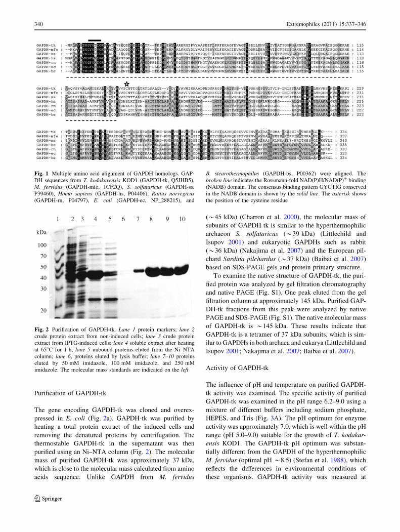
and Rattus norvegicus (rat, 13%) (Fig. 1). GAPDH-tk and
homologs share one conserved N-terminal domain: a
Rossmann-fold NAD(P)H/NAD(P)(?) binding (NADB)
domain. The NADB domain is found in many dehydro-
genases from metabolic pathways such as glycolysis, as
well as many other redox enzymes. GAPDH-tk contains
the characteristic GYGTIG sequence, a consensus binding
sequence in which the first two glycines participate in
NAD(P)-binding and the third facilitates close packing of
the helix to the adjacent beta-strand of the protein. In
contrast to GAPDH from S. solfataricus, which has three
cysteine residues, one in the nucleotide-binding domain
and two in the catalytic domain, GAPDH-tk has only one
cysteine, C141, which is conserved in the same topological
position as the active site C139 of the GAPDH from
S. solfataricus (Isupov et al. 1999; Littlechild and Isupov
2001).
Extremophiles (2011) 15:337–346 339
123
Purification of GAPDH-tk
The gene encoding GAPDH-tk was cloned and overex-
pressed in E. coli (Fig. 2a). GAPDH-tk was purified by
heating a total protein extract of the induced cells and
removing the denatured proteins by centrifugation. The
thermostable GAPDH-tk in the supernatant was then
purified using an Ni–NTA column (Fig. 2). The molecular
mass of purified GAPDH-tk was approximately 37 kDa,
which is close to the molecular mass calculated from amino
acids sequence. Unlike GAPDH from M. fervidus
(*45 kDa) (Charron et al. 2000), the molecular mass of
subunits of GAPDH-tk is similar to the hyperthermophilic
archaeon S. solfataricus (*39 kDa) (Littlechild and
Isupov 2001) and eukaryotic GAPDHs such as rabbit
(*36 kDa) (Nakajima et al. 2007) and the European pil-
chard Sardina pilchardus (*37 kDa) (Baibai et al. 2007)
based on SDS-PAGE gels and protein primary structure.
To examine the native structure of GAPDH-tk, the puri-
fied protein was analyzed by gel filtration chromatography
and native PAGE (Fig. S1). One peak eluted from the gel
filtration column at approximately 145 kDa. Purified GAP-
DH-tk fractions from this peak were analyzed by native
PAGE and SDS-PAGE (Fig. S1). The native molecular mass
of GAPDH-tk is *145 kDa. These results indicate that
GAPDH-tk is a tetramer of 37 kDa subunits, which is sim-
ilar to GAPDHs in both archaea and eukarya (Littlechild and
Isupov 2001; Nakajima et al. 2007; Baibai et al. 2007).
Activity of GAPDH-tk
The influence of pH and temperature on purified GAPDH-
tk activity was examined. The specific activity of purified
GAPDH-tk was examined in the pH range 6.2–9.0 using a
mixture of different buffers including sodium phosphate,
HEPES, and Tris (Fig. 3A). The pH optimum for enzyme
activity was approximately 7.0, which is well within the pH
range (pH 5.0–9.0) suitable for the growth of T. kodakar-
ensis KOD1. The GAPDH-tk pH optimum was substan-
tially different from the GAPDH of the hyperthermophilic
M. fervidus (optimal pH *8.5) (Stefan et al. 1988), which
reflects the differences in environmental conditions of
these organisms. GAPDH-tk activity was measured at
Fig. 1 Multiple amino acid alignment of GAPDH homologs. GAP-
DH sequences from T. kodakarensis KOD1 (GAPDH-tk, Q5JHB5),
M. fervidus (GAPDH-mfe, 1CF2Q), S. solfataricus (GAPDH-ss,
P39460), Homo sapiens (GAPDH-hs, P04406), Rattus norvegicus(GAPDH-rn, P04797), E. coli (GAPDH-ec, NP_288215), and
B. stearothermophilus (GAPDH-bs, P00362) were aligned. The
broken line indicates the Rossmann-fold NAD(P)H/NAD(P)? binding
(NADB) domain. The consensus binding pattern GYGTIG conserved
in the NADB domain is shown by the solid line. The asterisk shows
the position of the cysteine residue
100
70
50
40
30
20
kDa
1 2 3 4 5 6 7 8 9 10
Fig. 2 Purification of GAPDH-tk. Lane 1 protein markers; lane 2crude protein extract from non-induced cells; lane 3 crude protein
extract from IPTG-induced cells; lane 4 soluble extract after heating
at 65�C for 1 h; lane 5 unbound proteins eluted from the Ni–NTA
column; lane 6, proteins eluted by lysis buffer; lane 7–10 proteins
eluted by 50 mM imidazole, 100 mM imidazole, and 250 mM
imidazole. The molecular mass standards are indicated on the left
340 Extremophiles (2011) 15:337–346
123
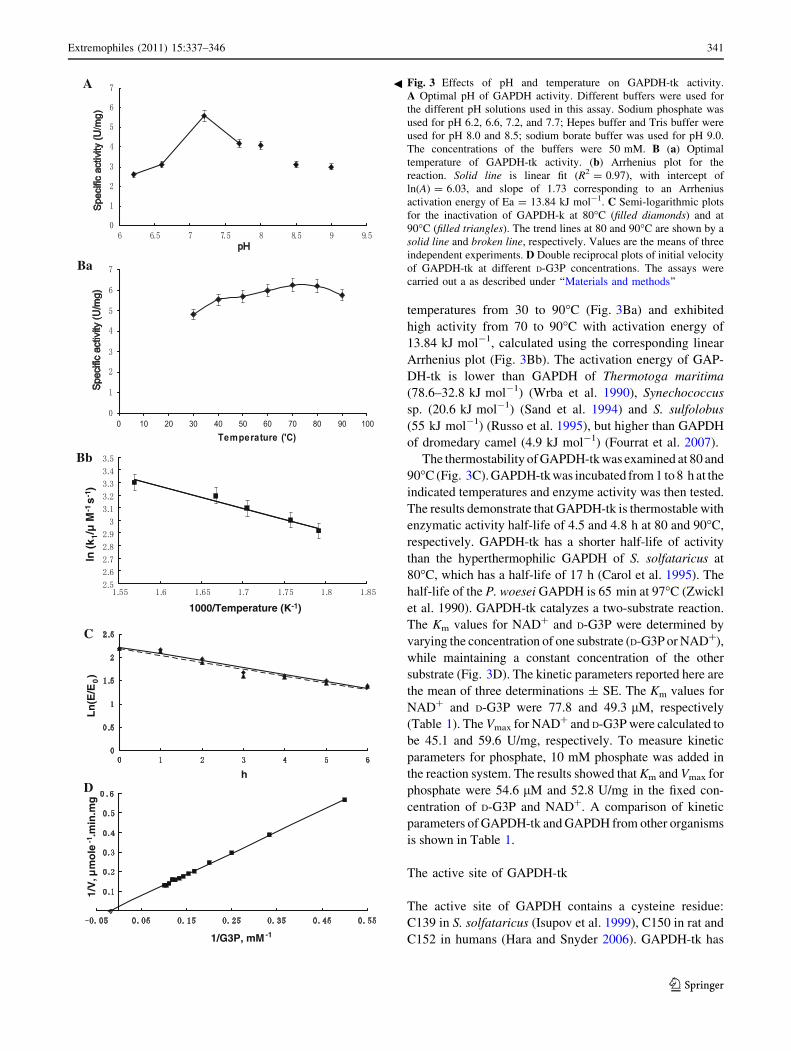
temperatures from 30 to 90�C (Fig. 3Ba) and exhibited
high activity from 70 to 90�C with activation energy of
13.84 kJ mol-1, calculated using the corresponding linear
Arrhenius plot (Fig. 3Bb). The activation energy of GAP-
DH-tk is lower than GAPDH of Thermotoga maritima
(78.6–32.8 kJ mol-1) (Wrba et al. 1990), Synechococcus
sp. (20.6 kJ mol-1) (Sand et al. 1994) and S. sulfolobus
(55 kJ mol-1) (Russo et al. 1995), but higher than GAPDH
of dromedary camel (4.9 kJ mol-1) (Fourrat et al. 2007).
The thermostability of GAPDH-tk was examined at 80 and
90�C (Fig. 3C). GAPDH-tk was incubated from 1 to 8 h at the
indicated temperatures and enzyme activity was then tested.
The results demonstrate that GAPDH-tk is thermostable with
enzymatic activity half-life of 4.5 and 4.8 h at 80 and 90�C,
respectively. GAPDH-tk has a shorter half-life of activity
than the hyperthermophilic GAPDH of S. solfataricus at
80�C, which has a half-life of 17 h (Carol et al. 1995). The
half-life of the P. woesei GAPDH is 65 min at 97�C (Zwickl
et al. 1990). GAPDH-tk catalyzes a two-substrate reaction.
The Km values for NAD? and D-G3P were determined by
varying the concentration of one substrate (D-G3P or NAD?),
while maintaining a constant concentration of the other
substrate (Fig. 3D). The kinetic parameters reported here are
the mean of three determinations ± SE. The Km values for
NAD? and D-G3P were 77.8 and 49.3 lM, respectively
(Table 1). The Vmax for NAD? and D-G3P were calculated to
be 45.1 and 59.6 U/mg, respectively. To measure kinetic
parameters for phosphate, 10 mM phosphate was added in
the reaction system. The results showed that Km and Vmax for
phosphate were 54.6 lM and 52.8 U/mg in the fixed con-
centration of D-G3P and NAD?. A comparison of kinetic
parameters of GAPDH-tk and GAPDH from other organisms
is shown in Table 1.
The active site of GAPDH-tk
The active site of GAPDH contains a cysteine residue:
C139 in S. solfataricus (Isupov et al. 1999), C150 in rat and
C152 in humans (Hara and Snyder 2006). GAPDH-tk has
A
Ba
Bb
C
D
ln(k
1/µ
M-1
s-1 )
1000/Temperature (K-1)
h
Ln
(E/E
0)
1/G3P, mM-1
1/V
, µm
ole
-1.m
in.m
gFig. 3 Effects of pH and temperature on GAPDH-tk activity.
A Optimal pH of GAPDH activity. Different buffers were used for
the different pH solutions used in this assay. Sodium phosphate was
used for pH 6.2, 6.6, 7.2, and 7.7; Hepes buffer and Tris buffer were
used for pH 8.0 and 8.5; sodium borate buffer was used for pH 9.0.
The concentrations of the buffers were 50 mM. B (a) Optimal
temperature of GAPDH-tk activity. (b) Arrhenius plot for the
reaction. Solid line is linear fit (R2 = 0.97), with intercept of
ln(A) = 6.03, and slope of 1.73 corresponding to an Arrhenius
activation energy of Ea = 13.84 kJ mol-1. C Semi-logarithmic plots
for the inactivation of GAPDH-k at 80�C (filled diamonds) and at
90�C (filled triangles). The trend lines at 80 and 90�C are shown by a
solid line and broken line, respectively. Values are the means of three
independent experiments. D Double reciprocal plots of initial velocity
of GAPDH-tk at different D-G3P concentrations. The assays were
carried out a as described under ‘‘Materials and methods’’
b
Extremophiles (2011) 15:337–346 341
123
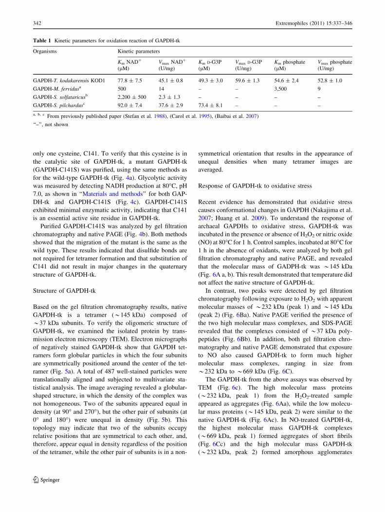
only one cysteine, C141. To verify that this cysteine is in
the catalytic site of GAPDH-tk, a mutant GAPDH-tk
(GAPDH-C141S) was purified, using the same methods as
for the wild-type GAPDH-tk (Fig. 4a). Glycolytic activity
was measured by detecting NADH production at 80�C, pH
7.0, as shown in ‘‘Materials and methods’’ for both GAP-
DH-tk and GAPDH-C141S (Fig. 4c). GAPDH-C141S
exhibited minimal enzymatic activity, indicating that C141
is an essential active site residue in GAPDH-tk.
Purified GAPDH-C141S was analyzed by gel filtration
chromatography and native PAGE (Fig. 4b). Both methods
showed that the migration of the mutant is the same as the
wild type. These results indicated that disulfide bonds are
not required for tetramer formation and that substitution of
C141 did not result in major changes in the quaternary
structure of GAPDH-tk.
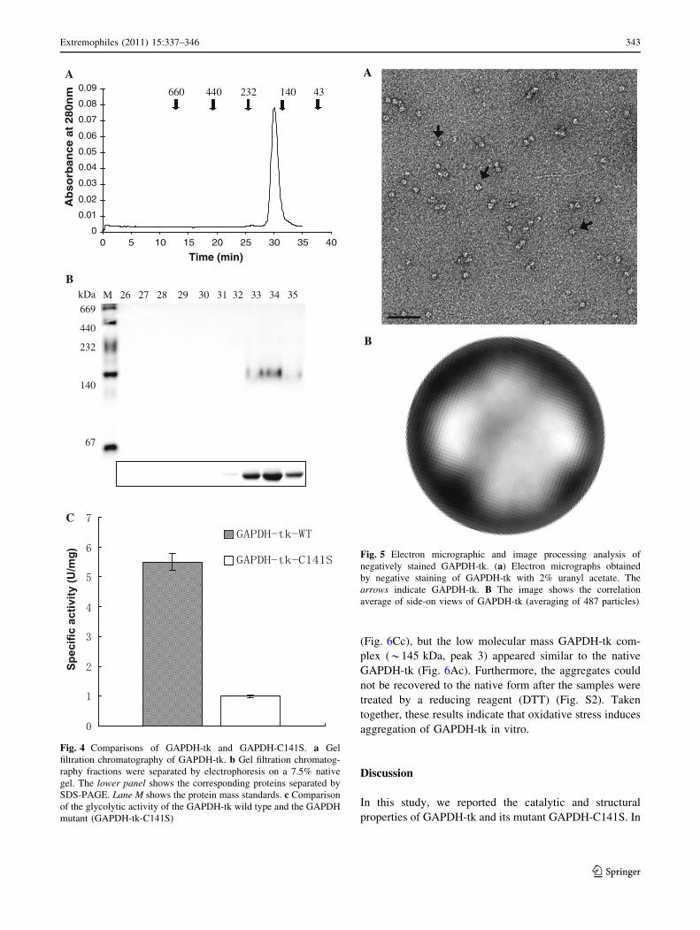
Structure of GAPDH-tk
Based on the gel filtration chromatography results, native
GAPDH-tk is a tetramer (*145 kDa) composed of
*37 kDa subunits. To verify the oligomeric structure of
GAPDH-tk, we examined the isolated protein by trans-
mission electron microscopy (TEM). Electron micrographs
of negatively stained GAPDH-tk show that GAPDH tet-
ramers form globular particles in which the four subunits
are symmetrically positioned around the center of the tet-
ramer (Fig. 5a). A total of 487 well-stained particles were
translationally aligned and subjected to multivariate sta-
tistical analysis. The image averaging revealed a globular-
shaped structure, in which the density of the complex was
not homogeneous. Two of the subunits appeared equal in
density (at 90� and 270�), but the other pair of subunits (at
0� and 180�) were unequal in density (Fig. 5b). This
topology may indicate that two of the subunits occupy
relative positions that are symmetrical to each other, and,
therefore, appear equal in density regardless of the position
of the tetramer, while the other pair of subunits is in a non-
symmetrical orientation that results in the appearance of
unequal densities when many tetramer images are
averaged.
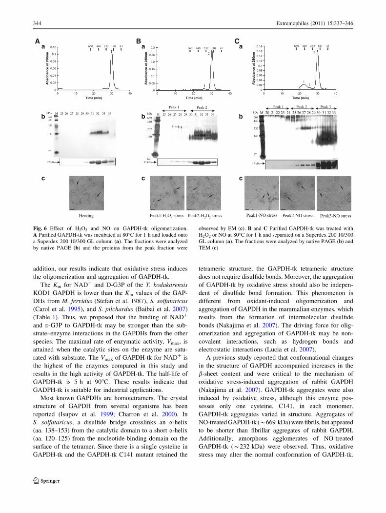
Response of GAPDH-tk to oxidative stress
Recent evidence has demonstrated that oxidative stress
causes conformational changes in GAPDH (Nakajima et al.
2007; Huang et al. 2009). To understand the response of
archaeal GAPDHs to oxidative stress, GAPDH-tk was
incubated in the presence or absence of H2O2 or nitric oxide
(NO) at 80�C for 1 h. Control samples, incubated at 80�C for
1 h in the absence of oxidants, were analyzed by both gel
filtration chromatography and native PAGE, and revealed
that the molecular mass of GADPH-tk was *145 kDa
(Fig. 6A a, b). This result demonstrated that temperature did
not affect the native structure of GAPDH-tk.
In contrast, two peaks were detected by gel filtration
chromatography following exposure to H2O2 with apparent
molecular masses of *232 kDa (peak 1) and *145 kDa
(peak 2) (Fig. 6Ba). Native PAGE verified the presence of
the two high molecular mass complexes, and SDS-PAGE
revealed that the complexes consisted of *37 kDa poly-
peptides (Fig. 6Bb). In addition, both gel filtration chro-
matography and native PAGE demonstrated that exposure
to NO also caused GAPDH-tk to form much higher
molecular mass complexes, ranging in size from
*232 kDa to *669 kDa (Fig. 6C).
The GAPDH-tk from the above assays was observed by
TEM (Fig. 6c). The high molecular mass proteins
(*232 kDa, peak 1) from the H2O2-treated sample
appeared as aggregates (Fig. 6Aa), while the low molecu-
lar mass proteins (*145 kDa, peak 2) were similar to the
native GAPDH-tk (Fig. 6Ac). In NO-treated GAPDH-tk,
the highest molecular mass GAPDH-tk complexes
(*669 kDa, peak 1) formed aggregates of short fibrils
(Fig. 6Cc) and the high molecular mass GAPDH-tk
(*232 kDa, peak 2) formed amorphous agglomerates
Table 1 Kinetic parameters for oxidation reaction of GAPDH-tk
Organisms Kinetic parameters
Km NAD?
(lM)
Vmax NAD?
(U/mg)
Km D-G3P
(lM)
Vmax D-G3P
(U/mg)
Km phosphate
(lM)
Vmax phosphate
(U/mg)
GAPDH-T. kodakarensis KOD1 77.8 ± 7.5 45.1 ± 0.8 49.3 ± 3.0 59.6 ± 1.3 54.6 ± 2.4 52.8 ± 1.0
GAPDH-M. fervidusa 500 14 – – 3,500 9
GAPDH-S. solfataricusb 2,200 ± 500 2.3 ± 1.3 – – – –
GAPDH-S. pilchardusc 92.0 ± 7.4 37.6 ± 2.9 73.4 ± 8.1 – – –
a, b, c From previously published paper (Stefan et al. 1988), (Carol et al. 1995), (Baibai et al. 2007)
‘‘–’’, not shown
342 Extremophiles (2011) 15:337–346
123
(Fig. 6Cc), but the low molecular mass GAPDH-tk com-
plex (*145 kDa, peak 3) appeared similar to the native
GAPDH-tk (Fig. 6Ac). Furthermore, the aggregates could
not be recovered to the native form after the samples were
treated by a reducing reagent (DTT) (Fig. S2). Taken
together, these results indicate that oxidative stress induces
aggregation of GAPDH-tk in vitro.
Discussion
In this study, we reported the catalytic and structural
properties of GAPDH-tk and its mutant GAPDH-C141S. In
0
0.01
0.02
0.03
0.04
0.05
0.06
0.07
0.08
0.09
0 5 10 15 20 25 30 35 40
Time (min)
Ab
so
rban
ce a
t 280n
m 660 440 232 140 43
A
M 26 27 28 29 30 31 32 33 34 35kDa
669
440
232
140
67
B
C
Fig. 4 Comparisons of GAPDH-tk and GAPDH-C141S. a Gel
filtration chromatography of GAPDH-tk. b Gel filtration chromatog-
raphy fractions were separated by electrophoresis on a 7.5% native
gel. The lower panel shows the corresponding proteins separated by
SDS-PAGE. Lane M shows the protein mass standards. c Comparison
of the glycolytic activity of the GAPDH-tk wild type and the GAPDH
mutant (GAPDH-tk-C141S)
Fig. 5 Electron micrographic and image processing analysis of
negatively stained GAPDH-tk. (a) Electron micrographs obtained
by negative staining of GAPDH-tk with 2% uranyl acetate. The
arrows indicate GAPDH-tk. B The image shows the correlation
average of side-on views of GAPDH-tk (averaging of 487 particles)
Extremophiles (2011) 15:337–346 343
123
addition, our results indicate that oxidative stress induces
the oligomerization and aggregation of GAPDH-tk.
The Km for NAD? and D-G3P of the T. kodakarensis
KOD1 GAPDH is lower than the Km values of the GAP-
DHs from M. fervidus (Stefan et al. 1987), S. solfataricus
(Carol et al. 1995), and S. pilchardus (Baibai et al. 2007)
(Table 1). Thus, we proposed that the binding of NAD?
and D-G3P to GAPDH-tk may be stronger than the sub-
strate–enzyme interactions in the GAPDHs from the other
species. The maximal rate of enzymatic activity, Vmax, is
attained when the catalytic sites on the enzyme are satu-
rated with substrate. The Vmax of GAPDH-tk for NAD? is
the highest of the enzymes compared in this study and
results in the high activity of GAPDH-tk. The half-life of
GAPDH-tk is 5 h at 90�C. These results indicate that
GADPH-tk is suitable for industrial applications.
Most known GAPDHs are homotetramers. The crystal
structure of GAPDH from several organisms has been
reported (Isupov et al. 1999; Charron et al. 2000). In
S. solfataricus, a disulfide bridge crosslinks an a-helix
(aa. 138–153) from the catalytic domain to a short a-helix
(aa. 120–125) from the nucleotide-binding domain on the
surface of the tetramer. Since there is a single cysteine in
GAPDH-tk and the GAPDH-tk C141 mutant retained the
tetrameric structure, the GAPDH-tk tetrameric structure
does not require disulfide bonds. Moreover, the aggregation
of GAPDH-tk by oxidative stress should also be indepen-
dent of disulfide bond formation. This phenomenon is
different from oxidant-induced oligomerization and
aggregation of GAPDH in the mammalian enzymes, which
results from the formation of intermolecular disulfide
bonds (Nakajima et al. 2007). The driving force for olig-
omerization and aggregation of GAPDH-tk may be non-
covalent interactions, such as hydrogen bonds and
electrostatic interactions (Lucia et al. 2007).
A previous study reported that conformational changes
in the structure of GAPDH accompanied increases in the
b-sheet content and were critical to the mechanism of
oxidative stress-induced aggregation of rabbit GAPDH
(Nakajima et al. 2007). GAPDH-tk aggregates were also
induced by oxidative stress, although this enzyme pos-
sesses only one cysteine, C141, in each monomer.
GAPDH-tk aggregates varied in structure. Aggregates of
NO-treated GAPDH-tk (*669 kDa) were fibrils, but appeared
to be shorter than fibrillar aggregates of rabbit GAPDH.
Additionally, amorphous agglomerates of NO-treated
GAPDH-tk (*232 kDa) were observed. Thus, oxidative
stress may alter the normal conformation of GAPDH-tk.
0
0.02
0.04
0.06
0.08
0.1
0.12 660 440 232 140 43
669440
232
140
67
M 25 26 27 28 29 30 31 32 33 34 kDa
37 kDa
Heating
A
669440
232
140
67
kDa M 25 26 27 28 29 30 31 32 33 34
Peak 2Peak 1
37 kDa
0
0.05
0.1
0.15
0.2
0.25
0.3
1
2
660 440 232 140 43
Peak1-H2O2 stress Peak2-H2O2 stress
Ba
b
c
a
b
c
0
0.02
0.04
0.06
0.08
0.1
0.12
0.14
0.16
0.18
0 10 20 30 40
Time (min)
Ab
sob
ance
at
280n
m
Ab
sob
ance
at
280n
m
Ab
sob
ance
at
280n
m660 440 232 140 43
12
3
669440
232
140
67
kDa M 20 21 22 23 24 25 26 27 28 29 30 31 32 33
Peak 3Peak 2Peak 1
37 kDa
Peak1-NO stress Peak2-NO stress Peak3-NO stress
Ca
b
c
0 10 20 30 40
Time (min)0 10 20 30 40
Time (min)
Fig. 6 Effect of H2O2 and NO on GAPDH-tk oligomerization.
A Purified GAPDH-tk was incubated at 80�C for 1 h and loaded onto
a Superdex 200 10/300 GL column (a). The fractions were analyzed
by native PAGE (b) and the proteins from the peak fraction were
observed by EM (c). B and C Purified GAPDH-tk was treated with
H2O2 or NO at 80�C for 1 h and separated on a Superdex 200 10/300
GL column (a). The fractions were analyzed by native PAGE (b) and
TEM (c)
344 Extremophiles (2011) 15:337–346
123
Our study is the first reported description of aggregates of
an archaeal GAPDH under oxidative stress. The mecha-
nisms of oxidative stress-induced aggregation of GAPDH-
tk will require further examination in future work.
References
Atomi H, Fukui T, Kanai T, Morikawa M, Imanaka T (2004)
Description of Thermococcus kodakaraensis sp. nov., a well
studied hyperthermophilic archaeon previously reported as
Pyrococcus sp. KOD1. Archaea 1:263–267
Baibai T, Oukhattar L, Moutaouakkil A, Soukri A (2007) Purification
and characterization of glyceraldehyde-3-phosphate dehydroge-
nase from European pilchard Sardina pilchardus. Acta Biochim
Biophys Sin 39:947–954
Brunner N, Brinkmann H, Siebers B, Hensel R (1998) NAD?-
dependent glyceraldehyde-3-phosphate dehydrogenase from
Thermoproteus tenax. J Biol Chem 273:6149–6156
Brunner N, Siebers B, Hensel R (2001) Role of two different
glyceraldehyde-3-phosphate dehydrogenases in controlling the
reversible Embden–Meyerhof–Parnas pathway in Thermopro-teus tenax: regulation on protein and transcript level. Extremo-
philes 5:101–109
Carol EJ, Toni MF, Don AC, Jennifer AL, Peter WP (1995) The
phosphoglycerate kinase and glyceraldehyde-3-phosphate dehy-
drogenase genes from the thermophilic archaeon Sulfolobussolfataricus overlap by 8-bp. Isolation, sequencing of the genes
and expression in Escherichia coli. Eur J Bio Chem
233:800–808
Charron C, Talfournier F, Isupov MN, Littlechild JA, Branlant G,
Vitoux B, Aubry A (2000) The crystal structure of
D-glyceraldehyde-3-phosphate dehydrogenase from the hyper-
thermophilic archaeon Methanothermus fervidus in the presence
of NADP? at 2.1 A resolution. J Mol Biol 29:481–500
Cleland WW (1963) The kinetics of enzyme-catalyzed reactions with
two or more substrates or products: II. Inhibition: nomenclature
and theory. Biochim Biophys Acta 67:173–187
Elkina Y, Kuravsky ML, El’darov MA, Stogov SV, Muronetz VI,
Schmalhausen EV (2010) Recombinant human sperm-specific
glyceraldehyde-3-phosphate dehydrogenase: structural basis for
enhanced stability. Biochim Biophys Acta 1804:2207–2212
Engel M, Seifert M, Theisinger B, Seyfert U, Welter C (1998)
Glyceraldehyde-3-phosphate dehydrogenase and Nm23–H1/
nucleoside diphosphate kinase A. J Biol Chem 273:20058–20065
Fabry S, Reinhar Hensel (1987) Purification and characterization of
D-glyceraldehyde-3-phosphate dehydrogenase from the thermo-
philic archaebacterium Methanothermus fervidus. Eur J Bio
Chem 165:147–155
Fourrat L, Iddar A, Soukri (2007) Purification and characterization of
cytosolic glyceraldehyde-3-phosphate dehydrogenase from the
dromedary camel. Acta Biochim Biophys Sin 39:148–154
Hara M, Snyder S (2006) Nitric oxide–GAPDH–Siah: a novel cell
death cascade. Cell Mol Neurobiol 26:525–536
Hara MR, Agrawal N, Kim SF, Cascio MB, Fujimuro M, Ozeki Y,
Takahashi M, Cheah JH, Tankou SK, Hester LD, Ferris CD,
Hayward SD, Snyder SH, Sawa A (2005) S-nitrosylated GAPDH
initiates apoptotic cell death by nuclear translocation following
Siah1 binding. Nat Cell Biol 7:665–674
Hegerl R (1996) The EM program package: a platform for image
processing in biological electron microscopy. J Struct Biol
116:30–34
Huang J, Hao L, Xiong N, Cao X, Liang Z, Sun S, Wang T (2009)
Involvement of glyceraldehyde-3-phosphate dehydrogenase in
rotenone-induced cell apoptosis: relevance to protein misfolding
and aggregation. Brain Res 1279:1–8
Isupov MN, Fleming TM, Dalby AR, Crowhurst GS, Bourne PC,
Littlechild JA (1999) Crystal structure of the glyceraldehyde-3-
phosphate dehydrogenase from the hyperthermophilic archaeon
Sulfolobus solfataricus. J Mol Biol 291:651–660
Kim KI, Cheong GW, Park SC, Ha JS, Woo KM, Choi SJ, Chung CH
(2000) Heptameric ring structure of the heat-shock protein ClpB,
a protein-activated ATPase in Escherichia coli. J Mol Biol
303:655–666
Launay JF, Jellali A, Vanier MT (1989) Glyceraldehyde-3-phosphate
dehydrogenase is a microtubule binding protein in a human
colon tumor cell line. Biochim Biophys Acta 996:103–109
Littlechild JA, Isupov M (2001) Glyceraldehyde-3-phosphate dehy-
drogenase from Sulfolobus solfataricus. Methods Enzymol
331:105–117
Lorentzen E, Hensel R, Knura T, Ahmed H, Pohl E et al (2004)
Structural basis of allosteric regulation and substrate specificity
of the non-phosphorylating glyceraldehyde 3-phosphate dehy-
drogenase from Thermoproteus tenax. J Mol Biol 341:815–828
Lucia B, Ivano B, Armando D, Stefania G, Edith BG, Manuele M,
Joan SV, Miguela V, Julian PW (2007) Metal-free superoxide
dismutase forms soluble oligomers under physiological condi-
tions: a possible general mechanism for familial ALS. Proc Natl
Acad Sci USA 104:11263–11267
Marco S, Urena D, Carrascosa JL, Waldmann T, Peters Jr, Hegerl R,
Pfeifer Gn, Sack-Kongehl H, Baumeister W (1994) The
molecular chaperone TF55: Assesment of symmetry. FEBS Lett
341:152–155
Nakajima H, Amano W, Fujita A, Fukuhara A, Azuma YT, Hata F,
Inui T, Takeuchi T (2007) The active site cysteine of the
proapoptotic protein glyceraldehyde-3-phosphate dehydrogenase
is essential in oxidative stress-induced aggregation and cell
death. J Biol Chem 282:26562–26574
Park SC, Jia B, Yang JK, Le Van D, Shao YG, Han SW, Jeon YJ,
Chung CH, Cheong GW (2006) Oligomeric structure of the
ATP-dependent protease La (Lon) of Escherichia coli. Mol Cells
21:129–134
Roitel O, Sergienko E, Branlant G (1999) Dimers generated from
tetrameric phosphorylating glyceraldehyde-3-phosphate dehy-
drogenase from Bacillus stearothermophilus are inactive but
exhibit cooperativity in NAD binding. Biochemistry
38:16084–6091
Russo AD, Rullo R, Masullo M, Ianniciello G, Arcari P, Bocchini V
(1995) Glyceraldehyde-3-phosphate dehydrogenase in the
hyperthermophilic archaeon Sulfolobus solfataricus: character-
ization and significance in glucose metabolism. Biochem Mol
Biol Int 36:123–135
Sand O, Petersen IM, Jørgen J, Iversen L (1994) Purification and
some properties of glyceraldehyde 3-phosphate dehydrogenase
from Synechococcus sp. Antonie Van Leeuwenhoek 65:133–142
Saxton WO, Pitt TJ, Horner M (1979) Digital image processing: the
Semper system. Ultramicroscopy 4:343–353
Sirover MA (1996) Emerging new functions of the glycolytic protein,
glyceraldehyde-3-phosphate dehydrogenase, in mammalian
cells. Life Sci 58:2271–2277
Stefan F, Anselm L, Wolfram B, Reinhard H (1988) Expression of the
glyceraldehyde-3-phosphate dehydrogenase gene from the
extremely thermophilic archaebacterium Methanothermus fer-vidus in E. coli. FEBS Lett 237:213–217
Tisdale EJ (2001) Glyceraldehyde-3-phosphate dehydrogenase is
required for vesicular transport in the early secretory pathway.
J Biol Chem 276:2480–2486
Extremophiles (2011) 15:337–346 345
123
Tristan C, Shahani N, Sedlak TW, Sawa A (2011) The diverse
functions of GAPDH: views from different subcellular compart-
ments. Cell Signal 23:317–323
Van Heel M, Frank J (1981) Use of multivariate statistics in analysing
the images of biological macromolecules. Ultramicroscopy
6:187–194
Wrba A, Schweiger A, Schultes V, Jaenicke R, Zavodszky P (1990)
Extremely thermostable D-glyceraldehyde-3-phosphate dehydro-
genase from the eubacterium Thermotoga maritima. Biochem-
istry 29:7584–7592
Yun M, Park CG, Kim JY, Park HW (2000) Structural analysis of
glyceraldehyde 3-phosphate dehydrogenase from Escherichiacoli: direct evidence of substrate binding and cofactor-induced
conformational changes. Biochemistry 39:10702–10710
Zwick P, Fabry S, Bogedain C, Haas A, Hensel R et al (1990)
Glyceraldehyde-3-phosphate dehydrogenase from the hyper-
thermophilic archaebacterium Pyrococcus woesei: characteriza-
tion of the enzyme, cloning and sequencing of the gene, and
expression in Escherichia coli. J Bacteriol 172:4329–4338
346 Extremophiles (2011) 15:337–346
123
Related Documents


