1 BIOCHEMICAL BASIS OF GLUCOKINASE ACTIVATION AND THE REGULATION BY GLUCOKINASE REGULATORY PROTEIN IN NATURALLY OCCURING MUTATIONS Vladi V. Heredia, Thomas J. Carlson, Erin Garcia and Shaoxian Sun From the Department of Biochemical Pharmacology, La Jolla Laboratories, Pfizer Global Research and Development, San Diego, California, 92121 Running Title: Glucokinase activating mutations and GKRP Address correspondence to: Shaoxian Sun, Department of Biochemical Pharmacology, La Jolla Laboratories, Pfizer Global Research & Development, 10628 Science Center Drive, San Diego, CA 92121, Tel: 858 526-4922; Fax 858 526-4240; E-mail: [email protected] Glucokinase (GK) has several known polymorphic activating mutations that increase the enzyme activity by enhancing glucose binding affinity and/or by alleviating the inhibition of glucokinase regulatory protein (GKRP), a key regulator of GK activity in the liver. Kinetic studies were undertaken to better understand the effect of these mutations on the enzyme mechanism of GK activation and GKRP regulation, and relate the enzyme properties to the associated clinical phenotype of hypoglycemia. Similar to wild type GK, the transient kinetics of glucose binding for activating mutations follows a general two-step mechanism: the formation of an enzyme- glucose complex followed by an enzyme conformational change. However, the kinetics for each step differed from wild type GK and could be grouped into specific types of kinetic changes. Mutations T65I, Y214C, and A456V accelerate glucose binding to the apo-enzyme form, while W99R, Y214C and V455M facilitate enzyme isomerization to the active form. Mutations that significantly enhance the glucose binding to the apo enzyme also disrupt the protein-protein interaction with GKRP to a large extent, suggesting these mutations may adopt a more compact conformation in the apo enzyme favorable for glucose binding. Y214C is the most active mutation (11 fold increase in k cat / h 0.5 K ) and exhibits the most severe clinical effects of hypoglycemia. In contrast, moderate activating mutation A456V nearly abolishes the GKRP inhibition (76 fold increase in K i ), but causes only mild hypoglycemia. This suggests that the alteration in GK enzyme activity may have a more profound biological impact than the alleviation of GKRP inhibition. Glucokinase (GK) plays a central role in maintaining glucose homeostasis (1-3). It serves as a glucose sensor due to its specific kinetic properties that include low affinity and positive cooperativity for glucose and a lack of inhibition by its product glucose-6-phosphate (4-6). In pancreatic β-cells, GK regulates glucose- dependent insulin secretion by modulation of the glycolytic pathway and subsequently the ATP/ADP ratio. In the liver, GK stimulates glucose disposal by converting glucose to glycogen for storage. Hepatic GK is tightly regulated by the 68 kDa glucokinase regulatory protein (GKRP) through the formation of a GKRP-GK complex followed by sequestration in the nucleus (7). Physiologically, the interaction between GK and GKRP is promoted by fructose- 6-phosphate and suppressed by fructose-1- phosphate. Upon increasing glucose concentrations, GK dissociates from GKRP and translocates from the nucleus to the cytoplasm resulting in an increase in GK activity. Alteration in GK activity and its regulation is associated with abnormal glycemia as evidenced in naturally occurring mutations. More than 190 inactivating GK mutations have been identified in patients with maturity-onset diabetes of the young (MODY2). In contrast, 5 activating GK mutations (T65I, W99R, Y214C, V455M and A456V) lead to hyperinsulinemic hypoglycemia (HH) (8-11). The degree of HH in the affected patients is variable. The mutation Y214C causes severe and possibly fatal hypoglycemia while other mutations are associated with mild hypoglycemia and are in some cases asymptomatic. Interestingly, all of these activating mutations cluster in an allosteric site of GK where small molecule activators bind, suggesting a critical role of the allosteric site in the http://www.jbc.org/cgi/doi/10.1074/jbc.M607987200 The latest version is at JBC Papers in Press. Published on November 2, 2006 as Manuscript M607987200 Copyright 2006 by The American Society for Biochemistry and Molecular Biology, Inc. by guest on September 5, 2016 http://www.jbc.org/ Downloaded from by guest on September 5, 2016 http://www.jbc.org/ Downloaded from by guest on September 5, 2016 http://www.jbc.org/ Downloaded from by guest on September 5, 2016 http://www.jbc.org/ Downloaded from by guest on September 5, 2016 http://www.jbc.org/ Downloaded from by guest on September 5, 2016 http://www.jbc.org/ Downloaded from

Welcome message from author
This document is posted to help you gain knowledge. Please leave a comment to let me know what you think about it! Share it to your friends and learn new things together.
Transcript
1
BIOCHEMICAL BASIS OF GLUCOKINASE ACTIVATION AND THE REGULATION BY GLUCOKINASE REGULATORY PROTEIN IN
NATURALLY OCCURING MUTATIONS Vladi V. Heredia, Thomas J. Carlson, Erin Garcia and Shaoxian Sun
From the Department of Biochemical Pharmacology, La Jolla Laboratories, Pfizer Global Research and Development, San Diego, California, 92121
Running Title: Glucokinase activating mutations and GKRP Address correspondence to: Shaoxian Sun, Department of Biochemical Pharmacology, La Jolla Laboratories, Pfizer Global Research & Development, 10628 Science Center Drive, San Diego, CA 92121, Tel: 858 526-4922; Fax 858 526-4240; E-mail: [email protected]
Glucokinase (GK) has several known
polymorphic activating mutations that increase the enzyme activity by enhancing glucose binding affinity and/or by alleviating the inhibition of glucokinase regulatory protein (GKRP), a key regulator of GK activity in the liver. Kinetic studies were undertaken to better understand the effect of these mutations on the enzyme mechanism of GK activation and GKRP regulation, and relate the enzyme properties to the associated clinical phenotype of hypoglycemia. Similar to wild type GK, the transient kinetics of glucose binding for activating mutations follows a general two-step mechanism: the formation of an enzyme-glucose complex followed by an enzyme conformational change. However, the kinetics for each step differed from wild type GK and could be grouped into specific types of kinetic changes. Mutations T65I, Y214C, and A456V accelerate glucose binding to the apo-enzyme form, while W99R, Y214C and V455M facilitate enzyme isomerization to the active form. Mutations that significantly enhance the glucose binding to the apo enzyme also disrupt the protein-protein interaction with GKRP to a large extent, suggesting these mutations may adopt a more compact conformation in the apo enzyme favorable for glucose binding. Y214C is the most active mutation (11 fold increase in kcat/ h
0.5K ) and exhibits the most severe clinical effects of hypoglycemia. In contrast, moderate activating mutation A456V nearly abolishes the GKRP inhibition (76 fold increase in Ki), but causes only mild hypoglycemia. This suggests that the alteration in GK enzyme activity may have a more profound biological impact than the alleviation of GKRP inhibition.
Glucokinase (GK) plays a central role in maintaining glucose homeostasis (1-3). It serves as a glucose sensor due to its specific kinetic properties that include low affinity and positive cooperativity for glucose and a lack of inhibition by its product glucose-6-phosphate (4-6). In pancreatic β-cells, GK regulates glucose-dependent insulin secretion by modulation of the glycolytic pathway and subsequently the ATP/ADP ratio. In the liver, GK stimulates glucose disposal by converting glucose to glycogen for storage. Hepatic GK is tightly regulated by the 68 kDa glucokinase regulatory protein (GKRP) through the formation of a GKRP-GK complex followed by sequestration in the nucleus (7). Physiologically, the interaction between GK and GKRP is promoted by fructose-6-phosphate and suppressed by fructose-1-phosphate. Upon increasing glucose concentrations, GK dissociates from GKRP and translocates from the nucleus to the cytoplasm resulting in an increase in GK activity.
Alteration in GK activity and its regulation is associated with abnormal glycemia as evidenced in naturally occurring mutations. More than 190 inactivating GK mutations have been identified in patients with maturity-onset diabetes of the young (MODY2). In contrast, 5 activating GK mutations (T65I, W99R, Y214C, V455M and A456V) lead to hyperinsulinemic hypoglycemia (HH) (8-11). The degree of HH in the affected patients is variable. The mutation Y214C causes severe and possibly fatal hypoglycemia while other mutations are associated with mild hypoglycemia and are in some cases asymptomatic. Interestingly, all of these activating mutations cluster in an allosteric site of GK where small molecule activators bind, suggesting a critical role of the allosteric site in the
http://www.jbc.org/cgi/doi/10.1074/jbc.M607987200The latest version is at JBC Papers in Press. Published on November 2, 2006 as Manuscript M607987200
Copyright 2006 by The American Society for Biochemistry and Molecular Biology, Inc.
by guest on September 5, 2016
http://ww
w.jbc.org/
Dow
nloaded from
by guest on September 5, 2016
http://ww
w.jbc.org/
Dow
nloaded from
by guest on September 5, 2016
http://ww
w.jbc.org/
Dow
nloaded from
by guest on September 5, 2016
http://ww
w.jbc.org/
Dow
nloaded from
by guest on September 5, 2016
http://ww
w.jbc.org/
Dow
nloaded from
by guest on September 5, 2016
http://ww
w.jbc.org/
Dow
nloaded from
2
regulation of GK activity. Both GK activators and activating mutations increase enzyme activity by enhancing the affinity for glucose as described by a decrease in K0.5 (8-12). In vivo, GK activators were shown to lower glucose levels and stimulate insulin release in animal models (12-14). The potential efficacy of GK activators in humans has drawn great interest as a promising therapeutic treatment for Type 2 Diabetes. Since the in vitro effects of small molecule GK activators are similar to the activating mutations in GK, a better understanding of the biochemical basis for the activation of GK activity by these mutations may provide insight for the design of GK activators as effective therapeutics.
Protein structural analyses have shown that substantial conformational changes in GK occur during glucose binding (15). Recently, we investigated the GK structural changes induced by glucose binding through transient kinetic studies and demonstrated that the slow enzyme conformational change contributed to the positive cooperativity and the low binding affinity for glucose (16). We also showed that the most clinically severe activating GK mutation Y214C facilitated the slow enzyme conformational changes to the active form by a factor of 7.5 fold. However, it is unknown if all the activating mutations at the allosteric site follow the same activation mechanism, how the mutations affect GKRP inhibitory regulation, and how the variation in their biochemical properties relate to the clinical phenotype.
In this paper, we extended our studies to include published naturally activating mutations. In addition, two engineered activating mutants at the allosteric site, Y214A and Y215A were included in the studies to further investigate the structure-function relationship of the allosteric site involved in the protein conformational change. The GK activation and GKRP binding for the GK activating mutations were characterized in vitro. We show that the structural perturbation at the allosteric site by single-point mutations alters the kinetic basis of enzyme activation as well as the protein-protein interactions with GKRP.
EXPERIMENTAL PROCEDURES Materials. Glucose, 2-deoxyglucose, pyruvate kinase/lactate dehydrogenase (PK/LDH),
phosphoenolpyruvate (PEP), dithiothreitol (DTT), and ATP were obtained from Sigma. All reagents were ACS grade or better. Expression and purification of recombinant proteins. The cloning, expression and purification of β-cell GK and Y214C were described previously (16,17). The oligonucleotide primers used to generate the rest of the GK allosteric-site mutations are listed as follows whereby the sites of the mutation are italicized and underlined: T65I forward: CCACCTACGTGCGCTCCATCCCAGAAGGCTCAGAAGTCGG T65I reverse: CCGACTTCTGAGCCTTCTGGGATGGAGCGCACGTAGGTGG W99R forward: GGTGAGGAGGGGCAGCGGAGCGTGAAGACCAAACACC W99R reverse: GGTGTTTGGTCTTCACGCTCCGCTGCCCCTCCTCACC V455M forward: GCCCTGGTCTCGGCGATGGCCTGTAAGAAGGCC V455M reverse: GGCCTTCTTACAGGCCATCGCCGAGACCAGGGC A456V forward: GCCCTGGTCTCGGCGGTGGTCTGTAAGAAGGCC A456V reverse: GGCCTTCTTACAGACCACCGCCGAGACCAGGGC Y214A forward: ACGATGATCTCCTGCGCCTACGAAGACCATCAG Y214A reverse: CTGATGGTCTTCGTAGGCGCAGGAGATCATCGT Y215A forward: ATGATCTCCTGCTACGCCGAAGACCATCAGTGC Y215A reverse: GCACTGATGGTCTTCGGCGTAGCAGGAGATCAT
The recombinant GK WT and mutations with a hexa-His-tag at the N-terminus were expressed in E. coli and purified using the protocol described previously. A complete protease inhibitor cocktail (Roche) was added to each step
by guest on September 5, 2016
http://ww
w.jbc.org/
Dow
nloaded from
3
of the purification for the A456V mutation to reduce the proteolysis of the protein. Purity of the enzyme was verified to be >95% by SDS-PAGE and QTOF-MS. The molecular weight of His-tagged GK was confirmed to be 55 kDa by QTOF-MS. Enzyme concentration was determined by the method of Bradford (18). Aliquots of WT and mutated GK were stored at –80o C in a pH 7.5 buffer containing 25 mM HEPES, 50 mM NaCl, 5 mM DTT and 5% glycerol.
The cloning of GKRP has been described previously (19). GKRP with a C-terminal FLAG tag was expressed in E. coli DH10B cells using a pFLAG-CTC expression vector. Expression was confirmed by western analysis using an anti-GKRP monoclonal antibody obtained from Santa Cruz Biotechnology. Bacterial cells were grown in a 10-L fermentation with TB media initially at 37o C containing 100 µg/mL ampicillin and inducted with 0.2 mM IPTG upon reaching A600 of 0.6. After overnight expression at 23o C, cells were harvested. Cell pellets were resuspended in 50 mM Tris-HCl, pH 7.4, 150 mM NaCl. Cells were subsequently lysed by microfluidization on ice. Purification was achieved by batch absorption using an anti-Flag monoclonal antibody Mab2 affinity gel (Sigma). Resin was then loaded onto a chromatography column where Flag-tagged GKRP was eluted from the resin by competition using 100 µg/mL Flag peptide (Sigma). Pooled fractions were then loaded onto a size-exclusion column where the peak at 72 kDa was collected. Purity of the enzyme was verified to be >95% by SDS-PAGE. Protein concentrations were determined by the method of Bradford (18). GKRP was stored in aliquots at –80o C in a pH 7.4 buffer containing 25 mM Tris-HCl, 150 mM NaCl, 20% glycerol and 5 mM DTT. Steady-state kinetics of GK activating mutations. Steady-state kinetic parameters of GK WT and activating mutations were determined as described previously using a pyruvate kinase/lactate dehydrogenase (PK/LDH) coupled assay (16). Experiments were performed with 0–50 mM glucose at a saturating concentration of 5 mM ATP in a buffer containing 50 mM HEPES, pH 8.0, 6 mM MgCl2, 25 mM KCl, 0.7 mM NADH, 2 mM DTT, 4 mM PEP and 1 unit/mL of PK/LDH at 25o C. Saturating concentration of MgCl2 was used in the assay to ensure majority of ATP (>98%) was present as MgATP. Control
experiments were carried out to demonstrate the coupling enzymes were in excess and the initial rate was linearly dependent on GK concentrations. To determine if activating mutations affect Km for ATP, initial rates were measured by varying ATP (0–5 mM) at a saturating concentration of 50 mM glucose at 25o C using pH 8.0 buffer. The kinetic parameters for substrate analog 2-deoxyglucose were determined at 25o C using pH 8.0 buffer by varying 2-deoxyglucose concentrations from 0–400 mM at 5 mM ATP. The steady-state kinetic parameters were obtained by fitting the initial rates to the Hill equation for sugar substrates due to the sigmoidal kinetics and to the Michaelis-Menten equation for ATP with hyperbolic kinetics using nonlinear regression analysis (Prism, Graph Pad Inc.);
Hill equation: hh
h
SKSV
v][][
5.0
max
+×
=
where Vmax is the maximal activity of GK, S is the sugar concentration, K0.5 is the sugar concentration at half-maximal activity, h is the Hill number, and v is the activity at a given sugar concentration S. Glucose binding to GK WT and activating mutations. Binding of glucose to the GK enzymes caused a change in the intrinsic enzyme fluorescence that was monitored with excitation at 290 nm. The equilibrium binding affinity of glucose was determined as described previously (16). Briefly, glucose binding to GK WT and the mutations was monitored at 330 nm by fluorescence spectrometry. The KD values were determined by fitting data to a binding isotherm using non-linear regression analysis
(][
][max
SKSceFluorescen
ceFluorescenD +
×= ).
The transient kinetics study of glucose binding to GK using a stopped-flow spectrophotometer was described in detail elsewhere (16). Final enzyme concentrations of 5, 10 or 20 µM were utilized for these measurements. Briefly, pre-steady state kinetics was monitored using a 320 nm cut off filter upon excitation at 290 nm by the rapid mixing of glucose and enzyme in separate syringes. The time traces were fit to a double exponential equation:
CeAeAY t)k(2
t)k(1
obs2obs1 ++= ×−×− to obtain the rate constants (kobs1 and kobs2), the amplitudes (A1 and A2) and the fitting variable C.
by guest on September 5, 2016
http://ww
w.jbc.org/
Dow
nloaded from
4
Inhibition of GK WT and the activating mutations by GKRP. GKRP binds to GK and inhibits the enzyme activity in a competitive manner with respect to glucose (20). Kinetic analyses were performed to determine the inhibition constant of GKRP with GK WT and the mutations by varying the glucose concentrations as described below and GKRP concentrations (0–500 nM). The range of glucose concentrations was adjusted to reflect the differences in glucose affinity of the mutations: 0–50 mM for GK WT, W99R, V455M, and Y214A; and 0–10 mM for T65I, Y214C, A456V and Y215A. GK and GKRP were mixed in a buffer containing 50 mM HEPES, pH 8.0, various concentrations of glucose, 6 mM MgCl2, 0.7 mM NADH, 2 mM DTT, 1 unit/ml PK/LDH, 4 mM phosphoenolpyruvate, 25 mM KCl and 0.2 mM sorbitol-6-phosphate at 25o C. Sorbitol-6-phosphate was used in the assay since it is a more potent analogue than fructose-6-phosphate in promoting the association of GKRP with GK. The assay mix was allowed to equilibrate for 10 min before the reaction was initiated by the addition of 5 mM ATP. The assay was conducted in a 96-well plate with a final volume of 100 µL. The apparent inhibition constant of GKRP, app
iK , at a given glucose concentration was determined by fitting initial rates to the Morrison equation (21):
][2]][[4)][]([)][]([
12
t
tappit
appit
o
i
EIEKIEKIE
vv −++−++
−=
and )K[S](1KK
mi
appi +=
where vi/vo is the fractional velocity of GK activity in the presence of GKRP relative to that in the absence of GKRP, Et is the total enzyme concentration, and I is the GKRP concentration. The inhibition constant Ki was determined by plotting app
iK against varying sugar substrate concentrations S. The experiments were performed in triplicates. The Ki values of GKRP were also determined using the non-cooperative substrate analog 2-deoxyglucose (0–300 mM). Kinetic analyses were performed as outlined above.
RESULTS
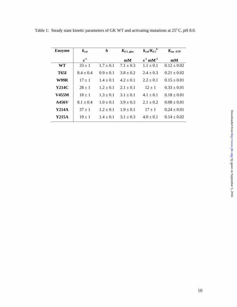
Steady-state kinetics of GK activating mutations. The central clinical feature of patients with HH as a result of GK activating mutations is low fasting glucose levels in the range of 1–3 mM compared to the normal 4–5.4 mM (8-11). To correlate the effects of activating mutations to the clinical consequence, steady-state kinetic properties of the recombinant activating mutations were characterized and compared to GK WT (Table 1). All of the activating mutations lowered the K0.5 for glucose and reduced the positive cooperativity of glucose as described by a decrease in Hill number (h). As a result, the catalytic efficiency with regards to glucose, kcat/ h
0.5K , is enhanced for the activating mutations. The Km values for ATP changed by 0.7–2.5 fold for the mutations.
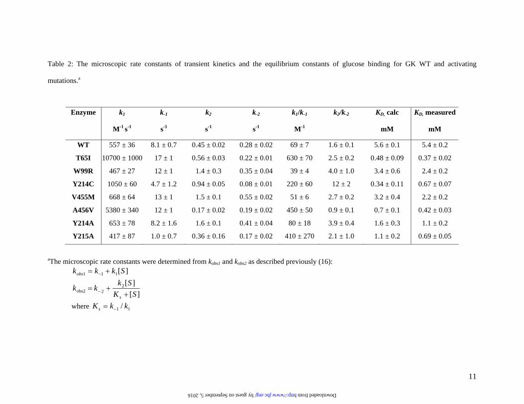
The kcat values for some of the mutations determined here were 2–3 fold lower than the reported results in earlier studies (8-11). The underlying cause of the discrepancy is unknown, but may be attributable to the experimental conditions, such as temperature and pH, or the GK construct since we used a hexa-histidine tag while previous reports utilized a GST-tag (8-11). Because of the large size of the GST-tag (26 kDa), we chose a 6-His-tag to minimize the potential disturbance of the native conformation of GK proteins. Identical kinetic properties were observed between the non-tagged and hexa-his-tagged GK enzymes (data not shown). Effects of GK activating mutations on glucose binding. Previous transient kinetic studies of glucose binding by stopped-flow spectrophotometry demonstrated that glucose binding to GK WT followed biphasic kinetics that fit best to a two-step reversible mechanism (16). The first kinetic phase was interpreted as a bi-molecular event where glucose loosely binds to the apo-enzyme form E*, followed by a second kinetic phase of enzyme conformational change from E*·S to E·S. The kinetic scheme was reported as follows: All of the activating mutations retained the two-step mechanism of glucose binding, but altered the individual microscopic rate constants (Table 2). Some mutations enhanced the first step of E*·glucose formation, as evidenced by either
E* + S E* • S E • S k1
k-1
k2
k-2
by guest on September 5, 2016
http://ww
w.jbc.org/
Dow
nloaded from
5
increasing k1 (observed in T65I and A456V) or decreasing k-1 (observed in Y215A). Some mutations favor E*·glucose to E·glucose transformation by increasing k2 (observed in W99R, Y214A, and V455M) or decreasing k-2 (observed in Y214C). The ratio of k1/k-1 and k2/k-2 represent the association constant of glucose binding to the apo enzyme E* and the degree of conversion of E*·S to E·S, respectively.
The overall equilibrium binding affinity of glucose can be calculated based on the individual microscopic rate constants of the two-step glucose binding using the following equation (22).
)k(k
kkkK
22
2
1
1D
−
−−
+×=
The results showed a good agreement with the experimentally determined KD (Table 2).
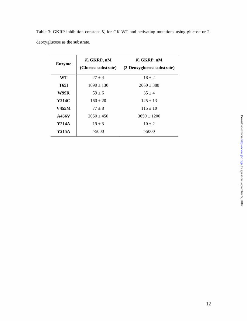
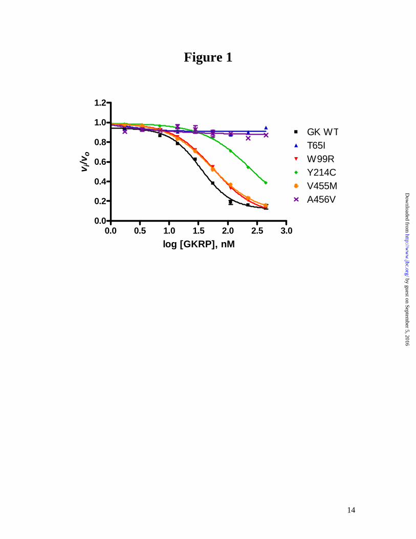
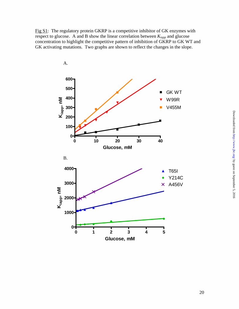
Based on the measured KD, T65I and A456V increased glucose binding the most (>10 fold), while W99R and V455M only increased the binding moderately (2–3 fold). Effects of GK activating mutations on GKRP inhibition. As shown in Figure 1, the naturally occurring GK activating mutations have different responses to GKRP inhibition at the physiological concentration of 5 mM glucose. At various glucose concentrations, the GK WT and mutations showed a glucose-dependent linear increase in IC50 value of GKRP, indicative of competitive inhibition of GKRP with respect to glucose (see Supplemental Data). It has been shown that GKRP is a competitive inhibitor for GK WT (20). To quantify the effects of the mutations on the protein-protein interaction with GKRP, the inhibition constant (Ki) of GKRP was determined for each mutation as shown in Table 3. W99R and Y214A retained similar binding interactions with GKRP compared to WT, whereas T65I, Y215A and A456V nearly abolished the GKRP interaction.
Because the GK WT and some of the mutations displayed sigmoidal kinetics for glucose that may complicate the inhibition data interpretation, the substrate analog 2-deoxyglucose was also utilized in the determination of Ki for GKRP since it exhibits little or no cooperativity for GK WT and the activating mutations. The Ki values obtained using both glucose and 2-deoxyglucose as the substrate were in close agreement with each other, consistent with a
similar competitive inhibition of GKRP by both sugar substrates (Table 3). The Ki value using glucose as the substrate was used for the rest of discussion.
DISCUSSION
The naturally-occurring GK activating mutations T65I, W99R, Y214C, V455M, and A456V were studied together with the engineered mutations Y214A and Y215A due to previous reports implicating the latter two residues as novel activating mutations (23,24). All of the activating mutations display an increased catalytic efficiency that is contributed mainly from the decrease in K0.5 for glucose. The 0.75–2.5 fold changes in the Km value of ATP for the activating mutations may not have significant biological consequence since the cellular ATP concentration is consistently maintained at mM ranges where the enzyme is nearly saturated by ATP. Due to its low affinity with glucose, GK does not operate at its maximal activity (kcat) under physiological conditions, therefore the catalytic efficiency, kcat/ h
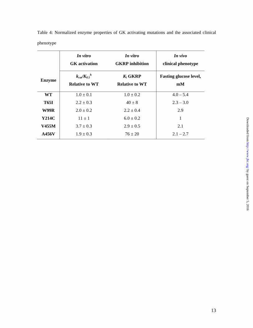
0.5K , may represent the most relevant rate constant of the enzyme’s activity. Correlation of in vitro biochemical properties with the clinical phenotype for GK activating mutations. Actions of GK in both glucose-dependent insulin regulation in β-cells and glucose disposal in the liver contribute to its role in maintaining glucose homeostasis. Therefore, to evaluate the correlation of the in vitro biochemical properties with the clinical phenotype, both effects of activating mutations on the enzyme activity and GKRP inhibitory interaction need to be considered. Table 4 summarizes the changes in the catalytic efficiency and the GKRP inhibition by the mutations relative to WT and the associated clinical phenotype.
Y214C is the most activating mutation with a catalytic efficiency of 11 fold higher than GK WT, and it also decreases the GKRP inhibitory interaction by 6 fold. In the clinical observations, the Y214C affected patient has the most severe hypoglycemia and hyperinsulinemia (9). Treatment with diazoxide and partial pancreatectomy did not relieve the symptoms, and eventually the patient’s brain was irreversibly damaged. Enlarged and hyperfunctional islets
by guest on September 5, 2016
http://ww
w.jbc.org/
Dow
nloaded from
6
were observed by pancreatic histology. This is consistent with the prediction by mathematical modeling based on in vitro analysis that Y214C has the lowest threshold for glucose stimulated insulin release (9). The patient also showed signs of accumulation of glycogen in the liver. This could be the result of both GK activation and decreased GKRP inhibitory regulation in the liver.
On the other hand, A456V moderately increases the catalytic efficiency, but decreases the GKRP binding by 76 fold. Significant glycogen storage in liver was observed in the patient by glucagon stimulation tests that showed rapid increases in blood glucose from low fasting levels. However, the significant disruption in GKRP inhibitory regulation in the liver does not translate into a severe clinical phenotype for A456V. Patients are either asymptomatic or can be treated by diazoxide, suggesting β-cell GK may be more essential in glucose homeostasis (8,9). This is consistent to the results of GK tissue specific knockout in mice. A β-cell specific knockout of GK in mice led to death within one week due to severe diabetes similar to the global GK knockout, while the liver specific GK knockout mice only experienced mild hyperglycemia but displayed pronounced defects in glycogen synthesis (25). Another possibility is that the alleviation of GK inhibition by GKRP in A456V may be offset by the loss of GK protection by GKRP from degradation by sequestering GK in the hepatocyte nucleus. A parallel loss of GK protein and activity has been observed in GKRP knockout mice (26). These results suggest that the alteration in GK enzyme activity has more profound biological impact than the change in GKRP binding.
The other 3 naturally occurring mutations T65I, W99R, and V455M have similar catalytic efficiency of 2–3 fold higher than WT. The fasting glucose levels are in the range of 2–3 mM for these patients, and the insulin levels vary (8,10,11). Although the functional characterization of GK activating mutations may explain some of the phenotype, the clinical symptoms and course of HH patients cover a broad spectrum from asymptomatic hypoglycemia to unconsciousness and seizures even within the same family, implicating a complex mechanism for GK regulation in vivo. Biochemical basis for GK activation and GKRP regulation for GK activating mutations. All of the
mutations studied here achieve their activation effects by enhancing overall glucose binding. The transient kinetic studies of glucose binding reveal that the mutations have a different biochemical basis for the increase in glucose binding affinity. The T65I, Y215A and A456V mutations mainly accelerate the initial bi-molecular interaction of glucose binding to the apo enzyme E* to form E*·S by an increase in the k1/k-1 ratio of 6–9 fold, while W99R, Y214A and V455M mainly enhance the enzyme conformational changes from E*·S to the catalytically competent E·S form by a moderate increase in the k2/k-2 ratio of 1.7–2.5 fold. Y214C has the most significant effect on k2/k-2 (7.5 fold relative to WT) and also moderate effect on k1/k-1 (3 fold relative to WT).
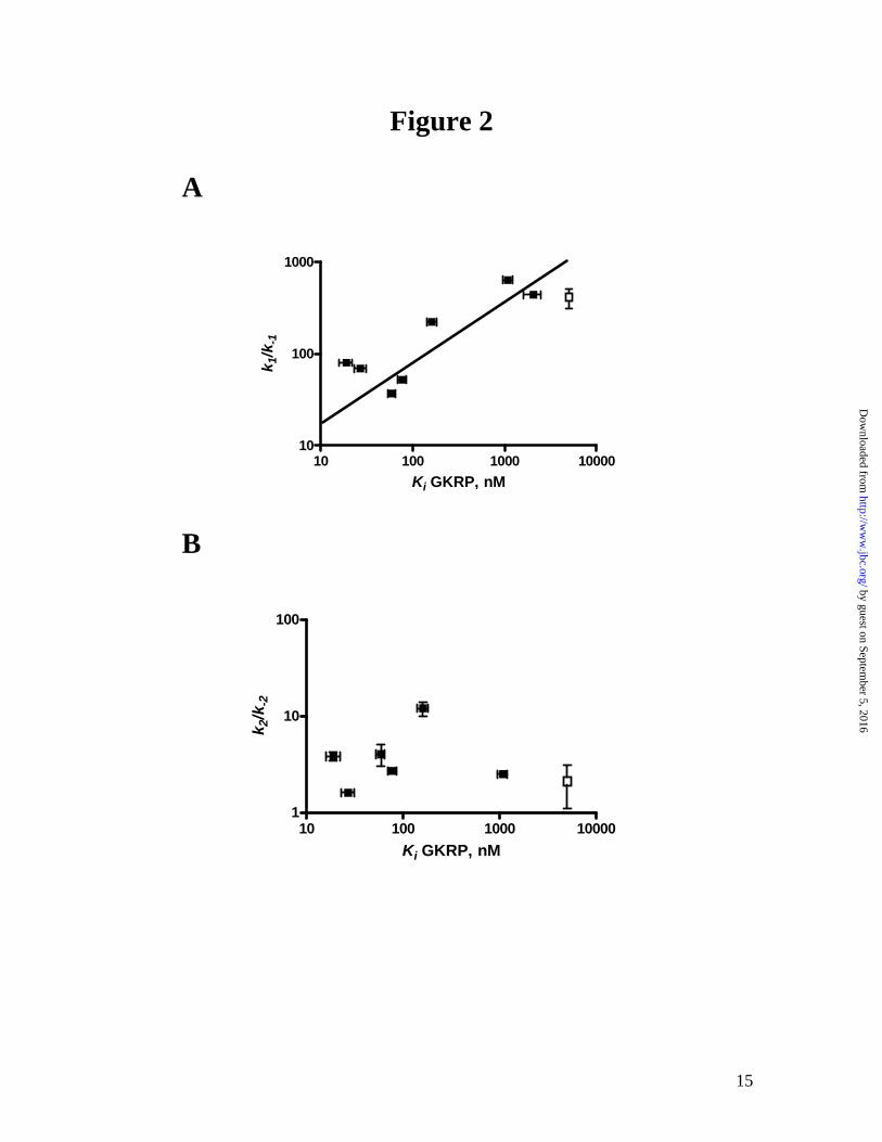
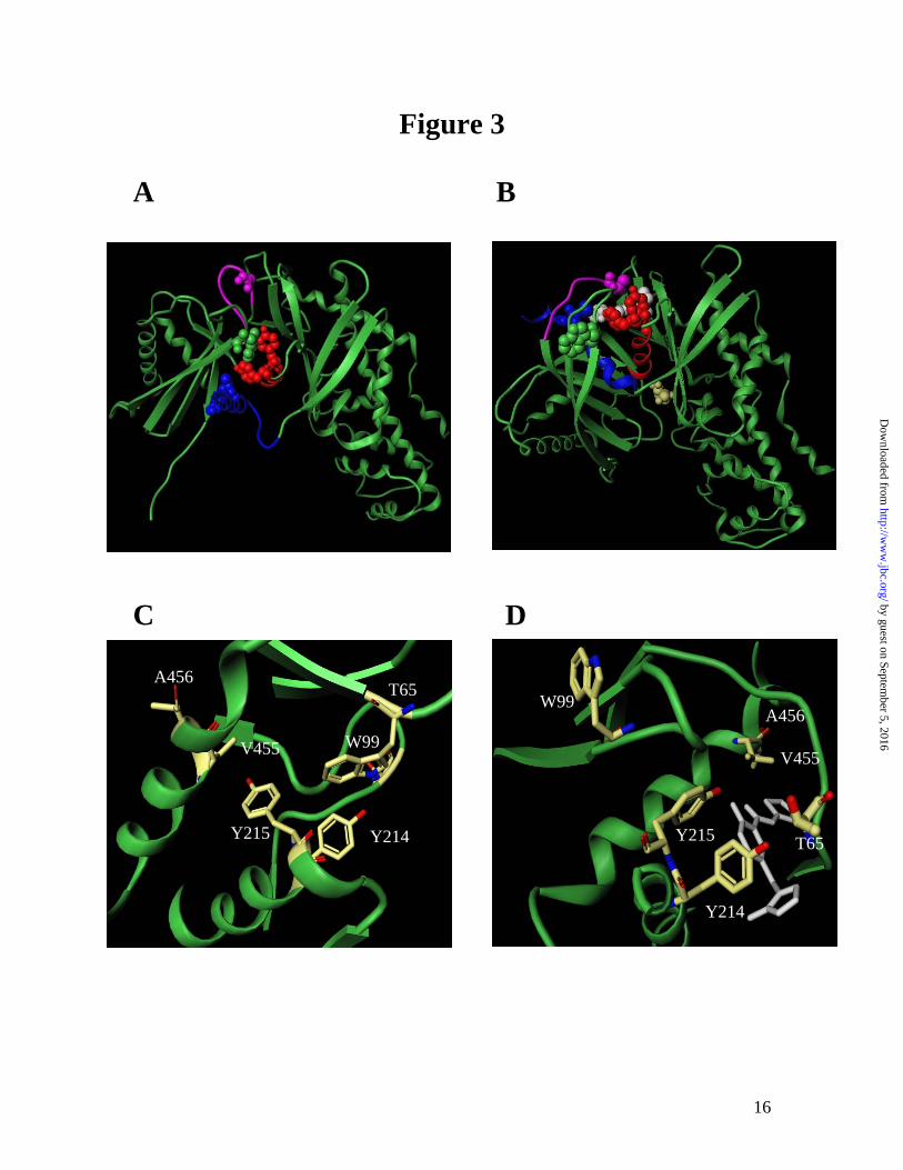
The different effects of the mutations on the two steps of glucose binding also reflect the protein-protein interaction with GKRP. One of the interesting observations here is the correlation between the k1/k-1 ratio in the transient kinetics of glucose binding and the Ki for GKRP as shown in Figure 2. GKRP binds to the super-open apo enzyme form competitively with respect to glucose. Since the k1/k-1 ratio describes the initial binding of glucose to the apo enzyme, a large increase in the k1/k-1 ratio suggests that these mutations (T65I, Y215A and A456V) adopt a more compact conformation in the apo enzyme which precludes GK-GKRP interaction. Mutations with small changes in the k1/k-1 ratio (W99R, V455M and Y214A) show minimal effects on GKRP interaction. Conversely, no such correlations between the slow conformational change step (k2/k-2) and Ki were observed. Implications of structure-function correlation in GK. The conformational changes in GK induced by glucose binding mainly involve the small domain through the rearrangement of the α13 helix and the connecting region between the large domain and small domain (Figure 3A and B). The domain closure forms the allosteric site and causes GKRP to no longer bind to the compact protein. GK mutations that alter the super-open apo conformation of GK may potentially disrupt a critical binding interface for the protein-protein interaction between GK and GKRP. Our findings suggest that T65, Y215 or A456 likely contributes to the integrity of the super-open conformation such that their mutation results in gross changes to
by guest on September 5, 2016
http://ww
w.jbc.org/
Dow
nloaded from
7
the overall protein structure, perhaps leading to a more compact form.
One interesting mutation that has the highest glucose binding affinity is T65I. It increases the on-rate k1 for glucose binding to the apo enzyme by nearly 20 fold and decreases the GKRP binding affinity by 40 fold. In addition, this mutation responds to further activation by GK activators, unlike most of the other activating mutations (Heredia and Sun, unpublished results). T65I is part of a highly flexible loop region that undergoes a substantial rearrangement going from the super-open to the closed form (Figure 3). The crystal structures suggest that the side chains of this loop region can adopt different spatial orientations in the presence of various small molecule GK activators suggesting a highly dynamic area (14,15). It is reasonable to hypothesize that the T65I mutation causes the protein to adopt a closed conformation that mimics the activated form of GK readily for glucose and activator binding. Further understanding of structure-function relation in T65I would be facilitated by the protein crystal structures.
The α5 and α13 helices are structurally important in the allosteric regulation of GK (24). V455 and A456 are adjacent residues located in the α13 helix. While the V455M mutation retains interaction with GKRP, A456V nearly abolishes GKRP binding. This is probably due to the difference in the spatial location of V455 versus A456 (Figure 3C). While V455 is buried in a hydrophobic pocket that includes W99, V101, I211, Y215 and L451, the sidechain of A456 is
mainly solvent exposed and accessible for protein interaction and may therefore play an important role in GKRP interaction. Y214A and Y215A show different effects on GKRP binding, where Y215A disrupts GKRP binding while Y214A has little effect. Y215 and Y214 are located in the α5 helix that forms the allosteric site and directly interact with GK activators (15). Y215 sits in a hydrophobic patch and forms a hydrogen bond with G72 in the apo-enzyme form, but loses most of its hydrophobic interactions in the closed form of the enzyme, while Y214 is located distal from the hydrophobic patch where Y215 is embedded. The hydrophobic interaction of Y215 may stabilize the super-open apo form which is thought to bind to GKRP. The Y215A mutation may therefore destabilize the apo form and prevent GKRP binding. The human GK activating mutations all show decreased fasting blood glucose in vivo and increased glucose binding affinity in vitro. The kinetic basis for the increased glucose binding differs among mutations as a result of their structural perturbation at the allosteric site. The first step of GK activation, formation of the loosely bound E*·S complex as described by k1/k-1 is shown to correlate with the level of GKRP inhibition. This set of activating mutations provides insight regarding the interrelation of kinetic parameters of GK, GKRP binding and the resulting clinical effects of altering the glucose sensor activity of GK.
REFERENCES 1. Cardenas, M. L. (1995) Glucokinase: Its Regulation and Role in Liver Metabolism, RG Landes,
Austin, Texas 2. Matschinsky, F. M. (1990) Diabetes 39, 647-652 3. Matschinsky, F. M. (2005) Curr Diab Rep 5, 171-176 4. Niemeyer, H., de la Luz Cardenas, M., Rabajille, E., Ureta, T., Clark-Turri, L., and Penaranda, J.
(1975) Enzyme 20, 321-333 5. Parry, M. J., and Walker, D. G. (1966) Biochem J 99, 266-274 6. Storer, A. C., and Cornish-Bowden, A. (1976) Biochem J 159, 7-14 7. van Schaftingen, E., Vandercammen, A., Detheux, M., and Davies, D. R. (1992) Adv Enzyme
Regul 32, 133-148 8. Christesen, H. B., Jacobsen, B. B., Odili, S., Buettger, C., Cuesta-Munoz, A., Hansen, T.,
Brusgaard, K., Massa, O., Magnuson, M. A., Shiota, C., Matschinsky, F. M., and Barbetti, F. (2002) Diabetes 51, 1240-1246
by guest on September 5, 2016
http://ww
w.jbc.org/
Dow
nloaded from
8
9. Cuesta-Munoz, A. L., Huopio, H., Otonkoski, T., Gomez-Zumaquero, J. M., Nanto-Salonen, K., Rahier, J., Lopez-Enriquez, S., Garcia-Gimeno, M. A., Sanz, P., Soriguer, F. C., and Laakso, M. (2004) Diabetes 53, 2164-2168
10. Glaser, B., Kesavan, P., Heyman, M., Davis, E., Cuesta, A., Buchs, A., Stanley, C. A., Thornton, P. S., Permutt, M. A., Matschinsky, F. M., and Herold, K. C. (1998) N Engl J Med 338, 226-230
11. Gloyn, A. L., Noordam, K., Willemsen, M. A., Ellard, S., Lam, W. W., Campbell, I. W., Midgley, P., Shiota, C., Buettger, C., Magnuson, M. A., Matschinsky, F. M., and Hattersley, A. T. (2003) Diabetes 52, 2433-2440
12. Grimsby, J., Sarabu, R., Corbett, W. L., Haynes, N. E., Bizzarro, F. T., Coffey, J. W., Guertin, K. R., Hilliard, D. W., Kester, R. F., Mahaney, P. E., Marcus, L., Qi, L., Spence, C. L., Tengi, J., Magnuson, M. A., Chu, C. A., Dvorozniak, M. T., Matschinsky, F. M., and Grippo, J. F. (2003) Science 301, 370-373
13. Efanov, A. M., Barrett, D. G., Brenner, M. B., Briggs, S. L., Delaunois, A., Durbin, J. D., Giese, U., Guo, H., Radloff, M., Gil, G. S., Sewing, S., Wang, Y., Weichert, A., Zaliani, A., and Gromada, J. (2005) Endocrinology 146, 3696-3701
14. Brocklehurst, K. J., Payne, V. A., Davies, R. A., Carroll, D., Vertigan, H. L., Wightman, H. J., Aiston, S., Waddell, I. D., Leighton, B., Coghlan, M. P., and Agius, L. (2004) Diabetes 53, 535-541
15. Kamata, K., Mitsuya, M., Nishimura, T., Eiki, J., and Nagata, Y. (2004) Structure (Camb) 12, 429-438
16. Heredia, V. V., Thomson, J., Nettleton, D., and Sun, S. (2006) Biochemistry 45, 7553-7562 17. Nishi, S., Stoffel, M., Xiang, K., Shows, T. B., Bell, G. I., and Takeda, J. (1992) Diabetologia 35,
743-747 18. Bradford, M. M. (1976) Anal Biochem 72, 248-254 19. Mookhtiar, K. A., Kalinowski, S. S., Brown, K. S., Tsay, Y. H., Smith-Monroy, C., and
Robinson, G. W. (1996) Diabetes 45, 1670-1677 20. Vandercammen, A., and Van Schaftingen, E. (1991) Eur J Biochem 200, 545-551 21. Morrison, J. F. (1969) Biochim Biophys Acta 185, 269-286 22. Lin, S. X., and Neet, K. A. (1990) J Biol Chem 265, 9670-9675 23. Moukil, M. A., Veiga-da-Cunha, M., and Van Schaftingen, E. (2000) Diabetes 49, 195-201 24. Pedelini, L., Garcia-Gimeno, M. A., Marina, A., Gomez-Zumaquero, J. M., Rodriguez-Bada, P.,
Lopez-Enriquez, S., Soriguer, F. C., Cuesta-Munoz, A. L., and Sanz, P. (2005) Protein Sci 14, 2080-2086
25. Grupe, A., Hultgren, B., Ryan, A., Ma, Y. H., Bauer, M., and Stewart, T. A. (1995) Cell 83, 69-78
26. Farrelly, D., Brown, K. S., Tieman, A., Ren, J., Lira, S. A., Hagan, D., Gregg, R., Mookhtiar, K. A., and Hariharan, N. (1999) Proc Natl Acad Sci U S A 96, 14511-14516
FOOTNOTES
We thank Janice Chin for kindly providing the GK WT and GKRP clones. We also thank Steve Grant and Jen Digits for critical reading of the manuscript and insightful discussion, and Junli Feng, Caroline Rodgers and Ketan Gajiwala for expert technical assistance. The on-line version of this article (available at http://www.jbc.org) contains supplemental information. The abbreviations used are: GK, glucokinase; GKRP, glucokinase regulatory protein; WT, wild type; MODY, maturity onset diabetes of the young; HH, hyperinsulinemic hypoglycemia; PK/LDH, pyruvate kinase/lactate dehydrogenase; QTOF-MS, quadrupole time-of-flight mass spectrometry; PEP, phosphoenolpyruvate; DTT, dithiothreitol; ATP, adenosine triphosphate; SDS-PAGE, sodium dodecyl sulfate-polyacrylamide gel electrophoresis.
by guest on September 5, 2016
http://ww
w.jbc.org/
Dow
nloaded from
9
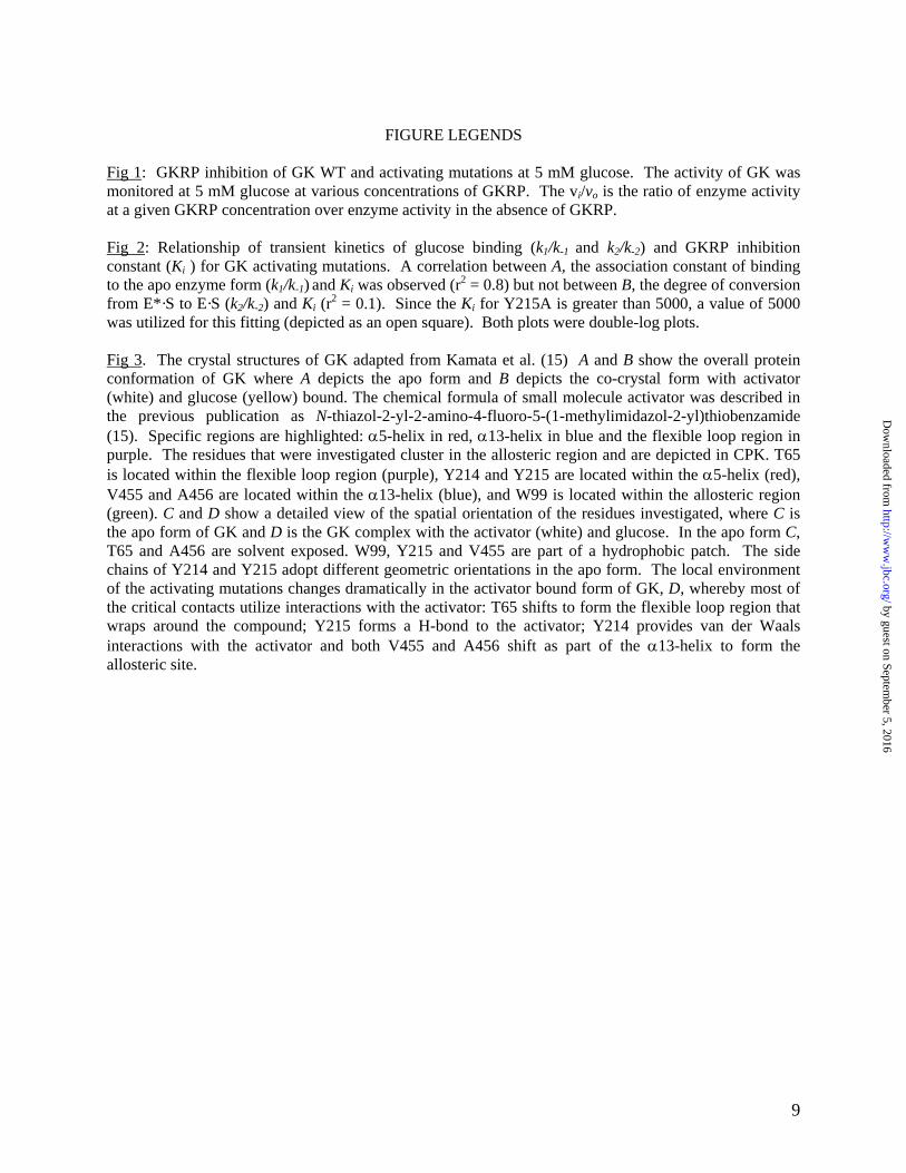
FIGURE LEGENDS
Fig 1: GKRP inhibition of GK WT and activating mutations at 5 mM glucose. The activity of GK was monitored at 5 mM glucose at various concentrations of GKRP. The vi/vo is the ratio of enzyme activity at a given GKRP concentration over enzyme activity in the absence of GKRP. Fig 2: Relationship of transient kinetics of glucose binding (k1/k-1 and k2/k-2) and GKRP inhibition constant (Ki ) for GK activating mutations. A correlation between A, the association constant of binding to the apo enzyme form (k1/k-1) and Ki was observed (r2 = 0.8) but not between B, the degree of conversion from E*·S to E·S (k2/k-2) and Ki (r2 = 0.1). Since the Ki for Y215A is greater than 5000, a value of 5000 was utilized for this fitting (depicted as an open square). Both plots were double-log plots. Fig 3. The crystal structures of GK adapted from Kamata et al. (15) A and B show the overall protein conformation of GK where A depicts the apo form and B depicts the co-crystal form with activator (white) and glucose (yellow) bound. The chemical formula of small molecule activator was described in the previous publication as N-thiazol-2-yl-2-amino-4-fluoro-5-(1-methylimidazol-2-yl)thiobenzamide (15). Specific regions are highlighted: α5-helix in red, α13-helix in blue and the flexible loop region in purple. The residues that were investigated cluster in the allosteric region and are depicted in CPK. T65 is located within the flexible loop region (purple), Y214 and Y215 are located within the α5-helix (red), V455 and A456 are located within the α13-helix (blue), and W99 is located within the allosteric region (green). C and D show a detailed view of the spatial orientation of the residues investigated, where C is the apo form of GK and D is the GK complex with the activator (white) and glucose. In the apo form C, T65 and A456 are solvent exposed. W99, Y215 and V455 are part of a hydrophobic patch. The side chains of Y214 and Y215 adopt different geometric orientations in the apo form. The local environment of the activating mutations changes dramatically in the activator bound form of GK, D, whereby most of the critical contacts utilize interactions with the activator: T65 shifts to form the flexible loop region that wraps around the compound; Y215 forms a H-bond to the activator; Y214 provides van der Waals interactions with the activator and both V455 and A456 shift as part of the α13-helix to form the allosteric site.
by guest on September 5, 2016
http://ww
w.jbc.org/
Dow
nloaded from
10
Table 1: Steady state kinetic parameters of GK WT and activating mutations at 25o C, pH 8.0.
Enzyme kcat
s-1
h K0.5, gluc
mM
kcat/K0.5h
s-1 mM-1
Km, ATP
mM WT 33 ± 1 1.7 ± 0.1 7.1 ± 0.3 1.1 ± 0.1 0.12 ± 0.02
T65I 8.4 ± 0.4 0.9 ± 0.1 3.8 ± 0.2 2.4 ± 0.3 0.21 ± 0.02
W99R 17 ± 1 1.4 ± 0.1 4.2 ± 0.1 2.2 ± 0.1 0.15 ± 0.01
Y214C 28 ± 1 1.2 ± 0.1 2.1 ± 0.1 12 ± 1 0.33 ± 0.01
V455M 18 ± 1 1.3 ± 0.1 3.1 ± 0.1 4.1 ± 0.1 0.18 ± 0.01
A456V 8.1 ± 0.4 1.0 ± 0.1 3.9 ± 0.3 2.1 ± 0.2 0.08 ± 0.01
Y214A 37 ± 1 1.2 ± 0.1 1.9 ± 0.1 17 ± 1 0.24 ± 0.01
Y215A 19 ± 1 1.4 ± 0.1 3.1 ± 0.3 4.0 ± 0.1 0.14 ± 0.02
by guest on September 5, 2016
http://ww
w.jbc.org/
Dow
nloaded from
11
Table 2: The microscopic rate constants of transient kinetics and the equilibrium constants of glucose binding for GK WT and activating
mutations.a
aThe microscopic rate constants were determined from kobs1 and kobs2 as described previously (16): ][111 Skkkobs += −
][
][ 2
2obs2 SKSk
kks +
+= −
where 11 / kkKs −=
Enzyme k1
M-1 s-1
k-1
s-1
k2
s-1
k-2
s-1
k1/k-1
M-1
k2/k-2
KD, calc
mM
KD, measured
mM
WT 557 ± 36 8.1 ± 0.7 0.45 ± 0.02 0.28 ± 0.02 69 ± 7 1.6 ± 0.1 5.6 ± 0.1 5.4 ± 0.2
T65I 10700 ± 1000 17 ± 1 0.56 ± 0.03 0.22 ± 0.01 630 ± 70 2.5 ± 0.2 0.48 ± 0.09 0.37 ± 0.02
W99R 467 ± 27 12 ± 1 1.4 ± 0.3 0.35 ± 0.04 39 ± 4 4.0 ± 1.0 3.4 ± 0.6 2.4 ± 0.2
Y214C 1050 ± 60 4.7 ± 1.2 0.94 ± 0.05 0.08 ± 0.01 220 ± 60 12 ± 2 0.34 ± 0.11 0.67 ± 0.07
V455M 668 ± 64 13 ± 1 1.5 ± 0.1 0.55 ± 0.02 51 ± 6 2.7 ± 0.2 3.2 ± 0.4 2.2 ± 0.2
A456V 5380 ± 340 12 ± 1 0.17 ± 0.02 0.19 ± 0.02 450 ± 50 0.9 ± 0.1 0.7 ± 0.1 0.42 ± 0.03
Y214A 653 ± 78 8.2 ± 1.6 1.6 ± 0.1 0.41 ± 0.04 80 ± 18 3.9 ± 0.4 1.6 ± 0.3 1.1 ± 0.2
Y215A 417 ± 87 1.0 ± 0.7 0.36 ± 0.16 0.17 ± 0.02 410 ± 270 2.1 ± 1.0 1.1 ± 0.2 0.69 ± 0.05
by guest on September 5, 2016 http://www.jbc.org/ Downloaded from
12
Table 3: GKRP inhibition constant Ki for GK WT and activating mutations using glucose or 2-
deoxyglucose as the substrate.
Enzyme Ki GKRP, nM
(Glucose substrate)
Ki GKRP, nM
(2-Deoxyglucose substrate)
WT 27 ± 4 18 ± 2
T65I 1090 ± 130 2050 ± 380
W99R 59 ± 6 35 ± 4
Y214C 160 ± 20 125 ± 13
V455M 77 ± 8 115 ± 10
A456V 2050 ± 450 3650 ± 1200
Y214A 19 ± 3 10 ± 2
Y215A >5000 >5000
by guest on September 5, 2016
http://ww
w.jbc.org/
Dow
nloaded from
13
Table 4: Normalized enzyme properties of GK activating mutations and the associated clinical
phenotype
In vitro
GK activation
In vitro
GKRP inhibition
In vivo
clinical phenotype
Enzyme kcat/K0.5
h
Relative to WT
Ki GKRP
Relative to WT
Fasting glucose level,
mM
WT 1.0 ± 0.1 1.0 ± 0.2 4.0 – 5.4
T65I 2.2 ± 0.3 40 ± 8 2.3 – 3.0
W99R 2.0 ± 0.2 2.2 ± 0.4 2.9
Y214C 11 ± 1 6.0 ± 0.2 1
V455M 3.7 ± 0.3 2.9 ± 0.5 2.1
A456V 1.9 ± 0.3 76 ± 20 2.1 – 2.7
by guest on September 5, 2016
http://ww
w.jbc.org/
Dow
nloaded from
14
Figure 1
0.0 0.5 1.0 1.5 2.0 2.5 3.00.0
0.2
0.4
0.6
0.8
1.0
1.2
GK WTT65IW99RY214CV455MA456V
log [GKRP], nM
v i/v
o
by guest on September 5, 2016
http://ww
w.jbc.org/
Dow
nloaded from
15
Figure 2
A
10 100 1000 1000010
100
1000
Ki GKRP, nM
k 1/k
-1
B
10 100 1000 100001
10
100
Ki GKRP, nM
k 2/k
-2
by guest on September 5, 2016
http://ww
w.jbc.org/
Dow
nloaded from
16
Figure 3
A B
C D
T65
V455
A456
Y214
W99
Y215
W99
Y215
Y214
A456
T65
V455
by guest on September 5, 2016
http://ww
w.jbc.org/
Dow
nloaded from
17
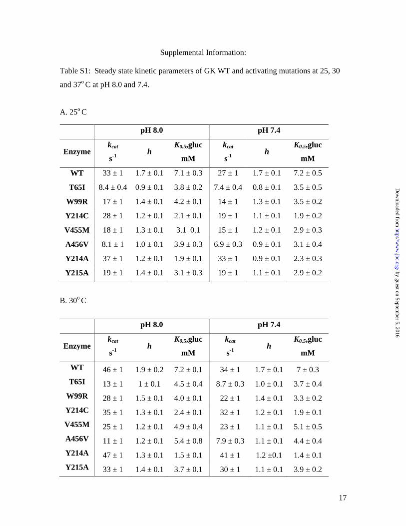
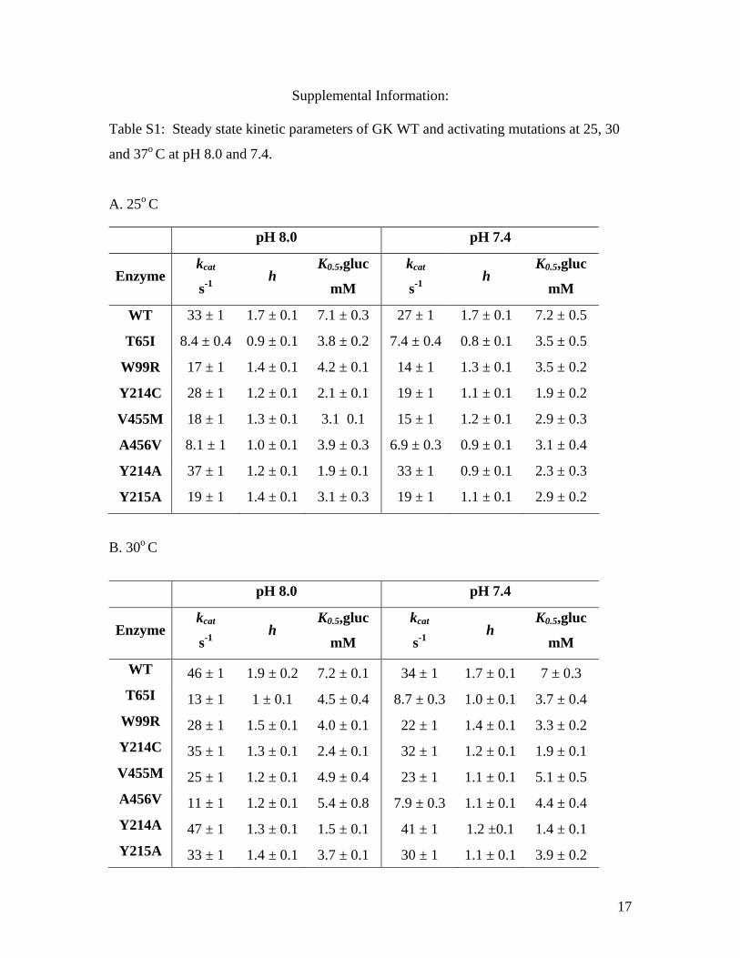
Supplemental Information: Table S1: Steady state kinetic parameters of GK WT and activating mutations at 25, 30
and 37o C at pH 8.0 and 7.4.
A. 25o C
B. 30o C
pH 8.0 pH 7.4
Enzyme kcat
s-1 h
K0.5,gluc
mM
kcat
s-1 h
K0.5,gluc
mM
WT 33 ± 1 1.7 ± 0.1 7.1 ± 0.3 27 ± 1 1.7 ± 0.1 7.2 ± 0.5
T65I 8.4 ± 0.4 0.9 ± 0.1 3.8 ± 0.2 7.4 ± 0.4 0.8 ± 0.1 3.5 ± 0.5
W99R 17 ± 1 1.4 ± 0.1 4.2 ± 0.1 14 ± 1 1.3 ± 0.1 3.5 ± 0.2
Y214C 28 ± 1 1.2 ± 0.1 2.1 ± 0.1 19 ± 1 1.1 ± 0.1 1.9 ± 0.2
V455M 18 ± 1 1.3 ± 0.1 3.1 0.1 15 ± 1 1.2 ± 0.1 2.9 ± 0.3
A456V 8.1 ± 1 1.0 ± 0.1 3.9 ± 0.3 6.9 ± 0.3 0.9 ± 0.1 3.1 ± 0.4
Y214A 37 ± 1 1.2 ± 0.1 1.9 ± 0.1 33 ± 1 0.9 ± 0.1 2.3 ± 0.3
Y215A 19 ± 1 1.4 ± 0.1 3.1 ± 0.3 19 ± 1 1.1 ± 0.1 2.9 ± 0.2
pH 8.0 pH 7.4
Enzyme kcat
s-1 h
K0.5,gluc
mM
kcat
s-1 h
K0.5,gluc
mM
WT 46 ± 1 1.9 ± 0.2 7.2 ± 0.1 34 ± 1 1.7 ± 0.1 7 ± 0.3 T65I 13 ± 1 1 ± 0.1 4.5 ± 0.4 8.7 ± 0.3 1.0 ± 0.1 3.7 ± 0.4
W99R 28 ± 1 1.5 ± 0.1 4.0 ± 0.1 22 ± 1 1.4 ± 0.1 3.3 ± 0.2 Y214C 35 ± 1 1.3 ± 0.1 2.4 ± 0.1 32 ± 1 1.2 ± 0.1 1.9 ± 0.1 V455M 25 ± 1 1.2 ± 0.1 4.9 ± 0.4 23 ± 1 1.1 ± 0.1 5.1 ± 0.5 A456V 11 ± 1 1.2 ± 0.1 5.4 ± 0.8 7.9 ± 0.3 1.1 ± 0.1 4.4 ± 0.4 Y214A 47 ± 1 1.3 ± 0.1 1.5 ± 0.1 41 ± 1 1.2 ±0.1 1.4 ± 0.1 Y215A 33 ± 1 1.4 ± 0.1 3.7 ± 0.1 30 ± 1 1.1 ± 0.1 3.9 ± 0.2
by guest on September 5, 2016
http://ww
w.jbc.org/
Dow
nloaded from
18
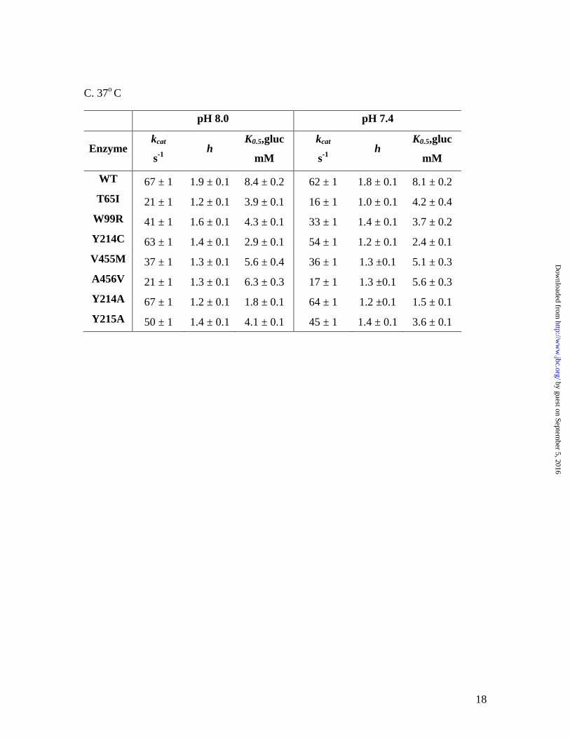
C. 37o C
pH 8.0 pH 7.4
Enzyme kcat
s-1 h
K0.5,gluc
mM
kcat
s-1 h
K0.5,gluc
mM
WT 67 ± 1 1.9 ± 0.1 8.4 ± 0.2 62 ± 1 1.8 ± 0.1 8.1 ± 0.2 T65I 21 ± 1 1.2 ± 0.1 3.9 ± 0.1 16 ± 1 1.0 ± 0.1 4.2 ± 0.4
W99R 41 ± 1 1.6 ± 0.1 4.3 ± 0.1 33 ± 1 1.4 ± 0.1 3.7 ± 0.2 Y214C 63 ± 1 1.4 ± 0.1 2.9 ± 0.1 54 ± 1 1.2 ± 0.1 2.4 ± 0.1 V455M 37 ± 1 1.3 ± 0.1 5.6 ± 0.4 36 ± 1 1.3 ±0.1 5.1 ± 0.3 A456V 21 ± 1 1.3 ± 0.1 6.3 ± 0.3 17 ± 1 1.3 ±0.1 5.6 ± 0.3 Y214A 67 ± 1 1.2 ± 0.1 1.8 ± 0.1 64 ± 1 1.2 ±0.1 1.5 ± 0.1 Y215A 50 ± 1 1.4 ± 0.1 4.1 ± 0.1 45 ± 1 1.4 ± 0.1 3.6 ± 0.1
by guest on September 5, 2016
http://ww
w.jbc.org/
Dow
nloaded from
19
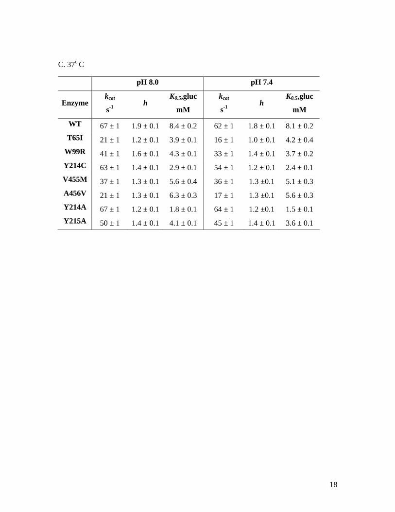
Table S2: Steady state kinetic parameters of GK using 2-deoxyglucose as substrate at 25o
C and pH 8.0
Enzyme kcat
s-1 h
K0.5,2DG
mM
WT 30 ± 1 1.0 ± 0.1 153 ± 19
T65I 7.3 ± 0.2 1.2 ± 0.1 24 ± 2
W99R 16 ± 0.2 1.1 ± 0.1 44 ± 1
Y214C 29 ± 1 1.0 ± 0.1 26 ± 1
V455M 17 ± 0.3 1.1 ± 0.1 42 ± 2
A456V 7.2 ± 0.2 1.1 ± 0.1 53 ± 4
Y214A 33 ± 0.8 1.0 ± 0.1 20 ± 1
Y215A 19 ± 0.3 1.2 ± 0.1 28 ± 1
by guest on September 5, 2016
http://ww
w.jbc.org/
Dow
nloaded from
20
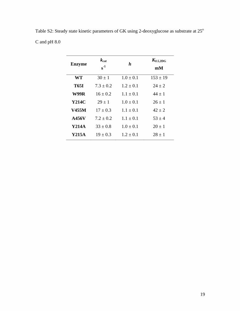
Fig S1: The regulatory protein GKRP is a competitive inhibitor of GK enzymes with respect to glucose. A and B show the linear correlation between Kiapp and glucose concentration to highlight the competitive pattern of inhibition of GKRP to GK WT and GK activating mutations. Two graphs are shown to reflect the changes in the slope.
A.
0 10 20 30 400
100
200
300
400
500
600
GK WTW99RV455M
Glucose, mM
Kia
pp, n
M
B.
0 1 2 3 4 50
1000
2000
3000
4000 T65IY214CA456V
Glucose, mM
Kia
pp, n
M
by guest on September 5, 2016
http://ww
w.jbc.org/
Dow
nloaded from
17
Supplemental Information: Table S1: Steady state kinetic parameters of GK WT and activating mutations at 25, 30
and 37o C at pH 8.0 and 7.4.
A. 25o C
B. 30o C
pH 8.0 pH 7.4
Enzyme kcat
s-1 h
K0.5,gluc
mM
kcat
s-1 h
K0.5,gluc
mM
WT 33 ± 1 1.7 ± 0.1 7.1 ± 0.3 27 ± 1 1.7 ± 0.1 7.2 ± 0.5
T65I 8.4 ± 0.4 0.9 ± 0.1 3.8 ± 0.2 7.4 ± 0.4 0.8 ± 0.1 3.5 ± 0.5
W99R 17 ± 1 1.4 ± 0.1 4.2 ± 0.1 14 ± 1 1.3 ± 0.1 3.5 ± 0.2
Y214C 28 ± 1 1.2 ± 0.1 2.1 ± 0.1 19 ± 1 1.1 ± 0.1 1.9 ± 0.2
V455M 18 ± 1 1.3 ± 0.1 3.1 0.1 15 ± 1 1.2 ± 0.1 2.9 ± 0.3
A456V 8.1 ± 1 1.0 ± 0.1 3.9 ± 0.3 6.9 ± 0.3 0.9 ± 0.1 3.1 ± 0.4
Y214A 37 ± 1 1.2 ± 0.1 1.9 ± 0.1 33 ± 1 0.9 ± 0.1 2.3 ± 0.3
Y215A 19 ± 1 1.4 ± 0.1 3.1 ± 0.3 19 ± 1 1.1 ± 0.1 2.9 ± 0.2
pH 8.0 pH 7.4
Enzyme kcat
s-1 h
K0.5,gluc
mM
kcat
s-1 h
K0.5,gluc
mM
WT 46 ± 1 1.9 ± 0.2 7.2 ± 0.1 34 ± 1 1.7 ± 0.1 7 ± 0.3 T65I 13 ± 1 1 ± 0.1 4.5 ± 0.4 8.7 ± 0.3 1.0 ± 0.1 3.7 ± 0.4
W99R 28 ± 1 1.5 ± 0.1 4.0 ± 0.1 22 ± 1 1.4 ± 0.1 3.3 ± 0.2 Y214C 35 ± 1 1.3 ± 0.1 2.4 ± 0.1 32 ± 1 1.2 ± 0.1 1.9 ± 0.1 V455M 25 ± 1 1.2 ± 0.1 4.9 ± 0.4 23 ± 1 1.1 ± 0.1 5.1 ± 0.5 A456V 11 ± 1 1.2 ± 0.1 5.4 ± 0.8 7.9 ± 0.3 1.1 ± 0.1 4.4 ± 0.4 Y214A 47 ± 1 1.3 ± 0.1 1.5 ± 0.1 41 ± 1 1.2 ±0.1 1.4 ± 0.1 Y215A 33 ± 1 1.4 ± 0.1 3.7 ± 0.1 30 ± 1 1.1 ± 0.1 3.9 ± 0.2
18
C. 37o C
pH 8.0 pH 7.4
Enzyme kcat
s-1 h
K0.5,gluc
mM
kcat
s-1 h
K0.5,gluc
mM
WT 67 ± 1 1.9 ± 0.1 8.4 ± 0.2 62 ± 1 1.8 ± 0.1 8.1 ± 0.2 T65I 21 ± 1 1.2 ± 0.1 3.9 ± 0.1 16 ± 1 1.0 ± 0.1 4.2 ± 0.4
W99R 41 ± 1 1.6 ± 0.1 4.3 ± 0.1 33 ± 1 1.4 ± 0.1 3.7 ± 0.2 Y214C 63 ± 1 1.4 ± 0.1 2.9 ± 0.1 54 ± 1 1.2 ± 0.1 2.4 ± 0.1 V455M 37 ± 1 1.3 ± 0.1 5.6 ± 0.4 36 ± 1 1.3 ±0.1 5.1 ± 0.3 A456V 21 ± 1 1.3 ± 0.1 6.3 ± 0.3 17 ± 1 1.3 ±0.1 5.6 ± 0.3 Y214A 67 ± 1 1.2 ± 0.1 1.8 ± 0.1 64 ± 1 1.2 ±0.1 1.5 ± 0.1 Y215A 50 ± 1 1.4 ± 0.1 4.1 ± 0.1 45 ± 1 1.4 ± 0.1 3.6 ± 0.1
19
Table S2: Steady state kinetic parameters of GK using 2-deoxyglucose as substrate at 25o
C and pH 8.0
Enzyme kcat
s-1 h
K0.5,2DG
mM
WT 30 ± 1 1.0 ± 0.1 153 ± 19
T65I 7.3 ± 0.2 1.2 ± 0.1 24 ± 2
W99R 16 ± 0.2 1.1 ± 0.1 44 ± 1
Y214C 29 ± 1 1.0 ± 0.1 26 ± 1
V455M 17 ± 0.3 1.1 ± 0.1 42 ± 2
A456V 7.2 ± 0.2 1.1 ± 0.1 53 ± 4
Y214A 33 ± 0.8 1.0 ± 0.1 20 ± 1
Y215A 19 ± 0.3 1.2 ± 0.1 28 ± 1
20
Fig S1: The regulatory protein GKRP is a competitive inhibitor of GK enzymes with respect to glucose. A and B show the linear correlation between Kiapp and glucose concentration to highlight the competitive pattern of inhibition of GKRP to GK WT and GK activating mutations. Two graphs are shown to reflect the changes in the slope.
A.
0 10 20 30 400
100
200
300
400
500
600
GK WTW99RV455M
Glucose, mM
Kia
pp, n
M
B.
0 1 2 3 4 50
1000
2000
3000
4000 T65IY214CA456V
Glucose, mM
Kia
pp, n
M
Vladi V. Heredia, Thomas J. Carlson, Erin Garcia and Shaoxian Sunregulatory protein in naturally occuring mutations
Biochemical basis of glucokinase activation and the regulation by glucokinase
published online November 2, 2006J. Biol. Chem.
10.1074/jbc.M607987200Access the most updated version of this article at doi:
Alerts:
When a correction for this article is posted•
When this article is cited•
to choose from all of JBC's e-mail alertsClick here
Supplemental material:
http://www.jbc.org/content/suppl/2006/11/02/M607987200.DC1.html
http://www.jbc.org/content/early/2006/11/02/jbc.M607987200.citation.full.html#ref-list-1
This article cites 0 references, 0 of which can be accessed free at
by guest on September 5, 2016
http://ww
w.jbc.org/
Dow
nloaded from
Related Documents