BIOC372x | AMN | 2014 34 Lecture L02b Surveying the Cells & Organs of the Immune System, part b I can assure you that peace will not be built on poor nutrition and human suffering. - Norman Borlag, 11/19/01 (from talk at Rice University)

Welcome message from author
This document is posted to help you gain knowledge. Please leave a comment to let me know what you think about it! Share it to your friends and learn new things together.
Transcript
BIOC372x | AMN | 2014 34
Lecture L02b Surveying the Cells & Organs of the Immune System, part b
I can assure you that peace will not be built on poor nutrition and human suffering. - Norman Borlag, 11/19/01 (from talk at
Rice University)

BIOC372x | AMN | 2014 35
I. Primary Organs of the Immune System
A. Bone Marrow (Figure 2B.1)
1. Location of HSCs, myeloid cell production, and initial division of lymphoid cells.
2. NK cells rise from here and B cells divide and rearrange their genes here.
3. T cells undergo initial commitment here, but then leave for the thymus to finish
rearranging genes and determining their specific roles.
4. Marrow of femur, humerus, hip bones and sternum are major sites.
Figure 2B.1: Bone Marrow
BIOC372x | AMN | 2014 36
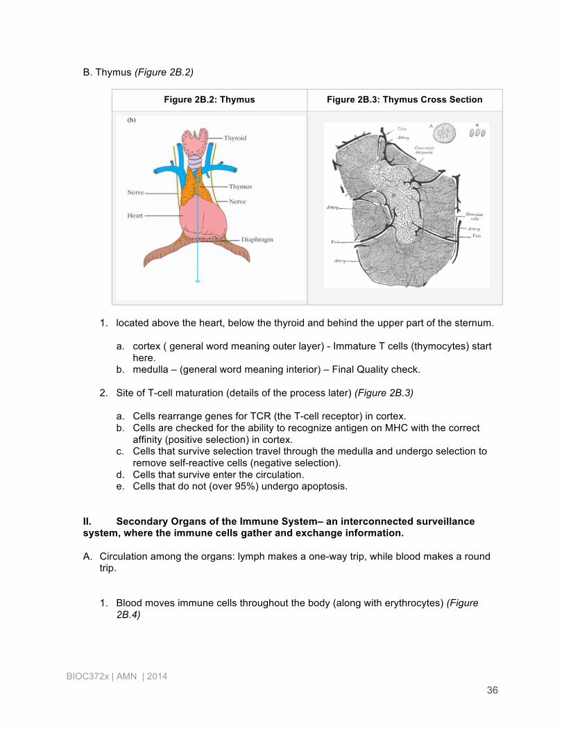
B. Thymus (Figure 2B.2)
Figure 2B.2: Thymus Figure 2B.3: Thymus Cross Section
1. located above the heart, below the thyroid and behind the upper part of the sternum.
a. cortex ( general word meaning outer layer) - Immature T cells (thymocytes) start
here. b. medulla – (general word meaning interior) – Final Quality check.
2. Site of T-cell maturation (details of the process later) (Figure 2B.3)
a. Cells rearrange genes for TCR (the T-cell receptor) in cortex. b. Cells are checked for the ability to recognize antigen on MHC with the correct
affinity (positive selection) in cortex. c. Cells that survive selection travel through the medulla and undergo selection to
remove self-reactive cells (negative selection). d. Cells that survive enter the circulation. e. Cells that do not (over 95%) undergo apoptosis.
II. Secondary Organs of the Immune System– an interconnected surveillance system, where the immune cells gather and exchange information.
A. Circulation among the organs: lymph makes a one-way trip, while blood makes a round
trip.
1. Blood moves immune cells throughout the body (along with erythrocytes) (Figure 2B.4)

BIOC372x | AMN | 2014 37
Figure 2B.4: Blood Movement
a. The lining of vessels (endothelium) responds to infections with inflammation and this directs neutrophils and other immune cells to the infected site.
b. Proteins in blood plasma include antibodies, clotting proteins and complement proteins that attack foreign cells.
c. Blood filtered by spleen, which recycles aged erythrocyte and picks of antigen and other detritus.
2. Lymph also provides transport of immune cells, primarily lymphocytes, but no erythrocytes (Figure 2B.5)
b. Drains interstitial fluid from tissues, picking up antigens and white cells (Figure
2B.6)
c. Lymph (fluid) filtered through lymph nodes, where antigen is trapped and acted on.
d. Vessels join into larger ones that empty into the thoracic duct, which in turn empties into left subclavian vein and then enters heart.

BIOC372x | AMN | 2014 38
Figure 2B.5: Return Figure 2B.6: Lymphatic System
B. Lymph nodes - trap antigen and provides sites for the lymphocytes to interact with
antigen.
1. Basic structure (Figure 2B.7-2B.8)
a. cortex receives incoming lymph (afferent) b. follicles embedded in cortex receive and hold B cells c. paracortex (immediately inside) hold T cells d. mature B cells leave through this e. exit out the efferent vessels
Figure 2B.7: Lymph Node Figure 2B.8: Node Cross Section

BIOC372x | AMN | 2014 39
2. Cell interactions
a. B cells activated by antigen migrate to the paracortex to alert T cells. Some get instruction to go forth and make antibodies.
b. Secondary follicle develops after antigen exposure. Has active germinal center where B cells develop in response to signal from follicular dendritic cells, TH cells and macrophages.
c. B cells that have spent time in a secondary follicle learn to make more effective antibodies.
C. Spleen (Figure 2B.9)
1. in abdomen, next to pancreas
2. filters blood, not lymph (Figure 2B.10)
3. red pulp with macrophages that recycle old red blood cells
4. white pulp (PALS) has T cells
5. marginal zone with B cells in follicles - system works like the lymph nodes:
6. Removing the spleen can increase a person's risk for bacterial infections, but there does seem to be some redundancy in the system as a whole.
Figure 2B.9: spleen Figure 2B.10: Spleen Cross Section
BIOC372x | AMN | 2014 40
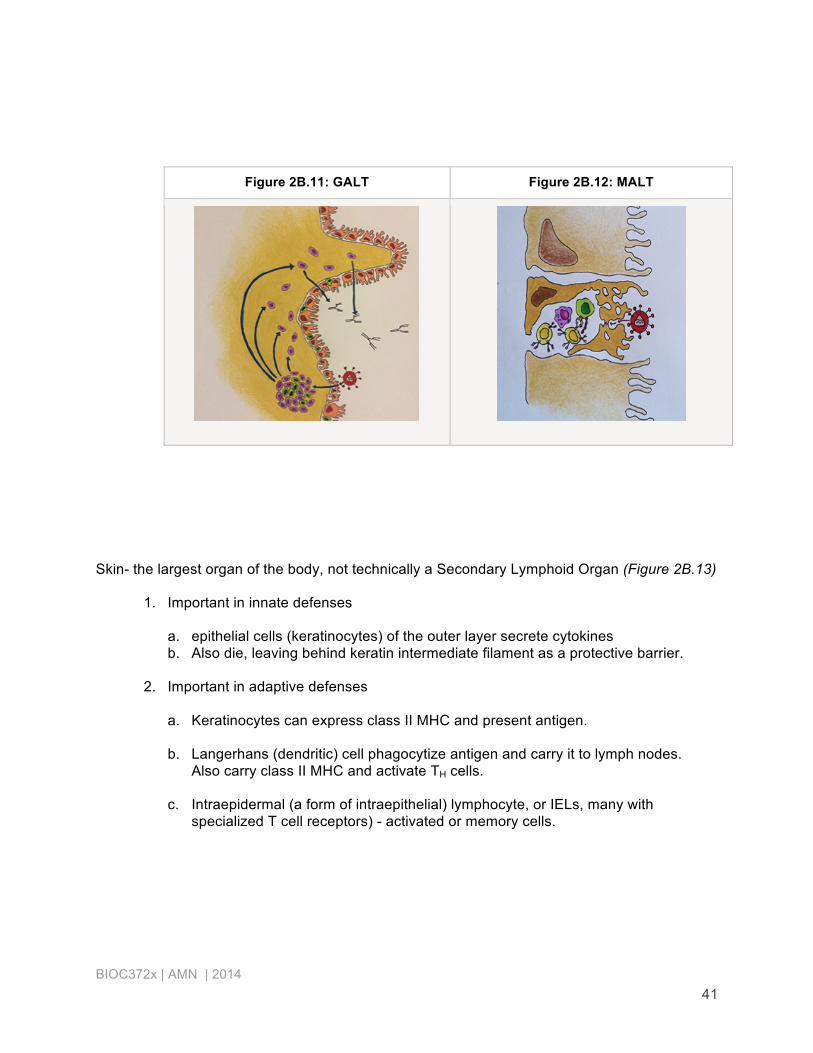
D. Mucosal-Associated Lymphoid Tissue – MALT (Figure 2B.11) Also gut associated – GALT, and bronchial (lung epithelia) – BALT, nasal – NALT
1. The mucosa of the digestive, respiratory, and urogenital systems represents
the major site of entry of most pathogens.
2. The epithelia of these systems contain defensive lymphoid tissues.
3. Organized structures present include tonsils, appendix, and Peyr's patches in the intestine (Figure 2B.12)
4. Epithelial cells of the mucosa deliver antigen samples from the lumen, delivering them via M cells
5. M cells are large epithelial cells, each with a number of smaller immune cells residing in the basolateral pocket it makes.
6. Antigen crosses the plasma membrane to these. The B cell then migrates to inductive sites.
BIOC372x | AMN | 2014 41
Figure 2B.11: GALT Figure 2B.12: MALT
Skin- the largest organ of the body, not technically a Secondary Lymphoid Organ (Figure 2B.13)
1. Important in innate defenses
a. epithelial cells (keratinocytes) of the outer layer secrete cytokines b. Also die, leaving behind keratin intermediate filament as a protective barrier.
2. Important in adaptive defenses
a. Keratinocytes can express class II MHC and present antigen.
b. Langerhans (dendritic) cell phagocytize antigen and carry it to lymph nodes.
Also carry class II MHC and activate TH cells.
c. Intraepidermal (a form of intraepithelial) lymphocyte, or IELs, many with specialized T cell receptors) - activated or memory cells.

BIOC372x | AMN | 2014 42
Figure 2B.13: Skin
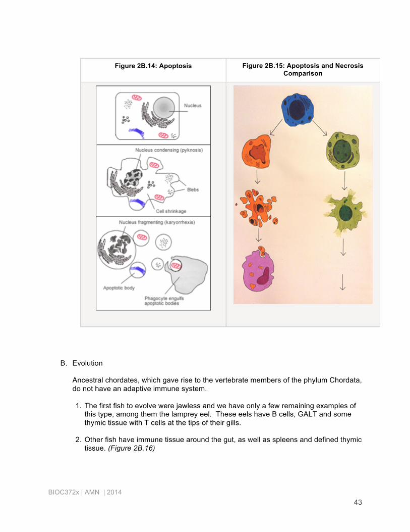
III. Final Issues: A. Apoptosis (Figure 2B.14)
One aspect of the immune system that makes it so energetically expensive is that it produces huge numbers of cells and then gets rid of the great majority of cells before they are even used.
1. Analogous to imploding a building.
a. Cell shrinks b. Chromatin condenses c. Membrane blebs d. Cell fragments into intact pieces, easily phagocytized
2. Necrosis – analogous to blowing up a building (Figure 2B.15)
a. Organelles swell and break down b. Cell disintegrates c. Contents released where they can cause tissue damage and inflammation d. Much harder to clean up after
BIOC372x | AMN | 2014 43
Figure 2B.14: Apoptosis Figure 2B.15: Apoptosis and Necrosis Comparison
B. Evolution
Ancestral chordates, which gave rise to the vertebrate members of the phylum Chordata, do not have an adaptive immune system. 1. The first fish to evolve were jawless and we have only a few remaining examples of
this type, among them the lamprey eel. These eels have B cells, GALT and some thymic tissue with T cells at the tips of their gills.
2. Other fish have immune tissue around the gut, as well as spleens and defined thymic tissue. (Figure 2B.16)

BIOC372x | AMN | 2014 44
3. Amphibians, reptiles, birds and mammals all have bone marrow, but their B cells mature in a variety of places.
4. So, while it’s true that reptiles, bird and mammals have B cells and T cell along with their innate defenses, there is a lot of variety in what gets made where and when.
5. Happily rodents and humans have reasonably similar immune systems, making mice and rats good lab models for the study of the immune response.
Figure 2B.16: Evolutionary Immunology