5/14/2018 bio.11-slidepdf.com http://slidepdf.com/reader/full/bio11 1/15 Review A thorough understanding o absorption, dis- tribution, metabolism, excretion and toxic- ity properties o drug candidates is essential in drug development and is the core ocus o drug metabolism and disposition departments. Knowledge o tissue distribution is linked to many other areas in drug development includ- ing pharmacology, pharmacokinetics and drug–drug interactions [1] . For a drug to have the desired pharmacological activity it must be highly associated with the intended biological target site [2]. Secondary pharmacology and toxicity can also result rom the unexpected association o parent drug or metabolites with nontargeted biological receptors, oten as the result o accumulation and localization in tis- sues. Furthermore, drugs and metabolites are typically not homogeneously distributed in tis- sues, and plasma concentrations do not neces- sarily reect tissue and sub-tissue compartment concentrations [3,4]. The beneft o tissue dis- tribution knowledge has been demonstrated in several areas, including oncology drugs where the ocus is on tumor tissue targets, and anti- biotics where eective treatment is dependent on getting the active drug to the specifc site o the inection [5,6]. For the most part, drug tissue distribution studies and the analytical methodologies have not changed much over the past 2–3 decades despite enormous advances in many analyti- cal areas. Most o our knowledge o drug tis- sue distribution is derived rom single-dose whole-body autoradiography (WBA) studies in a rodent species, and in some instances tissue homogenate analysis in other preclinical spe- cies [7]. Extrapolation rom these experiments is required to understand the complexity o drug tissue distribution in preclinical species as well as in humans. Tissue imaging o drug distributions based on autoradiography or autoradioluminography relies on the presence o a radiolabeled com- pound, typically 14 C or 3 H, to view drug-related material [8]. Because the detection method is based on the presence o a radiolabel only, there is no structural inormation associated with this technique. The images and quantifcation are strictly due to the presence o the radiolabel incorporated in the dosed drug. Thus, images broadly reect the distribution o drug-related material while the quantifcation results are con- verted to nanogram equivalences o the parent molecule. In cases where metabolic biotrans- ormation results in cleavage or ring scission, the radiolabel may no longer be associated with the pharmacophore portion o the parent drug. Discerning the amount o drug-related material in a tissue is valuable, but knowing whether the drug-related material is predominantly parent or consists o a large number o structurally diverse metabolites is critical in understand- ing drug distribution and the implications or pharmacology and saety. Despite these limi- tations, the good specifcity or drug-related material against the background, sensitivity, quantitative nature and large dynamic range have made this approach the industry standard and accepted by regulatory authorities [9]. For most WBA studies supporting a development MALDI imaging mass spectrometry: bridging biology and chemistry in drug development Our understanding of drug tissue distribution impacts a number of areas in drug development, including: pharmacology, pharmacokinetics, safety, drug–drug interactions, transport and metabolism. Despite their extensive use, autoradiography and tissue homogenate LC–MS analysis have limitations in providing a comprehensive assessment of tissue distributions. In the case of autoradiography, it is the inability to distinguish between parent drug and drug metabolites. In LC–MS analysis of tissue homogenate, all tissue localization information is lost. The emerging technique of MALDI imaging mass spectrometry has the capability to distinguish between parent and metabolites while maintaining spatial distribution in tissues. In this article, we will review the MALDI imaging MS methodology as applied to drug development and provide examples highlighting the impact of this important technique in drug development. Stephen Castellino †1 , M Reid Groseclose 1 & David Wagner 1 1 Department of Drug Metabolism & Pharmacokinetics, Platform Science & Technology, GlaxoSmithKline, 5 Moore Drive, RTP, NC 27709 USA † Author for correspondence: Tel.: +1 919 483 2262 Fax: +1 919 483 0443 E-mail: [email protected] 2427 ISSN 1757-6180 Bioanalysis (2011) 3(21), 24 27–2441 10.4155/BIO.11.232 © Stephen Castellino

Welcome message from author
This document is posted to help you gain knowledge. Please leave a comment to let me know what you think about it! Share it to your friends and learn new things together.
Transcript

5/14/2018 bio.11 - slidepdf.com
http://slidepdf.com/reader/full/bio11 1/15
Review
A thorough understanding o absorption, dis-tribution, metabolism, excretion and toxic-ity properties o drug candidates is essentialin drug development and is the core ocus o drug metabolism and disposition departments.Knowledge o tissue distribution is linked tomany other areas in drug development includ-ing pharmacology, pharmacokinetics anddrug–drug interactions [1]. For a drug to havethe desired pharmacological act ivity it must behighly associated with the intended biologicaltarget site [2]. Secondary pharmacology andtoxicity can also result rom the unexpectedassociation o parent drug or metabolites withnontargeted biological receptors, oten as theresult o accumulation and localization in tis-sues. Furthermore, drugs and metabolites aretypically not homogeneously distributed in tis-sues, and plasma concentrations do not neces-sarily reect tissue and sub-tissue compartmentconcentrations [3,4]. The beneft o tissue dis-tribution knowledge has been demonstrated in
several areas, including oncology drugs wherethe ocus is on tumor tissue targets, and anti-biotics where eective treatment is dependenton getting the active drug to the specifc site o the inection [5,6].
For the most part, drug tissue distributionstudies and the analytical methodologies havenot changed much over the past 2–3 decadesdespite enormous advances in many analyti-cal areas. Most o our knowledge o drug tis-sue distribution is derived rom single-dose whole-body autoradiography (WBA) studies in
a rodent species, and in some instances tissue
homogenate analysis in other preclinical spe-cies [7]. Extrapolation rom these experiments isrequired to understand the complexity o drugtissue distribution in preclinical species as wellas in humans.
Tissue imaging o drug distributions basedon autoradiography or autoradioluminography relies on the presence o a radiolabeled com-pound, typically 14C or 3H, to view drug-relatedmaterial [8]. Because the detection method isbased on the presence o a radiolabel only, thereis no structural inormation associated with thistechnique. The images and quantifcation arestrictly due to the presence o the radiolabelincorporated in the dosed drug. Thus, imagesbroadly reect the distribution o drug-relatedmaterial while the quantifcation results are con-verted to nanogram equivalences o the parentmolecule. In cases where metabolic biotrans-ormation results in cleavage or ring scission,the radiolabel may no longer be associated withthe pharmacophore portion o the parent drug.
Discerning the amount o drug-related materialin a tissue is valuable, but knowing whether thedrug-related material is predominantly parentor consists o a large number o structurally diverse metabolites is critical in understand-ing drug distribution and the implications orpharmacology and saety. Despite these limi-tations, the good specifcity or drug-relatedmaterial against the background, sensitivity,quantitative nature and large dynamic rangehave made this approach the industry standardand accepted by regulatory authorities [9]. For
most WBA studies supporting a development
MALDI imaging mass spectrometry: bridging
biology and chemistry in drug development
Our understanding of drug tissue distribution impacts a number of areas in drug development, including: pharmacology,
pharmacokinetics, safety, drug–drug interactions, transport and metabolism. Despite their extensive use,
autoradiography and tissue homogenate LC–MS analysis have limitations in providing a comprehensive assessment of
tissue distributions. In the case of autoradiography, it is the inability to distinguish between parent drug and drug
metabolites. In LC–MS analysis of tissue homogenate, all tissue localization information is lost. The emerging technique
of MALDI imaging mass spectrometry has the capability to distinguish between parent and metabolites while maintaining
spatial distribution in tissues. In this article, we will review the MALDI imaging MS methodology as applied to drug
development and provide examples highlighting the impact of this important technique in drug development.
Stephen Castellino†1,
M Reid Groseclose1
& David Wagner 1
1Department of Drug Metabolism& Pharmacokinetics, Platform
Science & Technology,GlaxoSmithKline, 5 Moore Drive,RTP, NC 27709 USA†Author for correspondence:
Tel.: +1 919 483 2262Fax: +1 919 483 0443E-mail: [email protected]
2427ISSN 1757-6180Bioanalysis (2011) 3(21), 2427–244110.4155/BIO.11.232 © Stephen Castellino

5/14/2018 bio.11 - slidepdf.com
http://slidepdf.com/reader/full/bio11 2/15
program, a time profle study design is employedin order to ollow the distribution and amountso drug-related material as a unction o timeater dosing. This study design provides thekinetics associated with lie time o drug-related
material in speciic tissues. Typical spatialresolution in WBA studies is between 50 and100 µm; however, spatial resolution as low as10 µm is possible with micro-autoradiography studies [10]. Micro-autoradiography studies aretechnically more challenging and not strictly quantitative and, thereore, are not routinely used in the pharmaceutical industry.
The other common method or establishingdrug tissue distribution is through LC–MSanalysis o tissue homogenates. Results romthis approach are typically reported in nano-
grams o drug or metabolite per gram o tissue.The value o determining tissue concentrationsby LC–MS analysis o tissue homogenate hasbeen debated or years [2,5]. Interpretation andextrapolation o these data can be very tenuousbecause the data provide no distribution inor-mation within a specifc tissue or organ, and inessence, treat all tissues as homogeneous com-ponents, ignoring the distinct compartmentsand unctions within tissues and organs. Thismethod structurally discriminates between par-ent and metabolites and is quantitative, pro-vided appropriate methods are employed.
MALDI imaging MS (IMS) provides the oppor-tunity to determine discreet tissue localizationor parent drug as well as metabolites [11–13].Combined with strategies to address quantifca-tion, this approach can oer signifcant advan-tages over autoradiography methods and tissuehomogenate analysis or determining drug andmetabolite tissue distribution in a preclinical set-ting. Recent advances in commercially availableinstrumentation ranging rom matrix applicationtools, matrix-assisted laser desorption/ionization(MALDI) imaging-centric mass spectrometers
and sotware are providing a pathway to main-stream this approach beyond the laboratorieso the pioneers in this area. This is an emerg-This is an emerg-ing and rapidly growing feld with demonstratedimpact in areas outside o drug development suchas proteomics and lipidomics. However, recentquestions have been raised about the readinesso MALDI IMS technology to be broadly usedin drug research [14]. Specifcally, the impact andunderstanding associated with sample prepara-tion, ion suppression and quantifcation will becritical or mainstream applications in all areas,
including drug distribution studies.
Overview of MALDI IMS
MALDI IMS has emerged as a powerul anddiverse technology or analyzing the spatial dis-tribution o endogenous and exogenous com-pounds directly rom a tissue section [15–18]. This
review will ocus mainly on the application o MALDI IMS or the analysis o tissue samples.Several recent reviews provide a broad overview o other MS imaging methods and applica-tions [19–25]. These include alternative ionizationtechniques such as secondary ion MS (SIMS),desorption electrospray ionization (DESI) andhybrid methods such as MALDI electrospray ionization (MALDESI).
In short, a typical MALDI IMS experimentis conducted by coating a thin tissue sectionmounted onto a target with an appropriate matrix
solution. This solution serves to extract analyteso interest rom the underlying tissue and uponsolvent evaporation the extracted molecules areco-crystallized with the matrix. The role o thematrix is to absorb the laser energy and acili-tate desorption/ionization o the analyte mol-ecules [26]. Mass spectra are then acquired acrossthe tissue at defned geometrical coordinates. Theresulting dataset contains hundreds to thousandso individual spectra consisting o all ions detectedat each location o acquisition. Custom sotwareis then used to compile the mass spectra into aormat where each spectrum represents a discretepixel and the distribution and intensity o any o the detected species can be viewed across thetissue as an ion density map or image (Figure 1).
In a single MALDI IMS experiment it ispossible to detect hundreds or even thousandso discrete signals across a tissue rom a diverseset o analytes. These analytes can range romendogenous biomolecules (e.g., proteins, pep-tides and lipids) to exogenous molecules (e.g.,pharmaceutical compounds). Thus, one o themajor advantages o using this technique is thatendogenous biomolecules are also automatically
detected, providing opportunities to evaluate theunderlying physiological state o a tissue [27].There are several aspects o imaging MS thatmerit critical evaluation and will be discussedin detail, including sample preparation, ionsuppression, sensitivity and data analysis.
Sample preparation
As with many analytical methods, a success-ul outcome is closely aligned with the rigorand attention paid to sample preparation steps.MALDI IMS sample preparation is unique in
that most practicing mass spectrometrists are
Key Terms
Spatial resolution: Center-to-center distancebetween adjacent acquisitionspots (pixels) in an MALDIimaging MS experiment (dened
here as: m/Dm where m = massand Dm = peak width at half maximum [50%]).
MALDI (matrix-assistedlaser desorption/ionization): Soft ionizationtechnique used in MS thatenables drugs and biomoleculesto be detected withoutsignicant fragmentation.
Imaging MS: Technology usedto evaluate the label-freedistribution of endogenous andexogenous molecules in a tissue.
Histology: Microscopicanatomical evaluation of thintissue sections and a criticalaspect in interpreting imagesgenerated by MALDI imaging MS.
Review | Castellino, Groseclose & Wagner
Bioanalysis (2011) 3(21)2428 future science group

5/14/2018 bio.11 - slidepdf.com
http://slidepdf.com/reader/full/bio11 3/15
typically not skilled in the art o tissue sec-tioning with a cryostat. Thus, many spectrome-trists will require assistance in this process romhistologists or toxicologists. Typically, resh-rozen tissues are sectioned at 10–12 µm. Therozen sections are then thaw mounted onto aMALDI target prior to desiccation and matrixapplication.
For whole body IMS experiments, prepara-
tion o tissue sections is dierent because thesample specimen (i.e., rat) is typically embed-ded in a block o ice or carboxymethylcellulose.Thicker sections (20–50 µm) are collected on a whole-body cryostat and then mounted onto theMALDI target with adhesive tape.
Although resh-rozen tissues are ideal, theanalysis o ormalin-fxed, parafn-embeddedtissues using MALDI IMS has been reportedor proteins and peptides [28,29] and also orsmall molecules [30]. However, due to the lim-ited number o publications in this area, the
compatibility and utility o these fxed tissues
or drug analysis with MALDI IMS remainsunclear. It is likely that the ormalin fxationprocess, where a tissue is placed into a orma-lin solution or several hours, as well as otherhistology processes such as alcohol dehydrationand parafn infltration, will result in diusionor even extraction o some drug compounds,rendering the tissue unusable or drug imag-ing experiments. In addition, the fxation pro-
cess, which orms methylene bridges betweenthe primary amines o lysine on proteins, may detrimentally aect the extractability o drugcompounds. It should be noted that the vastarchives o ormalin-fxed, parafn-embeddedtissues available at most pharmaceutical com-panies could represent an extremely valuablesource o samples i these technical difcultiescould be overcome. Embedding media that donot interere with the MALDI process haverecently been reported and can be useul inaiding cryosectioning o certain types o tissue
samples [31].
Mass spectrum acquired at each position
Matrix-coated
tissue sample
Ion density maps
Low High
Intensity
Laser
Figure 1. MALDI imaging mass spectrometer experimental workow. The MALDI imagingexperiment is initiated by mounting a tissue section onto a target, applying a matrix solution andrastering a laser across the surace o the tissue. At each discrete location where the laser is red, a
mass spectrum is acquired. By plotting the ion intensities as a unction o the x and y coordinates onthe tissue, ion images are generated.
MALDI imaging MS: bridging biology & chemistry in drug development | Review
www.future-science.com 2429future science group

5/14/2018 bio.11 - slidepdf.com
http://slidepdf.com/reader/full/bio11 4/15
A matrix solution when applied to the tissuesection serves to extract analytes o interest romthe underlying tissue, and upon solvent evapora-tion the extracted molecules are co-crystallized
with the matrix to acilitate subsequent MALDIMS analysis (Figure 2). The selection o the matrixsolution and method o application is primarily driven by empirical results. For small organic
MS imaging
H&E staining
Tissue sectioning
LC–MS/MSquantification
Figure 3. Serial sectioning strategy or correlating ion images, histology and LC–MSquantifcation. By taking serial sections rom tissue or MALDI imaging, H&E staining and LC–MSquantication, correlations between drug distribution, histology and drug quantication will beimproved. Drug and metabolite concentrations determined rom whole organ analysis may not berepresentative o sections used or MALDI imaging.H&E: Hematoxylin and eosin.
50/50
organic/H2O
Matrix absorbs at wavelength of laser resultingin desorption/ionization of co-crystallizedanalyte molecules+
++2,5-dihydroxybenzoic acid
OH
HO
OH
O
UV laser
Analytes co-crystallized withmatrix upon solvent evaporation
Analytes extracted from tissueinto matrix droplet
+
+
Bioanalysis © Future Science Group (2011)
Figure 2. Role o matrix application in MALDI imaging.
Review | Castellino, Groseclose & Wagner
Bioanalysis (2011) 3(21)2430 future science group

5/14/2018 bio.11 - slidepdf.com
http://slidepdf.com/reader/full/bio11 5/15
molecules the most commonly reported matrixcompounds are 2,5-dihydroxybenzoic acid,a-cyano-4-hydroxycinnamic acid and picolinicacid. As a starting point, the optimum matrix istypically determined by spotting drug standard
onto the MALDI target and observing whichmatrix provides the maximum S/N. Several otheractors, including undesired analyte ragmenta-tion, inhomogeneous crystallization and excessivematrix background peaks may also play a role inchoosing a desirable matrix. In our laboratory,2,5-dihydroxybenzoic acid tends to be the matrixo choice or the analysis o small molecules by MALDI IMS. The matrix is usually dissolvedin an acidifed mixture o organic solvent(s) and water (i.e., 50/50 methanol/water and 0.1% tri-uoroacetic acid). The matrix solution has two
critical unctions: extraction o analytes o inter-est rom the tissue and co-crystallization o theanalytes with the matrix to acilitate the desorp-tion and ionization processes. Discussion o theundamentals o the MALDI process is beyondthe scope o this review, but additional inorma-tion can be ound in literature ocused on thistopic [26,32–38].
In theory, the choice o solvent compositioncould be guided by the physicochemical proper-ties o the analyte or analyte class o interest wheremore polar analytes may preer higher percentageaqueous and nonpolar analytes a higher percent-age o organic solvent. While this is an importantconsideration, this parameter is complicated by several additional actors, including the complex-ity o the biomolecular interactions in a tissue,matrix solubility, and the solvent evaporation/matrix crystallization process [21]. As a result,selection o an optimal solvent composition may be determined empirically in many cases.
The fnal step o sample preparation is theapplication o the matrix solution to the target-mounted tissue. A variety o matrix applicationmethods have been successully used, including
microspotting [39], sublimation [40,41] and spray coating [42]. One o the key considerations inselecting an application method is the reproduc-ibility o the matrix application so that a num-ber o tissues can be analyzed with minimumvariability rom the matrix application. Severalautomated systems have been developed andmarketed including the HTX Imaging™ sprayer(nozzle spraying technology) [101], Bruker ImagePrep™ (vibrational vaporization) [102], ShimadzuCHIP-1000 (inkjet printing technology) [103],and Labcyte Portrait® (acoustic droplet technol-
ogy [104]. These systems permit accurate control
o application variables, such as coating cycles,drying times and matrix thickness, and thereore will provide increased reproducibility and optimi-zation relative to manual application. In general,imaging rom tissues with high analyte concentra-
tions and acquisition at low spatial resolution willbe less demanding on the optimization o matrixsolution selection and application method.
Ion suppression, sensitivity
& quantifcation
As is the case or any application o MS, theissues o ion suppression, sensitivity and quanti-fcation are important interrelated variables thatneed to be evaluated and addressed. In MALDIIMS, reproducibility and consistency in analyteextraction and matrix co-crystallization between
areas o a tissue containing dierent cell types orstructural morphologies are o greatest concern.Many o the questions surrounding the extento ion suppression in tissue imaging have beendiscussed extensively in the literature [14,43].
The eect o ion suppression is observed whencomparing the signal intensity o a standard spot-ted directly on the MALDI target versus the sig-nal rom the same amount spotted on the suraceo a control tissue section [13,14]. Typically, thisexperiment reveals a discrepancy in the observedsignal or equal amounts o an analyte wheresignal intensities are signifcantly higher whenacquired directly rom the target. Multiple actorsare likely to be contributing to the dierence inobserved signal including variation in matrix crys-tallization, ion suppression rom endogenous saltsand lipids, and molecular interactions betweenthe analyte and tissue biomolecules. In an imag-ing experiment, where interpretation o the iondensity map or an analyte is based on dierencesin intensity and is thereore undamentally quan-titative, it is important to establish that the varia-tion o signal intensity across the tissue reects theactual concentration and is not governed by the
eects o ion suppression.One strategy to evaluate ion suppression is to
uniormly coat the surace o a control tissue sec-tion with a standard and acquire the MALDIimage [1,44]. Ideally, this experiment would identiy any regions o a tissue section that may introducesignifcant ion suppression eects on the analytestandard by showing deviations rom a homog-enous distribution. However, accurate assessmento ion suppression requires careul experimentaldesign and it is critical that both the standard andthe matrix solution are applied homogenously and
reproducibly across the tissue section.
MALDI imaging MS: bridging biology & chemistry in drug development | Review
www.future-science.com 2431future science group
5/14/2018 bio.11 - slidepdf.com
http://slidepdf.com/reader/full/bio11 6/15
Another way to address the potential impact o ion suppression is to use a post-acquisition nor-malization o spectral intensities. Normalizationo the data in an IMS experiment is carried outby multiplying all spectra by an intensity scaling
actor. This scaling actor can be determined by a number o dierent methods (i.e., total ioncurrent), each having advantages and disadvan-tages; several recent publications have discussedthis topic in detail [45–47].
In the context o structurally characteriz-ing drug metabolites rom biological matrices,inevitably the issue o quantifcation will alwaysbe raised. This is because pharmacology andtoxicology are driven by knowledge o both theinherent reactivity o a molecule and the quan-tity at the receptor site. One strategy is to use
quantitative LC–MS data rom tissue homog-enates to try and correlate drug quantities withthe intensities detected in an ion image [48]. Thisstrategy assumes that tissues are homogenousand typically utilizes a large piece o tissue thatmay not be representative o the thin sectionused in the imaging experiment. Most tissuescontain sub-compartments, which may containhigh concentrations o particular metabolites.Consequently, the homogenate results may introduce a bias. Alternatively, serial sectioning with one section going to quantitative LC–MSand the other being used or imaging can reducethe likelihood o this bias (Figure 3) [49].
IMS studies employing radiolabels can takeadvantage o quantiication through auto-radiography in order to optimize both tissue dis-tribution and quantifcation [50]. Alternatively,spotting standards on control tissue prior tomatrix application to generate calibration curveshas also been used or IMS quantifcation [1].This methodology mimics the interaction o the tissue, analytes and matrix, but may not becompletely representative o the tissue extractionprocess. These recent literature reports are prom-
ising and eectively show that a reasonable levelo quantifcation can be obtained; however, sig-nifcant improvements will be required to reachthe validation level o a quantitative LC–MSassay or autoradiography. The issue o quantif-cation in MALDI IMS represents several uniquechallenges and should continue to draw atten-tion and become more refned as greater insightsinto the MALDI process are gained.
Evaluating limits o detection in IMS requiresrethinking the defnition o ‘sample’. It is clearin tissue homogenate work that homogenization
and extraction ollowed by quantitative LC–MS
analysis results in an analyte amount per vol-ume o extract that can then be converted to atissue concentration in µg per gram o tissue.However, in IMS the entire tissue section rep-resents an array o samples, where the sample is
defned as the individual area o tissue (pixel) where a MALDI mass spectrum is acquired.Thereore, it is important to defne a limit o detection (LOD) in this small volume o tissuebecause pixel intensities below this value will beregistered as containing no drug (Figure 4). Oneimmediate consequence o this is that greatersensitivity, LOD, will usually be obtained withlow spatial resolution (large laser spot or aver-aged raster area) compared with high spatialresolution (small laser spot or averaged rasterarea) because the amount o analyte will be pro-
portionally higher in a larger area. Thus, in mostcases the tradeos between signal intensity andspatial resolution must be taken into consider-ation when planning an experimental strategy.
Spectral resolution
Structural characterization o metabolites rombiological matrices using MS is typically car-ried out by employing a chromatographic sepa-ration method, such as HPLC or UPLC, priorto introducing the sample into the mass spec-trometer. The chromatographic separation addsanother dimension o analyte dierentiation orcomplex mixtures prior to mass spectrometricanalysis. The development o high-resolutionmass spectrometers [51–54] and data-miningsotware such as mass-deect fltering [55–57] has urther acilitated metabolite identifca-tion in biological matrices. MALDI IMS doesnot have the beneft o the initial chromato-graphic separation and the burden o resolvingall components depends on the mass analyzer.This is especially challenging in the low massrange (<1000 Da) where matrix backgroundand high levels o endogenous species can sig-
nifcantly interere with signals or drugs andmetabolites. Spectrometers with relatively low spectral resolution, including quadrupoles [58],ion traps [30] and time-o-ight [27] systems canbe employed in IMS, but these systems, may require MS/MS methods to provide enoughspecifcity to overcome intererence rom thematrix and endogenous compounds. The ana-lyte selectivity o MS/MS methods signifcantly narrows the scope o the IMS experiment to asingle target molecule o interest, and there-ore excludes additional drug-related material
or endogenous components rom detection.
Review | Castellino, Groseclose & Wagner
Bioanalysis (2011) 3(21)2432 future science group

5/14/2018 bio.11 - slidepdf.com
http://slidepdf.com/reader/full/bio11 7/15
However, it has recently been shown that a con-tinuous raster imaging mode where multipleion transitions are monitored sequentially cansignifcantly enhance the scope o this type o imaging experiment [58,59].
High-resolution (HR) mass spectrometerssuch as the Fourier transorm ion cyclotronresonance (FT–ICR or FT) and Orbitrap havethe ability to mass spectrally resolve drug andmetabolites rom endogenous compounds withthe same nominal mass. Consequently, HR-massspectrometer instruments can perorm IMSexperiments in ull scan mode allowing thesimultaneous detection o drug, metabolitesand potential biomarkers in one imaging experi-ment [1,60]. However, the acquisition times o these instruments are typically much slower thanlower resolution instrumentation, signifcantly increasing the total time or acquiring an image.
Several examples illustrate the importanceo high spectral resolution in drug distributionstudies. Figure 5 shows a region o the average
spectrum and the corresponding ion images ortwo metabolites rom a MALDI IMS experi-ment conducted on liver tissue rom a dogdosed with lapatinib. A resolution o >50 K isrequired to completely resolve these two metab-olites in a ull scan mass spectrum and conf-dently identiy their corresponding tissue dis-tributions. Another example o the importanceo mass resolution is captured in Figure 6. Here,our singly charged ions that span a range o 0.25 Da have been mapped as individual com-ponents and as a composite, simulating what
the image would look like i the ions were not
adequately resolved. The ability to resolve drug-related peaks rom background allows one tocomprehensively evaluate drug ti ssue distribu-tions in a single MALDI IMS experiment. It isimportant to note that in some cases (e.g., iso-baric metabolites), MS/MS wil l be required toconfrm a metabolite’s identity.
As a general strategy, instruments with lessresolving power and mass accuracy can beemployed successully in IMS i there is a singlemolecular species that is being targeted andMS/MS spectra can reliably provide sufcientresolution rom endogenous components. Sucha strategy might be well suited in a discovery setting where the goal is to image the potentialdrug against a biological target. However, whenthe goal is to understand the tissue distributiono a drug and its metabolites, instruments withhigh resolution and high mass accuracy can beessential or IMS studies. A relatively new typeo mass spectrometer, which incorporates post-ionization separation based on ion mobility prior
to detection has the ability to resolve even iso-baric molecular components o a tissue in thegas phase based on collision cross section and,thereore, has great potential or drug imagingstudies [61–64].
Spatial resolution
MALDI imaging generates 2D maps o ana-lyte intensities across the surace o a tissue.The term spatial resolution, or pixel resolution,generally reers to the center-to-center distancebetween two adjacent areas o acquisition (pix-
els). In this review we will use the terms spatial
Whole tissue Imaging MS acquisition at 50 µmspatial resolution
Tissue sectioned in z-plane
Tissue sectionArea: 50 mm2
Volume: 0.6 mm3
20,000 spots/spectra across tissueVolume of tissue
per acquisition spot
~23,500 µm3Laser fired/mass spectra
acquired at each position
12 µmthickness
12 µm
5 mm
50 µm
50 µm 50 µm
1 0 m m
Tissue homogenateconcentration: 50 ng/mg
Quantity per acquisition spot1 fg
x
y
z
Bioanalysis © Future Science Group (2011)
Figure 4. Limit o detection defned by MALDI pixel size.
MALDI imaging MS: bridging biology & chemistry in drug development | Review
www.future-science.com 2433future science group

5/14/2018 bio.11 - slidepdf.com
http://slidepdf.com/reader/full/bio11 8/15
resolution and lateral resolution interchangeably to describe the eective laser spot or pixel o thetissue image.
Spatial resolution is one o the most impor-tant parameters that defnes the utility andscope o MALDI imaging as applied to tissuedistribution studies in drug development. Incommercial instruments, the physical size o
the laser spot is limited to approximately 10 µm with most experiments using a laser spot sizebetween 50 and 250 µm. Recently, smaller laserspot sizes have been achieved in custom opti-cal systems used in IMS experiments and revealthe potential or urther development in the areao high-resolution imaging [60,65]. The physicallimitation on the choice o pixel size in an IMSexperiment is determined by the laser spot size;however, several additional actors, includingsample preparation, analyte tissue concentration,MALDI ionization efciency and heterogene-
ity o the tissue, are also important consider-ations. An oversampling method can be used toachieve spatial resolutions higher than the laserspot diameter. This is conducted by using a ras-ter increment that is smaller than the width o the laser beam and, thereore, only a raction o the beam is ablating matrix and generating themeasured analyte signal [66].
Average mass spectrum
Area of tissueLow
High
m/z 746.42 m/z 746.48
0.25 amu
746.30 746.40 746.50 746.60 746.80746.20 746.80
m/z 746.57 m/z 746.61Composite image
1 mm
m/z
Figure 6. Implications o spectral resolution on tissue images. Four endogenous ions (color coded) in the average mass spectrumspan 0.25 Da. An ion image or each o the our selected ions corresponding to the area shown in the optical image is shown. Eachion shows a unique distribution in the ion image. The composite image o all our selected ions demonstrates the result o poorspectral resolution.
m = 0.013 amu
473.1175
M2GW006
473.1045
473.000 473.050 473.100 473.150 473.200 m/z
Peak resolution (FWHM) = ~125,000
Figure 5. Two metabolites (GW006 and M2) o lapatinib rom dog liverresolved by Fourier transorm ion cyclotron resonance.
Review | Castellino, Groseclose & Wagner
Bioanalysis (2011) 3(21)2434 future science group
5/14/2018 bio.11 - slidepdf.com
http://slidepdf.com/reader/full/bio11 9/15
In practice, achieving higher spatial resolutioncan be technically challenging and time con-suming. Thereore it is important to considerthe minimum resolution needed to answer thequestion at hand prior to analyzing a sample.
Tissue sampling at 100–200 µm provides useulimages on the order o what would be expectedrom WBA and can provide valuable distribu-tion surveys o large tissue sections. Sampling with larger laser spot sizes has the advantage o speed (area sampled/time) and sensitivity (aver-aging spectra over a larger area). However, insome cases where analyte distributions are highly localized to specifc histological eatures (e.g.,liver bile ducts) then higher spatial resolutions(<50 µm) may be required.
In some cases the resolution needed to acquire
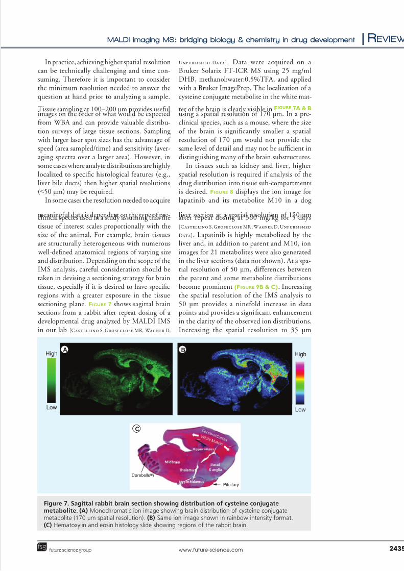
meaningul data is dependent on the type o pre-clinical species used in a study assuming that thetissue o interest scales proportionally with thesize o the animal. For example, brain tissuesare structurally heterogeneous with numerous well-defned anatomical regions o varying sizeand distribution. Depending on the scope o theIMS analysis, careul consideration should betaken in devising a sectioning strategy or braintissue, especially i it is desired to have specifcregions with a greater exposure in the tissuesectioning plane. Figure 7 shows sagittal brainsections rom a rabbit ater repeat dosing o adevelopmental drug analyzed by MALDI IMSin our lab [Castellino S, Groseclose MR, Wagner D,
Unpublished Data]. Data were acquired on aBruker Solarix FT-ICR MS using 25 mg/mlDHB, methanol:water:0.5%TFA, and applied with a Bruker ImagePrep. The localization o acysteine conjugate metabolite in the white mat-
ter o the brain is clearly visible in Figure 7A & B using a spatial resolution o 170 µm. In a pre-clinical species, such as a mouse, where the sizeo the brain is signifcantly smaller a spatialresolution o 170 µm would not provide thesame level o detail and may not be sufcient indistinguishing many o the brain substructures.
In tissues such as kidney and liver, higherspatial resolution is required i analysis o thedrug distribution into tissue sub-compartmentsis desired. Figure 8 displays the ion image orlapatinib and its metabolite M10 in a dog
liver section at a spatial resolution o 150 µmater repeat dosing at 360 mg/kg or 5 days[Castellino S, Groseclose MR , Wagner D, Unpublished
Data]. Lapatinib is highly metabolized by theliver and, in addition to parent and M10, ionimages or 21 metabolites were also generatedin the liver sections (data not shown). At a spa-tial resolution o 50 µm, dierences betweenthe parent and some metabolite distributionsbecome prominent (Figure 9B & C). Increasingthe spatial resolution o the IMS analysis to50 µm provides a nineold increase in datapoints and provides a signifcant enhancementin the clarity o the observed ion distributions.Increasing the spatial resolution to 35 µm
Figure 7. Sagittal rabbit brain section showing distribution o cysteine conjugatemetabolite. (A) Monochromatic ion image showing brain distribution o cysteine conjugatemetabolite (170 µm spatial resolution). (B) Same ion image shown in rainbow intensity ormat.(C) Hematoxylin and eosin histology slide showing regions o the rabbit brain.
A BHighHigh
Low Low
Pituitary
C
CerebellumCerebellu
MALDI imaging MS: bridging biology & chemistry in drug development | Review
www.future-science.com 2435future science group

5/14/2018 bio.11 - slidepdf.com
http://slidepdf.com/reader/full/bio11 10/15
reveals even urther refnement to the point where accurate correlation o the ion distribu-tions with the underlying tissue histology ispossible. For example, Figure 10 shows a metab-olite distribution localized predominately in thesmall bile ductules, which are typically between50 and 100 µm in diameter. Conducting IMSexperiments at this level o spatial resolutiongenerates discreet metabolite distributions intothe subcompartments o a liver tissue such asthe biliary tree, portal and central veins. Tissuesections serial to those analyzed by IMS can betreated with histology stains such as hematoxy-lin and eosin to identiy various subcompart-ments and enable correlation between the tissue
histology and the detected ion distributions. InFigure 11, inammation in the histopathology sections are correlated with metabolite M10tissue distribution.
In a similar ashion, dierentiated subcom-partment distributions o two drug metabolitesare shown in the mouse kidney at the 2 h timepoint ater 5 day repeat dosing o a develop-mental drug (Figure 12) [Castellino S, Groseclose
MR, Wagner D, Unpublished Data]. Note the cor-relation o metabolite distribution and histol-ogy o the tissue. In this study, metabolites aredierentially distributed between the medullaand cortex at 2 h ater the last dose. Romppet al. recently showed that at 10 µm spatial
Optical image of
region analyzed
m/z 581.14
Lapatinibm/z 649.14
M10
Low
A B CHigh
Figure 9. Optical and ion images o a dog liver section at a spatial resolution o 50 µm. (A) Optical image o liver section. (B) Monochromatic ion image o lapatinib, m/z 581.14. (C) Monochromatic ion image o metabolite M10, m/z 649.1.
M10 LapatinibLow
High
A
B C
Figure 8. Ion image o a dog liver section at a spatial resolution o 150 µm. (A) Opticalimage o liver section showing area o tissue where MALDI image was acquired. (B) Monochromatic ion image o metabolite M10, m/z 649.14. (C) Monochromatic ion image olapatinib, m/z 581.14.
Review | Castellino, Groseclose & Wagner
Bioanalysis (2011) 3(21)2436 future science group

5/14/2018 bio.11 - slidepdf.com
http://slidepdf.com/reader/full/bio11 11/15
resolution, urther dierentiation o the kid-ney morphology can be resolved through IMSo endogenous lipids as well as dosed drugs [60].
One o the direct consequences o high lateralresolution imaging experiments in combination with high spectral resolution is the resultantlarge data sets which may require expansive datastorage options. It is not uncommon to conductan IMS experiment where >10,000 spectraare acquired, and when using an FT-ICR MS,each o these fles can be several megabytes insize leading to datasets totaling >50 gigabytes.In addition, high perormance computers (4;8-processors) with enhanced memory read/
write capabilities may be needed to process andmanipulate these datasets efciently.
In the practical application o IMS in drugdevelopment high lateral resolution is not alwaysrequired. Rather, selection o the optimum spa-tial resolution should be based on tissue type,drug and metabolite concentration, MALDIionization efciency and the level o histology correlation necessary to answer key questions. Inmany ways the consideration o spatial resolutionin IMS is similar to how we use Google Earth® (Figure 13). A low spatial resolution image covers
much more area but with less detail while a zoom
can provide greater detail but over a smaller area.The pragmatic application o IMS in preclinicalapplications will typically involve initial tissuesection surveys at lower spatial resolution ol-lowed by higher spatial resolution images overselected tissue areas, potentially directed by histology analysis.
Conclusion
MALDI imaging is an emerging tool orenhancing our understanding o drug andmetabolite tissue distributions. The ability todierentiate parent drug and metabolites in thesame experiment without the need or labeling
sets it apart rom other methods. The impacto MALDI imaging in drug development stemsrom the unique ability o this method to corre-late chemistry and biology in preclinical mod-els. It provides the opportunity to careully link tissue histology and drug and metaboliteion maps at meaningul levels o spatial reso-lution. The ability to map pharmacologicalreceptors to drug or metabolite distribution istransormational in drug development. New insights into the mechanisms o pharmacology and toxicology will be possible. Furthermore,
the tissue imaging data will impact associated
H&E stainSerial tissue sections
Ion images
Bile ducts Bile ducts
Connective tissue
Portal vein
200 µm
m/z 656.14 - heme (+K)
m/z 597.13 - GSK042
m/z 578.20 - endogenous species
Figure 10. Correlation o histology and ion image in dog liver sections. A small area o thehistology section is magnied showing details o structural eatures. A composite ion map (50 µmspatial resolution), which corresponds to the same zoomed region, is constructed showing hemeions (potassium adduct) in the portal vein, a lapatinib metabolite (GSK042) in the bile ducts, andsurrounding area rom on endogenous species.H&E: Hematoxylin and eosin.
MALDI imaging MS: bridging biology & chemistry in drug development | Review
www.future-science.com 2437future science group

5/14/2018 bio.11 - slidepdf.com
http://slidepdf.com/reader/full/bio11 12/15
areas o physiologically based pharmacokineticmodeling as well as drug–drug interactions andtransporters.
Future perspective
The ability o MALDI imaging to provide agreater understanding o drug and metabolitetissue distribution is very exciting and at theheart o the widespread interest in this tech-nique. Linking biology through histology andthe intrinsic chemical properties o drugs andtheir metabolites could be translational in drugdevelopment. However, we must not let the early success o IMS in drug tissue distributions stud-ies obscure the need or greater understanding o the science behind MALDI imaging. Collecting
meaningul data requires careul experimentalexecution and an in-depth knowledge o the pro-cesses rom sample preparation to data analysis.Over the next 5 years, a greater understandingo the MALDI experiment will help guide prac-titioners in the successul application o IMS totissue distribution studies in drug development.In addition, venders and researchers will (con-tinue to urther develop and refne hardwareand sotware to make the IMS experiment morerobust and easier to implement. Researchers willalso continue to develop and explore compli-
mentary ionization methods that will broaden
H&E stainSerial tissue sections
Ion images
m/z 649.14 - M10
Inflammation
Central vein
50 µm
Figure 11. Correlation o histology and ion image in dog liver sections. A small area o thehistology section is magnied showing regions o infammation. An ion map (50 µm spatialresolution), which corresponds to the same zoomed region, shows the localization o lapatinibmetabolite M10, localized only in the regions associated with the infammation.H&E: Hematoxylin and eosin.
Medulla
Cortex
A B
150 µm
50 µm
M4 = 1421 ng/g
M8 = 1169 ng/g
Figure 12. Dierential metabolite distributions in mouse kidney at 2 h. (A) Ion images or two mouse kidney metabolites; (top) 150 µm spatial resolution(bottom) same section at 50 µm resolution. (B) Hematoxylin and eosin stain oserial section.
Review | Castellino, Groseclose & Wagner
Bioanalysis (2011) 3(21)2438 future science group

5/14/2018 bio.11 - slidepdf.com
http://slidepdf.com/reader/full/bio11 13/15
the scope o the IMS experiment. I we do notget ahead o ourselves, and continue to exploreand develop the science associated with imag-ing MS, in 10 years time this methodology could be as commonplace as LC–MS is in drug
development today.
Financial & competing interests disclosure
The authors wish to thank GlaxoSmithKline colleagues:
William Hardesty, Gary Bowers, Stephanie North, Gordon
Dear, Claire Beaumont and Donna Fraser or helpul sug-
gestions and review o the manuscript. The authors have no
other relevant aliations or nancial involvement with any
organization or entity with a nancial interest in or nancial
confict with the subject matter or materials d iscussed in the
manuscript apart rom those disclosed.
No writing assistance was utilized in the production o
this manuscript.
- Cover whole tissue- Hot spots- Good sensitivity
- Similar to whole-bodyautoradiography
- Large portion tissue- Some gross tissue features- Compares distributions
- Medium sensitivity
- Small portion of tissue- Histology overlays- Explore mechanisms
- Compare distributions- Low sensitivity
>100 µm <100 – >50 µm <50 µm
Central
vein
Areas of
necrosis
Figure 13. Impact o varying spatial resolution.
BibliographyPapers o special note have been highlighted as:
n o interest
nn o considerable interest
1 Marko-Varga G, Fehniger TE, Rezeli M et al.
Drug localization in dierent lung cancer
phenotypes by MALDI mass spectrometry
imaging. J. Proteomics 74(7), 982–992 (2011).
2 Lanao JM, Fraile MA. Drug tissue
distribution: study methods and therapeutic
implications. Curr. Pharm. Des. 11(29),
3829–3845 (2005).
nn Comparitive ana lysis o tissue distributions
with an emphasis on noninvasive methods in
human studies. Also examines
pharmacokinetic strategies or tissue
distribution ana lyses.
3 Langer O, Muller M. Methods to assess
tissue-specifc distribution and metabolism o
drugs. Curr. Drug Metab. 5(6), 463–481
(2004).
4 Monro AM. Interspecies comparisons in
toxicology: the utility and utility o plasma
concentrations o the test substance. Regul.
Toxicol. Pharmacol. 12(2), 137–160 (1990).
5 Mouton JW, Theuretzbacher U, Craig WA
et al. Tissue concentrations: do we ever learn?
J. Antimicrob. Chemother. 61(2), 235–237
(2008).
6 Ryan DM, Cars O, Hostedt B. The
use o antibiotic serum levels to predict
concentrations in tissues. Scand. J. Inect.
Dis. 18(5), 381–388 (1986).
7 Pellegatti M, Pagliarusco S. Drug and
metabolite concentrations in tissues in
relationship to tissue adverse fndings: a
review. Expert Opin. Drug Metab. Toxicol.
7(2), 137–146 (2011).
nn Critical review assessing the value o
preclinical drug and metabolite tissue
concentrations and the potential risk o
metabolite accumulation.
8 Solon EG, Schweitzer A, Stoeckli M et al.
Autoradiography, MALDI-MS and SIMS-MS
imaging in pharmaceutical discovery and
development. AAPS J. 12(1), 11–26 (2010).
nn Comprehensive review o molecular
imaging techniques used to study drug
tissue distributions.
9 Solon EG. Autoradiography: high-resolution
molecular imaging in pharmaceutical
discovery and development. Expert Opin.
Drug Discov. 2(4), 503–514 (2007).
10 Stump WE. Drug localization and
targeting with receptor microscopic
autoradiography. J. Pharmacol. Toxicol.
Methods 51(1), 25–40 (2005).
11 Sugiura Y, Setou M. Imaging mass
spectrometry or visua lization o drug
and endogenous metabolite distribution:
toward in situ pharmacometabolomes.
J. Neuro.Pharmacol. 5(1), 31–43 (2010).
12 Khatib-Shahidi S, Andersson M,
Herman JL et al. Direct molecularanalysis o whole-body animal tissue
sections by imaging MALDI mass
spectrometry. Anal. Chem. 78(18),
6448–6456 (2006).
13 Stoeckli M, Staab D, Schweitzer A.
Compound and metabolite distribution
measured by MALDI mass spectrometric
imaging in whole-body tissue sections. Int.
J. Mass Spec. 260(2–3), 195–202 (2006).
14 Heeren RM, Smith DF, Stauber J et al.
Imaging mass spectrometry: hype or hope?
J. Am. Soc.Mass Spectrom. 20(6), 1006–1014
(2009).
Executive summary
n MALDI imaging MS is emerging as a powerul tool or gaining a more complete picture o drug and metabolite tissue distribution in
preclinical drug development.
n Hardware and sotware developments will continue to rene and simpliy MALDI imaging.
n Strategies and methods or the quantication o imaging MS data will continue to be a ocal point o development.
n Current high-resolution mass spectrometers coupled with lasers capable o high spatial resolution (<50 µm) permit MALDI images to be
superimposed on histology sections taken by serial sectioning.
n This latter capability highlights the uniqueness o this technique to bridge biology and chemistry in the same experiment.
MALDI imaging MS: bridging biology & chemistry in drug development | Review
www.future-science.com 2439future science group
5/14/2018 bio.11 - slidepdf.com
http://slidepdf.com/reader/full/bio11 14/15
nn Discussion o the parameters that defne and
control the implications, challenges,
opportunities and (im)possibilities
associated with the application o
imaging MS.
15 Seeley EH, Caprioli RM. MALDI imaging
mass spectrometry o human tissue: methodchallenges and clinical perspectives. Trends
Biotechnol. 29(3), 136–143 (2011).
16 Schwamborn K, Caprioli RM. MALDI
imaging mass spectrometry – painting
molecular pictures. Mol. Oncol. 4(6),
529–538 (2010).
17 Schwamborn K, Caprioli RM. Molecular
imaging by mass spectrometry – looking
beyond classical histology. Nat. Rev. Cancer
10(9), 639–646 (2010).
18 Svatos A. Mass spectrometric imaging o
small molecules. Trends Biotechnol. 28(8),
425–434 (2010).
19 Goodwin RJ, Pitt AR. Mass spectrometry
imaging o pharmacological compounds in
tissue sections. Bioanalysis 2(2), 279–293
(2010).
20 Pol J, Strohalm M, Havlicek V et al.
Molecular mass spectrometry imaging in
biomedical and lie science research.
Histochem. Cell Biol. 134(5), 423–443
(2010).
21 Amstalden van Hove ER, Smith DF,
Heeren RM. A concise review o mass
spectrometry imaging. J. Chromatogr. A
1217(25), 3946–3954 (2010).
22 Vickerman JC. Molecular imaging and depth
profling by mass spectrometry – SIMS,
MALDI or DESI? Analyst 136(11),
2199–2217 (2011).
23 Goodwin RJ, Pitt AR. Mass spectrometry
imaging o pharmacological compounds in
tissue sections. Bioanalysis 2(2), 279–293
(2010).
nn Comprehensive review o MS-based
imaging technologies or the ana lysis o
drug distributions.
24 Greer T, Sturm R, Li L. Mass spectrometry
imaging or drugs and metabolites.
J. Proteomics doi:10.1016/j.jprot.2011.03.032
(2011) (Epub ahead o print).
25 Rubakhin SS, Sweedler JV. A mass
spectrometry primer or mass spectrometry
imaging. Methods Mol. Biol. 656, 21–49
(2010).
26 Knochenmuss R. Ion ormation mechanisms
in UV-MALDI. Analyst 131(9), 966–986
(2006).
nn In-depth review o the theories and
mechanisms describing ion ormation
in MALDI.
27 Chaurand P, Cornett DS, Angel PM et al.
From whole-body sections down to cellular
level, multiscale imaging o phospholipids by
MALDI mass spectrometry. Mol. Cell
Proteomics 10(2), O110.004259 (2011).
28 Lemaire R, Desmons A, Tabet JC et al. Direct
analysis and MALDI imaging o ormalin-fxed, parafn-embedded tissue sections.
J. Proteome. Res. 6(4), 1295–1305 (2007).
29 Groseclose MR, Massion PP, Chaurand P
et al. High-throughput proteomic analysis o
ormalin-fxed parafn-embedded tissue
microarrays using MALDI imaging mass
spectrometry. Proteomics 8(18), 3715–3724
(2008).
30 Drexler DM, Garrett TJ, Cantone JL
et al. Utility o imaging mass spectrometry
(IMS) by matrix-assisted laser desorption
ionization (MALDI) on an ion trap
mass spectrometer in the analysis o drugs and metabolites in biological tissues.
J. Pharmacol. Toxicol. Methods 55(3),
279–288 (2007).
31 Strohalm M, Strohalm J, Katan F et al.
Poly[N-2hydroxypropyl)methacrylamide]-
based tissue-embedding medium compatible
with MALDI mass spectrometry imaging
experiments. Anal. Chem. 83(13), 5458–5462
(2011).
32 Dreisewerd K. The desorption process in
MALDI. Chem. Rev. 103(2), 395–426
(2003).
33 Horneer V, Forsmann A, Strupat K et al.
Localization o analyte molecules in MALDI
preparations by conocal laser scanning
microscopy. Anal. Chem. 73(5), 1016–1022
(2001).
34 Knochenmuss R, Zenobi R. MALDI
ionization: the role o in-plume processes.
Chem. Rev. 103(2), 441–452 (2003).
35 Knochenmuss R. Photoionization pathways
and ree electrons in UV–MALDI. Anal.
Chem. 76(11), 3179–3184 (2004).
36 Knochenmuss R, Zhigilei LV. Molecular
dynamics simulations o MALDI: laser
uence and pulse width dependence o plume
characteristics and consequences or matrix
and analyte ionization. J. Mass Spectrom.
45(4), 333–346 (2010).
37 Hawkridge AM, Muddiman DC. Mass
spectrometry-based biomarker discovery:
toward a global proteome index o
individuality. Annu. Rev. Anal. Chem. (Palo.
Alto.Cali.) 2, 265–277 (2009).
38 Jaskolla T, Karas M. Compelling evidence
or lucky survivor and gas phase protonation:
the unifed MALDI analyte protonation
mechanism. J. Am.Soc. or Mass Spectrom.
22(6), 976–988 (2011).
39 Aerni HR, Cornett DS, Caprioli RM.
Automated acoustic matrix deposition or
MALDI sample preparation. Anal. Chem.
78(3), 827–834 (2006).
40 Murphy RC, Hankin JA, Barkley RM et al.
MALDI imaging o lipids ater matrix
sublimation/deposition. Biochim. Biophys. Acta DOI:10.1016/j.bbalip.2011.06.019
(2011) (Epub ahead o print).
41 Hankin JA, Barkley RM, Murphy RC.
Sublimation as a method o matrix
application or mass spectrometric imaging.
J. Am. Soc. Mass Spectrom. 18(9), 1646–1652
(2007).
42 Chaurand P, Schwartz SA, Capriolo RM.
Profling and imaging proteins in tissue
sections by MS. Anal. Chem. 76(5), 87A-93A
(2004).
43 Deininger SO, Cornett DS, Paape R
et al. Normalization in MALDI-TOFimaging datasets o proteins: practical
considerations. Anal. Bioanal. Chem. 401(1),
167–181 (2011).
44 Bonnel D, Legoue R, Willand et al. MALDI
imaging techniques dedicated to drug-
distribution studies. Bioanalysis 3(12),
1399–1406 (2011).
45 Borgaonkar SP, Hocker H, Shin H et al.
Comparison o normalization methods
or the identifcation o biomarkers using
MALDI-TOF and SELDI-TOF mass spectra.
OMICS 14(1), 115–126 (2010).
46 Deininger SO, Cornett DS, Paape R et al. Normalization in MALDI-TOF
imaging datasets o proteins: practical
considerations. Anal. Bioanal. Chem. 401(1),
167–181 (2011).
47 Meuleman W, Engwegen JY, Gast MC, et al.
Comparison o normalisation methods or
surace-enhanced laser desorption and
ionisation (SELDI) time-o-ight (TOF)
mass spectrometry data. BMC Bioinormatics
9, 88 (2008).
48 Reyzer ML , Hsieh Y, Ng K et al. Direct
analysis o drug candidates in tissue by
matrix-assisted laser desorption/ionization
mass spectrometry. J. Mass Spectrom. 38(10),
1081–1092 (2003).
49 Koeniger SL, Talaty N, Luo Y et al. A
quantitation method or mass spectrometry
imaging. Rapid Commun. Mass Spectrom.
25(4), 503–510 (2011).
50 Yamada Y, Hideumi K, Shion H et al.
Distribution o chloroquine in ocular tissue
o pigmented rat using matrix-assisted laser
desorption/ionization imaging quadrupole
time-o-ight tandem mas s spectrometry.
Rapid Commun. Mass Spectrom. 25(11),
1600–1608 (2011).
Review | Castellino, Groseclose & Wagner
Bioanalysis (2011) 3(21)2440 future science group

5/14/2018 bio.11 - slidepdf.com
http://slidepdf.com/reader/full/bio11 15/15
51 Hu Q, Noll RJ, Li H et al. The Orbitrap: a
new mass spectrometer. J. Mass Spectrom.
40(4), 430–443 (2005).
52 Perry RH, Cooks RG, Noll RJ. Orbitrap mass
spectrometry: instrumentation, ion motion
and applications. Mass Spectrom. Rev. 27(6),
661–699 (2008).
53 Marshall AG, Hendrickson CL, Jackson GS.
Fourier transorm ion cyclotron resonance
mass spectrometry: a primer. Mass Spectrom.
Rev. 17(1), 1–35 (1998).
54 Bristow AW. Accurate mass measurement or
the determination o elemental ormula – a
tutorial. Mass Spectrom. Rev. 25(1), 99–111
(2006).
55 Zhang H, Zhu M, Ray KL et al. Mass deect
profles o biological matrices and the general
applicability o mass deect fltering or
metabolite detection. Rapid Commun. Mass
Spectrom. 22(13), 2082–2088 (2008).56 Zhang H, Zhang D, Ray K et al. Mass deect
flter technique and its applications to drug
metabolite identifcation by high-resolution
mass spectrometry. J. Mass Spectrom. 44(7),
999–1016 (2009).
57 Zhang D, Cheng PT, Zhang H. Mass deect
fltering on high resolution LC–MS data as a
methodology or detecting metabolites with
unpredictable structures: identifcation o
oxazole-ring opened metabolites o
muraglitazar. Drug Metab. Lett. 1(4),
287–292 (2007).
58 Hopgartner G, Varesio E, Stoeckli M.
Matrix-assisted laser desorption/ionization
mass spectrometric imaging o complete rat
sections using a triple quadrupole linear ion
trap. Rapid Commun. Mass Spectrom. 23(6),
733–736 (2009).
59 Prideaux B, Dartois V, Staab D et al. High-sensitivity MALDI-MRM-MS imaging
o moxioxacin distribution in tuberculosis-
inected rabbit lungs and granulomatous
lesions. Anal. Chem. 83(6), 2112–2118
(2011).
60 Rompp A, Guenther S, Takats Z et al. Mass
spectrometry imaging with high resolution in
mass and space (HR(2) MSI) or reliable
investigation o drug compound distributions
on the cellular level. Anal. Bioanal. Chem.
401(1), 65–73 (2011).
n Combines high mass resolution and high
spatial resolution in drug imagingexperiments rom several tissues.
61 McLean JA, Fenn LS, Enders JR. Structurally
selective imaging mass spectrometry by
imaging ion mobility-mass spectrometry.
Methods Mol. Biol. 656, 363–383 (2010).
62 McLean JA, Ridenour WB, Caprioli RM.
Profling and imaging o tissues by imaging
ion mobility-mass spectrometry. J. Mass
Spectrom. 42(8), 1099–1105 (2007).
63 Kanu AB, Dwivedi P, Tam M et al. Ion
mobility-mass spectrometry. J. Mass Spectrom.
43(1), 1–22 (2008).
64 Woods AS, Jackson SN. The application and
potential o ion mobility mass spectrometry
in imaging MS with a ocus on lipids. Methods
Mol. Biol. 656, 99–111 (2010).
65 Chaurand P, Schriver KE, Caprioli RM.
Instrument design and characterization or
high resolution MALDI-MS imaging o tissuesections. J. Mass Spectrom. 42(4), 476–489
(2007).
66 Jurchen JC, Rubakhin SS, Sweedler JV.
MALDI-MS imaging o eatures smaller than
the size o the laser beam. J. Am. Soc. Mass
Spectrom. 16(10), 1654–1659 (2005).
n Websites
101 Specialized Scientifc Instrumentation or
Molecular Imaging.
http://htximaging.com/Content.
aspx?type=TMS
102 A new matrix application device or MALDI
tissue imaging.
www.maldi-msi.org/download/tech18_
ImagePrep.pd
103 Core technology or sample processing used
in MS tissue imaging and profling.
www.ssi.shimadzu.com/products/literature/
Biotech/CHIP-1000.pd
104 Labcyte.
www.labcyte.com/Portrait%C2%AE_630_
Spotter/Deault.92.html
MALDI imaging MS: bridging biology & chemistry in drug development | Review
www.future-science.com 2441future science group
Related Documents


