Citation: Pratelli, G.; Carlisi, D.; D’Anneo, A.; Maggio, A.; Emanuele, S.; Palumbo Piccionello, A.; Giuliano, M.; De Blasio, A.; Calvaruso, G.; Lauricella, M. Bio-Waste Products of Mangifera indica L. Reduce Adipogenesis and Exert Antioxidant Effects on 3T3-L1 Cells. Antioxidants 2022, 11, 363. https://doi.org/ 10.3390/antiox11020363 Academic Editors: Silvana Hrelia, Cristina Angeloni and Maria Cristina Barbalace Received: 10 January 2022 Accepted: 8 February 2022 Published: 11 February 2022 Publisher’s Note: MDPI stays neutral with regard to jurisdictional claims in published maps and institutional affil- iations. Copyright: © 2022 by the authors. Licensee MDPI, Basel, Switzerland. This article is an open access article distributed under the terms and conditions of the Creative Commons Attribution (CC BY) license (https:// creativecommons.org/licenses/by/ 4.0/). antioxidants Article Bio-Waste Products of Mangifera indica L. Reduce Adipogenesis and Exert Antioxidant Effects on 3T3-L1 Cells Giovanni Pratelli 1,† , Daniela Carlisi 1,† , Antonella D’Anneo 2, * , Antonella Maggio 3 , Sonia Emanuele 1 , Antonio Palumbo Piccionello 3 , Michela Giuliano 2 , Anna De Blasio 2 , Giuseppe Calvaruso 2 and Marianna Lauricella 1 1 Department of Biomedicine, Neurosciences and Advanced Diagnostics (BIND), Institute of Biochemistry, University of Palermo, 90127 Palermo, Italy; [email protected] (G.P.); [email protected] (D.C.); [email protected] (S.E.); [email protected] (M.L.) 2 Department of Biological, Chemical and Pharmaceutical Sciences and Technologies (STEBICEF), Laboratory of Biochemistry, University of Palermo, 90127 Palermo, Italy; [email protected] (M.G.); [email protected] (A.D.B.); [email protected] (G.C.) 3 Department of Biological, Chemical and Pharmaceutical Sciences and Technologies (STEBICEF), Section of Chemistry, University of Palermo, 90128 Palermo, Italy; [email protected] (A.M.); [email protected] (A.P.P.) * Correspondence: [email protected]; Tel.: +39-091-238-90-650 † These authors contributed equally to this work. Abstract: Several studies highlighted the beneficial value of natural compounds in the prevention and treatment of obesity. Here, we investigated the anti-obesity effects of extracts of peel and seed of mango (Mangifera indica L.) cultivated in Sicily (Italy) in 3T3-L1 cells. Mango Peel (MPE) and Mango Seed (MSE) extracts at a 100 μg/mL concentration significantly reduced lipid accumulation and triacylglycerol contents during 3T3-L1 adipocyte differentiation without toxicity. HPLC-ESI-MS analysis showed that both the extracts contain some polyphenolic compounds that can account for the observed biological effects. The anti-adipogenic effect of MPE and MSE was the result of down- regulation of the key adipogenic transcription factor PPARγ and its downstream targets FABP4/aP2, GLUT4 and Adipsin, as well SREBP-1c, a transcription factor which promotes lipogenesis. In addition, both MPE and MSE significantly activated AMPK with the consequent inhibition of Acetyl-CoA- carboxylase (ACC) and up-regulated PPARα. The addition of compound C, a specific AMPK inhibitor, reduced the effects of MPE and MSE on AMPK and ACC phosphorylation, suggesting a role of AMPK in mediating MPE and MSE anti-lipogenic effects. Notably, MPE and MSE possess an elevated radical scavenging activity, as demonstrated by DPPH radical scavenging assay, and reduced ROS content produced during adipocyte differentiation. This last effect could be a consequence of the increase in the antioxidant factors Nrf2, MnSOD and HO-1. In conclusion, MPE and MSE possesses both anti-adipogenic and antioxidant potential, thus suggesting that the bio-waste products of mango are promising anti-obesity natural compounds. Keywords: mango peel extracts; mango seed extracts; 3T3-L1 cells; adipogenesis; AMPK 1. Introduction In recent years, the incidence of obesity significantly increased worldwide representing a health problem [1]. The expansion of white adipose tissue (WAT) which characterize obese patients results from a combination of factors, including overnutrition, unhealthy diet, reduced physical activity and genetic predisposition [2]. In a normal healthy person excess calories are stored as triacylglycerols (TGs) in WAT. When energy intake exceeds en- ergy expenditure, this leads to a hypertrophic expansion of WAT which has been correlated with lipotoxicity and alteration of adipose tissue functionality [3,4]. Notably, a large body of literature reports that the hypertrophic WAT secretes adipokines with pro-inflammatory Antioxidants 2022, 11, 363. https://doi.org/10.3390/antiox11020363 https://www.mdpi.com/journal/antioxidants

Welcome message from author
This document is posted to help you gain knowledge. Please leave a comment to let me know what you think about it! Share it to your friends and learn new things together.
Transcript
�����������������
Citation: Pratelli, G.; Carlisi, D.;
D’Anneo, A.; Maggio, A.; Emanuele,
S.; Palumbo Piccionello, A.; Giuliano,
M.; De Blasio, A.; Calvaruso, G.;
Lauricella, M. Bio-Waste Products of
Mangifera indica L. Reduce
Adipogenesis and Exert Antioxidant
Effects on 3T3-L1 Cells. Antioxidants
2022, 11, 363. https://doi.org/
10.3390/antiox11020363
Academic Editors: Silvana Hrelia,
Cristina Angeloni and Maria
Cristina Barbalace
Received: 10 January 2022
Accepted: 8 February 2022
Published: 11 February 2022
Publisher’s Note: MDPI stays neutral
with regard to jurisdictional claims in
published maps and institutional affil-
iations.
Copyright: © 2022 by the authors.
Licensee MDPI, Basel, Switzerland.
This article is an open access article
distributed under the terms and
conditions of the Creative Commons
Attribution (CC BY) license (https://
creativecommons.org/licenses/by/
4.0/).
antioxidants
Article
Bio-Waste Products of Mangifera indica L. Reduce Adipogenesisand Exert Antioxidant Effects on 3T3-L1 CellsGiovanni Pratelli 1,† , Daniela Carlisi 1,† , Antonella D’Anneo 2,* , Antonella Maggio 3 , Sonia Emanuele 1,Antonio Palumbo Piccionello 3 , Michela Giuliano 2 , Anna De Blasio 2 , Giuseppe Calvaruso 2
and Marianna Lauricella 1
1 Department of Biomedicine, Neurosciences and Advanced Diagnostics (BIND), Institute of Biochemistry,University of Palermo, 90127 Palermo, Italy; [email protected] (G.P.); [email protected] (D.C.);[email protected] (S.E.); [email protected] (M.L.)
2 Department of Biological, Chemical and Pharmaceutical Sciences and Technologies (STEBICEF),Laboratory of Biochemistry, University of Palermo, 90127 Palermo, Italy; [email protected] (M.G.);[email protected] (A.D.B.); [email protected] (G.C.)
3 Department of Biological, Chemical and Pharmaceutical Sciences and Technologies (STEBICEF),Section of Chemistry, University of Palermo, 90128 Palermo, Italy; [email protected] (A.M.);[email protected] (A.P.P.)
* Correspondence: [email protected]; Tel.: +39-091-238-90-650† These authors contributed equally to this work.
Abstract: Several studies highlighted the beneficial value of natural compounds in the preventionand treatment of obesity. Here, we investigated the anti-obesity effects of extracts of peel and seedof mango (Mangifera indica L.) cultivated in Sicily (Italy) in 3T3-L1 cells. Mango Peel (MPE) andMango Seed (MSE) extracts at a 100 µg/mL concentration significantly reduced lipid accumulationand triacylglycerol contents during 3T3-L1 adipocyte differentiation without toxicity. HPLC-ESI-MSanalysis showed that both the extracts contain some polyphenolic compounds that can account forthe observed biological effects. The anti-adipogenic effect of MPE and MSE was the result of down-regulation of the key adipogenic transcription factor PPARγ and its downstream targets FABP4/aP2,GLUT4 and Adipsin, as well SREBP-1c, a transcription factor which promotes lipogenesis. In addition,both MPE and MSE significantly activated AMPK with the consequent inhibition of Acetyl-CoA-carboxylase (ACC) and up-regulated PPARα. The addition of compound C, a specific AMPK inhibitor,reduced the effects of MPE and MSE on AMPK and ACC phosphorylation, suggesting a role ofAMPK in mediating MPE and MSE anti-lipogenic effects. Notably, MPE and MSE possess an elevatedradical scavenging activity, as demonstrated by DPPH radical scavenging assay, and reduced ROScontent produced during adipocyte differentiation. This last effect could be a consequence of theincrease in the antioxidant factors Nrf2, MnSOD and HO-1. In conclusion, MPE and MSE possessesboth anti-adipogenic and antioxidant potential, thus suggesting that the bio-waste products of mangoare promising anti-obesity natural compounds.
Keywords: mango peel extracts; mango seed extracts; 3T3-L1 cells; adipogenesis; AMPK
1. Introduction
In recent years, the incidence of obesity significantly increased worldwide representinga health problem [1]. The expansion of white adipose tissue (WAT) which characterizeobese patients results from a combination of factors, including overnutrition, unhealthydiet, reduced physical activity and genetic predisposition [2]. In a normal healthy personexcess calories are stored as triacylglycerols (TGs) in WAT. When energy intake exceeds en-ergy expenditure, this leads to a hypertrophic expansion of WAT which has been correlatedwith lipotoxicity and alteration of adipose tissue functionality [3,4]. Notably, a large bodyof literature reports that the hypertrophic WAT secretes adipokines with pro-inflammatory
Antioxidants 2022, 11, 363. https://doi.org/10.3390/antiox11020363 https://www.mdpi.com/journal/antioxidants
Antioxidants 2022, 11, 363 2 of 19
roles, which may directly interfere with insulin signaling and recruit macrophages thatgenerate an inflamed state in the adipose tissue [5,6]. This status of low-grade of inflam-mation contributes to the development of different pathologies including type II diabetes,cardiovascular diseases and also certain forms of cancer [3,7–9].
The expansion of adipose tissue can be a consequence of two different events: accumu-lation of fat in existing adipocyte and differentiation of fibroblast such as pre-adipocytes inmature adipocyte by de novo adipogenesis. [10]. Therefore, the regulation of adipogenesisis significant for obesity prevention and treatment.
Several transcription factors regulating the expression of genes involved in adipocytedifferentiation are activated during adipogenesis [11]. Among them, CCAAT enhancerbinding protein alpha (C/EBPα) and peroxide proliferative activation receptor gamma(PPARγ) are master regulators [11]. They are involved in the stimulation of transcriptionfactors and enzymes which promote lipid accumulation within adipocytes, such as sterolregulatory element-binding protein-1c (SREBP-1c), adipocyte fatty acid-binding protein4 (FABP4), acetyl-CoA carboxylase (ACC) and fatty acid synthase (FAS) [12].
AMP-activated protein kinase (AMPK) is a nutrient sensor which is activated in re-sponse to cellular energy depletion [13]. To restore cellular ATP levels, AMPK stimulatesenergy-produced processes such as glycolysis, lipolysis and fatty acid oxidation, whileinhibits energy-consuming process such as lipogenesis [13]. In adipocytes the activation ofAMPK by phosphorylation of threonine 172 suppresses lipid biosynthesis. In particularAMPK phosphorylates and inactivates ACC, the enzyme involved in malonyl-CoA syn-thesis, as well as inhibits the expression of SREBP-1c, FABP4 and FAS [13]. This resultsin the attenuation of lipid accumulation in mature adipocytes. Recently, several studieshighlighted that AMPK is involved in adipocyte differentiation. Particularly, AMPK activa-tion inhibits adipogenesis by reducing the expression of C/EBPα and PPARγ in 3T3-L1cells [14]. Therefore, AMPK activation could be beneficial to counteract adipogenesis.
Nowadays, several plants, due to the presence of bioactive compounds, have shownbeneficial effects on the prevention and treatment of obesity by inhibiting adipogenesis,stimulating lipolysis and reducing chronic low-grade inflammation in adipocytes [15–19].
Mango (Mangifera indica L.) is a plant belonging to the Anacardiaceae family whosecultivation is widespread in tropical and subtropical areas of the world. In recent years,mango cultivation has also spread in different regions of Mediterranean area, includingthe South of Italy, which is characterized by a favorable subtropical climate and adaptedsoils for mango cultivation [20]. Mango fruit is highly appreciated all over the worldnot only for its aroma and pleasant taste, but because it is rich in active ingredients withan undisputed nutritional and nutraceutical value [20]. A vast literature highlights howdifferent parts of the plant (leaves, flowers and bark) and of the fruit (peel, pulp andseed) contain phytochemicals capable of exerting anti-inflammatory, anti-oxidant and anti-tumoral effects [20–23]. Furthermore, mango pulp and leaf extracts have been reported toinhibit adipogenesis in mouse 3T3-L1 cells and improve plasma levels of pro-inflammatorycytokines in obese patients [24,25]. The nutraceutical properties of mango are linked to thepresence of a wide range of polyphenols, including mangiferin, gallic acid, gallotannins,quercetin, isoquercetin, ellagic acid and β-glucogallin [20,22].
The edible part of the mango is only the pulp. Mango peel and seed are the mainbio-wastes from mango processing, representing a consistent part of the fruit (35% to 60%).However, several studies report that these parts of the fruit also contain high levels of health-enhancing compounds with antioxidant and anti-tumoral activity [23,26,27]. Recently, ithas been reported that mango peel extracts differently affect adipogenesis in 3T3-L1 cellsin relation to the differences in phytochemical composition of mango cultivars [28,29].However, the anti-adipogenic mechanism of mango peel needs to be clarified.
In the present study, we characterized bioactive compounds present in peel and seedof mango cultivated in Sicily (Italy) and examined the ability of these mango extracts ininhibiting adipogenesis in 3T3-L1 cells. This study provides evidence that both mangopeel (MPE) and mango seed (MSE) extracts exert anti-adipogenic effects which seem to be
Antioxidants 2022, 11, 363 3 of 19
mediated by downregulation of PPARγ and the activation of AMPK. In addition, our datahighlight that MPE and MSE exert an anti-oxidant effect, counteracting ROS productionduring adipocyte differentiation.
2. Materials and Methods2.1. Preparation of Mango Peel and Seed Extracts
Mango (Mangifera Indica L.) fruits grown in Sicily (Italy) were used in this study.Initially, the peel and seed were manually removed from the fruits, washed with distilledwater, cut and lyophilized (Hetosicc Lyophilizer Heto CD 52-1). Next, the lyophilizedproducts were powered using an electric blender, solubilized in ethanol-PBS 1:1 solutionand kept overnight at 37 ◦C in constant agitation. Final concentration of both Mango PeelExtracts (MPE) and Mango Seed Extracts (MSE) was 75 mg/mL. Subsequently, the extractswere centrifuged at 120× g for 10 min and the supernatants were subjected to a subsequentcentrifugation at 15–500× g for 10 min. MPE and MSE were stored in the dark at −20 ◦Cuntil use. The working solutions of MPE and MSE were diluted to final concentration inthe culture medium. The concentration of ethanol in the final solution did not exceed 0.06%of culture medium and was added as vehicle in control cells.
2.2. HPLC-ESI-MS Analysis
The lyophilized sample of Mango seed described above were solubilized as previouslyreported for mango peel [24]. The sample was subjected to ultrasound and vortex treatment,followed by filtration with 0.45 mm PTFE filters. The standard mangiferin calibration curveincluded 4 concentration points: 0.3, 0.45, 0.6 and 0.75 ppm. The standard gallic acidcalibration curve included 4 concentration points: 1.5, 4.5, 7.5 and 15 ppm. All sampleswere analyzed in LC-MS/MS using the instrumentation: Q-Exactive LCq/Orbitrap MS,interfaced with UHPLC Ultimate 3000 RS in ESI (Electrospray Ionization). All experimentswere performed in negative mode. The analyses were carried out using 2 different HPLCmethods as previously reported [23]. MS total ion counts (TIC) was employed to monitorthe eluate. Gallic acid and mangiferin standards were supplied by Sigma-Aldrich (St. Louis,MO, USA).
2.3. Cell Culture and Reagents
Mouse 3T3-L1 cell line was obtained from the American Type Culture Collection(ATCC). 3T3-L1 cells were cultured in complete DMEM (Euroclone, Pero, Italy) supple-mented with 10% (v/v) heat-inactivated fetal bovine serum (FBS; Euroclone, Pero, Italy),2 mM L-glutamine (BioWest, Nuaillé, France), 1% Non-Essential Amino Acids (BioWest,Nuaillé, France), 100 U/mL penicillin and 50 µg/mL streptomycin (Euroclone, Pero, Italy).The cells were maintained as monolayer in flasks of 75 cm2 at 37 ◦C in a 5% CO2 humidifiedincubator. When 3T3-L1 pre-adipocyte cells reached 80% of confluence, were detachedfrom tissue culture flask using trypsin-EDTA (0.5 mg/mL trypsin and 0.2 mg/mL EDTA)and seeded in accordance to the experimental conditions. All reagents and compounds,except where differently reported, were purchased from Sigma-Aldrich (Milan, Italy).
2.4. Adipocyte Differentiation and Treatments
To obtain mature adipocytes, 3T3-L1 pre-adipocyte cells (undifferentiated cells, Undif.cells) were seeded at 0.2 × 10⁵/well in 24-well plate or 0.8 × 10⁵/well in 6-well plate andmaintained in this state two days post-confluence. Then, confluent pre-adipocytes wereincubated for 3 days in differentiation medium (MDI) (DMEM supplemented with 10%(v/v) heat-inactivated fetal bovine serum, 2 mM L-glutamine, 1% Non-Essential AminoAcids, 100 U/mL penicillin and 50 µg/mL streptomycin, containing the pro-differentiativeagents 0.5 mM 3-isobutyl-1-methylxanthine (IBMX), 1 µM dexamethasone and 1 µg/mLinsulin). Then, the culture medium was replaced and the cells were incubated for addi-tional 5 days with maintenance medium (MM) (DMEM supplemented with 10% (v/v)heat-inactivated fetal bovine serum, 2 mM L-glutamine, 1% Non-Essential Amino Acids
Antioxidants 2022, 11, 363 4 of 19
100 U/mL penicillin and 50 µg/mL streptomycin containing 1 µg/mL insulin). To evaluatethe effects of MPE and MSE, different doses (25, 50 and or 100 µg/mL) of each extract wereadded to MDI and MM until complete adipocytes differentiation. The culture mediumand treatments were changed every two days and differentiation was completed at day8. At this time the cells exhibited characteristic of mature adipocytes. Undifferentiatedcells (Undif.) were grown in DMEM supplemented with 10% (v/v) heat-inactivated fetalbovine serum, 2 mM L-glutamine, 1% Non-Essential Amino Acids 100 U/mL penicillin and50 µg/mL streptomycin. Adipocyte differentiation was evaluated based on the expressionof adipogenic markers, Lipid droplets (LDs) formation and triglycerides accumulation.Control undifferentiated (Undif.) and differentiated adipocyte 3T3-L1 cells (Dif.) weretreated with vehicle containing 0.06% ethanol. This concentration did not exert any toxiceffects on the cells.
2.5. Cell Viability Assay
To evaluate cell viability, cells were treated with MTT 3-(4,5-dimethylthiazol-2-yl)-2,5-diphenyltetrazolium bromide which measures the activity of mitochondrial dehydro-genases as reported [30]. 3T3-L1 undifferentiated cells were plated in 96-well plate at adensity of 8 × 10³/well. After 24 h the cells were exposed to different concentrations ofMPE or MSE (25, 50, 75, 100, 150 and 200 µg/mL) for 8 days. MTT reagent (11 mg/mLin PBS, 20 µL) was added to each well and incubated for another 2 h at 37 ◦C. Then, thecolored crystal of produced formazan was dissolved in 100 µL of lysis buffer (20% sodiumdodecyl sulphate in 50% N,N-dimethylformamide, pH 4.0). The absorbance was measuredby a microplate reader (OPSYS MR, Dynex Technologies, Chantilly, VA, USA) at 540 nmwith a reference wavelength of 630 nm. Cell viability was measured as the percentage ofthe optical density (OD) values of treated cells compared with untreated cells as control.
2.6. Antioxidant Activity
Radical scavenging activity of MPE and MSE were determined by DPPH (1,1-diphenyl-2-picrylhydrazyl) radical. Different concentrations of MPE and MSE (25, 50, 75, 100 and200 µg/mL) were added to ethanol DPPH solution (100 µM) in a final volume of 1 mL. Eachconcentration of extracts (A1) was incubated for 30 min in the dark at room temperature;then the loss of absorbance was measured at 517 nm spectrophotometrically. DPPHradicals have a maximum absorption at 517 nm, the peak disappears with reduction byan antioxidant compound. In the same way the negative control (A0) was prepared withethanol DPPH solution, whereas a blank sample (A2) containing ethanol was used asreference. Radical scavenging activity (% of DPPH radical inhibition) was calculated usingthe following equation:
Inhibition (%) = 1 − (A1 − A2/A0 − A2) × 100
2.7. Western Blot Analysis
Protein levels were analyzed by Western blotting. After 8 days of adipocyte dif-ferentiation, cells were lysed as previously reported [31]. Protein concentration wasevaluated by Bradford Protein Assay (Bio-Rad Laboratories S.r.l., Segrate, Milan, Italy).Next, 30 µg/sample of total proteins were resolved by sodium dodecyl sulfate (SDS)–polyacrylamide gel electrophoresis (PAGE) and blotted on a nitrocellulose membrane(Bio-Rad). For all immunodetection analyses were used specific primary antibodies againstPPARγ (sc-7273), PPARα (sc-9000), FABP4/aP2 (sc-18661), Adipsin (sc-47683), GLUT4(sc-53566), mMSOD (sc-133254), purchased from Santa Cruz Biotechnology (Santa Cruz,CA, USA); phospho-ACC (#07-303) purchased from EMD Millipore Corporation (Temecula,CA, USA); AMPKα (#2532); Thr172-Phosphorylated AMPKα (#2535) purchased from CellSignaling (Danvers, MA, USA); SREBP-1c (#bs-1402R) purchased from BioSS (Dundee,United Kingdom); Nrf2 (NBP1-32822) purchased from Novus Biologicals (Bio-Techne SRL,Milan, Italy); HO-1, eme Oxygenase 1 (orb5455) purchased from Biorbyt Ltd. (Cambridge,
Antioxidants 2022, 11, 363 5 of 19
United Kingdom). Subsequently, filters were incubated with HPR-conjugated secondaryantibodies (Amersham, GE Healthcare Life Science, Milan, Italy), immunoreactive signalswere detected using enhanced chemiluminescence (ECL) reagents (Cyanagen, Bologna,Italy) and the signals obtained were performed with ChemiDoc XRS (Bio-Rad, Hercules, CA,USA). The intensity of the protein bands was quantified using Quantity One 1-D Analysissoftware (Bio-Rad) and β-actin (A5060; Sigma-Aldrich) was used for bands normalization.
2.8. Oil Red O (ORO) Staining
The effect of MPE and MSE on adipogenesis was evaluated through Oil Red O staining(Sigma-Aldrich, St. Luois, MO, USA). Mature 3T3-L1 adipocytes, differentiated in a 24-wellplate, were fixed with 10% formaldehyde for 1 h, washed with PBS and rinsed with 60%isopropanol for 5 min until completely dry. Next, the cells were stained with Oil Red Oworking solution (0.35 g in 100 mL isopropanol) for 10 min and then washed with dH2Oseveral times. The pictures were obtained by using Leica DM-IRB microscope and imageswere acquired on a Leica DC300F digital camera using Leica IM50 software, as represen-tative images of the experimental conditions. In addition, the pictures were analyzed inImageJ, converted into high-contrast black and white images to visualize lipid dropletsand scored as the percentage area per field [32]. Finally, Oil red O quantification was alsoperformed by extracting the dye by 100% isopropanol for 10 min and the absorbance of theOil Red O was measured at 490 nm; the percentage of the OD values of treated cells wascompared with untreated cells as control.
2.9. Detection of Reactive Oxygen Species Generation
The cell-permeant 2′,7′-dichlorodihydrofluorescein diacetate (H2DCFDA) (MolecularProbe, Life Technologies, Eugene, OR, USA) dye was used to quantify the production ofreactive oxygen species as previously reported [33]. For these experiments undifferentiated3T3-L1 cells (0.2 × 10⁵/well) were seeded in 24-well plates and grown until to completedifferentiation. At the end, the cells were washed with PBS and incubated with 10 µMH2DCFDA dye for 30 min in the dark in an incubator with 5% CO2 at 37 ◦C. Then, fluo-rochrome was removed, cells were washed in PBS and analyzed by fluorescence microscopyby using excitation and emission wavelengths that are appropriate for green fluorescence(FITC filter with λex = 485 nm and λem = 530 nm).
2.10. Triacylglycerol Accumulation Assay
3T3-L1 cells were differentiated for 8 days in the absence or presence of differentconcentrations of MPE or MSE (25, 50 or 100 µg/mL). Then, cells were lysed with 5%NP-40 and the triacylglycerol content of supernatants was quantified using a spectrophoto-metric commercial kit for Triglyceride determination (SENTINEL CH. SpA, Milan, Italy).Triacylglycerol concentrations were calculated based upon a standard curve made fromtriacylglycerol standard and normalized to total cellular protein content measured byBradford assay as reported [34].
2.11. Statistical Analysis
Each experiment and all determinations were performed in triplicate. The data wererepresented as mean ± S.D. The statistical significance of the differences between singlegroup and relative control was evaluated using a two-tailed Student’s t-test using MicrosoftExcel. A p value < 0.05 was considered the threshold for statistical significance. Where notspecified, the data is not significant with respect to the related control.
3. Results
Comprehensive characterization of the phenolic fraction of Sicilian mango seed wasperformed by HPLC-ESI-MS to compare the composition in water-soluble phenolic com-pounds in the two different parts of the fruit and the influence on the activity. As reportedin the literature, gallic acid and its derivatives were the largest family found in mango
Antioxidants 2022, 11, 363 6 of 19
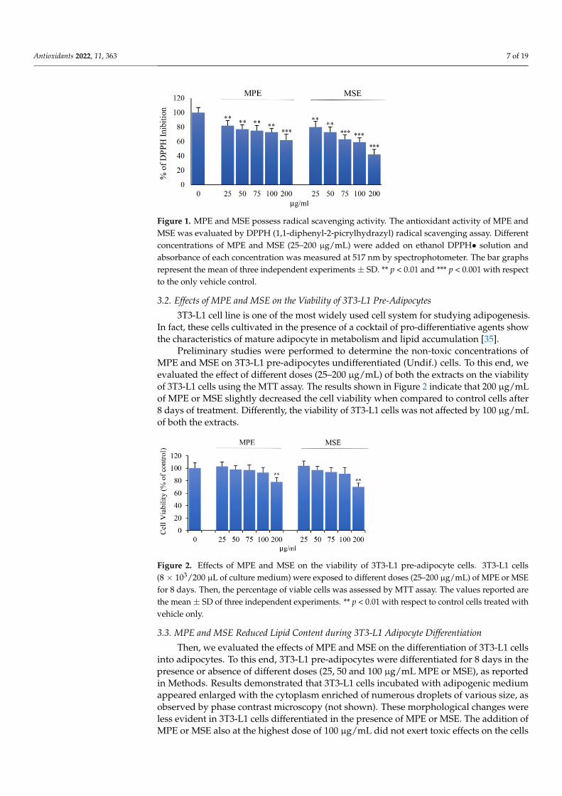
flesh, peel and seed kernel samples [20]. HPLC-ESI-MS analysis of Mango seed extracts(MSE) evidenced the presence of 8 polar compounds in MSE (see Table 1). Quantificationof mangiferin and gallic acid was performed with the calibration curves of their own stan-dards. According to previous studies, the gallates, gallotannins and maclurin derivativeswere quantified using the calibration curve of gallic acid.
Table 1. Composition of mango seed extract (MSE) HPLC-ESI-MS analysis of Mango seed extracts(MSE) evidenced the presence of 8 polar compounds in MSE. Quantification of mangiferin and gallicacid was performed with the calibration curves of their own standards.
Compound RT (min) ESI [M − H] (m/z) Molecular Formula ppm mg/100 g
Teor. Exp.
1 Disaccaride 1.5 341.1089 [M − H]− 341.1089 [M − H]− C12H22O11 trace trace
2 Quinic acid 1.5 191.0561 [M − H]− 191.0561 [M − H]− C7H12O6 trace trace
3 Glucosyl gallate 4.6 331.0671 [M − H]− 331.06707 [M − H]− C13H16O10 2.80 280
4 Gallic acid 5.8 169.0142 [M − H]− 169.01425 [M − H]− C7H6O5 1.3 130
5 Methylgallate 15.3 183.0299 [M − H]− 183.02990 [M − H]− C8H8O5 25.2 2520
6 Mangiferin 20.9 421.0776 [M − H]− 421.07763 [M − H]− C19H18O11 0.5 50
7 Methyl-digallateester isomer 22.6 335.04086 [M-H]- 12.1 1210
8 Maclurin tri-O-galloyl-glucoside 29.0 879.1262 [M − H]− 879.11651 [M − H]− C40H32O23 trace trace
Our previous studies demonstrated that in peel extracts (MPE) of Sicilian mangomethyl digallate (487.15 mg/100 g), methyl gallate (225.87 mg/100 g), gallic acid (118.57 mg/100 g) and glucosyl gallate (108.14 mg/100 g) were the principal phenolic compounds [23].Characterization of MSE evidenced higher concentration of polyphenols in MSE thanMPE. In particular, methyl gallate was the major component (2520 mg/100 g) and methyl-digallate ester isomer (1210 mg/100 g) the second most abundant compound.
3.1. MPE and MSE Possess ROS Scavenger Activities
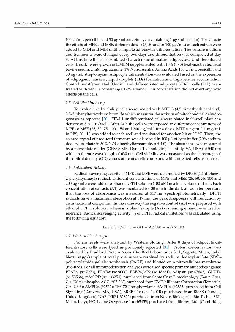
To ascertain the antioxidant activity of MPE and MSE we performed DPPH radicalscavenging assay, as reported in Methods. Figure 1 shows the effect of different concen-trations (25–200 µg/mL) of both the extracts. Our data demonstrated that both MPE andMSE possess an elevated radical scavenging activity. The inhibition of DPPH free radicalexerted by mango extracts was dose-dependent and resulted higher in MSE than MPE, inaccordance with the higher concentrations of polyphenols in MPE. In fact, at a concentrationof 100 µg/mL, the DPPH inhibition exerted by MPE was only by 73%, respect to that ofMSE that reached an inhibition value equal to 59%.
Antioxidants 2022, 11, 363 7 of 19
Antioxidants 2022, 11, x FOR PEER REVIEW 6 of 20
3. Results Comprehensive characterization of the phenolic fraction of Sicilian mango seed was
performed by HPLC-ESI-MS to compare the composition in water-soluble phenolic com-pounds in the two different parts of the fruit and the influence on the activity. As reported in the literature, gallic acid and its derivatives were the largest family found in mango flesh, peel and seed kernel samples [20]. HPLC-ESI-MS analysis of Mango seed extracts (MSE) evidenced the presence of 8 polar compounds in MSE (see Table 1). Quantification of mangiferin and gallic acid was performed with the calibration curves of their own standards. According to previous studies, the gallates, gallotannins and maclurin deriva-tives were quantified using the calibration curve of gallic acid.
Our previous studies demonstrated that in peel extracts (MPE) of Sicilian mango me-thyl digallate (487.15 mg/100 g), methyl gallate (225.87 mg/100 g), gallic acid (118.57 mg/100 g) and glucosyl gallate (108.14 mg/100 g) were the principal phenolic compounds [23]. Characterization of MSE evidenced higher concentration of polyphenols in MSE than MPE. In particular, methyl gallate was the major component (2520 mg/100 g) and methyl-digallate ester isomer (1210 mg/100 g) the second most abundant compound.
Table 1. Composition of mango seed extract (MSE) HPLC-ESI-MS analysis of Mango seed extracts (MSE) evidenced the presence of 8 polar compounds in MSE. Quantification of mangiferin and gallic acid was performed with the calibration curves of their own standards.
Compound RT (min) ESI [M-H] (m/z) Molecular formula ppm mg/100 g Teor. Exp. 1 Disaccaride 1.5 341.1089 [M − H]− 341.1089 [M − H]− C12H22O11 trace trace 2 Quinic acid 1.5 191.0561 [M − H]− 191.0561 [M − H]− C7H12O6 trace trace 3 Glucosyl gallate 4.6 331.0671 [M − H]− 331.06707 [M − H]− C13H16O10 2.80 280 4 Gallic acid 5.8 169.0142 [M − H]− 169.01425 [M − H]− C7H6O5 1.3 130 5 Methylgallate 15.3 183.0299 [M − H]− 183.02990 [M − H]− C8H8O5 25.2 2520 6 Mangiferin 20.9 421.0776 [M − H]− 421.07763 [M − H]− C19H18O11 0.5 50 7 Methyl-digallate ester isomer 22.6 335.04086 [M-H]- 12.1 1210 8 Maclurin tri-O-galloyl-glucoside 29.0 879.1262 [M − H]− 879.11651 [M − H]− C40H32O23 trace trace
3.1. MPE and MSE Possess ROS Scavenger Activities To ascertain the antioxidant activity of MPE and MSE we performed DPPH radical
scavenging assay, as reported in Methods. Figure 1 shows the effect of different concen-trations (25–200 µg/mL) of both the extracts. Our data demonstrated that both MPE and MSE possess an elevated radical scavenging activity. The inhibition of DPPH free radical exerted by mango extracts was dose-dependent and resulted higher in MSE than MPE, in accordance with the higher concentrations of polyphenols in MPE. In fact, at a concentra-tion of 100 µg/mL, the DPPH inhibition exerted by MPE was only by 73%, respect to that of MSE that reached an inhibition value equal to 59%.
Figure 1. MPE and MSE possess radical scavenging activity. The antioxidant activity of MPE and MSE was evaluated by DPPH (1,1-diphenyl-2-picrylhydrazyl) radical scavenging assay. Different
Figure 1. MPE and MSE possess radical scavenging activity. The antioxidant activity of MPE andMSE was evaluated by DPPH (1,1-diphenyl-2-picrylhydrazyl) radical scavenging assay. Differentconcentrations of MPE and MSE (25–200 µg/mL) were added on ethanol DPPH• solution andabsorbance of each concentration was measured at 517 nm by spectrophotometer. The bar graphsrepresent the mean of three independent experiments ± SD. ** p < 0.01 and *** p < 0.001 with respectto the only vehicle control.
3.2. Effects of MPE and MSE on the Viability of 3T3-L1 Pre-Adipocytes
3T3-L1 cell line is one of the most widely used cell system for studying adipogenesis.In fact, these cells cultivated in the presence of a cocktail of pro-differentiative agents showthe characteristics of mature adipocyte in metabolism and lipid accumulation [35].
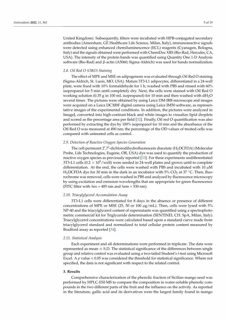
Preliminary studies were performed to determine the non-toxic concentrations ofMPE and MSE on 3T3-L1 pre-adipocytes undifferentiated (Undif.) cells. To this end, weevaluated the effect of different doses (25–200 µg/mL) of both the extracts on the viabilityof 3T3-L1 cells using the MTT assay. The results shown in Figure 2 indicate that 200 µg/mLof MPE or MSE slightly decreased the cell viability when compared to control cells after8 days of treatment. Differently, the viability of 3T3-L1 cells was not affected by 100 µg/mLof both the extracts.
Antioxidants 2022, 11, x FOR PEER REVIEW 7 of 20
concentrations of MPE and MSE (25–200 µg/mL) were added on ethanol DPPH• solution and ab-sorbance of each concentration was measured at 517 nm by spectrophotometer. The bar graphs rep-resent the mean of three independent experiments ±SD. ** p <0.01 and *** p <0.001 with respect to the only vehicle control.
3.2. Effects of MPE and MSE on the Viability of 3T3-L1 Pre-Adipocytes 3T3-L1 cell line is one of the most widely used cell system for studying adipogenesis.
In fact, these cells cultivated in the presence of a cocktail of pro-differentiative agents show the characteristics of mature adipocyte in metabolism and lipid accumulation [35].
Preliminary studies were performed to determine the non-toxic concentrations of MPE and MSE on 3T3-L1 pre-adipocytes undifferentiated (Undif.) cells. To this end, we evaluated the effect of different doses (25–200 µg/mL) of both the extracts on the viability of 3T3-L1 cells using the MTT assay. The results shown in Figure 2 indicate that 200 µg/mL of MPE or MSE slightly decreased the cell viability when compared to control cells after 8 days of treatment. Differently, the viability of 3T3-L1 cells was not affected by 100 µg/mL of both the extracts.
Figure 2. Effects of MPE and MSE on the viability of 3T3-L1 pre-adipocyte cells. 3T3-L1 cells (8 × 10³/200 µL of culture medium) were exposed to different doses (25–200 µg/mL) of MPE or MSE for 8 days. Then, the percentage of viable cells was assessed by MTT assay. The values reported are the mean ± SD of three independent experiments. ** p < 0.01 with respect to control cells treated with vehicle only.
3.3. MPE and MSE Reduced Lipid Content during 3T3-L1 Adipocyte Differentiation Then, we evaluated the effects of MPE and MSE on the differentiation of 3T3-L1 cells
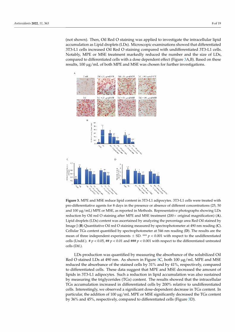
into adipocytes. To this end, 3T3-L1 pre-adipocytes were differentiated for 8 days in the presence or absence of different doses (25, 50 and 100 µg/mL MPE or MSE), as reported in Methods. Results demonstrated that 3T3-L1 cells incubated with adipogenic medium ap-peared enlarged with the cytoplasm enriched of numerous droplets of various size, as observed by phase contrast microscopy (not shown). These morphological changes were less evident in 3T3-L1 cells differentiated in the presence of MPE or MSE. The addition of MPE or MSE also at the highest dose of 100 µg/mL did not exert toxic effects on the cells (not shown). Then, Oil Red O staining was applied to investigate the intracellular lipid accumulation as Lipid droplets (LDs). Microscopic examinations showed that differenti-ated 3T3-L1 cells increased Oil Red O staining compared with undifferentiated 3T3-L1 cells. Notably, MPE or MSE treatment markedly reduced the number and the size of LDs, compared to differentiated cells with a dose dependent effect (Figure 3A,B). Based on these results, 100 µg/mL of both MPE and MSE was chosen for further investigations.
LDs production was quantified by measuring the absorbance of the solubilized Oil Red O stained LDs at 490 nm. As shown in Figure 3C, both 100 µg/mL MPE and MSE reduced the absorbance of the stained cells by 31% and by 41%, respectively, compared to differentiated cells. These data suggest that MPE and MSE decreased the amount of lipids
Figure 2. Effects of MPE and MSE on the viability of 3T3-L1 pre-adipocyte cells. 3T3-L1 cells(8 × 10³/200 µL of culture medium) were exposed to different doses (25–200 µg/mL) of MPE or MSEfor 8 days. Then, the percentage of viable cells was assessed by MTT assay. The values reported arethe mean ± SD of three independent experiments. ** p < 0.01 with respect to control cells treated withvehicle only.
3.3. MPE and MSE Reduced Lipid Content during 3T3-L1 Adipocyte Differentiation
Then, we evaluated the effects of MPE and MSE on the differentiation of 3T3-L1 cellsinto adipocytes. To this end, 3T3-L1 pre-adipocytes were differentiated for 8 days in thepresence or absence of different doses (25, 50 and 100 µg/mL MPE or MSE), as reportedin Methods. Results demonstrated that 3T3-L1 cells incubated with adipogenic mediumappeared enlarged with the cytoplasm enriched of numerous droplets of various size, asobserved by phase contrast microscopy (not shown). These morphological changes wereless evident in 3T3-L1 cells differentiated in the presence of MPE or MSE. The addition ofMPE or MSE also at the highest dose of 100 µg/mL did not exert toxic effects on the cells
Antioxidants 2022, 11, 363 8 of 19
(not shown). Then, Oil Red O staining was applied to investigate the intracellular lipidaccumulation as Lipid droplets (LDs). Microscopic examinations showed that differentiated3T3-L1 cells increased Oil Red O staining compared with undifferentiated 3T3-L1 cells.Notably, MPE or MSE treatment markedly reduced the number and the size of LDs,compared to differentiated cells with a dose dependent effect (Figure 3A,B). Based on theseresults, 100 µg/mL of both MPE and MSE was chosen for further investigations.
Antioxidants 2022, 11, x FOR PEER REVIEW 8 of 20
in 3T3-L1 adipocytes. Such a reduction in lipid accumulation was also sustained by meas-uring the triglycerides (TGs) content. The results showed that the intracellular TGs accu-mulation increased in differentiated cells by 200% relative to undifferentiated cells. Inter-estingly, we observed a significant dose-dependent decrease in TGs content. In particular, the addition of 100 µg/mL MPE or MSE significantly decreased the TGs content by 36% and 45%, respectively, compared to differentiated cells (Figure 3D).
Figure 3. MPE and MSE reduce lipid content in 3T3-L1 adipocytes. 3T3-L1 cells were treated with pro-differentiative agents for 8 days in the presence or absence of different concentrations (25, 50 and 100 µg/mL) MPE or MSE, as reported in Methods. Representative photographs showing LDs reduction by Oil red O staining after MPE and MSE treatment (200× original magnification) (A). Lipid droplets (LDs) content was ascertained by analyzing the percentage area Red Oil stained by Image J (B) Quantitative Oil red O staining measured by spectrophotometer at 490 nm reading (C). Cellular TGs content quantified by spectrophotometer at 546 nm reading (D). The results are the mean of three independent experiments ±SD. *** p < 0.001 with respect to the undifferentiated cells (Undif.). # p < 0.05, ## p < 0.01 and ### p < 0.001 with respect to the differentiated untreated cells (Dif.).
3.4. MPE and MSE Reduced the Expression of Key Factors of Adipogenic Differentiation and Lipid Accumulation
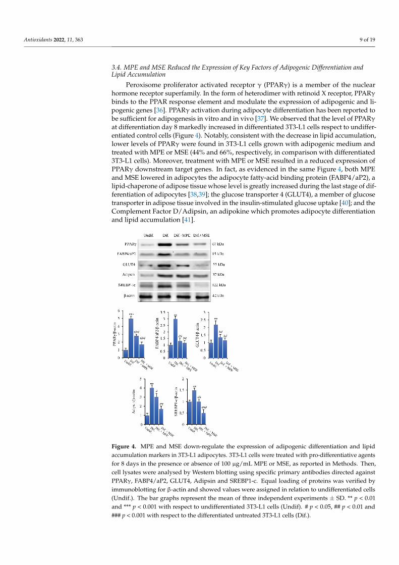
Peroxisome proliferator activated receptor γ (PPARγ) is a member of the nuclear hor-mone receptor superfamily. In the form of heterodimer with retinoid X receptor, PPARγ binds to the PPAR response element and modulate the expression of adipogenic and lipo-genic genes [36]. PPARγ activation during adipocyte differentiation has been reported to be sufficient for adipogenesis in vitro and in vivo [37]. We observed that the level of PPARγ at differentiation day 8 markedly increased in differentiated 3T3-L1 cells respect to undifferentiated control cells (Figure 4). Notably, consistent with the decrease in lipid accumulation, lower levels of PPARγ were found in 3T3-L1 cells grown with adipogenic
Figure 3. MPE and MSE reduce lipid content in 3T3-L1 adipocytes. 3T3-L1 cells were treated withpro-differentiative agents for 8 days in the presence or absence of different concentrations (25, 50and 100 µg/mL) MPE or MSE, as reported in Methods. Representative photographs showing LDsreduction by Oil red O staining after MPE and MSE treatment (200× original magnification) (A).Lipid droplets (LDs) content was ascertained by analyzing the percentage area Red Oil stained byImage J (B) Quantitative Oil red O staining measured by spectrophotometer at 490 nm reading (C).Cellular TGs content quantified by spectrophotometer at 546 nm reading (D). The results are themean of three independent experiments ± SD. *** p < 0.001 with respect to the undifferentiatedcells (Undif.). # p < 0.05, ## p < 0.01 and ### p < 0.001 with respect to the differentiated untreatedcells (Dif.).
LDs production was quantified by measuring the absorbance of the solubilized OilRed O stained LDs at 490 nm. As shown in Figure 3C, both 100 µg/mL MPE and MSEreduced the absorbance of the stained cells by 31% and by 41%, respectively, comparedto differentiated cells. These data suggest that MPE and MSE decreased the amount oflipids in 3T3-L1 adipocytes. Such a reduction in lipid accumulation was also sustainedby measuring the triglycerides (TGs) content. The results showed that the intracellularTGs accumulation increased in differentiated cells by 200% relative to undifferentiatedcells. Interestingly, we observed a significant dose-dependent decrease in TGs content. Inparticular, the addition of 100 µg/mL MPE or MSE significantly decreased the TGs contentby 36% and 45%, respectively, compared to differentiated cells (Figure 3D).
Antioxidants 2022, 11, 363 9 of 19
3.4. MPE and MSE Reduced the Expression of Key Factors of Adipogenic Differentiation andLipid Accumulation
Peroxisome proliferator activated receptor γ (PPARγ) is a member of the nuclearhormone receptor superfamily. In the form of heterodimer with retinoid X receptor, PPARγbinds to the PPAR response element and modulate the expression of adipogenic and li-pogenic genes [36]. PPARγ activation during adipocyte differentiation has been reported tobe sufficient for adipogenesis in vitro and in vivo [37]. We observed that the level of PPARγat differentiation day 8 markedly increased in differentiated 3T3-L1 cells respect to undiffer-entiated control cells (Figure 4). Notably, consistent with the decrease in lipid accumulation,lower levels of PPARγ were found in 3T3-L1 cells grown with adipogenic medium andtreated with MPE or MSE (44% and 66%, respectively, in comparison with differentiated3T3-L1 cells). Moreover, treatment with MPE or MSE resulted in a reduced expression ofPPARγ downstream target genes. In fact, as evidenced in the same Figure 4, both MPEand MSE lowered in adipocytes the adipocyte fatty-acid binding protein (FABP4/aP2), alipid-chaperone of adipose tissue whose level is greatly increased during the last stage of dif-ferentiation of adipocytes [38,39]; the glucose transporter 4 (GLUT4), a member of glucosetransporter in adipose tissue involved in the insulin-stimulated glucose uptake [40]; and theComplement Factor D/Adipsin, an adipokine which promotes adipocyte differentiationand lipid accumulation [41].
Antioxidants 2022, 11, x FOR PEER REVIEW 9 of 20
medium and treated with MPE or MSE (44% and 66%, respectively, in comparison with differentiated 3T3-L1 cells). Moreover, treatment with MPE or MSE resulted in a reduced expression of PPARγ downstream target genes. In fact, as evidenced in the same Figure 4, both MPE and MSE lowered in adipocytes the adipocyte fatty-acid binding protein (FABP4/aP2), a lipid-chaperone of adipose tissue whose level is greatly increased during the last stage of differentiation of adipocytes [38,39]; the glucose transporter 4 (GLUT4), a member of glucose transporter in adipose tissue involved in the insulin-stimulated glu-cose uptake [40]; and the Complement Factor D/Adipsin, an adipokine which promotes adipocyte differentiation and lipid accumulation [41].
We further examined the effects of mango extracts on the expression level of Sterol-regulatory element-binding protein-1c (SREBP-1c). SREBP-1c is a transcription factor up-regulated under adipocyte differentiation which regulates the expression of genes in-volved in de novo lipogenesis and triglyceride synthesis, including ATP-citrate lyase (ACL), Acetil-Coa Carboxylase (ACC), Fatty acid synthase (FAS), Stearoyl-CoA desatu-rase (SCD1) and Glycerol-3-phosphate acyltransferase (GPAT) [42–45]. We demonstrated that the level of SREBP-1c increased by 50% in differentiated 3T3-L1 cells in comparison with undifferentiated cells. Interestingly, the addition MPE or MSE in the differentiation medium lowered the level of this factor (33% and 67%, respectively, in comparison with 3T3-L1 differentiated cells (Figure 4).
Figure 4. MPE and MSE down-regulate the expression of adipogenic differentiation and lipid ac-cumulation markers in 3T3-L1 adipocytes. 3T3-L1 cells were treated with pro-differentiative agents for 8 days in the presence or absence of 100 µg/mL MPE or MSE, as reported in Methods. Then, cell
Figure 4. MPE and MSE down-regulate the expression of adipogenic differentiation and lipidaccumulation markers in 3T3-L1 adipocytes. 3T3-L1 cells were treated with pro-differentiative agentsfor 8 days in the presence or absence of 100 µg/mL MPE or MSE, as reported in Methods. Then,cell lysates were analysed by Western blotting using specific primary antibodies directed againstPPARγ, FABP4/aP2, GLUT4, Adipsin and SREBP1-c. Equal loading of proteins was verified byimmunoblotting for β-actin and showed values were assigned in relation to undifferentiated cells(Undif.). The bar graphs represent the mean of three independent experiments ± SD. ** p < 0.01and *** p < 0.001 with respect to undifferentiated 3T3-L1 cells (Undif). # p < 0.05, ## p < 0.01 and### p < 0.001 with respect to the differentiated untreated 3T3-L1 cells (Dif.).

Antioxidants 2022, 11, 363 10 of 19
We further examined the effects of mango extracts on the expression level of Sterol-regulatory element-binding protein-1c (SREBP-1c). SREBP-1c is a transcription factorupregulated under adipocyte differentiation which regulates the expression of genes in-volved in de novo lipogenesis and triglyceride synthesis, including ATP-citrate lyase (ACL),Acetil-Coa Carboxylase (ACC), Fatty acid synthase (FAS), Stearoyl-CoA desaturase (SCD1)and Glycerol-3-phosphate acyltransferase (GPAT) [42–45]. We demonstrated that the levelof SREBP-1c increased by 50% in differentiated 3T3-L1 cells in comparison with undifferen-tiated cells. Interestingly, the addition MPE or MSE in the differentiation medium loweredthe level of this factor (33% and 67%, respectively, in comparison with 3T3-L1 differentiatedcells (Figure 4).
3.5. MPE and MSE Increase the Levels of Lipolytic Factors
We further examined the ability of MPE and MSE to activate factors promoting lipoly-sis in adipocytes, such as PPARα and AMPK. Peroxisome proliferator-activated receptor-α(PPARα) is another member of PPAR family expressed mostly in tissues with high ratesof fatty acid oxidation, such as the liver, brown fat and muscle [46]. It has been shown toup-regulate the expression of genes involved in fatty acid oxidation, particularly when co-activated by PPAR coactivator 1 (PGC-1) [47]. Our data, shown in Figure 5, demonstratedthat the addition of MPE or MSE in the differentiation medium enhanced the expressionlevel of PPARα (40% and 70%, respectively).
Antioxidants 2022, 11, x FOR PEER REVIEW 10 of 20
lysates were analysed by Western blotting using specific primary antibodies directed against PPARγ, FABP4/aP2, GLUT4, Adipsin and SREBP1-c. Equal loading of proteins was verified by im-munoblotting for β-actin and showed values were assigned in relation to undifferentiated cells (Un-dif.). The bar graphs represent the mean of three independent experiments ±SD. ** p < 0.01 and *** p < 0.001 with respect to undifferentiated 3T3-L1 cells (Undif). # p < 0.05, ## p < 0.01 and ### p < 0.001 with respect to the differentiated untreated 3T3-L1 cells (Dif.).
3.5. MPE and MSE Increase the Levels of Lipolytic Factors We further examined the ability of MPE and MSE to activate factors promoting lipol-
ysis in adipocytes, such as PPARα and AMPK. Peroxisome proliferator-activated recep-tor-α (PPARα) is another member of PPAR family expressed mostly in tissues with high rates of fatty acid oxidation, such as the liver, brown fat and muscle [46]. It has been shown to up-regulate the expression of genes involved in fatty acid oxidation, particularly when co-activated by PPAR coactivator 1 (PGC-1) [47]. Our data, shown in Figure 5, demon-strated that the addition of MPE or MSE in the differentiation medium enhanced the ex-pression level of PPARα (40% and 70%, respectively).
Figure 5. MPE and MSE up-regulate PPARα and activate AMPK in 3T3-L1 adipocytes. 3T3-L1 cells were treated with pro-differentiative agents for 8 days in the presence or absence of 100 µg/mL MPE or MSE, as reported in Methods. Then, cell lysates were analysed by Western blotting using specific primary antibodies directed against PPARα, AMPK, phospho-AMPK and phospho-ACC. Equal loading of proteins was verified by immunoblotting for β-actin and showed values were assigned in relation to undifferentiated 3T3-L1 cells (Undif). The bar graphs represent the mean of three in-dependent experiments ±SD. # p < 0.05, ## p < 0.01 and ### p < 0.001 with respect to differentiated untreated cells (Dif.).
AMP-activated kinase (AMPK) is an energy sensor regulating glucose and lipid me-tabolism [13]. Upon activation in the phosphorylated form, AMPK reduces lipid synthesis by inhibiting the activity of the key enzymes of fatty acid de novo synthesis, such as ACC [13]. To investigate whether MPE and MSE are capable of activating AMPK, we examined by Western blotting analysis the levels of AMPK and its phosphorylated form. As shown in Figure 5, compared to differentiated 3T3-L1 cells, the presence of MPE or MSE in the
Figure 5. MPE and MSE up-regulate PPARα and activate AMPK in 3T3-L1 adipocytes. 3T3-L1 cellswere treated with pro-differentiative agents for 8 days in the presence or absence of 100 µg/mL MPEor MSE, as reported in Methods. Then, cell lysates were analysed by Western blotting using specificprimary antibodies directed against PPARα, AMPK, phospho-AMPK and phospho-ACC. Equalloading of proteins was verified by immunoblotting for β-actin and showed values were assignedin relation to undifferentiated 3T3-L1 cells (Undif). The bar graphs represent the mean of threeindependent experiments ± SD. # p < 0.05, ## p < 0.01 and ### p < 0.001 with respect to differentiateduntreated cells (Dif.).
AMP-activated kinase (AMPK) is an energy sensor regulating glucose and lipidmetabolism [13]. Upon activation in the phosphorylated form, AMPK reduces lipid syn-thesis by inhibiting the activity of the key enzymes of fatty acid de novo synthesis, suchas ACC [13]. To investigate whether MPE and MSE are capable of activating AMPK, we
Antioxidants 2022, 11, 363 11 of 19
examined by Western blotting analysis the levels of AMPK and its phosphorylated form.As shown in Figure 5, compared to differentiated 3T3-L1 cells, the presence of MPE orMSE in the differentiation medium enhanced the phosphorylation of AMPK by 23% and45%, respectively. The results also showed that MPE or MSE did not significantly modifythe basal levels of AMPK (Figure 5), thus supporting a role of mango extracts on AMPKactivation. Concomitantly, the expression of the phosphorylated and inactive form of ACCincreased following MPE and MSE treatment by 44% and 110%, respectively.
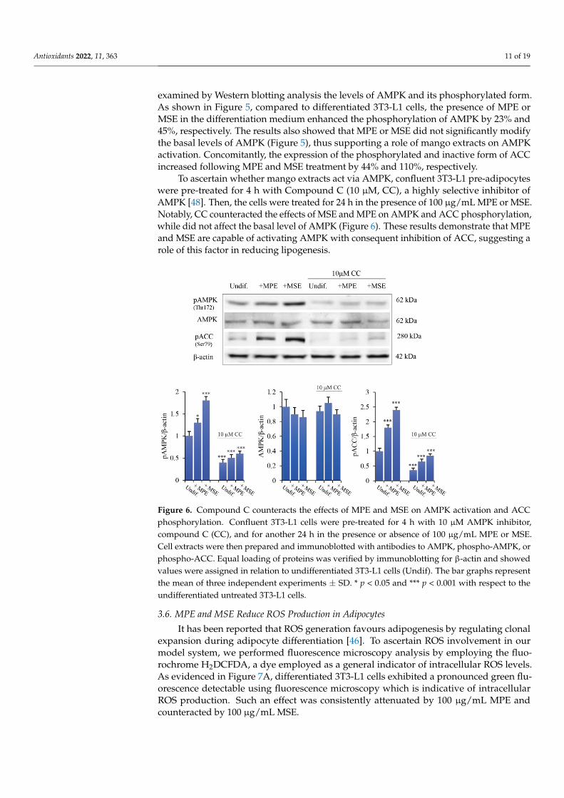
To ascertain whether mango extracts act via AMPK, confluent 3T3-L1 pre-adipocyteswere pre-treated for 4 h with Compound C (10 µM, CC), a highly selective inhibitor ofAMPK [48]. Then, the cells were treated for 24 h in the presence of 100 µg/mL MPE or MSE.Notably, CC counteracted the effects of MSE and MPE on AMPK and ACC phosphorylation,while did not affect the basal level of AMPK (Figure 6). These results demonstrate that MPEand MSE are capable of activating AMPK with consequent inhibition of ACC, suggesting arole of this factor in reducing lipogenesis.
Antioxidants 2022, 11, x FOR PEER REVIEW 11 of 20
differentiation medium enhanced the phosphorylation of AMPK by 23% and 45%, respec-tively. The results also showed that MPE or MSE did not significantly modify the basal levels of AMPK (Figure 5), thus supporting a role of mango extracts on AMPK activation. Concomitantly, the expression of the phosphorylated and inactive form of ACC increased following MPE and MSE treatment by 44% and 110%, respectively.
To ascertain whether mango extracts act via AMPK, confluent 3T3-L1 pre-adipocytes were pre-treated for 4 h with Compound C (10 µM, CC), a highly selective inhibitor of AMPK [48]. Then, the cells were treated for 24 h in the presence of 100 µg/mL MPE or MSE. Notably, CC counteracted the effects of MSE and MPE on AMPK and ACC phos-phorylation, while did not affect the basal level of AMPK (Figure 6). These results demon-strate that MPE and MSE are capable of activating AMPK with consequent inhibition of ACC, suggesting a role of this factor in reducing lipogenesis.
Figure 6. Compound C counteracts the effects of MPE and MSE on AMPK activation and ACC phosphorylation. Confluent 3T3-L1 cells were pre-treated for 4 h with 10 µM AMPK inhibitor, com-pound C (CC), and for another 24 h in the presence or absence of 100 µg/mL MPE or MSE. Cell extracts were then prepared and immunoblotted with antibodies to AMPK, phospho-AMPK, or phospho-ACC. Equal loading of proteins was verified by immunoblotting for β-actin and showed values were assigned in relation to undifferentiated 3T3-L1 cells (Undif). The bar graphs represent the mean of three independent experiments ±SD. * p < 0.05 and *** p < 0.001 with respect to the undifferentiated untreated 3T3-L1 cells.
3.6. MPE and MSE Reduce ROS Production in Adipocytes It has been reported that ROS generation favours adipogenesis by regulating clonal
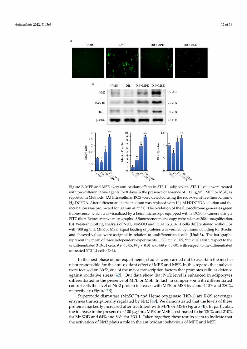
expansion during adipocyte differentiation [46]. To ascertain ROS involvement in our model system, we performed fluorescence microscopy analysis by employing the fluoro-chrome H2DCFDA, a dye employed as a general indicator of intracellular ROS levels. As evidenced in Figure 7A, differentiated 3T3-L1 cells exhibited a pronounced green fluores-cence detectable using fluorescence microscopy which is indicative of intracellular ROS production. Such an effect was consistently attenuated by 100 µg/mL MPE and counter-acted by 100 µg/mL MSE.
In the next phase of our experiments, studies were carried out to ascertain the mech-anism responsible for the anti-oxidant effect of MPE and MSE. In this regard, the analyses
Figure 6. Compound C counteracts the effects of MPE and MSE on AMPK activation and ACCphosphorylation. Confluent 3T3-L1 cells were pre-treated for 4 h with 10 µM AMPK inhibitor,compound C (CC), and for another 24 h in the presence or absence of 100 µg/mL MPE or MSE.Cell extracts were then prepared and immunoblotted with antibodies to AMPK, phospho-AMPK, orphospho-ACC. Equal loading of proteins was verified by immunoblotting for β-actin and showedvalues were assigned in relation to undifferentiated 3T3-L1 cells (Undif). The bar graphs representthe mean of three independent experiments ± SD. * p < 0.05 and *** p < 0.001 with respect to theundifferentiated untreated 3T3-L1 cells.
3.6. MPE and MSE Reduce ROS Production in Adipocytes
It has been reported that ROS generation favours adipogenesis by regulating clonalexpansion during adipocyte differentiation [46]. To ascertain ROS involvement in ourmodel system, we performed fluorescence microscopy analysis by employing the fluo-rochrome H2DCFDA, a dye employed as a general indicator of intracellular ROS levels.As evidenced in Figure 7A, differentiated 3T3-L1 cells exhibited a pronounced green flu-orescence detectable using fluorescence microscopy which is indicative of intracellularROS production. Such an effect was consistently attenuated by 100 µg/mL MPE andcounteracted by 100 µg/mL MSE.
Antioxidants 2022, 11, 363 12 of 19
Antioxidants 2022, 11, x FOR PEER REVIEW 12 of 20
were focused on Nrf2, one of the major transcription factors that promotes cellular defence against oxidative stress [42]. Our data show that Nrf2 level is enhanced in adipocytes dif-ferentiated in the presence of MPE or MSE. In fact, in comparison with differentiated con-trol cells the level of Nrf2 protein increases with MPE or MSE by about 110% and 280%, respectively (Figure 7B).
Figure 7. MPE and MSE exert anti-oxidant effects in 3T3-L1 adipocytes. 3T3-L1 cells were treated with pro-differentiative agents for 8 days in the presence or absence of 100 µg/mL MPE or MSE, as reported in Methods. (A) Intracellular ROS were detected using the redox-sensitive fluorochrome H2-DCFDA. After differentiation, the medium was replaced with 10 µM H2DCFDA solution and the incubation was protracted for 30 min at 37 °C. The oxidation of the fluorochrome generates green fluorescence, which was visualized by a Leica microscope equipped with a DC300F camera using a FITC filter. Representative micrographs of fluorescence microscopy were taken at 200× magnifica-tion. (B). Western blotting analysis of Nrf2, MnSOD and HO-1 in 3T3-L1 cells differentiated without or with 100 µg/mL MPE or MSE. Equal loading of proteins was verified by immunoblotting for β-actin and showed values were assigned in relation to undifferentiated cells (Undif.). The bar graphs represent the mean of three independent experiments ±SD. * p < 0.05, ** p < 0.01 with respect to the undifferentiated 3T3-L1 cells, # p < 0.05, ## p < 0.01 and ### p < 0.001 with respect to the differentiated untreated 3T3-L1 cells (Dif.).
Superoxide dismutase (MnSOD) and Heme oxygenase (HO-1) are ROS scavenger enzymes transcriptionally regulated by Nrf2 [49]. We demonstrated that the levels of these proteins markedly increased after treatment with MPE or MSE (Figure 7B). In particular, the increase in the presence of 100 µg/mL MPE or MSE is estimated to be 120% and 210% for MnSOD and 64% and 86% for HO-1. Taken together, these results seem to indicate that the activation of Nrf2 plays a role in the antioxidant behaviour of MPE and MSE.
Figure 7. MPE and MSE exert anti-oxidant effects in 3T3-L1 adipocytes. 3T3-L1 cells were treatedwith pro-differentiative agents for 8 days in the presence or absence of 100 µg/mL MPE or MSE, asreported in Methods. (A) Intracellular ROS were detected using the redox-sensitive fluorochromeH2-DCFDA. After differentiation, the medium was replaced with 10 µM H2DCFDA solution and theincubation was protracted for 30 min at 37 ◦C. The oxidation of the fluorochrome generates greenfluorescence, which was visualized by a Leica microscope equipped with a DC300F camera using aFITC filter. Representative micrographs of fluorescence microscopy were taken at 200×magnification.(B). Western blotting analysis of Nrf2, MnSOD and HO-1 in 3T3-L1 cells differentiated without orwith 100 µg/mL MPE or MSE. Equal loading of proteins was verified by immunoblotting for β-actinand showed values were assigned in relation to undifferentiated cells (Undif.). The bar graphsrepresent the mean of three independent experiments ± SD. * p < 0.05, ** p < 0.01 with respect to theundifferentiated 3T3-L1 cells, # p < 0.05, ## p < 0.01 and ### p < 0.001 with respect to the differentiateduntreated 3T3-L1 cells (Dif.).
In the next phase of our experiments, studies were carried out to ascertain the mecha-nism responsible for the anti-oxidant effect of MPE and MSE. In this regard, the analyseswere focused on Nrf2, one of the major transcription factors that promotes cellular defenceagainst oxidative stress [42]. Our data show that Nrf2 level is enhanced in adipocytesdifferentiated in the presence of MPE or MSE. In fact, in comparison with differentiatedcontrol cells the level of Nrf2 protein increases with MPE or MSE by about 110% and 280%,respectively (Figure 7B).
Superoxide dismutase (MnSOD) and Heme oxygenase (HO-1) are ROS scavengerenzymes transcriptionally regulated by Nrf2 [49]. We demonstrated that the levels of theseproteins markedly increased after treatment with MPE or MSE (Figure 7B). In particular,the increase in the presence of 100 µg/mL MPE or MSE is estimated to be 120% and 210%for MnSOD and 64% and 86% for HO-1. Taken together, these results seem to indicate thatthe activation of Nrf2 plays a role in the antioxidant behaviour of MPE and MSE.
Antioxidants 2022, 11, 363 13 of 19
4. Discussion
This paper aimed at investigating the anti-adipogenic effect of peel and seed frommango cultivated in Sicily in 3T3-L1 adipocytes. Sicily is a region of the Southern Italycharacterized by a favorable subtropical climate and adapted soils that favour mangocultivation conferring particular properties to the orchards [20].
It is worthwhile to mention that, although the chemical investigation of differentphytocomponents of mango fruit has been already published by other researchers, inthis paper we focused on cultivars of mango grown in the Sicilian rural areas to providean analysis of their specific composition. Indeed, many different factors can affect theplant phytochemical profile including environmental factors, mango variety, course offruit ripeness [50]. Such an aspect was also reported by Ajila et al. [51] providing evidencethat MPE polyphenolic content strongly depends on fruit maturity stage at the time ofharvest, favorable climatic conditions as well as growing location [52]. Interestingly, thecharacterization of the polyphenolic profile of MPE and MSE using HPLC/MS providedevidence that both these fractions of mango are rich in polyphenols. In particular, asdemonstrated in a previous study [23], methyl digallate, methyl gallate, gallic acid andglucosyl gallate were the principal phenolic compounds in MPE. The polyphenols profileof MSE is similar to MPE, with methyl digallate and methyl gallate representing the maincomponents. Notably, these polyphenols are at higher concentration in MSE than MPE.Gallic acid and its derivatives methyl gallate, methyl digallate and glucosyl gallate areplant secondary polyphenolic metabolites which possess strong anti-oxidant effects, due totheir ability to act as ROS scavenger [53]. Interestingly, gallic acid exerts protective effectsagainst obesity-related inflammation by reducing adipocyte size and the inflammationmarkers, as IL-6, NOS and COX2 [54]. Notably, methy gallate has been shown to exertanti-adipogenic effects in 3T3-L1 cells and human subcutaneous adipocytes, by reducingtriglyceride content and down-regulating adipocyte differentiation markers as C/EBPα,PPARγ and FABP4 [55].
Significantly, our results provided evidence that Sicilian MPE and MSE have strongeffects on reducing adipogenesis and lipid accumulation in 3T3-L1 adipocytes. Adipogene-sis resulting from differentiation of pre-adipocytes into adipocytes leads to intracellularlipid accumulation [10,56]. Interestingly, our data demonstrated that accumulation ofLipid droplets (LDs) and triacylglicerols (TGs) induced by adipocyte differentiation waslargely reduced by treatment of MPE and MSE compared with untreated differentiatedcells, thus indicating their promising role as anti-obesity agents by inhibition of lipidaccumulation. Notably, the highest dose of MPE and MSE (100 µg/mL) which exertsanti-adipogenic effects did not show toxic effects on both pre-adipocyte and differentiated3T3-L1 cells. Therefore, inhibition of TGs accumulation by mango extracts seems to berelated to reduction in adipogenesis without cytotoxicity.
To understand the molecular mechanism underlying the anti-adipogenic effect of MPEand MSE, we then focused our attention on some key factors involved in both adipocytedifferentiation and lipid metabolism. Adipogenesis is a complex process which is tightlyregulated by sequential activation of various transcriptional factors [11]. PPARγ is amember of the nuclear hormone receptor family expressed in adipose tissue. Its levelincreases at an early stage of this differentiation process to stimulate the expression of manyadipocyte-specific genes which control fatty acid metabolism [36]. Induction of PPARγ hasbeen shown to be necessary for adipogenesis both in vitro and in vivo and in many casessufficient to convert non-adipose cells to adipocyte-like cells [37,57]. Notably, both MPEand MSE significantly reduced the expression level of PPARγ consistent with the decreasein lipid accumulation compared with untreated adipocytes. In addition the presence ofmango extracts during adipogenic differentiation led to a reduction in the levels of FABP4and GLUT4, two markers of late adipogenesis which are transcriptionally regulated byPPARγ [58]. In addition, we found that MPE and MSE significantly lowered SREBP-1c, amember of the basic helix–loop–helix-leucine zipper family of transcription factors, whichhas been shown to have an important role in adipogenesis [42]. Taken together, our
Antioxidants 2022, 11, 363 14 of 19
data suggest that mango extracts could counteract adipogenesis by down-regulating theexpression of PPARγ and SREBP-1c.
In addition to downregulate transcription factors involved in the stimulation of adipo-genesis, our results demonstrated that MPE and MSE also up-regulated factors promotingcatabolic process in adipocytes. PPARα is another member of PPAR family mainly ex-pressed in liver and muscle cells [46] which promotes fatty acid oxidation [47]. Its level islow in white adipose tissue suggesting a limited role for this isotype during adipogene-sis [46]. Of note however, pharmacologic PPARα activators reduced adiposity in mousemodels of obesity [59]. Our data demonstrated that MPE and MSE up-regulated PPARαlevels, thus supporting a role of this transcription factor in reducing lipid accumulation in3T3-L1 adipocytes.
Interestingly, we also demonstrate that MPE and MSE increased the phosphorylatedand active form of AMPK. This result is in line with the observation that many polyphenolsderived from plants, such as resveratrol, quercetin, genistein and epigallocatechin gallate,are able to activate AMPK [60]. This activation seems to be mediated by the increase inAMP levels as a consequence of inhibition of mitochondrial ATP production [60]. No-tably, AMPK activation could favor the inhibition of adipogenesis induced by MPE andMSE. This conclusion is in accordance with observation that AMPK negatively regulateswhite adipocyte differentiation [61]. To this end, 5-Aminoimidazole-4-carboxamide-1-β-D-ribofuranoside (AICAR), an activator of AMPK, led to the inhibition of differentiationin 3T3-L1 pre-adipocytes and such an effect was accompanied by decreased PPARγ andC/EBPα [62]. Moreover, the anti-adipogenic effects of several natural compounds seem tobe mediated by AMPK activation [63–65].
In concomitance with activation of AMPK, we observed an increase in the phosphory-lated and inactive form of ACC. ACC is the key rate-limiting enzyme in the first stage offatty acid synthesis and it is inactivated via phosphorylation by AMPK [66]. Notably, weobserved that the addition of compound C, a specific AMPK inhibitor, counteracted theeffects of MPE and MSE on phosphorylation of both AMPK and ACC, thus demonstratingthe ability of mango extracts to activate AMPK and suggesting a role of this factor ininhibiting lipogenesis and adipogenesis in 3T3-L1 cells.
Reactive oxygen species (ROS) generation has been observed during adipogenesisand seems to promote adipocyte differentiation [67]. In particular, it has been shown thatROS increased PPARγ in early pre-adipocyte differentiation and promotes mitotic clonalexpansion of pre-adipocytes [68].
In addition, ROS production has been correlated with enhanced mitochondrial bio-genesis and metabolism during adipogenesis [69]. To support this conclusion it has beenshown that mitochondrial-targeted antioxidants inhibited adipocyte differentiation, whilethe addition of hydrogen peroxide restored it [70]. Notably, our results showed that bothMPE and MSE are capable of counteracting ROS production during 3T3-L1 adipocytedifferentiation. This is in line with the observation that mango extracts possess a strongscavenger activity, as demonstrated by the ability of MPE and MSE to significantly inhibitDPPH activity.
Nrf2 is the master regulator of the cellular antioxidant response, regulating the ex-pression of a battery of genes encoding for antioxidant and detoxifying factors [71]. Undernormal condition, kelch-like ECH-associated protein 1 (Keap1) binds Nrf2 in an inac-tive complex, leading to its ubiquitin-proteasomal degradation. Under oxidative stresscondition, ROS promote oxidation of Keap1 in a critical cysteine residue, promoting itsdissociation from Nrf2 [63]. Thus, Nrf2 traslocates into the nucleus where up-regulate apanel of antioxidant genes, including superoxide dismutase (SOD) and heme oxygenase(HO-1). In accordance with this observation, we demonstrated that during adipocytedifferentiation MPE or MSE upregulated Nrf2 and its targets HO-1 and MnSOD. Thus,the induction of these anti-oxidant enzymes could explain MPE- and MSE-induced ROSreduction in adipocytes. Several reports suggest that different natural compounds exertanti-oxidant effects by activating Nrf2 through different mechanisms including interaction
Antioxidants 2022, 11, 363 15 of 19
with cysteine residues on Keap1, disruption of Nrf2/Keap1 interaction or Nrf2 phospho-rylation [72]. Notably, Nrf2 and AMPK has been shown to be functionally connected andcollaborate to reduce oxidative stress. Induction of AMPK by natural compounds [73]or chemical activators [74] leads in turn to activation via phosphorylation of Nrf2. Thishas been correlated with a reduction in inflammation in several cell types, as adipocytes,macrophages and pancreatic cells [74,75]. Thus, MPE and MSE could explain antioxidanteffects by promoting activation of both AMPK and Nrf2 in 3T3-L1 adipocytes.
5. Conclusions
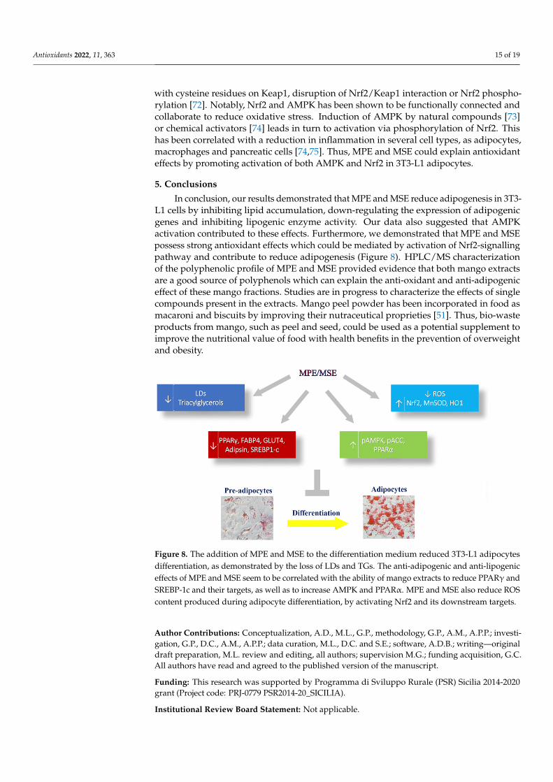
In conclusion, our results demonstrated that MPE and MSE reduce adipogenesis in 3T3-L1 cells by inhibiting lipid accumulation, down-regulating the expression of adipogenicgenes and inhibiting lipogenic enzyme activity. Our data also suggested that AMPKactivation contributed to these effects. Furthermore, we demonstrated that MPE and MSEpossess strong antioxidant effects which could be mediated by activation of Nrf2-signallingpathway and contribute to reduce adipogenesis (Figure 8). HPLC/MS characterizationof the polyphenolic profile of MPE and MSE provided evidence that both mango extractsare a good source of polyphenols which can explain the anti-oxidant and anti-adipogeniceffect of these mango fractions. Studies are in progress to characterize the effects of singlecompounds present in the extracts. Mango peel powder has been incorporated in food asmacaroni and biscuits by improving their nutraceutical proprieties [51]. Thus, bio-wasteproducts from mango, such as peel and seed, could be used as a potential supplement toimprove the nutritional value of food with health benefits in the prevention of overweightand obesity.
Antioxidants 2022, 11, x FOR PEER REVIEW 15 of 20
effects by activating Nrf2 through different mechanisms including interaction with cyste-ine residues on Keap1, disruption of Nrf2/Keap1 interaction or Nrf2 phosphorylation [72]. Notably, Nrf2 and AMPK has been shown to be functionally connected and collaborate to reduce oxidative stress. Induction of AMPK by natural compounds [73] or chemical acti-vators [74] leads in turn to activation via phosphorylation of Nrf2. This has been correlated with a reduction in inflammation in several cell types, as adipocytes, macrophages and pancreatic cells [74,75]. Thus, MPE and MSE could explain antioxidant effects by promot-ing activation of both AMPK and Nrf2 in 3T3-L1 adipocytes.
5. Conclusions In conclusion, our results demonstrated that MPE and MSE reduce adipogenesis in
3T3-L1 cells by inhibiting lipid accumulation, down-regulating the expression of adipo-genic genes and inhibiting lipogenic enzyme activity. Our data also suggested that AMPK activation contributed to these effects. Furthermore, we demonstrated that MPE and MSE possess strong antioxidant effects which could be mediated by activation of Nrf2-signal-ling pathway and contribute to reduce adipogenesis (Figure 8). HPLC/MS characteriza-tion of the polyphenolic profile of MPE and MSE provided evidence that both mango ex-tracts are a good source of polyphenols which can explain the anti-oxidant and anti-adi-pogenic effect of these mango fractions. Studies are in progress to characterize the effects of single compounds present in the extracts. Mango peel powder has been incorporated in food as macaroni and biscuits by improving their nutraceutical proprieties [51]. Thus, bio-waste products from mango, such as peel and seed, could be used as a potential sup-plement to improve the nutritional value of food with health benefits in the prevention of overweight and obesity.
Figure 8. The addition of MPE and MSE to the differentiation medium reduced 3T3-L1 adipocytes differentiation, as demonstrated by the loss of LDs and TGs. The anti-adipogenic and anti-lipogenic effects of MPE and MSE seem to be correlated with the ability of mango extracts to reduce PPARγ and SREBP-1c and their targets, as well as to increase AMPK and PPARα. MPE and MSE also reduce ROS content produced during adipocyte differentiation, by activating Nrf2 and its downstream tar-gets.
Author Contributions: Conceptualization, A.D., M.L., G.P., methodology, G.P., A.M., A.P.P.; inves-tigation, G.P., D.C., A.M., A.P.P.; data curation, M.L., D.C. and S.E.; software, A.D.B.; writing—orig-inal draft preparation, M.L. review and editing, all authors; supervision M.G.; funding acquisition, G.C. All authors have read and agreed to the published version of the manuscript.
Funding: This research was supported by Programma di Sviluppo Rurale (PSR) Sicilia 2014-2020 grant (Project code: PRJ-0779 PSR2014-20_SICILIA).
Figure 8. The addition of MPE and MSE to the differentiation medium reduced 3T3-L1 adipocytesdifferentiation, as demonstrated by the loss of LDs and TGs. The anti-adipogenic and anti-lipogeniceffects of MPE and MSE seem to be correlated with the ability of mango extracts to reduce PPARγ andSREBP-1c and their targets, as well as to increase AMPK and PPARα. MPE and MSE also reduce ROScontent produced during adipocyte differentiation, by activating Nrf2 and its downstream targets.
Author Contributions: Conceptualization, A.D., M.L., G.P., methodology, G.P., A.M., A.P.P.; investi-gation, G.P., D.C., A.M., A.P.P.; data curation, M.L., D.C. and S.E.; software, A.D.B.; writing—originaldraft preparation, M.L. review and editing, all authors; supervision M.G.; funding acquisition, G.C.All authors have read and agreed to the published version of the manuscript.
Funding: This research was supported by Programma di Sviluppo Rurale (PSR) Sicilia 2014-2020grant (Project code: PRJ-0779 PSR2014-20_SICILIA).
Institutional Review Board Statement: Not applicable.
Antioxidants 2022, 11, 363 16 of 19
Informed Consent Statement: Not applicable.
Data Availability Statement: The data presented in this study are available in this manuscript.
Acknowledgments: We thank Francesca Perrone’s farm, Balestrate, Sicily for mango fruits.
Conflicts of Interest: The authors declare no conflict of interest. The funders had no role in the designof the study; in the collection, analyses, or interpretation of data; in the writing of the manuscript, orin the decision to publish the results.
Abbreviations
ACC acetyl-CoA carboxylaseACL ATP-citrate lyaseAMPK AMP-activated protein kinaseC/EBPα CCAAT enhancer binding protein alphaCompound C CCDPPH 1,1-diphenyl-2-picrylhydrazyl radicalFABP4/aP2 adipocyte fatty acid-binding protein 4/adipocyte protein 2FAS fatty acid synthaseFBS fetal calf serumGLUT4 glucose transporter 4GPAT glycerol-3-phosphate acyltransferaseH2DCFDA 2′,7′-dichlorodihydrofluorescein diacetateHO-1 heme oxygenase
HPLC-ESI-MShigh-performance liquid chromatography/electrospray ionizationtandem mass spectrometry
IBMX 3-isobutyl-1-methylxanthineKeap1 kelch-like ECH-associated protein 1LDs lipid dropletsMM maintenance mediumMnSOD manganese superoxide dismutaseMDI differentiation mediumMPE mango peel extractMSE mango seed extractMTT 3-(4,5-dimethylthiazol-2-yl)-2,5-diphenyltetrazolium bromideNrf2 NF-E2–related factor 2ORO Oil Red OPGC-1 PPAR coactivator 1PPARα peroxide proliferative activation receptor alfaPPARγ peroxide proliferative activation receptor gammaROS Reactive oxygen species.SREBP-1c sterol regulatory element-binding protein-1cSCD1 stearoyl-CoA desaturaseTGs triacylglycerolsWAT white adipose tissue
References1. Chooi, Y.C.; Ding, C.; Magkos, F. The Epidemiology of Obesity. Metab. Clin. Exp. 2019, 92, 6–10. [CrossRef]2. Conway, B.; Rene, A. Obesity as a Disease: No Lightweight Matter. Obes. Rev. 2004, 5, 145–151. [CrossRef] [PubMed]3. Longo, M.; Zatterale, F.; Naderi, J.; Parrillo, L.; Formisano, P.; Raciti, G.A.; Beguinot, F.; Miele, C. Adipose Tissue Dysfunction as
Determinant of Obesity-Associated Metabolic Complications. Int. J. Mol. Sci. 2019, 20, 2358. [CrossRef] [PubMed]4. Engin, A.B. What Is Lipotoxicity? Adv. Exp. Med. Biol. 2017, 960, 197–220. [CrossRef] [PubMed]5. Yazıcı, D.; Sezer, H. Insulin Resistance, Obesity and Lipotoxicity. Adv. Exp. Med. Biol. 2017, 960, 277–304. [CrossRef]6. Sletten, A.C.; Peterson, L.R.; Schaffer, J.E. Manifestations and Mechanisms of Myocardial Lipotoxicity in Obesity. J. Intern. Med.
2018, 284, 478–491. [CrossRef]7. Mouton, A.J.; Li, X.; Hall, M.E.; Hall, J.E. Obesity, Hypertension, and Cardiac Dysfunction. Circ. Res. 2020, 126, 789–806.
[CrossRef]
Antioxidants 2022, 11, 363 17 of 19
8. Nakamura, M.; Sadoshima, J. Cardiomyopathy in Obesity, Insulin Resistance and Diabetes. J. Physiol. 2020, 598, 2977–2993.[CrossRef]
9. Avgerinos, K.I.; Spyrou, N.; Mantzoros, C.S.; Dalamaga, M. Obesity and Cancer Risk: Emerging Biological Mechanisms andPerspectives. Metabolism 2019, 92, 121–135. [CrossRef]
10. Berry, R.; Jeffery, E.; Rodeheffer, M.S. Weighing in on Adipocyte Precursors. Cell Metab. 2014, 19, 8–20. [CrossRef]11. Ambele, M.A.; Dhanraj, P.; Giles, R.; Pepper, M.S. Adipogenesis: A Complex Interplay of Multiple Molecular Determinants and
Pathways. Int. J. Mol. Sci. 2020, 21, 4283. [CrossRef] [PubMed]12. Choi, S.-S.; Park, J.; Choi, J.H. Revisiting PPARγ as a Target for the Treatment of Metabolic Disorders. BMB Rep. 2014, 47, 599–608.
[CrossRef] [PubMed]13. Herzig, S.; Shaw, R.J. AMPK: Guardian of Metabolism and Mitochondrial Homeostasis. Nat. Rev. Mol. Cell Biol. 2018, 19, 121–135.
[CrossRef] [PubMed]14. Habinowski, S.A.; Witters, L.A. The Effects of AICAR on Adipocyte Differentiation of 3T3-L1 Cells. Biochem. Biophys. Res.
Commun. 2001, 286, 852–856. [CrossRef]15. Ojulari, O.V.; Lee, S.G.; Nam, J.-O. Beneficial Effects of Natural Bioactive Compounds from Hibiscus Sabdariffa L. on Obesity.
Molecules 2019, 24, 210. [CrossRef]16. Bu, S.; Yuan, C.Y.; Xue, Q.; Chen, Y.; Cao, F. Bilobalide Suppresses Adipogenesis in 3T3-L1 Adipocytes via the AMPK Signaling
Pathway. Molecules 2019, 24, 3503. [CrossRef]17. Vinesh, D.; Neeru, V.; Sunil, S.; Ashok, K.; David, R. Lead Anti-Obesity Compounds from Nature. Endocr. Metab. Immune Disord.
Drug Targets 2020, 20, 1637–1653.18. De Blasio, A.; D’Anneo, A.; Lauricella, M.; Emanuele, S.; Giuliano, M.; Pratelli, G.; Calvaruso, G.; Carlisi, D. The Beneficial Effects
of Essential Oils in Anti-Obesity Treatment. Int. J. Mol. Sci. 2021, 22, 11832. [CrossRef]19. Ansary, J.; Forbes-Hernández, T.Y.; Gil, E.; Cianciosi, D.; Zhang, J.; Elexpuru-Zabaleta, M.; Simal-Gandara, J.; Giampieri, F.;
Battino, M. Potential Health Benefit of Garlic Based on Human Intervention Studies: A Brief Overview. Antioxidants 2020, 9, 619.[CrossRef]
20. Lauricella, M.; Emanuele, S.; Calvaruso, G.; Giuliano, M.; D’Anneo, A. Multifaceted Health Benefits of Mangifera Indica L.(Mango): The Inestimable Value of Orchards Recently Planted in Sicilian Rural Areas. Nutrients 2017, 9, 525. [CrossRef]
21. Mohan, C.; Deepak, M.; Viswanatha, G.; Savinay, G.; Hanumantharaju, V.; Rajendra, C.; Halemani, P.D. Anti-Oxidant andAnti-Inflammatory Activity of Leaf Extracts and Fractions of Mangifera Indica. Asian Pac. J. Trop. Med. 2013, 6, 311–314.[CrossRef]
22. García-Rivera, D.; Delgado, R.; Bougarne, N.; Haegeman, G.; Vanden Berghe, W. Gallic Acid Indanone and Mangiferin XanthoneAre Strong Determinants of Immunosuppressive Anti-Tumour Effects of Mangifera Indica L. Bark in MDA-MB231 Breast CancerCells. Cancer Lett. 2011, 305, 21–31. [CrossRef] [PubMed]
23. Lauricella, M.; Lo Galbo, V.; Cernigliaro, C.; Maggio, A.; Palumbo Piccionello, A.; Calvaruso, G.; Carlisi, D.; Emanuele, S.; Giuliano,M.; D’Anneo, A. The Anti-Cancer Effect of Mangifera Indica L. Peel Extract Is Associated to ΓH2AX-Mediated Apoptosis inColon Cancer Cells. Antioxidants 2019, 8, 422. [CrossRef] [PubMed]
24. Sferrazzo, G.; Palmeri, R.; Vanella, L.; Parafati, L.; Ronsisvalle, S.; Biondi, A.; Basile, F.; Li Volti, G.; Barbagallo, I. Mangifera IndicaL. Leaf Extract Induces Adiponectin and Regulates Adipogenesis. Int. J. Mol. Sci. 2019, 20, 3211. [CrossRef]
25. Fang, C.; Kim, H.; Barnes, R.C.; Talcott, S.T.; Mertens-Talcott, S.U. Obesity-Associated Diseases Biomarkers Are DifferentlyModulated in Lean and Obese Individuals and Inversely Correlated to Plasma Polyphenolic Metabolites After 6 Weeks of Mango(Mangifera indica L.) Consumption. Mol. Nutr. Food Res. 2018, 62, 1800129. [CrossRef]
26. Lo Galbo, V.; Lauricella, M.; Giuliano, M.; Emanuele, S.; Carlisi, D.; Calvaruso, G.; De Blasio, A.; Di Liberto, D.; D’Anneo, A.Redox Imbalance and Mitochondrial Release of Apoptogenic Factors at the Forefront of the Antitumor Action of Mango PeelExtract. Molecules 2021, 26, 4328. [CrossRef]
27. Morales, M.; Zapata, K.; Sagaste, C.A.; Angulo, A.A.; Rojano, B. Optimization of the Ultrasound-Assisted Extraction of Polyphenol,Mangiferin, and Its Antioxidant Expressionin Mango Peel (Mangifera Indica) Using Response Surface Methodology. Acta Sci. Pol.Technol. Aliment. 2020, 19, 5–14. [CrossRef]
28. Taing, M.-W.; Pierson, J.-T.; Hoang, V.L.T.; Shaw, P.N.; Dietzgen, R.G.; Gidley, M.J.; Roberts-Thomson, S.J.; Monteith, G.R. MangoFruit Peel and Flesh Extracts Affect Adipogenesis in 3T3-L1 Cells. Food Funct. 2012, 3, 828–836. [CrossRef]
29. Taing, M.-W.; Pierson, J.-T.; Shaw, P.N.; Dietzgen, R.G.; Roberts-Thomson, S.J.; Gidley, M.J.; Monteith, G.R. Mango (Mangiferaindica L.) Peel Extract Fractions from Different Cultivars Differentially Affect Lipid Accumulation in 3T3-L1 Adipocyte Cells. FoodFunct. 2013, 4, 481–491. [CrossRef]
30. Maggio, B.; Raimondi, M.V.; Raffa, D.; Plescia, F.; Scherrmann, M.-C.; Prosa, N.; Lauricella, M.; D’Anneo, A.; Daidone, G. Synthesisand Antiproliferative Activity of a Natural like Glycoconjugate Polycyclic Compound. Eur. J. Med. Chem. 2016, 122, 247–256.[CrossRef]
31. Emanuele, S.; Notaro, A.; Palumbo Piccionello, A.; Maggio, A.; Lauricella, M.; D’Anneo, A.; Cernigliaro, C.; Calvaruso, G.;Giuliano, M. Sicilian Litchi Fruit Extracts Induce Autophagy versus Apoptosis Switch in Human Colon Cancer Cells. Nutrients2018, 10, 1490. [CrossRef] [PubMed]
Antioxidants 2022, 11, 363 18 of 19
32. Grün, F.; Watanabe, H.; Zamanian, Z.; Maeda, L.; Arima, K.; Cubacha, R.; Gardiner, D.M.; Kanno, J.; Iguchi, T.; Blumberg, B.Endocrine-Disrupting Organotin Compounds Are Potent Inducers of Adipogenesis in Vertebrates. Mol. Endocrinol. 2006, 20,2141–2155. [CrossRef] [PubMed]
33. Celesia, A.; Morana, O.; Fiore, T.; Pellerito, C.; D’Anneo, A.; Lauricella, M.; Carlisi, D.; De Blasio, A.; Calvaruso, G.; Giuliano, M.;et al. ROS-Dependent ER Stress and Autophagy Mediate the Anti-Tumor Effects of Tributyltin (IV) Ferulate in Colon CancerCells. Int. J. Mol. Sci. 2020, 21, 8135. [CrossRef] [PubMed]
34. Roh, C.; Jung, U. Screening of Crude Plant Extracts with Anti-Obesity Activity. Int. J. Mol. Sci. 2012, 13, 1710–1719. [CrossRef]35. Green, H.; Meuth, M. An Established Pre-Adipose Cell Line and Its Differentiation in Culture. Cell 1974, 3, 127–133. [CrossRef]36. Mota de Sá, P.; Richard, A.J.; Hang, H.; Stephens, J.M. Transcriptional Regulation of Adipogenesis. Compr Physiol. 2017, 7, 635–674.
[CrossRef]37. Xiaoru, S.; Meiqi, W.; Xueqin, W.; Shuwen, D.; Na, F.; Qiang, P.; Yan, J.; Ling, Y.; Jiamin, X.; Yunfeng, L. Peroxisome Proliferator-
Activated Receptor-γ: Master Regulator of Adipogenesis and Obesity. Curr. Stem Cell Res. Ther. 2016, 11, 282–289.38. Amri, E.; Bertrand, B.; Ailhaud, G.; Grimaldi, P. Regulation of Adipose Cell Differentiation. I. Fatty Acids Are Inducers of the AP2
Gene Expression. J. Lipid Res. 1991, 32, 1449–1456. [CrossRef]39. Prentice, K.J.; Saksi, J.; Hotamisligil, G.S. Adipokine FABP4 Integrates Energy Stores and Counterregulatory Metabolic Responses.
J. Lipid Res. 2019, 60, 734–740. [CrossRef]40. Govers, R. Molecular Mechanisms of GLUT4 Regulation in Adipocytes. Diabetes Metab. 2014, 40, 400–410. [CrossRef]41. Song, N.-J.; Kim, S.; Jang, B.-H.; Chang, S.-H.; Yun, U.J.; Park, K.-M.; Waki, H.; Li, D.Y.; Tontonoz, P.; Park, K.W. Small Molecule-
Induced Complement Factor D (Adipsin) Promotes Lipid Accumulation and Adipocyte Differentiation. PLoS ONE 2016, 11,e0162228. [CrossRef] [PubMed]
42. Horton, J.D.; Goldstein, J.L.; Brown, M.S. SREBPs: Activators of the Complete Program of Cholesterol and Fatty Acid Synthesis inthe Liver. J. Clin. Investig. 2002, 109, 1125–1131. [CrossRef] [PubMed]
43. Tabor, D.E.; Kim, J.B.; Spiegelman, B.M.; Edwards, P.A. Transcriptional Activation of the Stearoyl-CoA Desaturase 2 Gene bySterol Regulatory Element-Binding Protein/Adipocyte Determination and Differentiation Factor 1. J. Biol. Chem. 1998, 273,22052–22058. [CrossRef] [PubMed]
44. Magaña, M.M.; Lin, S.S.; Dooley, K.A.; Osborne, T.F. Sterol Regulation of Acetyl Coenzyme A Carboxylase Promoter RequiresTwo Interdependent Binding Sites for Sterol Regulatory Element Binding Proteins. J. Lipid Res. 1997, 38, 1630–1638. [CrossRef]
45. Shimomura, I.; Shimano, H.; Korn, B.S.; Bashmakov, Y.; Horton, J.D. Nuclear Sterol Regulatory Element-Binding Proteins ActivateGenes Responsible for the Entire Program of Unsaturated Fatty Acid Biosynthesis in Transgenic Mouse Liver. J. Biol. Chem. 1998,273, 35299–35306. [CrossRef]
46. Pyper, S.R.; Viswakarma, N.; Yu, S.; Reddy, J.K. PPARα: Energy Combustion, Hypolipidemia, Inflammation and Cancer. Nucl.Recept. Signal. 2010, 8, nrs.08002. [CrossRef]
47. Haemmerle, G.; Moustafa, T.; Woelkart, G.; Büttner, S.; Schmidt, A.; van de Weijer, T.; Hesselink, M.; Jaeger, D.; Kienesberger, P.C.;Zierler, K.; et al. ATGL-Mediated Fat Catabolism Regulates Cardiac Mitochondrial Function via PPAR-α and PGC-1. Nat. Med.2011, 17, 1076–1085. [CrossRef]
48. Zhou, G.; Myers, R.; Li, Y.; Chen, Y.; Shen, X.; Fenyk-Melody, J.; Wu, M.; Ventre, J.; Doebber, T.; Fujii, N.; et al. Role ofAMP-Activated Protein Kinase in Mechanism of Metformin Action. J. Clin. Investig. 2001, 108, 1167–1174. [CrossRef]
49. Cernigliaro, C.; D’Anneo, A.; Carlisi, D.; Giuliano, M.; Marino Gammazza, A.; Barone, R.; Longhitano, L.; Cappello, F.; Emanuele,S.; Distefano, A.; et al. Ethanol-Mediated Stress Promotes Autophagic Survival and Aggressiveness of Colon Cancer Cells viaActivation of Nrf2/HO-1 Pathway. Cancers 2019, 11, 505. [CrossRef]
50. Manach, C.; Scalbert, A.; Morand, C.; Rémésy, C.; Jiménez, L. Polyphenols: Food Sources and Bioavailability. Am. J. Clin. Nutr.2004, 79, 727–747. [CrossRef]
51. Ajila, C.M.; Bhat, S.G.; Prasada Rao, U.J.S. Valuable Components of Raw and Ripe Peels from Two Indian Mango Varieties. FoodChem. 2007, 102, 1006–1011. [CrossRef]
52. Lebaka, V.R.; Wee, Y.-J.; Ye, W.; Korivi, M. Nutritional Composition and Bioactive Compounds in Three Different Parts of MangoFruit. Int. J. Environ. Res. Public Health 2021, 18, 741. [CrossRef] [PubMed]
53. Daglia, M.; Lorenzo, A.D.; Nabavi, S.F.; Talas, Z.S.; Nabavi, S.M. Polyphenols: Well Beyond The Antioxidant Capacity: GallicAcid and Related Compounds as Neuroprotective Agents: You Are What You Eat! Curr. Pharm. Biotechnol. 2014, 15, 362–372.[CrossRef]
54. Tanaka, M.; Sugama, A.; Sumi, K.; Shimizu, K.; Kishimoto, Y.; Kondo, K.; Iida, K. Gallic Acid Regulates Adipocyte Hypertrophyand Suppresses Inflammatory Gene Expression Induced by the Paracrine Interaction between Adipocytes and Macrophages inVitro and in Vivo. Nutr. Res. 2020, 73, 58–66. [CrossRef] [PubMed]
55. Rahman, N.; Jeon, M.; Kim, Y.-S. Methyl Gallate, a Potent Antioxidant Inhibits Mouse and Human Adipocyte Differentiationand Oxidative Stress in Adipocytes through Impairment of Mitotic Clonal Expansion. BioFactors 2016, 42, 716–726. [CrossRef][PubMed]
56. Cristancho, A.G.; Lazar, M.A. Forming Functional Fat: A Growing Understanding of Adipocyte Differentiation. Nat. Rev. Mol.Cell Biol. 2011, 12, 722–734. [CrossRef]
57. Rosen, E.D.; Sarraf, P.; Troy, A.E.; Bradwin, G.; Moore, K.; Milstone, D.S.; Spiegelman, B.M.; Mortensen, R.M. PPARγ Is Requiredfor the Differentiation of Adipose Tissue In Vivo and In Vitro. Mol. Cell 1999, 4, 611–617. [CrossRef]
Antioxidants 2022, 11, 363 19 of 19
58. Thompson, G.M.; Trainor, D.; Biswas, C.; LaCerte, C.; Berger, J.P.; Kelly, L.J. A High-Capacity Assay for PPARγ Ligand Regulationof Endogenous AP2 Expression in 3T3-L1 Cells. Anal. Biochem. 2004, 330, 21–28. [CrossRef]
59. Osinski, V.; Bauknight, D.K.; Dasa, S.S.K.; Harms, M.J.; Kroon, T.; Marshall, M.A.; Garmey, J.C.; Nguyen, A.T.; Hartman,J.; Upadhye, A.; et al. In Vivo Liposomal Delivery of PPARα/γ Dual Agonist Tesaglitazar in a Model of Obesity EnrichesMacrophage Targeting and Limits Liver and Kidney Drug Effects. Theranostics 2020, 10, 585–601. [CrossRef]
60. Kim, J.; Yang, G.; Kim, Y.; Kim, J.; Ha, J. AMPK Activators: Mechanisms of Action and Physiological Activities. Exp. Mol. Med.2016, 48, e224. [CrossRef]
61. Wang, S.; Liang, X.; Yang, Q.; Fu, X.; Rogers, C.J.; Zhu, M.; Rodgers, B.D.; Jiang, Q.; Dodson, M.V.; Du, M. Resveratrol InducesBrown-like Adipocyte Formation in White Fat through Activation of AMP-Activated Protein Kinase (AMPK) A1. Int. J. Obes.2015, 39, 967–976. [CrossRef] [PubMed]
62. Lee, H.; Kang, R.; Bae, S.; Yoon, Y. AICAR, an Activator of AMPK, Inhibits Adipogenesis via the WNT/β-Catenin Pathway in3T3-L1 Adipocytes. Int. J. Mol. Med. 2011, 28, 65–71. [CrossRef] [PubMed]
63. Wang, S.; Liang, X.; Yang, Q.; Fu, X.; Zhu, M.; Rodgers, B.D.; Jiang, Q.; Dodson, M.V.; Du, M. Resveratrol Enhances BrownAdipocyte Formation and Function by Activating AMP-Activated Protein Kinase (AMPK) A1 in Mice Fed High-Fat Diet. Mol.Nutr. Food Res. 2017, 61, 1600746. [CrossRef]
64. Han, M.H.; Kim, H.J.; Jeong, J.-W.; Park, C.; Kim, B.W.; Choi, Y.H. Inhibition of Adipocyte Differentiation by AnthocyaninsIsolated from the Fruit of Vitis Coignetiae Pulliat Is Associated with the Activation of AMPK Signaling Pathway. Toxicol. Res.2018, 34, 13–21. [CrossRef] [PubMed]
65. Choi, H.-S.; Jeon, H.-J.; Lee, O.-H.; Lee, B.-Y. Dieckol, a Major Phlorotannin in Ecklonia Cava, Suppresses Lipid Accumulation inthe Adipocytes of High-Fat Diet-Fed Zebrafish and Mice: Inhibition of Early Adipogenesis via Cell-Cycle Arrest and AMPKα
Activation. Mol. Nutr. Food Res. 2015, 59, 1458–1471. [CrossRef] [PubMed]66. Fullerton, M.D.; Galic, S.; Marcinko, K.; Sikkema, S.; Pulinilkunnil, T.; Chen, Z.; O’Neill, H.M.; Ford, R.J.; Palanivel, R.; O’Brien,
M.; et al. Single Phosphorylation Sites in Acc1 and Acc2 Regulate Lipid Homeostasis and the Insulin–Sensitizing Effects ofMetformin. Nat. Med. 2013, 19, 1649–1654. [CrossRef]
67. De Villiers, D.; Potgieter, M.; Ambele, M.A.; Adam, L.; Durandt, C.; Pepper, M.S. The Role of Reactive Oxygen Species inAdipogenic Differentiation. Adv. Exp. Med. Biol. 2018, 1083, 125–144. [CrossRef]
68. Lee, H.; Lee, Y.J.; Choi, H.; Ko, E.H.; Kim, J. Reactive Oxygen Species Facilitate Adipocyte Differentiation by Accelerating MitoticClonal Expansion. J. Biol. Chem. 2009, 284, 10601–10609. [CrossRef]
69. Wang, W.; Zhang, Y.; Lu, W.; Liu, K. Mitochondrial Reactive Oxygen Species Regulate Adipocyte Differentiation of MesenchymalStem Cells in Hematopoietic Stress Induced by Arabinosylcytosine. PLoS ONE 2015, 10, e0120629. [CrossRef]
70. Tormos, K.V.; Anso, E.; Hamanaka, R.B.; Eisenbart, J.; Joseph, J.; Kalyanaraman, B.; Chandel, N.S. Mitochondrial Complex III ROSRegulate Adipocyte Differentiation. Cell Metab. 2011, 14, 537–544. [CrossRef]
71. Emanuele, S.; Celesia, A.; D’Anneo, A.; Lauricella, M.; Carlisi, D.; De Blasio, A.; Giuliano, M. The Good and Bad of Nrf2: AnUpdate in Cancer and New Perspectives in COVID-19. Int. J. Mol. Sci. 2021, 22, 7963. [CrossRef] [PubMed]
72. Ooi, B.K.; Chan, K.-G.; Goh, B.H.; Yap, W.H. The Role of Natural Products in Targeting Cardiovascular Diseases via Nrf2 Pathway:Novel Molecular Mechanisms and Therapeutic Approaches. Front. Pharmacol. 2018, 9, 1308. [CrossRef]
73. Chen, X.; Jiang, Z.; Zhou, C.; Chen, K.; Li, X.; Wang, Z.; Wu, Z.; Ma, J.; Ma, Q.; Duan, W. Activation of Nrf2 by SulforaphaneInhibits High Glucose-Induced Progression of Pancreatic Cancer via AMPK Dependent Signaling. Cell. Physiol. Biochem. 2018, 50,1201–1215. [CrossRef] [PubMed]
74. Kong, L.; Zhang, H.; Lu, C.; Shi, K.; Huang, H.; Zheng, Y.; Wang, Y.; Wang, D.; Wang, H.; Huang, W. AICAR, an AMP-ActivatedProtein Kinase Activator, Ameliorates Acute Pancreatitis-Associated Liver Injury Partially Through Nrf2-Mediated AntioxidantEffects and Inhibition of NLRP3 Inflammasome Activation. Front. Pharmacol. 2021, 12, 724514. [CrossRef] [PubMed]
75. Vasileva, L.V.; Savova, M.S.; Amirova, K.M.; Dinkova-Kostova, A.T.; Georgiev, M.I. Obesity and NRF2-Mediated Cytoprotection:Where Is the Missing Link? Pharmacol. Res. 2020, 156, 104760. [CrossRef]
Related Documents