Binding of Orthosteric Ligands to the Allosteric Site of the M 2 Muscarinic Cholinergic Receptor Darya S. Redka, Luca F. Pisterzi, and James W. Wells Department of Pharmaceutical Sciences, Leslie Dan Faculty of Pharmacy, University of Toronto, Toronto, Ontario, Canada Received April 19, 2008; accepted June 13, 2008 ABSTRACT The M 2 muscarinic receptor has two topographically distinct sites: the orthosteric site and an allosteric site recognized by compounds such as gallamine. It also can exhibit cooperative effects in the binding of orthosteric ligands, presumably to the orthosteric sites within an oligomer. Such effects would be difficult to interpret, however, if those ligands also bound to the allosteric site. Monomers of the hemagglutinin (HA)- and FLAG- tagged human M 2 receptor therefore have been purified from coinfected Sf9 cells and examined for any effect of the antag- onist N-methyl scopolamine or the agonist oxotremorine-M on the rate at which N-[ 3 H]methyl scopolamine dissociates from the orthosteric site (k obsd ). The predominantly monomeric status was confirmed by coimmunoprecipitation and by cross- linking with bis(sulfosuccinimidyl)suberate. Both N-methyl sco- polamine and oxotremorine-M acted in a cooperative manner to decrease k obsd by 4.5- and 9.1-fold, respectively; the corre- sponding estimates of affinity (log K L ) are 2.55 0.13 and 2.29 0.14. Gallamine and the allosteric ligand obidoxime decreased k obsd by more than 100-fold (log K L 4.12 0.04) and by only 1.1-fold (log K L 1.73 0.91), respectively. Obidoxime reversed the effect of N-methyl scopolamine, oxotremorine-M, and gallamine in a manner that could be de- scribed by a model in which all four ligands compete for a common allosteric site. Ligands generally assumed to be ex- clusively orthosteric therefore can act at the allosteric site of the M 2 receptor, albeit at comparatively high concentrations. The M 2 muscarinic acetylcholine receptor is a member of the rhodopsin-like family of G protein-coupled receptors (GPCRs) and contains at least two ligand-binding sites: the orthosteric site and a topographically distinct allosteric site. The former is located within a pocket formed by the cluster of transmembrane helices (Curtis et al., 1989; Hulme et al., 1990), and binding involves amino acid residues that are well conserved among the five muscarinic subtypes (Curtis et al., 1989; Hulme et al., 2003). The allosteric site seems to be located at the entrance to the orthosteric pocket (Tuc ˇek and Pros ˇka, 1995), and it is thought to involve residues from the second and third extracellular loops (Ellis et al., 1993; Ellis and Seidenberg, 2000). Muscarinic receptors are known to form oligomers (Wreg- gett and Wells, 1995; Zeng and Wess, 1999; Park and Wells, 2004; Goin and Nathanson, 2006; Ma et al., 2007), a property that is common to many and perhaps all GPCRs (e.g., Gomes et al., 2001; Milligan, 2001; Angers et al., 2002; Park et al., 2004). Oligomerization results in two or more orthosteric and allosteric sites per multimer, and such an arrangement could allow for several types of cooperative interaction. Hetero- tropic cooperativity between allosteric and orthosteric sites could be either intra- or intermolecular with respect to indi- vidual molecules of the receptor. Homotropic cooperativity requires oligomers and could occur between orthosteric sites on the one hand or allosteric sites on the other. 1 Heterotropic cooperativity between allosteric modulators such as gallamine and orthosteric ligands such as N-methyl scopolamine is evident in the effect of the former on the rate 1 The terms hetero- and homocooperativity are used here according to the convention of Wyman and Gill (1990). Heterotropic cooperativity is taken to denote interactions between sites that bind different ligands, as in cooperat- ivity between the allosteric and orthosteric sites on the same or different molecules of receptor. Homotropic cooperativity denotes interactions between sites that bind the same ligand, as in cooperativity between the orthosteric sites on contiguous receptors. This work was supported by the Canadian Institutes for Health Research (grant MOP 43990) and by the Heart and Stroke Foundation of Ontario (grant T5650). D.S.R. and L.F.P. have received support from the Government of Ontario (Ontario Graduate Scholarship) and from the Natural Sciences and Engineering Research Council of Canada (Postgraduate Scholarship). Article, publication date, and citation information can be found at http://molpharm.aspetjournals.org. doi:10.1124/mol.108.048074. ABBREVIATIONS: HA, hemagglutinin; BS 3 , bis(sulfosuccinimidyl)suberate; Chin3/6, 9hexane-1,6-bis[dimethyl-3-{4-oxo-2-phenyl-3,4-dihydro- 2H-quinazolin-1-yl}propylammonium bromide]; NMS, N-methyl scopolamine; Obi, obidoxime; Oxo-M, oxotremorine-M; W84, hexane-1,6- bis[dimethyl-3-phthalimidopropylammonium bromide]; UH-AH 37, 6-chloro-5,10-dihydro-5-((1-methyl-4-piperidyl)acetyl)-11H-dibenzo(b,e)(1,4)- diazepine-11-one; AF-DX 116, otenzepad; AF-DX 384, 5,11-dihydro-11-(((2-(2-((dipropylamino)methyl)-1-piperidinyl)ethyl)amino)carbonyl)-6H- pyrido(2,3-b)(1,4)-benzodiazepin-6-one methanesulfonate. 0026-895X/08/7403-834 –843$20.00 MOLECULAR PHARMACOLOGY Vol. 74, No. 3 Copyright © 2008 The American Society for Pharmacology and Experimental Therapeutics 48074/3375879 Mol Pharmacol 74:834–843, 2008 Printed in U.S.A. 834 at ASPET Journals on July 29, 2016 molpharm.aspetjournals.org Downloaded from

Welcome message from author
This document is posted to help you gain knowledge. Please leave a comment to let me know what you think about it! Share it to your friends and learn new things together.
Transcript
Binding of Orthosteric Ligands to the Allosteric Site of the M2Muscarinic Cholinergic Receptor
Dar�ya S. Redka, Luca F. Pisterzi, and James W. WellsDepartment of Pharmaceutical Sciences, Leslie Dan Faculty of Pharmacy, University of Toronto, Toronto, Ontario, Canada
Received April 19, 2008; accepted June 13, 2008
ABSTRACTThe M2 muscarinic receptor has two topographically distinctsites: the orthosteric site and an allosteric site recognized bycompounds such as gallamine. It also can exhibit cooperativeeffects in the binding of orthosteric ligands, presumably to theorthosteric sites within an oligomer. Such effects would bedifficult to interpret, however, if those ligands also bound to theallosteric site. Monomers of the hemagglutinin (HA)- and FLAG-tagged human M2 receptor therefore have been purified fromcoinfected Sf9 cells and examined for any effect of the antag-onist N-methyl scopolamine or the agonist oxotremorine-M onthe rate at which N-[3H]methyl scopolamine dissociates fromthe orthosteric site (kobsd). The predominantly monomericstatus was confirmed by coimmunoprecipitation and by cross-
linking with bis(sulfosuccinimidyl)suberate. Both N-methyl sco-polamine and oxotremorine-M acted in a cooperative mannerto decrease kobsd by 4.5- and 9.1-fold, respectively; the corre-sponding estimates of affinity (log KL) are �2.55 � 0.13 and�2.29 � 0.14. Gallamine and the allosteric ligand obidoximedecreased kobsd by more than 100-fold (log KL � �4.12 � 0.04)and by only 1.1-fold (log KL � �1.73 � 0.91), respectively.Obidoxime reversed the effect of N-methyl scopolamine,oxotremorine-M, and gallamine in a manner that could be de-scribed by a model in which all four ligands compete for acommon allosteric site. Ligands generally assumed to be ex-clusively orthosteric therefore can act at the allosteric site of theM2 receptor, albeit at comparatively high concentrations.
The M2 muscarinic acetylcholine receptor is a member ofthe rhodopsin-like family of G protein-coupled receptors(GPCRs) and contains at least two ligand-binding sites: theorthosteric site and a topographically distinct allosteric site.The former is located within a pocket formed by the cluster oftransmembrane helices (Curtis et al., 1989; Hulme et al.,1990), and binding involves amino acid residues that are wellconserved among the five muscarinic subtypes (Curtis et al.,1989; Hulme et al., 2003). The allosteric site seems to belocated at the entrance to the orthosteric pocket (Tucek andProska, 1995), and it is thought to involve residues from thesecond and third extracellular loops (Ellis et al., 1993; Ellisand Seidenberg, 2000).
Muscarinic receptors are known to form oligomers (Wreg-
gett and Wells, 1995; Zeng and Wess, 1999; Park and Wells,2004; Goin and Nathanson, 2006; Ma et al., 2007), a propertythat is common to many and perhaps all GPCRs (e.g., Gomeset al., 2001; Milligan, 2001; Angers et al., 2002; Park et al.,2004). Oligomerization results in two or more orthosteric andallosteric sites per multimer, and such an arrangement couldallow for several types of cooperative interaction. Hetero-tropic cooperativity between allosteric and orthosteric sitescould be either intra- or intermolecular with respect to indi-vidual molecules of the receptor. Homotropic cooperativityrequires oligomers and could occur between orthosteric siteson the one hand or allosteric sites on the other.1
Heterotropic cooperativity between allosteric modulatorssuch as gallamine and orthosteric ligands such as N-methylscopolamine is evident in the effect of the former on the rate
1 The terms hetero- and homocooperativity are used here according to theconvention of Wyman and Gill (1990). Heterotropic cooperativity is taken todenote interactions between sites that bind different ligands, as in cooperat-ivity between the allosteric and orthosteric sites on the same or differentmolecules of receptor. Homotropic cooperativity denotes interactions betweensites that bind the same ligand, as in cooperativity between the orthostericsites on contiguous receptors.
This work was supported by the Canadian Institutes for Health Research(grant MOP 43990) and by the Heart and Stroke Foundation of Ontario (grantT5650). D.S.R. and L.F.P. have received support from the Government ofOntario (Ontario Graduate Scholarship) and from the Natural Sciences andEngineering Research Council of Canada (Postgraduate Scholarship).
Article, publication date, and citation information can be found athttp://molpharm.aspetjournals.org.
doi:10.1124/mol.108.048074.
ABBREVIATIONS: HA, hemagglutinin; BS3, bis(sulfosuccinimidyl)suberate; Chin3/6, 9hexane-1,6-bis[dimethyl-3�-{4-oxo-2-phenyl-3,4-dihydro-2H-quinazolin-1-yl}propylammonium bromide]; NMS, N-methyl scopolamine; Obi, obidoxime; Oxo-M, oxotremorine-M; W84, hexane-1,6-bis[dimethyl-3�-phthalimidopropylammonium bromide]; UH-AH 37, 6-chloro-5,10-dihydro-5-((1-methyl-4-piperidyl)acetyl)-11H-dibenzo(b,e)(1,4)-diazepine-11-one; AF-DX 116, otenzepad; AF-DX 384, 5,11-dihydro-11-(((2-(2-((dipropylamino)methyl)-1-piperidinyl)ethyl)amino)carbonyl)-6H-pyrido(2,3-b)(1,4)-benzodiazepin-6-one methanesulfonate.
0026-895X/08/7403-834–843$20.00MOLECULAR PHARMACOLOGY Vol. 74, No. 3Copyright © 2008 The American Society for Pharmacology and Experimental Therapeutics 48074/3375879Mol Pharmacol 74:834–843, 2008 Printed in U.S.A.
834
at ASPE
T Journals on July 29, 2016
molpharm
.aspetjournals.orgD
ownloaded from
of dissociation of the latter (Stockton et al., 1983; Lee andel-Fakahany, 1988; Waelbroeck et al., 1988). Homotropic co-operativity in the binding of orthosteric ligands has beeninferred from noncompetitive effects between the antagonistsN-methyl scopolamine and quinuclidinyl benzilate in studiesat thermodynamic equilibrium. A quantitative description ofthe data requires four interacting sites, presumably withina tetramer (Wreggett and Wells, 1995; Park et al., 2002; Maet al., 2007). When the receptor is purified as a monomer, thesame antagonists bind in a competitive manner (Park andWells, 2003; Ma et al., 2007). Homotropic cooperativityamong four orthosteric sites also can account for the guanylnucleotide-sensitive binding patterns revealed by muscarinicagonists, which raises the possibility that cooperativity has arole in the signaling process (Wreggett and Wells, 1995;Chidiac et al., 1997; Ma et al., 2007).
The notion that noncompetitive effects denote cooperativ-ity within an oligomer presupposes that muscarinic ligandssuch as N-methyl scopolamine, quinuclidinyl benzilate, andvarious agonists bind exclusively to the orthosteric site. Also,it generally is assumed that the effect of an allosteric modu-lator such as gallamine on the dissociation of a supposedorthosteric ligand such as N-methyl scopolamine is strictlyintramolecular. The interpretation of such effects is lessclear, however, if the ligands are less than wholly selectivefor one site or the other. Although gallamine seems to recog-nize only the allosteric site (Waelbroeck, 1994; Ellis andSeidenberg, 1999), the affinity of that site for orthostericligands and the consequences of any such interaction areunknown.
The present investigation was prompted by preliminaryevidence that the dissociation of N-[3H]methyl scopolaminefrom the orthosteric site of the M2 receptor was slowed byoxotremorine-M in a manner similar to that of gallamine.The effect was mechanistically ambiguous, however, owing tothe presence of oligomers. Two experimental tools thereforehave been used to examine whether supposed orthostericligands affect dissociation of the radioligand via the allostericsite or a neighboring orthosteric site. First, the effect hasbeen characterized in a preparation of monomeric M2 recep-tors purified from Sf9 cells (Park and Wells, 2003). Second,the pharmacological specificity associated with the effect hasbeen examined by means of the allosteric ligand obidoxime,which binds to the same site as gallamine but has little effecton the dissociation of N-methyl scopolamine from the ortho-steric site (Ellis and Seidenberg, 1992). The results indicatethat compounds commonly viewed as orthosteric ligands canbind to the allosteric site and mimic gallamine in their effecton the binding of N-methyl scopolamine.
Materials and MethodsLigands, Antibodies, and Other Materials. N-[3H]Methyl sco-
polamine was purchased from GE Healthcare (80 Ci/mmol; ChalfontSt. Giles, Buckinghamshire, UK). Unlabeled N-methyl scopolaminehydrobromide, oxotremorine-M, gallamine triethiodide, carbamoyl-choline chloride, and aminobenztropine were purchased from Sigma-Aldrich (St. Louis, MO). Obidoxime chloride was from Toronto Re-search Chemicals, Inc. (Toronto, ON, Canada).
Digitonin used for solubilization and purification of the receptorwas purchased from Wako Bioproducts (Richmond, VA) at puritynear 100%. Digitonin used for buffers to pre-equilibrate and eluteSephadex G-50 columns in binding assays was purchased from Cal-
biochem (San Diego, CA). Cholic acid was from Sigma-Aldrich. Seph-adex G-50 Fine was purchased from Sigma-Aldrich, and Fast-FlowDEAE-Sepharose was from GE Healthcare. Other chemicals wereobtained as follows: magnesium chloride and sodium chloride (BDH,Inc., Poole, Dorset, UK), dithiothreitol (Bioshop Canada, Burlington,ON, Canada), glycerol (Caledon Laboratories Ltd., Georgetown, ON,Canada), methanol, SDS, and potassium chloride (EMD Chemicals,Inc., San Diego, CA), BS3 (Pierce Biotechnology, Inc., Rockford, IL),HEPES (Roche Diagnostics, Indianapolis, IN), and EDTA, glycine,magnesium sulfate, phenylmethylsulfonyl fluoride, Tris (Trizmabase), and Tween 20 (Sigma-Aldrich).
Econo-Pacs and Econo-Columns were purchased from Bio-RadLaboratories (Mississauga, ON, Canada), and receptor was concen-trated using Centricon and Centriprep concentrators (Amicon) pur-chased from Millipore Corporation (Billerica, MA). Total proteinconcentration was estimated by means of bicinchoninic acid usingthe BCA Protein Assay Kit and bovine serum albumin, taken as thestandard, purchased from Pierce.
Anti-HA antibody conjugated to horseradish peroxidase (goat) waspurchased from Roche Diagnostics. Agarose-conjugated anti-FLAGantibody used for immunoprecipitation was from Sigma-Aldrich (M2,mouse).
Muscarinic Receptor. Human M2 muscarinic receptor bearingthe hemagglutinin (HA; YPYDVPDYA) or FLAG (DYKDDDDA)epitope at the amino terminus was expressed in Sf9 cells and puri-fied as described previously (Wreggett and Wells, 1995; Park et al.,2001; Ma et al., 2007). Sf9 cells were cultured at 27°C in Ex-Cell 400insect media (JRH Biosciences, Lenexa, KS) containing 2% fetalbovine serum, 1% Fungizone (amphotericin B), and 0.01% gentami-cin (all from Invitrogen Canada, Burlington, ON, Canada). Cellsgrowing at a density of 2 � 106 cells/ml were infected with one orboth baculoviruses and harvested 48 h later. The total multiplicity ofinfection was five. The membranes were solubilized in digitonin-cholate (0.86% digitonin, 0.17% cholate), and the receptor waspurified via successive passage on DEAE-Sepharose, 3-(2�-aminoben-zhydryloxy)tropane-Sepharose, and hydroxyapatite. The final concen-trations of digitonin and cholate were 0.1 and 0.02%, respectively.Purified receptor was stored at �75°C.
Cross-Linking. Samples of the unprocessed extract or the puri-fied receptor were supplemented with an aliquot of the cross-linkingreagent BS3 in deionized water (20 mM) to yield a final concentrationof 2 mM. The mixture was incubated for 30 min at room temperature,and the reaction was terminated by the addition of Tris-HCl (1 M, pH8.00) to a final concentration of 20 mM. After further incubation for15 min at room temperature, the sample was placed on ice pendingexamination by electrophoresis as described below. Samples lackingBS3 but treated in a parallel and otherwise identical manner wereused as controls.
Immunoprecipitation, Electrophoresis, and Western Blot-ting. Those procedures were carried out essentially as describedpreviously (Park et al., 2001; Park and Wells, 2003). To test forcoimmunoprecipitation, aliquots of tagged receptor (500 �l) weresupplemented with a 50% slurry of the agarose-conjugated anti-FLAG antibody (20 �l) and shaken overnight at 4°C. Immunoad-sorbed receptor was collected by centrifugation for 5 min at 4°C and1000g, and the precipitated beads were washed four times by resus-pension in 1 ml of buffer A (9.1 mM Na2HPO4, 1.7 mM NaH2PO4, and150 mM NaCl, adjusted to pH 7.40 with NaOH) and subsequentcentrifugation.
Samples for electrophoresis were heated at 65°C for 5 min beforeloading on precast polyacrylamide gels from Bio-Rad Laboratories(Ready Gel Tris-HCl, 10%). It has been shown previously that theseconditions do not induce aggregation of the M2 muscarinic receptorfrom Sf9 cells (Park and Wells, 2003). Resolved proteins were trans-ferred onto nitrocellulose membranes (Bio-Rad Laboratories, 0.45�m) in a Mini Trans-Blot Transfer Cell (Bio-Rad Laboratories). Themembranes were then treated with the horseradish peroxidase-con-jugated, anti-HA antibody for 2 h at a dilution of 1:1000. Proteins
Allosteric Site of the M2 Muscarinic Receptor 835
at ASPE
T Journals on July 29, 2016
molpharm
.aspetjournals.orgD
ownloaded from
were visualized by chemiluminescence using reagents and film pur-chased from GE Healthcare (ECL, Hyperfilm MP). Densitometrywas performed on images that were digitized at a resolution of 200dots per inch, and the intensities of the bands were quantified usingImageJ (http://rsb.info.nih.gov/ij).
Binding Assays. For binding at equilibrium, N-[3H]methyl sco-polamine and any unlabeled ligands were dissolved in buffer B (250mM HEPES, 0.8 mM EDTA, 10 mM MgCl2, and 0.1 mM phenyl-methylsulfonyl fluoride, adjusted to pH 7.40 with NaOH) supple-mented with 0.1% digitonin and 0.02% cholate. An aliquot (50 �l)was added to a sample of the purified receptor (3 �l) in a polypro-pylene microcentrifuge tube, and the reaction mixture was incubatedat 30°C for 45 min. Bound radioligand then was separated by apply-ing an aliquot (50 �l) to a column of Sephadex G-50 Fine (0.8 � 6.5cm) pre-equilibrated with buffer C (20 mM HEPES, 20 mM NaCl, 5mM MgSO4, and 1 mM EDTA, adjusted to pH 7.40 with NaOH)supplemented with 0.017% digitonin. The assays were performed intriplicate. Nonspecific binding was estimated in the presence of 1mM unlabeled N-methyl scopolamine.
To measure the time course of dissociation, a solution of N-[3H]methylscopolamine in buffer B (1509–1651 �l) was added to a sample of thepurified receptor (91–99 �l) in polypropylene microcentrifuge tubes.The final concentration of the radioligand was 6 to 10 nM. Thereaction mixture was incubated at 30°C for 45 min. Two aliquots (50�l) then were removed and applied to Sephadex G-50 as describedabove to obtain an estimate of initial binding (i.e., t � 0). Netdissociation of N-[3H]methyl scopolamine was initiated by the addi-tion of unlabeled N-methyl scopolamine either alone or together witha second ligand being examined for its allosteric effect on binding ofthe radioligand (i.e., oxotremorine-M, N-methyl scopolamine, obi-doxime, or gallamine). The final concentration of unlabeled N-methylscopolamine was 10 �M except in those assays when it was beingexamined for its allosteric effect. Incubation was continued at 30°C,and duplicate aliquots (50 �l) were removed at different times andapplied to Sephadex G-50 as described above. All concentrationswere corrected as required to accommodate an increase of 1 to 13% inthe volume of the reaction mixture upon the addition of the allostericmodulator. Nonspecific binding was measured in parallel assaysperformed at equilibrium as described above. The final concentrationof the radioligand was essentially the same as that in the timecourse, and any difference was accommodated by a linear correctionto the estimate of nonspecific binding.
Analysis of Data. Concentrations of receptor and ligand referthroughout to the concentration in the binding assay. Data wereanalyzed empirically in terms of the Hill equation, formulated as eq.1 or eq. 2.
Bobsd � Bmax
��P�t � BspnH
KnH ��P�t � BspnH
NS��P�t � Bsp (1)
The variable Bobsd in eq. 1 represents the total binding of N-[3H]methylscopolamine (P) at the total concentration [P]t, and Bsp is the corre-sponding value of specific binding. The parameter Bmax representsmaximal specific binding, nH is the Hill coefficient, and K is theconcentration of unbound radioligand that yields half-maximal oc-cupancy of the receptor. The parameter NS represents the fraction ofunbound radioligand that appears as nonspecific binding, which wasapproximately 0.012% for N-[3H]methyl scopolamine over the courseof the investigation (i.e., NS � 0.00012 � 0.00001, n � 30).
kobsd
k0� �Y�L��0 � Y�L�3�
EC50nH
EC50nH �L�nH
Y�L�3� (2)
In eq. 2, kobsd and k0 represent rate constants for the dissociation ofN-[3H]methyl scopolamine. Further details are described below. Theparameters Y[L]�0 and Y[L]3� represent the asymptotic values ofkobsd/k0, and EC50 is the concentration at which ligand L achieves ahalf-maximal reduction in kobsd/k0.
Net dissociation of N-[3H]methyl scopolamine was analyzed in
terms of a single exponential, as shown in eq. 3. The fits were notimproved by the addition of a second exponential. The variable Bobsd
represents total binding at time t after the addition of the allostericmodulator to the pre-equilibrated mixture of receptor and radioli-gand. The parameters Bt�0 and Bt3� represent binding immediatelybefore the addition of the modulator and at infinite time, respec-tively, and kobsd is the apparent rate constant. The value of Bt3� wasfixed throughout at the value obtained for nonspecific binding atequilibrium in the presence of 1 mM unlabeled N-methyl scopol-amine, corrected as required for any difference in the concentrationof the radioligand. This restraint generally was without appreciableeffect on the goodness of fit, as indicated by the weighted sum ofsquares, and it was required when the asymptote was not defined bythe data.
Bobsd � �Bt�0 � Bt3�e�kobsdt Bt3� (3)
Dose-dependent allosteric effects on the rate of dissociation ofN-[3H]methyl scopolamine were assessed in a manner similar to thatdescribed previously (Ellis and Seidenberg, 1992). The analyses werebased on Scheme 1, in which three ligands (A, B, and C) compete forthe allosteric site of a receptor (R) occupied at the orthosteric site bya radiolabeled probe (P). If the allosteric ligands equilibrate rapidlywith the receptor on the time scale of the binding assay, the observedrate constant for dissociation of the probe is given by
kobsd � kP
�RP�
�RP�t kA
�ARP�
�RP�t kB
�BRP�
�RP�t kC
�CRP�
�RP�t. (4)
The parameter kP in eq. 4 is the first-order rate constant for disso-ciation of the probe when the allosteric site is vacant, and theparameters kA, kB, and kC are the corresponding constants when theallosteric site is occupied by ligand A, B, or C, respectively. Thequantity [RP]t is defined by the equation of state for receptor boundwith probe at the orthosteric site; that is,
�RP�t � �RP� �ARP� �BRP� �CRP�. (5)
The ratio [RP]/[RP]t therefore can be expressed as
�RP�
�RP�t�
1
1 �A�
KA
�B�
KB
�C�
KC
, (6)
where [A], [B], and [C] represent the free concentrations of theallosteric ligands, and the parameters KA, KB, and KC represent theequilibrium dissociation constants for the interaction of each ligandwith the allosteric site (e.g., [A][RP]/[ARP] � KA). The differencebetween free and total concentration was negligible at the concen-trations used in the assays. Likewise, the ratio [ARP]/[RP]t can beexpressed as
Scheme 1. Dissociation of a radioligand from the orthosteric site of areceptor in the presence of three ligands that compete for a commonallosteric site. The radioligand was N-[3H]methyl scopolamine (P). Theallosteric ligands included various combinations of gallamine or ox-otremorine-M (A), obidoxime (B), and unlabeled N-methyl scopolamine(C). Further details are described under Analysis of Data.
836 Redka et al.
at ASPE
T Journals on July 29, 2016
molpharm
.aspetjournals.orgD
ownloaded from
�ARP�
�RP�t�
�A�
KA
1 �A�
KA
�B�
KB
�C�
KC
, (7)
and analogous equations can be written for [BRP]/[RP]t and [CRP]/[RP]t.
If the rate constant for dissociation in the presence of an allostericligand is expressed as a fraction of that in its absence (e.g., kA �fAkP), eq. 4 can be combined with eqs. 6 and 7 to obtain
kobsd � kP �1 fA
�A�
KA fB
�B�
KB fC
�C�
KC
1 �A�
KA
�B�
KB
�C�
KC
�. (8)
If the effect of the allosteric ligand is expressed as the change in kP
relative to the unmodulated value (e.g., mA � 1 � fA), eq. 8 can berewritten as
kobsd � kP �1 �1 � mA�A�
KA �1 � mB
�B�
KB �1 � mC
�C�
KC
1 �A�
KA
�B�
KB
�C�
KC
�. (9)
The value of kobsd was estimated throughout in the presence 10�M unlabeled N-methyl scopolamine, either alone or together withgallamine, obidoxime, oxotremorine-M, or additional N-methyl sco-polamine. Each experiment included up to four kinetic traces: onewith unlabeled N-methyl scopolamine alone at a concentration of 10�M, and those in the presence of an additional ligand or ligands. Thevalue of kobsd from the former trace has been designated k0 and wasused to normalize those from all other traces. The resulting values ofkobsd/k0 from several experiments then were pooled and analyzed interms of eq. 10 to obtain fitted estimates of kP/k0, mL, and KL (L' A,B, or C).
kobsd
k0�
kP
k0 �1 �1 � mA�A�
KA �1 � mB
�B�
KB �1 � mC
�C�
KC
1 �A�
KA
�B�
KB
�C�
KC
� (10)
Unlabeled N-methyl scopolamine was designated throughout as li-gand C, and the concentration was entered as either 10 �M or ahigher value as appropriate. Ligand B was obidoxime, and ligand Awas either oxotremorine-M or gallamine.
Statistical Procedures. All parameters were estimated by non-linear regression. The data were weighted according to the standarderror (Bobsd, eq. 1) or the error as estimated from the diagonalelement of the covariance matrix (kobsd, eq. 2 and 10). Means arepresented together with the standard error. Parametric values de-rived from a single analysis of one or more sets of data are presentedtogether with the errors as estimated from the covariance matrix. Inall figures, values plotted on the y-axis were normalized in thecontext of the fitted model as described previously [i.e., eq. 6 inChidiac et al. (1997)]. Further details regarding the statistical pro-cedures have been described previously (Chidiac et al., 1997; Ma etal., 2007; and references therein).
ResultsMonomers of the Purified Human M2 Muscarinic
Receptor. HA- and FLAG-tagged M2 muscarinic receptorswere coexpressed in Sf9 cells and purified by successivepassage on DEAE-Sepharose, 3-(2�-aminobenzhydryloxy)-tropane-Sepharose, and hydroxyapatite. The receptor was
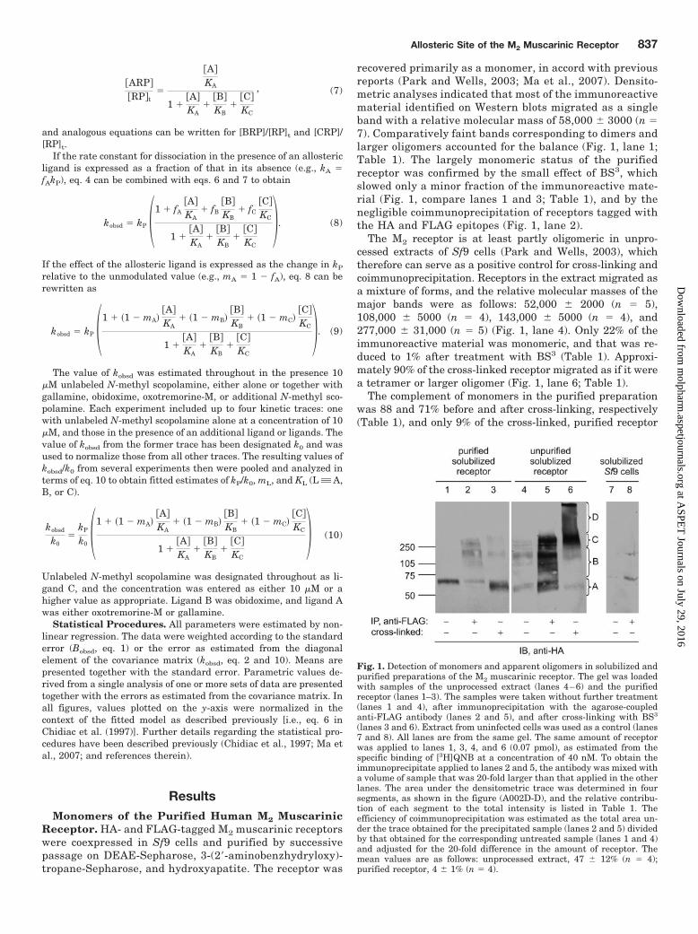
recovered primarily as a monomer, in accord with previousreports (Park and Wells, 2003; Ma et al., 2007). Densito-metric analyses indicated that most of the immunoreactivematerial identified on Western blots migrated as a singleband with a relative molecular mass of 58,000 � 3000 (n �7). Comparatively faint bands corresponding to dimers andlarger oligomers accounted for the balance (Fig. 1, lane 1;Table 1). The largely monomeric status of the purifiedreceptor was confirmed by the small effect of BS3, whichslowed only a minor fraction of the immunoreactive mate-rial (Fig. 1, compare lanes 1 and 3; Table 1), and by thenegligible coimmunoprecipitation of receptors tagged withthe HA and FLAG epitopes (Fig. 1, lane 2).
The M2 receptor is at least partly oligomeric in unpro-cessed extracts of Sf9 cells (Park and Wells, 2003), whichtherefore can serve as a positive control for cross-linking andcoimmunoprecipitation. Receptors in the extract migrated asa mixture of forms, and the relative molecular masses of themajor bands were as follows: 52,000 � 2000 (n � 5),108,000 � 5000 (n � 4), 143,000 � 5000 (n � 4), and277,000 � 31,000 (n � 5) (Fig. 1, lane 4). Only 22% of theimmunoreactive material was monomeric, and that was re-duced to 1% after treatment with BS3 (Table 1). Approxi-mately 90% of the cross-linked receptor migrated as if it werea tetramer or larger oligomer (Fig. 1, lane 6; Table 1).
The complement of monomers in the purified preparationwas 88 and 71% before and after cross-linking, respectively(Table 1), and only 9% of the cross-linked, purified receptor
Fig. 1. Detection of monomers and apparent oligomers in solubilized andpurified preparations of the M2 muscarinic receptor. The gel was loadedwith samples of the unprocessed extract (lanes 4–6) and the purifiedreceptor (lanes 1–3). The samples were taken without further treatment(lanes 1 and 4), after immunoprecipitation with the agarose-coupledanti-FLAG antibody (lanes 2 and 5), and after cross-linking with BS3
(lanes 3 and 6). Extract from uninfected cells was used as a control (lanes7 and 8). All lanes are from the same gel. The same amount of receptorwas applied to lanes 1, 3, 4, and 6 (0.07 pmol), as estimated from thespecific binding of [3H]QNB at a concentration of 40 nM. To obtain theimmunoprecipitate applied to lanes 2 and 5, the antibody was mixed witha volume of sample that was 20-fold larger than that applied in the otherlanes. The area under the densitometric trace was determined in foursegments, as shown in the figure (A002D-D), and the relative contribu-tion of each segment to the total intensity is listed in Table 1. Theefficiency of coimmunoprecipitation was estimated as the total area un-der the trace obtained for the precipitated sample (lanes 2 and 5) dividedby that obtained for the corresponding untreated sample (lanes 1 and 4)and adjusted for the 20-fold difference in the amount of receptor. Themean values are as follows: unprocessed extract, 47 � 12% (n � 4);purified receptor, 4 � 1% (n � 4).
Allosteric Site of the M2 Muscarinic Receptor 837
at ASPE
T Journals on July 29, 2016
molpharm
.aspetjournals.orgD
ownloaded from
was tetrameric or larger (Fig. 1, lane 3; Table 1). To theextent that the densitometric signal may have been nonlin-ear at higher levels of exposure, the fraction corresponding tomonomers is underestimated by the values listed in Table 1.In unprocessed extracts, approximately 47% of the HA-tagged receptor was precipitated by the anti-FLAG antibody(Fig. 1, lane 5); the corresponding value after purificationwas only 4% (Fig. 1, lane 2).
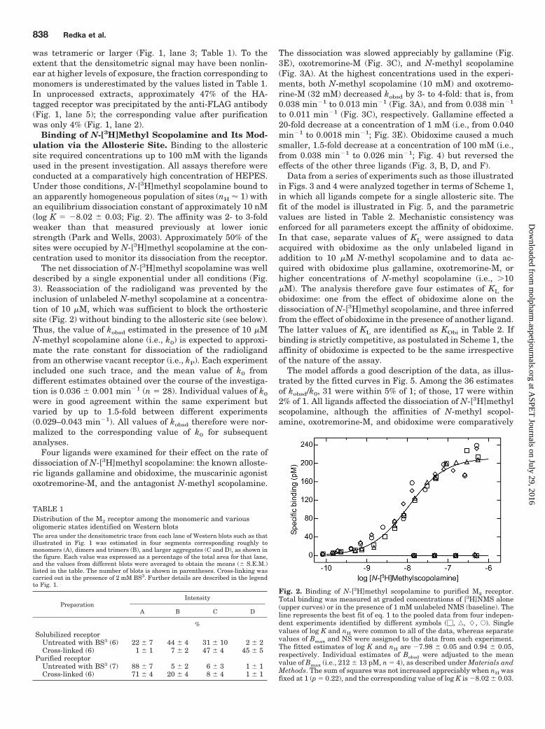
Binding of N-[3H]Methyl Scopolamine and Its Mod-ulation via the Allosteric Site. Binding to the allostericsite required concentrations up to 100 mM with the ligandsused in the present investigation. All assays therefore wereconducted at a comparatively high concentration of HEPES.Under those conditions, N-[3H]methyl scopolamine bound toan apparently homogeneous population of sites (nH � 1) withan equilibrium dissociation constant of approximately 10 nM(log K � �8.02 � 0.03; Fig. 2). The affinity was 2- to 3-foldweaker than that measured previously at lower ionicstrength (Park and Wells, 2003). Approximately 50% of thesites were occupied by N-[3H]methyl scopolamine at the con-centration used to monitor its dissociation from the receptor.
The net dissociation of N-[3H]methyl scopolamine was welldescribed by a single exponential under all conditions (Fig.3). Reassociation of the radioligand was prevented by theinclusion of unlabeled N-methyl scopolamine at a concentra-tion of 10 �M, which was sufficient to block the orthostericsite (Fig. 2) without binding to the allosteric site (see below).Thus, the value of kobsd estimated in the presence of 10 �MN-methyl scopolamine alone (i.e., k0) is expected to approxi-mate the rate constant for dissociation of the radioligandfrom an otherwise vacant receptor (i.e., kP). Each experimentincluded one such trace, and the mean value of k0 fromdifferent estimates obtained over the course of the investiga-tion is 0.036 � 0.001 min�1 (n � 28). Individual values of k0
were in good agreement within the same experiment butvaried by up to 1.5-fold between different experiments(0.029–0.043 min�1). All values of kobsd therefore were nor-malized to the corresponding value of k0 for subsequentanalyses.
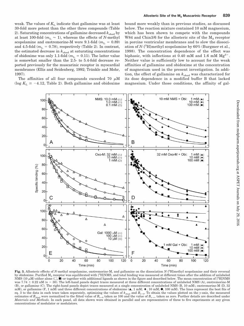
Four ligands were examined for their effect on the rate ofdissociation of N-[3H]methyl scopolamine: the known alloste-ric ligands gallamine and obidoxime, the muscarinic agonistoxotremorine-M, and the antagonist N-methyl scopolamine.
The dissociation was slowed appreciably by gallamine (Fig.3E), oxotremorine-M (Fig. 3C), and N-methyl scopolamine(Fig. 3A). At the highest concentrations used in the experi-ments, both N-methyl scopolamine (10 mM) and oxotremo-rine-M (32 mM) decreased kobsd by 3- to 4-fold: that is, from0.038 min�1 to 0.013 min�1 (Fig. 3A), and from 0.038 min�1
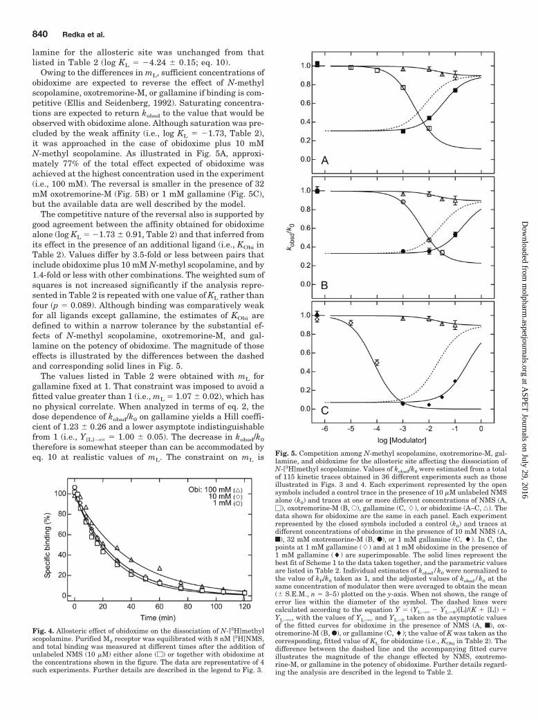
to 0.011 min�1 (Fig. 3C), respectively. Gallamine effected a20-fold decrease at a concentration of 1 mM (i.e., from 0.040min�1 to 0.0018 min�1; Fig. 3E). Obidoxime caused a muchsmaller, 1.5-fold decrease at a concentration of 100 mM (i.e.,from 0.038 min�1 to 0.026 min�1; Fig. 4) but reversed theeffects of the other three ligands (Fig. 3, B, D, and F).
Data from a series of experiments such as those illustratedin Figs. 3 and 4 were analyzed together in terms of Scheme 1,in which all ligands compete for a single allosteric site. Thefit of the model is illustrated in Fig. 5, and the parametricvalues are listed in Table 2. Mechanistic consistency wasenforced for all parameters except the affinity of obidoxime.In that case, separate values of KL were assigned to dataacquired with obidoxime as the only unlabeled ligand inaddition to 10 �M N-methyl scopolamine and to data ac-quired with obidoxime plus gallamine, oxotremorine-M, orhigher concentrations of N-methyl scopolamine (i.e., 10�M). The analysis therefore gave four estimates of KL forobidoxime: one from the effect of obidoxime alone on thedissociation of N-[3H]methyl scopolamine, and three inferredfrom the effect of obidoxime in the presence of another ligand.The latter values of KL are identified as KObi in Table 2. Ifbinding is strictly competitive, as postulated in Scheme 1, theaffinity of obidoxime is expected to be the same irrespectiveof the nature of the assay.
The model affords a good description of the data, as illus-trated by the fitted curves in Fig. 5. Among the 36 estimatesof kobsd/k0, 31 were within 5% of 1; of those, 17 were within2% of 1. All ligands affected the dissociation of N-[3H]methylscopolamine, although the affinities of N-methyl scopol-amine, oxotremorine-M, and obidoxime were comparatively
TABLE 1Distribution of the M2 receptor among the monomeric and variousoligomeric states identified on Western blotsThe area under the densitometric trace from each lane of Western blots such as thatillustrated in Fig. 1 was estimated in four segments corresponding roughly tomonomers (A), dimers and trimers (B), and larger aggregates (C and D), as shown inthe figure. Each value was expressed as a percentage of the total area for that lane,and the values from different blots were averaged to obtain the means (� S.E.M.)listed in the table. The number of blots is shown in parentheses. Cross-linking wascarried out in the presence of 2 mM BS3. Further details are described in the legendto Fig. 1.
PreparationIntensity
A B C D
%
Solubilized receptorUntreated with BS3 (6) 22 � 7 44 � 4 31 � 10 2 � 2Cross-linked (6) 1 � 1 7 � 2 47 � 4 45 � 5
Purified receptorUntreated with BS3 (7) 88 � 7 5 � 2 6 � 3 1 � 1Cross-linked (6) 71 � 4 20 � 4 8 � 4 1 � 1
Fig. 2. Binding of N-[3H]methyl scopolamine to purified M2 receptor.Total binding was measured at graded concentrations of [3H]NMS alone(upper curves) or in the presence of 1 mM unlabeled NMS (baseline). Theline represents the best fit of eq. 1 to the pooled data from four indepen-dent experiments identified by different symbols (�, ‚, �, E). Singlevalues of log K and nH were common to all of the data, whereas separatevalues of Bmax and NS were assigned to the data from each experiment.The fitted estimates of log K and nH are �7.98 � 0.05 and 0.94 � 0.05,respectively. Individual estimates of Bobsd were adjusted to the meanvalue of Bmax (i.e., 212 � 13 pM, n � 4), as described under Materials andMethods. The sum of squares was not increased appreciably when nH wasfixed at 1 (p � 0.22), and the corresponding value of log K is �8.02 � 0.03.
838 Redka et al.
at ASPE
T Journals on July 29, 2016
molpharm
.aspetjournals.orgD
ownloaded from
weak. The values of KL indicate that gallamine was at least30-fold more potent than the other three compounds (Table2). Saturating concentrations of gallamine decreased kobsd byat least 100-fold (mL � 1), whereas the effects of N-methylscopolamine and oxotremorine-M were 9.1-fold (mL � 0.89)and 4.5-fold (mL � 0.78), respectively (Table 2). In contrast,the estimated decrease in kobsd at saturating concentrationsof obidoxime was only 1.1-fold (mL � 0.11). The latter valueis somewhat smaller than the 2.5- to 5.4-fold decrease re-ported previously for the muscarinic receptor in myocardialmembranes (Ellis and Seidenberg, 1992; Trankle and Mohr,1997).
The affinities of all four compounds exceeded 70 �M(log KL � �4.12, Table 2). Both gallamine and obidoxime
bound more weakly than in previous studies, as discussedbelow. The reaction mixture contained 10 mM magnesium,which has been shown to compete with the compoundsW84 and Chin3/6 for the allosteric site of the M2 receptorin porcine ventricular membranes and to slow the dissoci-ation of N-[3H]methyl scopolamine by 60% (Burgmer et al.,1998). The concentration dependence of the effect wasbiphasic, with inflections at 0.40 mM and 1.6 mM Mg2.Neither value is sufficiently low to account for the weakaffinities of gallamine and obidoxime at the concentrationof magnesium used in the present investigation. In addi-tion, the effect of gallamine on kobsd was characterized forits dose dependence in a modified buffer B that lackedmagnesium. Under those conditions, the affinity of gal-
Fig. 3. Allosteric effects of N-methyl scopolamine, oxotremorine-M, and gallamine on the dissociation N-[3H]methyl scopolamine and their reversalby obidoxime. Purified M2 receptor was equilibrated with [3H]NMS, and total binding was measured at different times after the addition of unlabeledNMS (10 �M) either alone (�, f) or together with additional ligands as shown in the figure and described below. The mean concentration of [3H]NMSwas 7.74 � 0.22 nM (n � 30). The left-hand panels depict traces measured at three different concentrations of unlabeled NMS (A), oxotremorine-M(B), or gallamine (C). The right-hand panels depict traces measured at a single concentration of unlabeled NMS (B, 10 mM), oxotremorine-M (D, 32mM), or gallamine (F, 1 mM) and three different concentrations of obidoxime (Œ, 1 mM; �, 10 mM; F, 100 mM). The lines represent the best fits ofeq. 3 to the data in each trace taken separately, optimizing the values of kobsd and Bt�0. To obtain the values plotted on the y-axis, the measuredestimates of Bobsd were normalized to the fitted value of Bt�0 taken as 100 and the value of Bt3� taken as zero. Further details are described underMaterials and Methods. In each panel, all data shown were obtained in parallel and are representative of three to five experiments at any givenconcentrations of modulator or modulators.
Allosteric Site of the M2 Muscarinic Receptor 839
at ASPE
T Journals on July 29, 2016
molpharm
.aspetjournals.orgD
ownloaded from
lamine for the allosteric site was unchanged from thatlisted in Table 2 (log KL � �4.24 � 0.15; eq. 10).
Owing to the differences in mL, sufficient concentrations ofobidoxime are expected to reverse the effect of N-methylscopolamine, oxotremorine-M, or gallamine if binding is com-petitive (Ellis and Seidenberg, 1992). Saturating concentra-tions are expected to return kobsd to the value that would beobserved with obidoxime alone. Although saturation was pre-cluded by the weak affinity (i.e., log KL � �1.73, Table 2),it was approached in the case of obidoxime plus 10 mMN-methyl scopolamine. As illustrated in Fig. 5A, approxi-mately 77% of the total effect expected of obidoxime wasachieved at the highest concentration used in the experiment(i.e., 100 mM). The reversal is smaller in the presence of 32mM oxotremorine-M (Fig. 5B) or 1 mM gallamine (Fig. 5C),but the available data are well described by the model.
The competitive nature of the reversal also is supported bygood agreement between the affinity obtained for obidoximealone (log KL � �1.73 � 0.91, Table 2) and that inferred fromits effect in the presence of an additional ligand (i.e., KObi inTable 2). Values differ by 3.5-fold or less between pairs thatinclude obidoxime plus 10 mM N-methyl scopolamine, and by1.4-fold or less with other combinations. The weighted sum ofsquares is not increased significantly if the analysis repre-sented in Table 2 is repeated with one value of KL rather thanfour (p � 0.089). Although binding was comparatively weakfor all ligands except gallamine, the estimates of KObi aredefined to within a narrow tolerance by the substantial ef-fects of N-methyl scopolamine, oxotremorine-M, and gal-lamine on the potency of obidoxime. The magnitude of thoseeffects is illustrated by the differences between the dashedand corresponding solid lines in Fig. 5.
The values listed in Table 2 were obtained with mL forgallamine fixed at 1. That constraint was imposed to avoid afitted value greater than 1 (i.e., mL � 1.07 � 0.02), which hasno physical correlate. When analyzed in terms of eq. 2, thedose dependence of kobsd/k0 on gallamine yields a Hill coeffi-cient of 1.23 � 0.26 and a lower asymptote indistinguishablefrom 1 (i.e., Y[L]3� � 1.00 � 0.05). The decrease in kobsd/k0
therefore is somewhat steeper than can be accommodated byeq. 10 at realistic values of mL. The constraint on mL is Fig. 5. Competition among N-methyl scopolamine, oxotremorine-M, gal-
lamine, and obidoxime for the allosteric site affecting the dissociation ofN-[3H]methyl scopolamine. Values of kobsd/k0 were estimated from a totalof 115 kinetic traces obtained in 36 different experiments such as thoseillustrated in Figs. 3 and 4. Each experiment represented by the opensymbols included a control trace in the presence of 10 �M unlabeled NMSalone (k0) and traces at one or more different concentrations of NMS (A,�), oxotremorine-M (B, E), gallamine (C, �), or obidoxime (A–C, ‚). Thedata shown for obidoxime are the same in each panel. Each experimentrepresented by the closed symbols included a control (k0) and traces atdifferent concentrations of obidoxime in the presence of 10 mM NMS (A,f), 32 mM oxotremorine-M (B, F), or 1 mM gallamine (C, �). In C, thepoints at 1 mM gallamine (�) and at 1 mM obidoxime in the presence of1 mM gallamine (�) are superimposable. The solid lines represent thebest fit of Scheme 1 to the data taken together, and the parametric valuesare listed in Table 2. Individual estimates of kobsd/k0 were normalized tothe value of kP/k0 taken as 1, and the adjusted values of kobsd/k0 at thesame concentration of modulator then were averaged to obtain the mean(� S.E.M., n � 3–5) plotted on the y-axis. When not shown, the range oferror lies within the diameter of the symbol. The dashed lines werecalculated according to the equation Y � (YL3� � YL�0)[L]/(K [L]) YL3�, with the values of YL3� and YL�0 taken as the asymptotic valuesof the fitted curves for obidoxime in the presence of NMS (A, f), ox-otremorine-M (B, F), or gallamine (C, �); the value of K was taken as thecorresponding, fitted value of KL for obidoxime (i.e., KObi in Table 2). Thedifference between the dashed line and the accompanying fitted curveillustrates the magnitude of the change effected by NMS, oxotremo-rine-M, or gallamine in the potency of obidoxime. Further details regard-ing the analysis are described in the legend to Table 2.
Fig. 4. Allosteric effect of obidoxime on the dissociation of N-[3H]methylscopolamine. Purified M2 receptor was equilibrated with 8 nM [3H]NMS,and total binding was measured at different times after the addition ofunlabeled NMS (10 �M) either alone (�) or together with obidoxime atthe concentrations shown in the figure. The data are representative of 4such experiments. Further details are described in the legend to Fig. 3.
840 Redka et al.
at ASPE
T Journals on July 29, 2016
molpharm
.aspetjournals.orgD
ownloaded from
accompanied by a significant increase in the sum of squares(p � 0.00019). Virtually all of the increase derives from thedata for gallamine in the absence of obidoxime, which lieabove the values predicted by the model (Fig. 5C). The fittedcurves otherwise are superimposable with those shown inFig. 5, and the values of other parameters are essentially thesame as those listed in Table 2. The reason for this anomalyin the effect of gallamine is unclear.
DiscussionM2 muscarinic receptors from porcine atria and baculovi-
ral-infected Sf9 cells exhibit noncompetitive effects in thebinding of orthosteric ligands (Wreggett and Wells, 1995;Park et al., 2002; Ma et al., 2007). The data can be describedin terms of cooperative interactions among at least four sites,implying that the receptor is tetrameric or larger. In thesame vein, a complex containing the HA-, c-Myc-, and FLAG-tagged forms of the M2 receptor could be purified from Sf9cells coinfected with the three baculoviruses (Park and Wells,2004), suggesting that the receptor is at least trimeric. Inaddition, the M2 receptor has been shown by cross-linking toform tetramers when purified as a monomer from Sf9 cellsand reconstituted in phospholipid vesicles (Ma et al., 2007).
As inferred from cooperativity, the notion of a tetramer isbased on the assumption that ligands such as N-methyl sco-polamine and oxotremorine-M bind only to the single ortho-steric site within the cluster of transmembrane helices (cf.Hulme et al., 2003; Rasmussen et al., 2007). This disregardsthe possibility of an interaction with the allosteric site rec-ognized by compounds such as gallamine, but the preferenceof ligands for the two sites is not necessarily absolute. Allo-steric modulators of the M2 receptor are structurally diverse,and compounds such as UH-AH 37 (Ellis and Seidenberg,1999), methoctramine (Waelbroeck, 1994; Boselli and Grana,1995), and AF-DX 116 (Lee and el-Fakahany, 1991; Waelbro-eck, 1994) also may bind to the orthosteric site. Conversely,M2-selective antagonists, such as AF-DX 384, that bind to theorthosteric site (Miller et al., 1991) also may bind to theallosteric site (Trankle et al., 1998).
A tetramer of the M2 receptor would contain four ortho-steric and four allosteric sites, with considerable potential forhomo- and heterotropic cooperativity. Noncompetitive effectsbetween supposed orthosteric ligands would be difficult to
interpret if those ligands also bound to the allosteric site andmimicked the effects of gallamine. That possibility thereforehas been examined using a preparation of monomers, whichare expected to exhibit only heterotropic effects. HA- andFLAG-tagged M2 receptors extracted from coinfected Sf9cells were predominantly oligomeric, as indicated by theirdegree of coimmunoprecipitation and their electrophoreticmobility before and after cross-linking with BS3. In contrast,and in agreement with previous results (Park and Wells,2003; Ma et al., 2007), the affinity-purified receptor waspredominantly monomeric. There was little coimmunopre-cipitation of the tagged adducts, and most of the receptormigrated as a monomer after treatment with BS3.
Both oxotremorine-M and N-methyl scopolamine slowedthe dissociation of N-[3H]methyl scopolamine, which boundonly to the orthosteric site at the concentration of 6 to 10 nMused in the assays. Whereas the affinity of the radioligand forthe orthosteric site was 10 nM, that estimated for the allostericsite was at least 3 orders of magnitude weaker, as discussedbelow. Inasmuch as monomers are not expected to exhibitintermolecular interactions between orthosteric sites, the ef-fect of either compound on the dissociation of N-[3H]methylscopolamine was heterotropic in nature.
N-Methyl scopolamine, oxotremorine-M, gallamine, andobidoxime all seem to affect the dissociation of N-[3H]methylscopolamine via a common allosteric site. Scheme 1 providesa good description of the data, which yield four estimates forthe affinity of obidoxime: one estimate from the dependenceof kobsd on the concentration of obidoxime alone (KL) andthree from the potency of obidoxime in the presence of N-methyl scopolamine, oxotremorine-M, or gallamine (KObi).The fitted values of KL and KObi are indistinguishable, andobidoxime therefore was competitive with respect to theother three ligands. In the case of obidoxime and gallamine,the present results are consistent with the results of anearlier study in which the interaction between those twoligands also was found to be competitive (Ellis and Seiden-berg, 1992). It follows that N-methyl scopolamine andoxotremorine-M bound to the same site as gallamine andobidoxime.
The affinities of both obidoxime and gallamine for theallosteric site of the radioligand-occupied receptor were com-paratively weak. The present values of 19 mM and 76 �Mobtained for KL are at least 260-fold greater than thosereported for the effect of obidoxime and gallamine, respec-tively, on the dissociation of N-[3H]methyl scopolamine fromthe M2 receptor in rat myocardial membranes (Ellis andSeidenberg, 1992). In a more recent study on porcine myo-cardial membranes, the effect of obidoxime was found to bebiphasic, with inflections at 23 �M and 0.96 mM (Trankleand Mohr, 1997). In addition, the maximal decrease effectedby obidoxime in kobsd was only 11% in the present investiga-tion; in contrast, a decrease of at least 50% has been found instudies on the receptor in myocardial membranes (Ellis andSeidenberg, 1992; Trankle and Mohr, 1997).
The origin of such differences in the properties of obi-doxime and gallamine remains unclear. They apparently arenot caused by magnesium, and the reported effects of ionicstrength on the affinity of gallamine are comparatively small(Pedder et al., 1991). They may be related to the state ofaggregation of the receptor, which can exist as oligomers inwhole cells (Goin and Nathanson, 2006), in detergent-solubi-
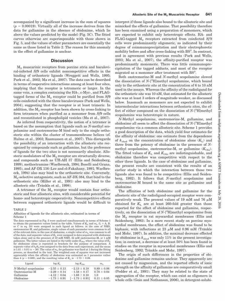
TABLE 2Affinities of ligands for the allosteric site, estimated in terms ofScheme 1The data represented in Fig. 5 were analyzed simultaneously in terms of Scheme 1to obtain the parametric values listed in the table. Values of KL and mL (L � A, B,or C in eq. 10) were assigned to each ligand as follows: in the case of NMS,oxotremorine-M, and gallamine, single values of each parameter were common to allof the relevant data; in the case of obidoxime, a single value of mL was common to allof the data, and separate values of KL were assigned to data acquired with obidoximetaken alone and in the presence of 10 mM NMS, 32 mM oxotremorine-M, or 1 mMgallamine. The latter values are listed in the table under KObi, where the value of KLfor obidoxime alone is reprinted in brackets for the purpose of comparison. Aseparate value of kP/k0 was assigned to the data from each experiment, and the meanis 1.01 � 0.01 (n � 36). The value of mL for gallamine was fixed at 1 during the fittingprocess, as described in the text. The weighted sum of squares was not increasedappreciably when the affinity of obidoxime was estimated as 1 parameter ratherthan 4 (p � 0.089), and the resulting value of KL is �1.73 � 0.08.
Ligand log KL log KObi mL
N-Methyl scopolamine �2.55 � 0.13 �2.13 � 0.18 0.89 � 0.06Oxotremorine-M �2.29 � 0.14 �1.58 � 0.17 0.78 � 0.04Gallamine �4.12 � 0.04 �1.68 � 0.10 1.0Obidoxime �1.73 � 0.91 ��1.73 � 0.91� 0.11 � 0.05
Allosteric Site of the M2 Muscarinic Receptor 841
at ASPE
T Journals on July 29, 2016
molpharm
.aspetjournals.orgD
ownloaded from
lized preparations (Park et al., 2001; Park and Wells, 2004),and after reconstitution (Ma et al., 2007). Such aggregationseems to have functional consequences, which may extend tothe binding and effects of allosteric ligands.
Under some conditions, the detergent-solubilized M2 recep-tor exhibits a pattern of noncompetitive behavior wherein thebinding of [3H]quinuclidinyl benzilate is inhibited byN-methyl scopolamine at sites that are inaccessible to thelatter (Park et al., 2002; Colozo et al., 2007). The effect can bedescribed quantitatively in terms of cooperativity among fouror more interacting and presumably orthosteric sites (Park etal., 2002). Such effects are absent from preparations of puri-fied monomers (Park and Wells, 2003; Ma et al., 2007), butthey are recovered upon reconstitution of the receptor inphospholipid vesicles (Ma et al., 2007). The parametric val-ues reported here for gallamine and obidoxime pertain to theallosteric site of a monomer. If the receptor in myocardialmembranes is predominantly oligomeric, the values of KL
and mL in those preparations may be affected by modes ofcooperativity and other factors that are absent from amonomer.
The allosteric effects of N-methyl scopolamine and ox-otremorine-M raise the possibility that such interactions con-tribute to events heretofore assumed to occur at the ortho-steric site. The affinity of N-methyl scopolamine for theallosteric site of the ligand-occupied monomer was 2.8 mM(KL, Table 2). Its affinity for the allosteric site of the vacantmonomer is unknown, but a cooperativity factor (�) of 46 hasbeen estimated for the degree of negative heterotropic coop-erativity between gallamine and N-[3H]methyl scopolamineat the cardiac muscarinic receptor (Trankle et al., 1999).Gallamine is one of the more efficacious allosteric modulatorsof N-[3H]methyl scopolamine (Mohr et al., 2003); accordingly,the value of � for the heterotropic interaction between N-methyl scopolamine and its radiolabeled analog seems un-likely to exceed 46. It follows that the equilibrium dissocia-tion constant for the binding of N-methyl scopolamine to theallosteric site of the vacant monomer is likely to exceed 0.06mM (i.e., KL/�).
Based on the lower limit estimated above, the affinity ofN-methyl scopolamine for the allosteric site is at least 2800-fold weaker than its affinity for the orthosteric site [10 nM,Fig. 2; � 22 nM, Ma et al. (2007)]. It is at least 1000-foldweaker than the affinity that emerges from the noncompet-itive inhibition of [3H]quinuclidinyl benzilate by N-methylscopolamine at the M2 receptor solubilized in cholate-NaCl[46 nM, Park et al. (2002)] or reconstituted in phospholipidvesicles [27 nM, Ma et al. (2007)]. These considerations sug-gest that the noncompetitive effect of N-methyl scopolamineat concentrations below 10 �M derives entirely from interac-tions among orthosteric sites. Such a system is potentiallycomplex, however, and the combined effect of several cooper-ativity factors on the affinity of N-methyl scopolamine for theallosteric sites is unknown.
In the case of oxotremorine-M, the affinity for the allostericsite of the ligand-occupied monomer was approximately 5mM (KL, Table 2). If the value of � for the heterotropicinteraction with N-[3H]methyl scopolamine is assumed onceagain to be 46 or less, the equilibrium dissociation constantfor oxotremorine-M at the allosteric site of the vacant recep-tor is 0.11 mM or more. That is approximately 170-foldgreater than the dissociation constant of oxotremorine-M for
the low-affinity form of the muscarinic receptor in myocar-dial membranes, as determined at equilibrium in the pres-ence of N-[3H]methyl scopolamine (0.66 �M; Ehlert, 1988),and it approximates the corresponding value obtained for thereconstituted M2 receptor in the presence of [3H]quinuclidi-nyl benzilate (0.35 mM; Ma et al., 2007). Oxotremorine-Mtherefore reveals comparatively little if any difference be-tween its estimated affinity for the allosteric site of themonomer and its affinity for at least some of the presumedorthosteric sites. It follows that the inhibitory effect of ox-otremorine-M on the binding of an antagonist such asN-[3H]methyl scopolamine may derive in part from an inter-action at the allosteric site. Such an effect would have impli-cations for our understanding of the GTP-sensitive heteroge-neity revealed by agonists at the muscarinic and perhapsother G protein-coupled receptors.
Acknowledgments
We are grateful to Rabindra V. Shivnaraine for helpful discus-sions.
ReferencesAngers S, Salahpour A, and Bouvier M (2002) Dimerization: An emerging concept for
G protein-coupled receptor ontogeny and function. Annu Rev Pharmacol Toxicol42:409–435.
Boselli C and Grana E (1995) Mode of antagonism of methoctramine, AF-DX 116 andhexahydrosiladifenidol in guinea-pig left atrium and ileum: Comparison of Schildand resultant analysis. J Auton Pharmacol 15:115–127.
Burgmer U, Schulz U, Trankle C, and Mohr K (1998) Interaction of Mg2 with theallosteric site of muscarinic M2 receptors. Naunyn Schmiedebergs Arch Pharmacol357:363–370.
Chidiac P, Green MA, Pawagi AB, and Wells JW (1997) Cardiac muscarinic recep-tors. Cooperativity as the basis for multiple states of affinity. Biochemistry 36:7361–7379.
Colozo AT, Park PS, Sum CS, Pisterzi LF, and Wells JW (2007) Cholesterol as adeterminant of cooperativity in the M2 muscarinic cholinergic receptor. BiochemPharmacol 74:236–255.
Curtis CA, Wheatley M, Bansal S, Birdsall NJ, Eveleigh P, Pedder EK, Poyner D,and Hulme EC (1989) Propylbenzilylcholine mustard labels an acidic residue intransmembrane helix 3 of the muscarinic receptor. J Biol Chem 264:489–495.
Ehlert FJ (1988) Correlation between the binding parameters of muscarinic agonistsand their inhibition of adenylate cyclase activity. Adv Exp Med Biol 236:265–276.
Ellis J and Seidenberg M (1999) Competitive and allosteric interactions of 6-chloro-5,10-dihydro-5-[(1-methyl-4-piperidinyl)acetyl]-11H-dibenzo[b,e][1,4]diazepine-11-one hydrochloride (UH-AH 37) at muscarinic receptors, via distinct epitopes.Biochem Pharmacol 57:181–186.
Ellis J and Seidenberg M (2000) Site-directed mutagenesis implicates a threonineresidue in TM6 in the subtype selectivities of UH-AH 37 and pirenzepine atmuscarinic receptors. Pharmacology 61:62–69.
Ellis J and Seidenberg M (1992) Two allosteric modulators interact at a common siteon cardiac muscarinic receptors. Mol Pharmacol 42:638–641.
Ellis J, Seidenberg M, and Brann MR (1993) Use of chimeric muscarinic receptors toinvestigate epitopes involved in allosteric interactions. Mol Pharmacol 44:583–588.
Goin JC and Nathanson NM (2006) Quantitative analysis of muscarinic acetylcho-line receptor homo- and heterodimerization in live cells: Regulation of receptordown-regulation by heterodimerization. J Biol Chem 281:5416–5425.
Gomes I, Jordan BA, Gupta A, Rios C, Trapaidze N, and Devi LA (2001) G proteincoupled receptor dimerization: Implications in modulating receptor function. J MolMed 79:226–242.
Hulme EC, Birdsall NJ, and Buckley NJ (1990) Muscarinic receptor subtypes. AnnuRev Pharmacol Toxicol 30:633–673.
Hulme EC, Lu ZL, Saldanha JW, and Bee MS (2003) Structure and activation ofmuscarinic acetylcholine receptors. Biochem Soc Trans 31:29–34.
Lee NH and el-Fakahany EE (1988) Influence of ligand choice on the apparentbinding profile of gallamine to cardiac muscarinic receptors. Identification of threemain types of gallamine-muscarinic receptor interactions. J Pharmacol Exp Ther246:829–838.
Lee NH and el-Fakahany EE (1991) Allosteric interactions at the M1, M2 and M3muscarinic receptor subtypes. J Pharmacol Exp Ther 256:468–479.
Ma AW, Redka DS, Pisterzi LF, Angers S, and Wells JW (2007) Recovery of oligomersand cooperativity when monomers of the M2 muscarinic cholinergic receptor arereconstituted into phospholipid vesicles. Biochemistry 46:7907–7927.
Miller JH, Gibson VA, and McKinney M (1991) Binding of [3H]AF-DX 384 to clonedand native muscarinic receptors. J Pharmacol Exp Ther 259:601–607.
Milligan G (2001) Oligomerisation of G-protein-coupled receptors. J Cell Sci 114:1265–1271.
Mohr K, Trankle C, and Holzgrabe U (2003) Structure/activity relationships of M2muscarinic allosteric modulators. Recept Channels 9:229–240.
842 Redka et al.
at ASPE
T Journals on July 29, 2016
molpharm
.aspetjournals.orgD
ownloaded from
Park P, Sum CS, Hampson DR, Van Tol HH, and Wells JW (2001) Nature of theoligomers formed by muscarinic M2 acetylcholine receptors in Sf9 cells. EurJ Pharmacol 421:11–22.
Park PS, Filipek S, Wells JW, and Palczewski K (2004) Oligomerization of G protein-coupled receptors: past, present, and future. Biochemistry 43:15643–15656.
Park PS, Sum CS, Pawagi AB, and Wells JW (2002) Cooperativity and oligomericstatus of cardiac muscarinic cholinergic receptors. Biochemistry 41:5588–5604.
Park PS and Wells JW (2004) Oligomeric potential of the M2 muscarinic cholinergicReceptor. J Neurochem 90:537–548.
Park PS and Wells JW (2003) Monomers and oligomers of the M2 muscariniccholinergic receptor purified from Sf9 cells. Biochemistry 42:12960–12971.
Pedder EK, Eveleigh P, Poyner D, Hulme EC, and Birdsall NJ (1991) Modulation ofthe structure-binding relationships of antagonists for muscarinic acetylcholinereceptor subtypes. Br J Pharmacol 103:1561–1567.
Rasmussen SG, Choi HJ, Rosenbaum DM, Kobilka TS, Thian FS, Edwards PC,Burghammer M, Ratnala VR, Sanishvili R, Fischetti RF, et al. (2007) Crystalstructure of the human �2 adrenergic G-protein-coupled receptor. Nature 450:383–387.
Stockton JM, Birdsall NJ, Burgen AS, and Hulme EC (1983) Modification of thebinding properties of muscarinic receptors by gallamine. Mol Pharmacol 23:551–557.
Trankle C, Andresen I, Lambrecht G, and Mohr K (1998) M2 receptor binding of theselective antagonist AF-DX 384: possible involvement of the common allostericsite. Mol Pharmacol 53:304–312.
Trankle C and Mohr K (1997) Divergent modes of action among cationic allostericmodulators of muscarinic M2 receptors. Mol Pharmacol 51:674–682.
Trankle C, Weyand O, Schroter A, and Mohr K (1999) Using a radioalloster to testpredictions of the cooperativity model for gallamine binding to the allosteric site ofmuscarinic acetylcholine M2 receptors. Mol Pharmacol 56:962–965.
Tucek S and Proska J (1995) Allosteric modulation of muscarinic acetylcholinereceptors. Trends Pharmacol Sci 16:205–212.
Waelbroeck M (1994) Identification of drugs competing with d-tubocurarine for anallosteric site on cardiac muscarinic receptors. Mol Pharmacol 46:685–692.
Waelbroeck M, Robberecht P, De Neef P, and Christophe J (1988) Effects ofd-tubocurarine on rat cardiac muscarinic receptors: a comparison with gallamine.J Recept Res 8:787–808.
Wreggett KA and Wells JW (1995) Cooperativity manifest in the binding propertiesof purified cardiac muscarinic receptors. J Biol Chem 270:22488–22499.
Wyman J and Gill SJ (1990) Binding and Linkage: Functional Chemistry of Biolog-ical Macromolecules. University Science Books, Mill Valley, California.
Zeng FY and Wess J (1999) Identification and molecular characterization of M3muscarinic receptor dimers. J Biol Chem 274:19487–19497.
Address correspondence to: James W. Wells, Department of Pharmaceuti-cal Sciences, Leslie Dan Faculty of Pharmacy, University of Toronto, 144College Street, Toronto, Ontario, Canada M5S 3M2. E-mail: [email protected]
Allosteric Site of the M2 Muscarinic Receptor 843
at ASPE
T Journals on July 29, 2016
molpharm
.aspetjournals.orgD
ownloaded from
Related Documents