Published: November 17, 2011 r2011 American Chemical Society 1111 dx.doi.org/10.1021/jp208567n | J. Phys. Chem. B 2012, 116, 1111–1119 ARTICLE pubs.acs.org/JPCB Binding of Inositol Stereoisomers To Model Amyloidogenic Peptides Grace Li, †,‡ Sarah Rauscher, †,‡ St ephanie Baud, § and R egis Pom es* ,†,‡ † Department of Biochemistry, University of Toronto, 27 King’s College Circle, Toronto, Ontario, Canada M5S 1A1 ‡ Molecular Structure and Function, The Hospital for Sick Children, 555 University Avenue, Toronto, Ontario, Canada M5G 1X8 § Laboratoire SiRMa, CNRS UMR MEDyC 6237, IFR 53 Biomol ecules, Universit e de Reims Champagne-Ardenne, Reims, France ’ INTRODUCTION Amyloid fibrils formed by various peptides and proteins are known to be associated with neurodegenerative diseases, type II diabetes, and prion-related disorders. 1 In particular, amyloid fibrils of Aβ peptides are found in the extracellular deposits of neuronal plaques and are thought to be central to the pathogen- esis of Alzheimer’s disease (AD), 1,2 a common and incurable neurodegenerative disease causing dementia and eventual death. In recent years, amyloid fibril formation was discovered to be a common phenomenon among many proteins in vitro; that is, under certain misfolding and denaturing conditions, proteins can self-aggregate to form amyloid fibrils. 1 When viewed with negatively stained transmission electron microscopy, amyloid fibrils appear as elongated, ropelike structures that are often 100 nm in length. 1 The core structure of all amyloid fibrils is the cross-β sheet. 1,3 At the molecular level, NMR 4,5 and X-ray crystallography 6 studies have revealed that the cross-β structure is comprised of extended polypeptides organized in highly ordered, in-register β-sheets. Although amyloid fibrils are a pathological hallmark of amyloid-based diseases, smaller nonfi- brillar oligomers as little as three or four peptides in size have been demonstrated to display higher cytoxicity than mature fibrils. 713 An important strategy to finding a cure to AD and other amyloid diseases is to derive new therapeutic candidates through the rational design of effective small-molecule inhibitors of amyloid formation. In recent years, a number of small molecules capable of preventing aggregation and/or fibril formation have been discovered and have emerged as potential therapeutic approaches for protein misfolding diseases. 1419 Interestingly, many of these small molecules share common chemical structur- al features, such as aromaticity and the presence of multiple hydrogen-bonding groups. 2023 However, the molecular basis of the structureactivity relationship of these small molecules is not understood, thus hindering drug development efforts for amyloid-based diseases. Recently, one such small molecule, scyllo-inositol, has shown promise as a therapeutic for AD. 24,25 scyllo-Inositol is one of nine stereoisomers that belongs to a class of cyclic polyols called cyclohexanehexols. Four stereoisomers, myo-, epi-, scyllo-, and chiro-inositol (Figure 1), are physiologically active. 26 myo-Inosi- tol, the most abundant stereoisomer, plays an important role in signal transduction as a precursor of phospholipid headgroups: once phosphorylated, myo-inositol phosphatides act as second messengers in intracellular signal transduction pathways. 26 Im- portantly for its therapeutic potential, inositol readily crosses the bloodbrain barrier. myo- And scyllo-inositol are found in tissues of the human central nervous system (CNS), with approximate concentrations of 5 and 0.10.5 mM, respectively. 27 Accord- ingly, they are also important osmolytes in the CNS, where alterations in their concentration have been associated with neuropathological conditions. 26,28 In vitro, inositol stereoisomers stabilize nonfibrillar β-struc- ture and prevent the formation of amyloid fibrils in a stereo- chemistry-dependent manner: scyllo-, epi-, and myo-inositol inhibit Aβ fibril formation, but not chiro-inositol. 24,2932 More- over, scyllo-inositol was also demonstrated to be the most effective stereosiomer in preventing and reversing AD-like symptoms in transgenic mice while reducing their brain plaque load. 25 Despite this progress, the molecular basis of amyloid Received: September 5, 2011 Revised: November 1, 2011 ABSTRACT: The self-aggregation of proteins into amyloid fibrils is a pathological hallmark of numerous incurable diseases such as Alzheimer’s disease. scyllo-Inositol is a stereochemistry-dependent in vitro inhibitor of amyloid formation. As the first step to elucidate its mechanism of action, we present molecular dynamics simulations of scyllo- inositol and its inactive stereoisomer, chiro-inositol, with simple peptide models, alanine dipeptide (ADP) and (Gly-Ala) 4 . We characterize molecular interactions and compute equilibrium binding constants between inositol and ADP as well as, successively, mono- mers, amorphous aggregates, and fibril-like β-sheet aggregates of (Gly-Ala) 4 . Inositol interacts weakly with all peptide systems considered, with millimolar to molar affinities, and displaces the conformational equilibria of ADP but not of the (Gly-Ala) 4 systems. However, scyllo- and chiro-inositol adopt different binding modes on the surface of β-sheet aggregates. These results suggest that inositol does not inhibit amyloid formation by breaking up preformed aggregates but rather by binding to the surface of prefibrillar aggregates.

Welcome message from author
This document is posted to help you gain knowledge. Please leave a comment to let me know what you think about it! Share it to your friends and learn new things together.
Transcript
Published: November 17, 2011
r 2011 American Chemical Society 1111 dx.doi.org/10.1021/jp208567n | J. Phys. Chem. B 2012, 116, 1111–1119
ARTICLE
pubs.acs.org/JPCB
Binding of Inositol Stereoisomers To Model Amyloidogenic PeptidesGrace Li,†,‡ Sarah Rauscher,†,‡ St�ephanie Baud,§ and R�egis Pom�es*,†,‡
†Department of Biochemistry, University of Toronto, 27 King’s College Circle, Toronto, Ontario, Canada M5S 1A1‡Molecular Structure and Function, The Hospital for Sick Children, 555 University Avenue, Toronto, Ontario, Canada M5G 1X8§Laboratoire SiRMa, CNRS UMR MEDyC 6237, IFR 53 Biomol�ecules, Universit�e de Reims Champagne-Ardenne, Reims, France
’ INTRODUCTION
Amyloid fibrils formed by various peptides and proteins areknown to be associated with neurodegenerative diseases, type IIdiabetes, and prion-related disorders.1 In particular, amyloidfibrils of Aβ peptides are found in the extracellular deposits ofneuronal plaques and are thought to be central to the pathogen-esis of Alzheimer’s disease (AD),1,2 a common and incurableneurodegenerative disease causing dementia and eventual death.
In recent years, amyloid fibril formation was discovered to be acommon phenomenon among many proteins in vitro; that is,under certain misfolding and denaturing conditions, proteins canself-aggregate to form amyloid fibrils.1 When viewed withnegatively stained transmission electron microscopy, amyloidfibrils appear as elongated, ropelike structures that are often100 nm in length.1 The core structure of all amyloid fibrils is thecross-β sheet.1,3 At the molecular level, NMR4,5 and X-raycrystallography6 studies have revealed that the cross-β structureis comprised of extended polypeptides organized in highlyordered, in-register β-sheets. Although amyloid fibrils are apathological hallmark of amyloid-based diseases, smaller nonfi-brillar oligomers as little as three or four peptides in size havebeen demonstrated to display higher cytoxicity than maturefibrils.7�13
An important strategy to finding a cure to AD and otheramyloid diseases is to derive new therapeutic candidates throughthe rational design of effective small-molecule inhibitors ofamyloid formation. In recent years, a number of small moleculescapable of preventing aggregation and/or fibril formation havebeen discovered and have emerged as potential therapeuticapproaches for protein misfolding diseases.14�19 Interestingly,many of these small molecules share common chemical structur-al features, such as aromaticity and the presence of multiple
hydrogen-bonding groups.20�23 However, the molecular basis ofthe structure�activity relationship of these small molecules isnot understood, thus hindering drug development efforts foramyloid-based diseases.
Recently, one such small molecule, scyllo-inositol, has shownpromise as a therapeutic for AD.24,25 scyllo-Inositol is one of ninestereoisomers that belongs to a class of cyclic polyols calledcyclohexanehexols. Four stereoisomers, myo-, epi-, scyllo-, andchiro-inositol (Figure 1), are physiologically active.26 myo-Inosi-tol, the most abundant stereoisomer, plays an important role insignal transduction as a precursor of phospholipid headgroups:once phosphorylated, myo-inositol phosphatides act as secondmessengers in intracellular signal transduction pathways.26 Im-portantly for its therapeutic potential, inositol readily crosses theblood�brain barrier.myo- And scyllo-inositol are found in tissuesof the human central nervous system (CNS), with approximateconcentrations of 5 and 0.1�0.5 mM, respectively.27 Accord-ingly, they are also important osmolytes in the CNS, wherealterations in their concentration have been associated withneuropathological conditions.26,28
In vitro, inositol stereoisomers stabilize nonfibrillar β-struc-ture and prevent the formation of amyloid fibrils in a stereo-chemistry-dependent manner: scyllo-, epi-, and myo-inositolinhibit Aβ fibril formation, but not chiro-inositol.24,29�32 More-over, scyllo-inositol was also demonstrated to be the mosteffective stereosiomer in preventing and reversing AD-likesymptoms in transgenic mice while reducing their brain plaqueload.25 Despite this progress, the molecular basis of amyloid
Received: September 5, 2011Revised: November 1, 2011
ABSTRACT: The self-aggregation of proteins into amyloid fibrils is a pathologicalhallmark of numerous incurable diseases such as Alzheimer’s disease. scyllo-Inositol is astereochemistry-dependent in vitro inhibitor of amyloid formation. As the first step toelucidate its mechanism of action, we present molecular dynamics simulations of scyllo-inositol and its inactive stereoisomer, chiro-inositol, with simple peptide models, alaninedipeptide (ADP) and (Gly-Ala)4. We characterize molecular interactions and computeequilibrium binding constants between inositol and ADP as well as, successively, mono-mers, amorphous aggregates, and fibril-like β-sheet aggregates of (Gly-Ala)4. Inositolinteracts weakly with all peptide systems considered, with millimolar to molar affinities,and displaces the conformational equilibria of ADP but not of the (Gly-Ala)4 systems.However, scyllo- and chiro-inositol adopt different binding modes on the surface of β-sheet aggregates. These results suggest thatinositol does not inhibit amyloid formation by breaking up preformed aggregates but rather by binding to the surface of prefibrillaraggregates.

1112 dx.doi.org/10.1021/jp208567n |J. Phys. Chem. B 2012, 116, 1111–1119
The Journal of Physical Chemistry B ARTICLE
inhibition by inositol is not understood. In vitro studies suggestthat inositol stereoisomers affect aggregation through directinteraction with Aβ peptides.24,29�31 However, it is not knownwhether inositol acts on monomeric peptides, nonfibrillar oligo-mers, or fibrillar aggregates.
Some small molecule inhibitors, including the osmolytes gly-cerol and trimethylamineN-oxide (TMAO), are known to inter-fere with in vitro aggregation of amyloidogenic peptides withdifferent sequences,21,33�37 suggesting that generic interactionscommon to all amyloid-forming peptides and proteins may playa role in the inhibition of amyloid formation. Indeed, smallorganic osmolytes are hypothesized to modulate protein foldingequilibria by interacting with the peptidic backbone, the chemicalgroup common to all polypeptides.38�40 Accordingly, the role ofbackbone solvation in the modulation of protein folding40,41 andaggregation equilibria has recently been highlighted.42 Further-more, several studies have shown that N-methylation of the back-bone of amyloidogenic peptides can abolish the formation ofamyloid fibrils by preventing intermolecular backbone hydrogenbonding.43,44
Experimental efforts to characterize the molecular interactionsof small molecules with amyloid oligomers and fibrils are oftenimpeded due to the noncrystalline, transient, and disorderednature of the aggregates involved. By contrast, molecular simula-tions are well-suited for studies of proteins involving disorder.45
Although several molecular dynamics (MD) simulation studieshave begun to examine the effect of small molecules on aggrega-tion and fibril formation,22,43,46,47 the role of backbone bindinghas not been considered systematically.
In this paper, we present an MD simulation study of theinteraction of inositol with simple model peptides to investigateits stereochemistry-dependent effect on amyloidogenic peptideaggregation and morphology. In a systematic approach, we firstcharacterize the binding equilibria of myo-, epi-, scyllo-, and chiro-inositol with alanine dipeptide, a model of the peptidic backbone.Next, to probe the stereochemistry-dependent effect of inositolbinding on amyloid aggregation, we study the interaction ofscyllo- and chiro-inositol, respectively active and inactive stereo-isomers in Aβ amyloid inhibition, succcessively with monomer,disordered, and fibrillar aggregates of (Gly-Ala)4 or (GA)4.(GA)4 is one of the simplest and shortest amyloidogenic peptidesthat is known to adopt an extended β-sheet structure both syn-thetically,48 as a metallocopolymer,49 and in nature, in crystallinedomains of spider silks.50,51 The repetitiveness and simplicity ofthe peptide sequence allow us to achieve statistically significant
estimates of the binding equilibrium from conventional samplingmethods while focusing on the effect of backbone interactions inpolypeptide self-aggregation.
’MATERIALS AND METHODS
Simulation Parameters and Protocol. Alanine dipeptide(ADP) was methylated at both the N- and C-terminus.The (GA)4 peptide was acetylated and amidated at the N- andC-terminus, respectively. The peptides were built using PyMol52
and modeled using the OPLS-AA/L force field.53 The extendedOPLS-AA force field for carbohydrates54 was used to modelinositol stereoisomers, and the TIP3P water model55 was used torepresent the solvent. Versions 3.3.1 and 3.3.3 of the GROMACSsoftware package56 were used to perform unrestrained all-atomMD simulations with the leapfrog algorithm using an integrationtime step of 2 fs. Unless otherwise noted, the following param-eters were used for all simulations in this study. Electrostaticinteractions were calculated using particle mesh Ewald (PME)summation with a grid size of 0.15 nm and a real-space cutoff of1.45 nm.57 The Lennard-Jones potential was computed up to1.3 nm and was switched to zero at 1.4 nm using the GROMACSswitch function. The temperature and pressure were controlledat 300 K and at 1 atm using the Berendsen thermostat andpressure coupling scheme, respectively.58 Covalent bonds invol-ving hydrogens were constrained using the SHAKE algorithm.59
All resultant simulation systems were first subjected to energyminimization and equilibration with isotropic pressure coupling.Replicas of every system were generated with different randomseeds for the choice of initial velocities. A trajectory frame waswritten to disk every picosecond, and all frames were used in thefinal data analysis. Additional details of simulation setup and totalsampling time for all systems performed in this study are listedin Table 1.Five initial starting conformations of ADP were obtained by
taking a frame every 20 ns from a 100-ns-long simulation of ADPin water. Sets of five independent simulations were carried outsuccessively in the presence of myo-, epi-, chiro-, and scyllo-inositol. The initial conformations of monomeric (GA)4 weretaken from an ensemble of monomeric structures generatedin water at 296 K by simulated tempering distributed replicasampling (STDR) from a previous study.60 STDR is a general-ized-ensemble simulation method developed in our laboratorythat allows each replica in the simulation to undergo a randomwalk in temperature to enhance conformational sampling.61 TheSTDR algorithm and implementation are described elsewhere.62
A representative set of 1117 structures was chosen from theSTDR ensemble at 296 K such that the end-to-end distanceprobability distribution of this selected subset is similar to thedistribution of the entire STDR ensemble of structures (about12 000 structures in total). These conformations were then usedas starting points for simulations at T = 300 K in the presence oftwo molecules of either scyllo- or chiro-inositol. A total of 5 μsof simulation time was generated for the monomeric systemswith either scyllo- or chiro-inositol (Table 1). The initial peptideconformations of disordered oligomeric systems were either dis-persed monomers drawn from the STDR ensemble at 296 Kor a preformed β-sheet oligomer of (GA)4 composed of fourpeptides.The (GA)4 peptide in the extended conformation was con-
structed using PyMol and was used to create the fibril-like β-sheetmodel. An eight-stranded antiparallel β-sheet was constructed by
Figure 1. Inositol stereoisomers most commonly found in nature. Stickfigures of the stereoisomers were drawn using the ChemDraw software.

1113 dx.doi.org/10.1021/jp208567n |J. Phys. Chem. B 2012, 116, 1111–1119
The Journal of Physical Chemistry B ARTICLE
first creating an antiparallel dimer of (GA)4. The principal axis ofthe dimer was then aligned with the x-dimension of the box andtranslated along the y-axis to form a single eight-stranded β-sheet.Two of these eight-stranded sheets were stacked in parallel in a“face-to-back” manner (with all Ala methyl groups facing up inthe z direction) and placed in the simulation box such that thefirst strand at the edge of the β-sheets was hydrogen-bondedin-register to the nearest periodic image of the eighth strand. Thefibril-like systems were first subjected to energy minimizationand a 500 ps equilibration stage. Production simulations wereperformed in the NVT ensemble with final box dimensionsof 4 nm � 3.8 nm � 4 nm. Three independent simulations ofthe (GA)4 fibril-like systems were performed for 100 ns each,successively in the presence and absence of scyllo- and chiro-inositol (see Table 1).Analysis Protocol. The DSSP geometry criteria63 were used
to determine the presence of a hydrogen bond: (1) the distancebetween donor (D) and acceptor (A) atoms is less than 0.35 nm,(2) the distance between the hydrogen and A is less than 0.25 nm,and (3) the angle formed by D�H�A is greater than 120�.Nonpolar contacts between inositol and peptide were defined bya separation between the center of mass of inositol and the Cβ
atom of alanine of less than 0.45 nm. The same cutoff was used tocompute protein�protein nonpolar contacts between Cβ atoms.All of the dissociation constants for inositol were calculated on
the basis of the presence of intermolecular contacts as definedabove. Then, assuming that the binding equilibrium of inositol is
protein 3 inositol sFRsKd
protein þ inositol
the dissociation constant is the equilibrium constant of thisreaction and is given by
Kd ¼ ½protein�½inositol�½protein 3 inositol�
¼ fub½inositol�
where fub is the fraction of unbound over bound peptide states.The DSSP algorithm was used for the analysis of secondary
structure of the disordered oligomer with N- and C-terminus of thepeptides excluded. The end-to-end distance for a (GA)4 peptidewas calculated as the distance between Cα atoms of the N- andC-terminus of the peptide. The spatial probability density ofinositol is the average spatial occupancy of the atoms of inositoland was computed using the VolMap tool from the visual mo-lecular dynamics (VMD) software package.64 The planar anglebetween inositol and the fibrillar model of (GA)4 was computed
using the g_sgangle program fromGROMACS analysis tools. Allplanar angles were corrected to a value between 0� and 90�, usingthe rule α = f(θ) = 180� θ, if θ > 90�, otherwise, f(θ) = θ. Theprobability distribution of the planar angles, P(α), was deter-mined for inositol molecules with atoms within 0.25 nm of thefibril. Average nonpolar and hydrogen bonding contacts in dis-ordered aggregates were computed using the last 70 ns of eachtrajectory. Error bars were determined by computing the stan-dard deviation of the averages obtained from trajectories of inde-pendent replicas. Block averaging was used whenever a singletrajectory was available.
’RESULTS
Inositol was found to bind weakly and reversibly to all thepeptidic systems considered in our simulations, allowing us tocharacterize binding equilibria from unbiased sampling.Alanine Dipeptide. Inositol stereoisomers bound weakly and
reversibly to alanine dipeptide with a molar Kd. A list of com-puted dissociation constants for each stereoisomer is shownin Table 2. Because all of the results for myo- (1.0( 0.1 M), epi-(1.2 ( 0.2 M), chiro- (1.0 ( 0.1 M), and scyllo-inositol (1.1 (0.1 M) were within error bars of one another, in this section weprovide detailed descriptions and data only for scyllo-inositol. Asingle molecule of scyllo-inositol can bind the peptidic backbonein either monodentate or bidentate fashion, as defined by thenumber of hydrogen bonds between hydroxyl groups of inositoland peptide groups. At a concentration of 250mM, scyllo-inositolwas bound in the monodentate and bidentate modes about 14(1% and 1.1 ( 0.1% of the time, respectively. The dominantbidentate binding modes of scyllo-inositol involved hydrogen
Table 1. Summary of Simulation Systems
system Npeptides Ninositol cpeptide(mM) cInositol(mM) Nreplicas time per replica (μs) total time (μs)
alanine dipeptide 1 0 61.5 0 5 0.100 0.500
with myo-, epi-, chiro-, or scyllo-inositol 1 4 61.5 246 5 0.100 0.500
(GA)4 monomer 1 0 61.5 0 1117 0.005 5.585
with chiro- or scyllo-inositol 1 2 61.5 123 1117 0.005 5.585
(GA)4 disordered aggregate (preformed) 4 0 246.0 0 5 0.100 0.500
with chiro- or scyllo-inositol 4 2 246.0 123 5 0.080 0.400
(GA)4 disordered aggregate (dispersed) 4 0 246.0 0 5 0.100 0.500
with scyllo-inositol 4 2 246.0 123 4 0.080 0.320
with chiro-inositol 4 2 246.0 123 3 0.080 0.240
(GA)4 fibrillar aggregate 16 0 437.0 0 3 0.100 0.300
with chiro- or scyllo-inositol 16 4 437.0 109 3 0.100 0.300
Table 2. Summary of Equilibrium Dissociation Constants(Kd) for Each System in the Study
system scylloa chiroa Ngroups,ADP/Ngroups scyllob chirob
alanine dipeptide 1100(100) 1000(100) 1.00 1100 1000
(GA)4 monomer 376(10) 362(16) 0.25 275 250
(GA)4 preformed 85(12) 89(8) 0.06 69 63
(GA)4 dispersed 87(21) 86(10) 0.06 69 63
(GA)4 fibrillar 51(3) 36(15) 0.02 17 16a Each dissociation constant is in units of mM. The standard error isshown within parentheses. b Kd, in units of mM, was estimated by scalingthe Kd of ADP by the ratio of the number of peptide groups in ADP tothe (GA)4 system
1114 dx.doi.org/10.1021/jp208567n |J. Phys. Chem. B 2012, 116, 1111–1119
The Journal of Physical Chemistry B ARTICLE
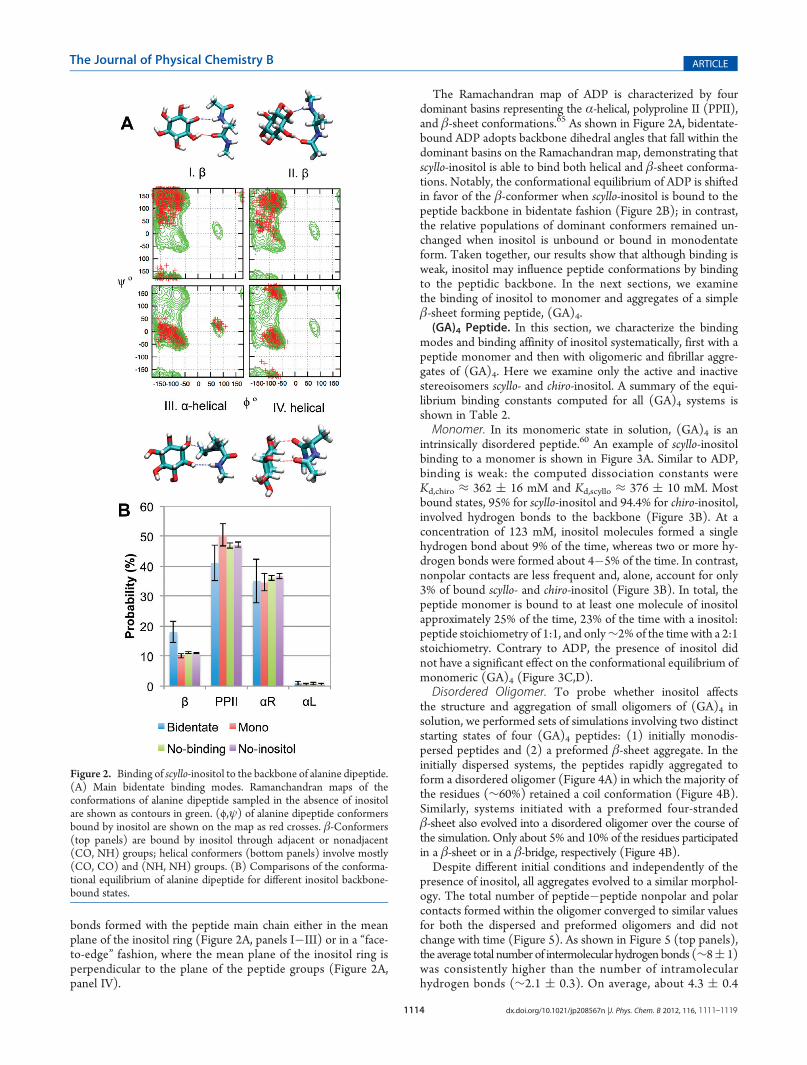
bonds formed with the peptide main chain either in the meanplane of the inositol ring (Figure 2A, panels I�III) or in a “face-to-edge” fashion, where the mean plane of the inositol ring isperpendicular to the plane of the peptide groups (Figure 2A,panel IV).
The Ramachandran map of ADP is characterized by fourdominant basins representing the α-helical, polyproline II (PPII),and β-sheet conformations.65 As shown in Figure 2A, bidentate-bound ADP adopts backbone dihedral angles that fall within thedominant basins on the Ramachandran map, demonstrating thatscyllo-inositol is able to bind both helical and β-sheet conforma-tions. Notably, the conformational equilibrium of ADP is shiftedin favor of the β-conformer when scyllo-inositol is bound to thepeptide backbone in bidentate fashion (Figure 2B); in contrast,the relative populations of dominant conformers remained un-changed when inositol is unbound or bound in monodentateform. Taken together, our results show that although binding isweak, inositol may influence peptide conformations by bindingto the peptidic backbone. In the next sections, we examinethe binding of inositol to monomer and aggregates of a simpleβ-sheet forming peptide, (GA)4.(GA)4 Peptide. In this section, we characterize the binding
modes and binding affinity of inositol systematically, first with apeptide monomer and then with oligomeric and fibrillar aggre-gates of (GA)4. Here we examine only the active and inactivestereoisomers scyllo- and chiro-inositol. A summary of the equi-librium binding constants computed for all (GA)4 systems isshown in Table 2.Monomer. In its monomeric state in solution, (GA)4 is an
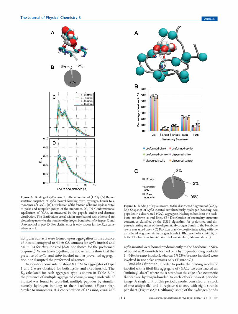
intrinsically disordered peptide.60 An example of scyllo-inositolbinding to a monomer is shown in Figure 3A. Similar to ADP,binding is weak: the computed dissociation constants wereKd,chiro ≈ 362 ( 16 mM and Kd,scyllo ≈ 376 ( 10 mM. Mostbound states, 95% for scyllo-inositol and 94.4% for chiro-inositol,involved hydrogen bonds to the backbone (Figure 3B). At aconcentration of 123 mM, inositol molecules formed a singlehydrogen bond about 9% of the time, whereas two or more hy-drogen bonds were formed about 4�5% of the time. In contrast,nonpolar contacts are less frequent and, alone, account for only3% of bound scyllo- and chiro-inositol (Figure 3B). In total, thepeptide monomer is bound to at least one molecule of inositolapproximately 25% of the time, 23% of the time with a inositol:peptide stoichiometry of 1:1, and only∼2% of the time with a 2:1stoichiometry. Contrary to ADP, the presence of inositol didnot have a significant effect on the conformational equilibrium ofmonomeric (GA)4 (Figure 3C,D).Disordered Oligomer. To probe whether inositol affects
the structure and aggregation of small oligomers of (GA)4 insolution, we performed sets of simulations involving two distinctstarting states of four (GA)4 peptides: (1) initially monodis-persed peptides and (2) a preformed β-sheet aggregate. In theinitially dispersed systems, the peptides rapidly aggregated toform a disordered oligomer (Figure 4A) in which the majority ofthe residues (∼60%) retained a coil conformation (Figure 4B).Similarly, systems initiated with a preformed four-strandedβ-sheet also evolved into a disordered oligomer over the course ofthe simulation. Only about 5% and 10% of the residues participatedin a β-sheet or in a β-bridge, respectively (Figure 4B).Despite different initial conditions and independently of the
presence of inositol, all aggregates evolved to a similar morphol-ogy. The total number of peptide�peptide nonpolar and polarcontacts formed within the oligomer converged to similar valuesfor both the dispersed and preformed oligomers and did notchange with time (Figure 5). As shown in Figure 5 (top panels),the average total number of intermolecular hydrogen bonds (∼8( 1)was consistently higher than the number of intramolecularhydrogen bonds (∼2.1 ( 0.3). On average, about 4.3 ( 0.4
Figure 2. Binding of scyllo-inositol to the backbone of alanine dipeptide.(A) Main bidentate binding modes. Ramanchandran maps of theconformations of alanine dipeptide sampled in the absence of inositolare shown as contours in green. (ϕ,ψ) of alanine dipeptide conformersbound by inositol are shown on the map as red crosses. β-Conformers(top panels) are bound by inositol through adjacent or nonadjacent(CO, NH) groups; helical conformers (bottom panels) involve mostly(CO, CO) and (NH, NH) groups. (B) Comparisons of the conforma-tional equilibrium of alanine dipeptide for different inositol backbone-bound states.
1115 dx.doi.org/10.1021/jp208567n |J. Phys. Chem. B 2012, 116, 1111–1119
The Journal of Physical Chemistry B ARTICLE
nonpolar contacts were formed upon aggregation in the absenceof inositol compared to 4.4 ( 0.5 contacts for scyllo-inositol and5.0 ( 0.4 for chiro-inositol (data not shown for the preformedoligomer). When taken together, the above results show that thepresence of scyllo- and chiro-inositol neither prevented aggrega-tion nor disrupted the preformed oligomer.Dissociation constants of about 80 mM to aggregates of type
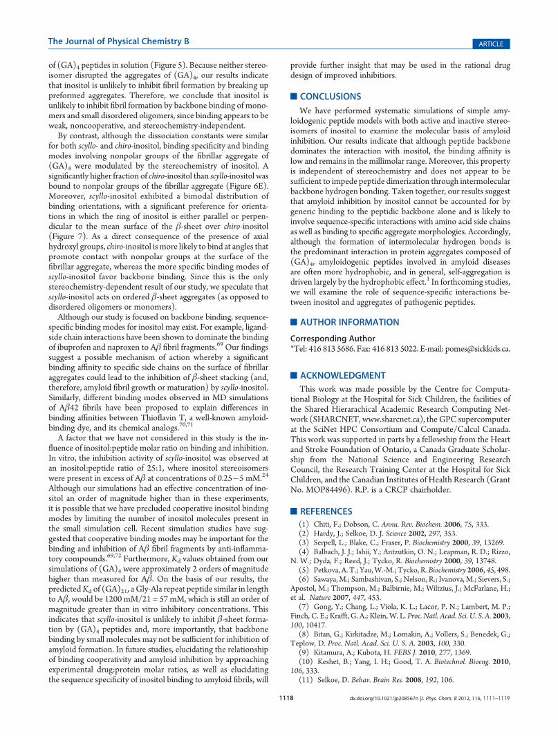
1 and 2 were obtained for both scyllo- and chiro-inositol. TheKd calculated for each aggregate type is shown in Table 2. Inthe presence of multiple aggregated chains, a single molecule ofinositol was found to cross-link multiple peptides by simulta-neously hydrogen bonding to their backbones (Figure 4A).Similar to monomers, at a concentration of 123 mM, chiro- and
scyllo-inositol were bound predominantly to the backbone:∼96%of bound scyllo-inositols formed only hydrogen-bonding contacts(∼94% for chiro-inositol), whereas 2% (3% for chiro-inositol) wereinvolved in nonpolar contacts only (Figure 4C).Fibril-like Oligomer. In order to probe the binding modes of
inositol with a fibril-like aggregate of (GA)4, we constructed an“infinite β-sheet”, where theβ-strands at the edge of an octamericβ-sheet are hydrogen-bonded to each other’s nearest periodicimage. A single unit of this periodic model consisted of a stackof two antiparallel and in-register β-sheets, with eight strandsper sheet (Figure 6A,B). Although some of the hydrogen bonds
Figure 4. Binding of scyllo-inositol to the disordered oligomer of (GA)4.(A) Snapshot of scyllo-inositol simultaneously hydrogen bonding twopeptides in a disordered (GA)4 aggregate. Hydrogen bonds to the back-bone are drawn as red lines. (B) Distribution of secondary structurecontent, as classified by the DSSP algorithm, for preformed and dis-persed starting states of the oligomer. Hydrogen bonds to the backboneare drawn as red lines. (C) Fraction of scyllo-inositol interacting with thedisordered oligomer via hydrogen bonds (HBs), nonpolar contacts, orboth. The fractions for chiro-inositol are similar (data not shown).
Figure 3. Binding of scyllo-inositol to the monomer of (GA)4. (A) Repre-sentative snapshot of scyllo-inositol forming three hydrogen bonds to amonomer of (GA)4. (B) Distribution of the fraction of bound scyllo-inositolto polar and nonpolar groups of the monomer. (C, D) Conformationalequilibrium of (GA)4 as measured by the peptide end-to-end distancedistribution. The distributions are all within error bars of each other and areplotted separately by the number of hydrogen bonds for scyllo- in part C andchiro-inositol in part D. For clarity, error is only shown for the Peed curvewhere n = 1.

1116 dx.doi.org/10.1021/jp208567n |J. Phys. Chem. B 2012, 116, 1111–1119
The Journal of Physical Chemistry B ARTICLE
defining the β-sheet structure occasionally broke, in the absenceof inositol the protofibril remained approximately planar andaggregated as an infinite fibril throughout the simulation.The spatial distribution of bound inositol molecules shows
that both chiro- and scyllo-inositol bind at the surface of the fibril(Figure 6C,D). chiro- And scyllo-inositol bound fibrillar aggre-gates of (GA)4 with aKd of 36( 15 and 51( 3mM, respectively.The apparent increase in affinity compared to amorphous aggre-gates can be attributed to the following reasons. First, the fibrillaraggregate presents a much larger effective surface area thanboth the monomer and the disordered oligomer (Figure 6C,D).Moreover, a larger fraction of the alanine side chains are com-pletely solvent-exposed in the fibril-like aggregate, increasing thefraction of bound conformations involving only nonpolar con-tacts by nearly an order of magnitude, from 2% in the disorderedoligomer to 12% for scyllo- and 18% for chiro-inositol in thefibrillar aggregate in the presence of 109 mM of inositol(Figures 3B and 6E). Accordingly, a higher fraction of scyllo-inositol, 83 ( 1% versus 77 ( 1% for chiro-inositol, was foundto form hydrogen bonds, where the 6% drop in the hydrogen-bonded-only population of chiro-inositol was compensated by acommensurate increase in the nonpolar-bound-only populationof chiro-inositol (Figure 6E).Thus, although chiro- and scyllo-inositol have similar binding
constants, they have different binding modes to fibrillar aggre-gates, a feature not previously observed for the monomer and thedisordered oligomer of (GA)4. Both scyllo- and chiro-inositolform nonpolar contacts and backbone hydrogen bonds in poseswhere the mean plane of the inositol ring lies parallel, at an angle,
or perpendicular to the plane of the fibril (Figure 7). Further-more, two or moremolecules of inositol may cluster together andbind at the surface of the sheet (Figure 7C,D). However, as shownin Figure 8A, scyllo-inositol adopts specific binding orientations,whereas chiro-inositol does not: scyllo-inositol preferentially bindsin either nearly flat (α = 20�) or upright (α = 65�) to the sheet,whereas chiro-inositol does not have such a bimodal preferenceand binds the fibril at an average angle of α = 45�. This stereo-chemistry-modulated difference in binding specificity explains the
Figure 6. Binding of scyllo- and chiro-inositol to the fibrillar aggregate of(GA)4. Different views of the initial starting structure of the fibril-likemodel. Top and bottom sheets are colored in gray and in cyan,respectively. A top down view is depicted in part A showing the backsideof the top (GA)4 sheet. A side view of the protofibril is shown in part B.The spatial probability density of bound scyllo-inositol (yellow) (C) andchiro-inositol (orange) (D) are shown overlapping with the fibril. Thedensity is shown at an occupancy isosurface value of 3% for both stereo-isomers. (E) The percentage of scyllo- and chiro-inositol bound to polarand nonpolar groups on the β-sheet.
Figure 5. Time evolution of peptide�peptide nonpolar and hydrogenbonding contacts in disordered aggregates in the presence and absenceof inositol (control). Each curve is smoothed using a running averageover a window with a length of 500 ps. Results for the dispersed mono-mer aggregates are shown on the left and the preformed β-sheet aggre-gates on the right. (Top) The total number of inter- and intramolecularhydrogen bonding contacts. (Bottom) Number of intermolecular non-polar contacts.

1117 dx.doi.org/10.1021/jp208567n |J. Phys. Chem. B 2012, 116, 1111–1119
The Journal of Physical Chemistry B ARTICLE
somewhat higher fraction of nonpolar binding by chiro-inositol(Figure 6E): chiro-inositol is more likely than scyllo-inositol to bindat angles of 30� <αe 60� (Figure 8A), where 24% of bound chiro-inositol (versus 16% for scyllo-inositol) is bound by nonpolarcontacts only (Figure 8B). For αe 30�, the distributions of scyllo-and chiro-inositol bound to polar and nonpolar groups are similar(data not shown). Moreover, because chiro-inositol has a partiallynonpolar edge, whereas scyllo-inositol does not, binding in theupright position also involvesmore nonpolar interactions for chiro-than for scyllo-inositol (Figure 8B). Finally, although inositol wasobserved to bind at the surface, binding did not change themorphology of the fibrillar aggregate.
’DISCUSSION
In the above analysis, we have systematically characterized theassociation of stereoisomers scyllo-, epi-, myo-, and chiro-inositolwith alanine dipeptide, a simple model of the peptidic backbone.Furthermore, we examined the binding of scyllo- and chiro-inositol to various aggregated states of (GA)4 to probe the role ofbackbone binding in amyloid inhibition. Our results showthat inositol exhibits weak binding with dissociation constantsin the range of 0.04�1 M to the different peptides and aggrega-tion states considered.
Furthermore, the Kd of inositol increases linearly with thenumber of peptide groups in the system (Table 2), indicatingthat inositol does not bind cooperatively to the monomer andaggregate states of (GA)4 considered. As expected, inositol bindsmost weakly to alanine dipeptide, with a value about 4 timessmaller than the Kd of urea to N-acetylalanine reported recentlyin the literature (0.3 M for urea66 vs 1.1 M for scyllo-inositol).Taken together, our results indicate that the activity of inositolstereoisomers is similar to that of osmolytes, which typically havebinding constants in the millimolar to molar range.38,66,67
The spacing of consecutive OH groups of inositol is well-suited to bidentate interactions with adjacent groups of the poly-peptide backbone (Figure 2A). Our findings shown in Figure 2Aare consistent with similar binding modes observed in a recentab initio simulation and IR spectroscopic study of the bindingof glucose epimers to the phenylalanine dipeptide backbone.68
Furthermore, inositol stereoisomers displace the backboneconformation of alanine dipeptide toward extended β-strandconformations (Figure 2B). However, neither scyllo- nor chiro-inositol had a significant effect on the conformational equilibriumof the (GA)4 monomer. Taken together, these results indicatethat inositol may not act as a drug by directly influencing mono-mer conformations. However, our results do not preclude thepossibility that inositol may block fibril elongation by prefer-entially binding to monomers that are constrained to extendedconformations, such as those at exposed edges of β-sheets(a factor not considered in this study).
Independently of the presence of inositol, both the preformedβ-sheet oligomer and the monodisperse solution of (GA)4evolved into a similar morphology (Figures 4B and 5) with onlya small amount of β-structure (Figure 4B), indicating that smallaggregates of (GA)4 are likely to be disordered. Unlike the hydro-phobic core of the Aβ peptide, (GA)4 is a shorter and more polarpeptide that is capable of forming more hydrogen bonds thannonpolar contacts. Our results show that peptide�peptidehydrogen bonding plays an important role in the aggregation
Figure 7. Example snapshots of scyllo- and chiro-inositol binding tothe fibril of (GA)4. Red and blue dashed lines denote hydrogen bonds.(A, B) Example binding modes of scyllo- and chiro-inositol binding at anangle to the surface. (C, D) Example binding modes of scyllo- and chiro-inositol binding face down on the sheet.
Figure 8. Binding mode and orientation of scyllo- and chiro-inositol tothe fibril of (GA)4. (A) The distribution of inositol to sheet planarangles. (B) Inositol binding to nonpolar and polar groups as classified byα, the angle at which inositol molecules bind at the surface of the fibrillar(GA)4 (see Materials and Methods).
1118 dx.doi.org/10.1021/jp208567n |J. Phys. Chem. B 2012, 116, 1111–1119
The Journal of Physical Chemistry B ARTICLE
of (GA)4 peptides in solution (Figure 5). Because neither stereo-isomer disrupted the aggregates of (GA)4, our results indicatethat inositol is unlikely to inhibit fibril formation by breaking uppreformed aggregates. Therefore, we conclude that inositol isunlikely to inhibit fibril formation by backbone binding of mono-mers and small disordered oligomers, since binding appears to beweak, noncooperative, and stereochemistry-independent.
By contrast, although the dissociation constants were similarfor both scyllo- and chiro-inositol, binding specificity and bindingmodes involving nonpolar groups of the fibrillar aggregate of(GA)4 were modulated by the stereochemistry of inositol. Asignificantly higher fraction of chiro-inositol than scyllo-inositol wasbound to nonpolar groups of the fibrillar aggregate (Figure 6E).Moreover, scyllo-inositol exhibited a bimodal distribution ofbinding orientations, with a significant preference for orienta-tions in which the ring of inositol is either parallel or perpen-dicular to the mean surface of the β-sheet over chiro-inositol(Figure 7). As a direct consequence of the presence of axialhydroxyl groups, chiro-inositol is more likely to bind at angles thatpromote contact with nonpolar groups at the surface of thefibrillar aggregate, whereas the more specific binding modes ofscyllo-inositol favor backbone binding. Since this is the onlystereochemistry-dependent result of our study, we speculate thatscyllo-inositol acts on ordered β-sheet aggregates (as opposed todisordered oligomers or monomers).
Although our study is focused on backbone binding, sequence-specific binding modes for inositol may exist. For example, ligand-side chain interactions have been shown to dominate the bindingof ibuprofen and naproxen to Aβ fibril fragments.69 Our findingssuggest a possible mechanism of action whereby a significantbinding affinity to specific side chains on the surface of fibrillaraggregates could lead to the inhibition of β-sheet stacking (and,therefore, amyloid fibril growth or maturation) by scyllo-inositol.Similarly, different binding modes observed in MD simulationsof Aβ42 fibrils have been proposed to explain differences inbinding affinities between Thioflavin T, a well-known amyloid-binding dye, and its chemical analogs.70,71
A factor that we have not considered in this study is the in-fluence of inositol:peptide molar ratio on binding and inhibition.In vitro, the inhibition activity of scyllo-inositol was observed atan inositol:peptide ratio of 25:1, where inositol stereoisomerswere present in excess of Aβ at concentrations of 0.25�5 mM.24
Although our simulations had an effective concentration of ino-sitol an order of magnitude higher than in these experiments,it is possible that we have precluded cooperative inositol bindingmodes by limiting the number of inositol molecules present inthe small simulation cell. Recent simulation studies have sug-gested that cooperative binding modes may be important for thebinding and inhibition of Aβ fibril fragments by anti-inflamma-tory compounds.69,72 Furthermore, Kd values obtained from oursimulations of (GA)4 were approximately 2 orders of magnitudehigher than measured for Aβ. On the basis of our results, thepredictedKd of (GA)21, a Gly-Ala repeat peptide similar in lengthto Aβ, would be 1200 mM/21 = 57 mM, which is still an order ofmagnitude greater than in vitro inhibitory concentrations. Thisindicates that scyllo-inositol is unlikely to inhibit β-sheet forma-tion by (GA)4 peptides and, more importantly, that backbonebinding by small molecules may not be sufficient for inhibition ofamyloid formation. In future studies, elucidating the relationshipof binding cooperativity and amyloid inhibition by approachingexperimental drug:protein molar ratios, as well as elucidatingthe sequence specificity of inositol binding to amyloid fibrils, will
provide further insight that may be used in the rational drugdesign of improved inhibitiors.
’CONCLUSIONS
We have performed systematic simulations of simple amy-loidogenic peptide models with both active and inactive stereo-isomers of inositol to examine the molecular basis of amyloidinhibition. Our results indicate that although peptide backbonedominates the interaction with inositol, the binding affinity islow and remains in the millimolar range. Moreover, this propertyis independent of stereochemistry and does not appear to besufficient to impede peptide dimerization through intermolecularbackbone hydrogen bonding. Taken together, our results suggestthat amyloid inhibition by inositol cannot be accounted for bygeneric binding to the peptidic backbone alone and is likely toinvolve sequence-specific interactions with amino acid side chainsas well as binding to specific aggregate morphologies. Accordingly,although the formation of intermolecular hydrogen bonds isthe predominant interaction in protein aggregates composed of(GA)4, amyloidogenic peptides involved in amyloid diseasesare often more hydrophobic, and in general, self-aggregation isdriven largely by the hydrophobic effect.1 In forthcoming studies,we will examine the role of sequence-specific interactions be-tween inositol and aggregates of pathogenic peptides.
’AUTHOR INFORMATION
Corresponding Author*Tel: 416 813 5686. Fax: 416 813 5022. E-mail: [email protected].
’ACKNOWLEDGMENT
This work was made possible by the Centre for Computa-tional Biology at the Hospital for Sick Children, the facilities ofthe Shared Hierarachical Academic Research Computing Net-work (SHARCNET, www.sharcnet.ca), the GPC supercomputerat the SciNet HPC Consortium and Compute/Calcul Canada.This work was supported in parts by a fellowship from the Heartand Stroke Foundation of Ontario, a Canada Graduate Scholar-ship from the National Science and Engineering ResearchCouncil, the Research Training Center at the Hospital for SickChildren, and the Canadian Institutes of Health Research (GrantNo. MOP84496). R.P. is a CRCP chairholder.
’REFERENCES
(1) Chiti, F.; Dobson, C. Annu. Rev. Biochem. 2006, 75, 333.(2) Hardy, J.; Selkoe, D. J. Science 2002, 297, 353.(3) Serpell, L.; Blake, C.; Fraser, P. Biochemistry 2000, 39, 13269.(4) Balbach, J. J.; Ishii, Y.; Antzutkin, O. N.; Leapman, R. D.; Rizzo,
N. W.; Dyda, F.; Reed, J.; Tycko, R. Biochemistry 2000, 39, 13748.(5) Petkova, A. T.; Yau,W.-M.; Tycko, R. Biochemistry 2006, 45, 498.(6) Sawaya,M.; Sambashivan, S.; Nelson, R.; Ivanova,M.; Sievers, S.;
Apostol, M.; Thompson, M.; Balbirnie, M.; Wiltzius, J.; McFarlane, H.;et al. Nature 2007, 447, 453.
(7) Gong, Y.; Chang, L.; Viola, K. L.; Lacor, P. N.; Lambert, M. P.;Finch, C. E.; Krafft, G. A.; Klein, W. L. Proc. Natl. Acad. Sci. U. S. A. 2003,100, 10417.
(8) Bitan, G.; Kirkitadze, M.; Lomakin, A.; Vollers, S.; Benedek, G.;Teplow, D. Proc. Natl. Acad. Sci. U. S. A. 2003, 100, 330.
(9) Kitamura, A.; Kubota, H. FEBS J. 2010, 277, 1369.(10) Keshet, B.; Yang, I. H.; Good, T. A. Biotechnol. Bioeng. 2010,
106, 333.(11) Selkoe, D. Behav. Brain Res. 2008, 192, 106.

1119 dx.doi.org/10.1021/jp208567n |J. Phys. Chem. B 2012, 116, 1111–1119
The Journal of Physical Chemistry B ARTICLE
(12) Lambert, M.; Barlow, A.; Chromy, B.; Edwards, C.; Freed, R.;Liosatos, M.; Morgan, T.; Rozovsky, I.; Trommer, B.; Viola, K.; et al.Proc. Natl. Acad. Sci. U. S. A. 1998, 95, 6448.(13) Caughey, B.; Baron, G.; Chesebro, B.; Jeffrey, M. Annu. Rev.
Biochem. 2009, 78, 177.(14) Necula, M.; Kayed, R.; Milton, S.; Glabe, C. J. Biol. Chem. 2007,
282, 10311.(15) Hawkes, C.; Ng, V.; McLaurin, J. Drug Develop Res. 2009, 70,
111.(16) Frid, P.; Anisimov, S.; Popovic, N. Brain Res. Rev. 2007, 53, 135.(17) LeVine, H.; Ding, Q.; Walker, J.; Voss, R.; Augelli-Szafran, C.
Neurosci. Lett. 2009, 465, 99.(18) Scherzer-Attali, R.; Pellarin, R.; Convertino, M.; Frydman-
Marom, A.; Egoz-Matia, N.; Peled, S.; Levy-Sakin, M.; Shalev, D. E.;Caflisch, A.; Gazit, E.; et al. PLoS one 2010, 5, e11101.(19) Sood, A.; Abid, M.; Hailemichael, S.; Foster, M.; T€or€ok, B.;
T€or€ok, M. Bioorg. Med. Chem. Lett. 2009, 19, 6931.(20) Porat, Y.; Abramowitz, A.; Gazit, E. Chem. Biol. Drug Des. 2006,
67, 27.(21) Ehrnhoefer, D. E.; Bieschke, J.; Boeddrich, A.; Herbst, M.;
Masino, L.; Lurz, R.; Engemann, S.; Pastore, A.; Wanker, E. E. Nat.Struct. Mol. Biol. 2008, 15, 558.(22) Liu, F.-F.; Ji, L.; Dong, X.-Y.; Sun, Y. J. Phys. Chem. B 2009,
113, 11320.(23) Liu, R.; Barkhordarian, H.; Emadi, S.; Park, C.; Sierks, M.
Neurobiol. Dis. 2005, 20, 74.(24) McLaurin, J.; Golomb, R.; Jurewicz, A.; Antel, J.; Fraser, P.
J. Biol. Chem. 2000, 275, 18495.(25) McLaurin, J.; Kierstead, M. E.; Brown, M. E.; Hawkes, C. A.;
Lambermon, M. H. L.; Phinney, A. L.; Darabie, A. A.; Cousins, J. E.;French, J. E.; Lan, M. F.; et al. Nat. Med. 2006, 12, 801.(26) Fisher, S.; Novak, J.; Agranoff, B. J. Neurochem. 2002, 82, 736.(27) Michaelis, T.; Helms, G.; Merboldt, K.-D.; H€anicke, W.; Bruhn,
H.; Frahm, J. NMR Biomed. 1993, 6, 105.(28) Michell, R. Nat. Rev. Mol. Cell. Biol. 2008, 9, 151.(29) McLaurin, J.; Franklin, T.; Chakrabartty, A.; Fraser, P. J. Mol.
Biol. 1998, 278, 183.(30) Nitz, M.; Fenili, D.; Darabie, A. A.; Wu, L.; Cousins, J. E.;
McLaurin, J. FEBS J. 2008, 275, 1663.(31) Sun, Y.; Zhang, G.; Hawkes, C. A.; Shaw, J. E.; McLaurin, J.;
Nitz, M. Bioorg. Med. Chem. 2008, 16, 7177.(32) Townsend, M.; Cleary, J. P.; Mehta, T.; Hofmeister, J.; Lesne,
S.; O’Hare, E.; Walsh, D. M.; Selkoe, D. J. Ann. Neurol. 2006, 60, 668.(33) Scaramozzino, F.; Peterson, D. W.; Farmer, P.; Gerig, J. T.;
Graves, D. J.; Lew, J. Biochemistry 2006, 45, 3684.(34) Yang, D. S.; Yip, C. M.; Huang, T. H.; Chakrabartty, A.; Fraser,
P. E. J. Biol. Chem. 1999, 274, 32970.(35) McLaurin, J.; Yang, D.; Yip, C. M.; Fraser, P. E. J. Struct. Biol.
2000, 130, 259.(36) Dasilva, K. A.; Shaw, J. E.; McLaurin, J. Exp. Neurol. 2010,
223, 311.(37) Bieschke, J.; Russ, J.; Friedrich, R.; Ehrnhoefer, D.; Wobst, H.;
Neugebauer, K.; Wanker, E. Proc. Natl. Acad. Sci. U.S.A. 2010, 107, 7710.(38) Street, T.; Bolen, D.; Rose, G. Proc. Natl. Acad. Sci. U. S. A. 2006,
103, 13997.(39) Hu, C. Y.; Lynch, G. C.; Kokubo, H.; Pettitt, B. M. Proteins
2010, 78, 695.(40) Auton, M.; Bolen, D. W.; R€osgen, J. Proteins 2008, 73, 802.(41) Rose, G. D.; Fleming, P. J.; Banavar, J. R.; Maritan, A. Proc. Natl.
Acad. Sci. U. S. A. 2006, 103, 16623.(42) Rauscher, S.; Baud, S.; Miao, M.; Keeley, F. W.; Pom�es, R.
Structure 2006, 14, 1667.(43) Takeda, T.; Klimov, D. K. J. Phys. Chem. B 2010, 114, 4755.(44) Soto, P.; Griffin, M.; Shea, J.-E. Biophys. J. 2007, 93, 3015.(45) Rauscher, S.; Pom�es, R. Biochem. Cell Biol. 2010, 88, 269.(46) Raman, E. P.; Takeda, T.; Klimov, D. K. Biophys. J. 2009, 97,
2070.(47) Lemkul, J.; Bevan, D. Biochemistry 2010, 49, 3935.
(48) Rathore, O.; Sogah, D. Y. Macromolecules 2001, 34, 1477.(49) Vandermeulen, G.; Kim, K.; Wang, Z.; Manners, I. Biomacro-
molecules 2006, 7, 1005.(50) Kenney, J. M.; Knight, D.; Wise, M. J.; Vollrath, F. Eur. J.
Biochem. 2002, 269, 4159.(51) Fossey, S. A.; N�emethy, G.; Gibson, K. D.; Scheraga, H. A.
Biopolymers 1991, 31, 1529.(52) The PyMOL Molecular Graphics System, 1.3r1; Schrodinger,
LLC, 2010.(53) Jorgensen, W. L.; Maxwell, D. S.; Tirado-Rives, J. J. Am. Chem.
Soc. 1996, 118, 11225.(54) Damm, W.; Frontera, A.; Tirado�Rives, J.; Jorgensen, W. L.
J. Comput. Chem. 1997, 18, 1955.(55) Jorgensen, W. L.; Chandrasekhar, J.; Madura, J. D.; Impey,
R. W.; Klein, M. L. J. Chem. Phys. 1983, 79, 926.(56) Van Der Spoel, D.; Lindahl, E.; Hess, B.; Groenhof, G.; Mark,
A. E.; Berendsen, H. J. C. J. Comput. Chem. 2005, 26, 1701.(57) Essmann, U.; Perera, L.; Berkowitz, M. L.; Darden, T.; Lee, H.;
Pedersen, L. G. J. Chem. Phys. 1995, 103, 8577.(58) Berendsen, H. J. C.; Postma, J. P. M.; Van Gunsteren, W. F.;
DiNola, A.; Haak, J. J. Chem. Phys. 1984, 81, 3684.(59) Ryckaert, J. P.; Ciccotti, G.; Berendsen, H. J. C. J. Comput. Phys.
1977, 23, 327.(60) Nikolic, A.; Baud, S.; Rauscher, S.; Pom�es, R. Proteins 2011,
79, 1.(61) Rodinger, T.; Howell, P.; Pom�es, R. J. Chem. Theory Comput
2006, 2, 725.(62) Rauscher, S.; Neale, C.; Pom�es, R. J. Chem. Theory Comput.
2009, 5, 2640.(63) Kabsch, W.; Sander, C. Biopolymers 1983, 22, 2577.(64) Humphrey, W.; Dalke, A.; Schulten, K. J. Mol. Graph. 1996,
14, 33.(65) Neale, C.; Rodinger, T.; Pomes, R. Chem. Phys. Lett. 2008,
460, 375.(66) Lee, S.; Shek, Y.; Chalikian, T. Biopolymers 2010, 93, 866.(67) R€osgen, J. Methods Enzymol. 2007, 428, 459.(68) Cocinero, E. J.; C-arc-abal, P.; Vaden, T. D.; Davis, B. G.; Simons,
J. P. J. Am. Chem. Soc. 2011, 133, 4548.(69) Kim, S.; Chang, W. E.; Kumar, R.; Klimov, D. K. Biophys. J.
2011, 100, 2024.(70) Mathis, C.; Wang, Y.; Holt, D.; Huang, G.-F.; Debnath, M.;
Klunk, W. J. Med. Chem. 2003, 46, 2740.(71) Wu, C.; Bowers, M.; Shea, J.-E. Biophys. J. 2011, 100, 1316.(72) Takeda, T.; Chang, W. E.; Raman, E. P.; Klimov, D. K. Proteins
2010, 78, 2849.
Related Documents