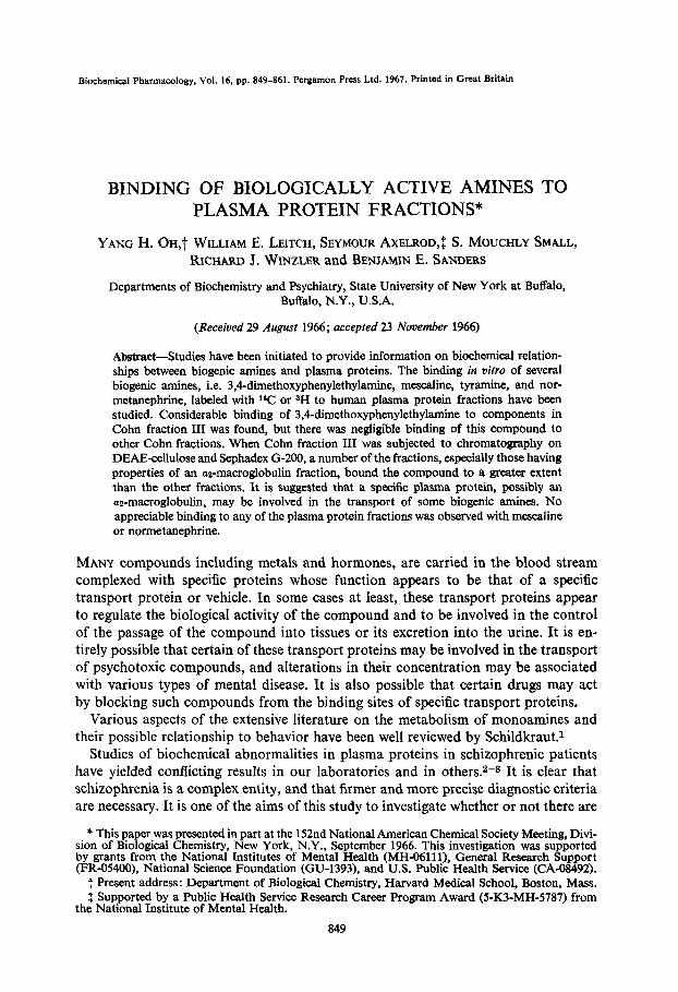
Biochemical Pharmacology, Vol. 16, pp. 849-861. Pergamon Press Ltd. 1967. Printed in Great Britain BINDING OF BIOLOGICALLY ACTIVE AMINES TO PLASMA PROTEIN FRACTIONS* YANG H. Or-&t WILLIAM E. LEITCH, SEYMOUR AXELROD,~S. MOUCHLY SMALL, RICHARD J. WINZLER and BENJAMIN E. SANDERS Departments of Biochemistry and Psychiatry, State University of New York at Buffalo, Buffalo, N.Y., U.S.A. (Received 29 August 1966; accepted 23 November 196Q Abstract-Studies have been initiated to provide information on biochemical relation- ships between biogenic amines and plasma proteins. The binding in vitro of several biogenic amines, i.e. 3,4-dimethoxyphenylethylamine, mescaline, tyramine, and nor- metanephrine, labeled with 1% or aH to human plasma protein fractions have been studied. Considerable binding of 3,4-dimethoxyphenylethylamine to components in Cohn fraction III was found, but there was negligible binding of this compound to other Cohn fractions. When Cohn fraction III was subjected to chromatography on DEAE-cellulose and Sephadex G-200, a number of the fractions, especially those having properties of an aa-maeroglobulin fraction, bound the compound to a greater extent than the other fractions. It is suggested that a specific plasma protein, possibly an az-maeroglobulin, may be involved in the transport of some biogenic amines. No appreciable binding to any of the plasma protein fractions was observed with mescaline or normetanephrine. MANY compounds including metals and hormones, are carried in the blood stream complexed with specific proteins whose function appears to be that of a specific transport protein or vehicle. In some cases at least, these transport proteins appear to regulate the biological activity of the compound and to be involved in the control of the passage of the compound into tissues or its excretion into the urine. It is en- tirely possible that certain of these transport proteins may be involved in the transport of psychotoxic compounds, and alterations in their concentration may be associated with various types of mental disease. It is also possible that certain drugs may act by blocking such compounds from the binding sites of specific transport proteins. Various aspects of the extensive literature on the metabolism of monoamines and their possible relationship to behavior have been well reviewed by Schildkraut.1 Studies of biochemical abnormalities in plasma proteins in schizophrenic patients have yielded confiicting results in our laboratories and in others.s-s It is clear that schizophrenia is a complex entity, and that firmer and more precise diagnostic criteria are necessary. It is one of the aims of this study to investigate whether or not there are * This paper was presented in part at the 152nd National American Chemical Society Meeting, Divi- sion of Biological Chemistry, New York, N.Y., September 1966. This investigation was supported by grants from the National Institutes of Mental Health (MH-06111), General Research Support (FR-O5400), National Science Foundation (GU-1393), and U.S. Public Health Service (CA-08492). t Present address: Department of Biological Chemistry, Harvard Medical School, Boston, Mass. $ Supported by a Public Health Service Research Career Program Award (5-K3-MH-5787) from the National Institute of Mental Health. 849

Welcome message from author
This document is posted to help you gain knowledge. Please leave a comment to let me know what you think about it! Share it to your friends and learn new things together.
Transcript
Biochemical Pharmacology, Vol. 16, pp. 849-861. Pergamon Press Ltd. 1967. Printed in Great Britain
BINDING OF BIOLOGICALLY ACTIVE AMINES TO PLASMA PROTEIN FRACTIONS*
YANG H. Or-&t WILLIAM E. LEITCH, SEYMOUR AXELROD,~ S. MOUCHLY SMALL, RICHARD J. WINZLER and BENJAMIN E. SANDERS
Departments of Biochemistry and Psychiatry, State University of New York at Buffalo, Buffalo, N.Y., U.S.A.
(Received 29 August 1966; accepted 23 November 196Q
Abstract-Studies have been initiated to provide information on biochemical relation- ships between biogenic amines and plasma proteins. The binding in vitro of several biogenic amines, i.e. 3,4-dimethoxyphenylethylamine, mescaline, tyramine, and nor- metanephrine, labeled with 1% or aH to human plasma protein fractions have been studied. Considerable binding of 3,4-dimethoxyphenylethylamine to components in Cohn fraction III was found, but there was negligible binding of this compound to other Cohn fractions. When Cohn fraction III was subjected to chromatography on DEAE-cellulose and Sephadex G-200, a number of the fractions, especially those having properties of an aa-maeroglobulin fraction, bound the compound to a greater extent than the other fractions. It is suggested that a specific plasma protein, possibly an az-maeroglobulin, may be involved in the transport of some biogenic amines. No appreciable binding to any of the plasma protein fractions was observed with mescaline or normetanephrine.
MANY compounds including metals and hormones, are carried in the blood stream complexed with specific proteins whose function appears to be that of a specific transport protein or vehicle. In some cases at least, these transport proteins appear to regulate the biological activity of the compound and to be involved in the control of the passage of the compound into tissues or its excretion into the urine. It is en-
tirely possible that certain of these transport proteins may be involved in the transport of psychotoxic compounds, and alterations in their concentration may be associated with various types of mental disease. It is also possible that certain drugs may act
by blocking such compounds from the binding sites of specific transport proteins. Various aspects of the extensive literature on the metabolism of monoamines and
their possible relationship to behavior have been well reviewed by Schildkraut.1 Studies of biochemical abnormalities in plasma proteins in schizophrenic patients
have yielded confiicting results in our laboratories and in others.s-s It is clear that schizophrenia is a complex entity, and that firmer and more precise diagnostic criteria are necessary. It is one of the aims of this study to investigate whether or not there are
* This paper was presented in part at the 152nd National American Chemical Society Meeting, Divi- sion of Biological Chemistry, New York, N.Y., September 1966. This investigation was supported by grants from the National Institutes of Mental Health (MH-06111), General Research Support (FR-O5400), National Science Foundation (GU-1393), and U.S. Public Health Service (CA-08492).
t Present address: Department of Biological Chemistry, Harvard Medical School, Boston, Mass. $ Supported by a Public Health Service Research Career Program Award (5-K3-MH-5787) from
the National Institute of Mental Health.
849
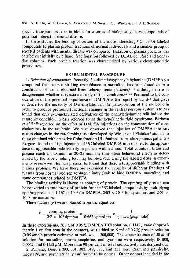
850 Y. H. OH, W. E. LEITCH, S. AXELROD, S. M. SMALL, R. J. WINZLER and B. E. SANDERS
specific transport proteins in blood for a series of biologically active compounds of potential interest in mental disease.
In these studies the binding of certain of the more interesting 14C- or sH-labeled compounds to plasma protein fractions of normal individuals and a smaller group of selected patients with mental disease was compared. Isolation of plasma proteins was carried out initially by ethanol fractionation followed by DEAE-cellulose and Sepha- dex columns. Each protein fraction was characterized by various electrophoretic procedures.
EXPERIMENTAL PROCEDURE
1. Selection of compounds. Recently, 3,4-dimethoxyphenylethylamine (DMPEA), a compound that bears a striking resemblance to mescaline, has been found to be a constituent of urine obtained from schizophrenic patients,s-r4 although there is disagreement whether it is excreted only in this condition.rs-17 Pertinent to the con- sideration of the potential importance of DMPEA is the report by Ernst18 that gives evidence for the necessity of 0-methylation in the para-position of the molecule in order to produce profound functional changes in the central nervous system. He has found that only p-0-methylated derivatives of the phenylethylamine will induce the catatonic condition in cats referred to as the hypokinetic rigid syndrome. Barbeau et d.lg-zo reported on the effect of DMPEA injections on the concentration of cate- cholamines in the rat brain. We have observed that injection of DMPEA into rats causes changes in the rat-climbing test developed by Winter and Flatakefl similar to those obtained with an active Cohn fraction III obtained from schizophrenic patients.7 Bergen21 found that i.p. injections of l4C-labeled DMPEA into rats led to the appear- ance of appreciable radioactivity in plasma within 5 min. Total counts in brain and plasma reach a maximum at 20-25 min, the time when behavioral effects as deter- mined by the rope-climbing test may be observed. Using the labeled drug in experi- ments in vitro with human plasma, he found that there was appreciable binding with plasma proteins. We have therefore examined the capacity of different fractions of plasma from normal and schizophrenic individuals to bind DMPEA, mescaline, and some compounds related to DMPEA.
The binding activity is shown as cpm/mg of protein. The cpm/mg of protein can be converted to I-l.moles/mg of protein for the r4C-labeled compounds by multiplying cpm/mgprotein x 1.167 x IO-7for DMPEA, 2.03 x 1O-7 for tyramine, and 2.51 x 10⁻⁷ for mescaline.
These factors (F) were obtained from the equation:
F- cpm/mg protein 1 1
-x 2.2 x 10s dpm/+
___~ >: -. O-667 cpm/dpm sp. act. (pc/pmole)
In these experiments, 50 pg of 0.05 % DMPEAeHCl solution, O-1 142 pmole (approxi- mately 1 million cpm in the counter), was added to 5 ml of 0*2’% protein solution (0.05 pmole protein estimated at mol. wt. - 200,000). The concentrations of 50 ~1 of solution for mescaline, normetanephrine, and tyramine were respectively : O*lOOO, 0.0027, and O-1 152 PM. More than 90 per cent of total radioactivity was dialyzed out.
2. Subjects. Donors 293, 301, 302, 318, 328, and 331 were examined physically, medically, and psychiatrically and found to be normal. Other donors included in the
Binding of amines to plasma proteins 851
normal group were selected volunteers from various walks of life. One subject had been on a tetracycline antibiotic a few days for minor skin irritation. The rest of the normal donors were not under medication.
Patients diagnosed as schizophrenic were 293, 297, 303, 306 and 329. Other donors included in this group were psychiatric patients presenting some features of the schizo- phrenic syndrome, though final diagnosis would not include them within this specific category. All the schizophrenic patients were on phenothiazone medication for brief periods at least.
3. Preparation of plasma fractions. The Cohn ethanol fractions were prepared from 200-250 ml plasma by modifications described be Lever et al.22 and by Sanders et aLa The fractions obtained were dissolved in 0.01 M sodium phosphate buffer, bH 7.8, one fourth the original plasma volume, and dialyzed overnight against the same buffer to remove alcohol and traces of insoluble protein.
4. Subfractionation and pur$cation. All procedures used in DEAE-cellulose chromatographic subfractionation of the Cohn fractions have been described in previous reports.23-25
The gel filtration experiments (Sephadex G-200) were performed essentially as described by Flodin and Killander. 2s The buffer used for elution was 0.01 M sodium phosphate, pH 7% The.columns were 4 x 75 cm without extension tube. The volume of the gel bed was 980 ml for 4 x 65 cm. The flow rate was maintained at 27 ml/hr. and the effluent was collected in 9-ml portions. Optimum sample size was 100-150 mg protein in 3-15 ml volume.
5. Determination of protein concentration. The protein concentration of each frac- tion was estimated by absorbance at 280 rnp (E:zm = 10) with a Beckman DU-2 spectrophotometer and also by the method described by Lowry et al.27
9. Electrophoretic characterization. The methods used for characterization of the protein fractions, microzone electrophoresis on cellulose acetate, disk electrophoresis. and immunoelectroyhoresis have been described previously.23
7. Binding studies. The DMPEA-814C (specific activity, 5.85 me/m-mole), the mesca- line (sp. act. 2.72 me/m-mole), and the tyramine (sp. act. 4.71 me/m-mole) were obtained from New England Nuclear Corp., Boston, Mass. Each of the labeled com- pounds was made 0.05 % solution with 0.01 M sodium phosphate pH 7.8. The norme- tanephrineJH (sp. act. 450 me/m-mole), obtained from Volk Radiochemical Co., Chicago, III., was made OWl % solution with the same phosphate buffer.
The protein fractions (5 ml of 0.2% protein solution) were incubated at 37” in 0.01 M sodium phosphate buffer, pH 7.8, and 50 pg of the prepared solution of labeled compound for I-hr. Incubation was carried out in a Dubnoff metabolic shaking bath at atmospheric conditions. Optimum conditions for these experiments, such as the period of incubation, temperature, pH, protein concentration, and addition of labeled compound, were previously determined.
Each incubated sample was dialyzed against the same buffer, 500-ml aliquots with four changes, for 48 hr at 4”; this time had previously been found sufficient to obtain a dialysate with no radioactivity.
Each sample (0.5 ml) was dissolved in 10 ml of a liquid scintillation solvent and counted in a Nuclear-Chicago liquid scintillation counter. The solvent was 1: 1, v/v, respectively, of toluene and 2-ethoxyethanol containing 0.66 % diphenyloxazol (PPO) and O-022 % 1,4-di [2-(phenyloxazolyl)]-benzene (POPOP).
852 Y. H. OH, W. E. LEITCH, S. AXELROD, S. M. SMALL, R. J, WINZLER and B. E. SAND~KS
RESULTS
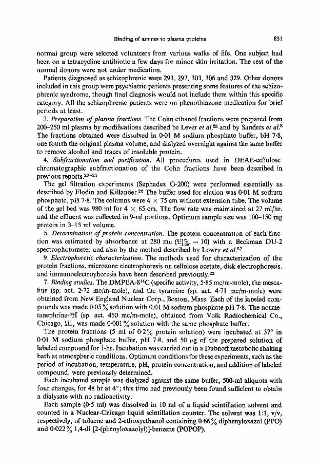
In previous work,24 the Cohn fraction III from individual human plasma has been successfully separated into thirteen globulin subfractions by employing a simple and rapid elution procedure on a DEAE-cellulose column. A typical separation of Cohn fraction III from normal plasma is shown in Fig. 1. Table 1 shows relative amounts of protein in the reproducible thirteen characteristic absorbance peaks (A.P.), pre- sented as each mean of 17 normal and 9 schizophrenic subjects.
I 2 3 4
Volume of effluent, L.
FIG. I. EfiIuent diagram of Cohn fraction III from normal human plasma. Approximately 760 mg of fraction III was applied to a column of DEAE-cefl~~lo~e (2.7 x 72 cm). How rate, 250 ml/hr;
temperature, 4”. Other conditions given in the previous paper.“3*!7,4
TABLE 1. RELATIVE AMOUNTS* 0~ PROTEIN IN EACH ABSORBANCE
PEAK UN DEAE-CELLULOSE FROM COHN FRACTION III
Normal (17) Patients (9)
Mean Standard Mean Standard (%) deviation (%) deviation
* The amount of protein in each absorbance peak is expressed as a percen- tage of the total amount recovered from the column and the mean of seventeen normal individuals and nine schizophrenic subjects. The range of protein introduced on column was 6OO-1200 mg.
Binding of amines to plasma proteins 853
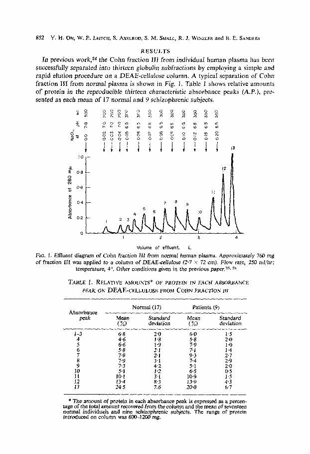
The original thirteen DEAE-cellulose peaks (Fig. 1) were individually concentrated, mixed with DMPEA as described above, and dialyzed to remove unbound DMPEA. Since many of these peaks contained little protein, it was usually found necessary to pool the samples from about three column runs. These pooled fractions (containing between 100 and 150 mg protein) were dialyzed after incubation with DMPEA and chromatographed on Sephadex G-200 columns. In these experiments, binding was confined to the 19/S peak (Fig. 2). When an excess of DMPEA was added to the undialyzed sample, the unbound DMPEA was recovered after approximately 1000 ml of the buffer had been passed through the Sephadex G-200 column.
300 500 700 300 500 700
Volume of Effluent , ml
FIG. 2. Effluent diagrams on Sephadex G-200 columns (4 x 65 cm) of Cohn fraction 111 and absorb- ance peaks 4, 7, and 11 with W-labeled DMPEA obtained from DEAE-cellulose chromatogram in Fig. 1. Approximately lOO-150 mg of each protein with DMPEA was applied to a column of Sepha- dex G-200. Flow rate, 27 mljhr; temperature, 4”. Other conditions as given in the text. Solid line represents absorbance at 280 w, dotted line shows cpm/ml efkent in radioactivity. Pooled original
plasmas used for these experiments were from donors 331, 332, and 337.
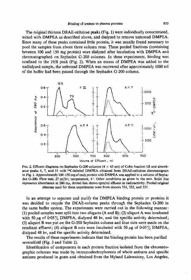
In an attempt to separate and purify the DMPEA binding protein or proteins it was decided to recycle the DEAE-column peaks through the Sephadex G-200 in the same buffer system. The experiments were carried out in the following manner: (1) pooled samples were split into two aliquots (A and B) ; (2) aliquot A was incubated with 50 pg of O*OS’A DMPEA, dialyzed 48 hr, and the specific activity determined; (3) aliquot B was put on the G-200 Sephadex column and four cuts were made on the resultant effluent; (4) aliquot B cuts were incubated with 50 pg of 0.05 ‘A DMPEA, dialyzed 48 hr, and the specific activity determined.
The results of these experiments indicate that the binding protein has been purified severalfold (Fig. 3 and Table 2).
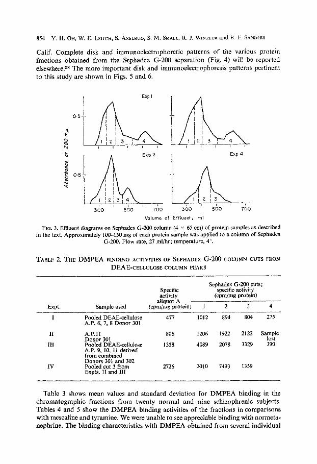
Identification of components in each protein fraction isolated from the chromato- graphic columns was made by immunoelectrophoresis of whole antisera and specific antisera produced in goats and obtained from the Hyland Laboratory, Los Angeles,
854 Y. H. OH, W. E. LEITCH, S. AXELROU, S. M. SMALL, K. J. WINZLEK and 13. F. SANIXRS
Calif. Complete disk and immunoelectrophoretic patterns of the various protein fractions obtained from the Sephadex G-200 separation (Fig. 4) will be reported elsewhere.28 The more important disk and immunoelectrophoresis patterns pertinent to this study are shown in Figs. 5 and 6.
f Expl I A
Exp 2 I Exp 4
Volume of Effluent, ml
Fro. 3. Effluent diagrams on Scphadex G-200 coIurnn (4 x 65 cm) of protein samples as described in the text, Approximately 100-150 mg of each protein sample was applied to a column of Sephadex
G-200. Flow rate, 27 ml/hr; temperature, 4”.
TABLE 2, THE DMPEA BINDING ACXVITIES OF SE~HADEX G-200 COLUMN CUTS FROM
DEAE-CELLULOSE COLUMN PEAKS
Sephadex G-200 cuts; Specific specific activity activity (cpm/mg protein)
Sample used aliquot A
Expt. (cpm/mg protein) 1 2 3 4- -
I Pooled DEAE-cellulose 477 1012 8Y4 804 275 A.P. 6,7, 8 Donor 301
II
III
IV
A.P.11 Donor 301 Pooled DEA~~efllllnse A.P. 9, 10, 11 derived from combined Donors 301 and 302 Pooled cut 3 from Expts. II and III
806 1206 1922 2122 sam,s’
1358 4089 2078 3329 390
2726 2010 7493 1359
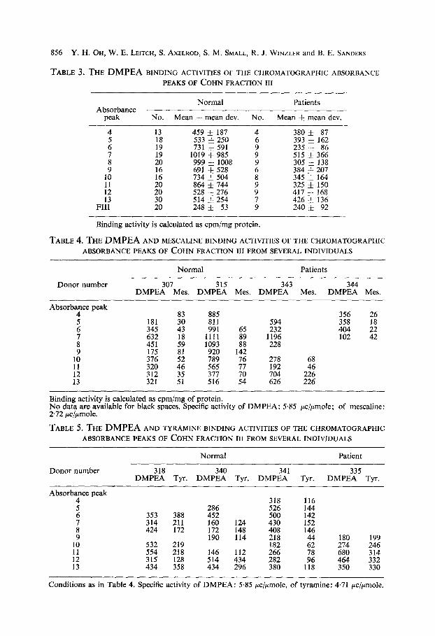
Table 3 shows mean values and standard deviation for DMPEA binding in the ~hromatographic fractions from twenty normal and nine schizophrenic subjects. Tables 4 and 5 show the DMPEA binding activities of the fractions in comparisons with mescaline and tyramine. We were unable to see appreciable binding with normeta- nephrine. The binding characteristics with DMPEA obtained from several individual
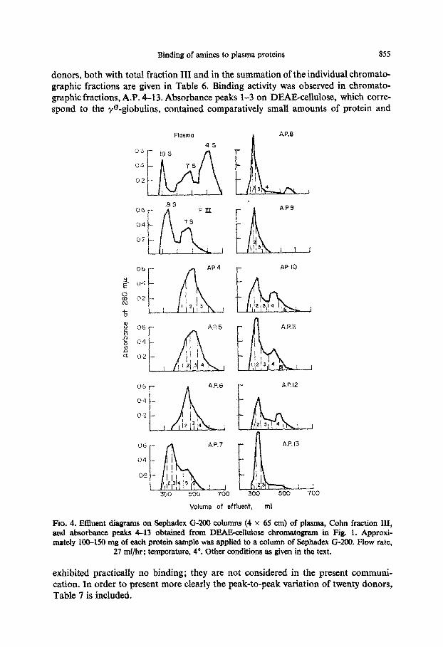
Binding of amines to plasma proteins 855
donors, both with total fraction III and in the summation of the individual chromato- graphic fractions are given in Table 6. Binding activity was observed in chromato- ~ap~c fractions, A.P. 4-13. Absorbance peaks 1-3 on DEAE-~llulose, which corre- spond to the yo-globulins, contained comparatively small amounts of protein and
Plosma
Volume of affhe&, ml
Fm, 4. Effluent diagrams on Sephadex G-200 columns (4 x 65 cm) of plasma, Cohn fraction III, and absorbance. peaks 4-13 obtained from lXAE~llulose chromatogram in Fig. 1. Approxi- mately 100-150 mg of each protein sample was applied to a column of Sephadex C-200. Wow rate,
27 ml/hr; temperature, 4”. Other conditions as given in the text.
exhibited practically no binding; they are not considered in the present communi- cation. In order to present more clearly the peak-to-peak variation of twenty donors, Table 7 is included.
856 Y. H. OH, W. E. LEITCH, S. AX~LROD, S. M. SMALL, R. J. WINZLER and B. E. SANDERS
TABLE 3. THE DMPEA BINDING ACTIVITIES OF THE CHROMATOGRAPHIC ABSORBANCE
PEAKS OF COHN FRACTION III
Absorbance peak
Normal Patients
No. Mean F mean dev. No. Mean * mean dev.
4 13 459 i_ 187 4 380 i_ 87
2 :9” 533 731 * k 250 591 : 235 393 + j, 162 86
;: ;! 1019 + 985 9 515 305 f & 366 138
1: :6” 734 Z$;!P f 504 6” 9 !
384 345 + i 207 164 :: ;: 864 528 & i 276 744 417 325 + f 168 150
FL:: :: 514 248 & -F_ 254 53 ; 426 240 & -r_ 136 92
Binding activity is calculated as cpm/mg protein.
TABLE 4. THE DMPEA AND MESCALINE BINDING ACTIVITIES OF THE CHROMATOGRAPIIIC
ABSORBANCE PEAKS OF COHN FRACTION III FROM SEVERAL INDIVIDUALS
Normal Patients
Donor number 307 315 343 344 DMPEA Mes. DMPEA Mes. DMPEA Mes. DMPEA Mes.
Absorby peak 83 885 356 26 5 181 30 811 594 358 18
6 345 43 991 232 404 s’ 451 632 :: 1111 1093 :;
1::
1196 228 102 Z
lo” 376 175 :: 789 920 11 320 46 565 7’:
278 192 4”:
12 312 13 321 ::
377 516 ;:
704 226 626 226
Binding activity is calculated as cpm/mg of protein. No data are available for black spaces. Specific activity of DMPEA: 5.85 &mole; of mescaline: 2.72 &pmole.
TABLE 5. THE DMPEA AND TYRAMINE BINDING ACTIVITIES 0~ THE CHR~MATOGRAPHIC
ABSORBANCE PEAKS OF COHN FRACTION III FROM SEVERAL INDIVIDUALS
Donor number
Normal Patient
318 340 341 335 DMPEA Tyr. DMPEA Tyr. DMPEA Tyr. DMPEA Tyr.
Absorby peak 318 116
2 353 388 286 452 526 500 144 142
Z 424 314 211 172 160 172 148 124 430 408 152 146 9 190 114 218 180 199
:: 532 554 218 219 2; 146 112 266 182
118 ;:
2:: 246 314
:: 434 315 128 358 434 514 434 296 282 380 464 350 332 330
Conditions as in Table 4. Specific activity of DMPEA: 5.85 &pmole, of tyramine: 4.71 &pmole.
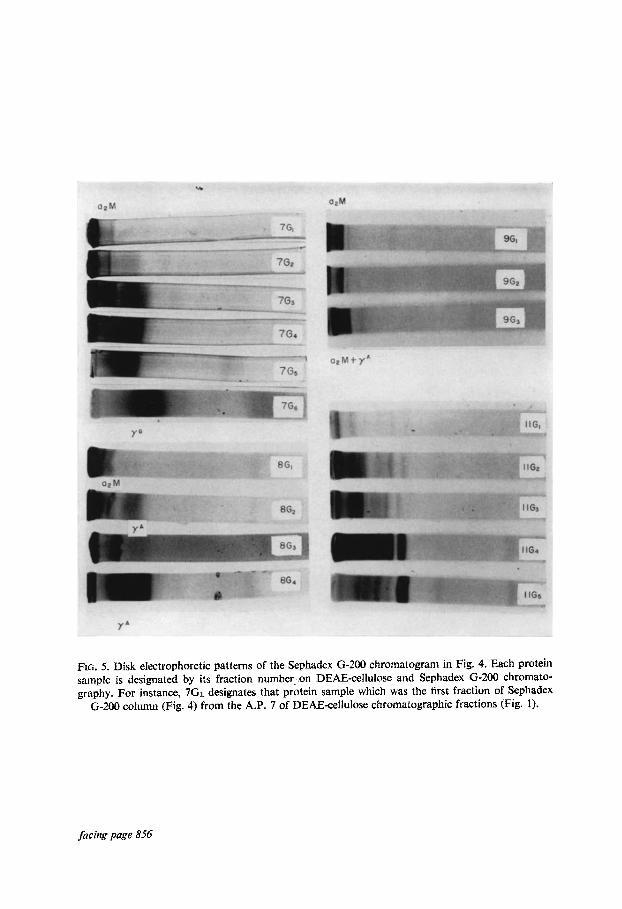
FIG. 5. Disk electrophoretic patterns of the Sephadex G-200 chromatogram in Fig. 4. Each protein sample is designated by its fraction number_.on DEAE-cellulose and Sephadex G-200 chromato- graphy. For instance, 7Gl designates that protein sample which was the first fraction of Sephadex
G-ZOO column (Fig. 4) from the A.P. 7 of DEAE-ce%ulose chromatographic fractions (Fig. 1).
facing page 856

. y ’ Cothers
I?+
Frc. 6. ~rntn~noelectro~boret~c patterns of absorbance peaks from DEAE-cellulose chroi~latogram (Fig. 1) and protein samples obtained from the Sephadex G-200 chromatograms (Fig. 4) against whole
antisera produced in goat (Myland Lab., Calif.). Designation of protein sample as given in Fig. 5.
Binding of amines to plasma proteins 857
Further experiments were performed with Cohn fraction III to which was added 14C-labeled DMPEA after a 1-hr incubation, and the sample was exhaustively dialyzed against O-01 M sodium phosphate buffer, pH 7.8. This DMPEA-labeled fraction III was then placed in a DEAE-cellulose column previously equilibrated with a pH 7.8.
TABLE 6. DMPEA-BINDING ACTIVITY OF COHN FRACTION
III FROM SEVERAL INDIVIDUAL DONORS
Donor number
Average of total* Binding activity binding capacity
(cpm)/mg (cpm) of A.P.s protein of F III from FIII/mg protein
Normals
tz 249 301 302 307 313 315 316 318 320 322 324 326 328 331 332 337 340 341 346 351
Mean & S.D.
Patients 293 297 303 306 329 335 338 343
Mean i%.D.
318 217 109 228 218 208 198 238 315 265 243 216 201 259
% 242 396
E 308 269 252 f 61
1:: 149
186
2% 350 254 240 & 92
2962 951 210
1351 654 354
2: 593 416 466 397 475 39i 501 371 232 286 382 336 274
1118 631 & 580
146 581 351 519 525 370 236 532 210 386 f 152
* A.P.Z 4-l 3 (Specific activity, cpmlmggrotein) x (mg Protein in psea ___~ A.P.C 4-13 (mg protein)
phosphate buffer at the same molarity. In these experiments all the radioactivity was recovered prior to the first protein peak during the washing step with 0.01 M sodium phosphate buffer at pH 7.8. The DMPEA-protein complex was therefore shown to be labile in ion-exchange chromatography.
The radioactivity did not vary markedly when the bound proteins were redialyzed against 0.01 M sodium phosphate at various pH values from 5.0 to 10-O. However,
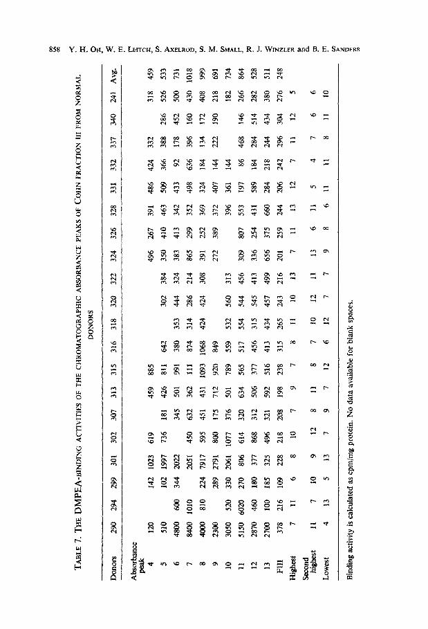
TA
BL
E 7
, T
HE
DM
PE
A-B
IND
ING
A
CT
IVIT
IES
OF
TH
E C
HR
OM
AT
OG
RA
PN
IC AB
SO
RB
AN
CE
PE
AK
S O
F C
OH
N F
RA
CT
ION
III
FR
OM
NO
RM
AL
DO
NO
RS
Don
ors
290
294
299
301
302
307
313
315
316
318
320
322
324
326
328
331
332
337
340
241
Avg
. _-
Abs
orba
nce
4 12
0 14
2 10
23
619
459
885
496
267
391
486
424
332
318
459
5 51
0 10
2 19
97
736
181
426
811
642
302
384
350
410
463
509
366
388
286
526
533
6 48
00
600
344
2022
34
5 50
1 99
1 38
0 35
3 44
4 32
4 38
3 41
3 34
2 43
3 92
17
8 45
2 S
O0
731
7 84
00
1010
20
51
450
632
362
111
874
314
286
214
865
299
352
498
636
396
160
430
1018
8 40
00
810
224
7917
59
5 45
1 43
1 10
93
1068
42
4 42
4 30
8 39
1 25
2 36
9 32
4 18
4 13
4 17
2 40
8 99
9
9 23
00
289
2791
80
0 17
5 71
2 92
0 84
9 27
2 38
9 37
2 40
7 14
4 22
2 19
0 21
8 69
1
10
3050
52
0 33
0 20
61
1077
37
6 S
O1
789
559
532
560
313
396
361
144
182
734
11
5150
60
20
270
806
614
320
634
565
517
554
544
456
309
807
553
197
86
468
146
266
864
12
2870
46
0 18
0 37
1 86
8 31
2 S
O6
371
456
315
545
413
336
254
431
589
184
284
514
282
528
13
2700
10
0 18
5 32
5 49
6 32
1 59
2 51
6 41
3 43
4 45
7 49
9 65
6 37
5 66
0 28
4 21
8 24
4 43
4 38
0 51
1
FII
I 37
8 21
6 10
9 22
8 21
8 20
8 19
8 23
8 31
5 26
5 24
3 21
6 20
1 25
9 24
4 20
6 24
2 29
6 30
4 27
6 24
8
Hig
hes
t 7
11
6 8
10
7 9
7 8
11
10
13
7 11
13
12
7
11
12
5
Sec
ond
hig
hes
t 11
7
10
9 12
8
11
8 7
10
12
11
13
6 11
5
4 7
6 6
Low
est
4 13
5
13
7 9
7 12
6
12
7 7
9 8
6 11
11
8
11
10
Bin
din
g ac
tivi
ty is
cal
cula
ted
as c
pm/m
g pr
otei
n. N
o da
ta a
vail
able
for
blan
k s
pace
s.
Binding- of amines to plasma proteins 859
a significant dissociation of DMPEA from A.P. 7 fraction occurred with dialyzed at pH 4.0. No appreciable difference was found in binding activity of the stored protein fractions at 4” for 2 or 3 weeks. Also, no difference in binding activities was observed when the buffer system was increased in ionic strength with NaCl up to 0.1 M by addition of NaCl to 0.01 M sodium phosphate buffer at pH 7%
Attempts were made to perform binding studies with 3,4-dihydroxyphenylethyla- mine (Dopamine) and 3,4_dihydroxyphenylalanine (Dopa); however, experiments with both compounds were unsuccessful because of their polymerization to form melanin derivatives in various buffer systems investigated.
Utilizing information from these studies we found either + or az-type macro- globulins present whenever good binding was demonstrated. Our most highly purified protein demonstrating DMPEA binding has the properties of az-macroglobulins. However, further purification of these protein fractions will be necessary before the binding molecule or protein can be identified. Other characterization data of all Sephadex G-200 chromatographic fractions are also reported elsewhere?s
DISCUSSION Preliminary studies of the binding of several biogenic amines such as DMPEA,
mescaline, tyramine, and normetanephrine to various Cohn fractions derived from normal human plasma indicated appreciable binding in Cohn fraction III but negli- gible binding of these compounds to other Cohn fractions. WC have therefore examined the capacity of Cohn fraction III of plasma from various individuals to bind these compounds. We found that the DMPEA appeared to bind more extensively to the Cohn fraction III than did other compounds tested.
It is seen that the highest activity was often found in the protein fractions which were eluted from a DEAE-cellulose column at 0.08 M NaCl (A.P. 7)-O-l 5 M NaCl (A.P. 11). These fractions were further separated by the use of Sephadex G-200 columns, and showed DMPEA-bound protein located in a 19 S peak, which is largely an az-macro- globulin, as characterized by various electrophoretic procedures. It appears that there may be more than one specific protein that will bind DMPEA if we consider the broad salt range over which the protein-bound material is eluted. There is already consider- able evidence for several different az-macroglobulins present in human plasma.28 At least one of these is known to bind proteins such as trypsinZe and insulin.30 There is thus evidence to propose that such protein-protein interaction may be what is com- plicating the present studies.
Accurate molecular weights and chemical compositions of proteins (from A.P.s 7-l 1) need to be determined after greater purification is achieved. The number of molecules of DMPEA bound per protein molecule, its binding sites, and factors affecting binding need to be determined. A specific activity of 1000 cpm/mg protein would indicate the binding of approximately O-1 mole of DMPEA to 1 mole of a macroglobulin protein with molecular weight of 800,000. Thus with one of our best preparations of an az-macroglobulin about 0.8 mole DMPEA is bound per mole protein. It is interesting but not easily explained why the mole ratio of DMPEA to protein is less than 1 in even our best preparation. It is possible that these proteins were already partially saturated with structurally similar compounds. Catechola- mines such as epinephrine and norepinephrine are known to be present in human plasma. Antoniades et aZ.31 have some data on catecholamines binding to albumin
860 Y. H. OH, W. E. LEITCH, S. AXELROD, S. M. SMALL, R. J. WINZLER and B. E. SANDERS
and other proteins. This same subject was considered again in an excellent review by Vendsalu.32 More recently, O’Hanlonss has reported on the concentration of these two catecholamines in plasma.
It is interesting, as shown in Table 3. that the binding capacities of A.P.s 6-l 1 were markedly different in different donors. This phenomenon appears to be genuine, since good duplication in binding activity was observed in separate columns of the same preparation of fraction III. On the other hand, a marked difference in binding activity was observed in fractions obtained from the blood of the same donor over a period of 3 months. More data are needed on individual variations before the significance of such variations can be assessed.
As shown in Tables 3 and 6, the mean amount of binding was less for the peaks from patients than from normal subjects. However, the groups are markedly hetero- geneous in variance; a t-test applied to the total scores in the last column of Table 6, indicates that the difference between the group means is not significant (0.05, < P < 0.1). The number of well-characterized schizophrenic patients is yet too small to permit firm conclusions.
No strong evidence is available about the nature and strength of the binding forces between DMPEA and the plasma proteins. This is an important area that merits further investigation.
The results of this study are suggestive enough to indicate that proteins in Cohn fraction III should be further studied by investigating their interactions with other phenylethylamine derivatives related to DMPEA. The important structural binding features should be evaluated, and the effect of selected drugs such as chloropromazine on the binding of DMPEA to the proteins should also be studied.
Acknowledgements-Grateful appreciation is due to Dr. Assar Corvin, Dr. Rapier McMenamy, Dr. Richard Wolin, Mr. Paul Kankolenski and Miss Karen Price for their cooperation and helpful
suggestions.
REFERENCES
1. J. J. SCHILDKRAUT, Am. J. Psychiaf. 122, 509 (1965). 2. R. G. HEATH, S. MARTENS, B. E. LEACH, M. COHEN and C. ANGEL, Am. J. Psychiut. 114,18 (1957). 3. R. G. HEATH, Serological Fractions in Schizophrenia. Harper & Row, New York (1963). 4. C. A. WINTER and L. FLATAKER, Archs Neural. Psychiat. 80, 441 (1958). 5. C. A, WINTER, L. FLATAKER, W. P. BOGER, E. V. C. SMITH and B. E. SANDERS, in Chemistry and
Pathology of the Nervous System, (Ed. J. FOLCH-PI), p. 641. Pergamon Press, Oxford (1961). 6. J. R. BERGEN, R. B. PENNELL, H. FREEMAN and H. HOAGLAND, Archs Neural. Psych&. 2, 146
(1960). 7. B. E. SANDERS, E. V. C. &ITH, L. FLATAKER and C. A. WINTER, Ann. N. Y. Acad. Sri. 96, 448
(1962). 8. B. E. SANDERS, S. M. SMALL, W. J. AYERS, Y. H. OH and S. AXELROD, Trans. N. Y. Acad. Sci. 28,
22 (1965). 9. A. J. FRIEDHOFF and E. VAN WINKLE, Nature, Lond. 194, 897 (1962).
10. A. J. FRIEDHOFF and E. VAN WINKLE, J. nerv. ment. Dis. 135, 135 (1962). 1 I. A. J. FRIEDHOFF and L. E. HOLLISTER, Biochem. Pharmac. 15, 269 (1966). 12. N. F. SENN and P. L. MCGEER, Biochem. biophys. Res. Commun. 14, 227 (1964). 13. F. A. KUEHL, M. HICHENS, R. E. ORMOND, M. A. P. MIESINGER, P. H. GALE, V. J. CIRILLO and
N. G. BRINK, Nature, Land. 203, 154 (1964). 14. R. E. BOURDILLON, C. A. CLARKE, A. P. RIDGES, P. M. SHEPPARD, P. HARPER and S. A. LESLIE,
Nature, Lond. 208, 453 (1965). 15. M. TAKESADA, Y. KAKIMOTO, I. SANO and 2. KANEKO, Nature, Lond. 199, 203 (1963).

Binding of amines to plasma proteins 861
16. J. L. PERRY, S. NASEN and L. MACINTYRE, Nature, Land. 202, 519 (1964). 17. A. BARBEAU, J. A. DEGROOT, J. G. JOLY, D. RAYMOND-TREMBLAY and J. PONALDSON, Reu. Can.
Biol. 22,469 (1963). 18. A. M. ERNST, Psychopharmacologiu, 7, 383 (1965). 19. A. BARBEAU, P. SINGH, P. GAUDREAU and M. JOUBERT, Rev. Can. Biol. 24, 229 (1965). 20. A. BARBEAU, L. TETREAULT, L. OLIVA, L. MORAZAIN and L. CARDIN, Nature, Land. 209,719 (1966). 21. J. R. BERGEN, Trans. N. Y. Acad. Sci. 28, 40 (1965). 22. W. F. LEVER, F. R. N. CURD, E. UROMA, R. K. BROWN, B. A. BARNES, K. SCHMID and E. L.
SCHULTZ, J. clin. Invest. 30, 99 (1951). 23. Y. H. OH and B. E. SANDERS, Anulyt. Biochem. 15, 232 (1966). 24. Y. H. OH, J. W. NAWROCKI, W. E. LEITCH and B. E. SANDERS, Analyt. Biochem. 16,220 (1966) 25. Y. H. OH and B. E. SANDERS, Life Sci. 5,827 (1966). 26. P. FLODIN and J. KILLANDER, Biochim. biophys. Actu. 63,403 (1962). 27.0. L. LOWRY, N. J. ROSEBROUGH, A. L. FARR and R. J. RANDALL, J. biol. Chem. 193 265 (1951). 28. Y. K. OH and B. E. SANDERS, submitted for publication.
27.0. L. LOWRY, N. J. ROSEBROUGH, A. L. FARR and R. J. RANDALL, J. biol. Chem. 193 265 (1951). 29. P. 0. GANROT, Clin. chim. Acta 13, 597 (1966). 30. E. KALLEE, Helv. med. Acta. 30, 510 (1963). 31. H. N. ANTONIADES, A. GOLDFIEN, S. ZIELI, and F. ELIMADIIAN, Proc. Sot. exp. Biol. Med. 97, 11
(1958). 32. A. VENDSALU, Acta physiol scand. 49, Suppl. 173 (1960). 33. J. F. O’HANLON, JR., Science. 150, 507 (1966).
Related Documents