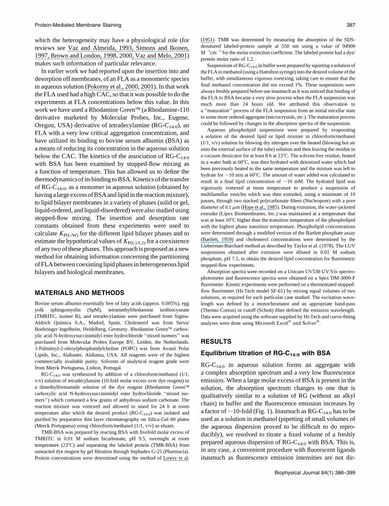
386 Biophysical Journal Volume 84 January 2003 386–399 Binding of a Fluorescent Lipid Amphiphile to Albumin and its Transfer to Lipid Bilayer Membranes Magda S. C. Abreu, Luı ´s M. B. B. Estronca, Maria Joa ˜o Moreno, and Winchil L. C. Vaz Departamento de Quı ´mica, Universidade de Coimbra, 3004-535 Coimbra, Portugal ABSTRACT Kinetics and thermodynamics of the binding of a fluorescent lipid amphiphile, Rhodamine Greenä- tetradecylamide (RG-C 14:0 ), to bovine serum albumin were characterized in an equilibrium titration and by stopped-flow fluorimetry. The binding equilibrium of RG-C 14:0 to albumin was then used to reduce its concentration in the aqueous phase to a value below its critical micelle concentration. Under these conditions, the only two species of RG-C 14:0 in the system were the monomer in aqueous solution in equilibrium with the protein-bound species. After previous determination of the kinetic and thermodynamic parameters for association of RG-C 14:0 with albumin, the kinetics of insertion of the amphiphile into and desorption off lipid bilayer membranes in different phases (solid, liquid-ordered, and liquid-disordered phases, presented as large unilamellar vesicles) were studied by stopped-flow fluorimetry at 308C. Insertion and desorption rate constants for association of the RG-C 14:0 monomer with the lipid bilayers were used to obtain lipid/water equilibrium partition coefficients for this fluorescent amphiphile. The direct measurement of these partition coefficients is shown to provide a new method for the indirect determination of the equilibrium partition coefficient of similar molecules between two defined lipid phases if they coexist in the same membrane. INTRODUCTION Serum albumins, abundant transport proteins found in blood plasma, are known to bind drugs and lipid amphiphiles with a high affinity (Peters, 1997). This binding has been well- studied and crystal structures of albumin and its complexes with fatty acids and drugs have become available in recent years (He and Carter, 1992, Curry et al., 1998, Bhattacharya et al., 2000a, Petitpas et al., 2001). Besides its obvious pharmacological interest, the binding of amphiphiles to serum albumins has been exploited in the labeling of cell surface membranes with fluorescent lipid amphiphiles (FLA) (Lipsky and Pagano, 1985, Pagano and Martin, 1988). The rationale of this utilization of serum albumins in cell biology lies in the fact that FLA form aggregates (microcrystals, micelles) in aqueous solution at concentrations above a critical value (solubility product, critical aggregation concentration or critical micelle concentration). This critical concentration, which we shall refer to generically as a critical aggregation concentration (CAC), can be quite low, so that under the experimental conditions required for an adequate staining of the cell plasma membrane most of the FLA in aqueous solution exists in an aggregated form. Interaction of the aggregate with the plasma membrane of the cell, when it does occur, can result in an undesirable localized (non-homoge- neous) staining. When serum albumin is present in the labeling solution, part of the FLA is bound to the albumin. Depending upon the albumin and FLA concentrations as well as on the equilibrium binding constant for the FLA binding to the protein, K a , a significant reduction of the effective free FLA concentration in aqueous solution can be achieved. Ideally, the free FLA concentration is reduced to a value below its CAC so that the FLA form that labels the cell surface is a monomer and the labeling is homogeneously achieved. Whereas the above rationale is very commonly used in staining of cell surfaces with FLA for fluorescence micros- copy, we are not aware of any systematic study on the binding of FLA to albumin in terms of the binding parameters (equilibrium binding constants, and the binding and de- sorption rate constants) and the detailed energetics of the process that a study of the temperature-dependence of these parameters can provide. Measurement of the rate constants for transfer of this monomeric FLA species to a lipid bilayer membrane then provide a direct estimation of the equilibrium partition coefficient, K P(L/W) , for partitioning of the FLA between the membrane and aqueous phases. This direct measurement of K P(L/W) can be performed for different lipid phases (solid, liquid-ordered, or liquid-disordered) using the same approach, thereby providing an indirect measurement of a hypothetical partition coefficient of the FLA for partitioning between any two of those lipid bilayer phases, K P(L1/L2) . The general acceptance of the concept that the biological membrane is a heterogeneous chemical system (has coexist- ing lipid phases, of which ‘‘rafts’’ may be a manifestation) in Submitted June 6, 2002, and accepted for publication September 16, 2002. Address reprint requests to Prof. Winchil L. C. Vaz, Departamento de Quı ´mica, Universidade de Coimbra, 3004-535 Coimbra, Portugal. Tel.: þ 351 239 824861; Fax.: þ 351 239 827703; E-mail: [email protected]. Abbreviations used: BSA, bovine serum albumin; CAC, critical aggrega- tion concentration, used here synonymously with solubility product or critical micelle concentration; FLA, fluorescent lipid amphiphile(s); K a , equilibrium binding constant for FLA to protein; K P(L/W) , equilibrium partition coefficient for partitioning of FLA between a membrane and aqueous phase; K P(L1/L2) , equilibrium partition coefficient for partitioning of FLA between two lipid phases; LUV, large unilamellar vesicles with an average diameter of 0.1 mm; POPC, 1-palmitoyl-2-oleoylphosphatidyl choline; RG-C 14:0 , Rhodamine Greenä-carboxylic acid tetradecylamide; SpM, Egg yolk sphingomyelin; TMRITC, Tetramethylrhodamine isothio- cyanate (isomer R); TMR-BSA, BSA labeled covalently with TMRITC at an average molar labeling ratio of 1. Ó 2003 by the Biophysical Society 0006-3495/03/01/386/14 $2.00

Welcome message from author
This document is posted to help you gain knowledge. Please leave a comment to let me know what you think about it! Share it to your friends and learn new things together.
Transcript
386 Biophysical Journal Volume 84 January 2003 386–399
Binding of a Fluorescent Lipid Amphiphile to Albumin and itsTransfer to Lipid Bilayer Membranes
Magda S. C. Abreu, Luıs M. B. B. Estronca, Maria Joao Moreno, and Winchil L. C. VazDepartamento de Quımica, Universidade de Coimbra, 3004-535 Coimbra, Portugal
ABSTRACT Kinetics and thermodynamics of the binding of a fluorescent lipid amphiphile, Rhodamine Green�-tetradecylamide (RG-C14:0), to bovine serum albumin were characterized in an equilibrium titration and by stopped-flowfluorimetry. The binding equilibrium of RG-C14:0 to albumin was then used to reduce its concentration in the aqueous phase toa value below its critical micelle concentration. Under these conditions, the only two species of RG-C14:0 in the system were themonomer in aqueous solution in equilibrium with the protein-bound species. After previous determination of the kinetic andthermodynamic parameters for association of RG-C14:0 with albumin, the kinetics of insertion of the amphiphile into anddesorption off lipid bilayer membranes in different phases (solid, liquid-ordered, and liquid-disordered phases, presented aslarge unilamellar vesicles) were studied by stopped-flow fluorimetry at 308C. Insertion and desorption rate constants forassociation of the RG-C14:0 monomer with the lipid bilayers were used to obtain lipid/water equilibrium partition coefficients forthis fluorescent amphiphile. The direct measurement of these partition coefficients is shown to provide a new method for theindirect determination of the equilibrium partition coefficient of similar molecules between two defined lipid phases if they coexistin the same membrane.
INTRODUCTION
Serum albumins, abundant transport proteins found in blood
plasma, are known to bind drugs and lipid amphiphiles with
a high affinity (Peters, 1997). This binding has been well-
studied and crystal structures of albumin and its complexes
with fatty acids and drugs have become available in recent
years (He and Carter, 1992, Curry et al., 1998, Bhattacharya
et al., 2000a, Petitpas et al., 2001). Besides its obvious
pharmacological interest, the binding of amphiphiles to
serum albumins has been exploited in the labeling of cell
surface membranes with fluorescent lipid amphiphiles (FLA)
(Lipsky and Pagano, 1985, Pagano and Martin, 1988). The
rationale of this utilization of serum albumins in cell biology
lies in the fact that FLA form aggregates (microcrystals,
micelles) in aqueous solution at concentrations above a critical
value (solubility product, critical aggregation concentration
or critical micelle concentration). This critical concentration,
which we shall refer to generically as a critical aggregation
concentration (CAC), can be quite low, so that under the
experimental conditions required for an adequate staining of
the cell plasma membrane most of the FLA in aqueous
solution exists in an aggregated form. Interaction of the
aggregate with the plasma membrane of the cell, when it does
occur, can result in an undesirable localized (non-homoge-
neous) staining. When serum albumin is present in the
labeling solution, part of the FLA is bound to the albumin.
Depending upon the albumin and FLA concentrations as well
as on the equilibrium binding constant for the FLA binding to
the protein, Ka, a significant reduction of the effective free
FLA concentration in aqueous solution can be achieved.
Ideally, the free FLA concentration is reduced to a value
below its CAC so that the FLA form that labels the cell surface
is a monomer and the labeling is homogeneously achieved.
Whereas the above rationale is very commonly used in
staining of cell surfaces with FLA for fluorescence micros-
copy, we are not aware of any systematic study on the binding
of FLA to albumin in terms of the binding parameters
(equilibrium binding constants, and the binding and de-
sorption rate constants) and the detailed energetics of the
process that a study of the temperature-dependence of these
parameters can provide. Measurement of the rate constants for
transfer of this monomeric FLA species to a lipid bilayer
membrane then provide a direct estimation of the equilibrium
partition coefficient, KP(L/W), for partitioning of the FLA
between the membrane and aqueous phases. This direct
measurement of KP(L/W) can be performed for different lipid
phases (solid, liquid-ordered, or liquid-disordered) using the
same approach, thereby providing an indirect measurement of
a hypothetical partition coefficient of the FLA for partitioning
between any two of those lipid bilayer phases, KP(L1/L2). The
general acceptance of the concept that the biological
membrane is a heterogeneous chemical system (has coexist-
ing lipid phases, of which ‘‘rafts’’ may be a manifestation) in
Submitted June 6, 2002, and accepted for publication September 16,
2002.
Address reprint requests to Prof. Winchil L. C. Vaz, Departamento de
Quımica, Universidade de Coimbra, 3004-535 Coimbra, Portugal. Tel.:
þ 351 239 824861; Fax.: þ 351 239 827703; E-mail: [email protected].
Abbreviations used: BSA, bovine serum albumin; CAC, critical aggrega-
tion concentration, used here synonymously with solubility product or
critical micelle concentration; FLA, fluorescent lipid amphiphile(s); Ka,
equilibrium binding constant for FLA to protein; KP(L/W), equilibrium
partition coefficient for partitioning of FLA between a membrane and
aqueous phase; KP(L1/L2), equilibrium partition coefficient for partitioning
of FLA between two lipid phases; LUV, large unilamellar vesicles with an
average diameter of 0.1 mm; POPC, 1-palmitoyl-2-oleoylphosphatidyl
choline; RG-C14:0, Rhodamine Green�-carboxylic acid tetradecylamide;
SpM, Egg yolk sphingomyelin; TMRITC, Tetramethylrhodamine isothio-
cyanate (isomer R); TMR-BSA, BSA labeled covalently with TMRITC at
an average molar labeling ratio of 1.
� 2003 by the Biophysical Society
0006-3495/03/01/386/14 $2.00
which the heterogeneity may have a physiological role (for
reviews see Vaz and Almeida, 1993, Simons and Ikonen,
1997, Brown and London, 1998, 2000, Vaz and Melo, 2001)
makes such information of particular relevance.
In earlier work we had reported upon the insertion into and
desorption off membranes, of an FLA as a monomeric species
in aqueous solution (Pokorny et al., 2000, 2001). In that work
the FLA used had a high CAC, so that it was possible to do the
experiments at FLA concentrations below this value. In this
work we have used a Rhodamine Green� (a Rhodamine-110
derivative marketed by Molecular Probes, Inc., Eugene,
Oregon, USA) derivative of tetradecylamine (RG-C14:0), an
FLA with a very low critical aggregation concentration, and
have utilized its binding to bovine serum albumin (BSA) as
a means of reducing its concentration in the aqueous solution
below the CAC. The kinetics of the association of RG-C14:0
with BSA has been examined by stopped-flow mixing as
a function of temperature. This has allowed us to define the
thermodynamics of its binding to BSA. Kinetics of the transfer
of RG-C14:0, as a monomer in aqueous solution (obtained by
having a large excess ofBSAand lipid in the reactionmixture),
to lipid bilayer membranes in a variety of phases (solid or gel,
liquid-ordered, and liquid-disordered) were also studied using
stopped-flow mixing. The insertion and desorption rate
constants obtained from these experiments were used to
calculate KP(L/W) for the different lipid bilayer phases and to
estimate the hypothetical values of KP(L1/L2) for a coexistence
of any two of these phases. This approach is proposed as a new
method for obtaining information concerning the partitioning
of FLA between coexisting lipid phases in heterogeneous lipid
bilayers and biological membranes.
MATERIALS AND METHODS
Bovine serum albumin essentially free of fatty acids (approx. 0.005%), egg
yolk sphingomyelin (SpM), tetramethylrhodamine isothiocyanate
(TMRITC, isomer R), and tetradecylamine were purchased from Sigma-
Aldrich Quımica S.A., Madrid, Spain. Cholesterol was from Serva/
Boehringer Ingelheim, Heidelberg, Germany. Rhodamine Green� carbox-
ylic acid N-hydroxysuccinimidyl ester hydrochloride ‘‘mixed isomers’’ was
purchased from Molecular Probes Europe BV, Leiden, the Netherlands.
1-Palmitoyl-2-oleoylphosphatidylcholine (POPC) was from Avanti Polar
Lipids, Inc., Alabaster, Alabama, USA. All reagents were of the highest
commercially available purity. Solvents of analytical reagent grade were
from Merck Portuguesa, Lisbon, Portugal.
RG-C14:0 was synthesized by addition of a chloroform/methanol (1/1,
v/v) solution of tetradecylamine (10-fold molar excess over dye reagent) to
a dimethylformamide solution of the dye reagent (Rhodamine Green�carboxylic acid N-hydroxysuccinimidyl ester hydrochloride ‘‘mixed iso-
mers’’) which contained a few grains of anhydrous sodium carbonate. The
reaction mixture was vortexed and allowed to stand for 24 h at room
temperature after which the desired product (RG-C14:0) was isolated and
purified by preparative thin layer chromatography on Silica Gel 60 plates
(Merck Portuguesa) using chloroform/methanol (1/1, v/v) as eluant.
TMR-BSA was prepared by reacting BSA with fivefold molar excess of
TMRITC in 0.01 M sodium bicarbonate, pH 9.5, overnight at room
temperature (238C) and separating the labeled protein (TMR-BSA) from
unreacted dye reagent by gel filtration through Sephadex G-25 (Pharmacia).
Protein concentrations were determined using the method of Lowry et al.
(1951). TMR was determined by measuring the absorption of the SDS-
denatured labeled-protein sample at 550 nm using a value of 94900
M�1cm�1 for the molar extinction coefficient. The labeled protein had a dye/
protein molar ratio of 1.2.
Suspensions of RG-C14:0 in buffer were prepared by squirting a solution of
the FLA in methanol (using a Hamilton syringe) into the desired volume of the
buffer, with simultaneous vigorous vortexing, taking care to ensure that the
final methanol concentration did not exceed 1%. These suspensions were
always freshly prepared before use inasmuch as it was noticed that binding of
the FLA to BSA became a very slow process when the FLA suspension was
much more than 24 hours old. We attributed this observation to
a ‘‘maturation’’ process of the FLA suspension from an initial micellar state
to some more ordered aggregate (microcrystals, etc.). The maturation process
could be followed by changes in the absorption spectra of the suspension.
Aqueous phospholipid suspensions were prepared by evaporating
a solution of the desired lipid or lipid mixture in chloroform/methanol
(1/1, v/v) solution by blowing dry nitrogen over the heated (blowing hot air
onto the external surface of the tube) solution and then leaving the residue in
a vacuum dessicator for at least 8 h at 238C. The solvent-free residue, heated
in a water bath at 608C, was then hydrated with deionized water which had
been previously heated to the same temperature and the mixture was left to
hydrate for ;10 min at 608C. The amount of water added was calculated to
result in a final lipid concentration of ;10 mM. The hydrated lipid was
vigorously vortexed at room temperature to produce a suspension of
multilamellar vesicles which was then extruded, using a minimum of 10
passes, through two stacked polycarbonate filters (Nucleopore) with a pore
diameter of 0.1 mm (Hope et al., 1985). During extrusion, the water-jacketed
extruder (Lipex Biomembranes, Inc.) was maintained at a temperature that
was at least 108C higher than the transition temperature of the phospholipid
with the highest phase transition temperature. Phospholipid concentrations
were determined through a modified version of the Bartlett phosphate assay
(Bartlett, 1959) and cholesterol concentrations were determined by the
Lieberman-Burchard method as described by Taylor et al. (1978). The LUV
suspensions obtained after extrusion were diluted in 0.01 M sodium
phosphate, pH 7.5, to obtain the desired lipid concentration for fluorimetric
stopped-flow experiments.
Absorption spectra were recorded on a Unicam UV530 UV/Vis spectro-
photometer and fluorescence spectra were obtained on a Spex DM-3000-F
fluorimeter. Kinetic experiments were performed on a thermostated stopped-
flow fluorimeter (Hi-Tech model SF-61) by mixing equal volumes of two
solutions, as required for each particular case studied. The excitation wave-
length was defined by a monochromator and an appropriate band-pass
(Thermo Corion) or cutoff (Schott) filter defined the emission wavelength.
Data were acquired using the software supplied by Hi-Tech and curve-fitting
analyses were done using Microsoft Excel� and Solver�.
RESULTS
Equilibrium titration of RG-C14:0 with BSA
RG-C14:0 in aqueous solution forms an aggregate with
a complex absorption spectrum and a very low fluorescence
emission. When a large molar excess of BSA is present in the
solution, the absorption spectrum changes to one that is
qualitatively similar to a solution of RG (without an alkyl
chain) in buffer and the fluorescence emission increases by
a factor of ;10-fold (Fig. 1). Inasmuch as RG-C14:0 has to be
used as a solution in methanol (pipetting of small volumes of
the aqueous dispersion proved to be difficult to do repro-
ducibly), we resolved to titrate a fixed volume of a freshly
prepared aqueous dispersion of RG-C14:0 with BSA. This is,
in any case, a convenient procedure with fluorescent ligands
inasmuch as fluorescence emission intensities are not dir-
Protein-Mediated Membrane Staining 387
Biophysical Journal 84(1) 386–399
ectly proportional to concentration when there are apprecia-
ble inner filter effects. The change in fluorescence emission
upon the binding of RG-C14:0 to BSA (increasing BSA
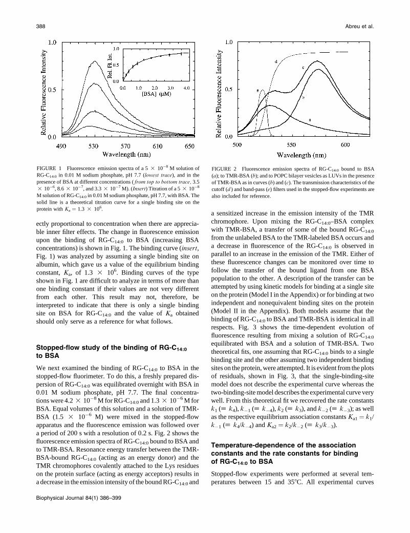
concentrations) is shown in Fig. 1. The binding curve (insert,Fig. 1) was analyzed by assuming a single binding site on
albumin, which gave us a value of the equilibrium binding
constant, Ka, of 1.3 3 106. Binding curves of the type
shown in Fig. 1 are difficult to analyze in terms of more than
one binding constant if their values are not very different
from each other. This result may not, therefore, be
interpreted to indicate that there is only a single binding
site on BSA for RG-C14:0 and the value of Ka obtained
should only serve as a reference for what follows.
Stopped-flow study of the binding of RG-C14:0
to BSA
We next examined the binding of RG-C14:0 to BSA in the
stopped-flow fluorimeter. To do this, a freshly prepared dis-
persion of RG-C14:0 was equilibrated overnight with BSA in
0.01 M sodium phosphate, pH 7.7. The final concentra-
tions were 4.2 3 10�8 M for RG-C14:0 and 1.3 3 10�6 M for
BSA. Equal volumes of this solution and a solution of TMR-
BSA (1.5 3 10�6 M) were mixed in the stopped-flow
apparatus and the fluorescence emission was followed over
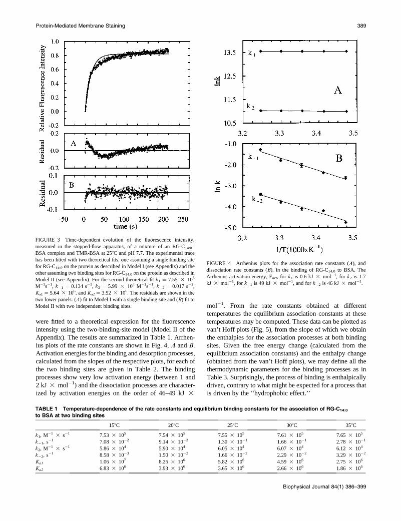
a period of 200 s with a resolution of 0.2 s. Fig. 2 shows the
fluorescence emission spectra of RG-C14:0 bound to BSA and
to TMR-BSA. Resonance energy transfer between the TMR-
BSA-bound RG-C14:0 (acting as an energy donor) and the
TMR chromophores covalently attached to the Lys residues
on the protein surface (acting as energy acceptors) results in
a decrease in the emission intensity of the bound RG-C14:0 and
a sensitized increase in the emission intensity of the TMR
chromophore. Upon mixing the RG-C14:0–BSA complex
with TMR-BSA, a transfer of some of the bound RG-C14:0
from the unlabeled BSA to the TMR-labeled BSA occurs and
a decrease in fluorescence of the RG-C14:0 is observed in
parallel to an increase in the emission of the TMR. Either of
these fluorescence changes can be monitored over time to
follow the transfer of the bound ligand from one BSA
population to the other. A description of the transfer can be
attempted by using kinetic models for binding at a single site
on the protein (Model I in the Appendix) or for binding at two
independent and nonequivalent binding sites on the protein
(Model II in the Appendix). Both models assume that the
binding of RG-C14:0 to BSA and TMR-BSA is identical in all
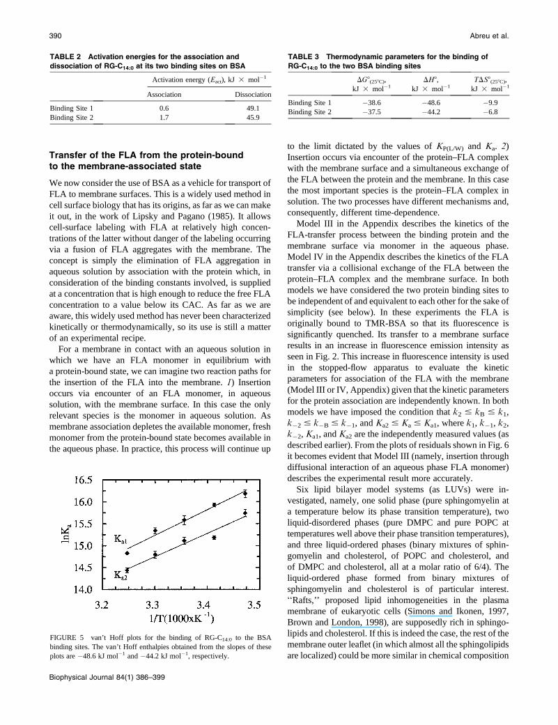
respects. Fig. 3 shows the time-dependent evolution of
fluorescence resulting from mixing a solution of RG-C14:0
equilibrated with BSA and a solution of TMR-BSA. Two
theoretical fits, one assuming that RG-C14:0 binds to a single
binding site and the other assuming two independent binding
sites on the protein, were attempted. It is evident from the plots
of residuals, shown in Fig. 3, that the single-binding-site
model does not describe the experimental curve whereas the
two-binding-site model describes the experimental curve very
well. From this theoretical fit we recovered the rate constants
k1 ([ k4), k�1 ([ k�4), k2 ([ k3), and k�2 ([ k�3); as well
as the respective equilibrium association constants Ka1 ¼ k1/
k�1 ([ k4/k�4) and Ka2 ¼ k2/k�2 ([ k3/k�3).
Temperature-dependence of the associationconstants and the rate constants for bindingof RG-C14:0 to BSA
Stopped-flow experiments were performed at several tem-
peratures between 15 and 358C. All experimental curves
FIGURE 1 Fluorescence emission spectra of a 5 3 10�8 M solution of
RG-C14:0 in 0.01 M sodium phosphate, pH 7.7 (lowest trace), and in the
presence of BSA at different concentrations ( from top to bottom trace, 3.5
3 10�6, 8.6 3 10�7, and 3.3 3 10�7 M). (Insert) Titration of a 5 3 10�8
M solution of RG-C14:0 in 0.01 M sodium phosphate, pH 7.7, with BSA. The
solid line is a theoretical titration curve for a single binding site on the
protein with Ka ¼ 1.3 3 106.
FIGURE 2 Fluorescence emission spectra of RG-C14:0 bound to BSA
(a); to TMR-BSA (b); and to POPC bilayer vesicles as LUVs in the presence
of TMR-BSA as in curves (b) and (c). The transmission characteristics of the
cutoff (d ) and band-pass (e) filters used in the stopped-flow experiments are
also included for reference.
388 Abreu et al.
Biophysical Journal 84(1) 386–399
were fitted to a theoretical expression for the fluorescence
intensity using the two-binding-site model (Model II of the
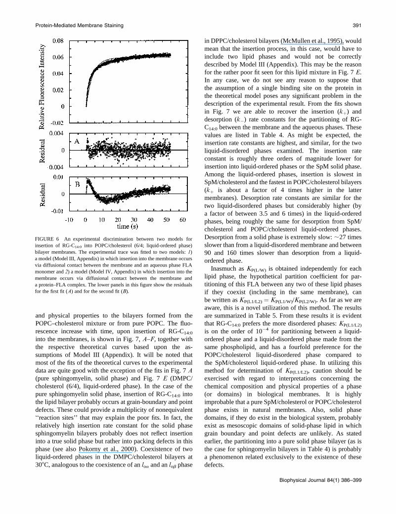
Appendix). The results are summarized in Table 1. Arrhen-
ius plots of the rate constants are shown in Fig. 4, A and B.
Activation energies for the binding and desorption processes,
calculated from the slopes of the respective plots, for each of
the two binding sites are given in Table 2. The binding
processes show very low activation energy (between 1 and
2 kJ 3 mol�1) and the dissociation processes are character-
ized by activation energies on the order of 46–49 kJ 3
mol�1. From the rate constants obtained at different
temperatures the equilibrium association constants at these
temperatures may be computed. These data can be plotted as
van’t Hoff plots (Fig. 5), from the slope of which we obtain
the enthalpies for the association processes at both binding
sites. Given the free energy change (calculated from the
equilibrium association constants) and the enthalpy change
(obtained from the van’t Hoff plots), we may define all the
thermodynamic parameters for the binding processes as in
Table 3. Surprisingly, the process of binding is enthalpically
driven, contrary to what might be expected for a process that
is driven by the ‘‘hydrophobic effect.’’
FIGURE 3 Time-dependent evolution of the fluorescence intensity,
measured in the stopped-flow apparatus, of a mixture of an RG-C14:0–
BSA complex and TMR-BSA at 258C and pH 7.7. The experimental trace
has been fitted with two theoretical fits, one assuming a single binding site
for RG-C14:0 on the protein as described in Model I (see Appendix) and the
other assuming two binding sites for RG-C14:0 on the protein as described in
Model II (see Appendix). For the second theoretical fit k1 ¼ 7.55 3 105
M�1s�1, k�1 ¼ 0.134 s�1, k2 ¼ 5.99 3 104 M�1s�1, k�2 ¼ 0.017 s�1,
Ka1 ¼ 5.64 3 106, and Ka2 ¼ 3.52 3 106. The residuals are shown in the
two lower panels: (A) fit to Model I with a single binding site and (B) fit to
Model II with two independent binding sites.
TABLE 1 Temperature-dependence of the rate constants and equilibrium binding constants for the association of RG-C14:0
to BSA at two binding sites
158C 208C 258C 308C 358C
k1, M�1 3 s�1 7.53 3 105 7.54 3 105 7.55 3 105 7.61 3 105 7.65 3 105
k�1, s�1 7.08 3 10�2 9.14 3 10�2 1.30 3 10�1 1.66 3 10�1 2.78 3 10�1
k2, M�1 3 s�1 5.86 3 104 5.90 3 104 6.05 3 104 6.07 3 104 6.12 3 104
k�2, s�1 8.58 3 10�3 1.50 3 10�2 1.66 3 10�2 2.29 3 10�2 3.29 3 10�2
Ka1 1.06 3 107 8.25 3 106 5.82 3 106 4.59 3 106 2.75 3 106
Ka2 6.83 3 106 3.93 3 106 3.65 3 106 2.66 3 106 1.86 3 106
FIGURE 4 Arrhenius plots for the association rate constants (A), and
dissociation rate constants (B), in the binding of RG-C14:0 to BSA. The
Arrhenius activation energy, Eact, for k1 is 0.6 kJ 3 mol�1, for k2 is 1.7
kJ 3 mol�1, for k�1 is 49 kJ 3 mol�1, and for k�2 is 46 kJ 3 mol�1.
Protein-Mediated Membrane Staining 389
Biophysical Journal 84(1) 386–399
Transfer of the FLA from the protein-boundto the membrane-associated state
We now consider the use of BSA as a vehicle for transport of
FLA to membrane surfaces. This is a widely used method in
cell surface biology that has its origins, as far as we can make
it out, in the work of Lipsky and Pagano (1985). It allows
cell-surface labeling with FLA at relatively high concen-
trations of the latter without danger of the labeling occurring
via a fusion of FLA aggregates with the membrane. The
concept is simply the elimination of FLA aggregation in
aqueous solution by association with the protein which, in
consideration of the binding constants involved, is supplied
at a concentration that is high enough to reduce the free FLA
concentration to a value below its CAC. As far as we are
aware, this widely used method has never been characterized
kinetically or thermodynamically, so its use is still a matter
of an experimental recipe.
For a membrane in contact with an aqueous solution in
which we have an FLA monomer in equilibrium with
a protein-bound state, we can imagine two reaction paths for
the insertion of the FLA into the membrane. 1) Insertion
occurs via encounter of an FLA monomer, in aqueous
solution, with the membrane surface. In this case the only
relevant species is the monomer in aqueous solution. As
membrane association depletes the available monomer, fresh
monomer from the protein-bound state becomes available in
the aqueous phase. In practice, this process will continue up
to the limit dictated by the values of KP(L/W) and Ka. 2)
Insertion occurs via encounter of the protein–FLA complex
with the membrane surface and a simultaneous exchange of
the FLA between the protein and the membrane. In this case
the most important species is the protein–FLA complex in
solution. The two processes have different mechanisms and,
consequently, different time-dependence.
Model III in the Appendix describes the kinetics of the
FLA-transfer process between the binding protein and the
membrane surface via monomer in the aqueous phase.
Model IV in the Appendix describes the kinetics of the FLA
transfer via a collisional exchange of the FLA between the
protein–FLA complex and the membrane surface. In both
models we have considered the two protein binding sites to
be independent of and equivalent to each other for the sake of
simplicity (see below). In these experiments the FLA is
originally bound to TMR-BSA so that its fluorescence is
significantly quenched. Its transfer to a membrane surface
results in an increase in fluorescence emission intensity as
seen in Fig. 2. This increase in fluorescence intensity is used
in the stopped-flow apparatus to evaluate the kinetic
parameters for association of the FLA with the membrane
(Model III or IV, Appendix) given that the kinetic parameters
for the protein association are independently known. In both
models we have imposed the condition that k2 # kB # k1,
k�2 # k�B # k�1, and Ka2 # Ka # Ka1, where k1, k�1, k2,
k�2, Ka1, and Ka2 are the independently measured values (as
described earlier). From the plots of residuals shown in Fig. 6
it becomes evident that Model III (namely, insertion through
diffusional interaction of an aqueous phase FLA monomer)
describes the experimental result more accurately.
Six lipid bilayer model systems (as LUVs) were in-
vestigated, namely, one solid phase (pure sphingomyelin at
a temperature below its phase transition temperature), two
liquid-disordered phases (pure DMPC and pure POPC at
temperatures well above their phase transition temperatures),
and three liquid-ordered phases (binary mixtures of sphin-
gomyelin and cholesterol, of POPC and cholesterol, and
of DMPC and cholesterol, all at a molar ratio of 6/4). The
liquid-ordered phase formed from binary mixtures of
sphingomyelin and cholesterol is of particular interest.
‘‘Rafts,’’ proposed lipid inhomogeneities in the plasma
membrane of eukaryotic cells (Simons and Ikonen, 1997,
Brown and London, 1998), are supposedly rich in sphingo-
lipids and cholesterol. If this is indeed the case, the rest of the
membrane outer leaflet (in which almost all the sphingolipids
are localized) could be more similar in chemical composition
TABLE 2 Activation energies for the association and
dissociation of RG-C14:0 at its two binding sites on BSA
Activation energy (Eact), kJ 3 mol�1
Association Dissociation
Binding Site 1 0.6 49.1
Binding Site 2 1.7 45.9
FIGURE 5 van’t Hoff plots for the binding of RG-C14:0 to the BSA
binding sites. The van’t Hoff enthalpies obtained from the slopes of these
plots are �48.6 kJ mol�1 and �44.2 kJ mol�1, respectively.
TABLE 3 Thermodynamic parameters for the binding of
RG-C14:0 to the two BSA binding sites
DG8(258C),
kJ 3 mol�1
DH8,
kJ 3 mol�1
TDS8(258C),
kJ 3 mol�1
Binding Site 1 �38.6 �48.6 �9.9
Binding Site 2 �37.5 �44.2 �6.8
390 Abreu et al.
Biophysical Journal 84(1) 386–399
and physical properties to the bilayers formed from the
POPC–cholesterol mixture or from pure POPC. The fluo-
rescence increase with time, upon insertion of RG-C14:0
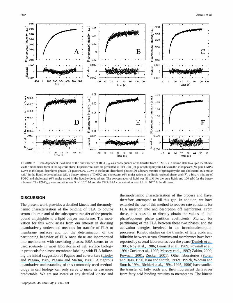
into the membranes, is shown in Fig. 7, A–F, together with
the respective theoretical curves based upon the as-
sumptions of Model III (Appendix). It will be noted that
most of the fits of the theoretical curves to the experimental
data are quite good with the exception of the fits in Fig. 7 A(pure sphingomyelin, solid phase) and Fig. 7 E (DMPC/
cholesterol (6/4), liquid-ordered phase). In the case of the
pure sphingomyelin solid phase, insertion of RG-C14:0 into
the lipid bilayer probably occurs at grain-boundary and point
defects. These could provide a multiplicity of nonequivalent
‘‘reaction sites’’ that may explain the poor fits. In fact, the
relatively high insertion rate constant for the solid phase
sphingomyelin bilayers probably does not reflect insertion
into a true solid phase but rather into packing defects in this
phase (see also Pokorny et al., 2000). Coexistence of two
liquid-ordered phases in the DMPC/cholesterol bilayers at
308C, analogous to the coexistence of an loa and an lob phase
in DPPC/cholesterol bilayers (McMullen et al., 1995), would
mean that the insertion process, in this case, would have to
include two lipid phases and would not be correctly
described by Model III (Appendix). This may be the reason
for the rather poor fit seen for this lipid mixture in Fig. 7 E.
In any case, we do not see any reason to suppose that
the assumption of a single binding site on the protein in
the theoretical model poses any significant problem in the
description of the experimental result. From the fits shown
in Fig. 7 we are able to recover the insertion (kþ) and
desorption (k�) rate constants for the partitioning of RG-
C14:0 between the membrane and the aqueous phases. These
values are listed in Table 4. As might be expected, the
insertion rate constants are highest, and similar, for the two
liquid-disordered phases examined. The insertion rate
constant is roughly three orders of magnitude lower for
insertion into liquid-ordered phases or the SpM solid phase.
Among the liquid-ordered phases, insertion is slowest in
SpM/cholesterol and the fastest in POPC/cholesterol bilayers
(kþ is about a factor of 4 times higher in the latter
membranes). Desorption rate constants are similar for the
two liquid-disordered phases but considerably higher (by
a factor of between 3.5 and 6 times) in the liquid-ordered
phases, being roughly the same for desorption from SpM/
cholesterol and POPC/cholesterol liquid-ordered phases.
Desorption from a solid phase is extremely slow: ;27 times
slower than from a liquid-disordered membrane and between
90 and 160 times slower than desorption from a liquid-
ordered phase.
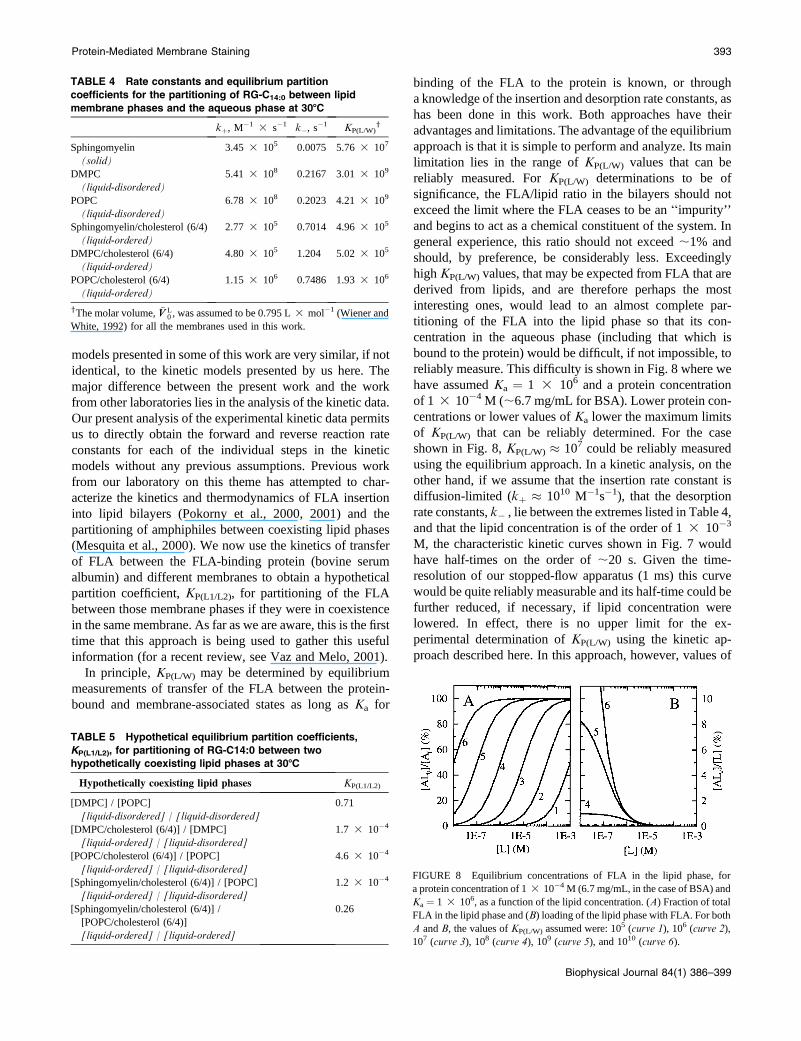
Inasmuch as KP(L/W) is obtained independently for each
lipid phase, the hypothetical partition coefficient for par-
titioning of this FLA between any two of these lipid phases
if they coexist (including in the same membrane), can
be written as KP(L1/L2) ¼ KP(L1/W)/KP(L2/W). As far as we are
aware, this is a novel utilization of this method. The results
are summarized in Table 5. From these results it is evident
that RG-C14:0 prefers the more disordered phases: KP(L1/L2)
is on the order of 10�4 for partitioning between a liquid-
ordered phase and a liquid-disordered phase made from the
same phospholipid, and has a fourfold preference for the
POPC/cholesterol liquid-disordered phase compared to
the SpM/cholesterol liquid-ordered phase. In utilizing this
method for determination of KP(L1/L2), caution should be
exercised with regard to interpretations concerning the
chemical composition and physical properties of a phase
(or domains) in biological membranes. It is highly
improbable that a pure SpM/cholesterol or POPC/cholesterol
phase exists in natural membranes. Also, solid phase
domains, if they do exist in the biological system, probably
exist as mesoscopic domains of solid-phase lipid in which
grain boundary and point defects are unlikely. As stated
earlier, the partitioning into a pure solid phase bilayer (as is
the case for sphingomyelin bilayers in Table 4) is probably
a phenomenon related exclusively to the existence of these
defects.
FIGURE 6 An experimental discrimination between two models for
insertion of RG-C14:0 into POPC/cholesterol (6/4; liquid-ordered phase)
bilayer membranes. The experimental trace was fitted to two models: 1)
a model (Model III, Appendix) in which insertion into the membrane occurs
via diffusional contact between the membrane and an aqueous phase FLA
monomer and 2) a model (Model IV, Appendix) in which insertion into the
membrane occurs via diffusional contact between the membrane and
a protein–FLA complex. The lower panels in this figure show the residuals
for the first fit (A) and for the second fit (B).
Protein-Mediated Membrane Staining 391
Biophysical Journal 84(1) 386–399
DISCUSSION
The present work provides a detailed kinetic and thermody-
namic characterization of the binding of FLA to bovine
serum albumin and of the subsequent transfer of the protein-
bound amphiphile to a lipid bilayer membrane. The moti-
vation for this work arises from our interest in devising
quantitatively understood methods for transfer of FLA to
membrane surfaces and for the determination of the
partitioning behavior of FLA once these are incorporated
into membranes with coexisting phases. BSA seems to be
used routinely in most laboratories of cell surface biology
in protocols for plasma membrane labeling with FLA follow-
ing the initial suggestion of Pagano and co-workers (Lipsky
and Pagano, 1985, Pagano and Martin, 1988). A rigorous
quantitative understanding of this commonly used method-
ology in cell biology can only serve to make its use more
predictable. We are not aware of any detailed kinetic and
thermodynamic characterization of the process and have,
therefore, attempted to fill this gap. In addition, we have
extended the use of this method to recover rate constants for
FLA insertion into and desorption off membranes. From
these, it is possible to directly obtain the values of lipid
phase/aqueous phase partition coefficients, KP(L/W), for
partitioning of the FLA between these two phases, and the
activation energies involved in the insertion/desorption
processes. Kinetic studies on the transfer of fatty acids and
bilirubin between serum albumins and membranes have been
reported by several laboratories over the years (Daniels et al.,
1985; Noy et al., 1986; Leonard et al., 1989; Pownall et al.,
1991; Zucker et al., 1995; Massey et al., 1997; Zakim, 2000;
Pownall, 2001; Zucker, 2001). Other laboratories (Storch
and Bass, 1990, Kim and Storch, 1992a, 1992b, Wootan and
Storch, 1994, Richieri et al., 1994, 1995, 1996) have studied
the transfer of fatty acids and their fluorescent derivatives
from fatty acid binding proteins to membranes. The kinetic
FIGURE 7 Time-dependent evolution of the fluorescence of RG-C14:0 as a consequence of its transfer from a TMR-BSA bound state to a lipid membrane
via the monomeric form in the aqueous phase. Experimental data are presented, at 308C, for (A), pure sphingomyelin LUVs in the solid phase; (B), pure DMPC
LUVs in the liquid-disordered phase; (C), pure POPC LUVs in the liquid-disordered phase; (D), a binary mixture of sphingomyelin and cholesterol (6/4 molar
ratio) in the liquid-ordered phase; (E), a binary mixture of DMPC and cholesterol (6/4 molar ratio) in the liquid-ordered phase; and (F), a binary mixture of
POPC and cholesterol (6/4 molar ratio) in the liquid-ordered phase. The concentration of lipid was 30 mM for the pure lipids and 100 mM for the binary
mixtures. The RG-C14:0 concentration was 5 3 10�8 M and the TMR-BSA concentration was 1.3 3 10�6 M in all cases.
392 Abreu et al.
Biophysical Journal 84(1) 386–399
models presented in some of this work are very similar, if not
identical, to the kinetic models presented by us here. The
major difference between the present work and the work
from other laboratories lies in the analysis of the kinetic data.
Our present analysis of the experimental kinetic data permits
us to directly obtain the forward and reverse reaction rate
constants for each of the individual steps in the kinetic
models without any previous assumptions. Previous work
from our laboratory on this theme has attempted to char-
acterize the kinetics and thermodynamics of FLA insertion
into lipid bilayers (Pokorny et al., 2000, 2001) and the
partitioning of amphiphiles between coexisting lipid phases
(Mesquita et al., 2000). We now use the kinetics of transfer
of FLA between the FLA-binding protein (bovine serum
albumin) and different membranes to obtain a hypothetical
partition coefficient, KP(L1/L2), for partitioning of the FLA
between those membrane phases if they were in coexistence
in the same membrane. As far as we are aware, this is the first
time that this approach is being used to gather this useful
information (for a recent review, see Vaz and Melo, 2001).
In principle, KP(L/W) may be determined by equilibrium
measurements of transfer of the FLA between the protein-
bound and membrane-associated states as long as Ka for
binding of the FLA to the protein is known, or through
a knowledge of the insertion and desorption rate constants, as
has been done in this work. Both approaches have their
advantages and limitations. The advantage of the equilibrium
approach is that it is simple to perform and analyze. Its main
limitation lies in the range of KP(L/W) values that can be
reliably measured. For KP(L/W) determinations to be of
significance, the FLA/lipid ratio in the bilayers should not
exceed the limit where the FLA ceases to be an ‘‘impurity’’
and begins to act as a chemical constituent of the system. In
general experience, this ratio should not exceed ;1% and
should, by preference, be considerably less. Exceedingly
high KP(L/W) values, that may be expected from FLA that are
derived from lipids, and are therefore perhaps the most
interesting ones, would lead to an almost complete par-
titioning of the FLA into the lipid phase so that its con-
centration in the aqueous phase (including that which is
bound to the protein) would be difficult, if not impossible, to
reliably measure. This difficulty is shown in Fig. 8 where we
have assumed Ka ¼ 1 3 106 and a protein concentration
of 1 3 10�4 M (;6.7 mg/mL for BSA). Lower protein con-
centrations or lower values of Ka lower the maximum limits
of KP(L/W) that can be reliably determined. For the case
shown in Fig. 8, KP(L/W) � 107 could be reliably measured
using the equilibrium approach. In a kinetic analysis, on the
other hand, if we assume that the insertion rate constant is
diffusion-limited (kþ � 1010 M�1s�1), that the desorption
rate constants, k� , lie between the extremes listed in Table 4,
and that the lipid concentration is of the order of 1 3 10�3
M, the characteristic kinetic curves shown in Fig. 7 would
have half-times on the order of ;20 s. Given the time-
resolution of our stopped-flow apparatus (1 ms) this curve
would be quite reliably measurable and its half-time could be
further reduced, if necessary, if lipid concentration were
lowered. In effect, there is no upper limit for the ex-
perimental determination of KP(L/W) using the kinetic ap-
proach described here. In this approach, however, values of
TABLE 5 Hypothetical equilibrium partition coefficients,
KP(L1/L2), for partitioning of RG-C14:0 between two
hypothetically coexisting lipid phases at 308C
Hypothetically coexisting lipid phases KP(L1/L2)
[DMPC] / [POPC]
[liquid-disordered] / [liquid-disordered]
0.71
[DMPC/cholesterol (6/4)] / [DMPC]
[liquid-ordered] / [liquid-disordered]
1.7 3 10�4
[POPC/cholesterol (6/4)] / [POPC]
[liquid-ordered] / [liquid-disordered]
4.6 3 10�4
[Sphingomyelin/cholesterol (6/4)] / [POPC]
[liquid-ordered] / [liquid-disordered]
1.2 3 10�4
[Sphingomyelin/cholesterol (6/4)] /
[POPC/cholesterol (6/4)]
[liquid-ordered] / [liquid-ordered]
0.26
FIGURE 8 Equilibrium concentrations of FLA in the lipid phase, for
a protein concentration of 1 3 10�4 M (6.7 mg/mL, in the case of BSA) and
Ka ¼ 1 3 106, as a function of the lipid concentration. (A) Fraction of total
FLA in the lipid phase and (B) loading of the lipid phase with FLA. For both
A and B, the values of KP(L/W) assumed were: 105 (curve 1), 106 (curve 2),
107 (curve 3), 108 (curve 4), 109 (curve 5), and 1010 (curve 6).
TABLE 4 Rate constants and equilibrium partition
coefficients for the partitioning of RG-C14:0 between lipid
membrane phases and the aqueous phase at 308C
kþ, M�1 3 s�1 k�, s�1 KP(L/W)y
Sphingomyelin
(solid)
3.45 3 105 0.0075 5.76 3 107
DMPC
(liquid-disordered)
5.41 3 108 0.2167 3.01 3 109
POPC
(liquid-disordered)
6.78 3 108 0.2023 4.21 3 109
Sphingomyelin/cholesterol (6/4)
(liquid-ordered)
2.77 3 105 0.7014 4.96 3 105
DMPC/cholesterol (6/4)
(liquid-ordered)
4.80 3 105 1.204 5.02 3 105
POPC/cholesterol (6/4)
(liquid-ordered)
1.15 3 106 0.7486 1.93 3 106
yThe molar volume, �VV L0 , was assumed to be 0.795 L 3 mol�1 (Wiener and
White, 1992) for all the membranes used in this work.
Protein-Mediated Membrane Staining 393
Biophysical Journal 84(1) 386–399
KP(L/W) � Ka would imply the use of impractically high
lipid concentrations. However, it is also probable that FLA
with very low KP(L/W) values aggregate in aqueous solutions
at reasonably high CAC so that KP(L/W) may be directly
determined without the necessity of using an FLA-binding
protein, as we have done elsewhere (Pokorny et al., 2000).
An added advantage of the kinetic approach used by us is
that the insertion and desorption rate constants may be
studied at varying temperatures so as to provide activation
energies for these processes if desired. This information can,
in principle, only be obtained from a kinetic study.
Serum albumins are known to bind amphiphiles of various
types, in particular, fatty acids, but also various drugs and
anesthetics (Peters, 1997). The binding sites for fatty acids,
anesthetics, and drugs have been characterized by high-
resolution x-ray diffraction structures in the case of human
serum albumin (He and Carter, 1992, Curry et al., 1998,
Bhattacharya et al., 2000a, 2000b, Petitpas et al., 2001).
There seems to be some degree of promiscuity with regard to
ligand binding at least in some of these sites (Bhattacharya et
al., 2000b). Considering the molecular structure of RG-C14:0
it could be expected that this FLA would bind either to
a typical fatty acid binding site or to a typical drug-binding
site on BSA. We tested the second hypothesis by trying to
displace the bound RG-C14:0 with several drugs (W.L.C.
Vaz, unpublished results) that are known to bind with
considerable affinity primarily to one (and in some cases,
both) of two well-described binding sites on the protein
(Sudlow et al., 1975, Peters, 1997). The displacement results
indicated an association constant of these drugs to the site or
sites at which RG-C14:0 was bound on the order of 102 to 103,
which is considerably less than the binding constants (on the
order of 105) known for the binding of these drugs to the
well-characterized drug-binding sites. We therefore con-
clude that RG-C14:0 does not bind to either of the known
drug-binding sites on albumin. Alternative binding sites for
this FLA on albumin are the high affinity fatty acid binding
sites (Curry et al., 1998). The equilibrium association
constants obtained by us in this work for the binding of
RG-C14:0 to BSA are compatible with some of the associa-
tion constants for myristic (Curry et al., 1998, Spector, 1975)
and parinaric acids (Sklar et al., 1977). We conclude that
RG-C14:0 probably binds to fatty acid binding sites on BSA
and are presently studying the competitive displacement of
RG-C14:0 from its binding sites on BSA by myristate. This
will be the subject of a future communication. The molecular
structural prerequisites (alkyl chain and terminal negative
charge) for binding to the fatty-acid binding sites are avail-
able in RG-C14:0. The kinetic analysis that we have used
clearly indicates that there are at least two binding sites (Fig.
3). Any further refinement of this type of analysis in terms of
increasing the number of possible binding sites would be
quite complex (see Model II, Appendix) without necessarily
improving our understanding of the system for purposes of
the present work and was, therefore, not attempted.
From the results presented in Table 3, it is apparent that
the driving force for the binding of RG-C14:0 to BSA is of an
enthalpic nature. There is, in fact, an apparent decrease in the
entropy of the system upon binding of the FLA to albumin.
The association of amphiphiles, like RG-C14:0, or fatty acids
with proteins is generally understood to be due to the
‘‘hydrophobic effect’’ and the driving force for this process is
generally understood to be entropic (Tanford, 1991). A
related study on the binding of fatty acids to cytosolic fatty
acid binding proteins (Richieri et al., 1994, 1995, 1996) also
indicated that the major contribution to the free energy of
binding was enthalpic. We present two possible explanations
for the lack of an entropic contribution to the free energy of
binding in our results. 1) A decrease in entropy of the system
resulting from the reduction of conformational states of the
FLA in water compared to its protein-bound state could
offset the entropy gain resulting from a breakdown of the
aqueous cage around the apolar molecule upon binding to the
protein. 2) Our experiment may be blind to a diffusion-
limited rapid step that involves the actual transfer of the FLA
from the aqueous phase to the protein surface. This
possibility is discussed further below.
A noteworthy result is the low values of activation energy
in the binding of the FLA to the albumin binding sites (Fig. 4
A). A simple explanation for this observation could be as
follows: binding of the FLA to the albumin binding sites is
a two-step process of the type:
A þ B� ½AB��AB:
In this scheme [AB] is an intermediate product whose
formation/dissociation is diffusion-limited and, therefore,
too fast to be observed by us. It could be a nonspecific asso-
ciation of the aqueous-phase FLA monomer with the protein
surface, the complex then undergoing a slow (rate-limiting)
reaction in which the FLA associates with its final binding
site. This second reaction rate would be the only one we
are able to see and the one for which the rate constants are
reported in Table 4.
The rather high negative binding enthalpy observed
experimentally also requires to be understood. One possible
explanation is a dipole–dipole interaction between the RG-
C14:0 dipole (m¼ 15.7 D, Estronca et al., 2002) and a protein
dipole resulting from helical segments in the proximity of the
binding site. Perhaps the same type of dipole–dipole
interaction may also be invoked in the case of fatty acid
binding to fatty acid binding proteins.
The next step in this study was analysis of the kinetics of
transfer of the protein-bound FLA to different homogeneous
lipid phases (solid, liquid-ordered, and liquid-disordered). In
the first place, we established that transfer of FLA to the
membranes occurred via the FLA monomer in the aqueous
phase and not through collisional transfer of the FLA–
protein complex and the membrane surface (Fig. 6). This
has also been shown to be the case for transfer of bilirubin
between albumin and small unilamellar vesicles (Zucker
394 Abreu et al.
Biophysical Journal 84(1) 386–399
et al., 1995). This mechanism of transfer of the FLA to the
membrane from its protein-bound state allowed us to
measure the rate constants for FLA insertion into and
desorption off the membrane surface unambiguously. The
results obtained are similar to our previous results on FLA
insertion into membranes in which the FLA was used at
concentrations below its CAC (Pokorny et al., 2000, 2001).
If the membrane is treated as a solvent phase, these rate
constants can be used to calculate an equilibrium partition
coefficient, KP(L/W), for partitioning of the FLA between the
membrane and the aqueous phases. The ratio of KP(L/W)
values between two lipid-water systems provides an indirect
estimation of the partition coefficient of the FLA between
those two lipid phases, KP(L1/L2), if they coexisted in the
same membrane. From the results summarized in Table 5 we
note that RG-C14:0 would appear to prefer the more dis-
ordered phases. This result agrees with the solubility of this
amphiphile in different lipid bilayer membrane phases
(Estronca et al., 2002). Considering the interest in cell mem-
brane heterogeneity manifested in the current literature
(Simons and Ikonen, 1997; Brown and London, 1998), we
obtained KP(L1/L2) values for RG-C14:0 partitioning between
sphingomyelin-cholesterol (6/4 molar ratio) and POPC-
cholesterol (6/4 molar ratio) liquid-ordered phases and
between a sphingomyelin-cholesterol (6/4 molar ratio)
liquid-ordered phase and a pure POPC liquid-disordered
phase (see Table 5). If ‘‘rafts’’ are indeed sphingolipid-
cholesterol-rich membrane domains, the sphingomyelin-
cholesterol (6/4) bilayer phase may be expected to simulate
the physical properties of a ‘‘raft’’ domain in a cell plasma
membrane outer leaflet. The rest of that leaflet may be
expected to have properties that are similar to or intermediate
between those of a POPC-cholesterol (6/4) bilayer and a pure
POPC bilayer.
In general terms, the present work reports a new method
for the determination of membrane phase/aqueous phase
partition coefficients for substances that form molecular
aggregates in water. The prerequisite is that these substances
should also bind to proteins with a reasonably high affinity
so that their aqueous phase concentrations can be reduced to
values below their critical aggregation concentrations. The
method has been specifically applied to amphiphiles but is, in
principle, equally applicable to the study of partitioning of
drugs and other molecules such as porphyrins between the
aqueous phase and membranes. The principle of the method
reported here does not differ significantly from previous
attempts, notably that of Feigenson (1997), to study similar
phenomena. The present method has the advantage that, as
long as the dispersing agent (in our case, albumin) does not
interact with the membranes, the only phenomenon of con-
cern is the partitioning process between the aqueous phase
and the membrane. We are presently extending this approach
to the study of partitioning of other amphiphiles between the
membrane and aqueous phases and between (hypothetically)
coexisting membrane phases.
APPENDIX
Model IWe consider the association of an FLA to BSA as occurring at a single
binding site (see text). In this case the experimental system in the stopped-
flow experiment may be defined by the following two equilibria:
A þ B)�����*k1
k�1
AB; Ka ¼k1
k�1
¼ ½AB�½A�½B�
A þ B�)�����*k�1
k��1
AB�; K�a ¼ k�
1
k��1
¼ ½AB��½A�½B�� ;
where A, B, and B* are the FLA, BSA, and TMR-BSA, respectively. It is
understood that [A], [B], [B*], [AB], and [AB*] are the equilibrium
concentrations of the respective entities. If [B] and [B*] are in a large molar
excess compared to [A] the system becomes pseudo-first order so that k1[B]
and k*1[B*] are constants. It is assumed that k1 ¼ k*1 and k�1 ¼ k*�1; i.e.,
labeling of BSA with TMRITC does not alter its affinities for the FLA.
Ka ¼ K�a ¼
k1
k�1
¼ k�1
k��1
:
The following system of differential equations describe the kinetics of the
model:
d½A�dt
¼ �k1½B� � k�1 ½B��
� �½A� þ k�1½AB� þ k�
�1½AB��
¼ �k1ð½B� þ ½B��Þ½A� þ k�1ð½AB� þ ½AB��Þ
d½AB�dt
¼ k1½A�½B� � k�1½AB�
d½AB��dt
¼ k�1 ½A�½B�� � k�
�1½AB�� ¼ k1½A�½B�� � k�1½AB��:
The above system of differential equations was resolved using matrix
algebra with the help of MapleV�-Release 4.0 (Gutfreund, 1995). The
general solution for the temporal evolution of each species can be
written in the form:
½A�ðtÞ ¼ a1 �a3ð½B� þ ½B��Þ
½B� eðl2tÞ
½AB�ðtÞ ¼a1k1½B�
k�1
� a2eðl1tÞ þ a3e
ðl2tÞ
½AB��ðtÞ ¼a1k1½B��
k�1
þ a2eðl1tÞ þ a3½B��
½B� eðl2tÞ;
where ai (i ¼ 1..3) are the amplitudes and are explicit functions of the initial
conditions of the experiment (concentrations at t¼ 0) and the rate constants.
li are the eigenvalues of the matrix and have the following values:
l0 ¼ 0
l1 ¼ �k�1
l2 ¼ �k�1 � k1ð½B� þ ½B��ÞThe temporal evolution of the measured fluorescence intensity F(t), using the
cutoff filter (see Fig. 2) is a function of the initial concentrations and of the
rate constants:
FðtÞ :¼ ½AB��ðtÞ � ½AB�ðtÞ ¼ fð½A�t¼0; ½B�; ½B��; k1; k�1Þ:
Protein-Mediated Membrane Staining 395
Biophysical Journal 84(1) 386–399
Model II
Here we consider the simultaneous association of an FLA to BSA and TMR-
BSA as occurring at two independent binding sites in each case (see text). In
this case, the experimental system may be defined by the following four
equilibria:
A þ B)�����*k1
k�1
ABI; Ka1 ¼k1
k�1
¼ ½ABI�½A�½B�
A þ B)�����*k2
k�2
ABII; Ka2 ¼k2
k�2
¼ ½ABII�½A�½B�
A þ B�)�����*k3
k�3
AB�II; Ka3 ¼
k3
k�3
¼ ½AB�II�
½A�½B��
A þ B�)�����*k4
k�4
AB�I ; Ka4 ¼
k4
k�4
¼ ½AB�I �
½A�½B�� ;
where the terms A, B, and B* have the same meanings as in Model I and it is
understood that [A], [B], and [B*] are the equilibrium concentrations of the
respective entities. [ABI] and [AB*I] are the equilibrium concentrations of
FLA bound to the first protein binding site, and [ABII] and [AB*II] are the
concentrations of FLA bound to the second protein binding site in BSA
and TMR-BSA, respectively. If [B] and [B*] are in a large molar excess
compared to [A] the system becomes pseudo-first order so that k1[B], k2[B],
k3[B*], and k4[B*] are constants. A result of having the protein in large
molar excess compared to the FLA also makes the probability of a single
protein molecule having both binding sites occupied by the FLA negligibly
small. As in Model I, it is further assumed that k1 ¼ k4, k�1 ¼ k�4, k2 ¼ k3,
and k�2 ¼ k�3; i.e., that the labeling of BSA with TMRITC does not change
its affinities for the FLA. We may, therefore, write:
Ka1 ¼ Ka4 ¼k1
k�1
¼ k4
k�4
Ka2 ¼ Ka3 ¼k2
k�2
¼ k3
k�3
:
The following system of differential equations describes the kinetics of the
model:
d½A�dt
¼�ðk1 þ k2Þ½B� � ðk3 þ k4Þ½B��½A� þ k�1½ABI�
þ k�2½ABII� þ k�3½AB�II� þ k�4½AB�
I �¼ �ðk1 þ k2Þð½B� þ ½B��Þ½A� þ k�1ð½ABI� þ ½AB�
I �Þþ k�2ð½ABII� þ ½AB�
II�Þd½ABI�
dt¼ k1½A�½B� � k�1½ABI�
d½ABII�dt
¼ k2½A�½B� � k�2½ABII�
d½AB�I �
dt¼ k4½A�½B��� k�4½AB�
I � ¼ k1½A�½B��� k�1½AB�I �
d½AB�II�
dt¼ k3½A�½B��� k�3½AB�
II� ¼ k2½A�½B��� k�2½AB�II�:
The general solution for the temporal evolution of each species can be
written in the form:
½A�ðtÞ ¼ a1 þa4ðl3þk�1Þ
k1½B�� eðl3tÞ þ a5ðl4 þ k�1Þk1½B�� eðl4tÞ
½ABI�ðtÞ ¼a1k1½B�
k�1
� a3eðl2tÞ þ a4½B�
½B�� eðl3tÞ þ a5½B�½B�� eðl4tÞ
½ABII�ðtÞ ¼a1k2½B�
k�2
� a2eðl1tÞ
� a4½B�ðl3þk�1 þ k1½B� þ k1½B��Þð½B� þ ½B��Þ½B��k1
eðl3tÞ
� a5½B�ðl4 þ k�1 þ k1½B� þ k1½B��Þð½B� þ ½B��Þ½B��k1
eðl4tÞ
½AB�I �ðtÞ ¼
a1k1½B��k�1
þ a3eðl2tÞ þ a4e
ðl3tÞ þ a5eðl4tÞ
½AB�II�ðtÞ ¼
a1k2½B��k�2
þ a2eðl1tÞ
� a4ðl3þk�1 þ k1½B� þ k1½B��Þð½B� þ ½B��Þk1
eðl3tÞ
� a5ðl4 þ k�1 þ k1½B� þ k1½B��ð½B� þ ½B��Þk1
eðl4tÞ;
where ai (i ¼ 1..5) and li have the same significance as in Model I, and li
have the following values:
l0 ¼ 0
l1 ¼ �k�2
l2 ¼ �k�1
l3 ¼ � 1
2k�2 �
1
2k�1 �
1
2k1½B�� � 1
2k2½B� �
1
2k2½B��
� 1
2k1½B� þ
1
2
ffiffiffix
p
l4 ¼ � 1
2k�2 �
1
2k�1 �
1
2k1½B�� � 1
2k2½B� �
1
2k2½B��
� 1
2k1½B� �
1
2
ffiffiffix
p;
where
x ¼ k2�2 þ k2
1 ½B�2 þ k2
1 ½B��2 þ k22 ½B�
2 þ k22 ½B��2 þ 2k2½B��k�2
þ 2k2½B�k�2 þ 2k1½B��k�1 þ 2k1½B�k�1 þ 4k1½B�k2½B��þ 2k2
1 ½B�½B�� þ 2k1½B�2k2 þ 2k1½B��2k2 � 2k�1k�2
� 2k1½B�k�2 � 2k1½B��k�2 � 2k2½B�k�1 � 2k2½B��k�1
þ 2k22 ½B�½B�� þ k2
�1:
The temporal evolution of the measured fluorescence intensity F(t), is
a function of the initial concentrations and of the rate constants and has the
following form:
FðtÞ :¼ ð½AB�I �ðtÞ þ ½AB�
II�ðtÞÞ � ð½ABI�ðtÞ þ ½ABII�ðtÞÞ¼ f ½A�t¼0; ½B�; ½B��; k1; k�1; k2; k�2
� �:
Model III
In this model we consider the simultaneous equilibria of a monomeric FLA
species in the aqueous phase between its binding site(s) on a protein and its
insertion into a lipid bilayer membrane that is present in the mixture. The
396 Abreu et al.
Biophysical Journal 84(1) 386–399
equilibrium between the FLA in the aqueous phase and the membrane phase
can be considered a simple process of partitioning between the two phases.
The protein is TMR-BSA (B*). In the case of the association of RG-C14:0
with TMR-BSA, the protein has two binding sites for the FLA (see text), so
that under conditions where the protein concentration is very much higher
than the FLA concentration, double binding of the FLA to the protein can be
ignored and the system may be defined by three equilibria that describe the
kinetics of the FLA-transfer process between the binding protein and the
membrane surface via monomer in the aqueous phase:
A þ B�)�����*k1
k�1
AB�I ; Ka1 ¼
k1
k�1
¼ ½AB�I �
½A�½B��
A þ B�)�����*k2
k�2
AB�II; Ka2 ¼
k2
k�2
¼ ½AB�II�
½A�½B��
A þ Lv )�����*kþk�
ALv; KaðLÞ ¼kþk�
¼ ½ALV�½A�½LV�
;
where [A] and [ALV] are the equilibrium concentrations of the aqueous FLA
and FLA in the lipid bilayer, respectively, [LV] is the concentration of lipid
vesicles, which, assuming 105 lipid molecules per vesicle, can be written as
[LV] ¼ [L] 3 10�5. kþ and k� are the rate constants for the FLA insertion
into and desorption off the membrane, and Ka(L) represents the equilibrium
association constant for the association of the FLA with the lipid bilayer
vesicles. It is related to the equilibrium partition coefficient, KP(L/W), for
partitioning of the FLA between the lipid and the aqueous phases (Pokorny
et al., 2002) by the expression:
KPðL=WÞ ¼KaðLÞ
VL0
;
where VL0 is the molar volume of the lipid. For the sake of simplicity we have
assumed the two binding sites on the protein to be independent and
equivalent so that the system may now be defined by just two equilibria:
A þ B�)�����*kB
k�B
AB�; KaðPÞ ¼kB
k�B
¼ ½AB��½A�½B��
A þ Lv )�����*kþk�
ALv; KaðLÞ ¼kþk�
¼ ½ALV�½A�½LV�
:
The condition imposed in the analysis was that k2 # kB # k1, k�2 # k�B
# k�1, and Ka2 # Ka(P) # Ka1, where k1, k�1, k2, k�2, Ka1, and Ka2 are the
independently experimentally measured values (see text). In the second
equilibrium above, ALV and LV function in an analogous manner with
respect to the association of A with the lipid vesicles so that, effectively, [LV]
does not change in the course of the reaction. Inasmuch as [B*] is in a large
molar excess compared to [A] and [LV] remains constant, both equilibria
become pseudo-first order so that kB[B*] and kþ[LV] are constants.
The following system of differential equations describes the kinetics of
the model:
d½A�dt
¼ f�kB½B�� � kþ½LV�g½A� þ k�B½AB�� þ k�½ALV�
d½AB��dt
¼ kB½A�½B�� � k�B½AB��
d½ALV�dt
¼ kþ½A�½LV� � k�½ALV�:
The general solution for the temporal evolution of each species can be
written in the form:
½A�ðtÞ ¼ a1 þa2ðl1 þ k�Þ
kþ½LV�eðl1tÞ þ a3ðl2 þ k�Þ
kþ½LV�eðl2tÞ
½AB��ðtÞ ¼a1kB½B��
k�B
� a2ðl1 þ kþ½LV� þ k�Þkþ½LV�
eðl1tÞ
� a3ðl2 þ kþ½LV� þ k�Þkþ½LV�
eðl2tÞ
½ALV�ðtÞ ¼a1kþ½LV�
k�þ a2e
ðl1tÞ þ a3eðl2tÞ;
where ai (i ¼ 1..3) are the amplitudes and are explicit functions of the initial
conditions of the experiment (concentrations at t ¼ 0 s) and the rate
constants.
li are the eigenvalues of the matrix and have the following values:
l0 ¼ 0
l1 ¼1
2�kB½B�� � kþ½LV� � k�B � k� þ
ffiffiffiffiw
p� �
l2 ¼1
2�kB½B�� � kþ½LV� � k�B � k� �
ffiffiffiffiw
p� �
with
w ¼ k2B½B��2 þ 2kB½B��kþ½LV� þ 2k�BkB½B��
� 2kB½B��k� þ k2þ½LV�2 � 2kþ½LV�k�B þ 2kþ½LV�k�
þ k2�B � 2k�Bk� þ k2
�:
The temporal evolution of the measured fluorescence intensity F(t), is
a function of the initial concentrations and of the rate constants:
FðtÞ :¼ ½ALV�ðtÞ � ½AB��ðtÞ¼ f ½A�
t¼0; ½B��; ½LV�; kB; k�B; kþ; k�
� �;
kþ and k� are the only fitting parameters.
Model IV
This model describes the kinetics of the FLA transfer via a collision between
protein–FLA complex and the membrane surface. Once again, for the sake
of simplicity we have assumed the two binding sites on the protein to be
independent and equivalent so that the system may now be defined by just
two equilibria:
A þ B�)�����*kB
k�B
AB�; KaðPÞ ¼kB
k�B
¼ ½AB��½A�½B��
AB� þ Lv )�����*kþk�
ALv þ B�; KaðLÞ ¼kþk�
¼ ½ALV�½B��½AB��½LV�:
The same conditions as in Model III are imposed in the analysis and the
following system of differential equations describes the kinetics of the
model:
d½A�dt
¼ �kB½A�½B�� þ k�B½AB��
d½AB��dt
¼ kB½A�½B�� � ðkþ½LV� þ k�BÞ½AB�� þ k�½B��½ALV�
d½ALV�dt
¼ kþ½AB��½LV� � k�½ALV�½B��:
The general solution for the temporal evolution of each species can be
written in the form:
Protein-Mediated Membrane Staining 397
Biophysical Journal 84(1) 386–399
½A�ðtÞ ¼a1k�Bk�kB½LV�kþ
� a2ðl1 þ kþ½LV� þ k�½B��Þkþ½LV�
eðl1tÞ
� a3ðl2 þ kþ½LV� þ k�½B��Þkþ½LV�
eðl2tÞ
½AB��ðtÞ ¼a1k�½B��kþ½Lv�
þ a2ðl1 þ k�½B��Þkþ½LV�
eðl1tÞ
þ a3ðl2 þ k�½B��Þkþ½LV�
eðl2tÞ
½ALV�ðtÞ ¼ a1 þ a2eðl1tÞ þ a3e
ðl2tÞ;
where ai (i ¼ 1..3) are the amplitudes and are explicit functions of the initial
conditions of the experiment (concentrations at t ¼ 0 s) and the rate
constants. li are the eigenvalues of the matrix and have the following values:
l0 ¼ 0
l1 ¼1
2�kB½B�� � kþ½LV� � k�B � k�½B�� þ ffiffiffiffi
up� �
l2 ¼1
2�kB½B�� � kþ½LV� � k�B � k�½B�� � ffiffiffiffi
up� �
with
u ¼ k2B½B��2 � 2kB½B��kþ½LV� þ 2k�BkB½B�� � 2kB½B��2k�
þ k2þ½LV�2 þ 2kþ½LV�k�½B�� � 2k�Bk�½B�� þ k2
�B
þ k2�½B��2:
The temporal evolution of the measured fluorescence intensity F(t), is
a function of the initial concentrations and of the rate constants:
FðtÞ :¼ ½ALV�ðtÞ � ½AB��ðtÞ¼ f ½A�t¼0; ½B��; ½LV�; kB; k�B; kþ; k�
� �
kþ and k� are, again, the only fitting parameters.
This work was supported in part by projects funded by the Portuguese
Ministry for Science and Technology (Fundacao para a Ciencia e
a Tecnologia) through the Praxis and Sapiens programs. Magda Abreu
and Luıs Estronca acknowledge support in the form of stipends for initiation
into scientific research (BIC) from the Fundacao para a Ciencia e
a Tecnologia.
REFERENCES
Bartlett, G. R. 1959. Phosphorous assay in column chromatography. J. Biol.
Chem. 234:466–468.
Bhattacharya, A. A., T. Grune, and S. Curry. 2000a. Crystallographic
analysis reveals common modes of binding of medium and long-chain
fatty acids to human serum albumin. J. Mol. Biol. 303:721–732.
Bhattacharya, A. A., S. Curry, and N. P. Franks. 2000b. Binding of the
general anesthetics Propofol and Halothane to human serum albumin.
J. Biol. Chem. 275:38731–38738.
Brown, D. A., and E. London. 1998. Functions of lipid rafts in biological
membranes. Annu. Rev. Cell Dev. Biol. 14:111–136.
Brown, D. A., and E. London. 2000. Structure and function of sphingolipid-
and cholesterol-rich membrane rafts. J. Biol. Chem. 275:17221–17224.
Curry, S., H. Mandelkow, P. Brick, and N. Francks. 1998. Crystal structure
of human serum albumin complexed with fatty acid reveals an
asymmetric distribution of binding sites. Nat. Struct. Biol. 5:827–835.
Daniels, C., N. Noy, and D. Zakim. 1985. Rates of hydration of fatty acidsbound to unilamellar vesicles of phosphatidylcholine or to albumin.Biochemistry. 24:3286–3292.
Estronca, L. M. B. B., M. J. Moreno, M. S. C. Abreu, E. Melo, and W. L.C. Vaz. 2002. Solubility of amphiphiles in membranes: influence ofphase properties and amphiphile head group. Biochem. Biophys. Res.Commun. 296:596–603.
Feigenson, G. W. 1997. Partitioning of a fluorescent phospholipid betweenfluid bilayers: dependence on host lipid acyl chains. Biophys. J.73:3112–3121.
Gutfreund, H. 1995. Kinetics for the Life Sciences: Receptors, Transmittersand Catalysts.Cambridge University Press, Cambridge, UK.
He, X. M., and D. C. Carter. 1992. Atomic structure and chemistry ofhuman serum albumin. Nature. 358:209–215.
Hope, M. J., M. B. Bally, G. Webb, and P. R. Cullis. 1985. Production oflarge unilamellar vesicles by a rapid extrusion procedure—characteriza-tion of size distribution, trapped volume and ability to maintaina membrane potential. Biochim. Biophys. Acta. 812:55–65.
Kim, H. K., and J. Storch. 1992a. Free fatty acid transfer from rat liver fattyacid-binding protein to phospholipid vesicles. Effect of ligand andsolution properties. J. Biol. Chem. 267:77–89.
Kim, H. K., and J. Storch. 1992b. Mechanism of free fatty acid transferfrom rat heart fatty acid-binding protein to phospholipid membranes.Evidence for a collisional process. J. Biol. Chem. 267:20051–20056.
Leonard, M., N. Noy, and D. Zakim. 1989. The interactions of bilirubinwith model and biological membranes. J. Biol. Chem. 264:5648–5652.
Lipsky, N. G., and R. E. Pagano. 1985. A vital stain for the Golgiapparatus. Science. 228:745–747.
Lowry, O. H., N. J. Rosebrough, A. L. Farr, and R. J. Randall. 1951.Protein measurement with the Folin phenol reagent. J. Biol. Chem.193:265–275.
Massey, J. B., D. H. Bick, and H. J. Pownall. 1997. Spontaneous transfer ofmonoacyl amphiphiles between lipid and protein surfaces. Biophys. J.72:1732–1743.
McMullen, T. P. W., R. N. A. H. Lewis, and R. N. McElhaney. 1995. Newaspects of the interaction of cholesterol with dipalmitoylphosphatidyl-choline bilayers as revealed by high-sensitivity differential scanningcalorimetry. Biochim. Biophys. Acta. 1234:90–98.
Mesquita, R. M. R. S., E. Melo, T. E. Thompson, and W. L. C. Vaz. 2000.Partitioning of amphiphiles between coexisting ordered and disorderedphases in two-phase lipid bilayer membranes. Biophys. J. 78:3019–3025.
Noy, N., T. M. Donnelly, and D. Zakim. 1986. Physical-chemical model forthe entry of water-insoluble compounds into cells. Studies of fatty aciduptake by liver. Biochemistry. 25:2013–2021.
Pagano, R. E., and O. C. Martin. 1988. A series of fluorescent N-acylsphingosines: synthesis, physical properties, and studies in culturedcells. Biochemistry. 27:4439–4445.
Peters, T., Jr. 1997. All About Albumin: Biochemistry, Genetics, andMedical Applications. Academic Press,San Diego, California.
Petitpas, I., A. A. Bhattacharya, S. Twine, M. East, and S. Curry.2001. Crystal structure analysis of Warfarin binding to humanserum albumin: anatomy of drug site I. J. Biol. Chem. 276:22804–22809.
Pokorny, A., P. F. F. Almeida, E. C. C. Melo, and W. L. C. Vaz. 2000.Kinetics of amphiphile association with two-phase lipid bilayer vesicles.Biophys. J. 78:267–280.
Pokorny, A., P. F. F. Almeida, and W. L. C. Vaz. 2001. Association ofa fluorescent amphiphile with lipid bilayer vesicles in regions of solid-liquid-disordered phase coexistence. Biophys. J. 80:1384–1394.
Pownall, H. J. 2001. Cellular transport of nonesterified fatty acids. J. Mol.Neurosci. 16:109–115.
Pownall, H. J., D. L. Bick, and J. B. Massey. 1991. Spontaneous phos-pholipid transfer: development of a quantitative model. Biochemistry.30:5696–5700.
398 Abreu et al.
Biophysical Journal 84(1) 386–399
Richieri, G. V., R. T. Ogata, and A. M. Kleinfeld. 1994. Equilibriumconstants for the binding of fatty acids with fatty acid-binding proteinsfrom adipocyte, intestine, heart, and liver measured with the fluorescentprobe ADIFAB. J. Biol. Chem. 269:23918–23930.
Richieri, G. V., R. T. Ogata, and A. M. Kleinfeld. 1995. Thermodynamicsof fatty acid binding to fatty acid-binding proteins and fatty acid partitionbetween water and membranes measured using the fluorescent probeADIFAB. J. Biol. Chem. 270:15076–15084.
Richieri, G. V., R. T. Ogata, and A. M. Kleinfeld. 1996. Kinetics of fattyacid interactions with fatty acid binding proteins from adipocyte, heart,and intestine. J. Biol. Chem. 271:11291–11300.
Simons, K., and E. Ikonen. 1997. Functional rafts in cell membranes.Nature. 387:569–572.
Sklar, L. A., B. S. Hudson, and R. D. Simoni. 1977. Conjugated polyenefatty acids as fluorescent probes: binding to bovine serum albumin.Biochemistry. 16:5100–5108.
Spector, A. 1975. Fatty acid binding to plasma albumin. J. Lipid Res.16:165–179.
Storch, J., and N. M. Bass. 1990. Transfer of fluorescent fatty acids fromliver and heart fatty acid binding proteins to model membranes. J. Biol.Chem. 265:7827–7831.
Sudlow, G., D. J. Birkett, and D. N. Wade. 1975. The characterizationof two specific drug binding sites on human serum albumin. Mol.Pharmacol. 11:824–832.
Tanford, C. 1991. The Hydrophobic Effect: Formation of Micelles andBiological Membranes, 2nd ed. Krieger Publishing Company, Malabar,Florida.
Taylor, R. P., A. V. Broccoli, and C. M. Grisham. 1978. Enzymatic and
colorimetric determination of total serum cholesterol. J. Chem. Educ.
55:63–64.
Vaz, W. L. C., and E. Melo. 2001. Fluorescence studies on phase
heterogeneity in lipid bilayer membranes. J. Fluorescence. 11:255–271.
Vaz, W. L. C., and P. F. F. Almeida. 1993. Phase topology and percolation
in multi-phase lipid bilayers. Is the biological membrane a domain
mosaic? Curr. Opin. Struct. Biol. 3:482–488.
Wiener, M. C., and S. H. White. 1992. Structure of a fluid dioleoylphos-
phatidylcholine bilayer determined by joint refinement of x-ray and
neutron diffraction data. II. Distribution and packing of terminal methyl
groups. Biophys. J. 61:428–433.
Wootan, M. G., and J. Storch. 1994. Regulation of fluorescent fatty acid
transfer from adipocyte and heart fatty acid-binding proteins by acceptor
membrane lipid composition and structure. J. Biol. Chem. 269:10517–
10523.
Zakim, D. 2000. Topical review: thermodynamics of fatty acid transfer.
J. Membr. Biol. 176:101–109.
Zucker, S. D. 2001. Kinetic model of protein-mediated ligand transport:
Influence of soluble binding proteins on the intermembrane diffusion of
a fluorescent fatty acid. Biochemistry. 40:977–986.
Zucker, S. D., W. Goessling, and J. L. Gollan. 1995. Kinetics of bilirubin
transfer between serum albumin and membrane vesicles. J. Biol. Chem.
270:1074–1081.
Protein-Mediated Membrane Staining 399
Biophysical Journal 84(1) 386–399
Related Documents