s87 Bile Acids and Cancer. , 2017; 16 (Suppl. 1): s87-s105 Bile Acids and Cancer: Direct and Environmental-Dependent Effects Agostino Di Ciaula,* David Q.-H. Wang, † Emilio Molina-Molina, ‡ Raquel Lunardi Baccetto, ‡ Giuseppe Calamita, § Vincenzo O. Palmieri, § Piero Portincasa ‡ * Division of Internal Medicine, Hospital of Bisceglie, Italy. † Department of Medicine, Division of Gastroenterology and Liver Diseases, Marion Bessin Liver Research Center, Albert Einstein College of Medicine, Bronx, NY, USA. ‡ Clinica Medica “A. Murri”, Department of Biomedical Sciences & Human Oncology, University of Bari Medical School, Bari, Italy. § Department of Biosciences, Biotechnologies and Biopharmaceutics, University of Bari. Italy. November, Vol. 16 (Suppl. 1), 2017: s87-s105 INTRODUCTION Lipids in bile include three species: bile acids (BAs), cholesterol and phospholipids. “Primary” BAs are synthe- tized in the liver from cholesterol as cholic acid (CA) and chenodeoxycholic acid (CDCA) starting from the host cytocrome P450 family enzymes (more than 200 enzymes) via the “classical” (CYP7A1) and “alternative” (CYP27A1) BA synthetic pathways (involving at least 14 enzymes). BAs in the liver are then conjugated to taurine and glycine (bile acid cholyl-CoA synthetase [BAC] activity and ami- dation at C24 to either glycine or taurine by the enzyme bile acid-CoA:amino acid N-acyltransferase [BAT]), 1 to be secreted as more hydrophilic molecules into the bile and stored and concentrated in the gallbladder, where the aqueous bile undergoes water reabsorption and concentra- tion. 2,3 The gallbladder is stimulated mainly after a meal by the entero-hormone cholecystokinin (CCK) and this step leads to biliary secretion of concentrated, BA-enriched bile into the duodenum which will flow down to the ile- um and the colon. Most of the BAs will be actively reab- sorbed as conjugated BAs in the terminal ileum and return to the liver through the portal blood circulation. About 15% of conjugated BAs will escape the terminal ileum ab- sorption and will enter the colon; the resident microbiota will provide deconjugation of the BA steroid nucleus from the amide-bond taurine and glycine (by the bile salt hy- drolases, BSH) and further biotransformation of unconju- gated primary hepatic BAs (CA, CDCA) into secondary (deoxycholic acid, DCA and lithocholic acid, LCA) and tertiary bile acids (ursodeoxycholic acid, UDCA). 4 The bile salt hydrolase (BSH) enzymes are essential in this re- The Official Journal of the Mexican Association of Hepatology, the Latin-American Association for Study of the Liver and the Canadian Association for the Study of the Liver Manuscript received: Manuscript received: Manuscript received: Manuscript received: Manuscript received: September 06, 2017. Manuscript accepted: Manuscript accepted: Manuscript accepted: Manuscript accepted: Manuscript accepted: September 06, 2017. DOI:10.5604/01.3001.0010.5501 ABSTRACT ABSTRACT ABSTRACT ABSTRACT ABSTRACT Bile acids (BAs) regulate the absorption of fat-soluble vitamins, cholesterol and lipids but have also a key role as signaling molecules and in the modulation of epithelial cell proliferation, gene expression and metabolism. These homeostatic pathways, when disrupted, are able to promote local inflammation, systemic metabolic disorders and, ultimately, cancer. The effect of hydrophobic BAs, in particular, can be linked with cancer in several digestive (mainly oesophagus, stomach, liver, pancreas, biliary tract, colon) and ex- tra-digestive organs (i.e. prostate, breast) through a complex series of mechanisms including direct oxidative stress with DNA dam- age, apoptosis, epigenetic factors regulating gene expression, reduced/increased expression of nuclear receptors (mainly farnesoid X receptor, FXR) and altered composition of gut microbiota, also acting as a common interface between environmental factors (includ- ing diet, lifestyle, exposure to toxics) and the molecular events promoting cancerogenesis. Primary prevention strategies (i.e. chang- es in dietary habits and lifestyle, reduced exposure to environmental toxics) mainly able to modulate gut microbiota and the epigenome, and the therapeutic use of hydrophilic BAs to counterbalance the negative effects of the more hydrophobic BAs might be, in the near future, part of useful tools for cancer prevention and management. Key words. Key words. Key words. Key words. Key words. Bile acids. Cancer. Microbiota. FXR. Environment. Epigenome. © 2019, Fundación Clínica Médica Sur, A.C. Published by Elsevier España S.L.U. This is an open access article under the CC BY-NC-ND license (http://creativecommons.org/licenses/by-nc-nd/4.0/).

Welcome message from author
This document is posted to help you gain knowledge. Please leave a comment to let me know what you think about it! Share it to your friends and learn new things together.
Transcript

s87Bile Acids and Cancer. , 2017; 16 (Suppl. 1): s87-s105
Bile Acids and Cancer:Direct and Environmental-Dependent Effects
Agostino Di Ciaula,* David Q.-H. Wang,† Emilio Molina-Molina,‡ Raquel Lunardi Baccetto,‡
Giuseppe Calamita,§ Vincenzo O. Palmieri,§ Piero Portincasa‡
* Division of Internal Medicine, Hospital of Bisceglie, Italy.† Department of Medicine, Division of Gastroenterology and Liver Diseases, Marion Bessin Liver Research Center,
Albert Einstein College of Medicine, Bronx, NY, USA.‡ Clinica Medica “A. Murri”, Department of Biomedical Sciences & Human Oncology, University of Bari Medical School, Bari, Italy.
§ Department of Biosciences, Biotechnologies and Biopharmaceutics, University of Bari. Italy.
November, Vol. 16 (Suppl. 1), 2017: s87-s105
INTRODUCTION
Lipids in bile include three species: bile acids (BAs),cholesterol and phospholipids. “Primary” BAs are synthe-tized in the liver from cholesterol as cholic acid (CA) andchenodeoxycholic acid (CDCA) starting from the hostcytocrome P450 family enzymes (more than 200 enzymes)via the “classical” (CYP7A1) and “alternative” (CYP27A1)BA synthetic pathways (involving at least 14 enzymes).BAs in the liver are then conjugated to taurine and glycine(bile acid cholyl-CoA synthetase [BAC] activity and ami-dation at C24 to either glycine or taurine by the enzymebile acid-CoA:amino acid N-acyltransferase [BAT]),1 tobe secreted as more hydrophilic molecules into the bileand stored and concentrated in the gallbladder, where theaqueous bile undergoes water reabsorption and concentra-
tion.2,3 The gallbladder is stimulated mainly after a meal bythe entero-hormone cholecystokinin (CCK) and this stepleads to biliary secretion of concentrated, BA-enrichedbile into the duodenum which will flow down to the ile-um and the colon. Most of the BAs will be actively reab-sorbed as conjugated BAs in the terminal ileum and returnto the liver through the portal blood circulation. About15% of conjugated BAs will escape the terminal ileum ab-sorption and will enter the colon; the resident microbiotawill provide deconjugation of the BA steroid nucleus fromthe amide-bond taurine and glycine (by the bile salt hy-drolases, BSH) and further biotransformation of unconju-gated primary hepatic BAs (CA, CDCA) into secondary(deoxycholic acid, DCA and lithocholic acid, LCA) andtertiary bile acids (ursodeoxycholic acid, UDCA).4 Thebile salt hydrolase (BSH) enzymes are essential in this re-
The Official Journal of the Mexican Association of Hepatology,
the Latin-American Association for Study of the Liver and
the Canadian Association for the Study of the Liver
Manuscript received:Manuscript received:Manuscript received:Manuscript received:Manuscript received: September 06, 2017. Manuscript accepted:Manuscript accepted:Manuscript accepted:Manuscript accepted:Manuscript accepted: September 06, 2017.
DOI:10.5604/01.3001.0010.5501
A B S T R A C TA B S T R A C TA B S T R A C TA B S T R A C TA B S T R A C T
Bile acids (BAs) regulate the absorption of fat-soluble vitamins, cholesterol and lipids but have also a key role as signaling moleculesand in the modulation of epithelial cell proliferation, gene expression and metabolism. These homeostatic pathways, when disrupted,are able to promote local inflammation, systemic metabolic disorders and, ultimately, cancer. The effect of hydrophobic BAs, inparticular, can be linked with cancer in several digestive (mainly oesophagus, stomach, liver, pancreas, biliary tract, colon) and ex-tra-digestive organs (i.e. prostate, breast) through a complex series of mechanisms including direct oxidative stress with DNA dam-age, apoptosis, epigenetic factors regulating gene expression, reduced/increased expression of nuclear receptors (mainly farnesoid Xreceptor, FXR) and altered composition of gut microbiota, also acting as a common interface between environmental factors (includ-ing diet, lifestyle, exposure to toxics) and the molecular events promoting cancerogenesis. Primary prevention strategies (i.e. chang-es in dietary habits and lifestyle, reduced exposure to environmental toxics) mainly able to modulate gut microbiota and theepigenome, and the therapeutic use of hydrophilic BAs to counterbalance the negative effects of the more hydrophobic BAs mightbe, in the near future, part of useful tools for cancer prevention and management.
Key words. Key words. Key words. Key words. Key words. Bile acids. Cancer. Microbiota. FXR. Environment. Epigenome.
© 2019, Fundación Clínica Médica Sur, A.C. Published by Elsevier España S.L.U. This is an open access article under the CC BY-NC-ND license (http://creativecommons.org/licenses/by-nc-nd/4.0/).

Di Ciaula A, et al. , 2017; 16 (Suppl. 1): s87-s105s88
spect. Both conjugated and unconjugated BAs will reachthe liver and after liver uptake, the secondary BAs arereconjugated with taurine and glycine to complete the BApool (i.e. glyco, tauro- CA, CDCA, DCA, LCA, UDCA).5
Only 5% (i.e. 0.2-0.6 g per day) of the BA secreted arelost in feces, and this portion equals to the amount of he-patic synthesis (0.2-0.6 g/day). The overall pool is there-fore calculated from 3 g of BA undergoing 4-12 cycles perday = 12-36 g/day.5
BAs are able to regulate their own synthesis via at leasttwo negative feedback mechanisms:
1) In the hepatocyte, binding of BAs to FXR in the nucle-us will activate the formation of the FXR/RXR het-erodimer and synthesis of the inhibitory SHP, whichwill inhibit the activity of the liver receptor homolo-gous-1 (LRH-1) and CYP7A1 transcription.6,7
2) In the enterocytes, the activation of FXR leads to thesecretion of the enterokyne FGF15/19, activation ofFGFR4 tyrosine kinase/ -klotho (a coexpressed mem-brane-bound glycosidase) signaling in the hepatocytebasolateral plasma membrane.8,9 The JNK-mediatedpathway will suppress CYP7A1 transcription.5,10,11
BAs play complex key roles in health and disease, as re-cently pointed out by Volle D.H.12
The physiologic functions of BAs include the intestinalsolubilization and absorption of fat-soluble vitamins, cho-lesterol and lipids.13 However, the role of BAs in humanmetabolism goes beyond that of pure fat emulsifier, be-cause of their chemical moieties as soluble amphiphiles.BAs also have distinct roles as signaling molecules withmetabolic effects via interaction with the nuclear receptorsfarnesoid X receptor (FXR), pregnane X receptor (PXR),and vitamin D receptor (VDR), G-protein coupled recep-tors such as the G-protein-coupled bile acid receptor-1(GPBAR-1, also known as TGR5), and cell signaling path-ways such as JNK and ERK.14 Through these interactions,BAs help regulate nutrient metabolism of energy, glucose,lipid and lipoprotein.13,15-17
The overall hydrophobicity scale of BA, which is di-rectly related to cytotoxicity is the following: UDCA <CA < CDCA < DCA < LCA. However, for BA-FXR in-teraction the rank order of potency is estimated to beCDCA > LCA = DCA > CA both in the conjugated andunconjugated forms18 and for BA-GPBAR-1 interactionthe rank order of potency is estimated to TLCA > TDCA> TCDCA > TCA.13 Thus, subtle quantitative or qualita-tive perturbations of the BA pool may greatly affect severalBA physiological functions in the body.1
Abnormalities in BA synthesis, secretion, absorptionand local and systemic effects have been implicated duringinflammation,16,19 metabolic disorders,16 liver diseases,19,20
and many other conditions.21,23 BAs play also a crucial roleas potential cancer-promoting agents24-27 and in regulatingthe proliferation of cancer cells of diverse origin.28-31 Acausal relationship between BAs (in particular DCA, oneof the components of the human BA pool) and cancer wasfirstly proposed in 1940.32 Only in the last decades the tox-ic and cancerogenic effects of BAs (mainly in terms of sec-ondary BAs) have been better elucidated.
In the current review we examine the main mecha-nisms linking BAs to both environmental stimuli and can-cer onset/progression, in order to dissect future lines ofresearch in primary prevention and therapy in oncology.
BAs AND CANCER:GENERAL CONSIDERATIONS
Pathways potentially linking BAs to cancer are being iden-tified and involve oxidative stress with DNA damage and ge-nomic instability,33 apoptosis,34 epigenetic factors,18,35-38
activation of nuclear receptors and metabolic and cellularhomeostasis,28,29,31,39-43 interactions with- and changes of gutmicrobiota.1,44 These mechanisms can also be secondary toenvironmental stimuli (i.e. diet, lifestyles, exposure to envi-ronmental toxics) and their relationships with cancer havebeen recognized as critical at different levels of the gastroin-testinal tract (oesophagus,36,40,45 stomach,46,47 liver,48-50 pancre-as,41,42,51 biliary tract,52 colon39) and in extra-digestive organs(i.e. prostate,31,53,54 breast43,55-58). Cooperative effects withother cancer-promoting agents (i.e. alcohol,59-63 smoking,64,65
environmental pollutants66-69) are also possible. Neverthe-less, recent observations suggest that some BAs might havebeneficial effects as anti-cancer agents as well, while modu-lating the same pathways which induce toxicity, i.e. apopto-sis,70,71 clonogenic potential,54,72,73 oxidative processesunderlying DNA damages.74
DIRECT EFFECTS OF BAs:FROM OXIDATIVE STRESS TO INFLAMMATIONAND MUTAGENIC PROCESSES
BAs have both hydrophilic and hydrophobic surfaces,are highly soluble, detergent-like amphiphilic molecules.While hydrophilic, less cytotoxic BAs play a protectiverole71 on gastrointestinal75-79 and liver80,81 cells, hydropho-bic BAs can be cytotoxic and can generate oxidative stressand DNA damage (genomic instability), which is a pre-disposing factor for cancer.24 The main general mecha-nisms involved are the increased intracellular productionof reactive oxygen and nitrogen species,24,27,82 and the al-tered expression of tumour suppressor/promotinggenes.47,83,84
CDCA (chenodeoxycholic acid) and DCA are able tosolubilise the cell membrane and to promote immuno-
s89Bile Acids and Cancer. , 2017; 16 (Suppl. 1): s87-s105
suppression and tissue damage.85 Dietary habits may have arole at different levels: the damaging effects of oral DCAon jejunum and colon (tissue-disrupting effect and in-creased permeability) are seen at concentrations inducedby a high-fat diet but not by a low-fat diet and are amelio-rated by administration of UDCA.76 In the liver, feedingvarious concentrations of BAs with diet to mice producedthe following hepatotoxicity: UDCA < CA < CDCA <DCA < LCA.86 Additional mechanisms of hydrophobicBAs include the induction of apoptosis (in the short term)or apoptosis resistance (in long term)33,87 and, ultimately,the direct activation of mutagenic processes involved incancer onset and progression.87,88
Since unconjugated BAs are produced by intestinalmicrobiota, the direct negative effects are mainly due tothe high concentrations reached in the gastrointestinal lu-men.27,39,82,83 For example, duodeno-gastro-oesophagealreflux of BA might play a cancer-promoting role both inthe stomach84,89,90 and in the oesophagus,91,92 and local pHis involved in this process.
BAs act also as signaling molecules involved in anumber of systemic processes,93,94 including metabolismand tumorigenesis.88 As previously mentioned, the twomain receptors are the FXR and the GPBAR-1. FXR isconsidered the intracellular sensor of BAs, is mainly ex-pressed in the entero-hepatic system, and regulates the ex-pression of genes involved in the control of BAs, lipid andglucose homeostasis95-97 as well as inflammatory process-es.95 FXR safeguards the maintenance of BA concentrationwithin a physiological range to prevent BA accumulationand cellular damage.18,97 The extent of FXR activation var-ies with BA affinity: the primary CDCA is the strongestagonist, the secondary LCA (lithocholic acid), DCA BAshave lower activity, while the more hydrophilic BAs donot activate this nuclear receptor.18
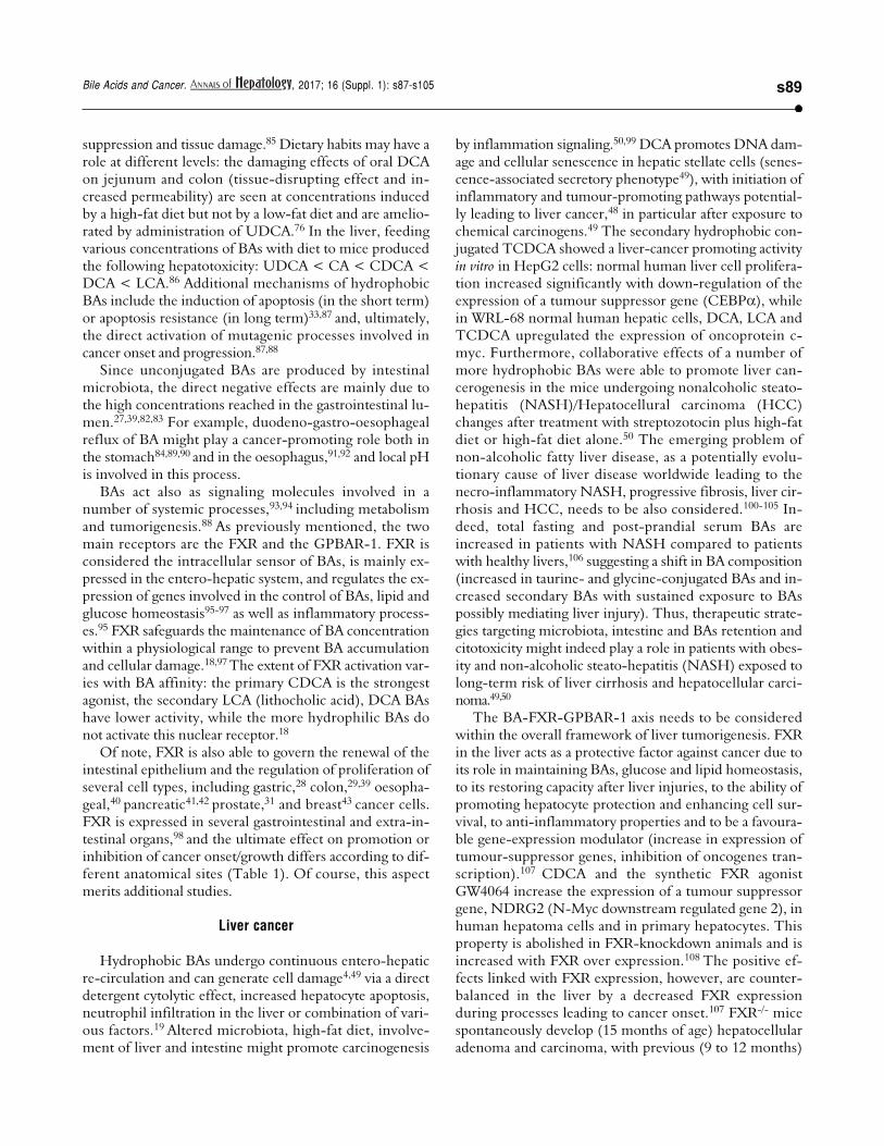
Of note, FXR is also able to govern the renewal of theintestinal epithelium and the regulation of proliferation ofseveral cell types, including gastric,28 colon,29,39 oesopha-geal,40 pancreatic41,42 prostate,31 and breast43 cancer cells.FXR is expressed in several gastrointestinal and extra-in-testinal organs,98 and the ultimate effect on promotion orinhibition of cancer onset/growth differs according to dif-ferent anatomical sites (Table 1). Of course, this aspectmerits additional studies.
Liver cancer
Hydrophobic BAs undergo continuous entero-hepaticre-circulation and can generate cell damage4,49 via a directdetergent cytolytic effect, increased hepatocyte apoptosis,neutrophil infiltration in the liver or combination of vari-ous factors.19 Altered microbiota, high-fat diet, involve-ment of liver and intestine might promote carcinogenesis
by inflammation signaling.50,99 DCA promotes DNA dam-age and cellular senescence in hepatic stellate cells (senes-cence-associated secretory phenotype49), with initiation ofinflammatory and tumour-promoting pathways potential-ly leading to liver cancer,48 in particular after exposure tochemical carcinogens.49 The secondary hydrophobic con-jugated TCDCA showed a liver-cancer promoting activityin vitro in HepG2 cells: normal human liver cell prolifera-tion increased significantly with down-regulation of theexpression of a tumour suppressor gene (CEBP ), whilein WRL-68 normal human hepatic cells, DCA, LCA andTCDCA upregulated the expression of oncoprotein c-myc. Furthermore, collaborative effects of a number ofmore hydrophobic BAs were able to promote liver can-cerogenesis in the mice undergoing nonalcoholic steato-hepatitis (NASH)/Hepatocellural carcinoma (HCC)changes after treatment with streptozotocin plus high-fatdiet or high-fat diet alone.50 The emerging problem ofnon-alcoholic fatty liver disease, as a potentially evolu-tionary cause of liver disease worldwide leading to thenecro-inflammatory NASH, progressive fibrosis, liver cir-rhosis and HCC, needs to be also considered.100-105 In-deed, total fasting and post-prandial serum BAs areincreased in patients with NASH compared to patientswith healthy livers,106 suggesting a shift in BA composition(increased in taurine- and glycine-conjugated BAs and in-creased secondary BAs with sustained exposure to BAspossibly mediating liver injury). Thus, therapeutic strate-gies targeting microbiota, intestine and BAs retention andcitotoxicity might indeed play a role in patients with obes-ity and non-alcoholic steato-hepatitis (NASH) exposed tolong-term risk of liver cirrhosis and hepatocellular carci-noma.49,50
The BA-FXR-GPBAR-1 axis needs to be consideredwithin the overall framework of liver tumorigenesis. FXRin the liver acts as a protective factor against cancer due toits role in maintaining BAs, glucose and lipid homeostasis,to its restoring capacity after liver injuries, to the ability ofpromoting hepatocyte protection and enhancing cell sur-vival, to anti-inflammatory properties and to be a favoura-ble gene-expression modulator (increase in expression oftumour-suppressor genes, inhibition of oncogenes tran-scription).107 CDCA and the synthetic FXR agonistGW4064 increase the expression of a tumour suppressorgene, NDRG2 (N-Myc downstream regulated gene 2), inhuman hepatoma cells and in primary hepatocytes. Thisproperty is abolished in FXR-knockdown animals and isincreased with FXR over expression.108 The positive ef-fects linked with FXR expression, however, are counter-balanced in the liver by a decreased FXR expressionduring processes leading to cancer onset.107 FXR-/- micespontaneously develop (15 months of age) hepatocellularadenoma and carcinoma, with previous (9 to 12 months)
Di Ciaula A, et al. , 2017; 16 (Suppl. 1): s87-s105s90
liver injury and inflammation. Also in this case, an alteredregulation of gene involved in the control of BAs levels ispresent, with high BAs concentration in both serum andliver. In this animal model, the role of endogenous BAs incancer promotion appears evident, since administration of2% cholestyramine is able to significantly reduce cancerlesions.109
Of note, a decreased FXR expression per se does notappear to be able, alone, to promote liver cancer onset andto maintain cancer proliferation if not associated with highlevels of BAs. While the FXR deficiency may have a role ascancer promoter, an increment in BA levels is requiredfor the promotion of cell proliferation and cancer forma-tion.110 Prospective metabolomics analysis of hepatocellu-lar carcinoma have clearly identified long term elevatedserum BAs levels as a risk factor for cancer develop-ment.111 Additionally, mice with hepatocyte-specific FXRdeficiency (FXR(hep-/-)) did not show spontaneous livercancer formation with aging, but cell proliferation andcancer formation were induced by cholic acid supple-mentation by diet, and were linked with increased basal
expression of tumour suppressor p53 protein and distur-bance of the mitogen-activated protein kinases (MAPK)and JAK/STAT3 signaling pathways.110 The MAPKs signal-ing pathways, in particular, have a pathogenic role in a se-ries of human diseases (including cancer) and theiractivation is secondary to cellular stress (also involvingoxidative damage promoted by Bas112) and to the presenceof proinflammatory cytokines.113 The activation of STAT3,on the other hand, is able to increase transcription ofgenes involved in suppression of anti-tumour immuni-ty,114 liver inflammation and cancer.114,115
An interesting animal model of FXR-null mice withre-expression of constitutively active FXR in enterocyteshas recently suggested that, in the presence of reduced he-patic FXR expression, the reactivation of intestinal FXRnormalized BA enterohepatic circulation through the fi-broblast growth factor 15 (FGF15)/cholesterol-7alpha-hy-droxylase enterohepatic axis, reducing BAs synthesis bythe liver, with a protective effect from spontaneous HCConset.116 Thus, in the case of reduced hepatic FXR expres-sion, the coexistence of adequate entero-hepatic signaling
Table 1. Effects of FXR overexpression at the level of different anatomical sites.
Organ Cancer protective/ Main effects References
promoting activity
Liver Protective - FXR activation linked with increased expression 107-110,116
of tumour-suppressor genes,inhibition of oncogenes transcription
Oesophagus Promoting - Altered function of genes regulating cell growth
(RAR- 2 and cyclooxygenase-2)- Over expression of FXR associated with high tumour grade,
large tumour size and lymph node metastasis
Colon Protective - Regulation of genes involved in cell proliferation and 39,154,155,
in inflammatory processes 158,159- FXR expression inversely correlated with clinical outcome
(higher FXR expression, lower carcinoma stage,
more favourable outcome)
Pancreas Promoting - Up-regulation of oncogenic MUC4 expression
- High FXR expression linked with poor prognosis- Elevated FXR levels in cancer cells responsible
for proliferation and migration
Breast Protective - High FXR expression in cancer tissue linked with 43,55,161Promoting smaller tumour size, better outcome
(in presence - Induction of apoptosis in cancer cellsoestrogen - In the presence of oestrogen receptor-positive breast cancer,receptor-positive positive correlation between FXR- and oestrogen
breast cancer) receptor expression
Prostate Protective - Up-regulation of tumour suppression gene 31,53- Inhibition of cell proliferation
Lung Promoting - In non-small cell lung cancer (NSCLC), FXR over expression 162and relation with poor outcomes
s91Bile Acids and Cancer. , 2017; 16 (Suppl. 1): s87-s105
pathways involving the FGF15/cholesterol-7alpha-hy-droxylase axis might be protective for liver cancer onset.
The role of aberrant signaling involving fibroblastgrowth factor 15/19, FGF receptor 4 (FGFR4) and beta-Klotho (KLB) co-receptor signaling system has been re-cently underlined in the onset of liver cancer,117 andaltered pathways involving these additional key regulatorsof BA synthesis and metabolism are able to promote HC-Cin mice and to influence the clinical outcome in HCCpatients.118
In mice, increased expression of FGF19 (fibroblastgrowth factor 19) promotes HCC development withFGFR4-dependent mechanisms and activating, also in thiscase, the STAT3 pathway.117 Higher concentrations of BAs(e.g. CDCA) might also explain in part the increased riskin men with Primary Biliary Cholangitis (PBC) for HCC,in particular in non-responders to UDCA therapy.119 FXRand the CDCA-dependent activation in the liver and intes-tine is likely involved.120,121
The GPBAR-1 receptor has also a key function in BAhomeostasis, LCA and taurolithocholic acid (TLCA) be-ing their most potent endogenous ligands.122-124 A BAs-stimulated GPBAR-1 expression is present Kupfer cells.125
Both FXR and GPBAR-1, once activated by BAs mightlead to suppression of NF- B factor and proinflammatorycytokines in the liver.126
Oesophageal cancer
Barrett’s oesophagus is characterized by the develop-ment of metaplastic columnar epithelium that replaces thenormal stratified squamous epithelium found in the distaloesophagus. Chronic gastroesophageal reflux disease(GERD) is the cause for Barrett's oesophagus, which is acondition predisposing to the development of adenocarci-noma of the oesophagus.
In tissues from human Barrett’s oesophagus, DCA gen-erated oxidative stress by inducing reactive oxygen and ni-trogen species after acting on intracellular NADPHoxidase and mitochondria and activation of the NF- Bpathway.74,77,78,127 Cells hosting the damaged DNA mightresist apoptosis.77 The BAs and acid-induced NF- B acti-vation in epithelial cells is dose- and time dependent andalso involves the induction of COX-2 promoter activity,potentially contributing to the onset of oesophageal can-cer.78,128 By contrast, the more hydrophilic UDCA (urso-deoxycholic acid) protects from DNA damage and NF- Bactivation.74,77,78 In a comprehensive study in patients withBarrett’s oesophagus, Peng, et al.74 showed that oral treat-ment with UDCA prevented the toxicity by DCA 250 μM(DNA damage, NF- B activation in the metaplastic mu-cosa of patients with Barrett's oesophagus). In vitro, UDCAactivated the NF-E2-related factor 2 (Nrf2) to upregulate
the expression of glutathione peroxidase 1 (GPX1) andcatalase antioxidants, a finding further confirmed in biopsyspecimens of Barrett’s metaplasia taken from patients after8 week treatment with oral UDCA. The DNA-damagingeffect might be operating with both glyco-conjugated BAsat acidic pH (pH = 4) but also with unconjugated BAs athigher pH (pH = 6). An overview on the role of second-ary BAs in neoplastic development in the oesophagus isavailable by Cronin, et al.91
FXR might play a role also in the context of Barrett’soesophagus: in the experimental mice model of oesopha-geal adenocarcinoma, the overexpression of FXR has beenassociated with higher tumour grade, larger tumour sizeand lymph node metastasis, and knockdown of FXR ex-pression suppressed tumour cell growth. Results fromthis study indicated that FXR expression mediated BAs-induced alterations of genes regulating cell growth (RAR-
2 and cyclooxygenase-2).40
Gastric cancer
Wang, et al.47 studied gastric cancer in mice and foundthat acidified bile acids induce tumour progression and te-lomerase activity both in vivo and in vitro, with mechanismsinvolving higher c-Myc transcription (a regulator genethat codes for a transcription factor and is involved in cellcycle progression, apoptosis and cellular transformation),with increased expression of human telomerase reversetranscriptase (hTERT) at the protein and mRNA levels. Inprimary human gastric adenocarcinoma cancer cell linesMKN28, MGC803 and SGC7901, the same authors foundthat 100 μg DCA and CDCA under acidified media acti-vate c-Myc that, in turn, increases hTERT expression.84 Inthe clinical setting, Tatsugami, et al.,90 studied 612 Japanesepatients positive for H. pylori infection using gastric biop-sies. The retrospective occurrence of gastric cancer wascalculated in 357 patients followed by endoscopic exami-nation for cancer screening for less than 3 years. BAs con-centration in gastric juice correlated with the extent ofgastric atrophy/intestinal metaplasia independently of in-flammatory cell infiltration. Also, the occurrence of gastriccancer was increased in patients with high- as compared tothose with low-BAs concentration. Exposure to acidifiedBAs (DCA and CDCA at pH 5.5) increased tumour pro-gression in MGC803 gastric cancer cell line.
GPBAR-1 expression has been linked with advancedstages of gastric cancer; GPBAR-1 expression correlateswith the expression of N-cadherin, a markers of epitheli-al-mesenchymal transition.129 Moderate to strong GPBAR-1 staining in gastric adenocarcinoma was associated withdecreased patient survival, and BAs increased cell prolifer-ation through activation of GPBAR-1 receptors and cou-pled G(q) and G (i-3) proteins.46
Di Ciaula A, et al. , 2017; 16 (Suppl. 1): s87-s105s92
Colon cancer
Colorectal cancer prevalence is dramatically risingworldwide.130 In the intestine, the replacement of intesti-nal villi cells is a crucial step. The process is completedevery 3-5 days and starts from the pluripotent cells locatedat the bottom of intestinal crypts, which transform intospecific enteroendocrine, absorptive, Gobleth and Panethcells. From the top of the villi, apoptotic cells are releasedinto the intestinal lumen at the end of the differentiationcycle. Several transcription factors are involved in theseprocesses, namely the caudal-related homeobox transcrip-tion factor (CDX2), E-cadherin, claudin-2, genes like Mu-cin 2 and sucrose isomaltase. Further signaling pathwaysinclude Wnt/ -catenin, the cytoplasmic protein -cateninand/or the tumour suppressor APC binding to -catenin.For colorectal cancer onset, several mutations are re-quired, starting from APC gene and also involving KRas,TP53, phosphoinositide 3-kinase (PI3K) and transforminggrowth factor (TGF ).39
Over-consumption of a Western-style diet can repre-sent a step linking BAs to colorectal cancer. The Western-style diet brings excess calories, is enriched withhighly-saturated fats and processed carbohydrates butlacks mono-polyunsaturated fatty acids and plant-derivedproteins and fibre.34,39,83 Following Western-style/high-fat/low-fibre diet, therefore, abnormally high levels of sec-ondary BAs might increase in the intestine,131,132 and thisstep leads to disruption of the complex mechanisms gov-erning the intestinal epithelial renewal. Elevated luminalconcentrations of secondary DCA and LCA (at variancewith the hydrophilic tertiary hydrophilic UDCA) mightprovoke intestinal cytotoxic damage which parallels theeffect of other genetic and environmental factors acting astumour promoter stepin the post-initiation early stages ofcolon carcinogenesis133 and acting as a tumour-promotingeffect.134 Even cholecystectomy, a condition which in-creases the exposure of intestinal mucosa to elevated BAlevels has been considered as a predisposing condition tocolorectal cancer.135 Mechanisms of BA-induced tumori-genesis include DNA oxidative damage, hyperprolifera-tion, NF- B activation and inflammation, -cateninsignaling and p53 degradation. Several additional mecha-nisms have been advocated and include BA-induced pro-liferative effect on undifferentiated epithelial cells ofintestine136 and colon cells,137 disrupted colonic mucosalintegrity,138 activation of extracellular signal-regulated ki-nase (ERK) signaling and epidermal growth factor recep-tor (EGFR)139 and stimulation of colonic epithelialproliferation via protein kinase C (PKC).140 Initiation ofapoptosis resistance by BAs such as DCA and LCA141
would imply mitochondrial damage with mitochondrialoxidative stress, generation of reactive oxygen species
(ROS), cytochrome C (cytC) release and activation of cy-tosolic caspases.71 Nuclear factor kappa (NF- B) path-way activation and release of arachidonic acid might workin concert with cytotoxic BAs in the colon.142
The intestinal microbiota is another important playerin the scenario mentioned above. Microbes populate thehuman gut reaching massive concentration in the colon(up to 1012 CFU/g luminal content),143 play a key role inBA biotransformation from primary to secondary mole-cules and can be easily modulated by factors like age, nu-trition, diseases, drugs and/or intestinal anatomy.144-146
Diet can heavily influence the microbial metabolic path-ways and gas production,143-147 since the saccharolytic fer-mentation of carbohydrates by microbiota producesshort-chain fatty acids (SCFAs) such as butyrate, propion-ate, acetate, and butyrate has anti-inflammatory and antine-oplastic properties148-150 while a high-fat diet wouldactivate pathways involving proteolysis, inflammation andtumorigenesis.151,152 Zeng, et al.83 demonstrated that bu-tyrate (the short-chain fatty acid and microbiota-depend-ent metabolite of dietary fibre) at a concentration of 0.5-2.0mM counteracted the detrimental effects of DCA (0.05-0.3mM) on colon cell proliferation. Although both butyrateand DCA inhibited cell proliferation and increased cell ap-optosis rate, only butyrate increased G1 and G2 fractions(vs. only G1 with DCA) with a concomitant drop in the S-phase fraction at cell cycle analyses. DCA but not butyrateincreased intracellular pathways including reactive oxygenspecies, genomic DNA breakage and the activation ofERK1/2, caspase-3 and PARP. Overall, the current datasuggest that both butyrate and DCA inhibit colonic cellproliferation. However, butyrate increases tumour sup-pressor gene expression, whereas DCA decreases tumoursuppressor activation in cell cycle and apoptosis path-ways.83 Similar mechanisms have been described in nor-mal and tumour human colon cells,34 as well as in the micemodel of colon cancer,24 where DCA and CDCA are ableto cause oxidative DNA damage27,82 and apoptosis153
through oxidative processes which can be limited by theconcomitant exposure of cells to antioxidants, i.e. beta-carotene, alpha-tocopherol, Na-butyrate, zinc and/or chlo-rogenic acid.24,27,82 Such findings point to a potentialprotective role, partly BA-mediated of healthy diets.
FXR expression has also a role in colon cancer,154,155
since mechanisms of cancerogenesis in the colon also in-volve Apc gene mutation, CDX2 inactivation and in-creased CpG methylation in the Fxr gene, resulting in lossof FXR in the colonic epithelium, increased mitotic activ-ity, cell hyperproliferation; all features associated with apro-tumorigenic phenotype.142,156,157 If FXR becomes defi-cient in the intestine, moreover, secondary BAs might beincreased and less detoxified in the liver. Loss of FXRgenerates high BAs concentrations and, in animal models,
s93Bile Acids and Cancer. , 2017; 16 (Suppl. 1): s87-s105
a pro-tumorigenic phenotype39 with pathways similar tothose observed for liver cancer. In an animal model, lossof FXR in the ApcMin/+ mice lead to early mortality and in-creased colon cancer progression, pointing to a protectiverole of FXR on intestinal cancer. However, the cancer-promoting effect was independent from intraluminal BAs,since it was not inhibited by treatment with cholesty-ramine.155 In mice, FXR deficiency also generates an up-regulation of genes involved in cell proliferation and ininflammatory processes, an increment in colon cell prolif-eration and a growth of small intestine adenocarcinomas inadenomatous polyposis coli mutant animals.158
In human colon cancer, FXR expression is repressedduring the transition of adenoma to carcinoma and is notexpressed in undifferentiated colon cancer cells SW480and in metastasis derived SW620 cells.159 A systematic im-munohistochemistry mapping on human intestinal muco-sa showed that FXR expression was reduced in coloncarcinomas as compared with non-neoplastic mucosa andthat a relationship was evident between the loss of FXRexpression and the grading of tumours in the right colon.FXR expression was inversely correlated with the clinicaloutcome of patients (higher FXR expression, lower carci-noma stage and more favourable outcome).154
Pancreatic cancer
A relationship between BAs and pancreatic cancer hasbeen suggested. BAs might reflux into the pancreatic ductand, on the other hand, are linked at a systemic level withobesity, diabetes and hypertriglyceridemia, all well knownrisk factors for pancreatic cancer.51 Elevated levels of BAshave been reported in serum and in pancreatic juice frompatients with pancreatic cancer, as compared with con-trols. This finding might be linked to up-regulation of on-cogenic MUC4 expression.42 High expression of FXR incolon154 and breast43 cancer relates with better clinical out-come of patients. However, for pancreatic cancer, highFXR expression is rather linked with poor prognosis andpoor survival. FXR elevation in pancreatic cancer cellsmight be responsible for cellular proliferation and migra-tion.41
Prostate cancer
Positive effects of FXR overexpression have also beendescribed in the case of prostate. FXR activity, in fact, ispresent in normal and cancer prostate epithelial cells andits stimulation by CDCA treatment is able to inhibit cellproliferation in prostate cancer.53 The suppression ofprostate tumour growth is associated with decreasedmRNA and protein levels of sterol regulatory elementbinding protein 1 (SREBP-1),53 and through an up-regula-
tion of the tumour suppression gene for the Phosphataseand tensin homolog (PTEN) induced by the FXR overex-pression.31
Breast cancer
FXR has been also detected in breast tissue.160 Simi-larly to that previously observed in colon cancer,154 inwomen with invasive breast carcinoma, high FXR ex-pression in cancer tissue was linked with smaller tumoursize and patients with high FXR expression had a betterclinical outcome (longer overall and disease-free surviv-al time) as compared with those with low FXR expres-sion.161 In vitro, the activation of FXR by CDCA or by asynthetic ligand (GW4064) induced cell death (mainly byintrinsic apoptotic pathway) in four distinct phenotypesof breast cancer cell lines, without stimulating migrationin cell lines.43 The effect of FXR overexpression onbreast cancer, however, seems to be different (opposite)in the presence of oestrogen receptor-positive breastcancer, where a positive correlation was found betweenFXR- and oestrogen receptor expression. In this case, in-creased FXR levels were also correlated with the prolif-eration marker Ki-67 and nodal metastasis inpostmenopausal women. The proliferation of oestrogenreceptor-positive breast cancer could be, in this case,secondary to a crosstalk between FXR and oestrogen re-ceptors, in particular during oestrogen deprivation (i.e.post-menopausal women, therapy with aromatase inhibi-tors).55
Lung cancer
A recent study has also depicted a negative role of FXRexpression in non-small cell lung cancer (NSCLC). Inthis case FXR is overexpressed and is related with pooroutcomes in patients, in particular in the presence of con-comitant over expression of cyclin D1,162 increment inCyclin D1 protein and mRNA expression.163
EPIGENETIC FACTORS
The pathway linking BAs and nuclear receptors withcancer onset is influenced by changes in gene expres-sion.42,47,50,83,84,95-97,107,164 This step leads to both benign andmalignant diseases and is also able to influence the clinicaloutcome in cancer patients.118,154,161,162
The expression of genes involved in BAs-dependentsignaling processes may be silenced, reduced or amplifiedby epigenetic mechanisms (mainly microRNA expres-sion, DNA methylation, histone/gene acetylation165) alsoinduced by dietary habits164 and various environmental fac-tors, without changes in DNA sequence.

Di Ciaula A, et al. , 2017; 16 (Suppl. 1): s87-s105s94
MicroRNA
MicroRNAs represent a class of small noncodingRNAs. They play a key role in a number of diseases (in-cluding human carcinogenesis) mainly through a down-regulation of various target genes.
MicroRNA-22 (miR-22) has a pronounced tumour-suppressive role in different organs166,167 including co-lon168 and liver cancer.169,170 The process is regulated byFXR expression in liver and colon.35 CDCA, due to itshigh affinity for FXR,18 increases miR-22 levels in liverand colon cells with a silencing effect on cyclin A2(CCNA2). In FXR-knockout mice low miR-22 levels areassociated with increased number of Ki-67-positive cellsin the colon and in the liver. In humans, levels of miR-22and CCNA2 are inversely correlated with colon and livercancers.35 Human oesophageal adenocarcinoma samplesdisplay increased levels of miRNA 221 and miRNA 222, ascompared with Barrett’s oesophagus samples taken fromthe same patients.36 Also, levels of both miRNA-221 and222 in cultured cells were related with FXR activity in re-sponse to BAs exposure and inhibited mRNA translationof p27Kip1, promoting degradation of the transcriptionfactor CDX2.36 It has to be underlined that altered expres-sion p27kip1 leads to deregulated cell growth/differentia-tion, promoting the development of a number of tumoursin humans.171
DNA methylation
In the rats and the mice, BAs like DCA, CDCA, CAand LCA introduced by diet induced DNA hypomethyla-tion in the colon. This effect was not induced by adminis-tration of the more hydrophilic UDCA.172 Other studiesclearly point to a relationship between DNA methylationand FXR expression. Mutations in the adenomatous poly-posis coli (APC) gene have been linked with the early de-velopment of colorectal cancer.37 Studies in APC deficientmice suggest that FXR expression is reduced; this silenc-ing effect is mainly linked to CpG methylation of theFxr 3/4 promoter.156 In the same study DCA loweredCpG methylation of FXR and induced FXR expression inhuman HCT-116 but not HT-29 colon cancer cells.156 Therelationship between DNA methylation and FXR silenc-ing was also described in a previous study in human coloncancer, demonstrating a reduced expression/function ofFXR in precancerous lesions and a silenced FXR in themajority of stage I-IV tumours.30 BAs are also able to affectDNA methylation in human oesophageal tissue. Exposureof human oesophageal epithelial cells to a mixture of sixdifferent forms of BAs (GCA, TCA, GCDCA, TCDCA,GDCA, and TDCA) induced Caudal-related homeobox 2(Cdx2) expression (as an early marker of Barrett’s
oesophagus) through promoter demethylation. Thismechanism contributes to the onset of intestinal metapla-sia, a premalignant lesion of oesophageal adenocarcino-ma.38 Over expression of Cdx2 was also described inhuman oesophageal tissues, in esophagitis and, in higherproportion, in samples from patients with Barrett’soesophagus and primary oesophageal adenocarcinoma.45
Histone acetylationand chromatin remodeling
Post-translational modifications of histones (i.e. his-tone acetylation/deacetylation) and chromatin remodelingare well-known epigenetic mechanisms173,174 workingwith transcriptional cofactors (i.e. sensing activities andsignaling pathways,175 as FXR176) and have a defined role inthe metabolism of lipids177 and in BA homeostasis andfunctions.178 The small heterodimer partner (SHP, an or-phan nuclear receptor) is an important epigenomic regu-lator of BA biosynthesis, mainly acting through chromatinremodeling179,180 and histone deacetylation.181,182 SHP hasbeen identified as having an antitumor role in liver can-cer183,184 due to its capacity to regulate cell proliferation,apoptosis, DNA methylation, and inflammation,184 and isalso involved (due to its strict relationships with FXR) incolon,156 gastric185 and breast160 cancer.
In an animal model Sirtuin 1 (SIRT1), a key regulatorof a number of metabolic processes (including BAs home-ostasis), has a critical role in the regulation of the regener-ative response in the liver by post-trascriptionalmodifications involving FXR activity (through theacetylation of FXR and neighboring histones) and mTOR,potentially contributing to liver cancer onset through dys-regulation of BA homeostasis by persistent FXRdeacetylation.181
BAs, MICROBIOTA,ENVIRONMENTAL POLLUTANTS
BAs undergo biotranformation especially in the colon,due to unique microbial enzymes which are encodedwithin the gut microbioma.1 Distribution of BSH en-zymes, essential in primary conjugated BA deconjugationin the colon, are found in Gram positive species Lactobacil-lus, Enterococcus, Clostridium spp, gram negative Bacteroides sppand in several bacterial strains (i.e., L. plantarum, L. acido-philus, L. salivarius, C. perfringens, etc.). BSH in bacteriamight confer a defensive mechanism against the effect ofBAs and provide glycine and taurine as bacterial energeticsource (glycine NH4+CO2 and taurine NH4+CO2+sulphate).1 Current knowledge suggests thatBSH influences several physiological processes in thehost and mark the BA signature with a control on meta-
s95Bile Acids and Cancer. , 2017; 16 (Suppl. 1): s87-s105
bolic, immunological, and receptorial functions.1,13 Fur-ther steps after bacterial deconjugation in the colon in-clude anaerobic bacterial re-amidation, redox reactions,desulfation186 (as prevention of BA loss in feces/urines),esterification, oligomerization from time-to-time byLactobacillus, Bacteroidetes, Eubacteria, Clostridium, etc.4,187,188
Bacterial stereospecific hydroxysteroid dehydrogenases(HSDH) control BA oxidation, epimerization and dehy-droxylation189 and, via Clostridium species, the biosynthesisof the tertiary UDCA from the secondary CDCA.190 Sev-eral other bacterial species will join such complex biosyn-thetic pathways.
Events pointing to qualitative or quantitative changes ofintestinal microbial community may heavily influencebacterial enzymes and, in turn, BA composition and func-tions. Paradigmatic situations include germ-free or antibi-otic treated animals,191,192 food consumption193,194 withchanges occurring even in the short-term (1 to 3 days195),aging,196 inflammatory bowel disease,186 even metabolicdisorders,197,198 functional disorders including irritablebowel syndrome,143,199 intestinal surgery including bariat-ric surgery in morbid obesity,200,201 primary sclerosingcholangitis202 and ingestion of environmental toxics con-tained in water or food.66-69,203-209
Forms of intestinal dysbiosis might also contribute totumorigenesis in different ways. Obesity is a major riskfactor for several types of common cancer,210 and obesitymight induce changes in gut microbiota,211 shift the BApool profile (i.e. increased DCA), and several hydropho-bic BAs might collaboratively promote carcinogenesis(not HCC initiation) via DNA damage,4 induction of se-nescence-associated secretory phenotype (SASP) in hepat-ic stellate cells (HSCs),49 Gram-negative activation oftoll-like receptor (TLR) 4 and bacterial production of li-popolysaccharide (LPS) in the intestine.212 In mice, pre-vention of liver cancerogenesis has been achieved byblocking DCA formation, and acting on gut microbiota49,50
with sterilization,212 increasing intestinal excretion of hy-drophobic BAs (i.e. with the bile acid sequestrant choles-tyramine50). Similar mechanisms involving disrupted BApool and dysbiosis might also operate in other sites of hu-man tumorigenesis. In the colon DCA and LCA would actas procarcinogenic bacterial metabolites but also promis-ing therapeutic targets.213 Both BAs might act as proin-flammatory agents, eliciting the production of reactiveoxygen and nitrogen species, as well as NF- B activationin intestinal epithelial cells.214-217 Moreover, chronic ex-posure to DCA induces the production of DNA adductswhich parallels enhanced epithelial cell proliferation anddecreased apoptosis.34
Tumorigenesis can also imply an impaired interactionbetween BAs and their receptors.14 FXR, for example pre-vents excessive inflammation in the liver and intestine218
(see also previous paragraphs on BAs and FXR). Thus,while changes in microbiota might be implicated in somesteps of tumorigenesis, inducible changes of microbiotamight also represent an additional clue to cancer thera-py.219,220 Much caution, however, is required in this field,until definitive prospective clinical/population studieswill clarify the true pathogenic role of this consortium ofactors in carcinogenesis.
Recent studies point to the marked effects on intestinalmicrobiota of some environmental pollutants as heavymetals (mainly arsenic, cadmium and lead) and persistentorganic pollutantsingested with contaminated water orfood,66,203-206 resulting in an increased toxicity (and poten-tial mutagenic properties) of the BAs pool. This inducesoxidative stress221 and strongly alters the intestinal micro-biota, by reducing the amount of both primary and sec-ondary BAs. This mechanism develops through adown-regulation of CA, UDCA and DCA levels.203 Amarked alteration of gut microbiota has been reported inthe animal model, after ingestion of arsenic in drinkingwater, which also increased the excretion of 7- -hydroxy-3-oxo-4-cholestenoate (involved in the biosynthesis ofprimary BAs) and reduced GCA in fecal samples of treat-ed animals.66 Of note, 7- -hydroxy-3-oxo-4-cholestenoateis believed to be, in humans, an important precursor ofCDCA,222 the strongest agonist involved in FXR activa-tion,18 and GCA has been linked by metabolomics withhepatocellular carcinoma.223,224
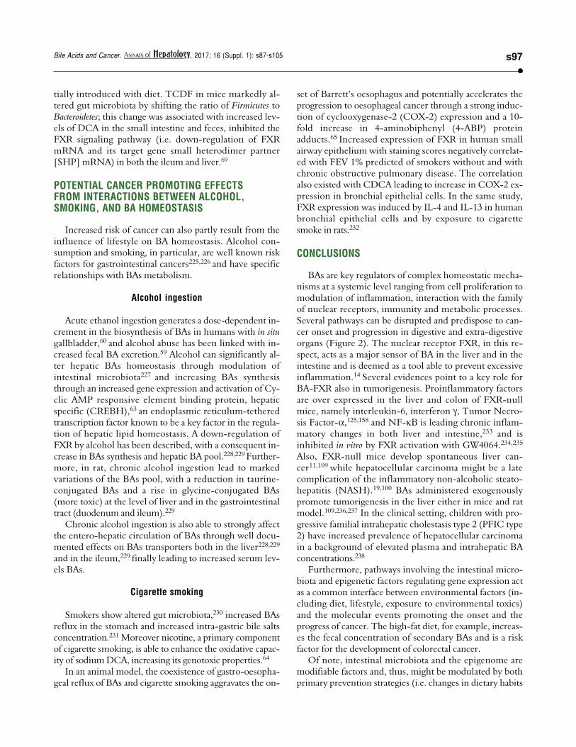
Pesticides such as chlorpyrifos,207-209 diazinon,67 and2,3,7,8-tetrachlorodibenzofuran (TCDF)69 can greatly al-ter microbiota composition58,157-159 (Figure 1).
Diazinon, a widely employed organophosphate pesti-cide able to contaminate ground water, drinking waterwells and food, in an animal study strongly altered gutmicrobiota and the related metabolic functions with dif-ferent sex-specific patterns (more pronounced responsesin male mice). Significant increments in BacteroidaceaeBacteroides (> 2,000-fold rise, bacteria with bile salt hydro-lase enzymes, BSHs) and Proteobacteria (+15-fold rise, bac-teria involved in BA transformation) were recorded intreated animal. As a consequence of this increased decon-jugation potential, a 4-fold and a 5-fold increment in LCAlevels was recorded in treated male and female mice, re-spectively and, in female mice, a significant increment(3.6-fold) of DCA was also noticed.67
Chlorpyrifos is an organophosphate pesticide whichacts on the nervous system by inhibiting acetylcholineste-rase. This compound promoted alterations in gut micro-biota composition and metabolome (including alterationsof the BAs pool) in mice. Changes were associated withhistological modifications in the colon of treated animals,intestinal inflammation and altered permeability.209 In oth-er animal models, chronic exposure to chlorpyrifos at low

Di Ciaula A, et al. , 2017; 16 (Suppl. 1): s87-s105s96
doses caused intestinal dysbiosis with proliferation (Ente-rococcus and Bacteroides) or decrement (lactic acid bacteria asLactobacillus and the bifidobacteria) of selected strains.207
Oral exposure of female rats during gestation to thesame pesticide caused marked gut dysbiosis and damagesto the intestinal epithelium in the pups.208
The SHIME® model also demonstrated that chlorpyri-
Figure 1. A. Figure 1. A. Figure 1. A. Figure 1. A. Figure 1. A. Chemical structure of the pesticide diazinon. B. B. B. B. B. Chemical structure of the organophosphate pesticide chlorpyrifos. C. C. C. C. C. Chemical structure of2,3,7,8-tetrachlorodibenzofuran.
AAAAA
BBBBB
CCCCC
fos is able to affect human colonic microbiota, with an in-crease in Enterobacteria, Bacteroides and Clostridia, and a de-crease in bifidobacterial counts following chronic low(below-threshold) doses of CPF (1 mg/day for one month,dissolved in rapeseed oil).68
Similar results were promoted by 2,3,7,8-tetrachlorod-ibenzofuran (TCDF), a persistent organic pollutant poten-
D i a z i n o nD i a z i n o nD i a z i n o nD i a z i n o nD i a z i n o n
O,O-diethyl O-(2-isopropyl-6-methylprimidin-4-yl) phosphorothiateChemical Formula: C12H21N2O3PS
Exact Mass: 304.10Molecular Weight: 304.34
Elemental Analysis: C: 47.36. H: 6.96. N: 9.20. O: 15.77. P: 10.18. S: 10.53
C h l o r p y r i f o sC h l o r p y r i f o sC h l o r p y r i f o sC h l o r p y r i f o sC h l o r p y r i f o s
O,O-diethyl O-(3,5,6-trichloropyridin-2-yl)phosphorothiate
Chemical Formula: C9H11Cl3 NO3PSExact Mass: 348.93
Molecualar Weight: 350.57Elemental Analysis:
C: 30.83. H: 3.16. Cl: 30.34. N: 4.00.O: 13.69. P. 8.84. S: 9.14.
2,3,7,8-tetracholorodibenzol [b,d] furanChemical Formula: C12H4Cl4O
Exact Mass: 303.90Molecular Weight: 305.96
Elemental Analysis: C: 47.11. H: 1.32. Cl: 46.35. O: 5.23.
s97Bile Acids and Cancer. , 2017; 16 (Suppl. 1): s87-s105
tially introduced with diet. TCDF in mice markedly al-tered gut microbiota by shifting the ratio of Firmicutes toBacteroidetes; this change was associated with increased lev-els of DCA in the small intestine and feces, inhibited theFXR signaling pathway (i.e. down-regulation of FXRmRNA and its target gene small heterodimer partner[SHP] mRNA) in both the ileum and liver.69
POTENTIAL CANCER PROMOTING EFFECTSFROM INTERACTIONS BETWEEN ALCOHOL,SMOKING, AND BA HOMEOSTASIS
Increased risk of cancer can also partly result from theinfluence of lifestyle on BA homeostasis. Alcohol con-sumption and smoking, in particular, are well known riskfactors for gastrointestinal cancers225,226 and have specificrelationships with BAs metabolism.
Alcohol ingestion
Acute ethanol ingestion generates a dose-dependent in-crement in the biosynthesis of BAs in humans with in situgallbladder,60 and alcohol abuse has been linked with in-creased fecal BA excretion.59 Alcohol can significantly al-ter hepatic BAs homeostasis through modulation ofintestinal microbiota227 and increasing BAs synthesisthrough an increased gene expression and activation of Cy-clic AMP responsive element binding protein, hepaticspecific (CREBH),63 an endoplasmic reticulum-tetheredtranscription factor known to be a key factor in the regula-tion of hepatic lipid homeostasis. A down-regulation ofFXR by alcohol has been described, with a consequent in-crease in BAs synthesis and hepatic BA pool.228,229 Further-more, in rat, chronic alcohol ingestion lead to markedvariations of the BAs pool, with a reduction in taurine-conjugated BAs and a rise in glycine-conjugated BAs(more toxic) at the level of liver and in the gastrointestinaltract (duodenum and ileum).229
Chronic alcohol ingestion is also able to strongly affectthe entero-hepatic circulation of BAs through well docu-mented effects on BAs transporters both in the liver228,229
and in the ileum,229 finally leading to increased serum lev-els BAs.
Cigarette smoking
Smokers show altered gut microbiota,230 increased BAsreflux in the stomach and increased intra-gastric bile saltsconcentration.231 Moreover nicotine, a primary componentof cigarette smoking, is able to enhance the oxidative capac-ity of sodium DCA, increasing its genotoxic properties.64
In an animal model, the coexistence of gastro-oesopha-geal reflux of BAs and cigarette smoking aggravates the on-
set of Barrett’s oesophagus and potentially accelerates theprogression to oesophageal cancer through a strong induc-tion of cyclooxygenase-2 (COX-2) expression and a 10-fold increase in 4-aminobiphenyl (4-ABP) proteinadducts.65 Increased expression of FXR in human smallairway epithelium with staining scores negatively correlat-ed with FEV 1% predicted of smokers without and withchronic obstructive pulmonary disease. The correlationalso existed with CDCA leading to increase in COX-2 ex-pression in bronchial epithelial cells. In the same study,FXR expression was induced by IL-4 and IL-13 in humanbronchial epithelial cells and by exposure to cigarettesmoke in rats.232
CONCLUSIONS
BAs are key regulators of complex homeostatic mecha-nisms at a systemic level ranging from cell proliferation tomodulation of inflammation, interaction with the familyof nuclear receptors, immunity and metabolic processes.Several pathways can be disrupted and predispose to can-cer onset and progression in digestive and extra-digestiveorgans (Figure 2). The nuclear receptor FXR, in this re-spect, acts as a major sensor of BA in the liver and in theintestine and is deemed as a tool able to prevent excessiveinflammation.14 Several evidences point to a key role forBA-FXR also in tumorigenesis. Proinflammatory factorsare over expressed in the liver and colon of FXR-nullmice, namely interleukin-6, interferon , Tumor Necro-sis Factor- ,125,158 and NF- B is leading chronic inflam-matory changes in both liver and intestine,233 and isinhibited in vitro by FXR activation with GW4064.234,235
Also, FXR-null mice develop spontaneous liver can-cer11,109 while hepatocellular carcinoma might be a latecomplication of the inflammatory non-alcoholic steato-hepatitis (NASH).19,100 BAs administered exogenouslypromote tumorigenesis in the liver either in mice and ratmodel.109,236,237 In the clinical setting, children with pro-gressive familial intrahepatic cholestasis type 2 (PFIC type2) have increased prevalence of hepatocellular carcinomain a background of elevated plasma and intrahepatic BAconcentrations.238
Furthermore, pathways involving the intestinal micro-biota and epigenetic factors regulating gene expression actas a common interface between environmental factors (in-cluding diet, lifestyle, exposure to environmental toxics)and the molecular events promoting the onset and theprogress of cancer. The high-fat diet, for example, increas-es the fecal concentration of secondary BAs and is a riskfactor for the development of colorectal cancer.
Of note, intestinal microbiota and the epigenome aremodifiable factors and, thus, might be modulated by bothprimary prevention strategies (i.e. changes in dietary habits
Di Ciaula A, et al. , 2017; 16 (Suppl. 1): s87-s105s98
and lifestyle, reduced exposure to environmental toxics)and therapeutic tools. Future studies are needed to betterclarify how these measures could influence pathogenicmechanisms leading to disease onset and progression andif they will also be able to ameliorate the efficacy of theavailable therapeutic tools.
On the other hand, the therapeutic role of hydrophilicBAs (mainly UDCA, TDCA) counterbalancing the direct(cytotoxicity) and indirect (mainly in term of geneexpression and activity of nuclear receptors) negativeeffects of the more hydrophobic BAs needs to be moreclearly assessed in both digestive and extra-digestivecancers.
ABBREVIATIONS
� AQPs: aquaporins.� BAs: bile acids.� CDCA: chenodeoxycholic acid.� DCA: deoxycholic acid.� FGF15: fibroblast growth factor 15.
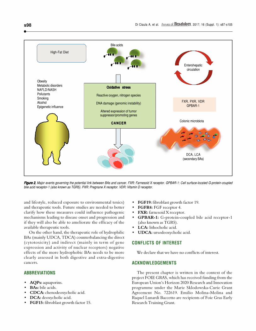
Figure 2. Figure 2. Figure 2. Figure 2. Figure 2. Major events governing the potential link between BAs and cancer. FXR: Farnesoid X receptor. GPBAR-1: Cell surface-located G-protein-coupledbile acid receptor-1 (also known as TGR5). PXR: Pregnane X receptor. VDR: Vitamin D receptor.
� FGF19: fibroblast growth factor 19.� FGFR4: FGF receptor 4.� FXR: farnesoid X receptor.� GPBAR-1: G-protein-coupled bile acid receptor-1
(also known as TGR5).� LCA: lithocholic acid.� UDCA: ursodeoxycholic acid.
CONFLICTS OF INTEREST
We declare that we have no conflicts of interest.
ACKNOWLEDGEMENTS
The present chapter is written in the context of theproject FOIE GRAS, which has received funding from theEuropean Union’s Horizon 2020 Research and Innovationprogramme under the Marie Sklodowska-Curie GrantAgreement No. 722619. Emilio Molina-Molina andRaquel Lunardi Baccetto are recipients of Foie Gras EarlyResearch Training Grant.
ObesityMetabolic disordersNAFLD/NASHPollutantsSmokingAlcoholEpigenetic influence
Bile acids
High-Fat Diet
OxidativeOxidativeOxidativeOxidativeOxidative stressstressstressstressstress
Reactive oxygen, nitrogen species
DNA damage (genomic instability)
Altered expression of tumorsuppressor/promoting genes
C A N C E RC A N C E RC A N C E RC A N C E RC A N C E R
Enterohepaticcirculation
FXR, PXR, VDRGPBAR-1
Colonic microbiota
DCA, LCA(secondary BAs)
s99Bile Acids and Cancer. , 2017; 16 (Suppl. 1): s87-s105
REFERENCES
1. Long SL, Gahan CGM, Joyce SA. Interactions between gut
bacteria and bile in health and disease. Mol Aspects Med
2017; 56: 54-65.
2. van Erpecum KJ, Wang DQ, Moschetta A, Ferri D, Svelto M,
Portincasa P, Hendrickx JJ, et al. Gallbladder histopathology
during murine gallstone formation: relation to motility and con-
centrating function. J Lipid Res 2006; 47: 32-41.
3. Portincasa P, Calamita G. Water channel proteins in bile for-
mation and flow in health and disease: when immiscible be-
comes miscible. Mol Aspects Med 2012; 33: 651-64.
4. Ridlon JM, Kang DJ, Hylemon PB. Bile salt biotransformations
by human intestinal bacteria. J Lipid Res 2006; 47: 241-59.
5. Wang DQH, Neuschwander-Tetri BA, Portincasa P. The Biliary
System. 2nd Ed. Morgan & Claypool Life Sciences; 2017.
6. Goodwin B, Jones SA, Price RR, Watson MA, McKee DD,
Moore LB, Galardi C, et al. A regulatory cascade of the nu-
clear receptors FXR, SHP-1, and LRH-1 represses bile acid
biosynthesis. Mol Cell 2000; 6: 517-26.
7. Miao J, Choi SE, Seok SM, Yang L, Zuercher WJ, Xu Y, Will-
son TM, et al. Ligand-Dependent Regulation of the Activity of
the Orphan Nuclear Receptor, Small Heterodimer Partner
(SHP), in the Repression of Bile Acid Biosynthetic CYP7A1
and CYP8B1 Genes. Mol Endocrinol 2011; 25: 1159-69.
8. Inagaki T, Choi M, Moschetta A, Peng L, Cummins CL, Mc-
Donald JG, Luo G, et al. Fibroblast growth factor 15 func-
tions as an enterohepatic signal to regulate bile acid
homeostasis. Cell Metab 2005; 2: 217-25.
9. Jones S. Mini-review: endocrine actions of fibroblast growth
factor 19. Mol Pharm 2008; 5: 42-8.
10. Holt JA, Luo G, Billin AN, Bisi J, McNeill YY, Kozarsky KF,
Donahee M, et al. Definition of a novel growth factor-de-
pendent signal cascade for the suppression of bile acid bio-
synthesis. Genes Dev 2003; 17: 1581-91.
11. Kim I, Ahn S-H, Inagaki T, Choi M, Ito S, Guo GL, Kliewer SA,
et al. Differential regulation of bile acid homeostasis by the
farnesoid X receptor in liver and intestine. J Lipid Res 2007;
48: 2664-72.
12. Volle DH. Bile acids, roles in integrative physiology and
pathophysiology. Mol Aspects Med 2017; 56: 1.
13. Li T, Chiang JYL. Bile Acid Signaling in Metabolic Disease and
Drug Therapy. Pharmacological Reviews 2014; 66: 948.
14. Martinot E, Sedes L, Baptissart M, Lobaccaro JM, Caira F,
Beaudoin C, Volle DH. Bile acids and their receptors. Mol As-
pects Med 2017; 56: 2-9.
15. Zhou H, Hylemon PB. Bile acids are nutrient signaling hor-
mones. Steroids 2014; 86: 62-8.
16. Chavez-Talavera O, Tailleux A, Lefebvre P, Staels B. Bile
Acid Control of Metabolism and Inflammation in Obesity, Type
2 Diabetes, Dyslipidemia, and Nonalcoholic Fatty Liver Dis-
ease. Gastroenterology 2017; 152: 1679-94 e3.
17. Vitek L, Haluzik M. The role of bile acids in metabolic regula-
tion. J Endocrinol 2016; 228: R85-96.
18. Parks DJ, Blanchard SG, Bledsoe RK, Chandra G, Consler
TG, Kliewer SA, Stimmel JB, et al. Bile acids: natural ligands
for an orphan nuclear receptor. Science 1999; 284: 1365-8.
19. Li M, Cai SY, Boyer JL. Mechanisms of bile acid mediated in-
flammation in the liver. Mol Aspects Med 2017; 56: 45-53.
20. Merlen G, Ursic-Bedoya J, Jourdainne V, Kahale N, Glenis-
son M, Doignon I, Rainteau D, et al. Bile acids and their re-
ceptors during liver regeneration: Dangerous protectors. Mol
Aspects Med 2017; 56: 25-33.
21. Sèdes L, Martinot E, Baptissart M, Baron S, Caira F, Beau-
doin C, Volle DH. Bile acids and male fertility: From mouse to
human? Mol Aspects Med 2017; 56: 101-9.
22. McIlvride S, Dixon PH, Williamson C. Bile acids and gestation.
Mol Aspects Med 2017; 56: 90-100.
23. Albaugh VL, Banan B, Ajouz H, Abumrad NN, Flynn CR. Bile
acids and bariatric surgery. Mol Aspects Med 2017; 56: 75-
89.
24. Bernstein C, Holubec H, Bhattacharyya AK, Nguyen H,
Payne CM, Zaitlin B, Bernstein H. Carcinogenicity of deoxy-
cholate, a secondary bile acid. Arch Toxicol 2011; 85: 863-
71.
25. Bernstein H, Bernstein C, Payne CM, Dvorakova K, Garewal
H. Bile acids as carcinogens in human gastrointestinal can-
cers. Mutat Res 2005; 589: 47-65.
26. Debruyne PR, Bruyneel EA, Li X, Zimber A, Gespach C,
Mareel MM. The role of bile acids in carcinogenesis. Mutat
Res 2001; 480-481: 359-69.
27. Rosignoli P, Fabiani R, De Bartolomeo A, Fuccelli R, Pelli MA,
Morozzi G. Genotoxic effect of bile acids on human normal
and tumour colon cells and protection by dietary antioxi-
dants and butyrate. Eur J Nutr 2008; 47: 301-9.
28. Duan JH, Fang L. MicroRNA-92 promotes gastric cancer cell
proliferation and invasion through targeting FXR. Tumour
Biol 2014; 35: 11013-9.
29. Peng Z, Raufman JP, Xie G. Src-mediated cross-talk be-
tween farnesoid X and epidermal growth factor receptors
inhibits human intestinal cell proliferation and tumorigenesis.
PLoS One 2012; 7: e48461.
30. Bailey AM, Zhan L, Maru D, Shureiqi I, Pickering CR, Kiriako-
va G, Izzo J, et al. FXR silencing in human colon cancer by
DNA methylation and KRAS signaling. Am J Physiol Gas-
trointest Liver Physiol 2014; 306: G48-58.
31. Liu J, Tong SJ, Wang X, Qu LX. Farnesoid X receptor inhibits
LNcaP cell proliferation via the upregulation of PTEN. Exp
Ther Med 2014; 8: 1209-12.
32. Cook JW. Cancer-Producing Chemical Compounds. Nature
1940; 145: 335-8.
33. Payne CM, Bernstein C, Dvorak K, Bernstein H. Hydrophobic
bile acids, genomic instability, Darwinian selection, and colon
carcinogenesis. Clin Exp Gastroenterol 2008; 1: 19-47.
34. Barrasa JI, Olmo N, Lizarbe MA, Turnay J. Bile acids in the
colon, from healthy to cytotoxic molecules. Toxicol In Vitro
2013; 27: 964-77.
35. Yang F, Hu Y, Liu HX, Wan YJ. MiR-22-silenced cyclin A ex-
pression in colon and liver cancer cells is regulated by bile
acid receptor. J Biol Chem 2015; 290: 6507-15.
36. Matsuzaki J, Suzuki H, Tsugawa H, Watanabe M, Hossain S,
Arai E, Saito Y, et al. Bile acids increase levels of microR-
NAs 221 and 222, leading to degradation of CDX2 during es-
ophageal carcinogenesis. Gastroenterology 2013; 145:
1300-11.
37. Powell SM, Zilz N, Beazer-Barclay Y, Bryan TM, Hamilton
SR, Thibodeau SN, Vogelstein B, et al. APC mutations occur
early during colorectal tumorigenesis. Nature 1992; 359:
235-7.
38. Liu T, Zhang X, So CK, Wang S, Wang P, Yan L, Myers R, et
al. Regulation of Cdx2 expression by promoter methylation,
and effects of Cdx2 transfection on morphology and gene
expression of human esophageal epithelial cells. Carcino-
genesis 2007; 28: 488-96.
39. Gadaleta RM, Garcia-Irigoyen O, Moschetta A. Bile acids and
colon cancer: Is FXR the solution of the conundrum? Mol
Aspects Med 2017.
40. Guan B, Li H, Yang Z, Hoque A, Xu X. Inhibition of farnesoid
X receptor controls esophageal cancer cell growth in vitro
and in nude mouse xenografts. Cancer 2013; 119: 1321-9.
41. Hu H, Wu LL, Han T, Zhuo M, Lei W, Cui JJ, Jiao F, et al. Cor-
related high expression of FXR and Sp1 in cancer cells con-
Di Ciaula A, et al. , 2017; 16 (Suppl. 1): s87-s105s100
fers a poor prognosis for pancreatic cancer: A study based
on TCGA and tissue microarray. Oncotarget 2017; 8: 33265-
75.
42. Joshi S, Cruz E, Rachagani S, Guha S, Brand RE, Pon-
nusamy MP, Kumar S, et al. Bile acids-mediated overexpres-
sion of MUC4 via FAK-dependent c-Jun activation in
pancreatic cancer. Mol Oncol 2016; 10: 1063-77.
43. Alasmael N, Mohan R, Meira LB, Swales KE, Plant NJ. Activa-
tion of the Farnesoid X-receptor in breast cancer cell lines
results in cytotoxicity but not increased migration potential.
Cancer Lett 2016; 370: 250-9.
44. Cao H, Xu M, Dong W, Deng B, Wang S, Zhang Y, Wang S, et
al. Secondary bile acid-induced dysbiosis promotes intestinal
carcinogenesis. Int J Cancer 2017; 140: 2545-56.
45. Vaninetti N, Williams L, Geldenhuys L, Porter GA, Guernsey
DL, Casson AG. Regulation of CDX2 expression in esopha-
geal adenocarcinoma. Molecular carcinogenesis 2009; 48:
965-74.
46. Cao W, Tian W, Hong J, Li D, Tavares R, Noble L, Moss SF,
et al. Expression of bile acid receptor TGR5 in gastric adeno-
carcinoma. Am J Physiol Gastrointest Liver Physiol 2013;
304: G322-7.
47. Wang X, Sun L, Wang X, Kang H, Ma X, Wang M, Lin S, et al.
Acidified bile acids enhance tumor progression and telomer-
ase activity of gastric cancer in mice dependent on c-Myc
expression. Cancer Med 2017; 6: 788-97.
48. Hara E. Relationship between Obesity, Gut Microbiome and
Hepatocellular Carcinoma Development. Dig Dis 2015; 33:
346-50.
49. Yoshimoto S, Loo TM, Atarashi K, Kanda H, Sato S, Oyad-
omari S, Iwakura Y, et al. Obesity-induced gut microbial me-
tabolite promotes liver cancer through senescence
secretome. Nature 2013; 499: 97-101.
50. Xie G, Wang X, Huang F, Zhao A, Chen W, Yan J, Zhang Y,
et al. Dysregulated hepatic bile acids collaboratively promote
liver carcinogenesis. Int J Cancer 2016; 139: 1764-75.
51. Feng HY, Chen YC. Role of bile acids in carcinogenesis of
pancreatic cancer: An old topic with new perspective.
World J Gastroenterol 2016; 22: 7463-77.
52. Kitamura T, Srivastava J, DiGiovanni J, Kiguchi K. Bile acid
accelerates erbB2-induced pro-tumorigenic activities in bil-
iary tract cancer. Molecular Carcinogenesis 2015; 54: 459-
72.
53. Liu N, Zhao J, Wang J, Teng H, Fu Y, Yuan H. Farnesoid X
receptor ligand CDCA suppresses human prostate cancer
cells growth by inhibiting lipid metabolism via targeting sterol
response element binding protein 1. Am J Transl Res 2016;
8: 5118-24.
54. Goldberg AA, Titorenko VI, Beach A, Sanderson JT. Bile ac-
ids induce apoptosis selectively in androgen-dependent and
-independent prostate cancer cells. PeerJ 2013; 1: e122.
55. Journe F, Durbecq V, Chaboteaux C, Rouas G, Laurent G,
Nonclercq D, Sotiriou C, et al. Association between far-
nesoid X receptor expression and cell proliferation in estro-
gen receptor-positive luminal-like breast cancer from
postmenopausal patients. Breast Cancer Res Treat 2009;
115: 523-35.
56. Spassieva S, Bieberich E. The gut-to-breast connection - in-
terdependence of sterols and sphingolipids in multidrug re-
sistance and breast cancer therapy. Anticancer Agents Med
Chem 2011; 11: 882-90.
57. Krishnamurthy K, Wang G, Rokhfeld D, Bieberich E. Deoxy-
cholate promotes survival of breast cancer cells by reduc-
ing the level of pro-apoptotic ceramide. Breast Cancer Res
2008; 10: R106.
58. Costarelli V, Sanders TA. Plasma deoxycholic acid concen-
tration is elevated in postmenopausal women with newly di-
agnosed breast cancer. Eur J Clin Nutr 2002; 56: 925-7.
59. Ackehed G, Hedenborg G, Wisen O, Norman A. Faecal bile
acid excretion during detoxification in patients with alcohol
abuse. Scand J Gastroenterol 1996; 31: 1205-10.
60. Axelson M, Mork B, Sjovall J. Ethanol has an acute effect on
bile acid biosynthesis in man. FEBS Lett 1991; 281: 155-9.
61. Kakiyama G, Hylemon PB, Zhou H, Pandak WM, Heuman DM,
Kang DJ, Takei H, et al. Colonic inflammation and secondary
bile acids in alcoholic cirrhosis. Am J Physiol Gastrointest
Liver Physiol 2014; 306: G929-37.
62. Ridlon JM, Alves JM, Hylemon PB, Bajaj JS. Cirrhosis, bile ac-
ids and gut microbiota: unraveling a complex relationship.
Gut Microbes 2013; 4: 382-7.
63. Chanda D, Kim YH, Li T, Misra J, Kim DK, Kim JR, Kwon J, et
al. Hepatic cannabinoid receptor type 1 mediates alcohol-in-
duced regulation of bile acid enzyme genes expression via
CREBH. PLoS One 2013; 8: e68845.
64. Crowley-Weber CL, Dvorakova K, Crowley C, Bernstein H,
Bernstein C, Garewal H, Payne CM. Nicotine increases oxi-
dative stress, activates NF-kappaB and GRP78, induces ap-
optosis and sensitizes cells to genotoxic/xenobiotic stresses
by a multiple stress inducer, deoxycholate: relevance to co-
lon carcinogenesis. Chem Biol Interact 2003; 145: 53-66.
65. Aiyer HS, Li Y, Harper N, Myers SR, Martin RC. Molecular
changes in the esophageal epithelium after a subchronic ex-
posure to cigarette smoke in the presence of bile-acid re-
flux. Inhal Toxicol 2011; 23: 304-11.
66. Lu K, Abo RP, Schlieper KA, Graffam ME, Levine S, Wishnok
JS, Swenberg JA, et al. Arsenic exposure perturbs the gut
microbiome and its metabolic profile in mice: an integrated
metagenomics and metabolomics analysis. Environ Health
Perspect 2014; 122: 284-91.
67. Gao B, Bian X, Mahbub R, Lu K. Sex-Specific Effects of Orga-
nophosphate Diazinon on the Gut Microbiome and Its Metabolic
Functions. Environ Health Perspect 2017; 125: 198-206.
68. Reygner J, Joly Condette C, Bruneau A, Delanaud S, Rhazi L,
Depeint F, Abdennebi-Najar L, et al. Changes in Composition
and Function of Human Intestinal Microbiota Exposed to
Chlorpyrifos in Oil as Assessed by the SHIME(R) Model. Int J
Environ Res Public Health 2016; 13.
69. Zhang L, Nichols RG, Correll J, Murray IA, Tanaka N, Smith
PB, Hubbard TD, et al. Persistent Organic Pollutants Modify
Gut Microbiota-Host Metabolic Homeostasis in Mice Through
Aryl Hydrocarbon Receptor Activation. Environ Health Per-
spect 2015; 123: 679-88.
70. Lee WS, Jung JH, Panchanathan R, Yun JW, Kim DH, Kim HJ,
Kim GS, et al. Ursodeoxycholic Acid Induces Death Recep-
tor-mediated Apoptosis in Prostate Cancer Cells. J Cancer
Prev 2017; 22: 16-21.
71. Amaral JD, Viana RJ, Ramalho RM, Steer CJ, Rodrigues CM.
Bile acids: regulation of apoptosis by ursodeoxycholic acid.
J Lipid Res 2009; 50: 1721-34.
72. Phelan JP, Reen FJ, Dunphy N, OConnor R, O'Gara F. Bile ac-
ids destabilise HIF-1alpha and promote anti-tumour pheno-
types in cancer cell models. BMC Cancer 2016; 16: 476.
73. Serfaty L, Bissonnette M, Poupon R. Ursodeoxycholic acid
and chemoprevention of colorectal cancer. Gastroenterol
Clin Biol 2010; 34: 516-22.
74. Peng S, Huo X, Rezaei D, Zhang Q, Zhang X, Yu C, Asanu-
ma K, et al. In Barrett's esophagus patients and Barrett's cell
lines, ursodeoxycholic acid increases antioxidant expres-
sion and prevents DNA damage by bile acids. Am J Physiol
Gastrointest Liver Physiol 2014; 307: G129-39.

s101Bile Acids and Cancer. , 2017; 16 (Suppl. 1): s87-s105
75. Araki Y, Andoh A, Bamba H, Yoshikawa K, Doi H, Komai
Y, Higuchi A, et al. The cytotoxicity of hydrophobic bile
acids is ameliorated by more hydrophilic bile acids in in-
testinal cell lines IEC-6 and Caco-2. Oncol Rep 2003; 10:
1931-6.
76. Stenman LK, Holma R, Eggert A, Korpela R. A novel mecha-
nism for gut barrier dysfunction by dietary fat: epithelial dis-
ruption by hydrophobic bile acids. Am J Physiol
Gastrointest Liver Physiol 2013; 304: G227-34.
77. Huo X, Juergens S, Zhang X, Rezaei D, Yu C, Strauch ED,
Wang JY, et al. Deoxycholic acid causes DNA damage while
inducing apoptotic resistance through NF-kappaB activation
in benign Barrett's epithelial cells. Am J Physiol Gastrointest
Liver Physiol 2011; 301: G278-86.
78. Abdel-Latif MM, Inoue H, Reynolds JV. Opposing effects of
bile acids deoxycholic acid and ursodeoxycholic acid on sig-
nal transduction pathways in oesophageal cancer cells. Eur
J Cancer Prev 2016; 25: 368-79.
79. Ojima E, Fujimura T, Oyama K, Tsukada T, Kinoshita J, Miyas-
hita T, Tajima H, et al. Chemoprevention of esophageal aden-
ocarcinoma in a rat model by ursodeoxycholic acid. Clin Exp
Med 2015; 15: 343-50.
80. Lim SC, Choi JE, Kang HS, Han SI. Ursodeoxycholic acid
switches oxaliplatin-induced necrosis to apoptosis by inhibit-
ing reactive oxygen species production and activating p53-
caspase 8 pathway in HepG2 hepatocellular carcinoma. Int J
Cancer 2010; 126: 1582-95.
81. Benz C, Angermuller S, Otto G, Sauer P, Stremmel W, Stiehl
A. Effect of tauroursodeoxycholic acid on bile acid-induced
apoptosis in primary human hepatocytes. Eur J Clin Invest
2000; 30: 203-9.
82. Smith AF, Longpre J, Loo G. Inhibition by zinc of deoxychola-
te-induced apoptosis in HCT-116 cells. J Cell Biochem
2012; 113: 650-7.
83. Zeng H, Claycombe KJ, Reindl KM. Butyrate and deoxycholic
acid play common and distinct roles in HCT116 human colon
cell proliferation. J Nutr Biochem 2015; 26: 1022-8.
84. Wang X, Zhou P, Sun X, Zheng J, Wei G, Zhang L, Wang H,
et al. Acidified bile acids increase hTERT expression via c-
myc activation in human gastric cancer cells. Oncol Rep
2015; 33: 3038-44.
85. Raimondi F, Santoro P, Barone MV, Pappacoda S, Barretta
ML, Nanayakkara M, Apicella C, et al. Bile acids modulate
tight junction structure and barrier function of Caco-2 monol-
ayers via EGFR activation. Am J Physiol Gastrointest Liver
Physiol 2008; 294: G906-13.
86. Song P, Zhang Y, Klaassen CD. Dose-response of five bile
acids on serum and liver bile Acid concentrations and hepa-
totoxicty in mice. Toxicol Sci 2011; 123: 359-67.
87. Bernstein H, Bernstein C, Payne CM, Dvorak K. Bile acids as
endogenous etiologic agents in gastrointestinal cancer.
World J Gastroenterol 2009; 15: 3329-40.
88. Kundu S, Kumar S, Bajaj A. Cross-talk between bile acids
and gastrointestinal tract for progression and development
of cancer and its therapeutic implications. IUBMB Life 2015;
67: 514-23.
89. Shi Y, Wei Y, Zhang T, Zhang J, Wang Y, Ding S. Deoxy-
cholic Acid Could Induce Apoptosis and Trigger Gastric Car-
cinogenesis on Gastric Epithelial Cells by Quantitative
Proteomic Analysis. Gastroenterol Res Pract 2016; 2016:
9638963.
90. Tatsugami M, Ito M, Tanaka S, Yoshihara M, Matsui H, Haru-
ma K, Chayama K. Bile acid promotes intestinal metaplasia
and gastric carcinogenesis. Cancer Epidemiol Biomarkers
Prev 2012; 21: 2101-7.
91. Cronin J, Williams L, McAdam E, Eltahir Z, Griffiths P, Baxter
J, Jenkins G. The role of secondary bile acids in neoplastic
development in the oesophagus. Biochem Soc Trans 2010;
38: 337-42.
92. Hong J, Behar J, Wands J, Resnick M, Wang LJ, Delellis
RA, Lambeth D, et al. Bile acid reflux contributes to devel-
opment of esophageal adenocarcinoma via activation of
phosphatidylinositol-specific phospholipase Cgamma2 and
NADPH oxidase NOX5-S. Cancer Res 2010; 70: 1247-55.
93. Martinot E, Sedes L, Baptissart M, Lobaccaro JM, Caira F,
Beaudoin C, Volle DH. Bile acids and their receptors. Mol
Aspects Med 2017.
94. Taoka H, Yokoyama Y, Morimoto K, Kitamura N, Tanigaki T,
Takashina Y, Tsubota K, et al. Role of bile acids in the reg-
ulation of the metabolic pathways. World J Diabetes 2016;
7: 260-70.
95. Ding L, Yang L, Wang Z, Huang W. Bile acid nuclear recep-
tor FXR and digestive system diseases. Acta Pharm Sin B
2015; 5: 135-44.
96. Gadaleta RM, Cariello M, Sabba C, Moschetta A. Tissue-
specific actions of FXR in metabolism and cancer. Biochim
Biophys Acta 2015; 1851: 30-9.
97. Makishima M, Okamoto AY, Repa JJ, Tu H, Learned RM, Luk
A, Hull MV, et al. Identification of a nuclear receptor for bile
acids. Science 1999; 284: 1362-5.
98. Huber RM, Murphy K, Miao B, Link JR, Cunningham MR,
Rupar MJ, Gunyuzlu PL, et al. Generation of multiple far-
nesoid-X-receptor isoforms through the use of alternative
promoters. Gene 2002; 290: 35-43.
99. Sun L, Beggs K, Borude P, Edwards G, Bhushan B,
Walesky C, Roy N, et al. Bile acids promote diethylnitro-
samine-induced hepatocellular carcinoma via increased in-
flammatory signaling. Am J Physiol Gastrointest Liver
Physiol 2016; 311: G91-G104.
100. Chow MD, Lee Y-H, Guo GL. The role of bile acids in nonal-
coholic fatty liver disease and nonalcoholic steatohepatitis.
Mol Aspects Med 2017; 56: 34-44.
101. Wong RJ, Cheung R, Ahmed A. Nonalcoholic steatohepati-
tis is the most rapidly growing indication for liver transplan-
tation in patients with hepatocellular carcinoma in the US.
Hepatology 2014; 59: 2188-95.
102. Bonfrate L, Grattagliano I, Palasciano G, Portincasa P. Dy-
namic carbon 13 breath tests for the study of liver func-
tion and gastric emptying. Gastroenterol Rep (Oxf) 2015;
3: 12-21.
103. Krawczyk M, Portincasa P, Lammert F. PNPLA3-associated
steatohepatitis: toward a gene-based classification of fatty
liver disease. Semin Liver Dis 2013; 33: 369-79.
104. Palasciano G, Moschetta A, Palmieri VO, Grattagliano I,
Iacobellis G, Portincasa P. Non-alcoholic fatty liver disease
in the metabolic syndrome. Curr Pharm Des 2007; 13:
2193-8.
105. Vecchione G, Grasselli E, Voci A, Baldini F, Grattagliano I,
Wang DQ, Portincasa P, et al. Silybin counteracts lipid ex-
cess and oxidative stress in cultured steatotic hepatic
cells. World J Gastroenterol 2016; 22: 6016-26.
106. Ferslew BC, Xie G, Johnston CK, Su M, Stewart PW, Jia W,
Brouwer KL, et al. Altered bile acid metabolome in patients
with nonalcoholic steatohepatitis. Digestive Diseases and
Sciences 2015; 60: 3318-28.
107. Huang XF, Zhao WY, Huang WD. FXR and liver carcinogen-
esis. Acta Pharmacol Sin 2015; 36: 37-43.
108. Langhi C, Pedraz-Cuesta E, Donate Y, Marrero PF, Haro D,
Rodriguez JC. Regulation of N-Myc downstream regulated
gene 2 by bile acids. Biochem Biophys Res Commun 2013;
434: 102-9.

Di Ciaula A, et al. , 2017; 16 (Suppl. 1): s87-s105s102
109. Yang F, Huang X, Yi T, Yen Y, Moore DD, Huang W. Spon-
taneous development of liver tumors in the absence of the
bile acid receptor farnesoid X receptor. Cancer Res 2007;
67: 863-7.
110. Kong B, Zhu Y, Li G, Williams JA, Buckley K, Tawfik O, Luy-
endyk JP, et al. Mice with hepatocyte-specific FXR defi-
ciency are resistant to spontaneous but susceptible to
cholic acid-induced hepatocarcinogenesis. Am J Physiol
Gastrointest Liver Physiol 2016; 310: G295-302.
111. Zhang W, Zhou L, Yin P, Wang J, Lu X, Wang X, Chen J, et
al. A weighted relative difference accumulation algorithm
for dynamic metabolomics data: long-term elevated bile ac-
ids are risk factors for hepatocellular carcinoma. Sci Rep
2015; 5: 8984.
112. Araki Y, Katoh T, Ogawa A, Bamba S, Andoh A, Koyama S,
Fujiyama Y, et al. Bile acid modulates transepithelial perme-
ability via the generation of reactive oxygen species in the
Caco-2 cell line. Free Radic Biol Med 2005; 39: 769-80.
113. Kim EK, Choi EJ. Compromised MAPK signaling in human
diseases: an update. Arch Toxicol 2015; 89: 867-82.
114. Yu H, Pardoll D, Jove R. STATs in cancer inflammation and
immunity: a leading role for STAT3. Nat Rev Cancer 2009;
9: 798-809.
115. He G, Karin M. NF-kappaB and STAT3 - key players in liver
inflammation and cancer. Cell Res 2011; 21: 159-68.
116. Degirolamo C, Modica S, Vacca M, Di Tullio G, Morgano A,
D'Orazio A, Kannisto K, et al. Prevention of spontaneous
hepatocarcinogenesis in farnesoid X receptor-null mice by
intestinal-specific farnesoid X receptor reactivation. Hepa-
tology 2015; 61: 161-70.
117. Zhou M, Wang X, Phung V, Lindhout DA, Mondal K, Hsu JY,
Yang H, et al. Separating Tumorigenicity from Bile Acid Reg-
ulatory Activity for Endocrine Hormone FGF19. Cancer Res
2014; 74: 3306-16.
118. Alvarez-Sola G, Uriarte I, Latasa MU, Urtasun R, Barcena-
Varela M, Elizalde M, Jimenez M, et al. Fibroblast Growth
Factor 15/19 in Hepatocarcinogenesis. Dig Dis 2017; 35:
158-65.
119. Aguilar-Olivos NE, Carrillo-Cordova D, Oria-Hernandez J,
Sanchez-Valle V, Ponciano-Rodriguez G, Ramirez-Jaramil-
lo M, Chable-Montero F, et al. The nuclear receptor FXR, but
not LXR, up-regulates bile acid transporter expression in
non-alcoholic fatty liver disease. Ann Hepatol 2015; 14:
487-93.
120. Fiorucci S, Distrutti E, Ricci P, Giuliano V, Donini A, Baldelli
F. Targeting FXR in cholestasis: hype or hope. Expert
opinion on therapeutic targets 2014; 18: 1449-59.
121. Trivedi PJ, Lammers WJ, van Buuren HR, Pares A, Floreani
A, Janssen HL, Invernizzi P, et al. Stratification of hepato-
cellular carcinoma risk in primary biliary cirrhosis: a multi-
centre international study. Gut 2016; 65: 321-9.
122. Sato H, Macchiarulo A, Thomas C, Gioiello A, Une M, Hof-
mann AF, Saladin R, et al. Novel potent and selective bile
acid derivatives as TGR5 agonists: biological screening,
structure-activity relationships, and molecular modeling
studies. J Med Chem 2008; 51: 1831-41.
123. Pellicciari R, Sato H, Gioiello A, Costantino G, Macchiarulo
A, Sadeghpour BM, Giorgi G, et al. Nongenomic actions of
bile acids. Synthesis and preliminary characterization of
23- and 6,23-alkyl-substituted bile acid derivatives as se-
lective modulators for the G-protein coupled receptor
TGR5. J Med Chem 2007; 50: 4265-8.
124. Nguyen A, Bouscarel B. Bile acids and signal transduction:
role in glucose homeostasis. Cell Signal 2008; 20: 2180-97.
125. Yang JI, Yoon JH, Myung SJ, Gwak GY, Kim W, Chung GE,
Lee SH, et al. Bile acid-induced TGR5-dependent c-Jun-N
terminal kinase activation leads to enhanced caspase 8 ac-
tivation in hepatocytes. Biochem Biophys Res Commun
2007; 361: 156-61.
126. Wang X, Fu X, Van Ness C, Meng Z, Ma X, Huang W. Bile
Acid Receptors and Liver Cancer. Curr Pathobiol Rep
2013; 1: 29-35.
127. Jolly AJ, Wild CP, Hardie LJ. Sodium deoxycholate causes
nitric oxide mediated DNA damage in oesophageal cells.
Free Radic Res 2009; 43: 234-40.
128. Abdel-Latif MM, Inoue H, Kelleher D, Reynolds JV. Factors
regulating nuclear factor-kappa B activation in esophageal
cancer cells: Role of bile acids and acid. J Cancer Res
Ther 2016; 12: 364-73.
129. Carino A, Graziosi L, D’Amore C, Cipriani S, Marchiano S,
Marino E, Zampella A, et al. The bile acid receptor GPBAR1
(TGR5) is expressed in human gastric cancers and pro-
motes epithelial-mesenchymal transition in gastric cancer
cell lines. Oncotarget 2016; 7: 61021-35.
130. GLOBOCAN 2012 v1.0. Cancer Incidence and Mortality
Worldwide. 2013. http://globocan.iarc.fr/Pages/
fact_sheets_ cancer.aspx?cancer¼colorectal.
131. Bajor A, Gillberg P-G, Abrahamsson H. Bile acids: short and
long term effects in the intestine. Scand J Gastroenterol
2010; 45: 645-64.
132. McGarr SE, Ridlon JM, Hylemon PB. Diet, anaerobic bacteri-
al metabolism, and colon cancer: a review of the literature.
J Clin Gastroenterol 2005; 39: 98-109.
133. Hori T, Matsumoto K, Sakaitani Y, Sato M, Morotomi M. Ef-
fect of dietary deoxycholic acid and cholesterol on fecal
steroid concentration and its impact on the colonic crypt
cell proliferation in azoxymethane-treated rats. Cancer Lett
1998; 124: 79-84.
134. Reddy BS, Watanabe K, Weisburger JH, Wynder EL. Pro-
moting effect of bile acids in colon carcinogenesis in germ-
free and conventional F344 rats. Cancer Res 1977; 37:
3238-42.
135. Giovannucci E, Colditz GA, Stampfer MJ. A meta-analysis
of cholecystectomy and risk of colorectal cancer. Gastro-
enterology 1993; 105: 130-41.
136. Zimber A, Gespach C. Bile acids and derivatives, their nu-
clear receptors FXR, PXR and ligands: role in health and
disease and their therapeutic potential. Anti-Cancer Agents
in Medicinal Chemistry (Formerly Current Medicinal
Chemistry-Anti-Cancer Agents) 2008; 8: 540-63.
137. Moschetta A, Portincasa P, van Erpecum KJ, Debellis L,
vanBerge-Henegouwen GP, Palasciano G. Sphingomyelin
protects against apoptosis and hyperproliferation induced
by deoxycholate: potential implications for colon cancer.
Dig Dis Sci 2003; 6 1094-101.
138. Rafter J, Eng V, Furrer R, Medline A, Bruce W. Effects of
calcium and pH on the mucosal damage produced by deox-
ycholic acid in the rat colon. Gut 1986; 27: 1320-9.
139. Cheng K, Raufman J-P. Bile acid-induced proliferation of a
human colon cancer cell line is mediated by transactivation
of epidermal growth factor receptors. Biochem Pharmacol
2005; 70: 1035-47.
140. Huang X, Fan X, Desjeux J, Castagna M. Bile acids, non-
phorbol-ester-type tumor promoters, stimulate the phos-
phorylation of protein kinase C substrates in human
platelets and colon cell line HT29. International Journal of
Cancer 1992; 52: 444-50.
141. Bernstein C, Bernstein H, Garewal H, Dinning P, Jabi R,
Sampliner RE, McCuskey MK, et al. A bile acid-induced
apoptosis assay for colon cancer risk and associated
quality control studies. Cancer Research 1999; 59:
2353-7.

s103Bile Acids and Cancer. , 2017; 16 (Suppl. 1): s87-s105
142. Ridlon JM, Bajaj JS. The human gut sterolbiome: bile acid-
microbiome endocrine aspects and therapeutics. Acta
Pharmaceutica Sinica B 2015; 5: 99-105.
143. Portincasa P, Bonfrate L, de Bari O, Lembo A, Ballou S. Irri-
table bowel syndrome and diet. Gastroenterol Rep (Oxf)
2017.
144. Arora T, Bäckhed F. The gut microbiota and metabolic dis-
ease: current understanding and future perspectives. J In-
tern Med 2016; 280: 339-49.
145. Arumugam M, Raes J, Pelletier E, Paslier D, Yamada T,
Mende DR, Fernandes GR, et al. Enterotypes of the human
gut microbiome. Nature 2011; 473: 174-80.
146. O'Keefe S. Diet, microorganisms and their metabolites, and
colon cancer. Nat Rev Gastroenterol Hepatol 2016; 13:
691-706.
147. Bonfrate L, Krawczyk M, Lembo A, Grattagliano I, Lammert
F, Portincasa P. Effects of dietary education, followed by a
tailored fructose-restricted diet in adults with fructose mal-
absorption. Eur J Gastroenterol Hepatol 2015; 27: 785-96.
148. Fung KY, Cosgrove L, Lockett T, Head R, Topping DL. A re-
view of the potential mechanisms for the lowering of color-
ectal oncogenesis by butyrate. Br J Nutr 2012; 108:
820-31.
149. Bultman SJ, Jobin C. Microbial-derived butyrate: an oncome-
tabolite or tumor-suppressive metabolite? Cell Host & Mi-
crobe 2014; 16: 143-5.
150. Beyer-Sehlmeyer G, Glei M, Hartmann E, Hughes R, Persin
C, Böhm V, Schubert R, et al. Butyrate is only one of sever-
al growth inhibitors produced during gut flora-mediated fer-
mentation of dietary fibre sources. British Journal of
Nutrition 2003; 90: 1057-70.
151. Clinton SK, Bostwick DG, Olson LM, Mangian HJ, Visek WJ.
Effects of ammonium acetate and sodium cholate on N-me-
thyl-N-nitro-N-nitrosoguanidine-induced colon carcinogene-
sis of rats. Cancer Research 1988; 48: 3035-9.
152. Windey K, De Preter V, Verbeke K. Relevance of protein
fermentation to gut health. Mol Nutr Food Res 2012; 56:
184-96.
153. Ignacio Barrasa J, Olmo N, Perez-Ramos P, Santiago-
Gomez A, Lecona E, Turnay J, Antonia Lizarbe M. Deoxy-
cholic and chenodeoxycholic bile acids induce apoptosis
via oxidative stress in human colon adenocarcinoma cells.
Apoptosis 2011; 16: 1054-67.
154. Lax S, Schauer G, Prein K, Kapitan M, Silbert D, Berghold A,
Berger A, et al. Expression of the nuclear bile acid recep-
tor/farnesoid X receptor is reduced in human colon carcino-
ma compared to nonneoplastic mucosa independent from
site and may be associated with adverse prognosis. Int J
Cancer 2012; 130: 2232-9.
155. Modica S, Murzilli S, Salvatore L, Schmidt DR, Moschetta A.
Nuclear bile acid receptor FXR protects against intestinal tu-
morigenesis. Cancer Res 2008; 68: 9589-94.
156. Selmin OI, Fang C, Lyon AM, Doetschman TC, Thompson
PA, Martinez JD, Smith JW, et al. Inactivation of Adenoma-
tous Polyposis Coli Reduces Bile Acid/Farnesoid X Recep-
tor Expression through Fxr gene CpG Methylation in Mouse
Colon Tumors and Human Colon Cancer Cells. J Nutr 2016;
146: 236-42.
157. Degirolamo C, Modica S, Palasciano G, Moschetta A. Bile
acids and colon cancer: Solving the puzzle with nuclear re-
ceptors. Trends Mol Med 2011; 17: 564-72.
158. Maran RR, Thomas A, Roth M, Sheng Z, Esterly N, Pinson
D, Gao X, et al. Farnesoid X receptor deficiency in mice
leads to increased intestinal epithelial cell proliferation and
tumor development. J Pharmacol Exp Ther 2009; 328:
469-77.
159. De Gottardi A, Touri F, Maurer CA, Perez A, Maurhofer
O, Ventre G, Bentzen CL, et al. The bile acid nuclear re-
ceptor FXR and the bile acid binding protein IBABP are
differently expressed in colon cancer. Dig Dis Sci
2004; 49: 982-9.
160. Swales KE, Korbonits M, Carpenter R, Walsh DT, Warner
TD, Bishop-Bailey D. The farnesoid X receptor is ex-
pressed in breast cancer and regulates apoptosis and aro-
matase expression. Cancer Res 2006; 66: 10120-6.
161. Giaginis C, Karandrea D, Alexandrou P, Giannopoulou I,
Tsourouflis G, Troungos C, Danas E, et al. High Farnesoid X
Receptor (FXR) expression is a strong and independent
prognosticator in invasive breast carcinoma. Neoplasma
2017; 64.
162. You W, Chen B, Liu X, Xue S, Qin H, Jiang H. Farnesoid X
receptor, a novel proto-oncogene in non-small cell lung can-
cer, promotes tumor growth via directly transactivating
CCND1. Sci Rep 2017; 7: 591.
163. Casaburi I, Avena P, Lanzino M, Sisci D, Giordano F, Maris
P, Catalano S, et al. Chenodeoxycholic acid through a
TGR5-dependent CREB signaling activation enhances cyc-
lin D1 expression and promotes human endometrial cancer
cell proliferation. Cell Cycle 2012; 11: 2699-710.
164. De Fabiani E, Mitro N, Gilardi F, Galmozzi A, Caruso D, Cre-
stani M. When food meets man: the contribution of epige-
netics to health. Nutrients 2010; 2: 551-71.
165. Mazzio EA, Soliman KF. Basic concepts of epigenetics: im-
pact of environmental signals on gene expression. Epige-
netics 2012; 7: 119-30.
166. Xin M, Qiao Z, Li J, Liu J, Song S, Zhao X, Miao P, et al. miR-
22 inhibits tumor growth and metastasis by targeting ATP cit-
rate lyase: evidence in osteosarcoma, prostate cancer,
cervical cancer and lung cancer. Oncotarget 2016; 7:
44252-65.
167. Koufaris C, Valbuena GN, Pomyen Y, Tredwell GD, Neve-
domskaya E, Lau CH, Yang T, et al. Systematic integration
of molecular profiles identifies miR-22 as a regulator of lipid
and folate metabolism in breast cancer cells. Oncogene
2016; 35: 2766-76.
168. Alvarez-Diaz S, Valle N, Ferrer-Mayorga G, Lombardia L,
Herrera M, Dominguez O, Segura MF, et al. MicroRNA-22 is
induced by vitamin D and contributes to its antiproliferative,
antimigratory and gene regulatory effects in colon cancer
cells. Hum Mol Genet 2012; 21: 2157-65.
169. Zhang J, Yang Y, Yang T, Liu Y, Li A, Fu S, Wu M, et al.
microRNA-22, downregulated in hepatocellular carcino-
ma and correlated with prognosis, suppresses cell pro-
liferation and tumourigenicity. Br J Cancer 2010; 103:
1215-20.
170. Qiao DD, Yang J, Lei XF, Mi GL, Li SL, Li K, Xu CQ, et al.
Expression of microRNA-122 and microRNA-22 in HBV-re-
lated liver cancer and the correlation with clinical features.
Eur Rev Med Pharmacol Sci 2017; 21: 742-7.
171. Clurman BE, Porter P. New insights into the tumor suppres-
sion function of P27(kip1). Proc Natl Acad Sci USA 1998;
95: 15158-60.
172. Pereira MA, Wang W, Kramer PM, Tao L. DNA hypomethyl-
ation induced by non-genotoxic carcinogens in mouse and
rat colon. Cancer Lett 2004; 212: 145-51.
173. Kouzarides T. Chromatin modifications and their function.
Cell 2007; 128: 693-705.
174. Narlikar GJ, Fan HY, Kingston RE. Cooperation between
complexes that regulate chromatin structure and transcrip-
tion. Cell 2002; 108: 475-87.
175. Rosenfeld MG, Lunyak VV, Glass CK. Sensors and sig-
nals: a coactivator/corepressor/epigenetic code for inte-
Di Ciaula A, et al. , 2017; 16 (Suppl. 1): s87-s105s104
grating signal-dependent programs of transcriptional re-
sponse. Genes Dev 2006; 20: 1405-28.
176. Kemper JK. Regulation of FXR transcriptional activity in
health and disease: Emerging roles of FXR cofactors and
post-translational modifications. Biochim Biophys Acta
2011; 1812: 842-50.
177. Ferrari A, Fiorino E, Giudici M, Gilardi F, Galmozzi A, Mitro
N, Cermenati G, et al. Linking epigenetics to lipid metabolism:
focus on histone deacetylases. Mol Membr Biol 2012; 29:
257-66.
178. Smith Z, Ryerson D, Kemper JK. Epigenomic regulation of
bile acid metabolism: emerging role of transcriptional cofac-
tors. Mol Cell Endocrinol 2013; 368: 59-70.
179. Miao J, Fang S, Lee J, Comstock C, Knudsen KE, Kemper
JK. Functional specificities of Brm and Brg-1 Swi/Snf AT-
Pases in the feedback regulation of hepatic bile acid bio-
synthesis. Mol Cell Biol 2009; 29: 6170-81.
180. Kemper JK, Kim H, Miao J, Bhalla S, Bae Y. Role of an
mSin3A-Swi/Snf chromatin remodeling complex in the feed-
back repression of bile acid biosynthesis by SHP. Mol Cell
Biol 2004; 24: 7707-19.
181. Garcia-Rodriguez JL, Barbier-Torres L, Fernandez-Alvarez
S, Gutierrez-de Juan V, Monte MJ, Halilbasic E, Herranz D,
et al. SIRT1 controls liver regeneration by regulating bile
acid metabolism through farnesoid X receptor and mamma-
lian target of rapamycin signaling. Hepatology 2014; 59:
1972-83.
182. Chanda D, Xie YB, Choi HS. Transcriptional corepressor
SHP recruits SIRT1 histone deacetylase to inhibit LRH-1
transactivation. Nucleic Acids Res 2010; 38: 4607-19.
183. Li G, Kong B, Zhu Y, Zhan L, Williams JA, Tawfik O, Kassel
KM, et al. Small heterodimer partner overexpression partial-
ly protects against liver tumor development in farnesoid X
receptor knockout mice. Toxicol Appl Pharmacol 2013;
272: 299-305.
184. Zou A, Lehn S, Magee N, Zhang Y. New Insights into Or-
phan Nuclear Receptor SHP in Liver Cancer. Nucl Recep-
tor Res 2015; 2.
185. Park MJ, Kim KH, Kim HY, Kim K, Cheong J. Bile acid induc-
es expression of COX-2 through the homeodomain tran-
scription factor CDX1 and orphan nuclear receptor SHP in
human gastric cancer cells. Carcinogenesis 2008; 29:
2385-93.
186. Duboc H, Rajca S, Rainteau D, Benarous D, Maubert MA,
Quervain E, Thomas G, et al. Connecting dysbiosis, bile-
acid dysmetabolism and gut inflammation in inflammatory
bowel diseases. Gut 2013; 62: 531-9.
187. Alnouti Y. Bile Acid sulfation: a pathway of bile acid elimi-
nation and detoxification. Toxicol Sci 2009; 108: 225-46.
188. Ridlon JM, Kang DJ, Hylemon PB, Bajaj JS. Bile acids and
the gut microbiome. Curr Opin Gastroenterol 2014; 30:
332-8.
189. Hofmann AF. The enterohepatic circulation of bile acids in
mammals: form and functions. Front Biosci 2009; 14: 2584-98.
190. Hofmann AF, Hagey LR. Bile acids: chemistry, pathochem-
istry, biology, pathobiology, and therapeutics. Cell Mol Life
Sci 2008; 65: 2461-83.
191. Swann JR, Want EJ, Geier FM, Spagou K, Wilson ID, Sida-
way JE, Nicholson JK, et al. Systemic gut microbial modula-
tion of bile acid metabolism in host tissue compartments.
Proc Natl Acad Sci USA 2011; 108(Suppl. 1): 4523-30.
192. Sayin SI, Wahlstrom A, Felin J, Jantti S, Marschall HU, Bam-
berg K, Angelin B, et al. Gut microbiota regulates bile acid
metabolism by reducing the levels of tauro-beta-muricholic
acid, a naturally occurring FXR antagonist. Cell Metab
2013; 17: 225-35.
193. Thaiss CA, Zeevi D, Levy M, Zilberman-Schapira G, Suez
J, Tengeler AC, Abramson L, et al. Transkingdom control of
microbiota diurnal oscillations promotes metabolic homeos-
tasis. Cell 2014; 159: 514-29.
194. Leone V, Gibbons SM, Martinez K, Hutchison AL, Huang
EY, Cham CM, Pierre JF, et al. Effects of diurnal variation
of gut microbes and high-fat feeding on host circadian
clock function and metabolism. Cell Host Microbe 2015;
17: 681-9.
195. Candela M, Biagi E, Maccaferri S, Turroni S, Brigidi P. Intes-
tinal microbiota is a plastic factor responding to environ-
mental changes. Trends Microbiol 2012; 20: 385-91.
196. Claesson MJ, Jeffery IB, Conde S, Power SE, O'Connor EM,
Cusack S, Harris HM, et al. Gut microbiota composition cor-
relates with diet and health in the elderly. Nature 2012; 488:
178-84.
197. Le Chatelier E, Nielsen T, Qin J, Prifti E, Hildebrand F, Falony
G, Almeida M, et al. Richness of human gut microbiome cor-
relates with metabolic markers. Nature 2013; 500: 541-6.
198. Karlsson FH, Tremaroli V, Nookaew I, Bergstrom G, Behre
CJ, Fagerberg B, Nielsen J, et al. Gut metagenome in Euro-
pean women with normal, impaired and diabetic glucose
control. Nature 2013; 498: 99-103.
199. Bonfrate L, Tack J, Grattagliano I, Cuomo R, Portincasa P.
Microbiota in health and irritable bowel syndrome: current
knowledge, perspectives and therapeutic options. Scand J
Gastroenterol 2013; 48: 995-1009.
200. Ryan KK, Tremaroli V, Clemmensen C, Kovatcheva-Datch-
ary P, Myronovych A, Karns R, Wilson-Perez HE, et al. FXR
is a molecular target for the effects of vertical sleeve gast-
rectomy. Nature 2014; 509: 183-8.
201. Liou AP, Paziuk M, Luevano JM, Jr., Machineni S, Turn-
baugh PJ, Kaplan LM. Conserved shifts in the gut microbio-
ta due to gastric bypass reduce host weight and adiposity.
Sci Transl Med 2013; 5: 178ra41.
202. Tabibian JH, O'hara SP, Trussoni CE, Tietz PS, Splinter PL,
Mounajjed T, Hagey LR, et al. Absence of the intestinal
microbiota exacerbates hepatobiliary disease in a murine
model of primary sclerosing cholangitis. Hepatology 2016;
63: 185-96.
203. Gao B, Chi L, Mahbub R, Bian X, Tu P, Ru H, Lu K. Multi-Om-
ics Reveals that Lead Exposure Disturbs Gut Microbiome
Development, Key Metabolites, and Metabolic Pathways.
Chem Res Toxicol 2017; 30: 996-1005.
204. Fazeli M, Hassanzadeh P, Alaei S. Cadmium chloride exhib-
its a profound toxic effect on bacterial microflora of the
mice gastrointestinal tract. Hum Exp Toxicol 2011; 30:
152-9.
205. Liu Y, Li Y, Liu K, Shen J. Exposing to cadmium stress
cause profound toxic effect on microbiota of the mice in-
testinal tract. PLoS One 2014; 9: e85323.
206. Breton J, Massart S, Vandamme P, De Brandt E, Pot B, Foli-
gne B. Ecotoxicology inside the gut: impact of heavy metals
on the mouse microbiome. BMC Pharmacol Toxicol 2013;
14: 62.
207. Joly C, Gay-Queheillard J, Leke A, Chardon K, Delanaud
S, Bach V, Khorsi-Cauet H. Impact of chronic exposure to
low doses of chlorpyrifos on the intestinal microbiota in
the Simulator of the Human Intestinal Microbial Ecosystem
(SHIME) and in the rat. Environ Sci Pollut Res Int 2013;
20: 2726-34.
208. Joly Condette C, Bach V, Mayeur C, Gay-Queheillard J,
Khorsi-Cauet H. Chlorpyrifos Exposure During Perinatal Pe-
riod Affects Intestinal Microbiota Associated With Delay of
Maturation of Digestive Tract in Rats. J Pediatr Gastroen-
terol Nutr 2015; 61: 30-40.
s105Bile Acids and Cancer. , 2017; 16 (Suppl. 1): s87-s105
209. Zhao Y, Zhang Y, Wang G, Han R, Xie X. Effects of chlo-
rpyrifos on the gut microbiome and urine metabolome in
mouse (Mus musculus). Chemosphere 2016; 153: 287-93.
210. Khandekar MJ, Cohen P, Spiegelman BM. Molecular mecha-
nisms of cancer development in obesity. Nat Rev Cancer
2011; 11: 886-95.
211. Ley RE, Turnbaugh PJ, Klein S, Gordon JI. Microbial ecolo-
gy: human gut microbes associated with obesity. Nature
2006; 444: 1022-3.
212. Dapito DH, Mencin A, Gwak GY, Pradere JP, Jang MK, Med-
eracke I, Caviglia JM, et al. Promotion of hepatocellular car-
cinoma by the intestinal microbiota and TLR4. Cancer Cell
2012; 21: 504-16.
213. Sears CL, Garrett WS. Microbes, microbiota, and colon can-
cer. Cell Host Microbe 2014; 15: 317-28.
214. Payne CM, Weber C, Crowley-Skillicorn C, Dvorak K, Bern-
stein H, Bernstein C, Holubec H, et al. Deoxycholate induc-
es mitochondrial oxidative stress and activates NF- Bthrough multiple mechanisms in HCT-116 colon epithelial
cells. Carcinogenesis 2007; 28: 215-22.
215. Mühlbauer M, Allard B, Bosserhoff A, Kiessling S, Herfarth
H, Rogler G, Schölmerich J, et al. Differential effects of de-
oxycholic acid and taurodeoxycholic acid on NF- B signaltransduction and IL-8 gene expression in colonic epithelial
cells. Am J Physiol Gastrointest Liver Physiol 2004; 286:
G1000-G8.
216. Lee DK, Park SY, Baik SK, Kwon SO, Chung JM, Oh ES,
Kim HS. [Deoxycholic acid-induced signal transduction in
HT-29 cells: role of NF-kappa B and interleukin-8]. Korean J
Gastroenterol 2004; 43: 176-85.
217. Da Silva M, Jaggers GK, Verstraeten SV, Erlejman AG, Fra-
ga CG, Oteiza PI. Large procyanidins prevent bile-acid-in-
duced oxidant production and membrane-initiated ERK1/2,
p38, and Akt activation in Caco-2 cells. Free Radic Biol
Med 2012; 52: 151-9.
218. Kim HI, Koh YK, Kim TH, Kwon SK, Im SS, Choi HS, Kim KS,
et al. Transcriptional activation of SHP by PPAR-gamma in
liver. Biochem Biophys Res. Commun 2007; 360: 301-6.
219. Dzutsev A, Goldszmid RS, Viaud S, Zitvogel L, Trinchieri G.
The role of the microbiota in inflammation, carcinogenesis,
and cancer therapy. Eur J Immunol 2015; 45: 17-31.
220. Perez-Chanona E, Trinchieri G. The role of microbiota in
cancer therapy. Curr Opin Immunol 2016; 39: 75-81.
221. Stohs SJ, Bagchi D. Oxidative mechanisms in the toxicity of
metal ions. Free Radic Biol Med 1995; 18: 321-36.
222. Axelson M, Sjovall J. Potential bile acid precursors in plas-
ma--possible indicators of biosynthetic pathways to cholic
and chenodeoxycholic acids in man. J Steroid Biochem
1990; 36: 631-40.
223. Ressom HW, Xiao JF, Tuli L, Varghese RS, Zhou B, Tsai
TH, Ranjbar MR, et al. Utilization of metabolomics to identify
serum biomarkers for hepatocellular carcinoma in patients
with liver cirrhosis. Anal Chim Acta 2012; 743: 90-100.
224. Zhang A, Sun H, Yan G, Han Y, Ye Y, Wang X. Urinary
metabolic profiling identifies a key role for glycocholic acid
in human liver cancer by ultra-performance liquid-chroma-
tography coupled with high-definition mass spectrometry.
Clin Chim Acta 2013; 418: 86-90.
225. Lee DH, Keum N, Giovannucci EL. Colorectal Cancer Epide-
miology in the Nurses' Health Study. Am J Public Health
2016; 106: 1599-607.
226. Mysuru Shivanna L, Urooj A. A Review on Dietary and
Non-Dietary Risk Factors Associated with Gastrointestinal
Cancer. J Gastrointest Cancer 2016; 47: 247-54.
227. Mutlu EA, Gillevet PM, Rangwala H, Sikaroodi M, Naqvi A,
Engen PA, Kwasny M, et al. Colonic microbiome is altered
in alcoholism. Am J Physiol Gastrointest Liver Physiol
2012; 302: G966-78.
228. Wu W, Zhu B, Peng X, Zhou M, Jia D, Gu J. Activation of
farnesoid X receptor attenuates hepatic injury in a murine
model of alcoholic liver disease. Biochem Biophys Res
Commun 2014; 443: 68-73.
229. Xie G, Zhong W, Li H, Li Q, Qiu Y, Zheng X, Chen H, et al.
Alteration of bile acid metabolism in the rat induced by
chronic ethanol consumption. FASEB J 2013; 27: 3583-93.
230. Biedermann L, Zeitz J, Mwinyi J, Sutter-Minder E, Rehman
A, Ott SJ, Steurer-Stey C, et al. Smoking cessation induces
profound changes in the composition of the intestinal
microbiota in humans. PLoS One 2013; 8: e59260.
231. Muller-Lissner SA. Bile reflux is increased in cigarette
smokers. Gastroenterology 1986; 90: 1205-9.
232. Chen B, You WJ, Xue S, Qin H, Zhao XJ, Zhang M, Liu XQ,
et al. Overexpression of farnesoid X receptor in small air-
ways contributes to epithelial to mesenchymal transition
and COX-2 expression in chronic obstructive pulmonary
disease. J Thorac Dis 2016; 8: 3063-74.
233. Rogler G, Brand K, Vogl D, Page S, Hofmeister R, Andus T,
Knuechel R, et al. Nuclear factor B is activated in macro-phages and epithelial cells of inflamed intestinal mucosa.
Gastroenterology 1998; 115: 357-69.
234. Gadaleta RM, van Erpecum KJ, Oldenburg B, Willemsen
EC, Renooij W, Murzilli S, Klomp LW, et al. Farnesoid X re-
ceptor activation inhibits inflammation and preserves the
intestinal barrier in inflammatory bowel disease. Gut
2011; 60: 463-72.
235. Wang YD, Chen WD, Wang M, Yu D, Forman BM, Huang W.
Farnesoid X receptor antagonizes nuclear factor kappaB in
hepatic inflammatory response. Hepatology 2008; 48:
1632-43.
236. Cameron RG, Imaida K, Tsuda H, Ito N. Promotive effects of
steroids and bile acids on hepatocarcinogenesis initiated by
diethylnitrosamine. Cancer Research 1982; 42: 2426-8.
237. Kitazawa S. Studies on initiating activity of secondary bile
acids for rat hepatocarcinogenesis. [Hokkaido Igaku
Zasshi] The Hokkaido Journal of Medical Science 1993;
68: 110-20.
238. Knisely A, Strautnieks SS, Meier Y, Stieger B, Byrne JA,
Portmann BC, Bull LN, et al. Hepatocellular carcinoma in ten
children under five years of age with bile salt export pump
deficiency. Hepatology 2006; 44: 478-86.
Correspondence and reprints request:
Prof. Piero Portincasa, M.D., PhD.Clinica Medica “Augusto Murri”
Department of Biomedical Sciences and Human OncologyUniversity of Bari Medical School -
Piazza Giulio Cesare 11 70124 Bari - ItalyTel. +39-80-5478.227; Fax +39-80.5478.232;
E-mail: [email protected]
Related Documents