Published Ahead of Print 5 May 2014. 2014, 82(7):3002. DOI: 10.1128/IAI.01664-14. Infect. Immun. Benjamin J. Koestler and Christopher M. Waters Vibrio cholerae Regulate Intracellular Cyclic di-GMP in Bile Acids and Bicarbonate Inversely http://iai.asm.org/content/82/7/3002 Updated information and services can be found at: These include: SUPPLEMENTAL MATERIAL Supplemental material REFERENCES http://iai.asm.org/content/82/7/3002#ref-list-1 at: This article cites 79 articles, 43 of which can be accessed free CONTENT ALERTS more» articles cite this article), Receive: RSS Feeds, eTOCs, free email alerts (when new http://journals.asm.org/site/misc/reprints.xhtml Information about commercial reprint orders: http://journals.asm.org/site/subscriptions/ To subscribe to to another ASM Journal go to: on June 25, 2014 by Univ of Texas Austin http://iai.asm.org/ Downloaded from on June 25, 2014 by Univ of Texas Austin http://iai.asm.org/ Downloaded from

Welcome message from author
This document is posted to help you gain knowledge. Please leave a comment to let me know what you think about it! Share it to your friends and learn new things together.
Transcript
Published Ahead of Print 5 May 2014. 2014, 82(7):3002. DOI: 10.1128/IAI.01664-14. Infect. Immun.
Benjamin J. Koestler and Christopher M. Waters Vibrio choleraeRegulate Intracellular Cyclic di-GMP in Bile Acids and Bicarbonate Inversely
http://iai.asm.org/content/82/7/3002Updated information and services can be found at:
These include:
SUPPLEMENTAL MATERIAL Supplemental material
REFERENCEShttp://iai.asm.org/content/82/7/3002#ref-list-1at:
This article cites 79 articles, 43 of which can be accessed free
CONTENT ALERTS more»articles cite this article),
Receive: RSS Feeds, eTOCs, free email alerts (when new
http://journals.asm.org/site/misc/reprints.xhtmlInformation about commercial reprint orders: http://journals.asm.org/site/subscriptions/To subscribe to to another ASM Journal go to:
on June 25, 2014 by Univ of T
exas Austin
http://iai.asm.org/
Dow
nloaded from
on June 25, 2014 by Univ of T
exas Austin
http://iai.asm.org/
Dow
nloaded from
Bile Acids and Bicarbonate Inversely Regulate Intracellular Cyclicdi-GMP in Vibrio cholerae
Benjamin J. Koestler, Christopher M. Waters
Department of Microbiology and Molecular Genetics, Michigan State University, East Lansing, Michigan, USA
Vibrio cholerae is a Gram-negative bacterium that persists in aquatic reservoirs and causes the diarrheal disease cholera uponentry into a human host. V. cholerae employs the second messenger molecule 3=,5=-cyclic diguanylic acid (c-di-GMP) to transi-tion between these two distinct lifestyles. c-di-GMP is synthesized by diguanylate cyclase (DGC) enzymes and hydrolyzed byphosphodiesterase (PDE) enzymes. Bacteria typically encode many different DGCs and PDEs within their genomes. Presumably,each enzyme senses and responds to cognate environmental cues by alteration of enzymatic activity. c-di-GMP represses the ex-pression of virulence factors in V. cholerae, and it is predicted that the intracellular concentration of c-di-GMP is low duringinfection. Contrary to this model, we found that bile acids, a prevalent constituent of the human proximal small intestine, in-crease intracellular c-di-GMP in V. cholerae. We identified four c-di-GMP turnover enzymes that contribute to increased intra-cellular c-di-GMP in the presence of bile acids, and deletion of these enzymes eliminates the bile induction of c-di-GMP and bio-film formation. Furthermore, this bile-mediated increase in c-di-GMP is quenched by bicarbonate, the intestinal pH buffersecreted by intestinal epithelial cells. Our results lead us to propose that V. cholerae senses distinct microenvironments withinthe small intestine using bile and bicarbonate as chemical cues and responds by modulating the intracellular concentration ofc-di-GMP.
Vibrio cholerae is a Gram-negative bacterium responsible forthe human diarrheal disease cholera. This bacterium primar-
ily resides in marine reservoirs and associates with aquatic organ-isms by preferentially forming biofilms on chitinous surfaces (1–3). V. cholerae multiplies in the small intestine and upregulatestwo key virulence factors, toxin-coregulated pilus (TCP) andcholera toxin (CT), to cause severe and acute diarrhea. Modula-tion of the second messenger 3=,5=-cyclic diguanylic acid (c-di-GMP) by V. cholerae is thought to be one mechanism by which V.cholerae mediates the transition from life in aquatic environmentsto a virulent state in the human host. It is well documented thatc-di-GMP regulates many bacterial phenotypes, including biofilmformation, motility, expression of virulence genes, and cell cycleprogression (4–9). It has been proposed that the intracellular c-di-GMP concentration in V. cholerae is relatively high in aquaticenvironments, leading to a sessile biofilm-forming lifestyle,whereas in the human host, reduced c-di-GMP concentrationsstimulate virulence factor expression (10). Due to the technicallimitations of measuring intracellular c-di-GMP, this model hasnot been directly tested, and the specific environmental cues thatregulate the transition between these two niches remain un-known.
c-di-GMP is synthesized by diguanylate cyclase (DGC) en-zymes and hydrolyzed by c-di-GMP-specific phosphodiesterase(PDE) enzymes. All DGCs contain a conserved C-terminalGGDEF domain with a GG[D/E]EF active site motif and a highlyvariable N terminus that often carries conserved signal recogni-tion domains such as GAF, PAS, or receiver domains (11, 12).Conversely, c-di-GMP PDEs contain either a conserved EAL or aHD-GYP domain in their C terminus (13, 14). Bacteria typicallyencode many DGCs and PDEs within their genome; V. choleraepossesses 61 predicted c-di-GMP turnover enzymes (15). It is notwell understood why V. cholerae encodes so many DGCs andPDEs, and the functions of the variable N-terminal domains formost of these enzymes have not been characterized. One hypoth-
esis is that each DGC or PDE senses and responds to a specificenvironmental signal by altering c-di-GMP synthesis or degrada-tion activity in the C-terminal domain. While a few environmen-tal cues, including light, oxygen, zinc, arginine, quorum sensing(QS) autoinducers, and norspermidine, have been shown to di-rectly regulate DGCs or PDEs in various bacteria (14, 16–24), theenvironmental signals recognized by the vast majority of DGCsand PDEs remain unidentified.
Upon entering the human host, it is imperative that V. choleraerecognize environmental signals to mediate the transition from anaquatic bacterium to a human pathogen. One signal prevalent inthe small intestine is bile. Bile is an antimicrobial substance se-creted from the liver into the proximal small intestine that aids indigestion by emulsifying lipids. The composition of secreted bile isheterogeneous and includes inorganic salts, cholesterol, phospho-lipids, pigments, and bile acids (25). Bile acids are derived fromcholesterol in the liver and are processed by hepatic cells to pro-duce a mixture dominated by taurine and glycine conjugates (25,26). Bile acids have detergent properties that enable the interac-tion between bile and digestive lipids. For a complete review onthe properties of bile, refer to the work of Begley et al. (27).
The interplay between enteric bacteria and bile is extensive. Inbacteria, there is evidence that bile causes oxidative stress and
Received 24 February 2014 Returned for modification 2 April 2014Accepted 24 April 2014
Published ahead of print 5 May 2014
Editor: A. Camilli
Address correspondence to Christopher M. Waters, [email protected].
Supplemental material for this article may be found at http://dx.doi.org/10.1128/IAI.01664-14.
Copyright © 2014, American Society for Microbiology. All Rights Reserved.
doi:10.1128/IAI.01664-14
3002 iai.asm.org Infection and Immunity p. 3002–3014 July 2014 Volume 82 Number 7
on June 25, 2014 by Univ of T
exas Austin
http://iai.asm.org/
Dow
nloaded from
DNA damage and perturbs the cell membrane (28–30). To coun-teract this stress, V. cholerae upregulates the porin protein ompUin a toxR-dependent manner to increase bile resistance (31). V.cholerae also employs six resistance-nodulation-division familyefflux pumps that have been implicated in bile resistance (32–34).Additionally, V. cholerae increases biofilm formation in a vpsR-dependent manner in the presence of bile acids, presumably toincrease resistance to the deleterious effects of bile (35). It is clearthat the sensing of bile by V. cholerae leads to distinct physiologicalchanges, but a role for c-di-GMP in this process has not beendescribed.
We hypothesized that bile is an environmental signal sensed byV. cholerae to recognize and adapt to growth in the human envi-ronment by modulating c-di-GMP signaling pathways. Based oncurrent models postulating that V. cholerae reduces intracellularc-di-GMP in the human host, we predicted that bile acids wouldreduce global c-di-GMP concentrations. Surprisingly, we discov-ered that bile acids increase intracellular c-di-GMP. A screen ofthe activity of all 61 V. cholerae c-di-GMP turnover enzymes in thepresence and absence of bile acids identified three DGCs thatshowed increased c-di-GMP synthesis in the presence of bile acids.Furthermore, a screen on the expression of all 61 V. cholerae c-di-GMP turnover enzymes revealed that bile acids inhibited the ex-pression of one PDE. Deletion of these four enzymes abolished theinduction of c-di-GMP by bile and negated the ability of V. chol-erae to form biofilms in the presence of bile. Bicarbonate, a bio-logical pH buffer secreted by intestinal epithelial cells in the smallintestine, suppressed the bile-induced increases in intracellularc-di-GMP. We propose that bile and bicarbonate inversely controlc-di-GMP levels in V. cholerae, allowing this bacterium to senseand adapt to local environmental niches within the small intes-tine.
MATERIALS AND METHODSGrowth conditions and molecular methods. The V. cholerae El Tor bio-type strain C6706str2 was used for all experiments (36), and Escherichiacoli strains DH10B (Invitrogen) and S17-�pir (37) were used to harborand conjugate plasmid DNA into V. cholerae. The construction of the�vpsL, �luxO, and �hapR strains has been described elsewhere (38–40).For all experiments, unless otherwise specified, cultures were grown inLuria-Bertani (LB) medium at 35°C with shaking at 220 rpm. When nec-essary, medium was supplemented with kanamycin (Sigma) at 100 �g/mlor chloramphenicol (Sigma) at 10 �g/ml. The inducer isopropyl-�-D-thiogalactopyranoside (IPTG) was added at 0.1 mM when required.
Synthetic human bile (SHB) is a mixture of six purified conjugated bileacids added to LB medium at physiologically relevant concentrations tomimic the human small intestine (25, 26, 41, 42). All bile acids werepurchased from Sigma. The conjugated bile acids added were tauro-cholate (0.46 mM), glycocholate (0.93 mM), taurochenodeoxycholate(0.46 mM), glycochenodeoxycholate (0.93 mM), taurodeoxycholate (0.32mM), and glycodeoxycholate (0.64 mM). Bovine bile (BV; Sigma) wasadded as a supplement at 0.4% (wt/vol). 3-[(3-Cholamidopropyl)dim-ethylammonio]-1-propanesulfonate (CHAPS) was added as a supple-ment at 0.4% (wt/vol), and sodium dodecyl sulfate (SDS) was added as asupplement at 0.01% (wt/vol) due to its potent bactericidal activity. Tau-rine and glycine were each added as a supplement at 4 mM. Sodiumbicarbonate (BiC) was added as a supplement when indicated at 0.3%(wt/vol) (49.2 mM), consistent with cholera toxin-inducing conditions(43), while Tris was added as a supplement when indicated at 0.6% (wt/vol) (49.5 mM).
All of the protocols used in this study for DNA manipulation andplasmid construction were performed as previously described (44), and
all strains, plasmids, and primers are listed in Table S1 in the supplementalmaterial. The DNA polymerase Phusion (Thermo Scientific) was used forall PCRs. The expression plasmids for the DGCs and PDEs were con-structed as described elsewhere (45). Briefly, these plasmids allow con-trolled expression of each DGC encoded by V. cholerae via induction ofthe Ptac promoter with IPTG. To construct the HD-GYP expression plas-mids, each HD-GYP gene was amplified from the V. cholerae chromo-some and then inserted into the pEVS143 vector using the EcoRI andBamHI insertion sites, as previously described (38). The identification ofthe c-di-GMP reporter plasmid 6:C9-lux is described elsewhere (46). TheDGC mutant allele plasmids were generated using the Lightning site-directed mutagenesis kit (Agilent) with the DGC expression plasmid asthe template using the ASM primers listed in Table S1. The VC2497-luxreporter was constructed by amplifying the promoter of VC2497 from theV. cholerae chromosome by PCR and then inserting it into the pBBRluxvector using the SacI and BamHI insertion sites.
To generate the DGC and PDE deletion strains, natural transforma-tion and homologous recombination were used. A PCR product was gen-erated that contained a chloramphenicol resistance cassette (cat) bor-dered by FLP recombination target (FRT) sites from the plasmid pKD3(47), flanked by 500 bp upstream and downstream of the targeted geneusing the primers KO1/KO2 and KO3/KO4. The PCR products generatedwith these promoters were fused to the cat gene using zipper PCR. Naturalcompetence was induced by ectopically expressing tfoX (VC1153) (48)from the Ptac promoter using the plasmid pANDA2. pANDA2 was con-structed by amplification of VC1153 with the primers CMW464 andCMW465 and insertion of this product into the EcoRI/BamHI sites of theplasmid pEVS143. Homologous recombination events were selected bygrowing the culture on LB medium supplemented with chloramphenicolat 1 �g/ml. The cat gene was then removed by ectopically expressing a FLPrecombinase on the vector pTL17 (49).
Quantifying the intracellular concentration of c-di-GMP. All c-di-GMP quantifications were analyzed using liquid chromatography cou-pled with tandem mass spectrometry (LC-MS/MS). Unless otherwisespecified, cultures were grown to an optical density at 600 nm (OD600)between 0.6 and 0.9, and a 1.5-ml aliquot of this culture was removed andcentrifuged for 30 s at maximum speed. The supernatant was immediatelyremoved, and the pellet was resuspended in 100 �l of cold extractionbuffer (40% acetonitrile, 40% methanol, 0.1 N formic acid) and incubatedfor 20 min at �20°C. The insoluble fraction was pelleted in a benchtopcentrifuge at 4°C for 5 min, and the supernatant was collected and storedat �80°C. Prior to mass spectrometry, the extraction buffer was evapo-rated using a vacuum manifold. The pellet was then resuspended in 100 �lwater. Ten microliters of each sample was then analyzed on a QuattroPremier XE mass spectrometer (Waters) coupled with an Acquity UltraPerformance LC system (Waters) as previously described (50). The intra-cellular concentration of c-di-GMP was calculated by dividing the intra-cellular c-di-GMP of the sample by the total volume of the extractedbacteria, which was estimated by multiplying the number of bacterial cellsin the extract by the average volume of the bacterial cell. The averagecellular volume was determined for each strain analyzed under eachgrowth condition by measuring individual cell dimensions using differ-ential image contrast microscopy and assuming the cells to be cylindrical.
Systematic screen of DGC and PDE activity. To determine the in vivoactivity of each V. cholerae DGC, each of the 40 DGC expression vectorswas conjugated into a �vpsL strain of V. cholerae harboring a separatereporter vector encoding a luciferase-transcriptional fusion of a c-di-GMP-inducible promoter located within the open reading frame (ORF)of VC1673 (6:C9-lux [46]). These strains were then grown in solid white96-well clear-bottom plates (Costar 3903) in 150 �l of LB medium or LBmedium with SHB in the presence of IPTG inoculated as a 1/100 dilutionfrom an overnight culture. Luminescence and OD600 values were re-corded in a SpectraMax M5 plate reader (Molecular Devices) after 8 h ofgrowth and were reported as relative luminescence units (RLU). Thescreen for PDE activity was performed in the same manner as the DGC
Bile and Cyclic di-GMP in Vibrio cholerae
July 2014 Volume 82 Number 7 iai.asm.org 3003
on June 25, 2014 by Univ of T
exas Austin
http://iai.asm.org/
Dow
nloaded from
assay with the addition of a third vector, pBRP02, which carries the qrgBallele of DGC under Ptac control. pBRP02 has the Vibrio harveyi qrgBallele cloned into the vector pMMB67eh (51), allowing it to coexist withthe PDE expression vectors and the 6:C9-lux reporter plasmid.
Systematic screen of DGC and PDE expression. DGC-gfp (GGDEFdomain proteins) and PDE-gfp (GGDEF � EAL and EAL domain pro-teins) transcriptional fusion plasmids were constructed as previously de-scribed (38). Strains containing the plasmids were grown in triplicateovernight and then inoculated 1/100 in 150 �l LB medium or LB mediumwith SHB in a Costar black, clear-bottom 96-well plate (catalog no. 3904).Cultures were grown for 8 h, and then fluorescence was quantified (exci-tation, 475 nm; emission, 510 nm) using an M5 SpectraMax plate reader(Molecular Devices). Relative fluorescence was quantified by dividing thefluorescence by the OD600 reading of the culture. PDE-lux transcriptionalfusion plasmids were constructed as previously described (52). Strainscontaining the plasmids were grown in triplicate overnight and then in-oculated 1/100 in 150 �l LB medium or LB medium with SHB in a Costarwhite, clear-bottom 96-well plate (catalog no. 3903). Cultures were grownfor 6 h, and then luminescence was quantified using an Envision platereader (PerkinElmer). Relative luminescence was quantified by dividingthe luminescence by the OD600 reading of the culture.
Biofilm quantification. Static biofilm formation was determined us-ing a protocol modified from that in reference 35. Overnight planktoniccultures of V. cholerae and V. cholerae DGC mutants were grown in LBmedium, and cultures were diluted 1:1,000 into 1 ml LB medium or LBmedium with BV as specified in 17- by 100-mm polystyrene test tubes (BDFalcon). These tubes were incubated at 35°C for 24 h without shaking. Thesupernatant was then removed, an absorbance reading of the supernatantwas taken (OD600), and the biofilm was gently washed with approximately2 ml of phosphate-buffered saline (PBS). The biofilm was then stainedwith 0.41% crystal violet in 12% ethanol for 3 min, followed by threewashes with PBS, elution of the crystal violet in 10 ml 95% ethanol, andOD570 measurement for each biofilm. Each OD570 biofilm measurementwas normalized to the OD600 measurement of the planktonic culture toaccount for differences in growth.
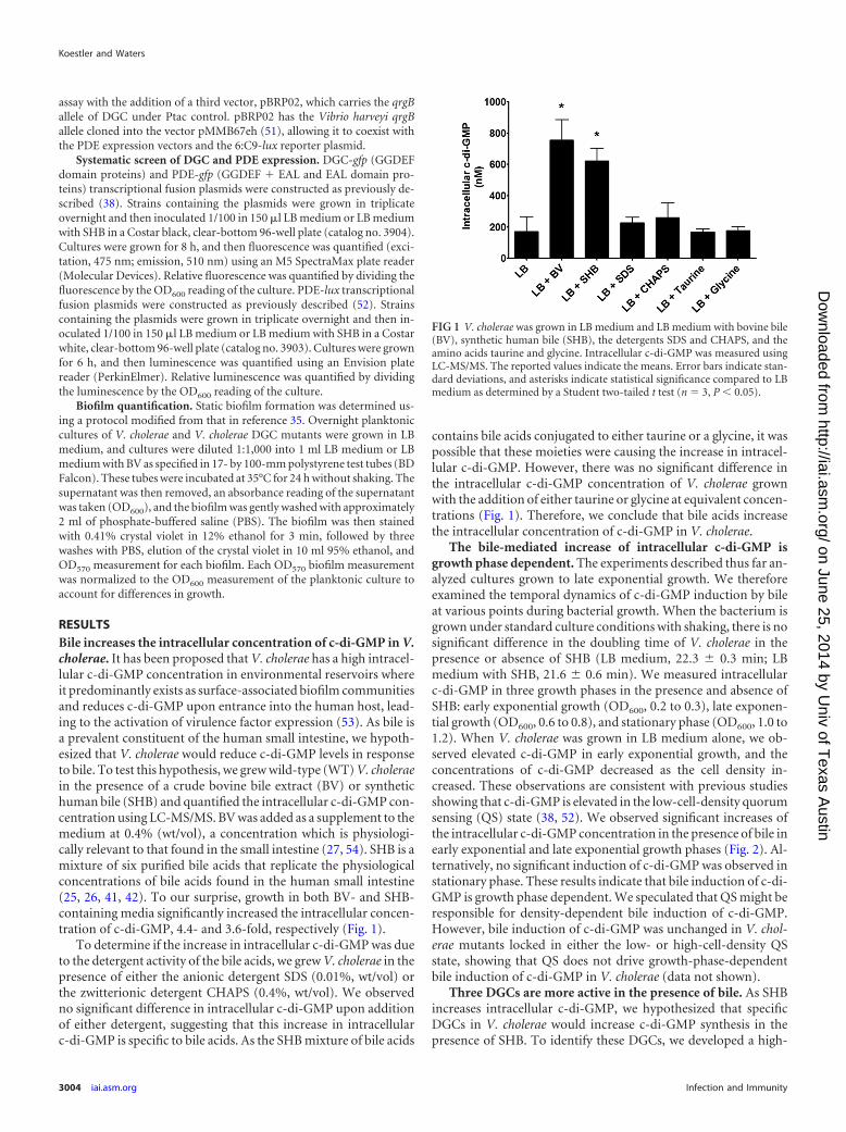
RESULTSBile increases the intracellular concentration of c-di-GMP in V.cholerae. It has been proposed that V. cholerae has a high intracel-lular c-di-GMP concentration in environmental reservoirs whereit predominantly exists as surface-associated biofilm communitiesand reduces c-di-GMP upon entrance into the human host, lead-ing to the activation of virulence factor expression (53). As bile isa prevalent constituent of the human small intestine, we hypoth-esized that V. cholerae would reduce c-di-GMP levels in responseto bile. To test this hypothesis, we grew wild-type (WT) V. choleraein the presence of a crude bovine bile extract (BV) or synthetichuman bile (SHB) and quantified the intracellular c-di-GMP con-centration using LC-MS/MS. BV was added as a supplement to themedium at 0.4% (wt/vol), a concentration which is physiologi-cally relevant to that found in the small intestine (27, 54). SHB is amixture of six purified bile acids that replicate the physiologicalconcentrations of bile acids found in the human small intestine(25, 26, 41, 42). To our surprise, growth in both BV- and SHB-containing media significantly increased the intracellular concen-tration of c-di-GMP, 4.4- and 3.6-fold, respectively (Fig. 1).
To determine if the increase in intracellular c-di-GMP was dueto the detergent activity of the bile acids, we grew V. cholerae in thepresence of either the anionic detergent SDS (0.01%, wt/vol) orthe zwitterionic detergent CHAPS (0.4%, wt/vol). We observedno significant difference in intracellular c-di-GMP upon additionof either detergent, suggesting that this increase in intracellularc-di-GMP is specific to bile acids. As the SHB mixture of bile acids
contains bile acids conjugated to either taurine or a glycine, it waspossible that these moieties were causing the increase in intracel-lular c-di-GMP. However, there was no significant difference inthe intracellular c-di-GMP concentration of V. cholerae grownwith the addition of either taurine or glycine at equivalent concen-trations (Fig. 1). Therefore, we conclude that bile acids increasethe intracellular concentration of c-di-GMP in V. cholerae.
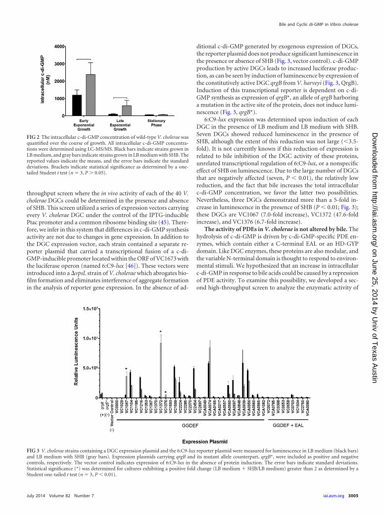
The bile-mediated increase of intracellular c-di-GMP isgrowth phase dependent. The experiments described thus far an-alyzed cultures grown to late exponential growth. We thereforeexamined the temporal dynamics of c-di-GMP induction by bileat various points during bacterial growth. When the bacterium isgrown under standard culture conditions with shaking, there is nosignificant difference in the doubling time of V. cholerae in thepresence or absence of SHB (LB medium, 22.3 � 0.3 min; LBmedium with SHB, 21.6 � 0.6 min). We measured intracellularc-di-GMP in three growth phases in the presence and absence ofSHB: early exponential growth (OD600, 0.2 to 0.3), late exponen-tial growth (OD600, 0.6 to 0.8), and stationary phase (OD600, 1.0 to1.2). When V. cholerae was grown in LB medium alone, we ob-served elevated c-di-GMP in early exponential growth, and theconcentrations of c-di-GMP decreased as the cell density in-creased. These observations are consistent with previous studiesshowing that c-di-GMP is elevated in the low-cell-density quorumsensing (QS) state (38, 52). We observed significant increases ofthe intracellular c-di-GMP concentration in the presence of bile inearly exponential and late exponential growth phases (Fig. 2). Al-ternatively, no significant induction of c-di-GMP was observed instationary phase. These results indicate that bile induction of c-di-GMP is growth phase dependent. We speculated that QS might beresponsible for density-dependent bile induction of c-di-GMP.However, bile induction of c-di-GMP was unchanged in V. chol-erae mutants locked in either the low- or high-cell-density QSstate, showing that QS does not drive growth-phase-dependentbile induction of c-di-GMP in V. cholerae (data not shown).
Three DGCs are more active in the presence of bile. As SHBincreases intracellular c-di-GMP, we hypothesized that specificDGCs in V. cholerae would increase c-di-GMP synthesis in thepresence of SHB. To identify these DGCs, we developed a high-
FIG 1 V. cholerae was grown in LB medium and LB medium with bovine bile(BV), synthetic human bile (SHB), the detergents SDS and CHAPS, and theamino acids taurine and glycine. Intracellular c-di-GMP was measured usingLC-MS/MS. The reported values indicate the means. Error bars indicate stan-dard deviations, and asterisks indicate statistical significance compared to LBmedium as determined by a Student two-tailed t test (n 3, P 0.05).
Koestler and Waters
3004 iai.asm.org Infection and Immunity
on June 25, 2014 by Univ of T
exas Austin
http://iai.asm.org/
Dow
nloaded from
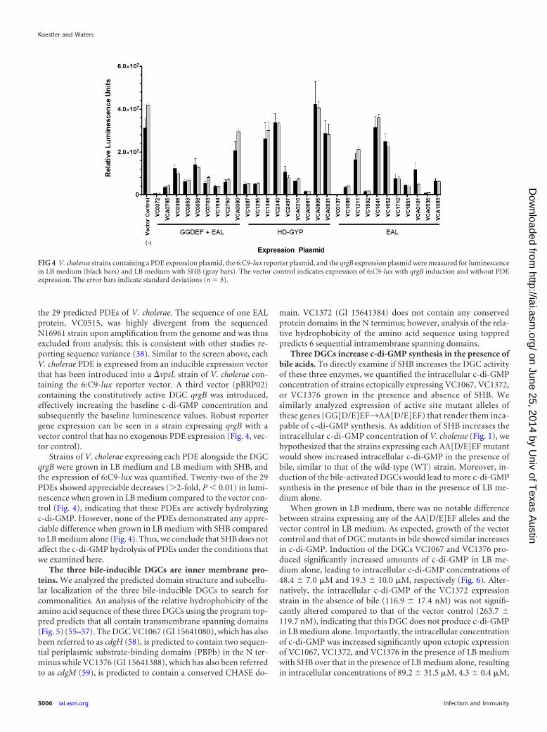
throughput screen where the in vivo activity of each of the 40 V.cholerae DGCs could be determined in the presence and absenceof SHB. This screen utilized a series of expression vectors carryingevery V. cholerae DGC under the control of the IPTG-induciblePtac promoter and a common ribosome binding site (45). There-fore, we infer in this system that differences in c-di-GMP synthesisactivity are not due to changes in gene expression. In addition tothe DGC expression vector, each strain contained a separate re-porter plasmid that carried a transcriptional fusion of a c-di-GMP-inducible promoter located within the ORF of VC1673 withthe luciferase operon (named 6:C9-lux [46]). These vectors wereintroduced into a �vpsL strain of V. cholerae which abrogates bio-film formation and eliminates interference of aggregate formationin the analysis of reporter gene expression. In the absence of ad-
ditional c-di-GMP generated by exogenous expression of DGCs,the reporter plasmid does not produce significant luminescence inthe presence or absence of SHB (Fig. 3, vector control). c-di-GMPproduction by active DGCs leads to increased luciferase produc-tion, as can be seen by induction of luminescence by expression ofthe constitutively active DGC qrgB from V. harveyi (Fig. 3, QrgB).Induction of this transcriptional reporter is dependent on c-di-GMP synthesis as expression of qrgB*, an allele of qrgB harboringa mutation in the active site of the protein, does not induce lumi-nescence (Fig. 3, qrgB*).
6:C9-lux expression was determined upon induction of eachDGC in the presence of LB medium and LB medium with SHB.Seven DGCs showed reduced luminescence in the presence ofSHB, although the extent of this reduction was not large (3.5-fold). It is not currently known if this reduction of expression isrelated to bile inhibition of the DGC activity of these proteins,unrelated transcriptional regulation of 6:C9-lux, or a nonspecificeffect of SHB on luminescence. Due to the large number of DGCsthat are negatively affected (seven, P 0.01), the relatively lowreduction, and the fact that bile increases the total intracellularc-di-GMP concentration, we favor the latter two possibilities.Nevertheless, three DGCs demonstrated more than a 5-fold in-crease in luminescence in the presence of SHB (P 0.01; Fig. 3);these DGCs are VC1067 (7.0-fold increase), VC1372 (47.6-foldincrease), and VC1376 (6.7-fold increase).
The activity of PDEs in V. cholerae is not altered by bile. Thehydrolysis of c-di-GMP is driven by c-di-GMP-specific PDE en-zymes, which contain either a C-terminal EAL or an HD-GYPdomain. Like DGC enzymes, these proteins are also modular, andthe variable N-terminal domain is thought to respond to environ-mental stimuli. We hypothesized that an increase in intracellularc-di-GMP in response to bile acids could be caused by a repressionof PDE activity. To examine this possibility, we developed a sec-ond high-throughput screen to analyze the enzymatic activity of
FIG 2 The intracellular c-di-GMP concentration of wild-type V. cholerae wasquantified over the course of growth. All intracellular c-di-GMP concentra-tions were determined using LC-MS/MS. Black bars indicate strains grown inLB medium, and gray bars indicate strains grown in LB medium with SHB. Thereported values indicate the means, and the error bars indicate the standarddeviations. Brackets indicate statistical significance as determined by a one-tailed Student t test (n 3, P � 0.05).
FIG 3 V. cholerae strains containing a DGC expression plasmid and the 6:C9-lux reporter plasmid were measured for luminescence in LB medium (black bars)and LB medium with SHB (gray bars). Expression plasmids carrying qrgB and its mutant allele counterpart, qrgB*, were included as positive and negativecontrols, respectively. The vector control indicates expression of 6:C9-lux in the absence of protein induction. The error bars indicate standard deviations.Statistical significance (*) was determined for cultures exhibiting a positive fold change (LB medium � SHB/LB medium) greater than 2 as determined by aStudent one-tailed t test (n 3, P 0.01).
Bile and Cyclic di-GMP in Vibrio cholerae
July 2014 Volume 82 Number 7 iai.asm.org 3005
on June 25, 2014 by Univ of T
exas Austin
http://iai.asm.org/
Dow
nloaded from
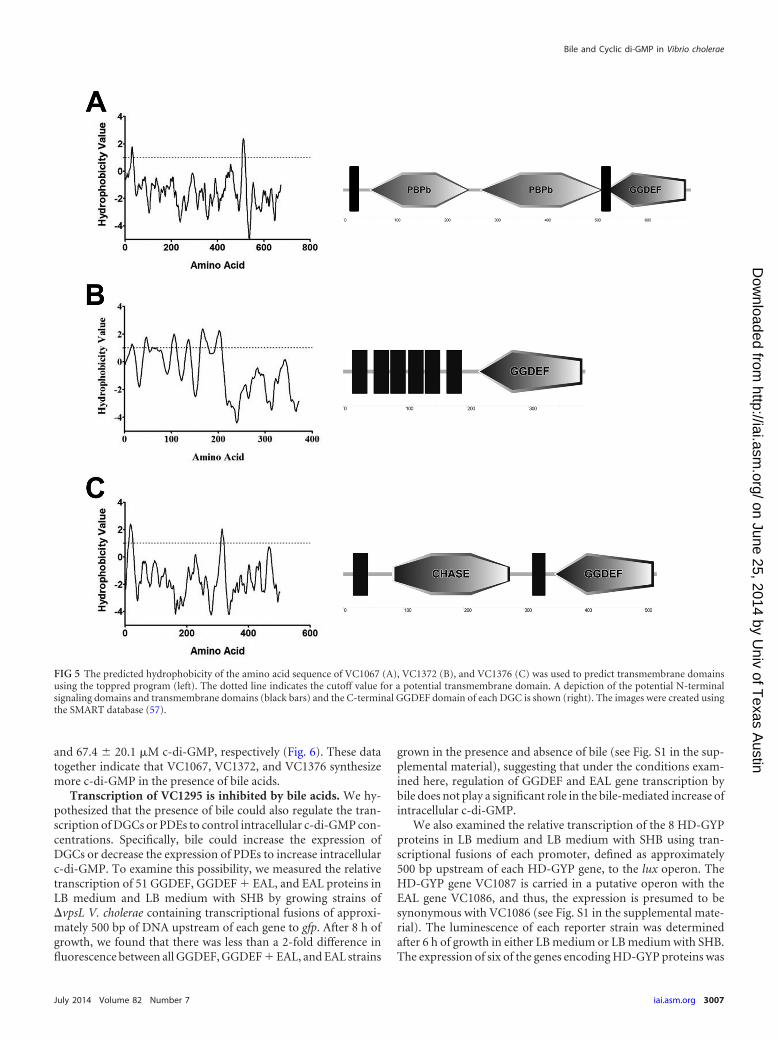
the 29 predicted PDEs of V. cholerae. The sequence of one EALprotein, VC0515, was highly divergent from the sequencedN16961 strain upon amplification from the genome and was thusexcluded from analysis; this is consistent with other studies re-porting sequence variance (38). Similar to the screen above, eachV. cholerae PDE is expressed from an inducible expression vectorthat has been introduced into a �vpsL strain of V. cholerae con-taining the 6:C9-lux reporter vector. A third vector (pBRP02)containing the constitutively active DGC qrgB was introduced,effectively increasing the baseline c-di-GMP concentration andsubsequently the baseline luminescence values. Robust reportergene expression can be seen in a strain expressing qrgB with avector control that has no exogenous PDE expression (Fig. 4, vec-tor control).
Strains of V. cholerae expressing each PDE alongside the DGCqrgB were grown in LB medium and LB medium with SHB, andthe expression of 6:C9-lux was quantified. Twenty-two of the 29PDEs showed appreciable decreases (�2-fold, P 0.01) in lumi-nescence when grown in LB medium compared to the vector con-trol (Fig. 4), indicating that these PDEs are actively hydrolyzingc-di-GMP. However, none of the PDEs demonstrated any appre-ciable difference when grown in LB medium with SHB comparedto LB medium alone (Fig. 4). Thus, we conclude that SHB does notaffect the c-di-GMP hydrolysis of PDEs under the conditions thatwe examined here.
The three bile-inducible DGCs are inner membrane pro-teins. We analyzed the predicted domain structure and subcellu-lar localization of the three bile-inducible DGCs to search forcommonalities. An analysis of the relative hydrophobicity of theamino acid sequence of these three DGCs using the program top-pred predicts that all contain transmembrane spanning domains(Fig. 5) (55–57). The DGC VC1067 (GI 15641080), which has alsobeen referred to as cdgH (58), is predicted to contain two sequen-tial periplasmic substrate-binding domains (PBPb) in the N ter-minus while VC1376 (GI 15641388), which has also been referredto as cdgM (59), is predicted to contain a conserved CHASE do-
main. VC1372 (GI 15641384) does not contain any conservedprotein domains in the N terminus; however, analysis of the rela-tive hydrophobicity of the amino acid sequence using toppredpredicts 6 sequential intramembrane spanning domains.
Three DGCs increase c-di-GMP synthesis in the presence ofbile acids. To directly examine if SHB increases the DGC activityof these three enzymes, we quantified the intracellular c-di-GMPconcentration of strains ectopically expressing VC1067, VC1372,or VC1376 grown in the presence and absence of SHB. Wesimilarly analyzed expression of active site mutant alleles ofthese genes (GG[D/E]EF¡AA[D/E]EF) that render them inca-pable of c-di-GMP synthesis. As addition of SHB increases theintracellular c-di-GMP concentration of V. cholerae (Fig. 1), wehypothesized that the strains expressing each AA[D/E]EF mutantwould show increased intracellular c-di-GMP in the presence ofbile, similar to that of the wild-type (WT) strain. Moreover, in-duction of the bile-activated DGCs would lead to more c-di-GMPsynthesis in the presence of bile than in the presence of LB me-dium alone.
When grown in LB medium, there was no notable differencebetween strains expressing any of the AA[D/E]EF alleles and thevector control in LB medium. As expected, growth of the vectorcontrol and that of DGC mutants in bile showed similar increasesin c-di-GMP. Induction of the DGCs VC1067 and VC1376 pro-duced significantly increased amounts of c-di-GMP in LB me-dium alone, leading to intracellular c-di-GMP concentrations of48.4 � 7.0 �M and 19.3 � 10.0 �M, respectively (Fig. 6). Alter-natively, the intracellular c-di-GMP of the VC1372 expressionstrain in the absence of bile (116.9 � 17.4 nM) was not signifi-cantly altered compared to that of the vector control (263.7 �119.7 nM), indicating that this DGC does not produce c-di-GMPin LB medium alone. Importantly, the intracellular concentrationof c-di-GMP was increased significantly upon ectopic expressionof VC1067, VC1372, and VC1376 in the presence of LB mediumwith SHB over that in the presence of LB medium alone, resultingin intracellular concentrations of 89.2 � 31.5 �M, 4.3 � 0.4 �M,
FIG 4 V. cholerae strains containing a PDE expression plasmid, the 6:C9-lux reporter plasmid, and the qrgB expression plasmid were measured for luminescencein LB medium (black bars) and LB medium with SHB (gray bars). The vector control indicates expression of 6:C9-lux with qrgB induction and without PDEexpression. The error bars indicate standard deviations (n 3).
Koestler and Waters
3006 iai.asm.org Infection and Immunity
on June 25, 2014 by Univ of T
exas Austin
http://iai.asm.org/
Dow
nloaded from
and 67.4 � 20.1 �M c-di-GMP, respectively (Fig. 6). These datatogether indicate that VC1067, VC1372, and VC1376 synthesizemore c-di-GMP in the presence of bile acids.
Transcription of VC1295 is inhibited by bile acids. We hy-pothesized that the presence of bile could also regulate the tran-scription of DGCs or PDEs to control intracellular c-di-GMP con-centrations. Specifically, bile could increase the expression ofDGCs or decrease the expression of PDEs to increase intracellularc-di-GMP. To examine this possibility, we measured the relativetranscription of 51 GGDEF, GGDEF � EAL, and EAL proteins inLB medium and LB medium with SHB by growing strains of�vpsL V. cholerae containing transcriptional fusions of approxi-mately 500 bp of DNA upstream of each gene to gfp. After 8 h ofgrowth, we found that there was less than a 2-fold difference influorescence between all GGDEF, GGDEF � EAL, and EAL strains
grown in the presence and absence of bile (see Fig. S1 in the sup-plemental material), suggesting that under the conditions exam-ined here, regulation of GGDEF and EAL gene transcription bybile does not play a significant role in the bile-mediated increase ofintracellular c-di-GMP.
We also examined the relative transcription of the 8 HD-GYPproteins in LB medium and LB medium with SHB using tran-scriptional fusions of each promoter, defined as approximately500 bp upstream of each HD-GYP gene, to the lux operon. TheHD-GYP gene VC1087 is carried in a putative operon with theEAL gene VC1086, and thus, the expression is presumed to besynonymous with VC1086 (see Fig. S1 in the supplemental mate-rial). The luminescence of each reporter strain was determinedafter 6 h of growth in either LB medium or LB medium with SHB.The expression of six of the genes encoding HD-GYP proteins was
FIG 5 The predicted hydrophobicity of the amino acid sequence of VC1067 (A), VC1372 (B), and VC1376 (C) was used to predict transmembrane domainsusing the toppred program (left). The dotted line indicates the cutoff value for a potential transmembrane domain. A depiction of the potential N-terminalsignaling domains and transmembrane domains (black bars) and the C-terminal GGDEF domain of each DGC is shown (right). The images were created usingthe SMART database (57).
Bile and Cyclic di-GMP in Vibrio cholerae
July 2014 Volume 82 Number 7 iai.asm.org 3007
on June 25, 2014 by Univ of T
exas Austin
http://iai.asm.org/
Dow
nloaded from

not significantly changed in LB medium with SHB from that in LBmedium alone (�2-fold), whereas the expression of VC2497 wasmodestly increased in the presence of SHB (2.2-fold, P 0.05).Importantly, the expression of VC1295 (GI 15641308) was de-creased 2.8-fold when grown in the presence of SHB (Fig. 7; P 0.05). VC1295 appears to be an active PDE when ectopicallyexpressed in V. cholerae (Fig. 4). This result indicates that bileacids decrease the expression of VC1295, possibly resulting in de-creased c-di-GMP hydrolysis contributing to increased intracel-lular c-di-GMP.
Three DGCs and one HD-GYP account for bile-mediatedc-di-GMP induction. To determine if the DGCs VC1067, VC1372,and VC1376 contribute to the bile-mediated increase of intracel-lular c-di-GMP in V. cholerae, we constructed unmarked V. chol-erae DGC deletion mutants and examined SHB-mediated induc-tion of c-di-GMP (Fig. 8). Similar to our previous findings, theintracellular c-di-GMP concentration of V. cholerae increased 4.1-fold in the presence of SHB in the WT strain. Both the �VC1067and �VC1376 single mutants showed a modest but significantreduction of c-di-GMP when grown in bile compared to the WTstrain, having 29.6% and 24.9% less c-di-GMP, respectively. The�VC1372 single mutant and �VC1372 �1376 double mutantwere not statistically different from the WT strain. Importantly,the �VC1067 �VC1372 �VC1376 triple mutant strain exhibitedthe greatest reduction of intracellular c-di-GMP in the presence ofbile, losing 46.8% of the intracellular c-di-GMP compared to theWT strain in LB medium with SHB (P 0.05). These resultssuggest that VC1067, VC1372, and VC1376 function redundantlyin the bile-mediated c-di-GMP induction.
As the expression of the HD-GYP VC1295 was inhibited by bileacids, we constructed an unmarked V. cholerae VC1295 deletionmutant and quantified intracellular c-di-GMP in the presence andabsence of SHB. In LB medium alone, the intracellular c-di-GMPwas modestly increased 1.8-fold in the �VC1295 mutant com-pared to the WT (Fig. 8; P 0.05). This result is expected, asdeletion of an active PDE will increase intracellular c-di-GMP.However, the intracellular c-di-GMP concentrations of the�VC1295 strain grown in the presence of SHB were indistinguish-able from those of the WT. We hypothesized that both activation
of DGC activity and transcriptional regulation of VC1295 con-tribute to bile induction of c-di-GMP. To test this, we created aquadruple �VC1295 �VC1067 �VC1372 �VC1376 mutant andmeasured intracellular c-di-GMP in the presence and absence ofSHB. Similar to the �VC1295 single mutant, when grown in LBmedium alone, the quadruple mutant had elevated intracellularc-di-GMP compared to the WT strain (2.4-fold, P 0.05). Im-portantly, this strain showed no change in intracellular c-di-GMP in the presence of SHB. This indicates that these fourproteins are responsible for the bile-mediated changes in intra-cellular c-di-GMP.
Deletion of the bile-responsive DGCs and PDE reduces bileinduction of V. cholerae biofilm formation. It has been previ-ously reported that BV (i.e., bovine bile) increases biofilm forma-tion of V. cholerae, and this induction is dependent on the tran-scriptional regulator vpsR (35). VpsR binds c-di-GMP to regulatethe transcription of biofilm genes (46). We wondered if the levelsof c-di-GMP measured in the various DGC and PDE mutantstrains with and without bile would correlate with biofilm forma-tion. To test this, we performed a static biofilm assay where thewild-type, �vpsL mutant, and the DGC and PDE V. cholerae mu-tant strains were grown in polystyrene test tubes containing LBmedium or LB medium with BV without shaking followed bycrystal violet staining of the resulting attached biofilm. The �vpsLmutant cannot produce exopolysaccharide and thus does notform biofilms. BV was used to induce biofilm formation to remainconsistent with prior studies (35, 60) and because it induced morerobust biofilm formation in this assay than did SHB (data notshown).
We observed that all cultures of V. cholerae grew to a signifi-cantly lower optical density, as measured by OD600, after staticgrowth in the presence of BV. To account for these growth differ-ences, the biofilm formation of each culture was normalized to theOD600 of the planktonic culture. Consistent with previous reports(35), BV increased biofilm formation in the WT strain 2.7-fold,and this response was eliminated in the �vpsL strain (Fig. 9).
FIG 6 Intracellular levels of c-di-GMP in V. cholerae expressing an emptyvector or DGC VC1067, VC1372, or VC1376 quantified with LC-MS/MS. Theintracellular c-di-GMP levels of strains expressing alleles containing mutationsin the active site motif of each DGC were also quantified. The black barsindicate strains grown in LB medium, while the gray bars indicate strainsgrown in LB medium with SHB. Error bars indicate standard deviations.Brackets indicate statistical significance, which was determined using a Stu-dent one-tailed t test (n 3, P 0.05).
FIG 7 V. cholerae strains containing a transcriptional fusion of each HD-GYPpromoter to luciferase were grown in LB medium (black) or LB medium withSHB (gray). Luminescence was quantified after 6 h of growth (n 4) undereach environmental condition. Each culture was normalized to an OD600 read-ing. Error bars indicate standard deviations. The asterisk indicates a statisti-cally significant differences from the LB medium condition, determined by aone-tailed Student t test (P 0.05).
Koestler and Waters
3008 iai.asm.org Infection and Immunity
on June 25, 2014 by Univ of T
exas Austin
http://iai.asm.org/
Dow
nloaded from
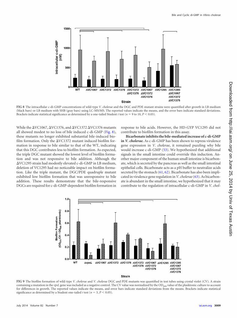
While the �VC1067, �VC1376, and �VC1372 �VC1376 mutantsall showed modest to no loss of bile induced c-di-GMP (Fig. 8),these mutants no longer exhibited substantial bile-induced bio-film formation. Only the �VC1372 mutant induced biofilm for-mation in response to bile similar to that of the WT, indicatingthat this DGC contributes less to biofilm formation. As expected,the triple DGC mutant showed the lowest level of biofilm forma-tion and was not responsive to bile addition. Although the�VC1295 strain had modestly elevated c-di-GMP in LB medium,deletion of VC1295 had no noticeable impact on biofilm forma-tion. Like the triple mutant, the DGC/PDE quadruple mutantexhibited low biofilm formation that was unresponsive to bileaddition. These results demonstrate that the bile-responsiveDGCs are required for c-di-GMP-dependent biofilm formation in
response to bile acids. However, the HD-GYP VC1295 did notcontribute to biofilm formation in this assay.
Bicarbonate inhibits the bile-mediated increase of c-di-GMPin V. cholerae. As c-di-GMP has been shown to repress virulencegene expression in V. cholerae, it remained puzzling why bilewould increase c-di-GMP (53). We hypothesized that additionalsignals in the small intestine could override this induction. An-other major component of the human small intestine is bicarbon-ate, which is secreted by the pancreas as well as the small intestinalepithelial cells. Bicarbonate acts as a pH buffer to neutralize acidssecreted by the stomach (61, 62). Bicarbonate has also been impli-cated in virulence gene regulation in V. cholerae (63). As bicarbon-ate is abundant in the small intestine, we hypothesized that it maycontribute to the regulation of intracellular c-di-GMP in V. chol-
FIG 8 The intracellular c-di-GMP concentrations of wild-type V. cholerae and the DGC and PDE mutant strains were quantified after growth in LB medium(black bars) or LB medium with SHB (gray bars) using LC-MS/MS. The reported values indicate the means, and the error bars indicate standard deviations.Brackets indicate statistical significance as determined by a one-tailed Student t test (n 9 to 10, P 0.05).
FIG 9 The biofilm formation of wild-type V. cholerae and V. cholerae DGC and PDE mutants was quantified in test tubes using crystal violet (CV). A straincontaining a mutation in the vpsL gene was included as a negative control. The CV value was normalized by the OD600 value of the planktonic culture to accountfor differences in growth. The reported values indicate the means, and error bars indicate standard deviations from the means. Brackets indicate statisticalsignificance as determined by a Student one-tailed t test (n 3, P 0.05).
Bile and Cyclic di-GMP in Vibrio cholerae
July 2014 Volume 82 Number 7 iai.asm.org 3009
on June 25, 2014 by Univ of T
exas Austin
http://iai.asm.org/
Dow
nloaded from
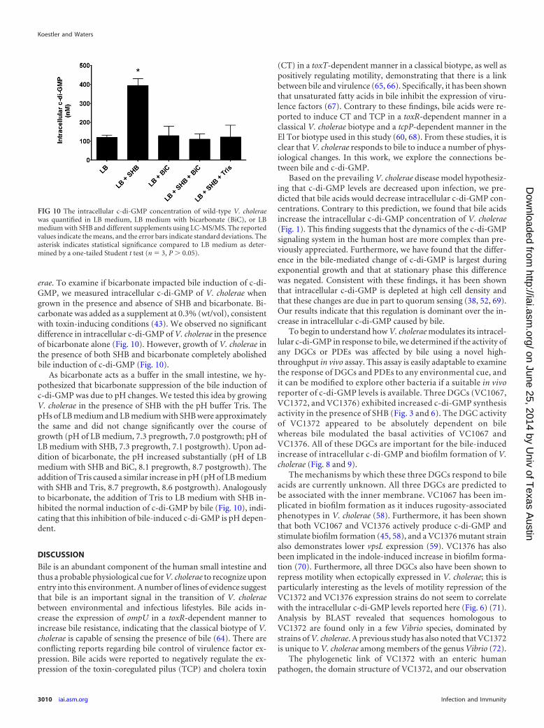
erae. To examine if bicarbonate impacted bile induction of c-di-GMP, we measured intracellular c-di-GMP of V. cholerae whengrown in the presence and absence of SHB and bicarbonate. Bi-carbonate was added as a supplement at 0.3% (wt/vol), consistentwith toxin-inducing conditions (43). We observed no significantdifference in intracellular c-di-GMP of V. cholerae in the presenceof bicarbonate alone (Fig. 10). However, growth of V. cholerae inthe presence of both SHB and bicarbonate completely abolishedbile induction of c-di-GMP (Fig. 10).
As bicarbonate acts as a buffer in the small intestine, we hy-pothesized that bicarbonate suppression of the bile induction ofc-di-GMP was due to pH changes. We tested this idea by growingV. cholerae in the presence of SHB with the pH buffer Tris. ThepHs of LB medium and LB medium with SHB were approximatelythe same and did not change significantly over the course ofgrowth (pH of LB medium, 7.3 pregrowth, 7.0 postgrowth; pH ofLB medium with SHB, 7.3 pregrowth, 7.1 postgrowth). Upon ad-dition of bicarbonate, the pH increased substantially (pH of LBmedium with SHB and BiC, 8.1 pregrowth, 8.7 postgrowth). Theaddition of Tris caused a similar increase in pH (pH of LB mediumwith SHB and Tris, 8.7 pregrowth, 8.6 postgrowth). Analogouslyto bicarbonate, the addition of Tris to LB medium with SHB in-hibited the normal induction of c-di-GMP by bile (Fig. 10), indi-cating that this inhibition of bile-induced c-di-GMP is pH depen-dent.
DISCUSSION
Bile is an abundant component of the human small intestine andthus a probable physiological cue for V. cholerae to recognize uponentry into this environment. A number of lines of evidence suggestthat bile is an important signal in the transition of V. choleraebetween environmental and infectious lifestyles. Bile acids in-crease the expression of ompU in a toxR-dependent manner toincrease bile resistance, indicating that the classical biotype of V.cholerae is capable of sensing the presence of bile (64). There areconflicting reports regarding bile control of virulence factor ex-pression. Bile acids were reported to negatively regulate the ex-pression of the toxin-coregulated pilus (TCP) and cholera toxin
(CT) in a toxT-dependent manner in a classical biotype, as well aspositively regulating motility, demonstrating that there is a linkbetween bile and virulence (65, 66). Specifically, it has been shownthat unsaturated fatty acids in bile inhibit the expression of viru-lence factors (67). Contrary to these findings, bile acids were re-ported to induce CT and TCP in a toxR-dependent manner in aclassical V. cholerae biotype and a tcpP-dependent manner in theEl Tor biotype used in this study (60, 68). From these studies, it isclear that V. cholerae responds to bile to induce a number of phys-iological changes. In this work, we explore the connections be-tween bile and c-di-GMP.
Based on the prevailing V. cholerae disease model hypothesiz-ing that c-di-GMP levels are decreased upon infection, we pre-dicted that bile acids would decrease intracellular c-di-GMP con-centrations. Contrary to this prediction, we found that bile acidsincrease the intracellular c-di-GMP concentration of V. cholerae(Fig. 1). This finding suggests that the dynamics of the c-di-GMPsignaling system in the human host are more complex than pre-viously appreciated. Furthermore, we have found that the differ-ence in the bile-mediated change of c-di-GMP is largest duringexponential growth and that at stationary phase this differencewas negated. Consistent with these findings, it has been shownthat intracellular c-di-GMP is depleted at high cell density andthat these changes are due in part to quorum sensing (38, 52, 69).Our results indicate that this regulation is dominant over the in-crease in intracellular c-di-GMP caused by bile.
To begin to understand how V. cholerae modulates its intracel-lular c-di-GMP in response to bile, we determined if the activity ofany DGCs or PDEs was affected by bile using a novel high-throughput in vivo assay. This assay is easily adaptable to examinethe response of DGCs and PDEs to any environmental cue, andit can be modified to explore other bacteria if a suitable in vivoreporter of c-di-GMP levels is available. Three DGCs (VC1067,VC1372, and VC1376) exhibited increased c-di-GMP synthesisactivity in the presence of SHB (Fig. 3 and 6). The DGC activityof VC1372 appeared to be absolutely dependent on bilewhereas bile modulated the basal activities of VC1067 andVC1376. All of these DGCs are important for the bile-inducedincrease of intracellular c-di-GMP and biofilm formation of V.cholerae (Fig. 8 and 9).
The mechanisms by which these three DGCs respond to bileacids are currently unknown. All three DGCs are predicted tobe associated with the inner membrane. VC1067 has been im-plicated in biofilm formation as it induces rugosity-associatedphenotypes in V. cholerae (58). Furthermore, it has been shownthat both VC1067 and VC1376 actively produce c-di-GMP andstimulate biofilm formation (45, 58), and a VC1376 mutant strainalso demonstrates lower vpsL expression (59). VC1376 has alsobeen implicated in the indole-induced increase in biofilm forma-tion (70). Furthermore, all three DGCs also have been shown torepress motility when ectopically expressed in V. cholerae; this isparticularly interesting as the levels of motility repression of theVC1372 and VC1376 expression strains do not seem to correlatewith the intracellular c-di-GMP levels reported here (Fig. 6) (71).Analysis by BLAST revealed that sequences homologous toVC1372 are found only in a few Vibrio species, dominated bystrains of V. cholerae. A previous study has also noted that VC1372is unique to V. cholerae among members of the genus Vibrio (72).
The phylogenetic link of VC1372 with an enteric humanpathogen, the domain structure of VC1372, and our observation
FIG 10 The intracellular c-di-GMP concentration of wild-type V. choleraewas quantified in LB medium, LB medium with bicarbonate (BiC), or LBmedium with SHB and different supplements using LC-MS/MS. The reportedvalues indicate the means, and the error bars indicate standard deviations. Theasterisk indicates statistical significance compared to LB medium as deter-mined by a one-tailed Student t test (n 3, P � 0.05).
Koestler and Waters
3010 iai.asm.org Infection and Immunity
on June 25, 2014 by Univ of T
exas Austin
http://iai.asm.org/
Dow
nloaded from
that the activity of VC1372 is absolutely dependent on the pres-ence of bile suggest that the physiological cue which controlsVC1372 is bile. Bile acids are known to interact with the cell mem-brane due to their detergent activity (29). Moreover, we observedbile-mediated activation of VC1372 in E. coli (data not shown), anorthologous system with no clear homolog to VC1372. Alterna-tively, as both VC1067 and VC1376 maintain robust activity uponexogenous expression even in the absence of bile, we postulate thatthese DGCs might be controlled indirectly by bile through sensingperturbations in the membrane.
Additionally, the expression of the PDE VC1295 was inhib-ited by bile acids (Fig. 7). This HD-GYP actively hydrolyzesc-di-GMP in both LB medium and LB medium with SHB, butbile acids do not affect this activity (Fig. 4). VC1295 is pre-dicted to be composed of 492 amino acids and contains 6 pre-dicted N-terminal transmembrane domains preceding a HAMP do-main linked to the HD-GYP domain (55–57). The mechanismgoverning this transcriptional regulation of VC1295 by bile remainsunknown. Analysis of the VC1295 promoter region reveals motifsresembling the ToxR binding site (�127 bp, TCAAA-N11-TTAAA[73]). While this gene is not listed among the known genes regu-lated by ToxR (74), there is evidence that the activity of ToxR isaltered by bile (60), suggesting that the ToxR regulon could bealtered when bile acids are present. We are currently investigatingthe connection between the transcriptional regulation of VC1295,bile, and ToxR. Although VC1295 contributed to bile-inducedc-di-GMP, we did not observe any effect of VC1295 on biofilmformation. We speculate that this result is due to the distinct ex-perimental growth conditions under which c-di-GMP and bio-film formation were measured.
Another important host-derived cue is bicarbonate, a biologi-cal pH buffer that is highly abundant in the human small intestine(62). Bicarbonate has been shown to be important for virulence, asbicarbonate is critical for in vitro production of CT (43). Bicar-bonate is capable of activating V. cholerae virulence gene expres-sion via the transcriptional regulator toxT (63). We have shownthat bicarbonate is able to suppress the bile-mediated induction ofc-di-GMP in V. cholerae. Furthermore, this regulation is driven bychanges in pH, as the bile-mediated induction is similarly re-
pressed by Tris (Fig. 10). It is possible that the change in pH altersthe structure of bile so that it no longer triggers the c-di-GMPsynthesis activity of the DGCs. Alternatively, the bicarbonatecould directly interact with DGCs or PDEs to competitively alterintracellular c-di-GMP.
As bile has strong antimicrobial properties (75), it may bephysiologically advantageous for V. cholerae to increase c-di-GMPto promote biofilm formation in order to grant elevated toleranceto bile acids and other stresses associated with the intestinal envi-ronment. Other studies have indicated that biofilm formation isimportant for increased acid shock tolerance and protection frombile acids (35, 76), and biofilms increase infectivity and intestinalcolonization in a mouse infection model, which has implicationsfor transmission (77). Indeed, we confirmed that BV induces bio-film formation in V. cholerae and showed that all three bile-re-sponsive DGCs were required for bile induction of biofilm forma-tion.
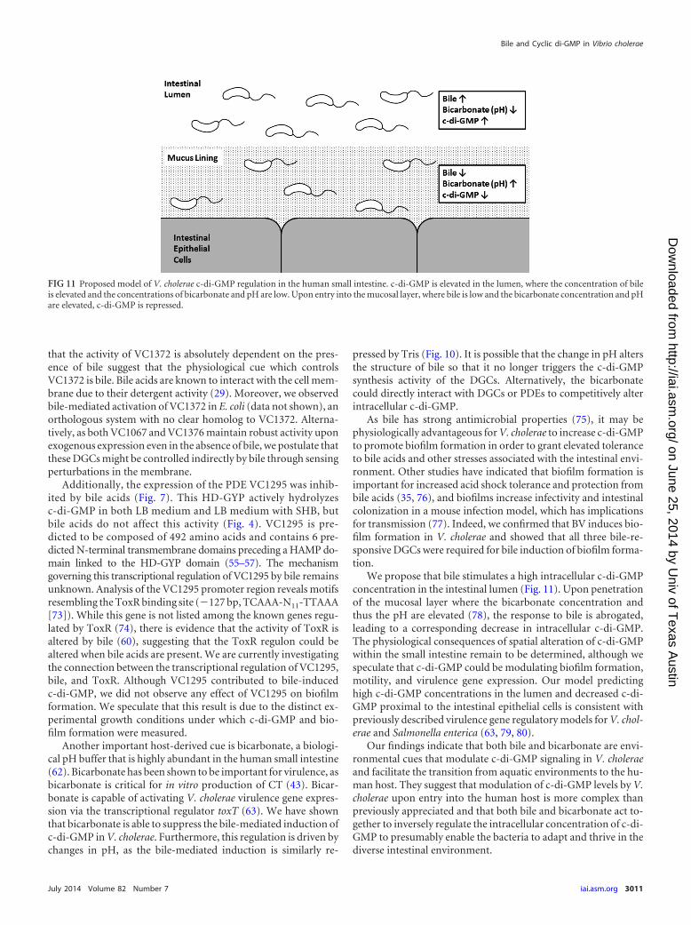
We propose that bile stimulates a high intracellular c-di-GMPconcentration in the intestinal lumen (Fig. 11). Upon penetrationof the mucosal layer where the bicarbonate concentration andthus the pH are elevated (78), the response to bile is abrogated,leading to a corresponding decrease in intracellular c-di-GMP.The physiological consequences of spatial alteration of c-di-GMPwithin the small intestine remain to be determined, although wespeculate that c-di-GMP could be modulating biofilm formation,motility, and virulence gene expression. Our model predictinghigh c-di-GMP concentrations in the lumen and decreased c-di-GMP proximal to the intestinal epithelial cells is consistent withpreviously described virulence gene regulatory models for V. chol-erae and Salmonella enterica (63, 79, 80).
Our findings indicate that both bile and bicarbonate are envi-ronmental cues that modulate c-di-GMP signaling in V. choleraeand facilitate the transition from aquatic environments to the hu-man host. They suggest that modulation of c-di-GMP levels by V.cholerae upon entry into the human host is more complex thanpreviously appreciated and that both bile and bicarbonate act to-gether to inversely regulate the intracellular concentration of c-di-GMP to presumably enable the bacteria to adapt and thrive in thediverse intestinal environment.
FIG 11 Proposed model of V. cholerae c-di-GMP regulation in the human small intestine. c-di-GMP is elevated in the lumen, where the concentration of bileis elevated and the concentrations of bicarbonate and pH are low. Upon entry into the mucosal layer, where bile is low and the bicarbonate concentration and pHare elevated, c-di-GMP is repressed.
Bile and Cyclic di-GMP in Vibrio cholerae
July 2014 Volume 82 Number 7 iai.asm.org 3011
on June 25, 2014 by Univ of T
exas Austin
http://iai.asm.org/
Dow
nloaded from
ACKNOWLEDGMENTS
This work was supported by NIH grants K22AI080937 and U19AI090872,membership within and support from the Region V “Great Lakes” RCE(NIH award 2U54AI057153), NSF grant MCB1253684, the NSF-spon-sored BEACON Science and Technology Center (cooperative agreementno. DBI-0939454), and Michigan State University to C.M.W. We alsoacknowledge the Rudolph Hugh Fellowship and the Russell B. DuVallScholarship to B.J.K.
We thank the MSU Mass Spectrometry Facility for assistance in mea-suring c-di-GMP. We also thank Robert Britton and Kristi Whitehead forproviding the mixture for SHB; Benjamin Pursley and Carolyn Chan forthe construction of pBRP2 and pANDA2, respectively; and Brian Ham-mer for generous sharing of the HD-GYP lux transcriptional fusions.
REFERENCES1. Halpern M, Broza YB, Mittler S, Arakawa E, Broza M. 2004. Chirono-
mid egg masses as a natural reservoir of Vibrio cholerae non-O1 and non-O139 in freshwater habitats. Microb. Ecol. 47:341–349. http://dx.doi.org/10.1007/s00248-003-2007-6.
2. Huq A, Small EB, West PA, Huq MI, Rahman R, Colwell RR. 1983.Ecological relationships between Vibrio cholerae and planktonic crusta-cean copepods. Appl. Environ. Microbiol. 45:275–283.
3. Tamplin ML, Gauzens AL, Huq A, Sack DA, Colwell RR. 1990. Attach-ment of Vibrio cholerae serogroup-O1 to zooplankton and phytoplanktonof Bangladesh waters. Appl. Environ. Microbiol. 56:1977–1980.
4. Tischler AD, Camilli A. 2004. Cyclic diguanylate (c-di-GMP) regulatesVibrio cholerae biofilm formation. Mol. Microbiol. 53:857– 869. http://dx.doi.org/10.1111/j.1365-2958.2004.04155.x.
5. Simm R, Morr M, Kader A, Nimtz M, Romling U. 2004. GGDEF andEAL domains inversely regulate cyclic di-GMP levels and transition fromsessility to motility. Mol. Microbiol. 53:1123–1134. http://dx.doi.org/10.1111/j.1365-2958.2004.04206.x.
6. Boehm A, Kaiser M, Li H, Spangler C, Kasper CA, Ackermann M,Kaever V, Sourjik V, Roth V, Jenal U. 2010. Second messenger-mediatedadjustment of bacterial swimming velocity. Cell 141:107–116. http://dx.doi.org/10.1016/j.cell.2010.01.018.
7. Kulasakara H, Lee V, Brencic A, Liberati N, Urbach J, Miyata S, LeeDG, Neely AN, Hyodo M, Hayakawa Y, Ausubel FM, Lory S. 2006.Analysis of Pseudomonas aeruginosa diguanylate cyclases and phosphodi-esterases reveals a role for bis-(3=-5=)-cyclic-GMP in virulence. Proc.Natl. Acad. Sci. U. S. A. 103:2839 –2844. http://dx.doi.org/10.1073/pnas.0511090103.
8. Hecht GB, Newton A. 1995. Identification of a novel response regulatorrequired for the swarmer-to-stalked-cell transition in Caulobacter crescen-tus. J. Bacteriol. 177:6223– 6229.
9. Aldridge P, Paul R, Goymer P, Rainey P, Jenal U. 2003. Role of theGGDEF regulator PleD in polar development of Caulobacter crescentus.Mol. Microbiol. 47:1695–1708. http://dx.doi.org/10.1046/j.1365-2958.2003.03401.x.
10. Tischler AD, Camilli A. 2005. Cyclic diguanylate regulates Vibrio choleraevirulence gene expression. Infect. Immun. 73:5873–5882. http://dx.doi.org/10.1128/IAI.73.9.5873-5882.2005.
11. Galperin MY. 2004. Bacterial signal transduction network in a genomicperspective. Environ. Microbiol. 6:552–567. http://dx.doi.org/10.1111/j.1462-2920.2004.00633.x.
12. Ryan RP, Fouhy Y, Lucey JF, Dow JM. 2006. Cyclic di-GMP signaling inbacteria: recent advances and new puzzles. J. Bacteriol. 188:8327– 8334.http://dx.doi.org/10.1128/JB.01079-06.
13. Schmidt AJ, Ryjenkov DA, Gomelsky M. 2005. The ubiquitous proteindomain EAL is a cyclic diguanylate-specific phosphodiesterase: enzymat-ically active and inactive EAL domains. J. Bacteriol. 187:4774 – 4781. http://dx.doi.org/10.1128/JB.187.14.4774-4781.2005.
14. Ryan RP, Fouhy Y, Lucey JF, Crossman LC, Spiro S, He YW, Zhang LH,Heeb S, Camara M, Williams P, Dow JM. 2006. Cell-cell signaling inXanthomonas campestris involves an HD-GYP domain protein that func-tions in cyclic di-GMP turnover. Proc. Natl. Acad. Sci. U. S. A. 103:6712–6717. http://dx.doi.org/10.1073/pnas.0600345103.
15. Galperin MY, Nikolskaya AN, Koonin EV. 2001. Novel domains ofthe prokaryotic two-component signal transduction systems. FEMSMicrobiol. Lett. 203:11–21. http://dx.doi.org/10.1111/j.1574-6968.2001.tb10814.x.
16. Bernier SP, Ha DG, Khan W, Merritt JH, O’Toole GA. 2011. Modula-tion of Pseudomonas aeruginosa surface-associated group behaviors byindividual amino acids through c-di-GMP signaling. Res. Microbiol. 162:680 – 688. http://dx.doi.org/10.1016/j.resmic.2011.04.014.
17. Carlson HK, Vance RE, Marletta MA. 2010. H-NOX regulation ofc-di-GMP metabolism and biofilm formation in Legionella pneumo-phila. Mol. Microbiol. 77:930 –942. http://dx.doi.org/10.1111/j.1365-2958.2010.07259.x.
18. Zähringer F, Lacanna E, Jenal U, Schirmer T, Boehm A. 13 June 2013.Structure and signaling mechanism of a zinc-sensory diguanylate cyclase.Structure http://dx.doi.org/10.1016/j.str.2013.04.026.
19. Kanazawa T, Ren S, Maekawa M, Hasegawa K, Arisaka F, Hyodo M,Hayakawa Y, Ohta H, Masuda S. 2010. Biochemical and physiologicalcharacterization of a BLUF protein-EAL protein complex involved in bluelight-dependent degradation of cyclic diguanylate in the purple bacteriumRhodopseudomonas palustris. Biochemistry 49:10647–10655. http://dx.doi.org/10.1021/bi101448t.
20. Ryan RP, McCarthy Y, Andrade M, Farah CS, Armitage JP, Dow JM.2010. Cell-cell signal-dependent dynamic interactions between HD-GYPand GGDEF domain proteins mediate virulence in Xanthomonas campes-tris. Proc. Natl. Acad. Sci. U. S. A. 107:5989 –5994. http://dx.doi.org/10.1073/pnas.0912839107.
21. Trimble MJ, McCarter LL. 2011. Bis-(3=-5=)-cyclic dimeric GMP-linkedquorum sensing controls swarming in Vibrio parahaemolyticus. Proc.Natl. Acad. Sci. U. S. A. 108:18079 –18084. http://dx.doi.org/10.1073/pnas.1113790108.
22. Tuckerman JR, Gonzalez G, Sousa EHS, Wan XH, Saito JA, Alam M,Gilles-Gonzalez MA. 2009. An oxygen-sensing diguanylate cyclase andphosphodiesterase couple for c-di-GMP control. Biochemistry 48:9764 –9774. http://dx.doi.org/10.1021/bi901409g.
23. Wan XH, Tuckerman JR, Saito JA, Freitas TAK, Newhouse JS, DeneryJR, Galperin MY, Gonzalez G, Gilles-Gonzalez MA, Alam M. 2009.Globins synthesize the second messenger bis-(3=-5=)-cyclic diguanosinemonophosphate in bacteria. J. Mol. Biol. 388:262–270. http://dx.doi.org/10.1016/j.jmb.2009.03.015.
24. Karatan E, Duncan TR, Watnick PI. 2005. NspS, a predicted polyaminesensor, mediates activation of Vibrio cholerae biofilm formation by nor-spermidine. J. Bacteriol. 187:7434 –7443. http://dx.doi.org/10.1128/JB.187.21.7434-7443.2005.
25. Dawson PA. 2010. Bile secretion and the enterohepatic circulation, p1075–1088. In Feldman M, Friedman LS, Brandt LJ (ed), Sleisenger andFordtran’s gastrointestinal and liver disease (9th), vol 1. WB Saunders,Philadelphia, PA.
26. O’Connor CJ, Wallace RG. 1985. Physicochemical behavior of bile-salts.Adv. Colloid Interface Sci. 22:1–111. http://dx.doi.org/10.1016/0001-8686(85)80002-6.
27. Begley M, Gahan CGM, Hill C. 2005. The interaction between bacteriaand bile. FEMS Microbiol. Rev. 29:625– 651. http://dx.doi.org/10.1016/j.femsre.2004.09.003.
28. Bernstein C, Bernstein H, Payne CM, Beard SE, Schneider J. 1999. Bilesalt activation of stress response promoters in Escherichia coli. Curr. Mi-crobiol. 39:68 –72. http://dx.doi.org/10.1007/s002849900420.
29. Schubert R, Schmidt KH. 1988. Structural changes in vesicle mem-branes and mixed micelles of various lipid compositions after bindingof different bile salts. Biochemistry 27:8787– 8794. http://dx.doi.org/10.1021/bi00424a015.
30. Cabral DJ, Small DM, Lilly HS, Hamilton JA. 1987. Transbilayer move-ment of bile-acids in model membranes. Biochemistry 26:1801–1804.http://dx.doi.org/10.1021/bi00381a002.
31. Provenzano D, Schuhmacher DA, Barker JL, Klose KE. 2000. Thevirulence regulatory protein ToxR mediates enhanced bile resistance inVibrio cholerae and other pathogenic Vibrio species. Infect. Immun. 68:1491–1497. http://dx.doi.org/10.1128/IAI.68.3.1491-1497.2000.
32. Colmer JA, Fralick JA, Hamood AN. 1998. Isolation and character-ization of a putative multidrug resistance pump from Vibrio cholerae.Mol. Microbiol. 27:63–72. http://dx.doi.org/10.1046/j.1365-2958.1998.00657.x.
33. Bina JE, Mekalanos JJ. 2001. Vibrio cholerae tolC is required for bileresistance and colonization. Infect. Immun. 69:4681– 4685. http://dx.doi.org/10.1128/IAI.69.7.4681-4685.2001.
34. Bina XR, Provenzano D, Nguyen N, Bina JE. 2008. Vibrio cholerae RNDfamily efflux systems are required for antimicrobial resistance, optimalvirulence factor production, and colonization of the infant mouse small
Koestler and Waters
3012 iai.asm.org Infection and Immunity
on June 25, 2014 by Univ of T
exas Austin
http://iai.asm.org/
Dow
nloaded from
intestine. Infect. Immun. 76:3595–3605. http://dx.doi.org/10.1128/IAI.01620-07.
35. Hung DT, Zhu J, Sturtevant D, Mekalanos JJ. 2006. Bile acids stimulatebiofilm formation in Vibrio cholerae. Mol. Microbiol. 59:193–201. http://dx.doi.org/10.1111/j.1365-2958.2005.04846.x.
36. Thelin KH, Taylor RK. 1996. Toxin-coregulated pilus, but not mannose-sensitive hemagglutinin, is required for colonization by Vibrio cholerae O1El Tor biotype and O139 strains. Infect. Immun. 64:2853–2856.
37. de Lorenzo V, Timmis KN. 1994. Analysis and construction of stablephenotypes in Gram-negative bacteria with Tn5-derived and Tn10-derived minitransposons. Methods Enzymol. 235:386 – 405. http://dx.doi.org/10.1016/0076-6879(94)35157-0.
38. Waters CM, Lu WY, Rabinowitz JD, Bassler BL. 2008. Quorum sensingcontrols biofilm formation in Vibrio cholerae through modulation of cy-clic di-GMP levels and repression of vpsT. J. Bacteriol. 190:2527–2536.http://dx.doi.org/10.1128/JB.01756-07.
39. Miller MB, Skorupski K, Lenz DH, Taylor RK, Bassler BL. 2002. Parallelquorum sensing systems converge to regulate virulence in Vibrio cholerae.Cell 110:303–314. http://dx.doi.org/10.1016/S0092-8674(02)00829-2.
40. Zhu J, Miller MB, Vance RE, Dziejman M, Bassler BL, Mekalanos JJ.2002. Quorum-sensing regulators control virulence gene expression inVibrio cholerae. Proc. Natl. Acad. Sci. U. S. A. 99:3129 –3134. http://dx.doi.org/10.1073/pnas.052694299.
41. Graham DY, Osato MS. 2000. H. pylori in the pathogenesis of duodenalulcer: interaction between duodenal acid load, bile, and H. pylori. Am.J. Gastroenterol. 95:87–91. http://dx.doi.org/10.1111/j.1572-0241.2000.01704.x.
42. Northfield TC, McColl I. 1973. Postprandial concentrations of free andconjugated bile acids down the length of the normal human small intes-tine. Gut 14:513–518. http://dx.doi.org/10.1136/gut.14.7.513.
43. Iwanaga M, Yamamoto K. 1985. New medium for the production ofcholera toxin by Vibrio cholerae 01 biotype El Tor. J. Clin. Microbiol.22:405– 408.
44. Sambrook J, Fritsch EF, Maniatis T. 1989. Molecular cloning: a labora-tory manual, 2nd ed. Cold Spring Harbor Laboratory Press, Cold SpringHarbor, NY.
45. Massie JP, Reynolds EL, Koestler BJ, Cong J-P, Agostoni M, WatersCM. 2012. Quantification of high-specificity cyclic diguanylate signaling.Proc. Natl. Acad. Sci. U. S. A. 109:12746 –12751. http://dx.doi.org/10.1073/pnas.1115663109.
46. Srivastava D, Harris RC, Waters CM. 2011. Integration of cyclic di-GMPand quorum sensing in the control of vpsT and aphA in Vibrio cholerae. J.Bacteriol. 193:6331– 6341. http://dx.doi.org/10.1128/JB.05167-11.
47. Datsenko KA, Wanner BL. 2000. One-step inactivation of chromosomalgenes in Escherichia coli K-12 using PCR products. Proc. Natl. Acad. Sci.U. S. A. 97:6640 – 6645. http://dx.doi.org/10.1073/pnas.120163297.
48. Meibom KL, Blokesch M, Dolganov NA, Wu CY, Schoolnik GK. 2005.Chitin induces natural competence in Vibrio cholerae. Science 310:1824 –1827. http://dx.doi.org/10.1126/science.1120096.
49. Long T, Tu KC, Wang YF, Mehta P, Ong NP, Bassler BL, Wingreen NS.2009. Quantifying the integration of quorum-sensing signals with single-cell resolution. PLoS Biol. 7:e68. http://dx.doi.org/10.1371/journal.pbio.1000068.
50. Bobrov AG, Kirillina O, Ryjenkov DA, Waters CM, Price PA, Fether-ston JD, Mack D, Goldman WE, Gomelsky M, Perry RD. 2011. Sys-tematic analysis of cyclic di-GMP signalling enzymes and their role inbiofilm formation and virulence in Yersinia pestis. Mol. Microbiol. 79:533–551. http://dx.doi.org/10.1111/j.1365-2958.2010.07470.x.
51. Fürste JP, Pansegrau W, Frank R, Blocker H, Scholz P, Bagdasarian M,Lanka E. 1986. Molecular cloning of the plasmid RP4 primase region in amulti-host-range tacP expression vector. Gene 48:119 –131. http://dx.doi.org/10.1016/0378-1119(86)90358-6.
52. Hammer BK, Bassler BL. 2009. Distinct sensory pathways in Vibrio chol-erae El Tor and classical biotypes modulate cyclic dimeric GMP levels tocontrol biofilm formation. J. Bacteriol. 191:169 –177. http://dx.doi.org/10.1128/JB.01307-08.
53. Tamayo R, Schild S, Pratt JT, Camilli A. 2008. Role of cyclic di-GMPduring el tor biotype Vibrio cholerae infection: characterization of the invivo-induced cyclic di-GMP phosphodiesterase CdpA. Infect. Immun. 76:1617–1627. http://dx.doi.org/10.1128/IAI.01337-07.
54. Hofmann AF. 1999. The continuing importance of bile acids in liver andintestinal disease. Arch. Intern. Med. 159:2647–2658. http://dx.doi.org/10.1001/archinte.159.22.2647.
55. Claros MG, Vonheijne G. 1994. TOPPRED-II—an improved softwarefor membrane-protein structure predictions. Comput. Appl. Biosci. 10:685– 686.
56. Neron B, Menager H, Maufrais C, Joly N, Maupetit J, Letort S, Carrere S,Tuffery P, Letondal C. 2009. Mobyle: a new full web bioinformatics framework.Bioinformatics 25:3005–3011. http://dx.doi.org/10.1093/bioinformatics/btp493.
57. Schultz J, Copley RR, Doerks T, Ponting CP, Bork P. 2000. SMART: aweb-based tool for the study of genetically mobile domains. Nucleic AcidsRes. 28:231–234. http://dx.doi.org/10.1093/nar/28.1.231.
58. Beyhan S, Odell LS, Yildiz FH. 2008. Identification and characteriza-tion of cyclic diguanylate signaling systems controlling rugosity inVibrio cholerae. J. Bacteriol. 190:7392–7405. http://dx.doi.org/10.1128/JB.00564-08.
59. Shikuma NJ, Fong JCN, Yildiz FH. 2012. Cellular levels and binding ofc-di-GMP control subcellular localization and activity of the Vibrio chol-erae transcriptional regulator VpsT. PLoS Pathog. 8:e1002719. http://dx.doi.org/10.1371/journal.ppat.1002719.
60. Hung DT, Mekalanos JJ. 2005. Bile acids induce cholera toxin expressionin Vibrio cholerae in a ToxT-independent manner. Proc. Natl. Acad. Sci.U. S. A. 102:3028 –3033. http://dx.doi.org/10.1073/pnas.0409559102.
61. Seidler U, Sjöblom M. 2012. Gastroduodenal bicarbonate secretion, p1311–1339. In Johnson LR (ed), Physiology of the gastrointestinal tract,5th ed, vol 2. Elsevier, San Diego, CA.
62. Hogan DL, Ainsworth MA, Isenberg JI. 1994. Gastroduodenal bicarbon-ate secretion. Aliment. Pharmacol. Ther. 8:475– 488.
63. Abuaita BH, Withey JH. 2009. Bicarbonate induces Vibrio cholerae vir-ulence gene expression by enhancing ToxT activity. Infect. Immun. 77:4111– 4120. http://dx.doi.org/10.1128/IAI.00409-09.
64. Provenzano D, Klose KE. 2000. Altered expression of the ToxR-regulated porins OmpU and OmpT diminishes Vibrio cholerae bile re-sistance, virulence factor expression, and intestinal colonization. Proc.Natl. Acad. Sci. U. S. A. 97:10220 –10224. http://dx.doi.org/10.1073/pnas.170219997.
65. Schuhmacher DA, Klose KE. 1999. Environmental signals modulateToxT-dependent virulence factor expression in Vibrio cholerae. J. Bacte-riol. 181:1508 –1514.
66. Gupta S, Chowdhury R. 1997. Bile affects production of virulence factorsand motility of Vibrio cholerae. Infect. Immun. 65:1131–1134.
67. Chatterjee A, Dutta PK, Chowdhury R. 2007. Effect of fatty acids andcholesterol present in bile on expression of virulence factors and motilityof Vibrio cholerae. Infect. Immun. 75:1946 –1953. http://dx.doi.org/10.1128/IAI.01435-06.
68. Yang MH, Liu Z, Hughes C, Stern AM, Wang H, Zhong ZT, Kan B,Fenical W, Zhu J. 2013. Bile salt-induced intermolecular disulfide bondformation activates Vibrio cholerae virulence. Proc. Natl. Acad. Sci. U. S. A.110:2348 –2353. http://dx.doi.org/10.1073/pnas.1218039110.
69. Koestler BJ, Waters CM. 2013. Exploring environmental control of cyclicdi-GMP signaling in Vibrio cholerae by using the ex vivo lysate cyclic di-GMP assay (TELCA). Appl. Environ. Microbiol. 79:5233–5241. http://dx.doi.org/10.1128/AEM.01596-13.
70. Mueller RS, Beyhan S, Saini SG, Yildiz FH, Bartlett DH. 2009. Indoleacts as an extracellular cue regulating gene expression in Vibrio cholerae. J.Bacteriol. 191:3504 –3516. http://dx.doi.org/10.1128/JB.01240-08.
71. Hunter JL, Severin GB, Koestler BJ, Waters CM. 2014. The Vibriocholerae diguanylate cyclase VCA0965 has an AGDEF active site and syn-thesizes cyclic di-GMP. BMC Microbiol. 14:22. http://dx.doi.org/10.1186/1471-2180-14-22.
72. Gu JY, Neary J, Cai H, Moshfeghian A, Rodriguez SA, Lilburn TG,Wang YF. 2009. Genomic and systems evolution in Vibrionaceae species.BMC Genomics 10(Suppl 1):S11. http://dx.doi.org/10.1186/1471-2164-10-S1-S11.
73. Goss TJ, Morgan SJ, French EL, Krukonis ES. 2013. ToxR recognizes adirect repeat element in the toxT, ompU, ompT and ctxA promoters ofVibrio cholerae to regulate transcription. Infect. Immun. 81:884 – 895.http://dx.doi.org/10.1128/IAI.00889-12.
74. Bina J, Zhu J, Dziejman M, Faruque S, Calderwood S, Mekalanos J.2003. ToxR regulon of Vibrio cholerae and its expression in vibrios shed bycholera patients. Proc. Natl. Acad. Sci. U. S. A. 100:2801–2806. http://dx.doi.org/10.1073/pnas.2628026100.
75. Begley M, Gahan CGM, Hill C. 2002. Bile stress response in Listeria
Bile and Cyclic di-GMP in Vibrio cholerae
July 2014 Volume 82 Number 7 iai.asm.org 3013
on June 25, 2014 by Univ of T
exas Austin
http://iai.asm.org/
Dow
nloaded from
monocytogenes LO28: adaptation, cross-protection, and identification ofgenetic loci involved in bile resistance. Appl. Environ. Microbiol. 68:6005–6012. http://dx.doi.org/10.1128/AEM.68.12.6005-6012.2002.
76. Zhu J, Mekalanos JJ. 2003. Quorum sensing-dependent biofilms enhancecolonization in Vibrio cholerae. Dev. Cell 5:647– 656. http://dx.doi.org/10.1016/S1534-5807(03)00295-8.
77. Tamayo R, Patimalla B, Camilli A. 2010. Growth in a biofilm induces ahyperinfectious phenotype in Vibrio cholerae. Infect. Immun. 78:3560 –3569. http://dx.doi.org/10.1128/IAI.00048-10.
78. Quigley EMM, Turnberg LA. 1987. pH of the microclimate lininghuman gastric and duodenal mucosa in vivo. Gastroenterology 92:1876 –1884.
79. Prouty AM, Gunn JS. 2000. Salmonella enterica serovar Typhimuriuminvasion is repressed in the presence of bile. Infect. Immun. 68:6763–6769. http://dx.doi.org/10.1128/IAI.68.12.6763-6769.2000.
80. Krukonis ES, DiRita VJ. 2003. From motility to virulence: sensing andresponding to environmental signals in Vibrio cholerae. Curr. Opin. Mi-crobiol. 6:186 –190. http://dx.doi.org/10.1016/S1369-5274(03)00032-8.
Koestler and Waters
3014 iai.asm.org Infection and Immunity
on June 25, 2014 by Univ of T
exas Austin
http://iai.asm.org/
Dow
nloaded from
Related Documents

![Replikation af Vibrio cholerae kromosom II origin, oriCII · Replikation af Vibrio cholerae kromosom II origin, oriCIIVc [Replication of Vibrio cholerae chromosome II origin, oriCIIVc]](https://static.cupdf.com/doc/110x72/5caeeeca88c99383228da77e/replikation-af-vibrio-cholerae-kromosom-ii-origin-oricii-replikation-af-vibrio.jpg)


![Vibrio cholerae - As Biological Weapons · Vibrio cholerae - as biological weapons Cholera (Vibrio cholerae) ± MDNREUR ELRORJLF]QD miotr Daniszewski Department of Invertebrate Zoology](https://static.cupdf.com/doc/110x72/60abe1c9df963953a24dc9d3/vibrio-cholerae-as-biological-weapons-vibrio-cholerae-as-biological-weapons.jpg)