U n ive rs ity J D en t S c ie 20 15 ; 1(2) : 6 7 -70 ABSTRACT: Rare occurrence and often misdiagnosed clinically ,we report a case of 42 year old male patient with very unusual diagnosis of superolateral dislocation of intact mandibular condyle bilaterally following traumatic insult to the mandible. Diffuse edema in the chin area and a left lateral deflection of the mandible, including an open bite and a crepitation in the Right parasymphyseal region. Three-dimensional computed tomography scans were taken, which presented a superior dislocation of both condyles hooked above the zygomatic arch laterally. Both condyles were reduced manually under general anaesthesia, right condyle was reduced but left condyle was repeatedly slipping into dislocated position. To prevent repeated early dislocation of condyle (dislocated superolaterally) the method of capsulorrhaphy by suturing split thickness temporalis Myofascial flap on lateral aspect of capsule was done followed by maxillomandibular fixation for 3 weeks and further active mouth opening exercise in order to prevent fibrosis. No case has been published in literature so far presented the method to prevent repeated slipping of condyle into superolateral dislocated position, ours is the first case with successful postoperative outcomes without any complication. 1 2 3 4 Kotak Rajkumar K, Sunil Sharma, Vikas Singh, Vishal Saini 1 2 3 4 Postgraduate Student, Professor and Head, Reader, Vishal Saini, Postgraduate Student, Department of Oral and Maxillofacial Surgery, Mahatma Gandhi Dental College and Hospital, Jaipur INTRODUCTION : Mandibular condyle fractures are common following maxillofacial trauma and dislocation of condyles as result of trauma out from the glenoid fossa is often associated with it. Displacement occurs anteriorly rather than posterior, laterally or superiorly. Superolateral Dislocation with intact mandibular condyle have often been misdiagnosed because of its rare occurrence. Yoshi et al1 suggested the possibility of unusual dislocation whenever signs and symptoms did not match any usual condylar fractures. Allen and Young2 first published in his series of case reports and gave classification of dislocation of condyle into type I (lateral subluxation),where the condyle is laterally displaced out of the glenoid fossa, and typeII (complete dislocation) where the condyle is displaced laterally as well as superiorly entering the temporal fossa Satoh et al3 also proposed a classification with modification an added subdivisions in it. dislocation into type IIA, in which the condyle is not hooked above the zygomatic arch; type IIB in which the condyle is hooked above the zygomatic arch; and type IIC in which the condyle is lodged inside the zygomatic arch, which is fractured. We present a case of Superolateral dislocation of intact mandibular condyle bilaterally associated with Right side parasymphysis fracture of mandible. Unilateral Open reduction for lateral Capsule repair of Temporomandibular joint was executed to prevent recurrence and hypermobilty of TMJ postoperatively with the help of conventional procedure of inferiorly based split thickness Temporalis myofascial Flap. CASE REPORT : A 42 years old male patient reported to emergency unit with the history of fall from height. Patient was conscious and was under the influence of alcohol all vital signs were normal with no history of loss of Consciousness. Patient was complaining of pain and inability to open mouth with presence of intraoral bleeding. No Positive history of vomiting and bleeding from ear or nose evaluated. On extraoral examination swelling and presence of laceration over the chin region with gross facial assymetry and left lateral deflection of the mandible was observed.(Figure 1) On palpation slight preauricular depression and condyles were palpated above the zygomatic arch. Marked Elevation at both the region above the zygomatic arch which was tender indicating the fracture displacement of condyle. On intraoral Examination classical anterior open bite with BILATERALLY SUPEROLATERAL DISLOCATION OF INTACT MANDIBULAR CONDYLE TREATED WITH UNILATERAL OPEN REDUCTION AND TMJ CAPSULORRHAPHY : A RARE CASE REPORT AND REVIEW OF LITERATURE. Journal of Dental Sciences University Key Words : Superolateral dislocation, Intact Mandibular Condyle, Temporomandibular Joint, Capsulorrhaphy. Source of support : Nil Conflict of interest : None Case Report 67

Welcome message from author
This document is posted to help you gain knowledge. Please leave a comment to let me know what you think about it! Share it to your friends and learn new things together.
Transcript
University J Dent Scie 2015; 1(2) : 67-70
ABSTRACT: Rare occurrence and often misdiagnosed clinically ,we report a case of 42 year old male patient with very unusual diagnosis of superolateral dislocation of intact mandibular condyle bilaterally following traumatic insult to the mandible. Diffuse edema in the chin area and a left lateral deflection of the mandible, including an open bite and a crepitation in theRight parasymphyseal region. Three-dimensional computed tomography scans were taken, which presented a superior dislocation of both condyles hooked above the zygomatic arch laterally. Both condyles were reduced manually under general anaesthesia, right condyle was reduced but left condyle was repeatedly slipping into dislocated position. To prevent repeated early dislocation of condyle (dislocated superolaterally) the method of capsulorrhaphy by suturing split thickness temporalis Myofascial flap on lateral aspect of capsule was done followed by maxillomandibular fixation for 3 weeks and further active mouth opening exercise in order to prevent fibrosis. No case has been published in literature so far presented the method to prevent repeated slipping of condyle into superolateral dislocated position, ours is the first case with successful postoperative outcomes without any complication.
1 2 3 4Kotak Rajkumar K, Sunil Sharma, Vikas Singh, Vishal Saini 1 2 3 4 Postgraduate Student, Professor and Head, Reader, Vishal Saini, Postgraduate Student, Department of Oral and Maxillofacial Surgery, Mahatma Gandhi Dental College and Hospital, Jaipur
INTRODUCTION : Mandibular condyle fractures are
common following maxillofacial trauma and dislocation of
condyles as result of trauma out from the glenoid fossa is often
associated with it. Displacement occurs anteriorly rather than
posterior, laterally or superiorly. Superolateral Dislocation
with intact mandibular condyle have often been misdiagnosed
because of its rare occurrence. Yoshi et al1 suggested the
possibility of unusual dislocation whenever signs and
symptoms did not match any usual condylar fractures. Allen
and Young2 first published in his series of case reports and
gave classification of dislocation of condyle into type I
(lateral subluxation),where the condyle is laterally displaced
out of the glenoid fossa, and typeII (complete dislocation)
where the condyle is displaced laterally as well as superiorly
entering the temporal fossa
Satoh et al3 also proposed a classification with modification
an added subdivisions in it. dislocation into type IIA, in which
the condyle is not hooked above the zygomatic arch; type IIB
in which the condyle is hooked above the zygomatic arch; and
type IIC in which the condyle is lodged inside the zygomatic
arch, which is fractured. We present a case of Superolateral
dislocation of intact mandibular condyle bilaterally
associated with Right side parasymphysis fracture of
mandible. Unilateral Open reduction for lateral Capsule
repair of Temporomandibular joint was executed to prevent
recurrence and hypermobilty of TMJ postoperatively with the
help of conventional procedure of inferiorly based split
thickness Temporalis myofascial Flap.
CASE REPORT : A 42 years old male patient reported to
emergency unit with the history of fall from height. Patient
was conscious and was under the influence of alcohol all vital
signs were normal with no history of loss of Consciousness.
Patient was complaining of pain and inability to open mouth
with presence of intraoral bleeding. No Positive history of
vomiting and bleeding from ear or nose evaluated. On
extraoral examination swelling and presence of laceration
over the chin region with gross facial assymetry and left
lateral deflection of the mandible was observed.(Figure 1)
On palpation slight preauricular depression and condyles
were palpated above the zygomatic arch. Marked Elevation at
both the region above the zygomatic arch which was tender
indicating the fracture displacement of condyle.
On intraoral Examination classical anterior open bite with
BILATERALLY SUPEROLATERAL DISLOCATION OF INTACTMANDIBULAR CONDYLE TREATED WITH UNILATERAL OPEN REDUCTION AND TMJ CAPSULORRHAPHY : A RARE CASE REPORT AND REVIEW OF LITERATURE.
Journal of Dental Sciences
University
Key Words : Superolateral dislocation, Intact Mandibular Condyle, Temporomandibular Joint, Capsulorrhaphy.
Source of support : NilConflict of interest : None
CaseReport
67
University J Dent Scie 2015; 1(2) : 67-70
bilateral posterior gagging along with unilateral cross bite on
left side and restricted mouth opening was observed. Patient
merely move or open the mouth with mouth opening of less
than 1 cm of interincisal distance. Flaring between lower right
1st & 2nd premolar without bucco-lingual displacement but
lower border of mandible was intact and tender suggested
right parasymphysis fracture only.
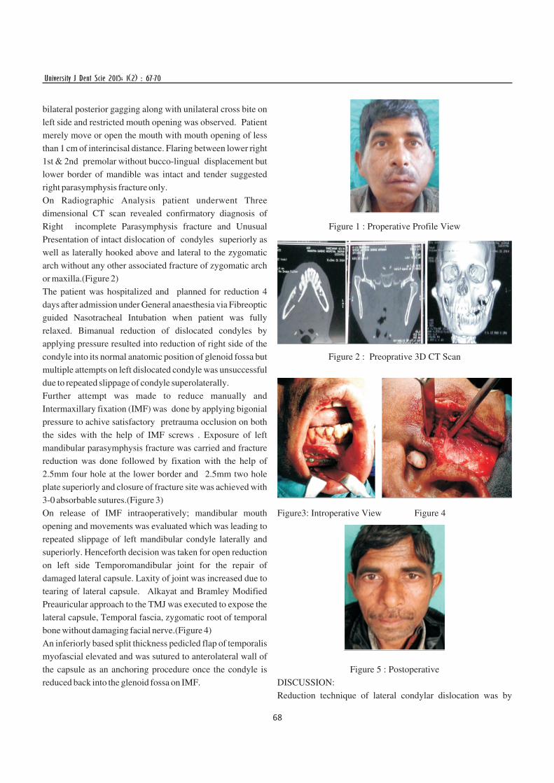
On Radiographic Analysis patient underwent Three
dimensional CT scan revealed confirmatory diagnosis of
Right incomplete Parasymphysis fracture and Unusual
Presentation of intact dislocation of condyles superiorly as
well as laterally hooked above and lateral to the zygomatic
arch without any other associated fracture of zygomatic arch
or maxilla.(Figure 2)
The patient was hospitalized and planned for reduction 4
days after admission under General anaesthesia via Fibreoptic
guided Nasotracheal Intubation when patient was fully
relaxed. Bimanual reduction of dislocated condyles by
applying pressure resulted into reduction of right side of the
condyle into its normal anatomic position of glenoid fossa but
multiple attempts on left dislocated condyle was unsuccessful
due to repeated slippage of condyle superolaterally.
Further attempt was made to reduce manually and
Intermaxillary fixation (IMF) was done by applying bigonial
pressure to achive satisfactory pretrauma occlusion on both
the sides with the help of IMF screws . Exposure of left
mandibular parasymphysis fracture was carried and fracture
reduction was done followed by fixation with the help of
2.5mm four hole at the lower border and 2.5mm two hole
plate superiorly and closure of fracture site was achieved with
3-0 absorbable sutures.(Figure 3)
On release of IMF intraoperatively; mandibular mouth
opening and movements was evaluated which was leading to
repeated slippage of left mandibular condyle laterally and
superiorly. Henceforth decision was taken for open reduction
on left side Temporomandibular joint for the repair of
damaged lateral capsule. Laxity of joint was increased due to
tearing of lateral capsule. Alkayat and Bramley Modified
Preauricular approach to the TMJ was executed to expose the
lateral capsule, Temporal fascia, zygomatic root of temporal
bone without damaging facial nerve.(Figure 4)
An inferiorly based split thickness pedicled flap of temporalis
myofascial elevated and was sutured to anterolateral wall of
the capsule as an anchoring procedure once the condyle is
reduced back into the glenoid fossa on IMF.
Figure 1 : Properative Profile View
Figure 2 : Preoprative 3D CT Scan
Figure3: Introperative View Figure 4
Figure 5 : Postoperative
DISCUSSION:
Reduction technique of lateral condylar dislocation was by
68

University J Dent Scie 2015; 1(2) : 67-70
described by Roberts4 in 1849. This consisted of strong
outward pressure on the ramus with inferior traction and
medial pressure on the condylar head.
In our case laceration at chin and wedge type fracture between
lower right first and second premolar suggest a high impact
force from the front and upward direction leads to
superolateral bilateral condylar dislocation, rupturing the
capsule & ligaments and associated with parasymphysis
fracture
It is postulated by the history of patient the mechanism of
dislocation might be due to sudden opening of the jaw on
frightening or screaming of patient, followed by chin hitting
on the floor. The force of the impact displaced the condyles
lateral to the glenoid fossa ,rupturing the capsular and
ligamentous attachments to the condylar head, and drove the
condyles superiorly, laterally to the zygomatic arches.
Allen and Young2, Satoh et al3, and Kapila and Lata5 support
this and emphasize that a fracture of the symphysis and/or
body of the mandible is a prerequisite for the lateral
dislocation of the mandibular condyle. But some authors6,7
reported intact condylar dislocation without mandibular
fracture.
In our case, the fracture of right parasymphysis caused the
flaring at dentoalveolar border, compression at lower border
and rotation of both the ramus leading to lateral dislocation of
the bilateral intact condyles.
Because it is rare often misdiagnosed certain diagnostic
features was addressed by the Worthington et al8 which were
(1) Malocclusion, (2) an open bite, (3) Persistent restriction of
mandibular movement, (4) An apparent loss of ramus height
with elevation of the ramus fragment, (5)Facial asymmetry.
Both the closed and open reduction are suggested for the
management of the same condition , type of treatment
depends on time of treatment after the injury. An early
diagnosis is imperative for successful management of the
dislocation.
Manual/closed reduction: -First choice, simple, less traumatic
& safe It depends on the time lapsed since the trauma. Mouth
props can be used- functioning as fulcrum in molar region
Open reduction -reduced by open traction through holes
drilled at lower border of the angle and downward traction at
the sigmoid notch with the help of channel retractor is also
one of the method published in literature. (Finck's
technique)9
Ferguson et al10. and Kapila and Lata5 were able to reduce
laterally dislocated condyles using strong traction and a wire
through the mandibular angle.
Kim et al11 used bone traction hook placed at the sigmoid notch
through stab incisiosn at the level of notch itself, and reduction
was achieved after applying an outward traction.
Two recent literature of case series by Shen et al12 and
Mishra.S et al13 debated the treatment outcome of open versus
closed reduction in their retrospective studies. Shen et al12
carried out retrospective clinical study on treatment modalities
of 10 patients of superolateral dislocation of condyle. Patients
who had dislocation for less than 1 week had condylar reduction
and rigid internal fixation of the fractures. Mandibular sagittal
split ramus osteotomy and articular reduction and fixation were
performed in seven cases. Maximum mouth opening and
occlusal relationships were compared following treatment
When the dislocated joint had become adherent to the
surrounding tissues and ankylosis developed, mandibular
sagittal split ramus osteotomy was performed with good results.
Mishra.S et13 al reported 7 case series in which six of all were
reduced manually under general anaesthesia and one case
underwent open reduction for the successful outcome.
In our case, both condyles were reduced manually, right condyle
was reduced but left condyle was repeatedly slipping into
dislocated position. In such cases of failure of closed reduction
the only alternative of open reduction proposed by many authors
2,5,13,14 was decided. Henceforth, decision was taken to open
the region to tighten the perforated capsule and fascia.
Capsulorrhaphy and temporal fascial flap are used in
strengthening of a lax capsule for treatment of hypermobile
joint, chronic subluxation and recurrent anterior dislocation.
Critical factors of success depends on the time between injury
and reduction. Delay induces fibro-osseous ankylosis
2,3,,15,16,17 necessitates open reduction, condylectomy with
or without arthroplasty.2 The delay in treatment can lead to
unsatisfactory results and imperfect reduction
In our case, the reduction was done on 4th day after injury
resulting in complete reduction
The average duration of postoperative immobilization
(maxillomandibular fixation) in the literature is 2 weeks 6. The
reduced condyle tends to return to the preoperative position. In
addition, immobilization facilitates healing of the presumably
damaged ligaments.
In our case, maxillomandibular immobilization was done for 3
weeks followed by active mouth opening exercise in order to
prevent fibrosis In published reports, no case has presented the
method to prevent repeated slipping of condyle into
superolateral dislocated position.
69

University J Dent Scie 2015; 1(2) : 67-70
Ours is the first case in which repeated superolateral
dislocation of condyle after manual reduction was
immobilized by capsulorrhaphy and further strengthened by
suturing temporal fascia on lateral aspect of capsule
CONCLUSION : Multiple factors determines the successful
outcome the time of reduction after the injury, and extent of
reduction either open or closed and postoperative
maxillomandibular fixation followed by patient compliance
in terms of postoperative aggressive physiotherapy, are the
major factors
The method of capsulorrhaphy and suturing temporal fascia
on lateral aspect of capsule can be used to prevent repeated
early dislocation of condyle (dislocated superolaterally). This
also prevents laxity of the capsule and reinforces the
temporomandibular ligament.
REFERENCES:
1. Yoshii T, Hamamoto Y, Muraoka S, Teranobu O,
Shigeta Y, Komori T. Traumatic dislocation of the
mandibular condyle into the temporal fossa in a child. J
Trauma. 2000 Oct;49(4):764-6.
2. Allen FJ, Young AH. Lateral displacement of the intact
mandibular condyle. A report of five cases. Br J Oral
Surg 1969;7:24–30
3. Satoh K, Suzuki H, Matsuzaki S. A type II lateral
dislocation of bilateral intact mandibular condyles with a
proposed new classification. Plast Reconstr Surg
1994;93:598–602.
4. Robert M (1849) Observation de luxation de la machoire
inferieur en haut ou dans la fosse temporale. Memoires de
la Societe de Chirurgie de Paris 1:456.
5. Kapila BK, Lata J. Superolateral dislocation of an intact
mandibular condyle into the temporal fossa: a case
report. J Trauma 1996; 41:351–352.
6. Bu SS, Jin SL, Yin L. Superolateral dislocation of the
intact mandibular condyle into the temporal fossa:
review of the literature and report of a case. Oral Surg
Oral Med Oral Pathol Oral Radiol Endod
2007;103:185–189.
7. Hegde S, Kamath VV, Deepa M, Priya A. Superolateral
dislocation of the mandibular condyle not associated
with fracture: a case report. J Maxillofac Oral Surg. 2010
Dec;9(4):424-7.
8. Worthington P. Dislocation of the mandibular condyle
into the temporal fossa. J Maxillofac Surg
1982;10:24–27.
9. Norman JE deB, Bramley P. Text Book and Colour Atlas of
the Temporomandibular Joint. London: Wolfe Med publ
Ltd; 1990:136–150.
10. Ferguson JW, Stewart IA, Whitley BD. Lateral
displacement of the intact mandibular condyle. Review of
literature and report of case with associated facial nerve
palsy. J Craniomaxillofac Surg 1989; 17:125–127.
11. Kim BC, Kang Samayoa SR, Kim HJ. Reduction of
superior-lateral intact mandibular condyle dislocation with
bone traction hook.J Korean Assoc Oral Maxillofac Surg.
2013 Oct;39(5):238-41.
12. Shen L, Li P, Li J, Long J, Tian W, Tang W. Management of
superolateral dislocation of the mandibular condyle: a
retrospective study of 10 cases. J Craniomaxillofac Surg.
2014 Jan;42(1):53-8.
13. Mishra.S, Mishra YC. Superolateral Dislocation of the
Mandibular Condyle: A Series of Seven Cases. J.
Maxillofac. Oral Surg. DOI 10.1007/s12663-015-0770-9
14. Prabhakar V, Singla S. Bilateral anterosuperior dislocation
of intact mandibular condyles in the temporal fossa. Int J
Oral Maxillofac Surg 2011;40:640–643
15. Rattan V. Superolateral dislocation of the mandibular
condyle: report of 2 cases and review of the literature. J Oral
Maxillofac Surg 2002;60:1366–9.
16. Li Z, Li ZB, Shang ZJ, Wu ZX. An unusual type of
superolateral dislocation of mandibular condyle: discussion
of the causative mechanisms and clinical characteristics. J
Oral Maxillofac Surg 2009;67:431–5.
17. Papadoupolos H, Edwards RS. Superolateral dislocation of
the condyle: report of rare case. Int J Oral Maxillofac Surg
2010;39:508–10.
CORRESPONDANCE:
Kotak Rajkumar K
IIIrd Yr Postgraduate Student
Mahatma Gandhi Dental College and Hospital, Jaipur.
E-mai : [email protected]
70
Related Documents