Copyright © Rebecca Rehder Wingerden Lecture Presentations for Biology Eighth Edition Neil Campbell and Jane Reece Pearson Education, Inc., publishing as Person Benjamin Cummings College Board, AP Biology Curriculum Framework 2012-2013 The processing of genetic information is imperfect and is a source of genetic variation. (3.C.3) Big Idea 3: Genetics and Information Transfer 19.1, 19.2, 27.2 Viral replication results in genetic variation, and viral infection can introduce genetic variation into the hosts. (3.C.3) a. Viral replication differs from other reproductive strategies and generates genetic variation via various mechanisms. 1. Viruses have highly efficient replicative capabilities that allow for rapid evolution and acquisition of new phenotypes. 2. Viruses replicate via a component assembly model allowing one virus to produce many progeny simultaneously via the lytic cycle. 3. Virus replication allows for mutations to occur through usual host pathways. The attach of an E. coli bacterial cell by numerous T4 bacteriophage. Most biologist studying viruses today would probably agree that they are not alive but exist in a shady area between life-forms and chemicals. Copyright © 2012 Rebecca Rehder Wingerden Viral structure. Viruses are made up of nucleic acid (DNA or RNA) enclosed in a protein coat (the capsid) and sometimes further wrapped in a membranous envelop. The individual protein subunits making up the capsid are called capsomeres. Although diverse in size and shape, viruses have common structural features, most of which appear in the four example shown here. (a) Tobacco mosaic virus has a helical capsid with the overall shape of a rigid rod. (b) Adenoviruses have an icosahedral capsid with a glycoprotein spike at each vertex. (c) Influenza viruses have an outer envelope studded with glycoprotein spikes. The genome consist of eight different RNA molecules, each wrapped in a helical capsid. (d) Bacteriophage T4, like other “T- even” phages, has a complex capsid consisting of an icosahedral head and a tail apparatus. Capsids and Envelopes Copyright © 2012 Rebecca Rehder Wingerden Copyright © 2012 Rebecca Rehder Wingerden Viral Reproductive Cycles (a) Tobacco mosaic virus has a helical capsid with the overall shape of a rigid rod. 2. Entry of phage DNA and degradation of host DNA. The sheath of the tail contracts, injecting the phage DNA into the cell and leaving an empty capsid outside. The cell’s DNA is hydrolyzed. 3. Synthesis of viral genomes and proteins. The phage DNA directs production of phage proteins and copies of the phage genome by host enzymes, using components within the cell. 4. Assembly. Three separate sets of proteins self-assemble to form phage heads, tails, and tail fibers. The phage genome is packaged inside the capsid as the head forms. 1. Attachment. The T4 phage uses its tail fibers to bind to specific receptor sites on the outer surface of an E. coli cell. 5. Release. The phage directs production of an enzyme that damages the bacterial cell wall, allowing fluid to enter. The cell swells and finally bursts, releasing 100 to 200 phage particles. The lytic cycle of phage T4, a virulent phage. Phage T4 has almost 300 genes, which are transcribed and translated using the host cell’s machinery. One of the first phage genes translated after the viral DNA enters the host cell codes for an enzyme that degrades the host cell’s DNA (step 2); the phage DNA is protected from breakdown because it contains a modified form of cytosine that is not recognized by the enzyme. The entire lytic cycle, from the phage’s first contact with the cell surface to cell lysis, takes only 20-30 minutes at 37°C. • Viruses lack metabolic enzymes and equipment for making proteins, such as ribosomes. They are obligate intracellular parasites; in other words, they can reproduce only within a host cell. • Each type of virus can infect cells of only a limited variety of hosts, called the host range of the virus. Viruses identify host cells by a “lock-and-key” fit between viral surface proteins and specific receptor molecules on the outside of cells. Copyright © 2012 Rebecca Rehder Wingerden Viral replication results in genetic variation, and viral infection can introduce genetic variation into the hosts. (3.C.3) 4. RNA viruses lack replication error-checking mechanisms, and thus have higher rates of mutation. 5. Related viruses can combine/recombine information if they infect the same host cell. 1. Glycoproteins on the viral envelope bind to specific receptor molecules (not shown) on the host cell, promoting viral entry into the cell. 2. The capsid and viral genome enter the cell. Digestion of the capsid by cellular enzymes releases the viral genome. 3. The viral genome (red) functions as a template for synthesis of complementary RNA strands (pink) by a viral enzyme. 4. New copies of viral genome RNA are made using complementary RNA strands as templates. 5. Complementary RNA strands also function as mRNA, which is translated into both capsid proteins (in the cytosol) and glycoproteins for the viral envelope (in the ER and Golgi apparatus). 6. Vesicles transport envelope glycoproteins to the plasma membrane. 7. A capsid assembles around each viral genome molecule. 8. Each new virus buds from the cel, its envelope studded with viral glycoproteins embedded in membrane derived from the host cell. • The RNA genome is transcribed into complementary RNA strands, which function both as mRNA and as templates for the synthesis of addition copies of genomic RNA. • Some enveloped viruses enter the host cell by fusion of the envelope with the cell’s plasma membrane; other enter by endocytosis. The reproductive cycle of an enveloped RNA virus. Shown here is a virus with a single-stranded RNA genome that functions as a template for synthesis of mRNA. Copyright © 2012 Rebecca Rehder Wingerden Viral replication results in genetic variation, and viral infection can introduce genetic variation into the hosts. (3.C.3) The reproductive cycle of HIV, the retrovirus that causes AIDS. Note in step 4 that DNA synthesized from the viral RNA genome is integrated into the host cell chromosomal DNA, a characteristic unique to retroviruses. 1. The envelope glycoproteins enable the virus to bind to specific receptors on certain white blood cells. 2. The virus fuses with the cell’s plasma membrane. The capsid proteins are removed, releasing the viral proteins and RNA. 3. Reverse transcriptase catalyzes the synthesis of a DNA strand complementary to the viral RNA. 4. Reverse transcriptase catalyzes the synthesis of a second DNA strand complementary to the first. 5. The double-stranded DNA is incorporated as a provirus into the cell’s DNA. 6. Proviral genes are transcribed into RNA molecules, which serve as genomes for the next viral generation and as mRNAs for translation into viral protein. 7. The viral proteins include capsid proteins and reverse transcriptase (made in the cytosol) and envelope glycoproteins (made in the ER). 8. Vesicles transport the glycoproteins to the cell’s plasma membrane. 9. Capsids are assembled around viral genomes and reverse transcriptase molecules. • The RNA animal viruses with the most complicated reproductive cycles are the retroviruses. • These viruses use an enzyme called reverse transcriptase, which transcribes an RNA template into DNA, the opposite of the usual direction. • The viral DNA integrates into the DNA of the host’s chromosome (provirus), becoming permanent part of the host’s genome. 10. New viruses bud off from the host cell.

Welcome message from author
This document is posted to help you gain knowledge. Please leave a comment to let me know what you think about it! Share it to your friends and learn new things together.
Transcript
Copyright © Rebecca Rehder Wingerden
Lecture Presentations for
Biology Eighth Edition
Neil Campbell and Jane Reece
Pearson Education, Inc., publishing as Person Benjamin Cummings College Board, AP Biology Curriculum Framework 2012-2013
The processing of genetic information is imperfect and is a source of genetic variation. (3.C.3)
Big Idea 3: Genetics and Information Transfer
19.1, 19.2, 27.2
Viral replication results in genetic variation, and viral infection can introduce genetic variation into the hosts. (3.C.3)
a.Viral replication differs from other reproductive strategies and generates genetic variation via various mechanisms.
1.Viruses have highly efficient replicative capabilities that allow for rapid evolution and acquisition of new phenotypes.
2.Viruses replicate via a component assembly model allowing one virus to produce many progeny simultaneously via the lytic cycle.
3.Virus replication allows for mutations to occur through usual host pathways.
The attach of an E. coli bacterial cell by numerous T4 bacteriophage. Most biologist studying viruses today would probably agree that they are not alive but exist in a shady area between life-forms and chemicals.
Copyright © 2012 Rebecca Rehder Wingerden
Viral structure. Viruses are made up of nucleic acid (DNA or RNA) enclosed in a protein coat (the capsid) and sometimes further wrapped in a membranous envelop. The individual protein subunits making up the capsid are called capsomeres. Although diverse in size and shape, viruses have common structural features, most of which appear in the four example shown here.
(a) Tobacco mosaic virus has a helical capsid with the overall shape of a rigid rod.
(b) Adenoviruses have an icosahedral capsid with a glycoprotein spike at each vertex.
(c) Influenza viruses have an outer envelope studded with glycoprotein spikes. The genome consist of eight different RNA molecules, each wrapped in a helical capsid.
(d) Bacteriophage T4, like other “T-even” phages, has a complex capsid consisting of an icosahedral head and a tail apparatus.
Capsids and Envelopes
Copyright © 2012 Rebecca Rehder Wingerden Copyright © 2012 Rebecca Rehder Wingerden
Viral Reproductive Cycles
(a) Tobacco mosaic virus has a helical capsid with the overall shape of a rigid rod.
2. Entry of phage DNA and degradation of host DNA. The sheath of the tail contracts, injecting the phage DNA into the cell and leaving an empty capsid outside. The cell’s DNA is hydrolyzed.
3. Synthesis of viral genomes and proteins. The phage DNA directs production of phage proteins and copies of the phage genome by host enzymes, using components within the cell.
4. Assembly. Three separate sets of proteins self-assemble to form phage heads, tails, and tail fibers. The phage genome is packaged inside the capsid as the head forms.
1. Attachment. The T4 phage uses its tail fibers to bind to specific receptor sites on the outer surface of an E. coli cell.
5. Release. The phage directs production of an enzyme that damages the bacterial cell wall, allowing fluid to enter. The cell swells and finally bursts, releasing 100 to 200 phage particles.
The lytic cycle of phage T4, a virulent phage. Phage T4 has almost 300 genes, which are transcribed and translated using the host cell’s machinery. One of the first phage genes translated after the viral DNA enters the host cell codes for an enzyme that degrades the host cell’s DNA (step 2); the phage DNA is protected from breakdown because it contains a modified form of cytosine that is not recognized by the enzyme. The entire lytic cycle, from the phage’s first contact with the cell surface to cell lysis, takes only 20-30 minutes at 37°C.
• Viruses lack metabolic enzymes and equipment for making proteins, such as ribosomes. They are obligate intracellular parasites; in other words, they can reproduce only within a host cell.
• Each type of virus can infect cells of only a limited variety of hosts, called the host range of the virus. Viruses identify host cells by a “lock-and-key” fit between viral surface proteins and specific receptor molecules on the outside of cells.
Copyright © 2012 Rebecca Rehder Wingerden
Viral replication results in genetic variation, and viral infection can introduce genetic variation into the hosts. (3.C.3)4. RNA viruses lack replication error-checking mechanisms, and thus
have higher rates of mutation. 5. Related viruses can combine/recombine information if they infect the
same host cell. 1. Glycoproteins on the viral envelope bind to specific receptor molecules (not shown) on the host cell, promoting viral entry into the cell.
2. The capsid and viral genome enter the cell. Digestion of the capsid by cellular enzymes releases the viral genome.
3. The viral genome (red) functions as a template for synthesis of complementary RNA strands (pink) by a viral enzyme.
4. New copies of viral genome RNA are made using complementary RNA strands as templates.
5. Complementary RNA strands also
function as mRNA, which is translated into
both capsid proteins (in the cytosol) and
glycoproteins for the viral envelope (in the
ER and Golgi apparatus).
6. Vesicles transport envelope
glycoproteins to the plasma membrane.
7. A capsid assembles around each viral genome molecule.
8. Each new virus buds from the cel, its envelope studded with viral glycoproteins embedded in membrane derived from the host cell.
• The RNA genome is transcribed into complementary RNA strands, which function both as mRNA and as templates for the synthesis of addition copies of genomic RNA.
• Some enveloped viruses enter the host cell by fusion of the envelope with the cell’s plasma membrane; other enter by endocytosis.
The reproductive cycle of an enveloped RNA virus. Shown here is a virus with a single-stranded RNA genome that functions as a template for synthesis of mRNA. Copyright © 2012 Rebecca Rehder Wingerden
Viral replication results in genetic variation, and viral infection can introduce genetic variation into the hosts. (3.C.3)
The reproductive cycle of HIV, the retrovirus that causes AIDS. Note in step 4 that DNA synthesized from the viral RNA genome is integrated into the host cell chromosomal DNA, a characteristic unique to retroviruses.
1. The envelope glycoproteins enable
the virus to bind to specific receptors on
certain white blood cells.
2. The virus fuses with the cell’s plasma membrane. The capsid proteins are removed, releasing the viral proteins and RNA.
3. Reverse transcriptase catalyzes the synthesis of a DNA strand complementary to the viral RNA.
4. Reverse transcriptase catalyzes the synthesis of a second DNA strand complementary to the first.
5. The double-stranded DNA is incorporated as a provirus into the cell’s DNA.
6. Proviral genes are transcribed into RNA molecules, which serve as genomes for the next viral generation and as mRNAs for translation into viral protein.
7. The viral proteins include capsid proteins and reverse transcriptase (made in the cytosol) and envelope glycoproteins (made in the ER).
8. Vesicles transport the glycoproteins to the cell’s plasma membrane.
9. Capsids are assembled around viral genomes and reverse transcriptase molecules.
• The RNA animal viruses with the most complicated reproductive cycles are the retroviruses.
• These viruses use an enzyme called reverse transcriptase, which transcribes an RNA template into DNA, the opposite of the usual direction.
• The viral DNA integrates into the DNA of the host’s chromosome (provirus), becoming permanent part of the host’s genome.
10. New viruses bud off from the host cell.
Viral replication results in genetic variation, and viral infection can introduce genetic variation into the hosts. (3.C.3)
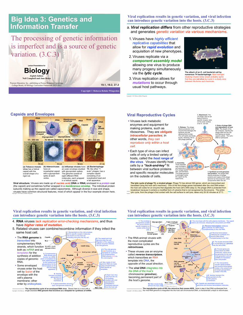
6. HIV is a well-studied system where the rapid evolution of a virus within the host contributes to the pathogenicity of viral infection.
Evolution of drug resistance in HIV. Rare resistant viruses multiplied quickly when each of these patients was treated with the anti-HIV drug 3TC. Within just a few weeks. 3TC-resistant organisms made up 100% of the virus population in each case.
HIV: The Ultimate Evolver. HIV is one of the fastest evolving entities known. It reproduces sloppily, accumulating lots of mutations when it copies its genetic material. It also reproduces at a lightning-fast rate - a single viruses can spawn billions of copies in just one day (http://evolution.berkeley.edu/evolibrary/article/medicine_04). Using a molecular clock, researchers at Los Alamos National Laboratory in New Mexico date the origin of HIV-1 M infection in humans during the 1930’s (p.550-51).
HIV shown budding from a white blood cell
1930’s
Copyright © 2012 Rebecca Rehder Wingerden
Viral replication results in genetic variation, and viral infection can introduce genetic variation into the hosts. (3.C.3)
1. Phage infects a bacterial cell that has alleles A+ and B+.
2. Host DNA (brown) is fragmented, and phage DNA and proteins are made. This is the donor cell.
3. A bacterial DNA fragment (in this case a fragment with the A+ allele) may be packaged in a phage capsid.
4. Phage with the A+ allele from the donor cell infects a recipient A-B- cell, and recombination between donor DNA (brown) and recipient DNA (green) occurs at two places (dotted lines).
5. The genotype of the resulting recombinant cell (A+B+) differs from the genotypes of both the donor (A+B+) and the recipient (A-B-)
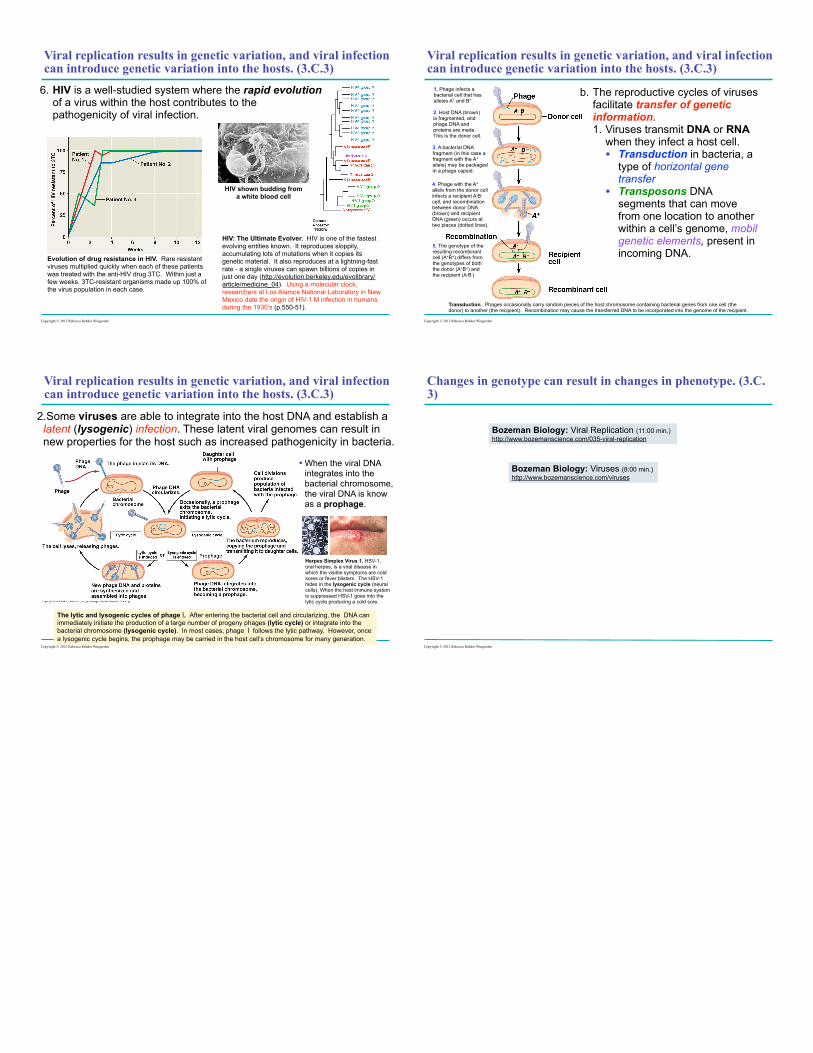
b. The reproductive cycles of viruses facilitate transfer of genetic information. 1. Viruses transmit DNA or RNA
when they infect a host cell. • Transduction in bacteria, a
type of horizontal gene transfer
• Transposons DNA segments that can move from one location to another within a cell’s genome, mobil genetic elements, present in incoming DNA.
Transduction. Phages occasionally carry random pieces of the host chromosome containing bacterial genes from one cell (the donor) to another (the recipient). Recombination may cause the transferred DNA to be incorporated into the genome of the recipient.
Copyright © 2012 Rebecca Rehder Wingerden
Copyright © 2012 Rebecca Rehder Wingerden
Viral replication results in genetic variation, and viral infection can introduce genetic variation into the hosts. (3.C.3)
2.Some viruses are able to integrate into the host DNA and establish a latent (lysogenic) infection. These latent viral genomes can result in new properties for the host such as increased pathogenicity in bacteria.
The lytic and lysogenic cycles of phage l. After entering the bacterial cell and circularizing, the DNA can immediately initiate the production of a large number of progeny phages (lytic cycle) or integrate into the bacterial chromosome (lysogenic cycle). In most cases, phage l follows the lytic pathway. However, once a lysogenic cycle begins, the prophage may be carried in the host cell’s chromosome for many generation.
• When the viral DNA integrates into the bacterial chromosome, the viral DNA is know as a prophage.
Herpes Simplex Virus 1. HSV-1, oral herpes, is a viral disease in which the visible symptoms are cold sores or fever blisters. The HSV-1 hides in the lysogenic cycle (neural cells). When the host immune system is suppressed HSV-1 goes into the lytic cycle producing a cold sore.
Changes in genotype can result in changes in phenotype. (3.C.3)
Bozeman Biology: Viruses (8:00 min.) http://www.bozemanscience.com/viruses
Bozeman Biology: Viral Replication (11:00 min.) http://www.bozemanscience.com/035-viral-replication
Copyright © 2012 Rebecca Rehder Wingerden
Related Documents


