Bifunctional nanoparticles for SERS monitoring and magnetic intervention of assembly and enzyme cutting of DNAs† Liqin Lin, ab Elizabeth Crew, a Hong Yan,‡ a Shiyao Shan, a Zakiya Skeete, a Derrick Mott, c Tatiana Krentsel, a Jun Yin, a Natasha A. Chernova, a Jin Luo, a Mark H. Engelhard, d Chongmin Wang, d Qingbiao Li b and Chuan-Jian Zhong * a The ability to harness the nanoscale structural properties is essential for the exploration of functional properties of nanomaterials. This report demonstrates a novel strategy exploring bifunctional nanoparticles for spectroscopic detection and magnetic intervention of DNA assembly, disassembly, and enzyme cutting processes in a solution phase. In contrast to existing single-function based approaches, this strategy exploits magnetic MnZn ferrite nanoparticles decorated with gold or silver on the surface to retain adequate magnetization while producing sufficient plasmonic resonance features to impart surface-enhanced Raman scattering (SERS) functions. The decoration of MnZn ferrite nanoparticles with Au or Ag (MZF/Au or MZF/Ag) was achieved by thermally activated deposition of Au or Ag atoms/ nanoparticles on MZF nanoparticles. Upon interparticle double-stranded DNA linkage of the MZF/Au (or MZF/Ag) nanoparticles with gold nanoparticles labeled with a Raman reporter, the resulting interparticle “hot spots” are shown to enable real time SERS monitoring of the DNA assembly, disassembly, or enzyme cutting processes, where the magnetic component provides an effective means for intervention of the biomolecular processes in the solution. The unique bifunctional combination of the SERS “hot spots” and the magnetic separation capability serves as the first example of bifunctional nanoprobes for biomolecular recognition and intervention. Introduction The ability to harness the nanoscale functional properties is essential for the exploration of technological applications of nanomaterials. In medical diagnostics and treatments, two of the functional properties that have been the subjects of broad interest for biomolecular recognition include signal trans- duction and activity intervention. Signal transduction of biomolecules such as DNAs, proteins, or enzymes has been a focal area of intensive research on various pathogens. 1–3 Many optical and spectroscopic tools are powerful for signal trans- duction due to their multiplexing or ngerprinting capabil- ities, 4–19 which oen involve immobilization of reporter labels on gold or silver nanoparticles (Au or Ag NPs) on a solid substrate. One example involves targeting of oligonucleotides using nanoparticles functionalized with a specic sequence of oligonucleotides along with a dye label. 4 For most of the existing approaches which involved planar gold or silver substrates as supports for the nanoparticles, 4,6,7,12–14,17 the uncontrollable aggregation of the nanoparticles constitutes a major compli- cation. In contrast, the interparticle plasmonic coupling, a phenomenon originating from the formation of small clusters of NPs such as dimers and trimers, 12 as described in the present report, functions as a spectroscopic nanoprobe to biomolecular processes in solutions where the interparticle chemistry can be well dened by the biomolecules. On the other hand, an effec- tive means is needed for activity intervention of the biomolec- ular processes in a solution. While the magnetic properties of magnetic nanoparticles (MNPs) have been exploited for bio- separation, controlled delivery, specic targeting, or magnetic resonance imaging (MRI), 20–22 the direct use of many existing metal oxides (e.g., iron or cobalt oxides) in biological uids is problematic considering their potential toxicity and limited surface chemistry. To impart the desired biocompatibility and surface functionality to MNPs, the surface functionalization of MNPs with gold or silver is one important approach in view of the rich surface chemistry for DNAs, proteins and other a Department of Chemistry, State University of New York at Binghamton, Binghamton, New York 13902, USA. E-mail: [email protected] b Environmental Science Research Center, College of Environment and Ecology, Xiamen University, Xiamen 361005, China c School of Materials Science, Japan Advanced Institute of Science and Technology, 1-1 Asahidai, Nomi, 923-1292 Ishikawa, Japan d EMSL, Pacic Northwest National Laboratory, Richland, Washington 99352, USA † Electronic supplementary information (ESI) available: Additional data for the synthesis and the experimental measurements. See DOI: 10.1039/c3tb20446d ‡ Present address: Institute of Materials Research and Engineering, A*STAR, 3 Research Link, Singapore 117602. Cite this: J. Mater. Chem. B, 2013, 1, 4320 Received 29th March 2013 Accepted 27th June 2013 DOI: 10.1039/c3tb20446d www.rsc.org/MaterialsB 4320 | J. Mater. Chem. B, 2013, 1, 4320–4330 This journal is ª The Royal Society of Chemistry 2013 Journal of Materials Chemistry B PAPER Published on 17 July 2013. Downloaded by Agency for Science, Technology & Research (A*STAR) on 14/08/2013 09:37:19. View Article Online View Journal | View Issue

Welcome message from author
This document is posted to help you gain knowledge. Please leave a comment to let me know what you think about it! Share it to your friends and learn new things together.
Transcript
Journal ofMaterials Chemistry B
PAPER
Publ
ishe
d on
17
July
201
3. D
ownl
oade
d by
Age
ncy
for
Scie
nce,
Tec
hnol
ogy
& R
esea
rch
(A*S
TA
R)
on
14/0
8/20
13 0
9:37
:19.
View Article OnlineView Journal | View Issue
aDepartment of Chemistry, State University
New York 13902, USA. E-mail: cjzhong@binbEnvironmental Science Research Center, Co
University, Xiamen 361005, ChinacSchool of Materials Science, Japan Advance
Asahidai, Nomi, 923-1292 Ishikawa, JapandEMSL, Pacic Northwest National Laborato
† Electronic supplementary informationsynthesis and the experimental measurem
‡ Present address: Institute of MaterialsResearch Link, Singapore 117602.
Cite this: J. Mater. Chem. B, 2013, 1,4320
Received 29th March 2013Accepted 27th June 2013
DOI: 10.1039/c3tb20446d
www.rsc.org/MaterialsB
4320 | J. Mater. Chem. B, 2013, 1, 43
Bifunctional nanoparticles for SERS monitoring andmagnetic intervention of assembly and enzyme cuttingof DNAs†
Liqin Lin,ab Elizabeth Crew,a Hong Yan,‡a Shiyao Shan,a Zakiya Skeete,a Derrick Mott,c
Tatiana Krentsel,a Jun Yin,a Natasha A. Chernova,a Jin Luo,a Mark H. Engelhard,d
Chongmin Wang,d Qingbiao Lib and Chuan-Jian Zhong*a
The ability to harness the nanoscale structural properties is essential for the exploration of functional
properties of nanomaterials. This report demonstrates a novel strategy exploring bifunctional
nanoparticles for spectroscopic detection and magnetic intervention of DNA assembly, disassembly, and
enzyme cutting processes in a solution phase. In contrast to existing single-function based approaches,
this strategy exploits magnetic MnZn ferrite nanoparticles decorated with gold or silver on the surface
to retain adequate magnetization while producing sufficient plasmonic resonance features to impart
surface-enhanced Raman scattering (SERS) functions. The decoration of MnZn ferrite nanoparticles with
Au or Ag (MZF/Au or MZF/Ag) was achieved by thermally activated deposition of Au or Ag atoms/
nanoparticles on MZF nanoparticles. Upon interparticle double-stranded DNA linkage of the MZF/Au (or
MZF/Ag) nanoparticles with gold nanoparticles labeled with a Raman reporter, the resulting
interparticle “hot spots” are shown to enable real time SERS monitoring of the DNA assembly,
disassembly, or enzyme cutting processes, where the magnetic component provides an effective means
for intervention of the biomolecular processes in the solution. The unique bifunctional combination of
the SERS “hot spots” and the magnetic separation capability serves as the first example of bifunctional
nanoprobes for biomolecular recognition and intervention.
Introduction
The ability to harness the nanoscale functional properties isessential for the exploration of technological applications ofnanomaterials. In medical diagnostics and treatments, two ofthe functional properties that have been the subjects of broadinterest for biomolecular recognition include signal trans-duction and activity intervention. Signal transduction ofbiomolecules such as DNAs, proteins, or enzymes has been afocal area of intensive research on various pathogens.1–3 Manyoptical and spectroscopic tools are powerful for signal trans-duction due to their multiplexing or ngerprinting capabil-ities,4–19 which oen involve immobilization of reporter labels
of New York at Binghamton, Binghamton,
ghamton.edu
llege of Environment and Ecology, Xiamen
d Institute of Science and Technology, 1-1
ry, Richland, Washington 99352, USA
(ESI) available: Additional data for theents. See DOI: 10.1039/c3tb20446d
Research and Engineering, A*STAR, 3
20–4330
on gold or silver nanoparticles (Au or Ag NPs) on a solidsubstrate. One example involves targeting of oligonucleotidesusing nanoparticles functionalized with a specic sequence ofoligonucleotides along with a dye label.4 Formost of the existingapproaches which involved planar gold or silver substrates assupports for the nanoparticles,4,6,7,12–14,17 the uncontrollableaggregation of the nanoparticles constitutes a major compli-cation. In contrast, the interparticle plasmonic coupling, aphenomenon originating from the formation of small clustersof NPs such as dimers and trimers,12 as described in the presentreport, functions as a spectroscopic nanoprobe to biomolecularprocesses in solutions where the interparticle chemistry can bewell dened by the biomolecules. On the other hand, an effec-tive means is needed for activity intervention of the biomolec-ular processes in a solution. While the magnetic properties ofmagnetic nanoparticles (MNPs) have been exploited for bio-separation, controlled delivery, specic targeting, or magneticresonance imaging (MRI),20–22 the direct use of many existingmetal oxides (e.g., iron or cobalt oxides) in biological uids isproblematic considering their potential toxicity and limitedsurface chemistry. To impart the desired biocompatibility andsurface functionality to MNPs, the surface functionalization ofMNPs with gold or silver is one important approach in view ofthe rich surface chemistry for DNAs, proteins and other
This journal is ª The Royal Society of Chemistry 2013
Paper Journal of Materials Chemistry B
Publ
ishe
d on
17
July
201
3. D
ownl
oade
d by
Age
ncy
for
Scie
nce,
Tec
hnol
ogy
& R
esea
rch
(A*S
TA
R)
on
14/0
8/20
13 0
9:37
:19.
View Article Online
biomolecules.23 While this approach is supported by our earlierstudies on gold-coated MNPs in biomolecular separation fromsolutions to a solid substrate,12,13 and other studies such asMNPs on a planar gold substrate,24 Fe3O4@Au NPs labeled withdyes,25 silver-embedded MNPs for cancer-cell targeting andimaging,26,27 and SERS detection of DNAs via various nano-structures,28–32 most of them rely either on using solidsubstrates with immobilized NPs or magnetic enrichment ofDNA-particle aggregates separated from the solution state forthe detection. There has been no real demonstration of signaltransduction and bioactivity intervention through the directplasmonic coupling of DNA-linked magnetic nanoparticles inthe solution and in real time.
We demonstrate herein the rst example of a novel bifunc-tional strategy for signal transduction and activity interventionusing Ag- and Au-decoratedmagnetic MnZn ferrite NPs (MZF/Agand MZF/Au). In our previous work, MnZn ferrite type NPs wereshown to form a core–shell nanostructure consisting of a Fe3O4
core and a MnZnFe2O4 type shell.33 In comparison with Fe3O4
NPs,34,35 the magnetic properties of such magnetic nano-particles are tunable by Mn/Zn doping in the shell. In the
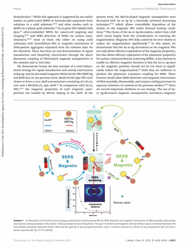
Scheme 1 An illustration of the bifunctional strategy exploiting the multifunctionaand enzyme cutting processes in the solution. These processes involve interparticle “htheoretically calculated enhanced electric field and the spacing of Ag nanoparticlecertain spacing (see Fig. S1† for details).
This journal is ª The Royal Society of Chemistry 2013
present work, the Mn/Zn-doped magnetic nanoparticles weredecorated with Au or Ag by a thermally activated processingtechnique,13,14 which allows controllable deposition of themetals on the magnetic NPs under thermal heating condi-tions.13 The choice of the Au or Ag decoration, rather than a fullshell, stems largely from the consideration of retaining themagnetization. Magnetic NPs fully coated by Au were shown toreduce the magnetization signicantly.34 In this report, wedemonstrate that the Au or Ag decoration on the magnetic NPsnot only allows effective exploitation of the magnetic properties,but also allows effective exploration of the plasmonic propertiesfor surface-enhanced Raman scattering (SERS). A key element toenable an effective magnetic function is that the Au or Ag layeron the magnetic particles should not be too thick to signi-cantly reduce the magnetization,34 while they are sufficient toproduce the plasmonic resonance coupling for SERS. Thesefeatures would allow SERS detection and magnetic interventionof DNA assembly, disassembly, and enzyme cutting processes inaqueous solutions. In contrast to the previous studies,28–32 thereare several important attributes in our strategy. The use of Au-or Ag-decorated magnetic nanoparticles introduces magnetic
l NPs for SERS detection and magnetic intervention of DNA assembly, disassembly,ot-spot” formation andmagnetic removal. Bottom panel: correlation between thedimers. Inset: a contour external to a dimer of Ag nanoparticles (60 nm) with a
J. Mater. Chem. B, 2013, 1, 4320–4330 | 4321
Journal of Materials Chemistry B Paper
Publ
ishe
d on
17
July
201
3. D
ownl
oade
d by
Age
ncy
for
Scie
nce,
Tec
hnol
ogy
& R
esea
rch
(A*S
TA
R)
on
14/0
8/20
13 0
9:37
:19.
View Article Online
functionality in the direct recognition of two DNA strandsanchored respectively to two different nanoparticles, i.e.,Raman-labeled Au NPs (“Raman probe”) and Au- or Ag-deco-rated magnetic NPs (“Magnetic (Mag) probe”), which are illus-trated in Scheme 1 for the biomolecular processes involvingassembly and disassembly of two complementary ss-DNAs(DNA1 and DNA2), and the enzyme cutting of the ds-DNA. Oness-DNA is attached to Au NPs labeled with a Raman labelmolecule (Au NPs-R) and the other being anchored to themagnetic MZF/Au or MZF/Ag NPs. The interparticle plasmoniccoupling as a result of the ds-DNA linkage creates a “hot-spot”which provides a means for SERS detection of the assembly andthe disassembly either by restriction enzyme cleavage of the ds-DNA or by thermal removal of the hydrogen-bonding betweenthe base pairs. The magnetic component of the nanoprobesprovides the capabilities of magnetic intervention, biosepara-tion, resuspendability, and potential recyclable or sustainableuses.
The foundation for the SERS function stems from theoreticalcalculation of the local electric eld enhancement (i.e., “hot-spot”) for a dimer of NPs using the discrete dipole approxima-tion method,36 which reveals an E-eld enhancement that isdependent on the interparticle spacing in the dimer (see ESI,Fig. S1†). The E-eld enhancement increases sharply with thereduction of the interparticle spacing. When the interparticlespacing increases, the E-eld enhancement weakens. For adimer of DNA1–MNP and MBA-Au NPs by DNA2 via binding ofthe complementary base pairs as described in this report, theinterparticle spacing under an ideal interparticle ds-DNA link-ing is estimated to be about 10 nm in length, which falls into the“hot spot” region (ESI, Fig. S1†). When an enzyme cuts the ds-DNA at a specic site, or the solution is heated to induce adisassembly of the dimer, the interparticle spacing increasesand eventually the two NPs are separated, leading to weakeningand disappearance of the “hot spot”. This type of “hot spot”formation/removal, coupled with the magnetic functionality,constitutes the basis of the SERS detection and activity inter-vention in the DNA assembly and disassembly processes. Incomparison with the SERS-only detection using pure Au NPs,37
this “Raman-Magnetic” probe could enable interparticle “hotspot” formation and bio-separation capability for monitoringDNA assembly and cutting processes in the solution phase andin real time, which is to our knowledge the rst example of thiskind of bifunctionality.
ExperimentalChemicals and nanoparticles
Hydrogen tetrachloroaurate(III) hydrate (HAuCl4$xH2O), silvernitrate (AgNO3), sodium hydroxide, sodium acrylate (97%),sodium chloride (NaCl, 99%), ethylenediaminetetraacetic acid(EDTA), 4-mercaptobenzoic acid (MBA), oleylamine (OAM,70%), benzyl ether, 1-decanethiol (DT, 99%), and 11-mercap-toundecanoic acid (MUA, 97%) were purchased from Sigma-Aldrich (Milwaukee, WI) and used as received. Iron(III) acetyla-cetonate (Fe(acac)3, 99%, Lancaster), manganese(II) acetylacet-onate (Mn(acac)2, 95% Strem), zinc acetylacetonate (Zn(acac)2,
4322 | J. Mater. Chem. B, 2013, 1, 4320–4330
98%, Strem), and oleic acid (OAC, 99%, Alfa Aesar) were alsoused for the synthesis. Phosphate buffer was purchased fromFisher Scientic (Pittsburgh, PA). Thiol modied DNAs withstandard desalting purication were purchased from IntegratedDNA Technologies, Inc. (Coralville, IA). NAP-5 columns werepurchased from GE Healthcare (Uppsala, Sweden) and MspIfrom New England Biolabs (Beverly, MA). The solventsincluding hexane (99.9%) and toluene (99.8%) were fromFisher. Water was puried with a Millipore Milli-Q watersystem.
Nanoparticle synthesis
Au NPs and Ag NPs of 2 nm in size encapsulated with DTmonolayer capping molecules were synthesized based on thestandard and modied two-phase method.13,38,52
The synthesis of MZF nanoparticles was based on a methoddeveloped in our laboratory,33,39 which involved thermaldecomposition of metal acetylacetonate compounds, e.g.,0.469 g Fe(acac)3, 0.081 g Mn(acac)2, and 0.087 g Zn(acac)2 in20 mL of benzyl ether with 2 mL of oleic acid and 2 mL ofoleylamine. The mixture was reuxed for 60 min. The productwas collected using a magnet.
For the preparation of MZF/Au and MZF/Ag nanoparticles, amodied strategy of the thermally activated processing protocolwas used.13 In a typical synthesis, 1.3 mL of concentrated Au-DT(or Ag-DT) and MZF nanoparticles (e.g., stock solutions ofDT-capped Au (2 nm, 33 mM) and OAM/OAC-capped MZF (8 nm,2.6 mM)) in toluene with a certain ratio was placed in a reactiontube. The tube was then placed in a preheated Yamato DX400gravity convection oven at 150 �C for 3 h. Temperature variationfrom this set point was limited to 1.5 �C. Aer the thermaltreatment, the reaction tube was allowed to cool down, and theparticles were re-dispersed in toluene.
DNA assembly/disassembly procedures
The as-synthesized DT-capped MZF/Au and MZF/Ag particleswere transferred to water by ligand exchange using mercap-toundecanoic acid (MUA) by following a procedure reported byGittins and Caruso with a slight modication.53 Then the NPswere further modied with DNA for assembly and SERS detec-tion. The detailed procedures for the assembly and the restric-tion enzymes cutting areas were reported previously,12,46 and aresummarized below.
MZF/Au (or MZF/Ag) and Au NP assembly based oncomplementary oligonucleotides
To demonstrate the viability of assembly between MZF/Au (orMZF/Ag) and Au NPs, two different DNAs (DNA1: 50-/5ThioMC6-D/AGGCCAGACCTGCCCGGGCAAGCCTTGGCA-30 (bottom strand)and DNA2: 50-/5ThioMC6-D/TGCCAAGGCTTGCCCGGGCAGGTCTGGCCT-30 (top strand)) were used. Acrylate-capped Au NPswith an average size of 39.7� 1.8 nmwere synthesized followingthe procedure reported previously.8 DNA1 and DNA2 were rstdissolved in 0.1 M phosphate buffer (pH 8) at a concentrationranging from 300 to 370 mM. The disulde bonds in DNA1 andDNA2 were cleaved using an approach similar to the reported
This journal is ª The Royal Society of Chemistry 2013
Paper Journal of Materials Chemistry B
Publ
ishe
d on
17
July
201
3. D
ownl
oade
d by
Age
ncy
for
Scie
nce,
Tec
hnol
ogy
& R
esea
rch
(A*S
TA
R)
on
14/0
8/20
13 0
9:37
:19.
View Article Online
procedure,46 where dithiothreitol (DTT) at a nal concentrationof 0.1 M was added to �10 OD of the nucleotides in a nalvolume of 400 mL. The solution was allowed to react at roomtemperature for 2 h, then poured through a NAP-5 column andan aliquot of 1.1 mL phosphate buffer (pH 8) was added to thecolumn to elute the cleaved oligonucleotide. The nal concen-tration of the cleaved DNAs was 10 mM with an OD 260 nm of3.6. The exact concentrations of DNAs varied slightly dependingon the specic experiment.
The surface of MZF/Au (or MZF/Ag) was functionalized withthe cleaved DNA1 similar to the reported procedure46 to formMZF/Au–DNA1 (or MZF/Ag–DNA1). Briey, 0.176 mL of thecleaved DNA1 was added to 3 mL of MZF/Au nanoparticles (orMZF/Ag nanoparticles). The solution was le standing at roomtemperature for 16 h, aer which it was diluted to 20 mM NaCland 10 mM phosphate buffer (pH 7) and allowed to stand foranother 40 h at room temperature. The DNA1-capped nano-particles were then centrifuged and washed twice at 14 000 rpm(18 620g) for 25 min (each time the solution was re-dispersed ina 20 mM NaCl/10 mM phosphate buffer (pH 7) solution) beforebeing re-dispersed in its nal (20 mM NaCl/10 mM phosphatebuffer/0.01% sodium azide (pH 7)) solution and stored at roomtemperature.
The MBA-Au NPs: a controlled volume of 0.1 mM MBA for a50% surface coverage was added to 3 mL of Au NPs (stockconcentration 0.1 nM). The solution was le standing at roomtemperature overnight before use. The estimate of this coveragewas based on the measurement of an adsorption isotherm, i.e.,the SERS intensity vs. concentration of MBA.12
To study the assembly of the DNA capped MZF/Au (or MZF/Ag) nanoparticles with 39 nm Au NPs, 17 mL of DNA2 was addedto 300 mL of MZF/Au–DNA1 (or MZF/Ag–DNA1) in the presenceof 300 mL of MBA-labeled Au NPs. The reaction was monitoredby UV-Vis and Raman spectroscopy.
Restriction enzyme cutting
For the enzyme cutting experiment, the restriction enzymeMspI(100 units per mL) was utilized. 5 mL of restriction enzyme wasadded to 350 mL of the assembled solution, along with thebuffer for the restriction enzyme. The solution was incubated at37 �C (MspI) with constant stirring and the Raman spectrumwas taken at different time intervals.
Instrumentation and measurements
UV-Visible (UV-Vis) spectra were acquired with a Hewlett Pack-ard 8453 spectrophotometer.
Transmission electron microscopy (TEM) was performed ona Hitachi H-7000 Electron Microscope (100 kV). The TEMsamples were prepared by taking a solution sample and castingit onto a carbon-coated copper grid sample holder followed byevaporation in air at room temperature. HRTEM analysis wascarried out using a JEOL JEM 2010F at an acceleration voltage of200 kV. The nanoparticles were diluted in hexane solvent anddrop cast onto a carbon-coated copper grid, followed by solventevaporation in air at room temperature. HRTEM in PNNLanalysis was carried out on a JEOL JEM 2010Fmicroscope with a
This journal is ª The Royal Society of Chemistry 2013
specied point-to-point resolution of 0.194 nm. The operatingvoltage of the microscope was 200 kV. High-angle annular dark-eld scanning TEM (HAADFSTEM) imaging for morphologycharacterization and energy dispersive X-ray spectroscopy (EDS)for elemental mapping were carried out on an JEOL JEM-ARM200F instrument operated at 200 kV with a sphericalaberration corrector.
Surface-enhanced Raman scattering (SERS) spectra wererecorded using an Advantage 200A Raman spectrometer(DeltaNu) and the datawere collected from200 to 2000 cm�1. Thelaser power and wavelength were 5 mW and 632.8 nm, respec-tively. The scattering geometry utilized was backscattering.
Inductively coupled plasma-optical emission spectroscopy(ICP-OES) was used to analyze the nanoparticle composition. Itwas performed on a Perkin-Elmer 2000 DV ICP-OES with thefollowing parameters: plasma, 18.0 L of Ar(g) min�1; auxiliary,0.3 L of Ar(g) min�1; nebulizer, 0.73 L of Ar(g) min�1; power,1500 W; and peristaltic pump rate, 1.40 mL min�1. Reportedvalues of <1.0 mg L�1 were analyzed using aMeinhard nebulizercoupled to a cyclonic spray chamber to increase the analytesensitivity. Elemental concentrations were determined bymeasuring one or more emission lines (in nm) to check forinterferences.
X-ray Photoelectron Spectroscopy (XPS) measurements wereperformed on a Physical Electronics 5000 versa probe scanningESCA microprobe. The system uses a focused monochromaticAl Ka X-ray (1486.7 eV) source for excitation and a sphericalsection analyzer. The instrument has a 16-element multi-channel detection system. Wide scan data were collected usinga pass energy of 187.85 eV. The binding energy (BE) scale wascalibrated using Cu 2p3/2 line at 932.58 � 0.05 eV and Au 4f7/2line at 84.01� 0.05 eV. The sample experienced variable degreesof charging by low energy electrons at�1.5 eV, 20 mA so that lowenergy Ar+ ions needed to be used to minimize this charging.The percentages of individual elements detected were deter-mined from the relative composition analysis of the areas of theXPS lines.
Superconducting quantum interference device (SQUID): Thedc magnetization and magnetic susceptibility were measuredusing a magnetometer Quantum Design MPMS XL-5. Before themeasurements, a possible remnant magnetic eld was removedusing the ultralow-eld option at 298 K. The resultant remnanteld was less than 3 mOe. The measurements of the dcmagnetization and magnetic susceptibility were carried outaer appropriate cooling procedures. The details of the coolingprotocol for each measurement are described in the Discussion.
Results and discussionCharacterization of MZF/Au and MZF/Ag NPs
The as-synthesized MZF NPs feature about 8 nm in size androughly cubical shape as reported recently.33 MZF NPs of severalslightly different ratios were utilized for the decoration by Au orAg. For example, an MZF with an Fe : Mn : Zn ratio of 84 : 8 : 8consisted of a Fe3O4 core and a (Mn0.5Zn0.5)Fe2O4 shell with ashell to core volume ratio of 1 : 1.1 on the basis of our previousstudy.33 The shell was about 48% (vol) (i.e., % of the volume of
J. Mater. Chem. B, 2013, 1, 4320–4330 | 4323
Journal of Materials Chemistry B Paper
Publ
ishe
d on
17
July
201
3. D
ownl
oade
d by
Age
ncy
for
Scie
nce,
Tec
hnol
ogy
& R
esea
rch
(A*S
TA
R)
on
14/0
8/20
13 0
9:37
:19.
View Article Online
the shell relative to the whole core–shell volume), which is�0.8 nm in shell thickness for the 8 nm MZF NP. The MZF/Auand MZF/Ag NPs, prepared by the thermally activated process-ing of the MZF NPs and Au (2 nm) or Ag NPs (2 nm) at or neartheir nanoscale melting temperatures,13,14,38 featured 11 and9 nm in sizes. Based on the elemental composition from ICPdata, the shell thickness was estimated to be �0.3 nm for Auand�0.1 nm for Ag, which appeared to be quite consistent withthe EDS compositionmapping data (as described later). This setof MZF/Au and MZF/Ag NPs was mainly used for the demon-stration of the bifunctional properties for assembly, disas-sembly and enzyme cutting of DNAs.
In another set of MZF/Au and MZF/Ag NPs, MZF of a similarcomposition was used for the decoration by Au or Ag, whichcorresponds to the Fe3O4 core and the (Mn0.5Zn0.5)Fe2O4 shellwith a shell to core ratio of 1 : 3.2. This shell is about 24 vol%,which is 0.4 nm in shell thickness for the 8 nm MZF NP. TheMZF/Au and MZF/Ag NPs, prepared by the same thermallyactivated processing,13,14,38 also featured 11 and 9 nm in sizes.Based on the elemental composition from ICP data, the shellthickness was estimated to be �1.0 nm for Au and �1.4 nm forAg, which appeared much larger than those found by the EDSmapping analysis. Due to the presence of pure Au or Ag nano-particles in the Au or Ag decorated MZF samples, the actualthickness is believed to be much smaller, as supported byHRTEM and EDSmapping data. This set of MZF/Au andMZF/AgNPs was mainly used for the determination of the magneticproperties as discussed next.
The functional properties of the MZF/Au and MZF/Ag NPswere rst assessed by monitoring the changes in the surfaceplasmon resonance bands from the Au or Ag component andthe magnetic function from the MZF component for a solutionof the nanoparticles in a cell (see ESI, Fig. S2 and S3†). Theexperiment data revealed clearly a gradual decrease of thesurface plasmon resonance bands upon applying a magneticbar to the solution cell (Fig. S2 and S3†), similar to thosereported recently.13,39 The fact that the experimental plasmonicresonance absorption spectra spanned to the near IR region isconsistent with the theoretical simulation results that thesurface plasmonic (SP) resonance band shis from the visible tonear infrared region as shell thickness decreases (see ESI,Fig. S4†), suggesting that the MZF/Au or MZF/Ag nanoparticleslikely have various shell thicknesses.
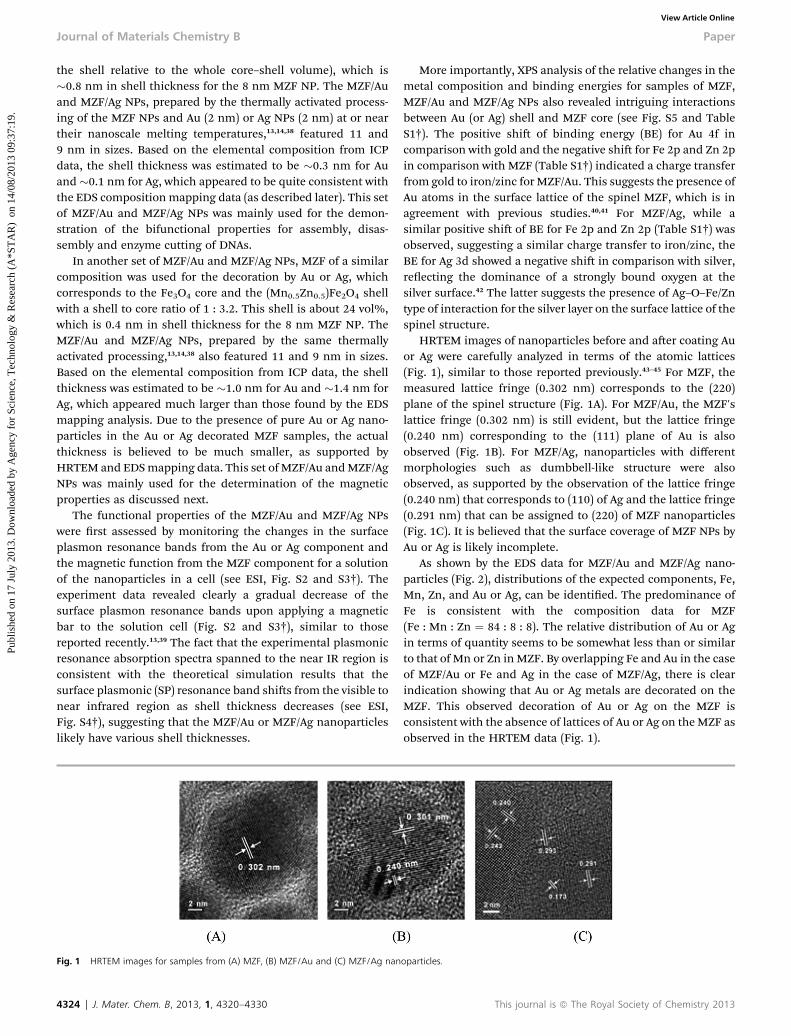
Fig. 1 HRTEM images for samples from (A) MZF, (B) MZF/Au and (C) MZF/Ag nan
4324 | J. Mater. Chem. B, 2013, 1, 4320–4330
More importantly, XPS analysis of the relative changes in themetal composition and binding energies for samples of MZF,MZF/Au and MZF/Ag NPs also revealed intriguing interactionsbetween Au (or Ag) shell and MZF core (see Fig. S5 and TableS1†). The positive shi of binding energy (BE) for Au 4f incomparison with gold and the negative shi for Fe 2p and Zn 2pin comparison with MZF (Table S1†) indicated a charge transferfrom gold to iron/zinc for MZF/Au. This suggests the presence ofAu atoms in the surface lattice of the spinel MZF, which is inagreement with previous studies.40,41 For MZF/Ag, while asimilar positive shi of BE for Fe 2p and Zn 2p (Table S1†) wasobserved, suggesting a similar charge transfer to iron/zinc, theBE for Ag 3d showed a negative shi in comparison with silver,reecting the dominance of a strongly bound oxygen at thesilver surface.42 The latter suggests the presence of Ag–O–Fe/Zntype of interaction for the silver layer on the surface lattice of thespinel structure.
HRTEM images of nanoparticles before and aer coating Auor Ag were carefully analyzed in terms of the atomic lattices(Fig. 1), similar to those reported previously.43–45 For MZF, themeasured lattice fringe (0.302 nm) corresponds to the (220)plane of the spinel structure (Fig. 1A). For MZF/Au, the MZF'slattice fringe (0.302 nm) is still evident, but the lattice fringe(0.240 nm) corresponding to the (111) plane of Au is alsoobserved (Fig. 1B). For MZF/Ag, nanoparticles with differentmorphologies such as dumbbell-like structure were alsoobserved, as supported by the observation of the lattice fringe(0.240 nm) that corresponds to (110) of Ag and the lattice fringe(0.291 nm) that can be assigned to (220) of MZF nanoparticles(Fig. 1C). It is believed that the surface coverage of MZF NPs byAu or Ag is likely incomplete.
As shown by the EDS data for MZF/Au and MZF/Ag nano-particles (Fig. 2), distributions of the expected components, Fe,Mn, Zn, and Au or Ag, can be identied. The predominance ofFe is consistent with the composition data for MZF(Fe : Mn : Zn ¼ 84 : 8 : 8). The relative distribution of Au or Agin terms of quantity seems to be somewhat less than or similarto that of Mn or Zn in MZF. By overlapping Fe and Au in the caseof MZF/Au or Fe and Ag in the case of MZF/Ag, there is clearindication showing that Au or Ag metals are decorated on theMZF. This observed decoration of Au or Ag on the MZF isconsistent with the absence of lattices of Au or Ag on the MZF asobserved in the HRTEM data (Fig. 1).
oparticles.
This journal is ª The Royal Society of Chemistry 2013
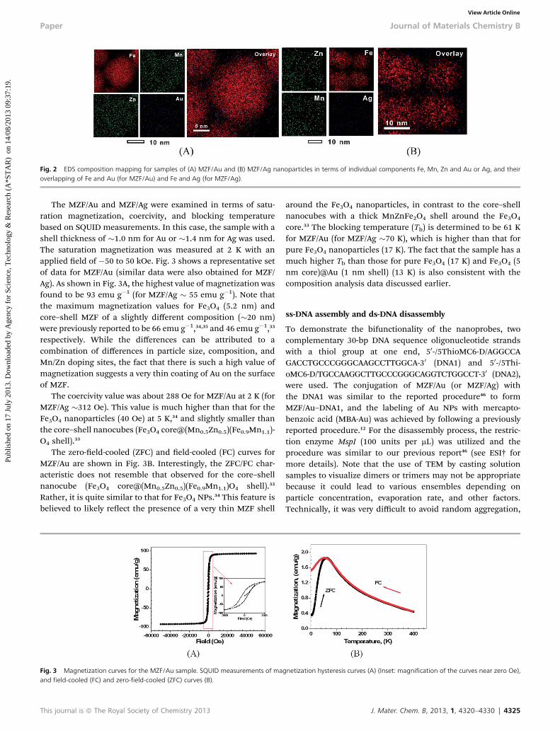
Fig. 2 EDS composition mapping for samples of (A) MZF/Au and (B) MZF/Ag nanoparticles in terms of individual components Fe, Mn, Zn and Au or Ag, and theiroverlapping of Fe and Au (for MZF/Au) and Fe and Ag (for MZF/Ag).
Paper Journal of Materials Chemistry B
Publ
ishe
d on
17
July
201
3. D
ownl
oade
d by
Age
ncy
for
Scie
nce,
Tec
hnol
ogy
& R
esea
rch
(A*S
TA
R)
on
14/0
8/20
13 0
9:37
:19.
View Article Online
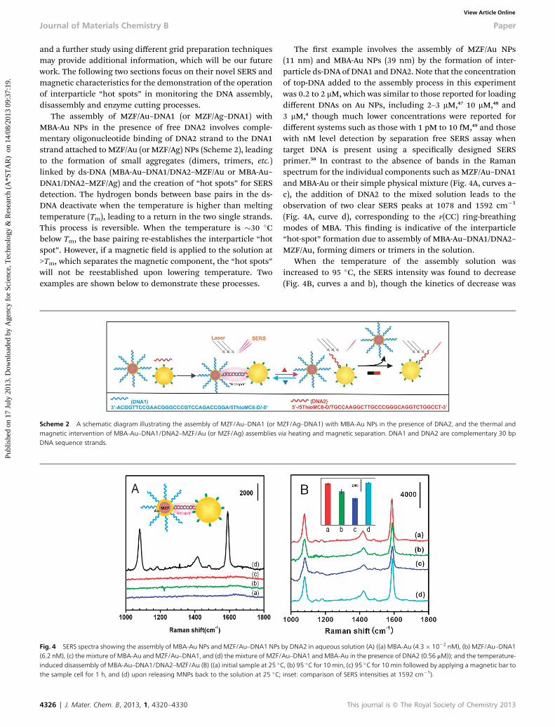
The MZF/Au and MZF/Ag were examined in terms of satu-ration magnetization, coercivity, and blocking temperaturebased on SQUID measurements. In this case, the sample with ashell thickness of �1.0 nm for Au or �1.4 nm for Ag was used.The saturation magnetization was measured at 2 K with anapplied eld of �50 to 50 kOe. Fig. 3 shows a representative setof data for MZF/Au (similar data were also obtained for MZF/Ag). As shown in Fig. 3A, the highest value of magnetization wasfound to be 93 emu g�1 (for MZF/Ag � 55 emu g�1). Note thatthe maximum magnetization values for Fe3O4 (5.2 nm) andcore–shell MZF of a slightly different composition (�20 nm)were previously reported to be 66 emu g�1,34,35 and 46 emu g�1,33
respectively. While the differences can be attributed to acombination of differences in particle size, composition, andMn/Zn doping sites, the fact that there is such a high value ofmagnetization suggests a very thin coating of Au on the surfaceof MZF.
The coercivity value was about 288 Oe for MZF/Au at 2 K (forMZF/Ag �312 Oe). This value is much higher than that for theFe3O4 nanoparticles (40 Oe) at 5 K,34 and slightly smaller thanthe core–shell nanocubes (Fe3O4 core@(Mn0.5Zn0.5)(Fe0.9Mn1.1)-O4 shell).33
The zero-eld-cooled (ZFC) and eld-cooled (FC) curves forMZF/Au are shown in Fig. 3B. Interestingly, the ZFC/FC char-acteristic does not resemble that observed for the core–shellnanocube (Fe3O4 core@(Mn0.5Zn0.5)(Fe0.9Mn1.1)O4 shell).33
Rather, it is quite similar to that for Fe3O4 NPs.34 This feature isbelieved to likely reect the presence of a very thin MZF shell
Fig. 3 Magnetization curves for the MZF/Au sample. SQUID measurements of maand field-cooled (FC) and zero-field-cooled (ZFC) curves (B).
This journal is ª The Royal Society of Chemistry 2013
around the Fe3O4 nanoparticles, in contrast to the core–shellnanocubes with a thick MnZnFe2O4 shell around the Fe3O4
core.33 The blocking temperature (Tb) is determined to be 61 Kfor MZF/Au (for MZF/Ag �70 K), which is higher than that forpure Fe3O4 nanoparticles (17 K). The fact that the sample has amuch higher Tb than those for pure Fe3O4 (17 K) and Fe3O4 (5nm core)@Au (1 nm shell) (13 K) is also consistent with thecomposition analysis data discussed earlier.
ss-DNA assembly and ds-DNA disassembly
To demonstrate the bifunctionality of the nanoprobes, twocomplementary 30-bp DNA sequence oligonucleotide strandswith a thiol group at one end, 50-/5ThioMC6-D/AGGCCAGACCTGCCCGGGCAAGCCTTGGCA-30 (DNA1) and 50-/5Thi-oMC6-D/TGCCAAGGCTTGCCCGGGCAGGTCTGGCCT-30 (DNA2),were used. The conjugation of MZF/Au (or MZF/Ag) withthe DNA1 was similar to the reported procedure46 to formMZF/Au–DNA1, and the labeling of Au NPs with mercapto-benzoic acid (MBA-Au) was achieved by following a previouslyreported procedure.12 For the disassembly process, the restric-tion enzyme MspI (100 units per mL) was utilized and theprocedure was similar to our previous report46 (see ESI† formore details). Note that the use of TEM by casting solutionsamples to visualize dimers or trimers may not be appropriatebecause it could lead to various ensembles depending onparticle concentration, evaporation rate, and other factors.Technically, it was very difficult to avoid random aggregation,
gnetization hysteresis curves (A) (Inset: magnification of the curves near zero Oe),
J. Mater. Chem. B, 2013, 1, 4320–4330 | 4325
Journal of Materials Chemistry B Paper
Publ
ishe
d on
17
July
201
3. D
ownl
oade
d by
Age
ncy
for
Scie
nce,
Tec
hnol
ogy
& R
esea
rch
(A*S
TA
R)
on
14/0
8/20
13 0
9:37
:19.
View Article Online
and a further study using different grid preparation techniquesmay provide additional information, which will be our futurework. The following two sections focus on their novel SERS andmagnetic characteristics for the demonstration of the operationof interparticle “hot spots” in monitoring the DNA assembly,disassembly and enzyme cutting processes.
The assembly of MZF/Au–DNA1 (or MZF/Ag–DNA1) withMBA-Au NPs in the presence of free DNA2 involves comple-mentary oligonucleotide binding of DNA2 strand to the DNA1strand attached to MZF/Au (or MZF/Ag) NPs (Scheme 2), leadingto the formation of small aggregates (dimers, trimers, etc.)linked by ds-DNA (MBA-Au–DNA1/DNA2–MZF/Au or MBA-Au–DNA1/DNA2–MZF/Ag) and the creation of “hot spots” for SERSdetection. The hydrogen bonds between base pairs in the ds-DNA deactivate when the temperature is higher than meltingtemperature (Tm), leading to a return in the two single strands.This process is reversible. When the temperature is �30 �Cbelow Tm, the base pairing re-establishes the interparticle “hotspot”. However, if a magnetic eld is applied to the solution at>Tm, which separates the magnetic component, the “hot spots”will not be reestablished upon lowering temperature. Twoexamples are shown below to demonstrate these processes.
Scheme 2 A schematic diagram illustrating the assembly of MZF/Au–DNA1 (or Mmagnetic intervention of MBA-Au–DNA1/DNA2–MZF/Au (or MZF/Ag) assemblies vDNA sequence strands.
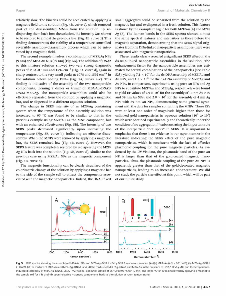
Fig. 4 SERS spectra showing the assembly of MBA-Au NPs and MZF/Au–DNA1 NPs(6.2 nM), (c) the mixture of MBA-Au andMZF/Au–DNA1, and (d) the mixture of MZF/induced disassembly of MBA-Au–DNA1/DNA2–MZF/Au (B) ((a) initial sample at 25 �
the sample cell for 1 h, and (d) upon releasing MNPs back to the solution at 25 �C;
4326 | J. Mater. Chem. B, 2013, 1, 4320–4330
The rst example involves the assembly of MZF/Au NPs(11 nm) and MBA-Au NPs (39 nm) by the formation of inter-particle ds-DNA of DNA1 and DNA2. Note that the concentrationof top-DNA added to the assembly process in this experimentwas 0.2 to 2 mM, which was similar to those reported for loadingdifferent DNAs on Au NPs, including 2–3 mM,47 10 mM,48 and3 mM,4 though much lower concentrations were reported fordifferent systems such as those with 1 pM to 10 fM,49 and thosewith nM level detection by separation free SERS assay whentarget DNA is present using a specically designed SERSprimer.50 In contrast to the absence of bands in the Ramanspectrum for the individual components such as MZF/Au–DNA1and MBA-Au or their simple physical mixture (Fig. 4A, curves a–c), the addition of DNA2 to the mixed solution leads to theobservation of two clear SERS peaks at 1078 and 1592 cm�1
(Fig. 4A, curve d), corresponding to the n(CC) ring-breathingmodes of MBA. This nding is indicative of the interparticle“hot-spot” formation due to assembly of MBA-Au–DNA1/DNA2–MZF/Au, forming dimers or trimers in the solution.
When the temperature of the assembly solution wasincreased to 95 �C, the SERS intensity was found to decrease(Fig. 4B, curves a and b), though the kinetics of decrease was
ZF/Ag–DNA1) with MBA-Au NPs in the presence of DNA2, and the thermal andia heating and magnetic separation. DNA1 and DNA2 are complementary 30 bp
by DNA2 in aqueous solution (A) ((a) MBA-Au (4.3 � 10�2 nM), (b) MZF/Au–DNA1Au–DNA1 andMBA-Au in the presence of DNA2 (0.56 mM)); and the temperature-C, (b) 95 �C for 10 min, (c) 95 �C for 10 min followed by applying a magnetic bar toinset: comparison of SERS intensities at 1592 cm�1).
This journal is ª The Royal Society of Chemistry 2013
Paper Journal of Materials Chemistry B
Publ
ishe
d on
17
July
201
3. D
ownl
oade
d by
Age
ncy
for
Scie
nce,
Tec
hnol
ogy
& R
esea
rch
(A*S
TA
R)
on
14/0
8/20
13 0
9:37
:19.
View Article Online
relatively slow. The kinetics could be accelerated by applying amagnetic eld to the solution (Fig. 4B, curve c), which removedpart of the disassembled MNPs from the solution. By re-dispersing them back into the solution, the intensity was shownto be restored to almost the previous level (Fig. 4B, curve d). Thisnding demonstrates the viability of a temperature-controlledreversible assembly–disassembly process which can be inter-vened by a magnetic eld.
The second example involves a combination of MZF/Ag NPs(9 nm) andMBA-Au NPs (39 nm) (Fig. 5A). The addition of DNA2to this mixture solution showed two very strong diagnosticpeaks of MBA at 1078 and 1592 cm�1 (Fig. 5A, curve d), again insharp contrast to the very small peaks at 1078 and 1592 cm�1 inthe solution before adding DNA2 (Fig. 5A, curves a–c). Thisnding is indicative of the assembly of the two nanoparticlecomponents, forming a dimer or trimer of MBA-Au–DNA1/DNA2–MZF/Ag. The nanoparticle assemblies could also beeffectively separated from the solution by applying a magneticbar, and re-dispersed in a different aqueous solution.
The change in SERS intensity of an MZF/Ag containingsystem when the temperature of the assembly solution wasincreased to 95 �C was found to be similar to that in theprevious example using MZF/Au as the MNP component, butwith an enhanced effectiveness (Fig. 5B). The intensity of twoSERS peaks decreased signicantly upon increasing thetemperature (Fig. 5B, curve b), indicating an effective disas-sembly. When the MNPs were removed by applying a magneticbar, the SERS remained low (Fig. 5B, curve c). However, theSERS feature was completely restored by redispersing the MZF/Ag NPs back into the solution (Fig. 5B, curve d), similar to theprevious case using MZF/Au NPs as the magnetic component(Fig. 4B, curve d).
The magnetic functionality can be clearly visualized of thecolorimetric change of the solution by applying a magnetic barto the side of the sample cell to attract the components asso-ciated with the magnetic nanoparticles. Indeed, the DNA-linked
Fig. 5 SERS spectra showing the assembly of MBA-Au NPs and MZF/Ag–DNA1 NPs(3.0 nM), (c) the mixture of MBA-Au andMZF/Ag–DNA1, and (d) the mixture of MZF/induced disassembly of MBA-Au–DNA1/DNA2–MZF/Ag (B) ((a) initial sample at 25 �
the sample cell for 1 h, and (d) upon releasing magnetic components back to the s
This journal is ª The Royal Society of Chemistry 2013
small aggregates could be separated from the solution by themagnetic bar and re-dispersed in a fresh solution. This featureis shown by the example in Fig. 6 for both MZF/Au (A) and MZF/Ag (B). The Raman bands in the SERS spectra showed almostthe same spectral features and intensities as those before themagnetic separation, demonstrating that the SERS signal orig-inates from the DNA-linked nanoparticle assemblies there wereassociated with magnetic nanoparticles.
These results clearly revealed a signicant SERS effect on theds-DNA-linked nanoparticle assemblies in the solution. Theenhancement factor for the nanoparticle assemblies was esti-mated for several combinations of the nanoparticles (see TableS2†), yielding 7.1 � 104 for the ds-DNA assembly of MZF/Au andAu NPs, and 1.5 � 105 for the ds-DNA assembly of MZF/Ag andAu NPs. In comparison, experiments using pure Au NPs and AgNPs to substitute MZF/Au and MZF/Ag, respectively were foundto yield EF values of 2.9 � 105 for the assembly of 13 nm Au NPsand 39 nm Au NPs, and 2.8 � 105 for the assembly of 4 nm AgNPs with 39 nm Au NPs, demonstrating some general agree-ment with the data for samples containing the MNPs. These EFswere at least one order of magnitude higher than those forunlinked gold nanoparticles in aqueous solution (102 to 103)which were obtained experimentally and theoretically under thecondition of no aggregation,51 substantiating the important roleof the interparticle “hot spots” in SERS. It is important toemphasize that there is no evidence in our experiment or in theliterature indicating the SERS effect of the pure magneticnanoparticles, which is consistent with the lack of effectiveplasmonic coupling for the pure magnetic particles. As evi-denced by the UV-Vis data, the plasmonic band of the pure AuNP is larger than that of the gold-coated magnetic nano-particles. Thus, the plasmonic coupling of the pure Au NPs isapparently greater than that of the gold-decorated magneticnanoparticles, leading to an increased enhancement. We didnot study the particle size effect at this point, which will be partof our future study.
by DNA2 in aqueous solution (A) ((a) MBA-Au (4.3 � 10�2 nM), (b) MZF/Ag–DNA1Ag–DNA1 andMBA-Au in the presence of DNA2 (0.56 mM)); and the temperature-C; (b) 95 �C for 10 min, and (c) 95 �C for 10 min followed by applying a magnet toolution at room temperature).
J. Mater. Chem. B, 2013, 1, 4320–4330 | 4327
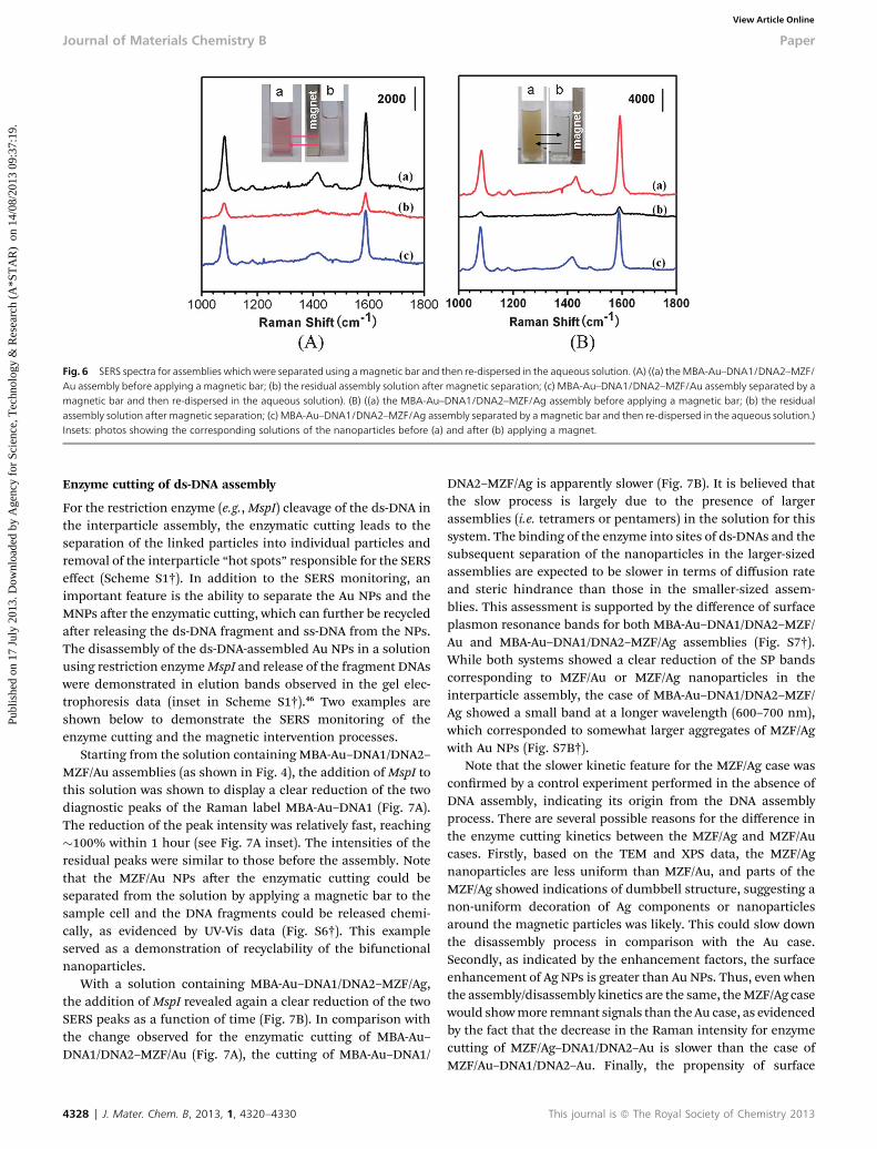
Fig. 6 SERS spectra for assemblies which were separated using amagnetic bar and then re-dispersed in the aqueous solution. (A) ((a) theMBA-Au–DNA1/DNA2–MZF/Au assembly before applying a magnetic bar; (b) the residual assembly solution after magnetic separation; (c) MBA-Au–DNA1/DNA2–MZF/Au assembly separated by amagnetic bar and then re-dispersed in the aqueous solution). (B) ((a) the MBA-Au–DNA1/DNA2–MZF/Ag assembly before applying a magnetic bar; (b) the residualassembly solution after magnetic separation; (c) MBA-Au–DNA1/DNA2–MZF/Ag assembly separated by amagnetic bar and then re-dispersed in the aqueous solution.)Insets: photos showing the corresponding solutions of the nanoparticles before (a) and after (b) applying a magnet.
Journal of Materials Chemistry B Paper
Publ
ishe
d on
17
July
201
3. D
ownl
oade
d by
Age
ncy
for
Scie
nce,
Tec
hnol
ogy
& R
esea
rch
(A*S
TA
R)
on
14/0
8/20
13 0
9:37
:19.
View Article Online
Enzyme cutting of ds-DNA assembly
For the restriction enzyme (e.g.,MspI) cleavage of the ds-DNA inthe interparticle assembly, the enzymatic cutting leads to theseparation of the linked particles into individual particles andremoval of the interparticle “hot spots” responsible for the SERSeffect (Scheme S1†). In addition to the SERS monitoring, animportant feature is the ability to separate the Au NPs and theMNPs aer the enzymatic cutting, which can further be recycledaer releasing the ds-DNA fragment and ss-DNA from the NPs.The disassembly of the ds-DNA-assembled Au NPs in a solutionusing restriction enzymeMspI and release of the fragment DNAswere demonstrated in elution bands observed in the gel elec-trophoresis data (inset in Scheme S1†).46 Two examples areshown below to demonstrate the SERS monitoring of theenzyme cutting and the magnetic intervention processes.
Starting from the solution containing MBA-Au–DNA1/DNA2–MZF/Au assemblies (as shown in Fig. 4), the addition ofMspI tothis solution was shown to display a clear reduction of the twodiagnostic peaks of the Raman label MBA-Au–DNA1 (Fig. 7A).The reduction of the peak intensity was relatively fast, reaching�100% within 1 hour (see Fig. 7A inset). The intensities of theresidual peaks were similar to those before the assembly. Notethat the MZF/Au NPs aer the enzymatic cutting could beseparated from the solution by applying a magnetic bar to thesample cell and the DNA fragments could be released chemi-cally, as evidenced by UV-Vis data (Fig. S6†). This exampleserved as a demonstration of recyclability of the bifunctionalnanoparticles.
With a solution containing MBA-Au–DNA1/DNA2–MZF/Ag,the addition of MspI revealed again a clear reduction of the twoSERS peaks as a function of time (Fig. 7B). In comparison withthe change observed for the enzymatic cutting of MBA-Au–DNA1/DNA2–MZF/Au (Fig. 7A), the cutting of MBA-Au–DNA1/
4328 | J. Mater. Chem. B, 2013, 1, 4320–4330
DNA2–MZF/Ag is apparently slower (Fig. 7B). It is believed thatthe slow process is largely due to the presence of largerassemblies (i.e. tetramers or pentamers) in the solution for thissystem. The binding of the enzyme into sites of ds-DNAs and thesubsequent separation of the nanoparticles in the larger-sizedassemblies are expected to be slower in terms of diffusion rateand steric hindrance than those in the smaller-sized assem-blies. This assessment is supported by the difference of surfaceplasmon resonance bands for both MBA-Au–DNA1/DNA2–MZF/Au and MBA-Au–DNA1/DNA2–MZF/Ag assemblies (Fig. S7†).While both systems showed a clear reduction of the SP bandscorresponding to MZF/Au or MZF/Ag nanoparticles in theinterparticle assembly, the case of MBA-Au–DNA1/DNA2–MZF/Ag showed a small band at a longer wavelength (600–700 nm),which corresponded to somewhat larger aggregates of MZF/Agwith Au NPs (Fig. S7B†).
Note that the slower kinetic feature for the MZF/Ag case wasconrmed by a control experiment performed in the absence ofDNA assembly, indicating its origin from the DNA assemblyprocess. There are several possible reasons for the difference inthe enzyme cutting kinetics between the MZF/Ag and MZF/Aucases. Firstly, based on the TEM and XPS data, the MZF/Agnanoparticles are less uniform than MZF/Au, and parts of theMZF/Ag showed indications of dumbbell structure, suggesting anon-uniform decoration of Ag components or nanoparticlesaround the magnetic particles was likely. This could slow downthe disassembly process in comparison with the Au case.Secondly, as indicated by the enhancement factors, the surfaceenhancement of Ag NPs is greater than Au NPs. Thus, even whenthe assembly/disassembly kinetics are the same, theMZF/Ag casewould showmore remnant signals than the Au case, as evidencedby the fact that the decrease in the Raman intensity for enzymecutting of MZF/Ag–DNA1/DNA2–Au is slower than the case ofMZF/Au–DNA1/DNA2–Au. Finally, the propensity of surface
This journal is ª The Royal Society of Chemistry 2013
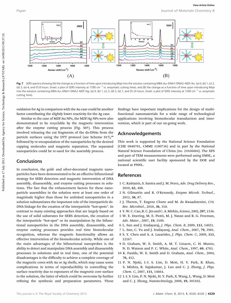
Fig. 7 SERS spectra showing (A) the change as a function of time upon introducingMspI into the solution containingMBA-Au–DNA1/DNA2–MZF/Au: (a) 0, (b) 1, (c) 2,(d) 3, (e) 4, and (f) 8 hours. (Inset: a plot of SERS intensity at 1590 cm�1 vs. enzymatic cutting time); and (B) the change as a function of time upon introducing MspIinto the solution containing MBA-Au–DNA1/DNA2–MZF/Ag: (a) 0, (b) 1, (c) 3, (d) 5, (e) 7, and (f) 24 hours. (Inset: a plot of SERS intensity at 1590 cm�1 vs. enzymaticcutting time).
Paper Journal of Materials Chemistry B
Publ
ishe
d on
17
July
201
3. D
ownl
oade
d by
Age
ncy
for
Scie
nce,
Tec
hnol
ogy
& R
esea
rch
(A*S
TA
R)
on
14/0
8/20
13 0
9:37
:19.
View Article Online
oxidation for Ag in comparison with the Au case could be anotherfactor contributing the slightly lower reactivity for the Ag case.
Similar to the case of MZF/Au NPs, the MZF/Ag NPs were alsodemonstrated to be recyclable by the magnetic interventionaer the enzyme cutting process (Fig. S8†). This processinvolved releasing the cut fragments of the ds-DNAs from theparticle surfaces using the DTT protocol (see Scheme S1†),46
followed by re-encapsulation of the nanoparticles by the desiredcapping molecules and magnetic separation. The separatednanoparticles could be re-used for the assembly process.
Conclusions
In conclusion, the gold- and silver-decorated magnetic nano-particles have been demonstrated to be an effective bifunctionalstrategy for SERS detection and magnetic intervention of DNAassembly, disassembly, and enzyme cutting processes in solu-tions. The fact that the enhancement factors for these nano-particle assemblies in the solution were at least one order ofmagnitude higher than those for unlinked nanoparticles in asolution substantiates the important role of the interparticle ds-DNA linkage for the creation of the interparticle “hot-spots”. Incontrast to many existing approaches that are largely based onthe use of solid substrates for SERS detection, the creation ofthe interparticle “hot-spot” or its manipulation by the bifunc-tional nanoparticles in the DNA assembly, disassembly, andenzyme cutting processes provides real time biomolecularrecognition, whereas the magnetic functionality allows aneffective intervention of the biomolecular activity. While one ofthe main advantages of the bifunctional nanoprobes is theability to detect andmanipulate DNA assembly and disassemblyprocesses in solutions and in real time, one of the potentialdisadvantages is the difficulty to achieve a complete coverage ofthe magnetic cores with Au or Ag shells, which may cause somecomplications in terms of reproducibility in controlling thesurface reactivity due to exposures of the magnetic core surfaceto the solution, the latter of which could be overcome by furtherrening the synthesis and preparation parameters. These
This journal is ª The Royal Society of Chemistry 2013
ndings have important implications for the design of multi-functional nanomaterials for a wide range of technologicalapplications involving biomolecular transduction and inter-vention, which is part of our on-going work.
Acknowledgements
This work is supported by the National Science Foundation(CHE 0848701, CMMI 1100736) and in part by the NationalNatural Science Foundation of China (no. 21036004). The XPSand part of TEM measurements were performed using EMSL, anational scientic user facility sponsored by the DOE andlocated at PNNL.
References
1 C. Kaittanis, S. Santra and J. M. Perez, Adv. Drug Delivery Rev.,2010, 62, 408.
2 N. Gilmartin and R. O'Kennedy, Enzyme Microb. Technol.,2012, 50, 87.
3 J. Theron, T. Eugene Cloete and M. de Kwaadsteniet, Crit.Rev. Microbiol., 2010, 36, 318.
4 Y.W.C.Cao,R.C. Jin andC.A.Mirkin,Science, 2002,297, 1536.5 W. E. Doering, M. E. Piotti, M. J. Natan and R. G. Freeman,Adv. Mater., 2007, 19, 3100.
6 L. Sun and J. Irudayaraj, J. Phys. Chem. B, 2009, 113, 14021.7 L. Sun, C. Yu and J. Irudayaraj, Anal. Chem., 2007, 79, 3981.8 S. Y. Chen and A. A. Lazarides, J. Phys. Chem. C, 2009, 113,12167.
9 D. Graham, W. E. Smith, A. M. T. Linacre, C. H. Munro,N. D. Watson and P. C. White, Anal. Chem., 1997, 69, 4703.
10 K. Faulds, W. E. Smith and D. Graham, Anal. Chem., 2004,76, 412.
11 P. N. Njoki, I.-I. S. Lim, D. Mott, H. Y. Park, B. Khan,S. Mishra, R. Sujakumar, J. Luo and C. J. Zhong, J. Phys.Chem. C, 2007, 111, 14664.
12 I.-I. S. Lim, P. N. Njoki, H. Y. Park, X. Wang, L. Wang, D. Mottand C. J. Zhong, Nanotechnology, 2008, 19, 305102.
J. Mater. Chem. B, 2013, 1, 4320–4330 | 4329
Journal of Materials Chemistry B Paper
Publ
ishe
d on
17
July
201
3. D
ownl
oade
d by
Age
ncy
for
Scie
nce,
Tec
hnol
ogy
& R
esea
rch
(A*S
TA
R)
on
14/0
8/20
13 0
9:37
:19.
View Article Online
13 H. Y. Park, M. J. Schadt, L. Y. Wang, I.-I. S. Lim, P. N. Njoki,S. H. Kim, M. Y. Jang, J. Luo and C. J. Zhong, Langmuir, 2007,23, 9050.
14 I.-I. S. Lim and C. J. Zhong, Acc. Chem. Res., 2009, 42, 798.15 K. Okamoto and D. Beach, EMBO J., 1994, 13, 4816.16 Y. Huang, S. Zhao, M. Shi, J. Chen, Z. F. Chen and H. Liang,
Anal. Chem., 2011, 83, 8913.17 H. Yan, I.-I. S. Lim, L. C. Zhang, S. C. Gao, D. Mott, Y. Le,
D. L. An and C. J. Zhong, J. Mater. Chem., 2011, 21, 1890.18 R. A. Alvarez-Puebla and L. M. Liz-Marzan, Chem. Soc. Rev.,
2012, 41, 43.19 S. Nie and S. R. Emery, Science, 1997, 275, 1102.20 K. L. Hultman, A. J. Raffo, A. L. Grzenda, P. E. Harris,
T. R. Brown and S. O'Brien, ACS Nano, 2008, 2, 477.21 H.W.Gu,K.M.Xu,C. J.XuandB.Xu,Chem.Commun., 2006,941.22 M. Mahmoudi, A. Simchi, M. Imani, A. S. Milani and
P. Stroeve, J. Phys. Chem. B, 2008, 112, 14470.23 M. R. Jones, K. D. Osberg, R. J. Macfarlane, M. R. Langille
and C. A. Mirkin, Chem. Rev., 2011, 111, 3736.24 L. Li, T. Hutter, A. S. Finnemore, F. M. Huang,
J. J. Baumberg, S. R. Elliott, U. Steiner and S. Mahajan,Nano Lett., 2012, 12, 4242.
25 X. Zhou, W. L. Xu, Y. Wang, Q. Kuang, Y. F. Shi, L. B. Zhongand Q. Q. Zhang, J. Phys. Chem. C, 2010, 114, 19607.
26 B.-H. Jun, M. S. Noh, J. Y. Kim, G. S. Kim, H. M. Kang,M. S. Kim, Y. T. Seo, J. H. Baek, J. H. Kim, J. Y. Park,S. Y. Kim, Y. K. Kim, T. W. Hyeon, M. H. Cho, D. H. Jeongand Y. S. Lee, Small, 2010, 6, 119.
27 C. A. Tao, Q. An, W. Zhu, H. W. Yang, W. N. Li, C. X. Lin,D. Xu and G. T. Li, Chem. Commun., 2011, 47, 9867.
28 X. M. Qian, X. Zhou and S. M. Nie, J. Am. Chem. Soc., 2008,130, 14934.
29 Z. L. Zhang, Y. Q. Wen, Y. Ma, J. Luo, L. Jiang and Y. L. Song,Chem. Commun., 2011, 47, 7407.
30 Y. Liang, J. L. Gong, Y. Huang, Y. Zheng, J. H. Jiang,G. L. Shen and R. Q. Yu, Talanta, 2007, 72, 443.
31 H. Zhang, M. H. Harpster, W. C. Wilson and P. A. Johnson,Langmuir, 2012, 28, 4030.
32 H. Zhang, M. H. Harpster, H. J. Park and P. A. Johnson, Anal.Chem., 2011, 83, 254.
33 L. Y. Wang, X. Wang, J. Luo, B. N. Wanjala, C. Wang,N. Chernova, M. H. Engelhard, I.-T. Bae, Y. Liu andC. J. Zhong, J. Am. Chem. Soc., 2010, 132, 17686.
4330 | J. Mater. Chem. B, 2013, 1, 4320–4330
34 L. Y. Wang, J. Luo, Q. Fan, M. Suzuki, I. S. Suzuki,M. H. Engelhard, Y. Lin, N. Kim, J. Q. Wang andC. J. Zhong, J. Phys. Chem. B, 2005, 109, 21593.
35 L. Y. Wang, J. Luo, M. M. Maye, Q. Fan, Q. Rendeng,M. H. Engelhard, C. Wang, Y. Lin and C. J. Zhong,J. Mater. Chem., 2005, 15, 1821.
36 E. Hao and C. G. Schatz, J. Chem. Phys., 2004, 120, 357.37 E. Crew, H. Yan, L. Q. Lin, Z. Skeete, T. Kotlyar, N. Tchah,
J. Lee, M. Bellavia, I. Goodshaw, P. Joseph, J. Luo, S. Galand C. J. Zhong, Analyst, 2013, DOI: 10.1039/c3an00683b.
38 M. J. Hostetler, C. J. Zhong, B. K. H. Yen, J. Anderegg,S. M. Gross, N. D. Evans, M. D. Porter and R. W. Murray,J. Am. Chem. Soc., 1998, 120, 9396.
39 L. Y. Wang, J. Luo, S. Y. Shan, E. Crew, J. Yin, B. Wallek,S. Wong and C. J. Zhong, Anal. Chem., 2011, 83, 8688.
40 H. Yu, M. Chen, P. M. Rice, S. X. Wang, R. L. White andS. Sun, Nano Lett., 2005, 5, 379.
41 C. Wang, C. Xu, H. Zeng and S. Sun, Adv. Mater., 2009, 21,3045.
42 X. Bao, M. Muhler, T. Schedel-Niedrig and R. Schlogl, Phys.Rev. B: Condens. Matter Mater. Phys., 1996, 54, 2249.
43 J. Salado, M. Insausti, L. Lezama, I. Gil de Muro, M. Moros,B. Pelaz, V. Grazu, J. M. de la Fuente and T. Rojo,Nanotechnology, 2012, 23, 315102.
44 H. L. Liu, J. H. Wu, J. H. Min and Y. K. Kim, J. Alloys Compd.,2012, 537, 60.
45 H. M. Song, Q. S. Wei, Q. K. Ong and A. Wei, ACS Nano, 2010,4, 5163.
46 I.-I. S. Lim, U. Chandrachud, L. Y. Wang, S. Gal andC. J. Zhong, Anal. Chem., 2008, 80, 6038.
47 J. Wang, L. Wu, J. Ren and X. Qu, Small, 2012, 8, 259.48 X. Wang, X. Zhang, P. He and Y. Fang, Biosens. Bioelectron.,
2011, 26, 3608.49 K. J. Jang, H. Lee, H. L. Jin, Y. Park and J. M. Nam, Small,
2009, 5, 2665.50 D. van Lierop, K. Faulds and D. Graham, Anal. Chem., 2011,
83, 5817.51 V. Joseph, A. Matschulat, J. Polte, S. Rolf, F. Emmerling and
J. Kneipp, J. Raman Spectrosc., 2011, 42, 1736.52 N. N. Kariuki, J. Luo, M. M. Maye, A. Hassan, T. Menard,
H. R. Naslund, Y. Lin, C. Wang, M. H. Engelhard andC. J. Zhong, Langmuir, 2004, 20, 11240.
53 D. I. Gittins and F. Caruso, ChemPhysChem, 2002, 3, 110.
This journal is ª The Royal Society of Chemistry 2013
Related Documents