BEVACIZUMAB (AVASTIN) DOES NOT HARM RETINAL FUNCTION AFTER INTRAVITREAL INJECTION AS SHOWN BY ELECTRORETINOGRAPHY IN ADULT MICE PETER HEIDUSCHKA, PHD,* SYLVIE JULIEN, PHD,* SABINE HOFMEISTER,* KARL ULRICH BARTZ-SCHMIDT, PHD,† ULRICH SCHRAERMEYER, PHD* Purpose: Scavenging of VEGF by specific antibodies is a promising way to treat ocular conditions connected with neovascularization. Intravitreal injections of Avastin (bevacizumab) are performed frequently as a treatment of such conditions. In this study, the authors examine whether the retinal function in wild-type mice is affected by an intravitreal injection of Avastin. Methods: Electroretinography was performed in four different experimental groups of wild-type C57BL/6 mice before treatment and 1, 4, 12, and 25 days afterwards. The first group was injected intravitreally with BSS, the second one received injections of a vehicle solution, and the third group was injected with the commercial Avastin solution. In a fourth group, sham surgery was performed. Immunohistochemistry was performed in some eyes to evaluate penetration of the bevacizumab molecule through the retina. Results: In all four groups, a similar behavior of the ERG parameters could be detected. One day after the injections, the amplitudes showed a clear decrease. Later on, they recovered gradually. No difference could be seen between eyes injected with Avastin or vehicle solution. Bevacizumab immunoreactivity was already present in the whole retina half an hour after the intravitreal injection and was not detectable 25 days later. Moreover, binding of bevacizumab to endogenous mouse VEGF could be shown. Conclusions: Based on the electroretinographic findings, the authors conclude that bevacizumab does not have any toxic effects on the mouse retina and its function. The bevacizumab molecule penetrates the retina quickly. Therefore, it can act safely and very quickly, also in deeper retinal layers after its injection. RETINA 28:46 –55, 2008 N eovascularization occurs in several diseases of the eye, such as wet age-related macular degen- eration, proliferative diabetic retinopathy, rubeosis, or retinopathy of prematurity. One of the major factors inducing formationke of new vessels is the vascular endothelial growth factor (VEGF). 1 Two main VEGF inhibitors have been launched recently for the treatment of ocular neovasculariza- tion: pegaptanib (Macugen, Eye Tech/Pfizer) and ranibizumab (rhuFab V2, Lucentis, Genentech/Novar- tis). Another VEGF-binding compound is bevaci- zumab (Avastin, Roche/Genentech), which is a com- plete humanized murine monoclonal antibody against all isoforms of VEGF. 2,3 Bevacizumab has been ap- proved by the Food and Drug Administration for the From the †Centre of Ophthalmology and *Experimental Vitreo- retinal Surgery, University of Tu ¨bingen Eye Hospital, Germany. None of the authors has proprietary interest. Presented in part at the annual ARVO meeting; May 2006; Ft. Lauderdale, Florida. Reprint requests: Dr. Peter Heiduschka, University Eye Hospital, Experimental Vitreoretinal Surgery, Schleichstr. 12/1, D–72076 Tu ¨bingen, Germany; e-mail: [email protected] 46

Welcome message from author
This document is posted to help you gain knowledge. Please leave a comment to let me know what you think about it! Share it to your friends and learn new things together.
Transcript

BEVACIZUMAB (AVASTIN) DOES NOTHARM RETINAL FUNCTION AFTERINTRAVITREAL INJECTION AS SHOWN BYELECTRORETINOGRAPHY IN ADULT MICEPETER HEIDUSCHKA, PHD,* SYLVIE JULIEN, PHD,*SABINE HOFMEISTER,* KARL ULRICH BARTZ-SCHMIDT, PHD,†ULRICH SCHRAERMEYER, PHD*
Purpose: Scavenging of VEGF by specific antibodies is a promising way to treat ocularconditions connected with neovascularization. Intravitreal injections of Avastin (bevacizumab)are performed frequently as a treatment of such conditions. In this study, the authors examinewhether the retinal function in wild-type mice is affected by an intravitreal injection of Avastin.
Methods: Electroretinography was performed in four different experimental groups ofwild-type C57BL/6 mice before treatment and 1, 4, 12, and 25 days afterwards. The firstgroup was injected intravitreally with BSS, the second one received injections of a vehiclesolution, and the third group was injected with the commercial Avastin solution. In a fourthgroup, sham surgery was performed. Immunohistochemistry was performed in some eyesto evaluate penetration of the bevacizumab molecule through the retina.
Results: In all four groups, a similar behavior of the ERG parameters could be detected.One day after the injections, the amplitudes showed a clear decrease. Later on, theyrecovered gradually. No difference could be seen between eyes injected with Avastin orvehicle solution. Bevacizumab immunoreactivity was already present in the whole retinahalf an hour after the intravitreal injection and was not detectable 25 days later. Moreover,binding of bevacizumab to endogenous mouse VEGF could be shown.
Conclusions: Based on the electroretinographic findings, the authors conclude thatbevacizumab does not have any toxic effects on the mouse retina and its function. Thebevacizumab molecule penetrates the retina quickly. Therefore, it can act safely and veryquickly, also in deeper retinal layers after its injection.
RETINA 28:46–55, 2008
Neovascularization occurs in several diseases ofthe eye, such as wet age-related macular degen-
eration, proliferative diabetic retinopathy, rubeosis, orretinopathy of prematurity. One of the major factors
inducing formationke of new vessels is the vascularendothelial growth factor (VEGF).1
Two main VEGF inhibitors have been launchedrecently for the treatment of ocular neovasculariza-tion: pegaptanib (Macugen, Eye Tech/Pfizer) andranibizumab (rhuFab V2, Lucentis, Genentech/Novar-tis). Another VEGF-binding compound is bevaci-zumab (Avastin, Roche/Genentech), which is a com-plete humanized murine monoclonal antibody againstall isoforms of VEGF.2,3 Bevacizumab has been ap-proved by the Food and Drug Administration for the
From the †Centre of Ophthalmology and *Experimental Vitreo-retinal Surgery, University of Tubingen Eye Hospital, Germany.
None of the authors has proprietary interest.Presented in part at the annual ARVO meeting; May 2006; Ft.
Lauderdale, Florida.Reprint requests: Dr. Peter Heiduschka, University Eye Hospital,
Experimental Vitreoretinal Surgery, Schleichstr. 12/1, D–72076Tubingen, Germany; e-mail: [email protected]
46

treatment of metastatic colorectal cancer and is in PhaseIII trials for advanced breast and renal cancers.4–7 Com-pared to the above mentioned VEGF inhibitors, bevaci-zumab is by far the less cost-intensive treatment, whichis important when considering the required reinjectionsin 4- to 6-week intervals. Systemically or intravitreallyapplied bevacizumab significantly reduced macularedema and neovascularization in patients with age-related macular degeneration, central vein occlusion, andretinal angiomatous proliferation.8–16
VEGF is not only an important angiogenic factor,but is also important for the proliferation of neuronalprecursors, development and survival of a variety ofneurons in the central nervous system, including theretina,17–22 where it is expressed by neurones and theMuller cells.23–25 Therefore, it is of great interest tomake sure that depletion of VEGF does not affectsurvival and normal function of retinal neurons. Onthe other hand, binding of bevacizumab to murineVEGF is rather weak, so it is of general interestwhether the intravitreal injection of a foreign proteincan harm the retinal integrity and function.
The purpose of this study is to test if intravitreallyinjected bevacizumab in its commercial formulation(Avastin) has any detrimental side effects on retinalfunction and/or structure. The commercial formula-tion contains approximately 0.04% of the detergentpolysorbate (Tween) 20, and it has to be clarifiedwhether it harms the retina and its function. Moreover,it will be checked immunohistochemically if the pro-tein molecule bevacizumab penetrates deeper layers ofthe retina after intravitreal injection, which is crucialfor its use as an antiangiogenic agent.
Materials and Methods
Animals and Intravitreal Injections
We used wild-type mice, strain C57BL/6. The animalswere anesthetized with a mixture of ketamine and xyla-zine (120 mg/kg ketamine, 10 mg/kg xylazine), and theeye was desensitized by a drop of Novesine (NovartisOphthalmics). Four experimental groups were estab-lished: three in which different intravitreal injectionswere performed, and one sham group.
For the intravitreal injections, a small incision wasmade into the conjunctiva at the outer corner of theeyes. The eyeball was rotated by grasping the con-junctiva with a pair of fine tweezers and gentle pull-ing. The conjunctiva was incised to allow direct ac-cess to the sclera. A small hole was made into thesclera using a 30 gauge needle. A volume of 1 �L ofBSS (Alcon, balanced saline solution used for bothintra- and extraocular irrigation, n � 10), commercial
Avastin solution (Roche Pharma AG, Grenzach, Ger-many, 25 mg bevacizumab per mL, n � 17), orvehicle solution (n � 14) was injected through thehole intravitreally using a Hamilton syringe with a 32gauge needle. Four milliliters of the vehicle solutioncontains 240 mg a,a-trehalose · 2 H2O, 23.2 mgNa2HPO4 · H2O, 4.8 mg NaH2PO4, and 1.6 mg poly-sorbate 20. Therefore, the vehicle solution constitutesthe formulation of commercial Avastin solution, ex-cept that it does not contain the bevacizumab protein.
After the injection, the needle remained in the eyefor an additional 3 or 4 seconds to reduce reflux andwas then drawn back. The eyeball was brought backinto its normal position, and the antibiotic ointmentGentamytrex (Dr. Mann Pharma, Berlin) was appliedto the eye. The whole procedure was performed usinga surgical microscope equipped with illumination. Theperson performing the injections was blinded aboutthe kind of injected solution.
In the sham group (n � 3), the same manipula-tions were performed with the animals, except forthe injections.
Electroretinography
Electroretinograms were recorded from anesthe-tized animals according to standard procedures usingthe commercial RetiPort32 device from Roland Con-sult Systems, Germany. After a dark adaptation periodof at least 12 hours, animals were anesthetized asdescribed above. The cornea was desensitized by adrop of Novesine (Novartis Ophthalmics). The pupilswere dilated by a drop of tropicamide (Novartis Oph-thalmics). Gold wire ring electrodes served as work-ing and reference electrodes, respectively, and wereput onto the cornea of the eyes and into the mouth. Astainless steel needle electrode was inserted into thetail of the animals for grounding. All these manipula-tions were performed under dim red light, withoutbringing the animal into ambient light after overnightdark adaptation. After an additional 5 minutes to allowthe pupil to dilate, standard electroretinographic mea-surements were performed, with scotopic flash ERG atfive different light intensities, an additional run forscotopic oscillatory potentials, photopic flash ERG after10 minutes of light adaptation, and photopic 30 HzFlicker. The maximum light intensity used for the flasheswas 3 cd. The time of measurement was 160 msec,sample rate 3.2 kHz, and frequency range of 0.5 to 200Hz for both scotopic and photopic flash ERG, 50 to 500Hz for oscillatory potentials, and 10 to 50 Hz for 30 HzFlicker. The body temperature of the animals was kept at37°C during the measurement. ERG recordings wereperformed before the intravitreal injection, and then 1, 4,
47BEVACIZUMAB WITHOUT EFFECT ON MOUSE ERG ● HEIDUSCHKA ET AL
12, and 25 days after the injection. The person whoperformed the measurements did not know which eyereceived which injection.
For data evaluation, changes in amplitudes and la-tencies of scotopic a- and b-waves, scotopic oscilla-tory potentials, photopic b-waves, and photopic 30-HzFlicker ERG were analyzed by setting the valuesobtained before the treatments to 100% as a baseline.Significance was tested using the t-test.
Immunohistochemistry
Animals were enucleated 30 minutes, 1 day, and 25days after the intravitreal injection of either Avastin orvehicle solution. The eyes were fixed in formalin,embedded in paraffin wax, cut to 5 �m sections, anddeparaffinized according to standard procedures. Todetect the bevacizumab in the retina, the sections wereincubated overnight with a biotinylated polyclonal rabbitantihuman IgG antibody (dilution 1:2000, E0482, Dako-Cymation, Glostrup, Denmark). The bound antibody wasthen visualized using the Dako ChemMate Detection KitAlkaline Phosphatase/Red.
To compare binding of bevacizumab to mouseVEGF compared to a regular VEGF antibody, paraffinsections obtained from an untreated mouse eye wereincubated with either Avastin (concentration 25 �g/mL) or an antimouse VEGF antibody (R&D Systems,AF-493-NA, dilution 1:15). Donkey antihuman IgGlabeled with Cy3 (Jackson Immunoresearch, 709-166-149, dilution 1:500) was used for detection of boundbevacizumab. This polyclonal antibody binds to manyepitopes of both Fc and Fab portions of human IgG.The bound antimouse VEGF antibody was detectedusing donkey antigoat IgG labeled with Alexa488(Molecular Probes, A11055, dilution 1:400). In anadditional experiment, Avastin was preincubated withthe recombinant mouse VEGF protein (R&D Systems,493-MV) for 2 hours before it was given onto paraffinsections of an untreated mouse eye.
Results
Electroretinography
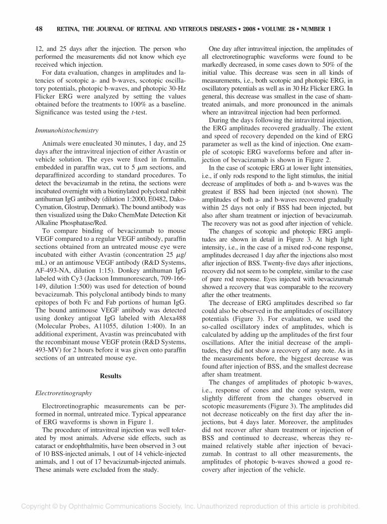
Electroretinographic measurements can be per-formed in normal, untreated mice. Typical appearanceof ERG waveforms is shown in Figure 1.
The procedure of intravitreal injection was well toler-ated by most animals. Adverse side effects, such ascataract or endophthalmitis, have been observed in 3 outof 10 BSS-injected animals, 1 out of 14 vehicle-injectedanimals, and 1 out of 17 bevacizumab-injected animals.These animals were excluded from the study.
One day after intravitreal injection, the amplitudes ofall electroretinographic waveforms were found to bemarkedly decreased, in some cases down to 50% of theinitial value. This decrease was seen in all kinds ofmeasurements, i.e., both scotopic and photopic ERG, inoscillatory potentials as well as in 30 Hz Flicker ERG. Ingeneral, this decrease was smallest in the case of sham-treated animals, and more pronounced in the animalswhere an intravitreal injection had been performed.
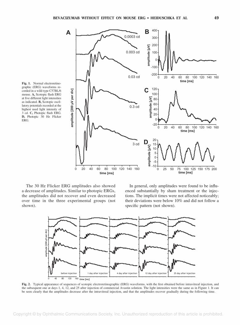
During the days following the intravitreal injection,the ERG amplitudes recovered gradually. The extentand speed of recovery depended on the kind of ERGparameter as well as the kind of injection. One exam-ple of scotopic ERG waveforms before and after in-jection of bevacizumab is shown in Figure 2.
In the case of scotopic ERG at lower light intensities,i.e., if only rods respond to the light stimulus, the initialdecrease of amplitudes of both a- and b-waves was thegreatest if BSS had been injected (not shown). Theamplitudes of both a- and b-waves recovered graduallywithin 25 days not only if BSS had been injected, butalso after sham treatment or injection of bevacizumab.The recovery was not as good after injection of vehicle.
The changes of scotopic and photopic ERG ampli-tudes are shown in detail in Figure 3. At high lightintensity, i.e., in the case of a mixed rod-cone response,amplitudes decreased 1 day after the injections also mostafter injection of BSS. Twenty-five days after injections,recovery did not seem to be complete, similar to the caseof pure rod response. Eyes injected with bevacizumabshowed a recovery that was comparable to the recoveryafter the other treatments.
The decrease of ERG amplitudes described so farcould also be observed in the amplitudes of oscillatorypotentials (Figure 3). For evaluation, we used theso-called oscillatory index of amplitudes, which iscalculated by adding up the amplitudes of the first fouroscillations. After the initial decrease of the ampli-tudes, they did not show a recovery of any note. As inthe measurements before, the biggest decrease wasfound after injection of BSS, and the smallest decreaseafter sham treatment.
The changes of amplitudes of photopic b-waves,i.e., response of cones and the cone system, wereslightly different from the changes observed inscotopic measurements (Figure 3). The amplitudes didnot decrease noticeably on the first day after the in-jections, but 4 days later. Moreover, the amplitudesdid not recover after sham treatment or injection ofBSS and continued to decrease, whereas they re-mained relatively stable after injection of bevaci-zumab. In contrast to all other measurements, theamplitudes of photopic b-waves showed a good re-covery after injection of the vehicle.
48 RETINA, THE JOURNAL OF RETINAL AND VITREOUS DISEASES ● 2008 ● VOLUME 28 ● NUMBER 1
The 30 Hz Flicker ERG amplitudes also showeda decrease of amplitudes. Similar to photopic ERGs,the amplitudes did not recover and even decreasedover time in the three experimental groups (notshown).
In general, only amplitudes were found to be influ-enced substantially by sham treatment or the injec-tions. The implicit times were not affected noticeably;their deviations were below 10% and did not follow aspecific pattern (not shown).
Fig. 1. Normal electroretino-graphic (ERG) waveforms re-corded in a wild-type C57BL/6mouse. A, Scotopic flash ERGat five different light intensitiesas indicated. B, Scotopic oscil-latory potentials recorded at thehighest used light intensity of3 cd. C, Photopic flash ERG.D, Photopic 30 Hz FlickerERG.
Fig. 2. Typical appearance of sequences of scotopic electroretinographic (ERG) waveforms, with the first obtained before intravitreal injection, andthe subsequent one at days 1, 4, 12, and 25 after injection of commercial Avastin solution. The light intensities were the same as in Figure 1. It canbe seen clearly that the amplitudes decrease after the intravitreal injection, and that the amplitudes recover gradually during the following time.
49BEVACIZUMAB WITHOUT EFFECT ON MOUSE ERG ● HEIDUSCHKA ET AL

Immunohistochemistry
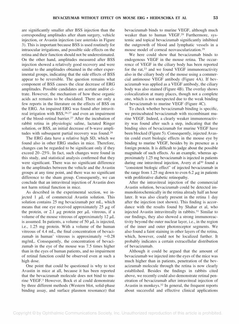
To check whether the bevacizumab molecule rec-ognizes endogenous murine VEGF, paraffin sectionsof untreated eyes were incubated with Avastin. Forcomparison, an antimouse VEGF antibody was alsoused to label VEGF in the eye sections. VEGF immu-noreactivity (IR) visualized by the VEGF antibodywas seen both in the ciliary body and the retina. WhenAvastin was used, a staining of the ciliary body wasalso found. In both cases, no specific labeling wasseen in the negative controls. Furthermore, a double-labeling of retinal paraffin section was performed us-ing the antimouse VEGF antibody and Avastin, re-spectively (Figure 4). Again, VEGF IR can be seen inthe ciliary body, after both antimouse VEGF antibodyand Avastin treatment (Figure 4, A and B, respec-tively). Overlay of both stainings shows clear colocal-ization at many places (Figure 4C).
As a next experiment, we tried blocking of thebevacizumab molecule with murine VEGF before giv-ing it onto the mouse eye sections. The results areshown in Figure 5. Whereas there is no specific stain-ing of the retina in the negative control, several retinallayers are stained after incubation with Avastin. IfAvastin is incubated with recombinant murine VEGF
before its application on the eye section, most of thestaining is gone (Figure 5).
Further histochemical analysis was performed tocheck if the intravitreally injected bevacizumab pen-etrates the retina (Figure 6). The paraffin sections ofthe retina do not show any signs of damage; the layersof the retina and the cells appear to be intact except fora slight occasional loosening of the dense packing inthe inner and outer nuclear layers, respectively.
Bevacizumab immunoreactivity could already be de-tected in the retina half an hour after the injection, evenin deeper layers. The staining was also present 1 dayafter the injection and then faded away gradually, until ithad diminished completely 25 days after the injection.
Discussion
C57BL/6 mice are a well-established animal modelbecause their retina mimics a large part of the mor-phologic, biochemical, and physiologic properties ofthe human retina (except for the macula, which doesnot exist in rodents). This is also true for electroreti-nographic measurements that yield waveforms com-parable to those achieved in humans. For this reason,this mouse strain is frequently used for a wide variety
Fig. 3. Changes of several kinds of electroretinographic (ERG) amplitudes as indicated in the diagrams after the different kinds of intravitrealinjections (black: sham-treated animals, white: injection of BSS, light gray: injection of vehicle solution, dark gray: injection of commercial Avastinsolution). Error bars indicate SD. In the lower part of the columns, significance of the changes compared to base line is indicated by diagonal crosses,with � � P � 0.05, �� � P � 0.01, ��� � P � 0.001. Asterisks indicate significance of differences between treatments at single time points,with *P � 0.05 and **P � 0.01.
50 RETINA, THE JOURNAL OF RETINAL AND VITREOUS DISEASES ● 2008 ● VOLUME 28 ● NUMBER 1
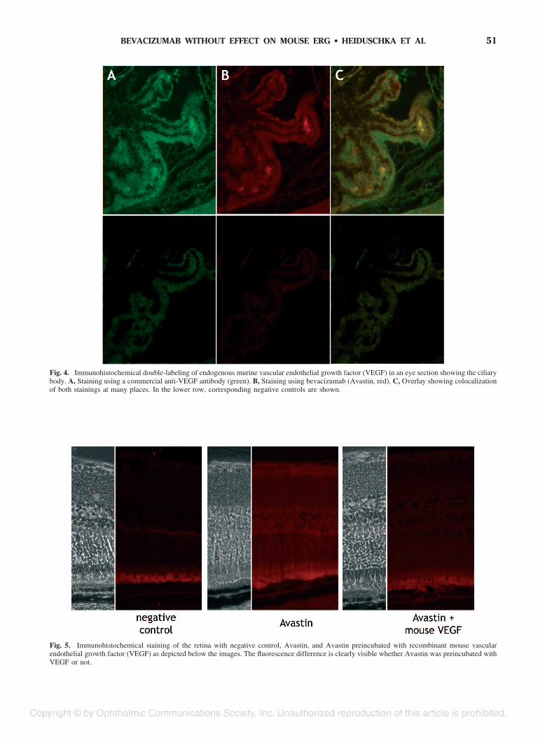
Fig. 4. Immunohistochemical double-labeling of endogenous murine vascular endothelial growth factor (VEGF) in an eye section showing the ciliarybody. A, Staining using a commercial anti-VEGF antibody (green). B, Staining using bevacizumab (Avastin, red). C, Overlay showing colocalizationof both stainings at many places. In the lower row, corresponding negative controls are shown.
Fig. 5. Immunohistochemical staining of the retina with negative control, Avastin, and Avastin preincubated with recombinant mouse vascularendothelial growth factor (VEGF) as depicted below the images. The fluorescence difference is clearly visible whether Avastin was preincubated withVEGF or not.
51BEVACIZUMAB WITHOUT EFFECT ON MOUSE ERG ● HEIDUSCHKA ET AL

of studies, among them to test compounds on theirneuroprotective, neuromodulatory, or toxic properties.
Intravitreal injections are relatively safe in micedespite the small size of their eyes. Using the tech-nique described above, only a few incidents of inflam-mation or cataract had to be registered, and theseanimals were excluded from the study.
The first crucial issue of this study is the dramaticdecrease of ERG amplitudes 1 day after the intravit-real injection, an observation we also made in otherstudies using this technique. Two main reasons can besuggested for this finding. First, the procedure ofintravitreal injection is performed using a microscopeequipped with a bright lamp. Therefore, a severebleaching of photoreceptors has to be taken into ac-count, if not even a certain light-induced damage ofthe photoreceptors. Secondly, the intravitreal injectionis carried out by penetrating the sclera and the retina,which represents a local trauma to the eye damagingthe retina at this site.
As described in the Results, the ERG amplitudesrecovered gradually after their initial decrease. Tocheck if intravitreally injected bevacizumab has anytoxic side effects on retinal function, the extent of boththe initial decrease and recovery of amplitudes had tobe evaluated. As the main result of this study, wecould show that the ERG amplitudes observed afterintravitreal injection of bevacizumab behave similarly toERG amplitudes after injection of BSS or vehicle. Therecovery of the amplitudes after bevacizumab injectioneven tends to be better than after injection of BSS or
vehicle in the scotopic ERGs, and clearly better thanafter sham treatment or BSS injection in the photopicERG. Therefore, we can conclude that intravitreal injec-tion of bevacizumab (Avastin) in mice appears to be safewith respect to retinal function.
A point that requires further consideration is thedifferent behavior of ERG parameters after differentinjections. Whereas ERG amplitudes were clearly de-creased in rod-dominated scotopic ERG after injectionof the vehicle and recovered later on, the amplitudesin photopic ERG showed no decrease on the first dayafter injection and decreased later. This opposite be-havior was seen in particular after sham treatment orinjection of BSS, i.e., a recovery of amplitudes inscotopic ERG and a further decrease of amplitudes inphotopic ERG. It seems that the cones are injured bythe bright light during the surgery, leading to theirgradual inactivation and/or degeneration, which is re-flected by the continued decrease of photopic amplitudesafter sham treatment or BSS injection. It is not clear atthe moment why this decrease was not seen after injec-tion of vehicle or Avastin. Probably the trehalose, whichis present in the commercial Avastin formulation andhence also in the vehicle, has some protective effect onthe function of cones. However, this could not be exam-ined further in this study and remains speculative.
One striking finding is the sharp decrease of ERGamplitudes 1 day after intravitreal injection of BSS.Whereas there is no significant difference between thetreatment groups 4 days after injection or later, thescotopic b-wave and oscillatory potential amplitudes
Fig. 6. Immunohistochemical detection of bevacizumab in the retina 30 minutes (0 days) and 25 days after intravitreal injection of Avastin solution.It can be clearly seen that the bevacizumab molecule penetrates the mouse retina quickly after the injection. No trace of bevacizumab can be found25 days after the injection.
52 RETINA, THE JOURNAL OF RETINAL AND VITREOUS DISEASES ● 2008 ● VOLUME 28 ● NUMBER 1
are significantly smaller after BSS injection than thecorresponding amplitudes after sham surgery, vehicleinjection, or Avastin injection (see asterisks in Figure3). This is important because BSS is used routinely forintraocular irrigations, and possible side effects on theretina and their function should not be underestimated.On the other hand, amplitudes measured after BSSinjection showed a relatively good recovery and weresimilar to the amplitudes obtained in the other exper-imental groups, indicating that the side effects of BSSappear to be reversible. The question remains whatcomponent of BSS causes the clear decrease of ERGamplitudes. Possible candidates are acetate and/or ci-trate. However, the mechanism of how these organicacids act remains to be elucidated. There are only afew reports in the literature on the effects of BSS onthe ERG. An impaired ERG was found after intravit-real irrigation with BSS,26,27 and even an impairmentof the blood–retinal barrier.27 After the incubation ofrabbit retina in physiologic saline, lactated Ringersolution, or BSS, an initial decrease of b-wave ampli-tudes with subsequent partial recovery was found.28
The ERG data have a relative high SD, which wefound also in other ERG studies in mice. Therefore,changes can be regarded to be significant only if theyexceed 20–25%. In fact, such changes were found inthis study, and statistical analysis confirmed that theywere significant. There was no significant differencein the amplitudes between the vehicle and the Avastingroups at any time point, and there was no significantdifference to the sham group. Consequently, we canconclude that an intravitreal injection of Avastin doesnot harm retinal function in mice.
As described in the experimental section, we in-jected 1 �L of commercial Avastin solution. Thissolution contains 25 mg bevacizumab per mL, whichmeans that one eye received approximately 25 �g ofthe protein, or 2.1 �g protein per �L vitreous, if avolume of the mouse vitreous of approximately 12 �Lis assumed. In patients, a volume of 50 �L is injected,i.e., 1.25 mg protein. With a volume of the humanvitreous of 4.4 mL, the final concentration of bevaci-zumab in human’ vitreous is approximately �0.28mg/mL. Consequently, the concentration of bevaci-zumab in the eye of the mouse was 7.5 times higherthan in the eyes of human patients, and no impairmentof retinal function could be observed even at such ahigh dose.
One point that could be questioned is why to testAvastin in mice at all, because it has been reportedthat the bevacizumab molecule does not bind to mu-rine VEGF.3 However, Bock et al could demonstrateby three different methods (Western blot, solid-phasebinding assay, and surface plasmon resonance) that
bevacizumab binds to murine VEGF, although muchweaker than to human VEGF.29 Furthermore, sys-temic and topical bevacizumab significantly inhibitedthe outgrowth of blood and lymphatic vessels in amouse model of corneal neovascularization.29
We here could show that bevacizumab binds toendogenous VEGF in the mouse retina. The occur-rence of VEGF in the ciliary body has been reportedfor the rat,23 and we found VEGF immunoreactivityalso in the ciliary body of the mouse using a commer-cial antimouse VEGF antibody (Figure 4A). If bev-acizumab was applied as a VEGF antibody, the ciliarybody was also stained (Figure 4B). The overlay showscolocalization at many places, though not a completeone, which is not unexpected due to the weak bindingof bevacizumab to murine VEGF (Figure 4C).
To check whether bevacizumab binding is specific,we preincubated bevacizumab with recombinant mu-rine VEGF. Indeed, a clearly weaker immunoreactiv-ity was found after such a step, indicating that thebinding sites of bevacizumab for murine VEGF havebeen blocked (Figure 5). Consequently, injected Avas-tin could exert biologic effects in the mouse eye bybinding to murine VEGF, besides by its presence as aforeign protein. It is difficult to judge about the possiblemagnitude of such effects. As an example, whereas ap-proximately 1.25 mg bevacizumab is injected in patientsduring one intravitreal injection, Avery et al30 found a“consistent biologic effect” of injected bevacizumab inthe range from 1.25 mg down to even 6.2 �g in patientswith proliferative diabetic retinopathy.
After the intravitreal injection of the commercialAvastin solution, bevacizumab could be detected im-munohistochemically in the retina already half an hourlater. It was also clearly present in the retina 1 dayafter the injection (not shown). This finding is accor-dance with the results found by Shahar et al, whoinjected Avastin intravitreally in rabbits.31 Similar toour findings, they also showed a strong immunoreac-tivity beyond the outer nuclear layer, i.e., in the regionof the inner and outer photoreceptor segments. Wealso found a faint staining in other layers of the retina,which, however, could not be localized further. Itprobably indicates a certain extracellular distributionof bevacizumab.
Although it could be argued that the amount ofbevacizumab we injected into the eyes of the mice wasmuch higher than in patients, penetration of the bev-acizumab molecule through the retina is now clearlyestablished. Besides the findings in rabbits citedabove, we recently could also demonstrate retinal pen-etration of bevacizumab after intravitreal injection ofAvastin in monkeys.32 In general, the frequent reportsabout successful and effective clinical applications
53BEVACIZUMAB WITHOUT EFFECT ON MOUSE ERG ● HEIDUSCHKA ET AL
suggest that the bevacizumab molecule penetrates thehuman retina to reach its site of action.
Avastin injections in patients are repeated monthly,if necessary. In our study, no bevacizumab immuno-reactivity could be detected 25 days after injection.Similarly, Shahar et al reported that no bevacizumablabeling was found 4 weeks after the intravitreal in-jection.31 Therefore, the time schedule of monthlyinjections seems to be appropriate.
Shahar et al31 suggest that clinically observed ef-fects of intravitreally injected Avastin may be relatedto a thinning of both the inner limiting membrane andthe retina at the fovea. In our laboratory, we found aquick penetration (i.e., within 1 day) of bevacizumabthrough the retina in mice, and also in rabbits andcynomolgus monkeys.32 Mice and rabbits do not havea fovea, which rules out the possibility of increasedpenetration at this site. The Tween 20 contained in thecommercial formulation of bevacizumab could be onemajor factor promoting quick penetration of bevaci-zumab through the retina. As an example, applicationof detergents to allow antibodies to penetrate the ret-ina belongs to the standard routines in retinal whole-mount immunohistochemistry. Therefore, it is not thatsurprising to find bevacizumab penetrating the retinasof our experimental animals.
As a summary, no adverse side effects on retinalfunction could be detected after intravitreal injectionof the commercial Avastin solution in mice. In thecontext with other studies where the effect of bevaci-zumab on the ERG had been tested in vitro and invivo,32–36 intravitreal injection of Avastin seems to besafe with regard to retinal function.
Key words: Avastin, bevacizumab, BSS, electro-retinography, immunohistochemistry, intravitreal in-jection, mouse, retinal function, toxicity testing.
References
1. Witmer AN, Vrensen FJM, Van Noorden CJF, SchlingemannRO. Vascular endothelial growth factors and angiogenesis ineye disease. Progr Retin Eye Res 2003;22:1–29.
2. Presta LG, Chen H, O’Connor SJ, et al. Humanization of ananti-vascular endothelial growth factor monoclonal antibodyfor the therapy of solid tumors and other disorders. CancerRes 1997;57:4593–4599.
3. Ferrara N, Hillan KJ, Gerber H-P, Novotny W. Discoveryand development of bevacizumab, an anti-VEGF antibody fortreating cancer. Nature Rev Drug Discov 2004;3:391–400.
4. Yang JC, Haworth L, Sherry RM, et al. A randomized trial ofbevacizumab, an anti-vascular endothelial growth factor an-tibody, for metastatic renal cancer. N Engl J Med 2003;349:427–434.
5. Miller KD, Chap LI, Holmes FA, et al. Randomized phase IIItrial of capecitabine compared with bevacizumab plus cape-citabine in patients with previously treated metastatic breastcancer. J Clin Oncol 2005;23:792–799.
6. Ferrara N, Hillan KJ, Novotny W. Bevacizumab (Avastin), ahumanized anti-VEGF monoclonal antibody for cancer ther-apy. Biochem Biophys Res Commun 2005;333:328–335.
7. Hurwitz H. Integrating the anti–VEGF-A humanized monoclo-nal antibody bevacizumab with chemotherapy in advanced colo-rectal cancer. Clin Colorectal Cancer 2004;2:S62–S68.
8. Michels S, Rosenfeld PJ, Puliafito CA, et al. MARINA study;Systemic bevacizumab (Avastin) therapy for neovascularage-related macular degeneration twelve-week results of anuncontrolled open-label clinical study. Ophthalmology 2005;112:1035–1047.
9. Reichel E. Intravitreal bevacizumab for choroidal neovascu-larization and cystoid macular edema: a cost-effective treat-ment? Ophthalmic Surg Lasers Imaging 2005;36:270–271.
10. Rosenfeld PJ, Fung AE, Puliafito CA. Optical coherencetomography findings after an intravitreal injection of bevaci-zumab (Avastin) for macular edema from central retinal veinocclusion. Ophthalmic Surg Lasers Imaging 2005;36:336–339.
11. Rosenfeld PJ, Moshfeghi AA, Puliafito CA. Optical coherencetomography findings after an intravitreal injection of bevaci-zumab (Avastin) for neovascular age-related macular degener-ation. Ophthalmic Surg Lasers Imaging 2005;36:331–335.
12. Avery RL, Pieramici DJ, Rabena MD, et al. Intravitrealbevacizumab (Avastin) for neovascular age-related maculardegeneration. Ophthalmology 2006;113:363–372.
13. Rabena MD, Pieramici DJ, Castellarin AA, et al. Intravitrealbevacizumab (Avastin) in the treatment of macular edemasecondary to branch retinal vein occlusion. Retina 2007;27:419–425.
14. Emerson MV, Lauer AK, Flaxel CJ, et al. Intravitreal bev-acizumab (Avastin) treatment of neovascular age-relatedmacular degeneration. Retina 2007;27:439–444.
15. Joeres S, Heussen FMA, Treziak T, et al. Bevacizumab(Avastin) treatment in patients with retinal angiomatous pro-liferation. Graefes Arch Clin Exp Ophthalmol 2008 (in press).
16. Meyerle CB, Freund KB, Iturralde D, et al. Intravitreal bev-acizumab (Avastin) for retinal angiomatous proliferation.Retina 2007;27:451–457.
17. Matsuzaki H, Tamatani M, Yamaguchi A, et al. Vascularendothelial growth factor rescues hippocampal neurons fromglutamate-induced toxicity: signal transduction cascades.FASEB J 2001;15:1218–1220.
18. Robinson GS, Ju M, Shih SC, et al. Nonvascular role forVEGF: VEGFR-1, 2 activity is critical for neural retinaldevelopment. FASEB J 2001;15:1215–1217.
19. Carmeliet P, Storkebaum E. Vascular and neuronal effects ofVEGF in the nervous system: implications for neurologicaldisorders. Seminars Cell Dev Biol 2002;13:39–53.
20. Jin K, Zhu Y, Sun Y, et al. Vascular endothelial growth factor(VEGF) stimulates neurogenesis in vitro and in vivo. ProcNat Acad Sci USA 2002;99:11946–11950.
21. Ogunshola OO, Antic A, Donoghue MJ, et al. Paracrine andautocrine functions of neuronal vascular endothelial growthfactor (VEGF) in the central nervous system. J Biol Chem2002;277:11410–11415.
22. Storkebaum E, Lambrechts D, Carmeliet P. VEGF: onceregarded as a specific angiogenic factor, now implicated inneuroprotection. BioEssays 2004;26:943–954.
23. Ueda H, Kashiwagi K, Iizuka Y. Vascular endothelial growthfactor and its receptors expression in the rat eye. Acta His-tochem. Cytochem 2001;34:329–335.
24. Famiglietti EV, Stopa EG, McGookin ED, et al. Immunocy-tochemical localization of vascular endothelial growth factor
54 RETINA, THE JOURNAL OF RETINAL AND VITREOUS DISEASES ● 2008 ● VOLUME 28 ● NUMBER 1
in neurons and glial cells of human retina. Brain Res 2003;969:195–204.
25. Gariano RF, Hu D, Helms J. Expression of angiogenesis-related genes during retinal development. Gene Expr Patterns2006;6:187–192.
26. Moorhead LC, Redburn DA, Merritt J, Garcia CA. Theeffects of intravitreal irrigation during vitrectomy on theelectroretinogram. Am J Ophthalmol 1979;88:239–245.
27. Garner WH, Scheib S, Berkowitz BA, et al. The effect ofpartial vitrectomy on blood-ocular barrier function in therabbit. Curr Eye Res 2001;23:372–381.
28. Negi A, Honda Y, Kawano S. Effects of intraocular irrigatingsolutions on the electroretinographic b-wave. Am J Ophthal-mol 1981;92:28–37.
29. Bock F, Onderka J, Dietrich T, et al. Bevacizumab as a potentinhibitor of inflammatory corneal angiogenesis and lymphangio-genesis. Invest Ophthalmol Vis Sci 2007;48:2545–2552.
30. Avery RL, Pearlman J, Pieramici DJ, et al. Intravitreal bev-acizumab (Avastin) in the treatment of proliferative diabeticretinopathy. Ophthalmology 2006;113:1695–1705.
31. Shahar J, Avery RL, Heilweil G, et al. Electrophysiologic andretinal penetration studies following intravitreal injection ofbevacizumab (Avastin). Retina 2006;26:262–269.
32. Heiduschka P, Fietz H, Hofmeister S, et al. Penetration ofbevacizumab through the retina after intravitreal injectionin the monkey. Invest Ophthalmol Vis Sci 2007;48:2814–2823.
33. Luke M, Warga M, Ziemssen F, et al. Effects of bevacizumabon retinal function in isolated vertebrate retina. Br J Ophthal-mol 2006;90:1178–1182.
34. Manzano RP, Peyman GA, Khan P, Kivilcim M. Testingintravitreal toxicity of bevacizumab (Avastin). Retina 2006;26:257–261.
35. Maturi RK, Bleau LA, Wilson DL. Electrophysiologic find-ings after intravitreal bevacizumab (Avastin) treatment. Ret-ina 2006;26:270–274.
36. Inan OO, Avci B, Kusbeci T, et al. Preclinical safety evalu-ation of intravitreal injection of full-length humanized vas-cular endothelial growth factor antibody in rabbit eyes. InvestOphthalmol Vis Sci 2007;48:1773–1781.
55BEVACIZUMAB WITHOUT EFFECT ON MOUSE ERG ● HEIDUSCHKA ET AL
Related Documents