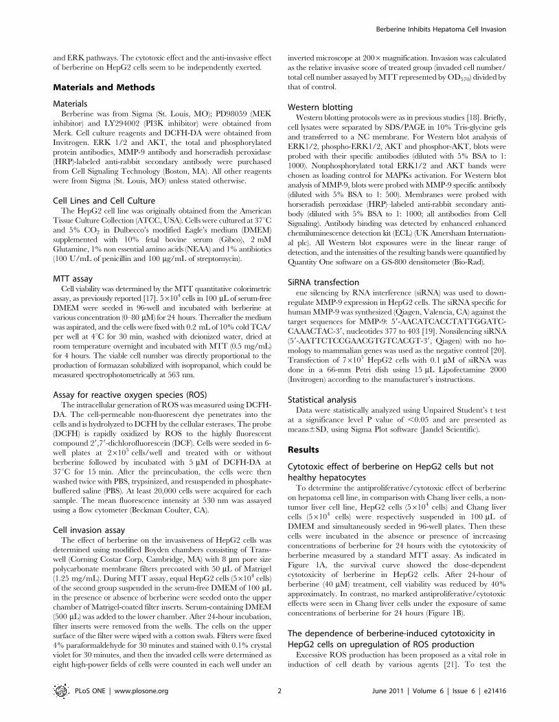
Berberine Inhibits Human Hepatoma Cell Invasion without Cytotoxicity in Healthy Hepatocytes Bing Liu 1. , Genshu Wang 2. , Jie Yang 3. , Xuediao Pan 1 , Zhicheng Yang 1 , Linquan Zang 1 * 1 Department of Pharmacology, School of Pharmacy, Guangdong Pharmaceutical University, Guangzhou, People’s Republic of China, 2 Department of Liver Surgery, the Third Affiliated Hospital, Sun Yat-Sen University, Guangzhou, People’s Republic of China, 3 Department of Pharmacology, School of Pharmacy, Guangxi Medical University, Nanning, People’s Republic of China Abstract Conventional chemotherapy fails to cure metastatic hepatoma mainly due to its high hepatotoxicity. Many plant-derived agents have been accepted to effectively inhibit hepatoma cell invasion. However, the investigation that whether effectual plant-derived agents against invasive hepatoma cells exert unexpected cytotoxicity in healthy hepatocytes has been ignored. This study demonstrated that berberine exhibited significant cytotoxicity in HepG2 cells mainly through upregulation of reactive oxygen species (ROS) production but was ineffective in normal Chang liver cells. Berberine exerted anti-invasive effect on HepG2 cells through suppression of matrix metalloproteinase-9 (MMP-9) expression. Moreover, berberine could significantly inhibit the activity of PI3K-AKT and ERK pathways. Combination treatment of ERK pathway inhibitor PD98059 or AKT pathway inhibitor LY294002 and berberine could result in a synergistic reduction on MMP-9 expression along with an inhibition of cell invasion. Enhancement of ROS production by berberine had no influence on its suppressive effects on the activity of PI3K-AKT and ERK pathways, as well as MMP-9 expression and HepG2 cell invasion. In conclusion, our results suggest that berberine may be a potential alternative against invasive hepatoma cells through PI3K- AKT and ERK pathways-dependent downregulation of MMP-9 expression. This study also provides a previously neglected insight into the investigation of plant-derived agents-based therapy against tumor invasion with the consideration of damage to healthy cells. Citation: Liu B, Wang G, Yang J, Pan X, Yang Z, et al. (2011) Berberine Inhibits Human Hepatoma Cell Invasion without Cytotoxicity in Healthy Hepatocytes. PLoS ONE 6(6): e21416. doi:10.1371/journal.pone.0021416 Editor: Alfons Navarro, University of Barcelona, Spain Received December 10, 2010; Accepted June 1, 2011; Published June 24, 2011 Copyright: ß 2011 Liu et al. This is an open-access article distributed under the terms of the Creative Commons Attribution License, which permits unrestricted use, distribution, and reproduction in any medium, provided the original author and source are credited. Funding: The authors have no support or funding to report. Competing Interests: The authors have declared that no competing interests exist. * E-mail: [email protected] . These authors contributed equally to this work. Introduction Hepatoma is one of the most frequent and death-leading visceral neoplasms worldwide [1]. Most hepatoma patients have underlying hepatic dysfunction, which complicates safe administration of systemic therapy and conduction of trials of new agents [2]. Invasive phenotypes are fundamental components of malignant hepatoma, and thus become critical targets of anti-cancer agents development [3]. Most advanced hepatoma cells, which become progressively dedifferentiated, have high proliferative activity and progress to give rise to invasion and metastasis [4]. The most widely used agent against invasive hepatoma cells is doxorubicin, either as a single agent or in combination with other chemother- apeutics like cisplatin. However, this conventional chemotherapy has shown only a minimal survival advantage, as the hepatotox- icity of doxorubicin, which may undoubtedly aggratate hepatic dysfunction, remains a severe concern [5]. Many plant-derived agents with few adverse effects have been accepted as potential alternatives to the therapy for invasive hepatoma [6,7,8,9]. However, most of these agents have failed to exert antiproliferative/cytotoxic effects on hepatoma cells within their suited anti-invasive doses [8,9]. More importantly, to date, the investigation that whether effectual plant-derived agents against invasive hepatoma cells have unexpected cytotoxicity in healthy hepatocytes has been neglected. Berberine, a clinically important natural isoquinoline alkaloid derived from Berberis species, has been reported to exhibit multiple pharmacological activities including anti-cancer effect [10]. A recent report indicated that berberine could induce hepatoma cell apoptosis through a mitochondria/caspases path- way while elicit no cytotoxic effects in healthy hepatocytes [11]. Yet the exact mechanism underlying the different effects of berberine on highly proliferative hepatoma cells and normal hepatocytes has not been fully elucidated. Specifically, berberine has gradually entered the limelight for its potentially therapeutic effect against invasion and metastasis of various lines of cancers such as glioma, lung cancer and nasopharyngeal carcinoma [12,13,14]. Very recently, berberine was firstly reported to inhibit melanoma cell migration, an essential step in invasion, by inhibition of COX-2, PGE 2 and PGE2 receptors [15]. Neverthe- less, no information about the action of berberine on invasive hepatoma cells has been addressed. In this study, we explored the effects of berberine on malignant invasive phenotypes of HepG2 cells (a highly invasive human hepatoma cell line [16]). Our results demonstrate the critical component of ROS production in berberine-induced inconsistent cytotoxic effects on HepG2 cells and normal Chang liver cells. Specifically, berberine, without any cytotoxic effect on normal hepatocytes, inhibits HepG2 cell invasion through suppression of MMP-9 expression by concomitant inactivation of the PI3K-AKT PLoS ONE | www.plosone.org 1 June 2011 | Volume 6 | Issue 6 | e21416

Welcome message from author
This document is posted to help you gain knowledge. Please leave a comment to let me know what you think about it! Share it to your friends and learn new things together.
Transcript
Berberine Inhibits Human Hepatoma Cell Invasionwithout Cytotoxicity in Healthy HepatocytesBing Liu1., Genshu Wang2., Jie Yang3., Xuediao Pan1, Zhicheng Yang1, Linquan Zang1*
1 Department of Pharmacology, School of Pharmacy, Guangdong Pharmaceutical University, Guangzhou, People’s Republic of China, 2 Department of Liver Surgery, the
Third Affiliated Hospital, Sun Yat-Sen University, Guangzhou, People’s Republic of China, 3 Department of Pharmacology, School of Pharmacy, Guangxi Medical University,
Nanning, People’s Republic of China
Abstract
Conventional chemotherapy fails to cure metastatic hepatoma mainly due to its high hepatotoxicity. Many plant-derivedagents have been accepted to effectively inhibit hepatoma cell invasion. However, the investigation that whether effectualplant-derived agents against invasive hepatoma cells exert unexpected cytotoxicity in healthy hepatocytes has beenignored. This study demonstrated that berberine exhibited significant cytotoxicity in HepG2 cells mainly throughupregulation of reactive oxygen species (ROS) production but was ineffective in normal Chang liver cells. Berberine exertedanti-invasive effect on HepG2 cells through suppression of matrix metalloproteinase-9 (MMP-9) expression. Moreover,berberine could significantly inhibit the activity of PI3K-AKT and ERK pathways. Combination treatment of ERK pathwayinhibitor PD98059 or AKT pathway inhibitor LY294002 and berberine could result in a synergistic reduction on MMP-9expression along with an inhibition of cell invasion. Enhancement of ROS production by berberine had no influence on itssuppressive effects on the activity of PI3K-AKT and ERK pathways, as well as MMP-9 expression and HepG2 cell invasion. Inconclusion, our results suggest that berberine may be a potential alternative against invasive hepatoma cells through PI3K-AKT and ERK pathways-dependent downregulation of MMP-9 expression. This study also provides a previously neglectedinsight into the investigation of plant-derived agents-based therapy against tumor invasion with the consideration ofdamage to healthy cells.
Citation: Liu B, Wang G, Yang J, Pan X, Yang Z, et al. (2011) Berberine Inhibits Human Hepatoma Cell Invasion without Cytotoxicity in Healthy Hepatocytes. PLoSONE 6(6): e21416. doi:10.1371/journal.pone.0021416
Editor: Alfons Navarro, University of Barcelona, Spain
Received December 10, 2010; Accepted June 1, 2011; Published June 24, 2011
Copyright: � 2011 Liu et al. This is an open-access article distributed under the terms of the Creative Commons Attribution License, which permits unrestricteduse, distribution, and reproduction in any medium, provided the original author and source are credited.
Funding: The authors have no support or funding to report.
Competing Interests: The authors have declared that no competing interests exist.
* E-mail: [email protected]
. These authors contributed equally to this work.
Introduction
Hepatoma is one of the most frequent and death-leading visceral
neoplasms worldwide [1]. Most hepatoma patients have underlying
hepatic dysfunction, which complicates safe administration of
systemic therapy and conduction of trials of new agents [2].
Invasive phenotypes are fundamental components of malignant
hepatoma, and thus become critical targets of anti-cancer agents
development [3]. Most advanced hepatoma cells, which become
progressively dedifferentiated, have high proliferative activity and
progress to give rise to invasion and metastasis [4]. The most
widely used agent against invasive hepatoma cells is doxorubicin,
either as a single agent or in combination with other chemother-
apeutics like cisplatin. However, this conventional chemotherapy
has shown only a minimal survival advantage, as the hepatotox-
icity of doxorubicin, which may undoubtedly aggratate hepatic
dysfunction, remains a severe concern [5].
Many plant-derived agents with few adverse effects have been
accepted as potential alternatives to the therapy for invasive
hepatoma [6,7,8,9]. However, most of these agents have failed to
exert antiproliferative/cytotoxic effects on hepatoma cells within
their suited anti-invasive doses [8,9]. More importantly, to date,
the investigation that whether effectual plant-derived agents
against invasive hepatoma cells have unexpected cytotoxicity in
healthy hepatocytes has been neglected.
Berberine, a clinically important natural isoquinoline alkaloid
derived from Berberis species, has been reported to exhibit
multiple pharmacological activities including anti-cancer effect
[10]. A recent report indicated that berberine could induce
hepatoma cell apoptosis through a mitochondria/caspases path-
way while elicit no cytotoxic effects in healthy hepatocytes [11].
Yet the exact mechanism underlying the different effects of
berberine on highly proliferative hepatoma cells and normal
hepatocytes has not been fully elucidated. Specifically, berberine
has gradually entered the limelight for its potentially therapeutic
effect against invasion and metastasis of various lines of cancers
such as glioma, lung cancer and nasopharyngeal carcinoma
[12,13,14]. Very recently, berberine was firstly reported to inhibit
melanoma cell migration, an essential step in invasion, by
inhibition of COX-2, PGE2 and PGE2 receptors [15]. Neverthe-
less, no information about the action of berberine on invasive
hepatoma cells has been addressed.
In this study, we explored the effects of berberine on malignant
invasive phenotypes of HepG2 cells (a highly invasive human
hepatoma cell line [16]). Our results demonstrate the critical
component of ROS production in berberine-induced inconsistent
cytotoxic effects on HepG2 cells and normal Chang liver cells.
Specifically, berberine, without any cytotoxic effect on normal
hepatocytes, inhibits HepG2 cell invasion through suppression of
MMP-9 expression by concomitant inactivation of the PI3K-AKT
PLoS ONE | www.plosone.org 1 June 2011 | Volume 6 | Issue 6 | e21416
and ERK pathways. The cytotoxic effect and the anti-invasive effect
of berberine on HepG2 cells seem to be independently exerted.
Materials and Methods
MaterialsBerberine was from Sigma (St. Louis, MO); PD98059 (MEK
inhibitor) and LY294002 (PI3K inhibitor) were obtained from
Merk. Cell culture reagents and DCFH-DA were obtained from
Invitrogen. ERK 1/2 and AKT, the total and phosphorylated
protein antibodies, MMP-9 antibody and horseradish peroxidase
(HRP)-labeled anti-rabbit secondary antibody were purchased
from Cell Signaling Technology (Boston, MA). All other reagents
were from Sigma (St. Louis, MO) unless stated otherwise.
Cell Lines and Cell CultureThe HepG2 cell line was originally obtained from the American
Tissue Culture Collection (ATCC, USA). Cells were cultured at 37uCand 5% CO2 in Dulbecco’s modified Eagle’s medium (DMEM)
supplemented with 10% fetal bovine serum (Gibco), 2 mM
Glutamine, 1% non essential amino acids (NEAA) and 1% antibiotics
(100 U/mL of penicillin and 100 mg/mL of streptomycin).
MTT assayCell viability was determined by the MTT quantitative colorimetric
assay, as previously reported [17]. 56104 cells in 100 mL of serum-free
DMEM were seeded in 96-well and incubated with berberine at
various concentration (0–80 mM) for 24 hours. Thereafter the medium
was aspirated, and the cells were fixed with 0.2 mL of 10% cold TCA/
per well at 4uC for 30 min, washed with deionized water, dried at
room temperature overnight and incubated with MTT (0.5 mg/mL)
for 4 hours. The viable cell number was directly proportional to the
production of formazan solubilized with isopropanol, which could be
measured spectrophotometrically at 563 nm.
Assay for reactive oxygen species (ROS)The intracellular generation of ROS was measured using DCFH-
DA. The cell-permeable non-fluorescent dye penetrates into the
cells and is hydrolyzed to DCFH by the cellular esterases. The probe
(DCFH) is rapidly oxidized by ROS to the highly fluorescent
compound 29,79-dichlorofluorescein (DCF). Cells were seeded in 6-
well plates at 26105 cells/well and treated with or without
berberine followed by incubated with 5 mM of DCFH-DA at
37uC for 15 min. After the preincubation, the cells were then
washed twice with PBS, trypsinized, and resuspended in phosphate-
buffered saline (PBS). At least 20,000 cells were acquired for each
sample. The mean fluorescence intensity at 530 nm was assayed
using a flow cytometer (Beckman Coulter, CA).
Cell invasion assayThe effect of berberine on the invasiveness of HepG2 cells was
determined using modified Boyden chambers consisting of Trans-
well (Corning Costar Corp, Cambridge, MA) with 8 mm pore size
polycarbonate membrane filters precoated with 50 mL of Matrigel
(1.25 mg/mL). During MTT assay, equal HepG2 cells (56104 cells)
of the second group suspended in the serum-free DMEM of 100 mL
in the presence or absence of berberine were seeded onto the upper
chamber of Matrigel-coated filter inserts. Serum-containing DMEM
(500 mL) was added to the lower chamber. After 24-hour incubation,
filter inserts were removed from the wells. The cells on the upper
surface of the filter were wiped with a cotton swab. Filters were fixed
4% paraformaldehyde for 30 minutes and stained with 0.1% crystal
violet for 30 minutes, and then the invaded cells were determined as
eight high-power fields of cells were counted in each well under an
inverted microscope at 2006magnification. Invasion was calculated
as the relative invasive score of treated group (invaded cell number/
total cell number assayed by MTT represented by OD570) divided by
that of control.
Western blottingWestern blotting protocols were as in previous studies [18]. Briefly,
cell lysates were separated by SDS/PAGE in 10% Tris-glycine gels
and transferred to a NC membrane. For Western blot analysis of
ERK1/2, phospho-ERK1/2, AKT and phosphor-AKT, blots were
probed with their specific antibodies (diluted with 5% BSA to 1:
1000). Nonphosphorylated total ERK1/2 and AKT bands were
chosen as loading control for MAPKs activation. For Western blot
analysis of MMP-9, blots were probed with MMP-9 specific antibody
(diluted with 5% BSA to 1: 500). Membranes were probed with
horseradish peroxidase (HRP)–labeled anti-rabbit secondary anti-
body (diluted with 5% BSA to 1: 1000; all antibodies from Cell
Signaling). Antibody binding was detected by enhanced enhanced
chemiluminescence detection kit (ECL) (UK Amersham Internation-
al plc). All Western blot exposures were in the linear range of
detection, and the intensities of the resulting bands were quantified by
Quantity One software on a GS-800 densitometer (Bio-Rad).
SiRNA transfectionene silencing by RNA interference (siRNA) was used to down-
regulate MMP-9 expression in HepG2 cells. The siRNA specific for
human MMP-9 was synthesized (Qiagen, Valencia, CA) against the
target sequences for MMP-9: 59-AACATCACCTATTGGATC-
CAAACTAC-39, nucleotides 377 to 403 [19]. Nonsilencing siRNA
(59-AATTCTCCGAACGTGTCACGT-39, Qiagen) with no ho-
mology to mammalian genes was used as the negative control [20].
Transfection of 76105 HepG2 cells with 0.1 mM of siRNA was
done in a 66-mm Petri dish using 15 mL Lipofectamine 2000
(Invitrogen) according to the manufacturer’s instructions.
Statistical analysisData were statistically analyzed using Unpaired Student’s t test
at a significance level P value of ,0.05 and are presented as
means6SD, using Sigma Plot software (Jandel Scientific).
Results
Cytotoxic effect of berberine on HepG2 cells but nothealthy hepatocytes
To determine the antiproliferative/cytotoxic effect of berberine
on hepatoma cell line, in comparison with Chang liver cells, a non-
tumor liver cell line, HepG2 cells (56104 cells) and Chang liver
cells (56104 cells) were respectively suspended in 100 mL of
DMEM and simultaneously seeded in 96-well plates. Then these
cells were incubated in the absence or presence of increasing
concentrations of berberine for 24 hours with the cytotoxicity of
berberine measured by a standard MTT assay. As indicated in
Figure 1A, the survival curve showed the dose-dependent
cytotoxicity of berberine in HepG2 cells. After 24-hour of
berberine (40 mM) treatment, cell viability was reduced by 40%
approximately. In contrast, no marked antiproliferative/cytotoxic
effects were seen in Chang liver cells under the exposure of same
concentrations of berberine for 24 hours (Figure 1B).
The dependence of berberine-induced cytotoxicity inHepG2 cells on upregulation of ROS production
Excessive ROS production has been proposed as a vital role in
induction of cell death by various agents [21]. To test the
Berberine Inhibits Hepatoma Cell Invasion
PLoS ONE | www.plosone.org 2 June 2011 | Volume 6 | Issue 6 | e21416

hypothesis that berberine-induced cytotoxicity in HepG2 cells is
also initiated through upregulation of ROS level, we first used the
DCFH-DA flow cytometry system to detect the effect of berberine
on ROS production [22]. As shown in Figure 2A, treatment of
HepG2 cells with berberine for 24 hours resulted in a dose-
dependent increase in ROS generation compared with non-
berberine-treated cells, which was demonstrated by the increase in
intensity of DCF fluorescence. Pretreatment with DPI (10 mM), an
inhibitor of NADPH oxidase [23], blocked the berberine-
increased ROS production (Figure 2B). Next, we found that
elimination of increased ROS by pretreatment with DPI (10 mM)
reversed berberine-induced cytotoxic effect on HepG2 cells
(Figure 2C). These results suggested that berberine exerted
cytotoxic effect on HepG2 cells through enhancement of ROS
production. On the contrary, berberine had no effect on ROS
production in Chang liver cells (Figure 2D).
Inhibitory effect of berberine on HepG2 cell invasionthrough suppressing MMP-9 expression
Berberine has been shown to exert inhibitory effect on invasion
of multiple human cancer cells. To test whether berberine also has
the same effect in hepatoma cells, HepG2 cell invasion with
berberine treatment was analyzed by Matrigel invasion assay.
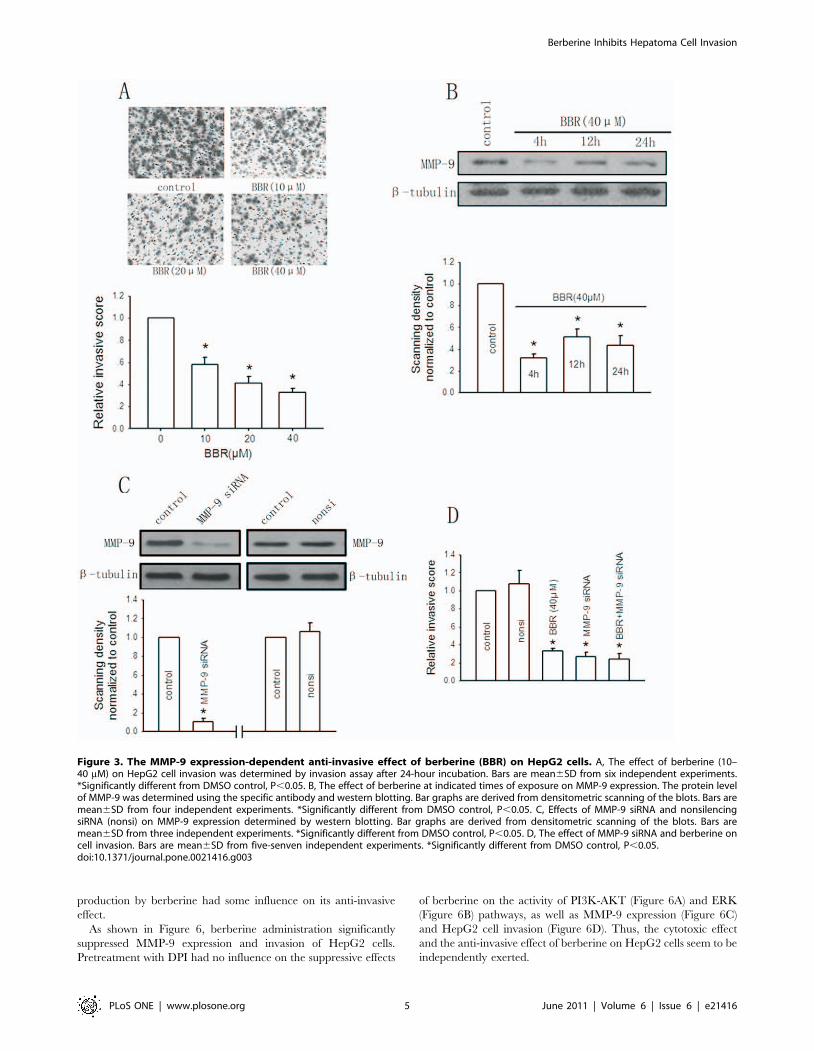
Figure 3A showed that berberine administration led to concen-
tration-dependent decrease in cell invasion after 24-hour incuba-
tion. Berberine (40 mM) diminished the invasive ability of HepG2
cells substantially up to 32.8% of the control.
To rule out the possibility that decreased cell invasion after
berberine treatment might be caused by decreased total cell
number, in fact, we plated cells at the same density and culture
medium volume as in the MTT assay into transwell chambers for
invasion assay (see the section‘Cell invasion assay’ of methods). At
the same time, moreover, equal cells of berberine-treated and
-untreated groups were plated to 96-well plates for total cell
number assay (MTT) represented by OD570. The invasiveness of
HepG2 cells was expressed by the invasive score (number of
invaded cells/total cell number).
MMP-9 has been described to be closely participated in
capsular infiltration in hepatoma cells [24]. The present study
further investigated the effect of berberine on MMP-9 expression
in HepG2 cells for determination of the mechanism for berberine-
induced suppressive effect on cell invasion. Figure 3B showed that
treatment of cells with berberine (40 mM) significantly suppressed
MMP-9 expression and the decrease in MMP-9 level relative to
that of non-berberine-treated group was approximately 40% after
a 24-hour incubation period.
To confirm a causal link between berberine-mediated downreg-
ulation of MMP-9 expression and decreased invasion, the
expression of MMP-9 was blocked by transfecting cells with
MMP-9 siRNA. Figure 3C showed that siRNA to MMP-9 at the
concentrations of 0.1 mM decreased MMP-9 expression by 89% as
compared with control. The nonsilencing siRNA had no effect on
MMP-9 expression. Both MMP-9 siRNA and the nonsilencing
siRNA exerted no cytotoxic effect on HepG2 cells (data not shown).
Knockdown of MMP-9 expression with MMP-9 siRNA resulted in
a significant reduction of HepG2 cell invasion. Berberine
significantly inhibited HepG2 cell invasion, but this effect was not
seen when cells were pretreated with MMP-9 siRNA (Figure 3D).
Altogether, these results suggest that berberine inhibits HepG2 cell
invasion through suppression of MMP-9 expression.
Involvement of the PI3K-AKT and ERK pathways ininhibitory effects of berberine on HepG2 cell invasionand MMP-9 expression
To investigate the mechanism for anti-invasive effect of
berberine on HepG2 cells, we determined whether interfering
with the PI3K-AKT and ERK pathways affected the inhibition of
cell invasion and MMP-9 expression by berberine.
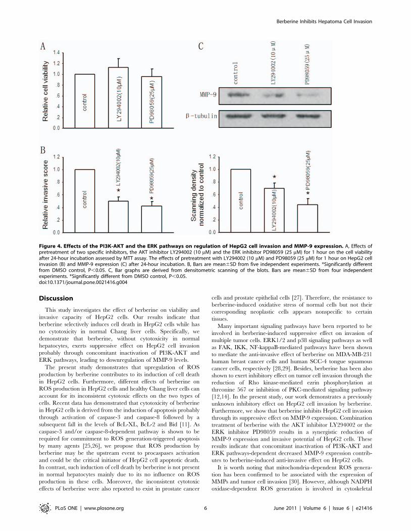
We first determined the specific effect of the PI3K-AKT
inhibitor LY294002 and ERK inhibitor PD98059. After 24-hour
incubation, both LY294002 and PD98059 at the indicated
concentrations had no impact on HepG2 cell growth as assessed
by MTT assay (Figure 4A). Pretreatment with LY294002 (10 mM)
and PD98059 (25 mM) for 1 hour significantly decreased the
invasive ability of the cells, as the relative invasive score was
reduced by near 50% and 58%, respectively (Figure 4B). Figure 4C
showed that pretreatment with two inhibitors substantially
downregulated MMP-9 expression by 31% and 56% respectively
after 24-hour incubation, which suggested the critical contribution
of PI3K-AKT and ERK pathways-dependent MMP-9 expression
to the invasive phenotype of HepG2 cells.
To exclude the possibility that the PI3K-AKT and ERK
pathways-dependent MMP-9 expression is restricted to hepatoma
cells, experiments were also performed in normal Chang liver cells.
Like HepG2 cells, PI3K-AKT and ERK pathways-dependent
regulation of MMP-9 expression may also exist in normal Chang
liver cells (See Text S1 and Figure S1, available online).
Figure 1. The effect of berberine (BBR) on the cell viability ofHepG2 cells and Chang liver cells. HepG2 cells (A) or Chang livercells (B) were treated with either 0.1% DMSO (as control) or berberine(10-40 mM) for 24 hours, and the proportion of surviving cells wasmeasured by the MTT assay. Bars are mean6SD from six independentexperiments. *Significantly different from control, P,0.05.doi:10.1371/journal.pone.0021416.g001
Berberine Inhibits Hepatoma Cell Invasion
PLoS ONE | www.plosone.org 3 June 2011 | Volume 6 | Issue 6 | e21416
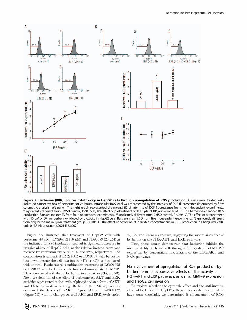
Figure 5A illustrated that treatment of HepG2 cells with
berberine (40 mM), LY294002 (10 mM) and PD98059 (25 mM) at
the indicated time of incubation resulted in significant decrease in
invasive ability of HepG2 cells, as the relative invasive score was
reduced by approximately 67%, 50% and 42%, respectively. The
combination treatment of LY294002 or PD98059 with berberine
could even reduce the cell invasion by 83% or 85%, as compared
with control. Furthermore, combination treatment of LY294002
or PD98059 with berberine could further downregulate the MMP-
9 level compared with that of berberine treatment only (Figure 5B).
Next, we determined the effect of berberine on AKT and ERK
activities represented as the levels of phosphorylated forms of AKT
and ERK by western blotting. Berberine (40 mM) significantly
decreased the levels of p-AKT (Figure 5C) and p-ERK1/2
(Figure 5D) with no changes on total AKT and ERK levels under
4-, 12-, and 24-hour exposure, suggesting the suppressive effect of
berberine on the PI3K-AKT and ERK pathways.
Thus, these results demonstrate that berberine inhibits the
invasive ability of HepG2 cells through downregulation of MMP-9
expression by concomitant inactivation of the PI3K-AKT and
ERK pathways.
No involvement of upregulation of ROS production byberberine in its suppressive effects on the activity ofPI3K-AKT and ERK pathways, as well as MMP-9 expressionand HepG2 cell invasion
To explore whether the cytotoxic effect and the anti-invasive
effect of berberine on HepG2 cells are independently exerted or
have some crosslinks, we determined if enhancement of ROS
Figure 2. Berberine (BBR) induces cytotoxicity in HepG2 cells through upregulation of ROS production. A, Cells were treated withindicated concentrations of berberine for 24 hours. Intracellular ROS level was represented by the intensity of DCF fluorescence determined by flowcytometric analysis (left panel). The right graph represented the mean6SD of intensity of DCF fluorescence from five independent experiments.*Significantly different from DMSO control, P,0.05. B, The effect of pretreatment with 10 mM of DPI,a scavenger of ROS, on berberine-enhanced ROSproduction. Bars are mean6SD from four independent experiments. *Significantly different from DMSO control, P,0.05. C, The effect of pretreatmentwith 10 mM of DPI on berberine-induced cytotoxicity in HepG2 cells. Bars are mean6SD from five independent experiments. *Significantly differentfrom only berberine (40 mM) treatment group, P,0.05. D, The effect of berberine of indicated concentrations on ROS production in Chang liver cells.doi:10.1371/journal.pone.0021416.g002
Berberine Inhibits Hepatoma Cell Invasion
PLoS ONE | www.plosone.org 4 June 2011 | Volume 6 | Issue 6 | e21416
production by berberine had some influence on its anti-invasive
effect.
As shown in Figure 6, berberine administration significantly
suppressed MMP-9 expression and invasion of HepG2 cells.
Pretreatment with DPI had no influence on the suppressive effects
of berberine on the activity of PI3K-AKT (Figure 6A) and ERK
(Figure 6B) pathways, as well as MMP-9 expression (Figure 6C)
and HepG2 cell invasion (Figure 6D). Thus, the cytotoxic effect
and the anti-invasive effect of berberine on HepG2 cells seem to be
independently exerted.
Figure 3. The MMP-9 expression-dependent anti-invasive effect of berberine (BBR) on HepG2 cells. A, The effect of berberine (10–40 mM) on HepG2 cell invasion was determined by invasion assay after 24-hour incubation. Bars are mean6SD from six independent experiments.*Significantly different from DMSO control, P,0.05. B, The effect of berberine at indicated times of exposure on MMP-9 expression. The protein levelof MMP-9 was determined using the specific antibody and western blotting. Bar graphs are derived from densitometric scanning of the blots. Bars aremean6SD from four independent experiments. *Significantly different from DMSO control, P,0.05. C, Effects of MMP-9 siRNA and nonsilencingsiRNA (nonsi) on MMP-9 expression determined by western blotting. Bar graphs are derived from densitometric scanning of the blots. Bars aremean6SD from three independent experiments. *Significantly different from DMSO control, P,0.05. D, The effect of MMP-9 siRNA and berberine oncell invasion. Bars are mean6SD from five-senven independent experiments. *Significantly different from DMSO control, P,0.05.doi:10.1371/journal.pone.0021416.g003
Berberine Inhibits Hepatoma Cell Invasion
PLoS ONE | www.plosone.org 5 June 2011 | Volume 6 | Issue 6 | e21416
Discussion
This study investigates the effect of berberine on viability and
invasive capacity of HepG2 cells. Our results indicate that
berberine selectively induces cell death in HepG2 cells while has
no cytotoxicity in normal Chang liver cells. Specifically, we
demonstrate that berberine, without cytotoxicity in normal
hepatocytes, exerts suppressive effect on HepG2 cell invasion
probably through concomitant inactivation of PI3K-AKT and
ERK pathways, leading to downregulation of MMP-9 levels.
The present study demonstrates that upregulation of ROS
production by berberine contributes to its induction of cell death
in HepG2 cells. Furthermore, different effects of berberine on
ROS production in HepG2 cells and healthy Chang liver cells can
account for its inconsistent cytotoxic effects on the two types of
cells. Recent data has demonstrated that cytotoxicity of berberine
in HepG2 cells is derived from the induction of apoptosis probably
through activation of caspase-3 and caspase-8 followed by a
subsequent fall in the levels of BcL-XL, BcL-2 and Bid [11]. As
caspase-3 and/or caspase-8-dependent pathway is shown to be
required for commitment to ROS generation-triggered apoptosis
by many agents [25,26], we propose that ROS production by
berberine may be the upstream event to procaspases activation
and could be the critical initiator of HepG2 cell apoptotic death.
In contrast, such induction of cell death by berberine is not present
in normal hepatocytes mainly due to its no influence on ROS
production in these cells. Moreover, the inconsistent cytotoxic
effects of berberine were also reported to exist in prostate cancer
cells and prostate epithelial cells [27]. Therefore, the resistance to
berberine-induced oxidative stress of normal cells but not their
corresponding neoplastic cells appears nonspecific to certain
tissues.
Many important signaling pathways have been reported to be
involved in berberine-induced suppressive effect on invasion of
multiple tumor cells. ERK1/2 and p38 signaling pathways as well
as FAK, IKK, NF-kappaB-mediated pathways have been shown
to mediate the anti-invasive effect of berberine on MDA-MB-231
human breast cancer cells and human SCC-4 tongue squamous
cancer cells, respectively [28,29]. Besides, berberine has been also
shown to exert inhibitory effect on tumor cell invasion through the
reduction of Rho kinase-mediated ezrin phosphorylation at
threonine 567 or inhibition of PKC-mediated signaling pathway
[12,14]. In the present study, our work demonstrates a previously
unknown inhibitory effect on HepG2 cell invasion by berberine.
Furthermore, we show that berberine inhibits HepG2 cell invasion
through its suppressive effect on MMP-9 expression. Combination
treatment of berberine with the AKT inhibitor LY294002 or the
ERK inhibitor PD98059 results in a synergistic reduction of
MMP-9 expression and invasive potential of HepG2 cells. These
results indicate that concomitant inactivation of PI3K-AKT and
ERK pathways-dependent decreased MMP-9 expression contrib-
utes to berberine-induced anti-invasive effect on HepG2 cells.
It is worth noting that mitochondria-dependent ROS genera-
tion has been confirmed to be associated with the expression of
MMPs and tumor cell invasion [30]. However, although NADPH
oxidase-dependent ROS generation is involved in cytoskeletal
Figure 4. Effects of the PI3K-AKT and the ERK pathways on regulation of HepG2 cell invasion and MMP-9 expression. A, Effects ofpretreatment of two specific inhibitors, the AKT inhibitor LY294002 (10 mM) and the ERK inhibitor PD98059 (25 mM) for 1 hour on the cell viabilityafter 24-hour incubation assessed by MTT assay. The effects of pretreatment with LY294002 (10 mM) and PD98059 (25 mM) for 1 hour on HepG2 cellinvasion (B) and MMP-9 expression (C) after 24-hour incubation. B, Bars are mean6SD from five independent experiments. *Significantly differentfrom DMSO control, P,0.05. C, Bar graphs are derived from densitometric scanning of the blots. Bars are mean6SD from four independentexperiments. *Significantly different from DMSO control, P,0.05.doi:10.1371/journal.pone.0021416.g004
Berberine Inhibits Hepatoma Cell Invasion
PLoS ONE | www.plosone.org 6 June 2011 | Volume 6 | Issue 6 | e21416

remodeling [31], extravasation and angiogenesis [32], there has
been no direct evidence for its role in MMPs expression and
invasion of tumor cells [33]. In our study, pretreatment with DPI
blocked the berberine-increased ROS production (Fig. 2B), which
suggests that enhanced ROS level by berberine is likely NADPH
oxidase-dependent rather than mitochondria-dependent, as DPI is
an inhibitor of NADPH oxidase [23]. DPI administration has no
influence on the suppressive effects of berberine on the activity of
PI3K-AKT and ERK pathways, as well as MMP-9 expression and
HepG2 cell invasion (Fig. 6). Thus, it is suggested from our results
that NADPH oxidase-dependent ROS generation may not exert
any direct influence on MMP-9 expression and invasion of HepG2
cells. Moreover, the cytotoxic effect and the anti-invasive effect of
berberine on HepG2 cells seem to be independently exerted.
The expression MMP-9 is regulated by the upstream promoter
sequence, of which the activator protein-1 (AP-1) and nuclear
factorkB (NF-kB) binding sites are centrally involved [34]. AP-1
and NF-kB are well accepted to be involved in many pathological
processes including tumor cell migration and invasion [35]. Cheng
et al. showed that NF-kB modulates the radiation-enhanced
MMP-9 activity and cell invasion in HepG2 cells [36]. Chia-Jui
Weng et al. reported that induction of MMP-9 expression by PMA
Figure 5. Involvement of the PI3K-AKT and ERK pathways in the inhibitory effect of berberine (BBR) on HepG2 cell invasion andMMP-9 expression. The effects of combination treatment of AKT inhibitor LY294002 (10 mM) or ERK inhibitor PD98059 (25 mM) with berberine onHepG2 cell invasion (A) and MMP-9 expression (B) after 24-hour incubation. Bar graphs are derived from densitometric scanning of the blots (B). Barsare mean6SD from four-seven independent experiments. *Significantly different from combination treatment of LY294002 (10 mM) and berberine(40 mM) group; #significantly different from combination treatment of PD98059 (25 mM) and berberine (40 mM) group, P,0.05. The effects ofberberine (40 mM) on AKT (C) and ERK (D) activities represented as the levels of phosphorylated forms of AKT and ERK by western blotting at theindicated times of treatment. Bar graphs are derived from densitometric scanning of the blots. Bars are mean6SD from four independentexperiments. *Significantly different from DMSO control, P,0.05.doi:10.1371/journal.pone.0021416.g005
Berberine Inhibits Hepatoma Cell Invasion
PLoS ONE | www.plosone.org 7 June 2011 | Volume 6 | Issue 6 | e21416

Figure 6. The effect of enhancement of ROS production by berberine (BBR) on its suppressive effects on the activity of PI3K-AKTand ERK pathways, as well as MMP-9 expression and HepG2 cell invasion. The effect of pretreatment with 10 mM of DPI on berberine-suppressed AKT (A) and ERK (B) activities represented as the levels of phosphorylated forms of AKT and ERK by western blotting after 24-hourincubation. Bar graphs are derived from densitometric scanning of the blots. Bars are mean6SD from three independent experiments. *Significantlydifferent from DMSO control, P,0.05. C, The effect of pretreatment with 10 mM of DPI on berberine-suppressed MMP-9 expression after 24-hourincubation. Bar graphs are derived from densitometric scanning of the blots. Bars are mean6SD from three independent experiments. *Significantlydifferent from DMSO control, P,0.05. D, The effect of pretreatment with 10 mM of DPI on berberine-suppressed HepG2 cell invasion after 24-hourincubation. Bars are mean6SD from five independent experiments. *Significantly different from DMSO control, P,0.05.doi:10.1371/journal.pone.0021416.g006
Berberine Inhibits Hepatoma Cell Invasion
PLoS ONE | www.plosone.org 8 June 2011 | Volume 6 | Issue 6 | e21416
and LAB treatment possibly through regulation of the AP-1 and
NF-kB DNA-binding activities [16]. Thus, as AP-1 and NF-kB are
the downstream targets of ERK and AKT pathways [16,37], it is
suggested from our results that berberine inhibits ERK and AKT
pathways-dependent MMP-9 expression probably through resul-
tant suppression of AP-1 and NF-kB activities.
MMP-9 and MMP-2 play critical roles in the degradation of
type IV collagen, a major constituent of the basement membrane,
and are closely related to the invasion and metastasis of various
cancer cells [38,39]. Specially for hepatoma cell invasion, MMP-9
has been shown to be more likely to participate in hepatoma cell
invasion than MMP-2 for its destruction of tumor capsule [24].
This work showing that specific inhibition of MMP-9 expression
by siRNA substantially suppresses the high invasive potential of
HepG2 cells also suggests the decisive role of MMP-9 in basal
hepatoma cell invasion.
Human hepatoma cells have been shown to exhibit high
expression and enhanced activity of ERK and AKT [40,41,42].
Activation of the ERK and AKT pathways are considered to
contribute to increased tumor aggressiveness, yet the exact
mechanisms remain obscure [36]. In this study, our results
showing the dependence of MMP-9 expression and invasive
capacity of hepatoma cells on activity of AKT and ERK pathways
can partly account for the high hepatoma cell aggressiveness.
Besides, many studies have also linked the activity of PI3K-AKT
and ERK pathways to MMP-9 expression in other tumor cell
lines. O-charoenrat et al. showed that beta-cellulin induced MMP-
9 production and invasion in head-and-neck squamous carcinoma
cells through activation of EGFR, MAPK and AKT [43]. In
ovarian cancer cells, Thant et al. suggested that both ERK and
AKT were required for the fibronectin-dependent activation of
MMP-9 secretion and the resultant cell invasiveness [44]. Thus,
activation of AKT and ERK pathways-dependent MMP-9
expression may be widespread in tumor cells and contribute to
tumor progression. Besides, the result that activation of ERK and
PI3K-AKT pathways significantly increased the level of MMP-9
in Chang liver cells (See Figure S1B) suggests that AKT and ERK
pathways-dependent MMP-9 expression may also exist in Chang
liver cells and not be specific to tumor cells.
Currently berberine is administrated orally in clinical practice,
for example, treatment of cardiac arrhythmia [45]. Research of
the anti-tumor effect of berberine still remains preclinical.
Numerous studies, including the present study, have shown that
berberine inhibits cell growth and invasion of various tumor cell
lines in vitro with a large concentrations ranging from 20 mM up to
300 mM [14,29,46]. However, pharmacokinetic studies in humans
have shown that berberine is poorly absorbed, and difficult to
maintain 40 mM plasma concentration after oral administration
[45]. Alternatively, the preparation of the micro-emulsion
formulation could be a potential approach to achieve a high Cmax
of berberine, as it can significantly increase the rate and extent of
absorption [47]. Specifically, intravenous administration may be
another potential alternative to achieve a high Cmax in terms of
anti-tumor effect [48] . Nevertheless, more works is definitely
needed to translate the preclinical data into clinical practice and
make our in vitro observation on the anti-tumor effect of berberine
clinically relevant.
Our work has some limitations. It is not known to what extent
the present principal finding can be generalized to cell types other
than HepG2 cells and Chang liver cells examined in this study.
Notwithstanding the limitation, our study does demonstrate the
therapeutic potential of berberine against hepatoma invasion with
the advantage of no unexpected cytotoxicity in healthy liver cells.
Specifically, our study also provides a previously neglected insight
into the investigation of plant-derived agents-based therapy against
tumor invasion with the consideration of damage to healthy cells.
Supporting Information
Figure S1 Effects of the PI3K-AKT and the ERK pathways on
regulation of MMP-9 expression in normal Chang liver cells. A,
The effects of pretreatment with LY294002 (10 mM) and PD98059
(25 mM) for 1 hour on MMP-9 expression of normal Chang liver
cells after 24-hour incubation. B, The effects of pretreatment of
Chang liver cells with 740 Y-P (20 mg/ml), a specific activator of
PI3K, for 6 hours or enterostatin (100 nM), a nonspecific activator
of ERK, for 1 hour on MMP-9 expression of normal Chang liver
cells after 24-hour incubation. Bar graphs are derived from
densitometric scanning of the blots. Bars are mean6SD from
three-four independent experiments. *Significantly different from
DMSO control, P,0.05.
(TIF)
Text S1 The PI3K-AKT and ERK pathways-dependent
downregulation of MMP-9 expression exists in Chang liver cells.
(DOC)
Author Contributions
Conceived and designed the experiments: LZ. Performed the experiments:
BL GW JY XP ZY. Analyzed the data: XP ZY. Wrote the paper: BL GW
JY.
References
1. Sener SF, Grey N (2005) The global burden of cancer. J Surg Oncol 92: 1–3.
2. Thomas MB, O’Beirne JP, Furuse J, Chan AT, Abou-Alfa G, et al. (2008)
Systemic therapy for hepatocellular carcinoma: cytotoxic chemotherapy,
targeted therapy and immunotherapy. Ann Surg Oncol 15: 1008–1014.
3. Pervaiz S (2002) Anti-cancer drugs of today and tomorrow: are we close to
making the turn from treating to curing cancer? Curr Pharm Des 8: 1723–
1734.
4. Monvoisin A, Neaud V, De Ledinghen V, Dubuisson L, Balabaud C, et al.
(1999) Direct evidence that hepatocyte growth factor-induced invasion of
hepatocellular carcinoma cells is mediated by urokinase. J Hepatol 30: 511–
518.
5. Chung TW, Lee YC, Kim CH (2004) Hepatitis B viral HBx induces matrix
metalloproteinase-9 gene expression through activation of ERK and PI-3K/
AKT pathways: involvement of invasive potential. FASEB J 18: 1123–1125.
6. Kozuki Y, Miura Y, Yagasaki K (2000) Inhibitory effects of carotenoids on the
invasion of rat ascites hepatoma cells in culture. Cancer Lett 151: 111–115.
7. Zhang G, Miura Y, Yagasaki K (2000) Suppression of adhesion and invasion of
hepatoma cells in culture by tea compounds through antioxidative activity.
Cancer Lett 159: 169–173.
8. Yagasaki K, Miura Y, Okauchi R, Furuse T (2000) Inhibitory effects of
chlorogenic acid and its related compounds on the invasion of hepatoma cells in
culture. Cytotechnology 33: 229–235.
9. Kozuki Y, Miura Y, Yagasaki K (2001) Resveratrol suppresses hepatoma cell
invasion independently of its anti-proliferative action. Cancer Lett 167:
151–156.
10. Jantova S, Cipak L, Cernakova M, Kost’alova D (2003) Effect of berberine on
proliferation, cell cycle and apoptosis in HeLa and L1210 cells. J Pharm
Pharmacol 55: 1143–1149.
11. Hwang JM, Kuo HC, Tseng TH, Liu JY, Chu CY (2006) Berberine induces
apoptosis through a mitochondria/caspases pathway in human hepatoma cells.
Arch Toxicol 80: 62–73.
12. Lin TH, Kuo HC, Chou FP, Lu FJ (2008) Berberine enhances inhibition of
glioma tumor cell migration and invasiveness mediated by arsenic trioxide.
BMC Cancer 8: 58.
13. Peng PL, Hsieh YS, Wang CJ, Hsu JL, Chou FP (2006) Inhibitory effect of
berberine on the invasion of human lung cancer cells via decreased productions
of urokinase-plasminogen activator and matrix metalloproteinase-2. Toxicol
Appl Pharmacol 214: 8–15.
Berberine Inhibits Hepatoma Cell Invasion
PLoS ONE | www.plosone.org 9 June 2011 | Volume 6 | Issue 6 | e21416

14. Tang F, Wang D, Duan C, Huang D, Wu Y, et al. (2009) Berberine inhibits
metastasis of nasopharyngeal carcinoma 5-8F cells by targeting Rho kinase-
mediated Ezrin phosphorylation at threonine 567. J Biol Chem 284:
27456–27466.
15. Singh T, Vaid M, Katiyar N, Sharma S, Katiyar SK (2011) Berberine, an
isoquinoline alkaloid, inhibits melanoma cancer cell migration by reducing the
expressions of cyclooxygenase-2, prostaglandin E2 and prostaglandin E2
receptors. Carcinogenesis 32: 86–92.
16. Weng CJ, Chau CF, Hsieh YS, Yang SF, Yen GC (2008) Lucidenic acid inhibits
PMA-induced invasion of human hepatoma cells through inactivating MAPK/
ERK signal transduction pathway and reducing binding activities of NF-kappaB
and AP-1. Carcinogenesis 29: 147–156.
17. Kim WH, Chon CY, Moon YM, Kang JK, Park IS, et al. (1993) Effect of
anticancer drugs and desferrioxamine in combination with radiation on
hepatoma cell lines. Yonsei Med J 34: 45–56.
18. He B, Tong X, Wang L, Wang Q, Ye H, et al. (2009) Tramadol and
flurbiprofen depress the cytotoxicity of cisplatin via their effects on gap junctions.
Clin Cancer Res 15: 5803–5810.
19. Sanceau J, Truchet S, Bauvois B (2003) Matrix metalloproteinase-9 silencing by
RNA interference triggers the migratory-adhesive switch in Ewing’s sarcoma
cells. J Biol Chem 278: 36537–36546.
20. Deryugina EI, Zijlstra A, Partridge JJ, Kupriyanova TA, Madsen MA, et al.
(2005) Unexpected effect of matrix metalloproteinase down-regulation on
vascular intravasation and metastasis of human fibrosarcoma cells selected in
vivo for high rates of dissemination. Cancer Res 65: 10959–10969.
21. Kamata H, Hirata H (1999) Redox regulation of cellular signalling. Cell Signal
11: 1–14.
22. Nishikawa T, Edelstein D, Du XL, Yamagishi S, Matsumura T, et al. (2000)
Normalizing mitochondrial superoxide production blocks three pathways of
hyperglycaemic damage. Nature 404: 787–790.
23. Foreman J, Demidchik V, Bothwell JH, Mylona P, Miedema H, et al. (2003)
Reactive oxygen species produced by NADPH oxidase regulate plant cell
growth. Nature 422: 442–446.
24. Arii S, Mise M, Harada T, Furutani M, Ishigami S, et al. (1996) Overexpression
of matrix metalloproteinase 9 gene in hepatocellular carcinoma with invasive
potential. Hepatology 24: 316–322.
25. Oh SH, Lim SC (2006) A rapid and transient ROS generation by cadmium
triggers apoptosis via caspase-dependent pathway in HepG2 cells and this is
inhibited through N-acetylcysteine-mediated catalase upregulation. Toxicol
Appl Pharmacol 212: 212–223.
26. Kim JH, Lee SY, Oh SY, Han SI, Park HG, et al. (2004) Methyl jasmonate
induces apoptosis through induction of Bax/Bcl-XS and activation of caspase-3
via ROS production in A549 cells. Oncol Rep 12: 1233–1238.
27. Meeran SM, Katiyar S, Katiyar SK (2008) Berberine-induced apoptosis in
human prostate cancer cells is initiated by reactive oxygen species generation.
Toxicol Appl Pharmacol 229: 33–43.
28. Kim S, Choi JH, Kim JB, Nam SJ, Yang JH, et al. (2008) Berberine suppresses
TNF-alpha-induced MMP-9 and cell invasion through inhibition of AP-1
activity in MDA-MB-231 human breast cancer cells. Molecules 13: 2975–2985.
29. Ho YT, Yang JS, Li TC, Lin JJ, Lin JG, et al. (2009) Berberine suppresses in
vitro migration and invasion of human SCC-4 tongue squamous cancer cells
through the inhibitions of FAK, IKK, NF-kappaB, u-PA and MMP-2 and -9.
Cancer Lett 279: 155–162.
30. van Waveren C, Sun Y, Cheung HS, Moraes CT (2006) Oxidative
phosphorylation dysfunction modulates expression of extracellular matrix--remodeling genes and invasion. Carcinogenesis 27: 409–418.
31. Groth S, Schulze M, Kalthoff H, Fandrich F, Ungefroren H (2005) Adhesion
and Rac1-dependent regulation of biglycan gene expression by transforminggrowth factor-beta. Evidence for oxidative signaling through NADPH oxidase.
J Biol Chem 280: 33190–33199.32. Deem TL, Cook-Mills JM (2004) Vascular cell adhesion molecule 1 (VCAM-1)
activation of endothelial cell matrix metalloproteinases: role of reactive oxygen
species. Blood 104: 2385–2393.33. Wu WS (2006) The signaling mechanism of ROS in tumor progression. Cancer
Metastasis Rev 25: 695–705.34. Takahra T, Smart DE, Oakley F, Mann DA (2004) Induction of myofibroblast
MMP-9 transcription in three-dimensional collagen I gel cultures: regulation byNF-kappaB, AP-1 and Sp1. Int J Biochem Cell Biol 36: 353–363.
35. Bahassi el M, Karyala S, Tomlinson CR, Sartor MA, Medvedovic M, et al.
(2004) Critical regulation of genes for tumor cell migration by AP-1. Clin ExpMetastasis 21: 293–304.
36. Cheng JC, Chou CH, Kuo ML, Hsieh CY (2006) Radiation-enhancedhepatocellular carcinoma cell invasion with MMP-9 expression through
PI3K/Akt/NF-kappaB signal transduction pathway. Oncogene 25: 7009–7018.
37. Abiru S, Nakao K, Ichikawa T, Migita K, Shigeno M, et al. (2002) Aspirin andNS-398 inhibit hepatocyte growth factor-induced invasiveness of human
hepatoma cells. Hepatology 35: 1117–1124.38. Kohn EC, Liotta LA (1995) Molecular insights into cancer invasion: strategies
for prevention and intervention. Cancer Res 55: 1856–1862.39. Itoh Y, Nagase H (2002) Matrix metalloproteinases in cancer. Essays Biochem
38: 21–36.
40. Ito Y, Sasaki Y, Horimoto M, Wada S, Tanaka Y, et al. (1998) Activation ofmitogen-activated protein kinases/extracellular signal-regulated kinases in
human hepatocellular carcinoma. Hepatology 27: 951–958.41. Xu G, Zhang W, Bertram P, Zheng XF, McLeod H (2004) Pharmacogenomic
profiling of the PI3K/PTEN-AKT-mTOR pathway in common human tumors.
Int J Oncol 24: 893–900.42. Scheving LA, Buchanan R, Krause MA, Zhang X, Stevenson MC, et al. (2007)
Dexamethasone modulates ErbB tyrosine kinase expression and signalingthrough multiple and redundant mechanisms in cultured rat hepatocytes.
Am J Physiol Gastrointest Liver Physiol 293: G552–559.43. P Oc, Wongkajornsilp A, Rhys-Evans PH, Eccles SA (2004) Signaling pathways
required for matrix metalloproteinase-9 induction by betacellulin in head-and-
neck squamous carcinoma cells. Int J Cancer 111: 174–183.44. Thant AA, Nawa A, Kikkawa F, Ichigotani Y, Zhang Y, et al. (2000) Fibronectin
activates matrix metalloproteinase-9 secretion via the MEK1-MAPK and thePI3K-Akt pathways in ovarian cancer cells. Clin Exp Metastasis 18: 423–428.
45. Ye M, Fu S, Pi R, He F (2009) Neuropharmacological and pharmacokinetic
properties of berberine: a review of recent research. J Pharm Pharmacol 61:831–837.
46. Tsang CM, Lau EP, Di K, Cheung PY, Hau PM, et al. (2009) Berberine inhibitsRho GTPases and cell migration at low doses but induces G2 arrest and
apoptosis at high doses in human cancer cells. Int J Mol Med 24: 131–138.47. Gui SY, Wu L, Peng DY, Liu QY, Yin BP, et al. (2008) Preparation and
evaluation of a microemulsion for oral delivery of berberine. Pharmazie 63:
516–519.48. Tsai PL, Tsai TH (2004) Hepatobiliary excretion of berberine. Drug Metab
Dispos 32: 405–412.
Berberine Inhibits Hepatoma Cell Invasion
PLoS ONE | www.plosone.org 10 June 2011 | Volume 6 | Issue 6 | e21416
Related Documents