Beneficial effects of hydroxyapatite on enamel subjected to 30% hydrogen peroxide Tao Jiang a,b,1 , Xiao Ma a,1 , Zhejun Wang a , Hua Tong b , Jiming Hu b , Yining Wang a, * a Key Laboratory for Oral Biomedical Engineering, Ministry of Education, School and Hospital of Stomatology, Wuhan University, 237 Luoyu Road, Wuhan 430079, PR China b Institute of Analytical and Biomedical Science, Wuhan University, Luojia Hill, Wuhan 430072, PR China 1. Introduction Vital tooth bleaching is a well-accepted method of treating discolored tooth. 1 The method is based upon hydrogen peroxide (HP) as the active agent. 2,3 HP may be applied directly, or produced in a chemical reaction from carbamide peroxide (CP). It acts as a strong oxidizing agent through the formation of free radicals, reactive oxygen molecules, and HP anions. 2,3 It is suggested that these reactive molecules interact with chromophore molecules and oxidize the macromole- cules and pigment stains. 2,3 Although there is little question about their efficacy, a primary concern is that the enamel structure may be weakened by the bleaching agent. Numerous studies have evaluated the effects of peroxide-containing products on the physical and chemical properties of tooth enamel. However, the research in this area has been controversial. Some studies reported that there was no evident change in enamel microhardness and morphology after bleaching treatment. 4–7 But others have found calcium loss, 8–10 alterations of surface morphology, 11–15 changes in chemical composition, 10,15–18 decrease in hard- ness 10,15,19–22 and fracture resistance 23 of enamel. The diver- journal of dentistry 36 (2008) 907–914 article info Article history: Received 8 April 2008 Received in revised form 14 July 2008 Accepted 20 July 2008 Keywords: Hydroxyapatite Hydrogen peroxide Enamel Tooth bleaching abstract Objectives: To evaluate the effect of combination of hydroxyapatite (HA) and hydrogen peroxide (HP) on color, microhardness and morphology of human tooth enamel. Methods: Forty-eight human dental blocks were obtained from 12 pairs of premolars and were randomly divided into four groups. Group DW was treated with distilled water, group HP with 30% HP, group HA + DW with HA mixed with distilled water and group HA + HP with HA mixed with 30% HP. Baseline and final color measurements and microhardness test were carried out before and after bleaching experiments. Two specimens from each group were selected for morphological investigation after final tests. Results: The DE of group HP and HA + HP were significantly higher than those of group DW ( p = 0.000 and p = 0.000) and group HA + DW ( p = 0.000 and p = 0.000). The percentage microhardness loss of group HA + HP was significantly lower than that of group HP ( p = 0.047), but significantly higher than those of group DW ( p = 0.000) and group HA + DW ( p = 0.000). The obvious variation of morphology was only observed on enamel surfaces in group HP. Conclusions: This study suggested that combination of HA and HP was effective in tooth whitening. HA could significantly reduce the microhardness loss of enamel caused by 30% HP and keep enamel surface morphology almost unchanged. # 2008 Elsevier Ltd. All rights reserved. * Corresponding author. Tel.: +86 27 87646696; fax: +86 27 87873260. E-mail address: [email protected] (Y. Wang). 1 Contributed equally to this work. available at www.sciencedirect.com journal homepage: www.intl.elsevierhealth.com/journals/jden 0300-5712/$ – see front matter # 2008 Elsevier Ltd. All rights reserved. doi:10.1016/j.jdent.2008.07.005

Welcome message from author
This document is posted to help you gain knowledge. Please leave a comment to let me know what you think about it! Share it to your friends and learn new things together.
Transcript
j o u r n a l o f d e n t i s t r y 3 6 ( 2 0 0 8 ) 9 0 7 – 9 1 4
Beneficial effects of hydroxyapatite on enamel subjectedto 30% hydrogen peroxide
Tao Jiang a,b,1, Xiao Ma a,1, Zhejun Wang a, Hua Tong b, Jiming Hu b, Yining Wang a,*aKey Laboratory for Oral Biomedical Engineering, Ministry of Education, School and Hospital of Stomatology, Wuhan University,
237 Luoyu Road, Wuhan 430079, PR Chinab Institute of Analytical and Biomedical Science, Wuhan University, Luojia Hill, Wuhan 430072, PR China
a r t i c l e i n f o
Article history:
Received 8 April 2008
Received in revised form
14 July 2008
Accepted 20 July 2008
Keywords:
Hydroxyapatite
Hydrogen peroxide
Enamel
Tooth bleaching
a b s t r a c t
Objectives: To evaluate the effect of combination of hydroxyapatite (HA) and hydrogen
peroxide (HP) on color, microhardness and morphology of human tooth enamel.
Methods: Forty-eight human dental blocks were obtained from 12 pairs of premolars and
were randomly divided into four groups. Group DW was treated with distilled water, group
HP with 30% HP, group HA + DW with HA mixed with distilled water and group HA + HP with
HA mixed with 30% HP. Baseline and final color measurements and microhardness test were
carried out before and after bleaching experiments. Two specimens from each group were
selected for morphological investigation after final tests.
Results: The DE of group HP and HA + HP were significantly higher than those of group DW
( p = 0.000 and p = 0.000) and group HA + DW ( p = 0.000 and p = 0.000). The percentage
microhardness loss of group HA + HP was significantly lower than that of group HP
( p = 0.047), but significantly higher than those of group DW ( p = 0.000) and group HA + DW
( p = 0.000). The obvious variation of morphology was only observed on enamel surfaces in
group HP.
Conclusions: This study suggested that combination of HA and HP was effective in tooth
whitening. HA could significantly reduce the microhardness loss of enamel caused by 30%
HP and keep enamel surface morphology almost unchanged.
# 2008 Elsevier Ltd. All rights reserved.
avai lab le at www.sc iencedi rec t .com
journal homepage: www. int l .e lsev ierhea l th .com/ journa ls / jden
1. Introduction
Vital tooth bleaching is a well-accepted method of treating
discolored tooth.1 The method is based upon hydrogen
peroxide (HP) as the active agent.2,3 HP may be applied
directly, or produced in a chemical reaction from carbamide
peroxide (CP). It acts as a strong oxidizing agent through the
formation of free radicals, reactive oxygen molecules, and HP
anions.2,3 It is suggested that these reactive molecules interact
with chromophore molecules and oxidize the macromole-
cules and pigment stains.2,3
* Corresponding author. Tel.: +86 27 87646696; fax: +86 27 87873260.E-mail address: [email protected] (Y. Wang).
1 Contributed equally to this work.0300-5712/$ – see front matter # 2008 Elsevier Ltd. All rights reserveddoi:10.1016/j.jdent.2008.07.005
Although there is little question about their efficacy, a
primary concern is that the enamel structure may be weakened
by the bleaching agent. Numerous studies have evaluated the
effects of peroxide-containing products on the physical and
chemical properties of tooth enamel. However, the research in
this area has been controversial. Some studies reported that
there was no evident change in enamel microhardness and
morphology after bleaching treatment.4–7 But others have
found calcium loss,8–10 alterations of surface morphology,11–15
changes in chemical composition,10,15–18 decrease in hard-
ness10,15,19–22 and fracture resistance23 of enamel. The diver-
.

j o u r n a l o f d e n t i s t r y 3 6 ( 2 0 0 8 ) 9 0 7 – 9 1 4908
gence in these results is probably related to different meth-
odologies (in vivo or in vitro, time of evaluation, bleaching
agents used, time of application, immersion of the specimens in
artificial saliva between treatments, type of storage, bleaching
agent pH, usage of fluoride, etc.).6,24,25
The oxidative effect, pH and composition of the bleaching
agents are claimed to produce possible side effects during
tooth bleaching.23 As the oxidative effect is not specific, the
bleaching agents may not only oxide the chromogen of
the discolored teeth but also attack the organic matter of the
teeth.15,19 The acidity of HP is probably the main cause for
the demineralization effect.25 When the pH falls below 5.2,
enamel demineralization26 and root resorption27,28 have been
reported. On the other hand, however, different concentra-
tions of CP are also capable of causing enamel surface
alterations regardless of being close to neutral pH level.13,14,29
Urea, which is the by-product of such bleaching agents, could
remove enamel proteins and related mineral elements.14,23,30
Glycerin or carbopol, the most common carriers in the
bleaching formula, may also act as a demineralizing
agent.21,22
It should be pointed out that the remineralization and
protective benefits of saliva may overcome the detrimental
bleaching effects in vivo.25 And no clinical studies or case
reports in the literature have documented macroscopically
or clinically visible damage due to vital bleaching.31
However, it is still necessary to minimize the risk of even
minor damage caused by the agents. To achieve this goal,
some ingredients such as fluoride,31 calcium32 or amor-
phous calcium phosphate33 are added into the bleaching gel
by manufacturers.
Synthetic hydroxyapatite (HA) is an attractive biomaterial
for bone and tooth implants, owing to its chemical and
structural similarity with natural bone and tooth mineral.34,35
Recent research indicated that HA paste containing concen-
trated H3PO4 and HP can repair early caries lesion.36,37 It was
shown that the hydroxyapatite nanocrystals seamlessly grew
in the interface between the paste and tooth enamel despite
low pH of the paste.36,37 Meanwhile, HA is an alkaline salt38
and it may increase the pH value of the compound when it
mixed with acidic HP. The increased pH may reduce the
demineralization effect of acidic HP and facilitate the bleach-
ing procedure2 by accelerating formation of free radicals from
HP. Furthermore, addition of hydroxyapatite to toothpaste
could result in a marked increase in tooth whitening.39
Thus, we hypothesize that combination of HA and HP may
bring better whitening effect and reduce enamel deminer-
alization caused by acidic HP alone. The purpose of the study
was to evaluate the effect of combination of HA and HP on
color, microhardness and morphology of human tooth
enamel.
2. Materials and methods
The study protocol was reviewed and approved by the Ethics
Committee of the School and Hospital of Stomatology, Wuhan
University. Patients from whom teeth were being extracted
were asked to read and sign a consent form prior to the
extraction.
2.1. Synthesis of HA
HA was synthesized in a wet condition according to the
method of Boanini et al.41 with some modifications. Twenty
milliliters of 1.08 M Ca(NO3)2�4H2O solution at pH adjusted to
10 with NH4OH. The solution was heated at 90 8C and 20 ml of
0.65 M (NH4)2HPO4 solution, pH 10 adjusted with NH4OH, was
added drop-wise under stirring. The precipitate was main-
tained in contact with the reaction solution for 5 h at 90 8C
under stirring, and then centrifuged at 1800 g for 10 min. The
precipitate was repeatedly washed with distilled water and
centrifuged for 6 times, and then dried at 37 8C overnight.
2.2. Characterization of the precipitate
The precipitate was characterized by the scanning electron
microscopy (SEM), Fourier transform infrared (FTIR) spectro-
scopy and X-ray diffraction (XRD). Morphological investigation
was performed using SEM (Fei QUANTA-200, Eindhoven, the
Netherlands). For FTIR analysis, 1 mg powder was mixed with
potassium bromide powder and pressed into disks. The
spectra were recorded on Thermo Nicolet 5700 spectrometer
(Nicolet, Madison, Wis., USA). The crystal phase was char-
acterized by wide angle XRD (D/MAX-RB, Rigaku, Miniflex,
Japan). The scanning range was 20–708, with a scanning speed
of 48/min. The radiation utilized was CuKa at 40 kV and 50 mA.
2.3. Tooth selection
Twelve pairs of premolars, extracted for orthodontic reasons,
were selected. Buccal surfaces were devoid of stain, enamel
cracks or fractures, caries or other defects. The teeth were
cleaned thoroughly and stored in 0.5% thymol at 4 8C until
required.
2.4. Sample preparation
Two dental blocks (2 mm � 3 mm � 4 mm) were obtained
from middle 1/3 of buccal halves of each tooth by a low speed
saw (Isomet, Buehler Ltd., Lake Bluff, IL, USA) under water-
cooling. The dental blocks were individually embedded in
colorless translucent acrylic resin with enamel surface
exposing for bleaching applications. The specimens were
serially polished by means of 600–3000 grit SiC papers with
water as a cooler to obtain flat standardized enamel surfaces.
Subsequently, they were polished with diamond spray (1 mm,
0.5 mm) and polishing cloths. After then the specimens were
ultrasonic cleaned for 5 min with distilled water to remove
smear layer. The initial color measurement and microhard-
ness test were performed within 24 h after preparation. The
specimens were stored in Hank’s balanced salts solution
(HBSS)40 during the interval of the preparation and before
testing.
2.5. Bleaching procedure
All specimens were washed under running distilled water for
30 s and dried by compressed air for 3 s before treatment
started. Four specimens prepared from each pair were
randomly divided into four groups and treated as follow:

j o u r n a l o f d e n t i s t r y 3 6 ( 2 0 0 8 ) 9 0 7 – 9 1 4 909
Group DW (n = 12, pH �6.8): As a control group. The
specimens were immersed in 2 ml distilled water for
15 min.
Group HP (n = 12, pH �3.2): The specimens were immersed
in a 2 ml 30% solution of HP (Sinopharm Chemcial Reagent
Co. Ltd., Shanghai, China) for 15 min.
Group HA + DW (n = 12, pH �7.6): HA mixed with distilled
water in a ratio of 2 g powder to 1 ml liquid. A similar
quantity (about 0.1 g HA and 0.05 ml liquid) of HA paste was
applied to the enamel. After then the specimens were
immersed in 2 ml distilled water for 15 min.
Group HA + HP (n = 12, pH �5.4): HA mixed with HP in the
same ratio for group HA + DW and a similar quantity was
applied to the enamel surface. Then the specimens were
immersed in 2 ml 30% solution of HP for 15 min.
During the treatment period, all the specimens were stored
at 37 8C. Between treatments, the residual treatment solution
and HA were removed under running distilled water with
cotton wool pellets. Then the next 15 min treatment was
carried out immediately. Wet cotton pellets were put on the
surfaces of specimens to diminish dehydration between
treatments. The treatment procedure was repeated 4 times
so that the total application time was 60 min. The pH values of
solutions were measured with a digital pH meter (Sartorius,
Gottingen, Germany).
2.6. Color measurement
Baseline and final color measurements were carried out before
and after bleaching experiments. The color distributions (L*, a*
and b*) of each specimen were measured with a spectro-
photometer (Spectrascan PR650, Photo Research, California,
USA). A circular area with 1.0 mm in diameter was measured
at the middle third region of the specimen. Before each
measurement session, the spectrophotometer was calibrated
with a white reflectance standard according to the manufac-
turer’s protocol. A custom sample holder was used to position
the specimens to insure that every measurement was on the
same area of each sample.
The L*, a* and b* color space system was defined by the
Commission International de l’Eclairage in 1979 and is referred
to as CIELAB (International Commission on Illumination,
1978). The L* represents the value where white is 100 and black
is 0. A positive a* value indicates the red direction, a negative a*
value the green direction, a positive b* value the yellow
direction and a negative b* value the blue direction. The
difference between L*, a* and b* at baseline and final of the
experiment were expressed as DL*, Da*and Db*. In addition, the
overall color difference DE of the specimens in each group was
calculated by the following expression:
DE ¼ ðDL�2 þ Da�2 þ Db�2Þ1=2
2.7. Microhardness test
Microhardness was measured with a hardness tester (HXD-
1000TMC/LCD, Taiming Inc. Shanghai, China) with a load of
200 g for 15 s. Five baseline indentations were performed on
each specimen and averaged. The indentations were made in
five widely separated locations. The final measurements were
made near the baseline indentations (space = 100 mm) to
minimize interactions between neighboring marks. The
percentage microhardness loss (PML) was calculated using
the following calculation: PML (%) = (VHN(B) � VHN(F))/VHN(B),
where VHN(B) is the average of the baseline microhardness
measurements, and VHN(F) is the average of final microhard-
ness values.
2.8. SEM analysis
To observe the morphological alterations of enamel surface
after 60-min treatment, two specimens from each group were
selected for SEM analysis after final microhardness measure-
ments. Specimens were ultrasonic cleaned for 5 min with
distilled water, then desiccated and sputter coated with gold in
a vacuum evaporator (JEOL JFC1600, Japan). Micrographs of
central areas were taken using a field emission scanning
electron microscope (JEOL JSM-6700F, Japan).
To investigate the adherence ability of HA to the enamel
surface, another four specimens were prepared. Two were
treated the same as those in group DW + HA, the other two as
those in group HP + HA. After final treatment, the specimens
were washed only with running water (without cotton wool
pellets or ultrasonic). Then these specimens were prepared for
SEM observation.
2.9. Statistical analysis
Statistical analyses were performed with the use of SPSS 10.0
for WINDOWS. Normality was examined by using the
Kolmogorov–Smirnov test, and homogeneity of variance
was assessed by using Levene’s test. One-way ANOVA was
used for normally distributed data. This was followed by
post-hoc multiple comparisons using Tukey’s honestly
significant difference test for cases with equal variances
and Tamhane’s T2 test for cases with unequal variances.
Data were reported as the mean � S.D. Statistical signifi-
cance was set at p < 0.05.
3. Results
3.1. Characterization of HA
The SEM micrograph of HA is shown in Fig. 1a. The precipitates
are rod-like nanocrystals. The FTIR spectrum of the powder is
showed in Fig. 1b. The bands at 1092, 1032, 962, 604, 564 and
470 cm�1 were assigned to vibrations of PO43�, 1458, 1421 and
875 cm�1 to CO32�, 3568 and 631 cm�1 to OH� and 1638 cm�1 to
water. The X-ray diffraction pattern of the precipitate was
identified as HA and the peaks were in agreement with those
in the standard HA diffraction pattern (Fig. 1c).
3.2. Color measurement
The L*, a* and b* and DE values of the four groups are shown in
Table 1. There was no significant difference on L*, a* and b*
values among the four groups at the baseline ( p = 0.996,

Fig. 1 – SEM micrograph (a), FTIR spectrum (b) and XRD pattern of the precipitate.
Table 1 – Mean values (standard deviations) of baseline and final L*, a* and b* for each group and DE
Group L* a* b* DE
Baseline Final Baseline Final Baseline Final
DW 68.87 (1.71) 69.87 (1.40) �0.61 (0.57) �0.84 (0.55) 18.60 (2.02) 18.03 (2.28) 1.27 (0.63)a
HP 68.71 (2.42) 73.53 (2.73) �0.30 (1.34) �0.63 (1.09) 17.11 (3.14) 10.73 (2.30) 8.07 (2.32)b
HA + DW 68.68 (1.34) 68.95 (1.50) �0.62 (0.42) �0.87 (0.47) 18.29 (2.74) 19.23 (2.48) 1.70 (1.29)a
HA + HP 68.81 (3.08) 72.86 (2.86) �0.23 (1.09) �0.55 (0.93) 17.31 (3.94) 11.99 (3.84) 6.76 (3.14)b
Different superscripts (a and b) indicate mean values that are significantly different.
Table 2 – Mean values (standard deviations) of baselineand final microhardness measurements for each groupand the percentage of microhardness loss (%)
Group VHN(B) VHN(F) PML (%)
DW 355.6 (15.6) 356.5 (20.1) �0.3 (3.6)a
HP 357.6 (16.2) 329.3 (20.7) 7.9 (2.8)b
HA + DW 351.2 (31.0) 354.7 (25.5) �1.2 (2.6)a
HA + HP 354.4 (19.0) 338.0 (17.5) 4.6 (3.0)c
VHN(B) is the average of the baseline microhardness measure-
ments, VHN(F) the average of final microhardness values and PML
percentage of microhardness loss. Different superscripts (a, b and
c) indicate mean values that are significantly different.
j o u r n a l o f d e n t i s t r y 3 6 ( 2 0 0 8 ) 9 0 7 – 9 1 4910
p = 0.641 and p = 0.699). The difference on DE value was
significant among the four groups after 1 h treatment
(p = 0.000). The DE of group HP and group HA + HP were
significantly higher than that of group DW (p = 0.000 and
p = 0.000) and group HA + DW (p = 0.000 and p = 0.000). There
was no significant difference between group HP and group
HA + HP (p = 0.903), and between group DW and group
HA + DW (p = 0.837).
3.3. Microhardness test
The VHN(B), VHN(F) and PML of four groups are shown in
Table 2. There was no difference on VHN(B) among four groups
(p = 0.905). Significant difference on PML was found among
four groups after 1 h treatment (p = 0.000). The PML of group
HA + HP was significantly lower than that of group HP
(p = 0.047), but significantly higher than that of group DW
(p = 0.000) and group HA + DW (p = 0.000). There was no
significant difference between group DW and group HA + DW
(p = 0.884).
3.4. SEM analysis
Representative SEM micrographs of enamel surfaces in four
groups after ultrasonic cleaned are shown in Fig. 2. The
obvious variation of morphology was observed on enamel

Fig. 2 – SEM micrographs of enamel after ultrasonic cleaning in group DW (a and b), group HP (c and d), group HA + DW (e and
f) and group HA + HP (g and h) at 2000T (left) and 20,000T (right) magnifications.
j o u r n a l o f d e n t i s t r y 3 6 ( 2 0 0 8 ) 9 0 7 – 9 1 4 911
surfaces in group HP when compared to the other three
groups. It showed distinct structures of enamel, which
included enamel rods and narrow interrods. Under higher
magnification, the nanocrystals in rods and interrods became
distinguishable from each other. No special alteration was
found on the enamel surface in group HA + HP compared with
those in group DW and group HA + DW under lower
magnification. A smooth, flat and polished surface was
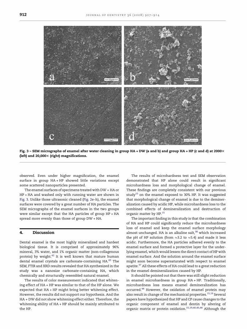
Fig. 3 – SEM micrographs of enamel after water cleaning in group HA + DW (a and b) and group HA + HP (c and d) at 2000T
(left) and 20,000T (right) magnifications.
j o u r n a l o f d e n t i s t r y 3 6 ( 2 0 0 8 ) 9 0 7 – 9 1 4912
observed. Even under higher magnification, the enamel
surface in group HA + HP showed little variations except
some scattered nanoparticles presented.
The enamel surfaces of specimens treated with DW + HA or
HP + HA and washed only with running water are shown in
Fig. 3. Unlike those ultrasonic cleaned (Fig. 2e–h), the enamel
surfaces were covered by a great number of HA particles. The
SEM micrographs of the enamel surfaces in the two groups
were similar except that the HA particles of group HP + HA
spread more evenly than those of group DW + HA.
4. Discussion
Dental enamel is the most highly mineralized and hardest
biological tissue. It is comprised of approximately 96%
mineral, 3% water, and 1% organic matter (non-collagenous
protein) by weight.42 It is well known that mature human
dental enamel crystals are carbonate-containing HA.43 The
SEM, FTIR and XRD results revealed that HA synthesized in the
study was a nanosize carbonate-containing HA, which
chemically and structurally resembled natural enamel.
The results of color measurement indicated that whiten-
ing effect of HA + HP was similar to that of the HP alone. We
expected that HA + HP might bring better whitening effect.
However, the results did not support our hypothesis. And the
HA + DW did not show whitening effect either. Therefore, the
whitening ability of HA + HP should be mainly attributed to
the HP.
The results of microhardness test and SEM observation
demonstrated that HP alone could result in significant
microhardness loss and morphological change of enamel.
These findings are completely consistent with our previous
study15 on the enamel exposed to 30% HP. It was suggested
that morphological change of enamel is due to the deminer-
alization caused by acidic HP, while microhardness loss to the
combined effects of demineralization and destruction of
organic matter by HP.15
The important finding in this study is that the combination
of HA and HP could significantly reduce the microhardness
loss of enamel and keep the enamel surface morphology
almost unchanged. HA is an alkaline salt,38 which increased
the pH of HP solution (from �3.2 to �5.4) and made it less
acidic. Furthermore, the HA particles adhered evenly to the
enamel surface and formed a protective layer for the under-
lying enamel, which would lessen the direct contact of HP with
enamel surface. And the solution around the enamel surface
might soon become supersaturated with respect to enamel
apatite.37 All these effects of HA could lead to a great reduction
in the enamel demineralization caused by HP.
It should be pointed out that there was still slight reduction
in enamel microhardness in group HA + HP. Traditionally,
microhardness loss means enamel demineralization has
occurred.44 However, the oxidation of enamel protein may
also result in change of the mechanical properties.15,19 Several
papers have hypothesized that HP and CP cause changes to the
organic component of enamel and dentin by altering of
organic matrix or protein oxidation.15,19,40,45,46 Although the
j o u r n a l o f d e n t i s t r y 3 6 ( 2 0 0 8 ) 9 0 7 – 9 1 4 913
protein comprises only a minor part of enamel, it is contained
in the spaces between mineral crystals, where it serves as a
‘‘glue’’ between crystallites.47 It is reasonable to assume that
the degradation of the ‘‘glue’’ will lead to the microhardness
loss of enamel.
One limitation of this study was the use of the highly
concentrated solution of HP. However, it had been chosen in
many in vitro studies.10,13,15,18 In present study, it was chosen
instead of gel used in the clinical just because we wanted to
explore the protective ability of HA under relatively rigorous
condition.
Another limitation of the study is that the enamel surfaces
were polished and flattened before bleaching. This procedure
was performed to provide a more uniform surface to improve
the precision of the indentations. However, it probably also
removed the upper aprismatic surface layer from enamel,
which is generally more highly mineralized than the subsurface
and thus more resistant to demineralization.48 For these
reasons, we assume that the change of enamel with aprismatic
surface layer would probably be less severe when treated by HP.
Some in vitro studies used artificial saliva or fluoride
products between or after the treatments, for these elements
are known to be an important factor to simulate clinical
situations. However, the aim of this study was to investigate
the protective effects of HA on the enamel surface subjected to
HP. We did not employ these elements in order to prevent the
influences of any other remineralization factors except HA.
Nevertheless, it is necessary to involve these factors in the
future studies to investigate the beneficial effects of HA under
typical clinical conditions.
5. Conclusions
The 30% HP solution resulted in significant microhardness loss
and morphological change of enamel. HA could significantly
reduce the microhardness loss of enamel caused by 30% HP
and keep the enamel surface morphology almost unchanged.
However, combination of HA and HP could not bring better
whitening effect than HP alone. The HA could be a potential
biomaterial used for tooth bleaching.
Acknowledgement
This study was supported by National Natural Science
Foundation of China (no. 30400507 and 30740019) and Key
Technologies R&D Program of Hubei Province (no.
2007AA301B28).
r e f e r e n c e s
1. Matis BA, Wang Y, Jiang T, Eckert GJ. Extended at-homebleaching of tetracycline-stained teeth with differentconcentrations of carbamide peroxide. QuintessenceInternational 2002;33:645–55.
2. Dahl JE, Pallesen U. Tooth bleaching—a critical review of thebiological aspects. Critical Reviews in Oral Biology and Medicine2003;14:292–304.
3. Joiner A. The bleaching of teeth: a review of the literature.Journal of Dentistry 2006;34:412–9.
4. Justino LM, Tames DR, Demarco FF. In situ and in vitroeffects of bleaching with carbamide peroxide on humanenamel. Operative Dentistry 2004;29:219–25.
5. Sulieman M, Addy M, Macdonald E, Rees JS. A safety study invitro for the effects of an in-office bleaching system on theintegrity of enamel and dentine. Journal of Dentistry2004;32:581–90.
6. Maia E, Baratieri LN, Caldeira de Andrada MA, Monteiro Jr S,Vieira LC. The influence of two home-applied bleachingagents on enamel microhardness: an in situ study. Journal ofDentistry 2008;36:2–7.
7. Cadenaro M, Breschi L, Nucci C, Antoniolli F, Visintini E,Prati C, et al. Effect of two in-office whitening agents on theenamel surface in vivo: a morphological and non-contactprofilometric study. Operative Dentistry 2008;33:127–34.
8. McCracken MS, Haywood VB. Demineralization effects of 10percent carbamide peroxide. Journal of Dentistry 1996;24:395–8.
9. Tezel H, Ertas OS, Ozata F, Dalgar H, Korkut ZO. Effect ofbleaching agents on calcium loss from the enamel surface.Quintessence International 2007;38:339–47.
10. Al-Salehi SK, Wood DJ, Hatton PV. The effect of 24 h non-stop hydrogen peroxide concentration on bovine enameland dentine mineral content and microhardness. Journal ofDentistry 2007;35:845–50.
11. Bitter NC. A scanning electron microscopy study of theeffect of bleaching agents on enamel: a preliminary report.Journal of Prosthetic Dentistry 1992;67:852–5.
12. Flaitz CM, Hicks MJ. Effects of carbamide peroxidewhitening agents on enamel surfaces and caries-like lesionformation: an SEM and polarized light microscopic in vitrostudy. ASDC Journal of Dentistry for Children 1996;63:249–56.
13. Hegedus C, Bistey T, Flora-Nagy E, Keszthelyi G, Jenei A. Anatomic force microscopy study on the effect of bleachingagents on enamel surface. Journal of Dentistry 1999;27:509–15.
14. Cavalli V, Arrais CA, Giannini M, Ambrosano GM. High-concentrated carbamide peroxide bleaching agents effectson enamel surface. Journal of Oral Rehabilitation 2004;31:155–9.
15. Jiang T, Ma X, Wang Y, Tong H, Shen X, Hu Y, et al.Investigation of the effects of 30% hydrogen peroxide onhuman tooth enamel by Raman scattering and laser-induced fluorescence. Journal of Biomedical Optics2008;13:014019.
16. Oltu U, Gurgan S. Effects of three concentrations ofcarbamide peroxide on the structure of enamel. Journal ofOral Rehabilitation 2000;27:332–40.
17. Cimilli H, Pameijer CH. Effect of carbamide peroxidebleaching agents on the physical properties and chemicalcomposition of enamel. American Journal of Dentistry2001;14:63–6.
18. Bistey T, Nagy IP, Sim A, Hegedus C. In vitro FT-IR study ofthe effects of hydrogen peroxide on superficial toothenamel. Journal of Dentistry 2007;35:325–30.
19. Seghi RR, Denry I. Effects of external bleaching onindentation and abrasion characteristics of human enamelin vitro. Journal of Dental Research 1992;71:1340–4.
20. Basting RT, Rodrigues Junior AL, Serra MC. The effect of 10%carbamide peroxide bleaching material on microhardnessof sound and demineralized enamel and dentin in situ.Operative Dentistry 2001;26:531–9.
21. Rodrigues JA, Marchi GM, Ambrosano GM, Heymann HO,Pimenta LA. Microhardness evaluation of in situ vitalbleaching on human dental enamel using a novel studydesign. Dental Materials 2005;21:1059–67.
22. Basting RT, Rodrigues Jr AL, Serra MC. The effects of sevencarbamide peroxide bleaching agents on enamel

j o u r n a l o f d e n t i s t r y 3 6 ( 2 0 0 8 ) 9 0 7 – 9 1 4914
microhardness over time. Journal of the American DentalAssociation 2003;134:1335–42.
23. Cavalli V, Giannini M, Carvalho RM. Effect of carbamideperoxide bleaching agents on tensile strength of humanenamel. Dental Materials 2004;20:733–9.
24. Marson FC, Sensi LG, Vieira LC, Araujo E. Clinical evaluationof in-office dental bleaching treatments with and withoutthe use of light-activation sources. Operative Dentistry2008;33:15–22.
25. Joiner A. Review of the effects of peroxide on enamel anddentine properties. Journal of Dentistry 2007;35:889–96.
26. Driessens FC, Theuns HM, Borggreven JM, van Dijk JW.Solubility behaviour of whole human enamel. CariesResearch 1986;20:103–10.
27. Rotstein I, Friedman S. pH variation among materials usedfor intracoronal bleaching. Journal of Endodontics1991;17:376–9.
28. Weiger R, Kuhn A, Lost C. Effect of various types of sodiumperborate on the pH of bleaching agents. Journal ofEndodontics 1993;19:239–41.
29. Efeoglu N, Wood D, Efeoglu C. Microcomputerisedtomography evaluation of 10% carbamide peroxide appliedto enamel. Journal of Dentistry 2005;33:561–7.
30. Arends J, Jongebloed WL, Goldberg M, Schuthof J. Interactionof urea and human enamel. Caries Research 1984;18:17–24.
31. Attin T, Betke H, Schippan F, Wiegand A. Potential offluoridated carbamide peroxide gels to support post-bleaching enamel re-hardening. Journal of Dentistry2007;35:755–9.
32. de Oliveira R, Paes Leme AF, Giannini M. Effect of acarbamide peroxide bleaching gel containing calcium orfluoride on human enamel surface microhardness. BrazilianDental Journal 2005;16:103–6.
33. Giniger M, MacDonald J, Ziemba S, Felix H. The clinicalperformance of professionally dispensed bleaching gel withadded amorphous calcium phosphate. Journal of the AmericanDental Association 2005;136:383–92.
34. Legeros RZ. Calcium phosphate materials in restorativedentistry: a review. Advances in Dental Research 1988;2:164–80.
35. Sun L, Berndt CC, Gross KA, Kucuk A. Materialfundamentals and clinical performance of plasma-sprayedhydroxyapatite coatings: a review. Journal of BiomedicalMaterials Research 2001;58:570–92.
36. Yamagishi K, Onuma K, Suzuki T, Okada F, Tagami J, OtsukiM, et al. Materials chemistry: a synthetic enamel for rapidtooth repair. Nature 2005;433:819.
37. Onuma K, Yamagishi K, Oyane A. Nucleation and growth ofhydroxyapatite nanocrystals for nondestructive repair ofearly caries lesions. Journal of Crystal Growth 2005;282:199–207.
38. Chow LC, Sun L, Hockey B. Properties of nanostructuredhydroxyapatite prepared by a spray drying technique.Journal of Research of the National Institute of Standards andTechnology 2004;109:543–51.
39. Niwa M, Sato T, Li W, Aoki H, Aoki H, Daisaku T. Polishingand whitening properties of toothpaste containinghydroxyapatite. Journal of Materials Science Materials inMedicine 2001;12:277–81.
40. Jiang T, Ma X, Wang Y, Zhu Z, Tong H, Hu J. Effects ofhydrogen peroxide on human dentin structure. Journal ofDental Research 2007;86:1040–5.
41. Boanini E, Torricelli P, Gazzano M, Giardino R, Bigi A.Nanocomposites of hydroxyapatite with aspartic acid andglutamic acid and their interaction with osteoblast-likecells. Biomaterials 2006;27:4428–33.
42. Deakins M, Volker JF. Amount of organic matter in enamelfrom several types of human teeth. Journal of Dental Research1941;20:117–21.
43. Sydney-Zax M, Mayer I, Deutsch D. Carbonate content indeveloping human and bovine enamel. Journal of DentalResearch 1991;70:913–6.
44. Featherstone JD, ten Cate JM, Shariati M, Arends J.Comparison of artificial caries-like lesions by quantitativemicroradiography and microhardness profiles. CariesResearch 1983;17:385–91.
45. Rotstein I, Lehr Z, Gedalia I. Effect of bleaching agents oninorganic components of human dentin and cementum.Journal of Endodontics 1992;18:290–3.
46. Rotstein I, Dankner E, Goldman A, Heling I, Stabholz A,Zalkind M. Histochemical analysis of dental hard tissuesfollowing bleaching. Journal of Endodontics 1996;22:23–5.
47. Maas MC, Dumont ER. Built to last: the structure, function,and evolution of primate dental enamel. EvolutionaryAnthropology Issues News and Reviews 1999;8:133–52.
48. Whittaker DK. Structural variations in the surface zone ofhuman tooth enamel observed by scanning electronmicroscopy. Archives of Oral Biology 1982;27:383–92.
Related Documents