Bell Work What is connective tissue? Where can it be found?

Bell Work What is connective tissue? Where can it be found?
Dec 17, 2015
Welcome message from author
This document is posted to help you gain knowledge. Please leave a comment to let me know what you think about it! Share it to your friends and learn new things together.
Transcript

Bell Work
What is connective tissue? Where can it be found?

Connective Tissue Most diverse and
abundant tissue Main classes:
Connective tissue proper
CartilageBone tissueBlood

Connective Tissue- General Features
Components of connective tissue:Cells (varies according to tissue)Extracellular Matrix (in btw. Cells)
○ Protein Fibers ○ Ground substance
Other Characteristics:Not on body surfacesHighly vascular (except cartilage)

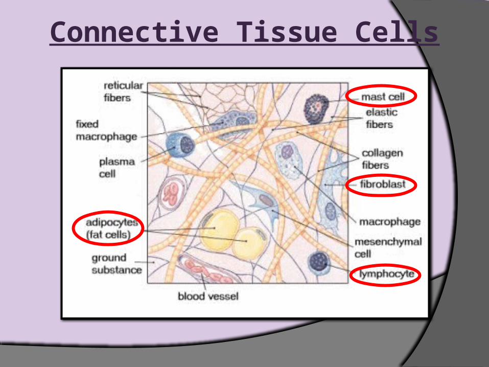
Connective Tissue Cells1. “Blast” Cells
Immature class of cells- blast cells have ability to divide & secrete extracellular matrix
Called:○ Fibroblasts in loose & dense connective tissue○ Chondroblasts in cartilage○ Osteoblasts in bone Once EC matrix is
produced, “blast” cells become “cyte” cells &
maintain the matrix
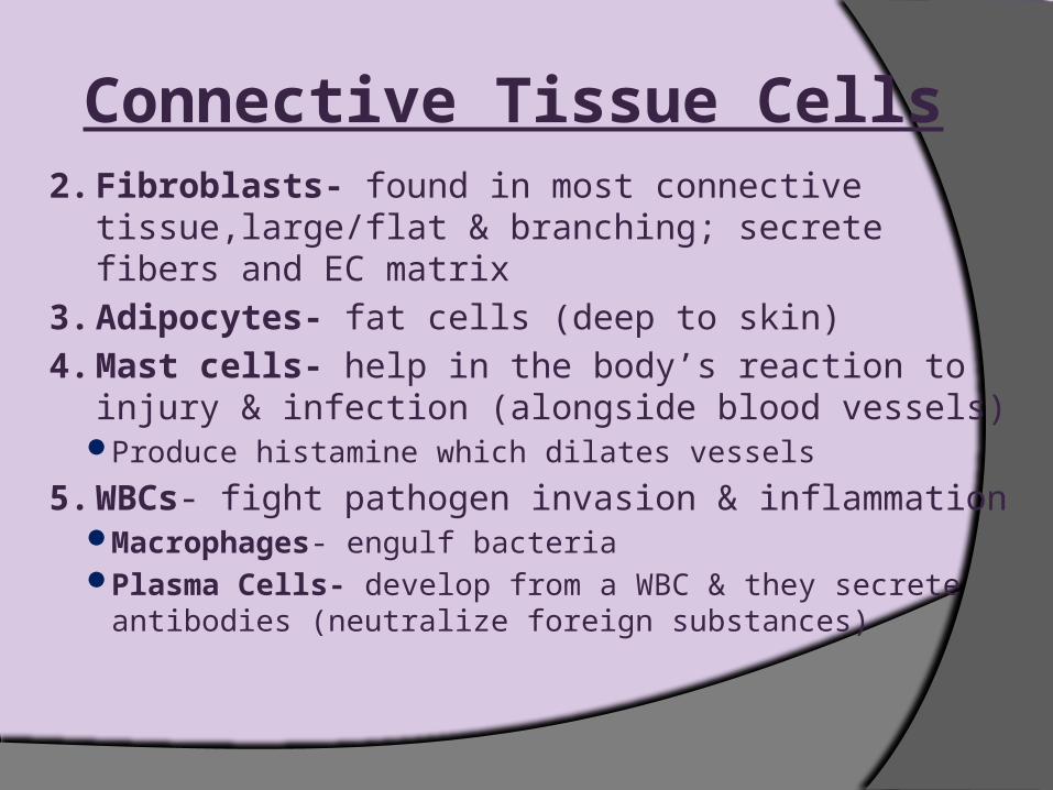
Connective Tissue Cells2. Fibroblasts- found in most connective
tissue,large/flat & branching; secrete fibers and EC matrix
3. Adipocytes- fat cells (deep to skin)
4. Mast cells- help in the body’s reaction to injury & infection (alongside blood vessels)
Produce histamine which dilates vessels
5. WBCs- fight pathogen invasion & inflammationMacrophages- engulf bacteriaPlasma Cells- develop from a WBC & they secrete
antibodies (neutralize foreign substances)
Connective Tissue Cells

Extracellular Matrix Definition: material btw cells Functions:
Supports cells/ binds cells togetherStores waterProvides a medium for exchange of substances
btw. cells & blood Components:
Fibers & Ground Substance
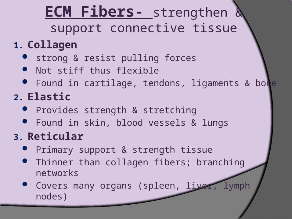
ECM Fibers- strengthen & support connective tissue
1. Collagen strong & resist pulling forces Not stiff thus flexible Found in cartilage, tendons, ligaments & bone
2. Elastic Provides strength & stretching Found in skin, blood vessels & lungs
3. Reticular Primary support & strength tissue Thinner than collagen fibers; branching networks Covers many organs (spleen, liver, lymph nodes)

Collagen Fibers

Elastic Fibers

Reticular Fibers

Ground Substance- connective tissue btw. cells
Characteristics:May be fluid, semifluid, gelatinous, or calcified
Composed of Glycosaminoglycans (GAGs) Polysaccharides Attract water Lubricate/support
Types: Chondroitin Sulfate- supports/adheres skin,
cartilage, tendons, bone & blood vessels Hyaluronic Acid- slippery & binds cells together,
lubricates joints, & maintains structure shape

Do Now:
What components make up extracellular matrix? Describe
each component.

Entrance Slip
1. Name three cells found in connective tissue?
2. What type of fiber has characteristics of strength and flexibility?
3. What is ground substance? Give a characteristic of it.

Connective Tissue Proper Loose Connective Tissue- loosely
intertwined fibers btw. cellsAreolarReticularAdipose
Dense Connective Tissue- thicker, denser fibers btw. fewer cells RegularIrregularElastic

Areolar Connective Tissue Description
Gel-like matrix w/:○ 3 Fibers: collagen, reticular, & elastic for support○ Ground substance is made up by many GAGs
Cells – fibroblasts, macrophages, mast cells, white blood cells, adipocytes
Function Wraps & cushions organsBinds the skin to underlying organs & fills space
between musclesImportant role in inflammation main battlefield in fight
against infection

Areolar Connective Tissue
Location Widely distributed under epithelia (has many blood
vessels so it nourishes epithelial cells)Packages organsSurrounds capillaries

Adipose Tissue Description
Closely packed adipocytes Nucleus pushed to one side
by fat droplet Function
Provides reserve food fuelInsulates against heat lossSupports & protects organs
LocationFound by areolar tissueUnder skin- insulates the
body & protects organsCushions jointsAround kidneys, between
muscles, behind eyeballs, within abdomen and in breasts


Reticular Connective Tissue
Description – network of reticular fibers in loose ground substance
Function – form a soft, internal skeleton (stroma- covers soft organs)
Location – lymphoid organs Lymph nodes, bone
marrow, and spleen
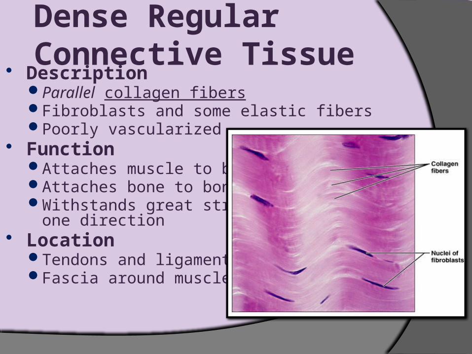
Dense Regular Connective Tissue
DescriptionParallel collagen fibersFibroblasts and some elastic fibersPoorly vascularized
FunctionAttaches muscle to boneAttaches bone to boneWithstands great stress in
one direction Location
Tendons and ligamentsFascia around muscles


Dense Irregular Connective Tissue
Description Irregularly arranged collagen
fibers Some elastic fibers and
fibroblasts Function
Withstands tension Provides structural strength
Location Dermis of skin Heart valves Surrounds cartilage & bone Submucosa of digestive tract Fibrous capsules of joints and
organs

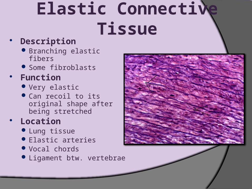
Elastic Connective Tissue
Description Branching elastic fibers Some fibroblasts
Function Very elastic Can recoil to its original shape
after being stretched Location
Lung tissue Elastic arteries Vocal chords Ligament btw. vertebrae


Connective Tissue Catch Phrase Set the timer. Give the stack of cards to someone
on Team 1. One person from Team 1 will try to give their team mates clues to their phrase. If Team 1 guesses the phrase, Team 2 gets the stack of cards and tries to guess their phrase.
Continue the game by going back and forth, as each team guesses the correct phrases. If the buzzer sounds during your turn, the other team gets a point and has the chance to earn a bonus point if they guess your phrase.

Bell Work
What are the three types of loose and dense
connective tissue?

Cartilage Characteristics:
Firm, flexible tissueContains no blood vessels or nervesMatrix contains up to 80% waterMainly collagen & elastic fibersCell type – chondrocyte
Types:HyalineFibrocartilageElastic


Hyaline Cartilage Description
Collagen fibers not in the matrix (hyaline = glassy)
Chodroblasts produce matrixChondrocytes lie in lacunae (space in mature
cartilage) Function
Supports and reinforcesResilient cushion/ Resists repetitive stressReduces friction

Hyaline Cartilage Location
Fetal skeletonEnds of long bonesCostal cartilage of ribsCartilages of nose tip,
trachea, and larynxJoints
*weakest of the 3 cartilages


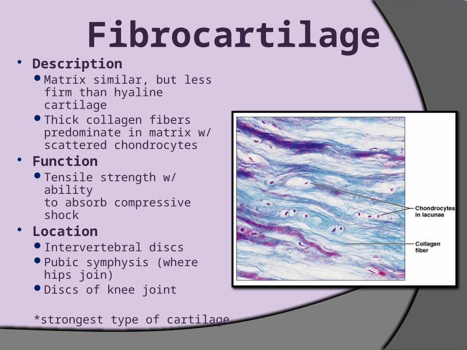
Fibrocartilage Description
Matrix similar, but less firm than hyaline cartilage
Thick collagen fibers predominate in matrix w/ scattered chondrocytes
FunctionTensile strength w/ ability
to absorb compressive shock
LocationIntervertebral discsPubic symphysis (where hips
join)Discs of knee joint
*strongest type of cartilage
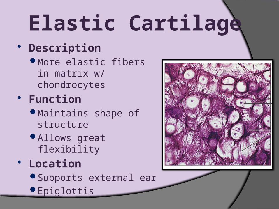
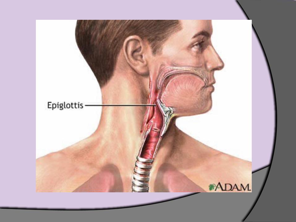
Elastic Cartilage Description
More elastic fibers in matrix w/ chondrocytes
Function Maintains shape of
structureAllows great flexibility
LocationSupports external earEpiglottis

Bone Tissue Function
Supports and protects organsProvides levers and
attachment site for musclesStores calcium and other
mineralsStores fatMarrow is site for blood cell
formation Characteristics
ETC matrix= lamellae rings of mineral salts
Lacunae- spaces in lamellae containing osteocytes
Location Bones


Blood Tissue Description
red and white blood cells in a fluid matrix
Functiontransport of respiratory
gases, nutrients, and wastes Location
within blood vessels Characteristics
An atypical connective tissue
Consists of cells surrounded by nonliving matrix (blood plasma- mainly water & dissolved nutrients)

Do Now:
What is the proper name of a bone cell, and what is the
name of the space in which a bone cell lies?

Do Now:
What are the three types of dense connective tissue?

Muscle Tissue Types
Skeletal muscle tissueCardiac muscle tissue Smooth muscle tissue

Skeletal Muscle Tissue
CharacteristicsLong, cylindrical cellsMultinucleateObvious striations
Function Voluntary movementManipulation of
environmentFacial expression
LocationSkeletal muscles
attached to bones (occasionally to skin)

Long, cylindrical cells that tend to have more than one nuclei.

Cardiac Muscle Tissue Function
Contracts to propel blood into circulatory system
CharacteristicsBranching cellsUninucleateIntercalated discs
LocationOccurs in walls of heart

Long, cylindrical cells that are shorter than skeletal muscle cells.
They have only one nucleus per cell.
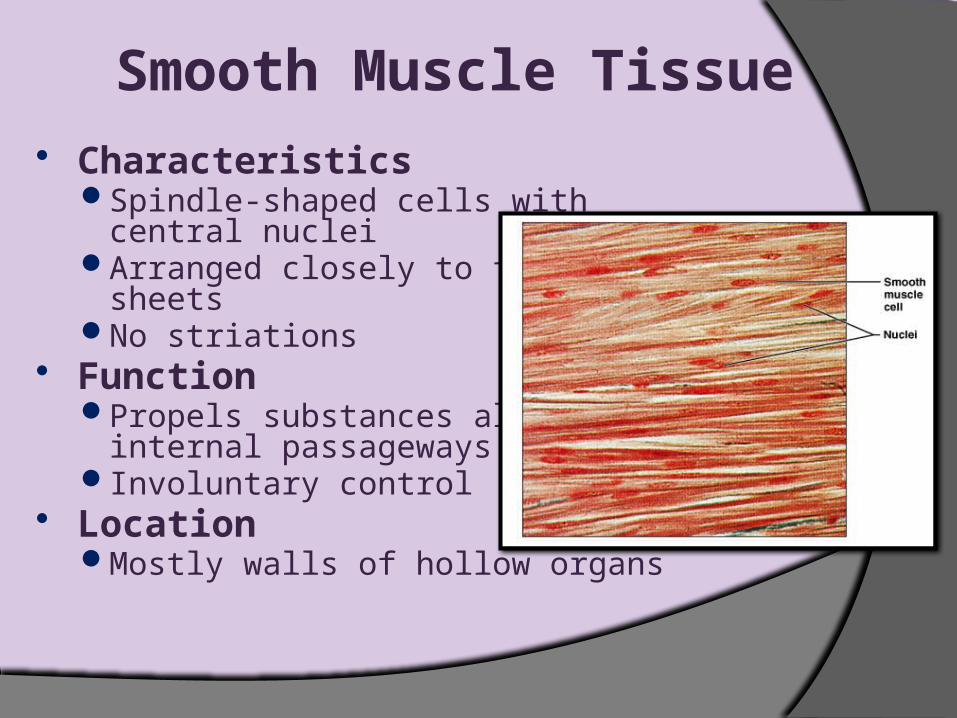
Smooth Muscle Tissue Characteristics
Spindle-shaped cells withcentral nuclei
Arranged closely to form sheets
No striations Function
Propels substances along internal passageways
Involuntary control Location
Mostly walls of hollow organs

These cells are tapered at the ends, giving them a spindle appearance. They have one
nucleus and are not striated.

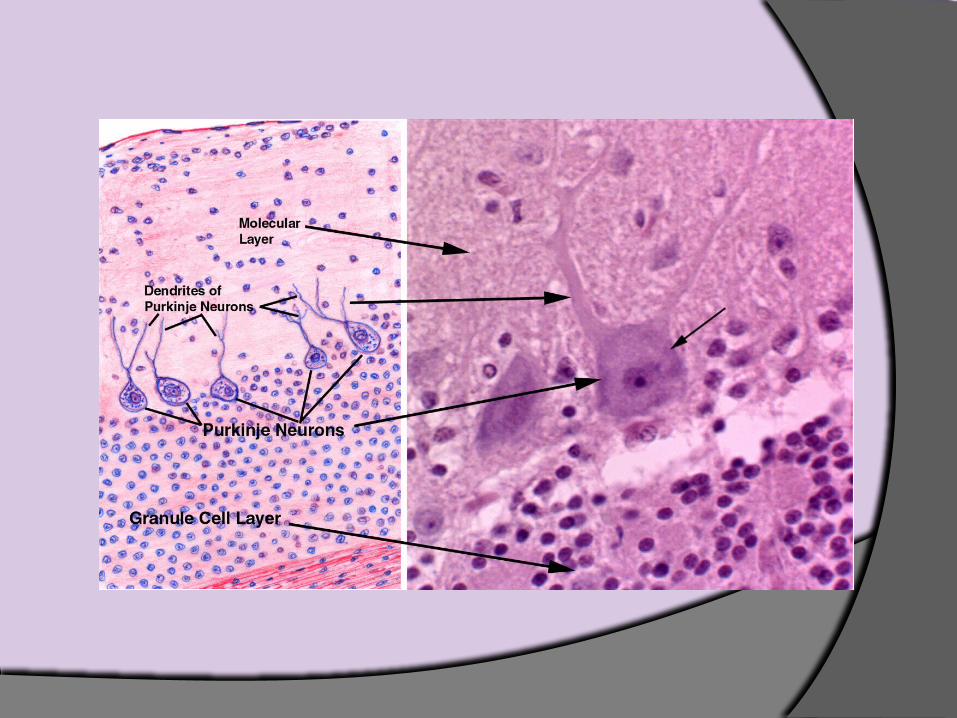
Nervous Tissue Function
Transmit electrical signals from sensory receptors to effectors
LocationBrain, spinal cord, and
nerves Description
Main components of brain, spinal cord, & nerves
Contains two types of cells○ Neurons – excitatory cells○ Supporting cells (neuroglial
cells)

Neurons consist of the cell body, which does basic cell activities, and dendrites (which receive impulses) and
axons (one per cell, conducting impulses away from cell body).

Tissue Response to Injury
Inflammatory response – non-specific, local responseLimits damage to injury site
Immune response – takes longer to develop and very specificDestroys particular microorganisms at site of
infection

The Tissues Throughout Life
At the end of second month of development:Primary tissue types have appearedMajor organs are in place
AdulthoodOnly a few tissues regenerateMany tissues still retain populations of stem cells
○ Stem cells- divide/ differentiate into specialized cell types
With increasing age:Epithelia thin Collagen decreasesBones, muscles, and nervous tissue begin to atrophyPoor nutrition and poor circulation – poor health of tissues
Covering and Lining Membranes
Combine epithelial tissues and connective tissues
Cover broad areas within body Consist of epithelial sheet plus
underlying connective tissue

Three Types of Membranes
Cutaneous membrane – skin Mucous membrane
Lines hollow organs that open to surface of bodyAn epithelial sheet underlain with layer of lamina
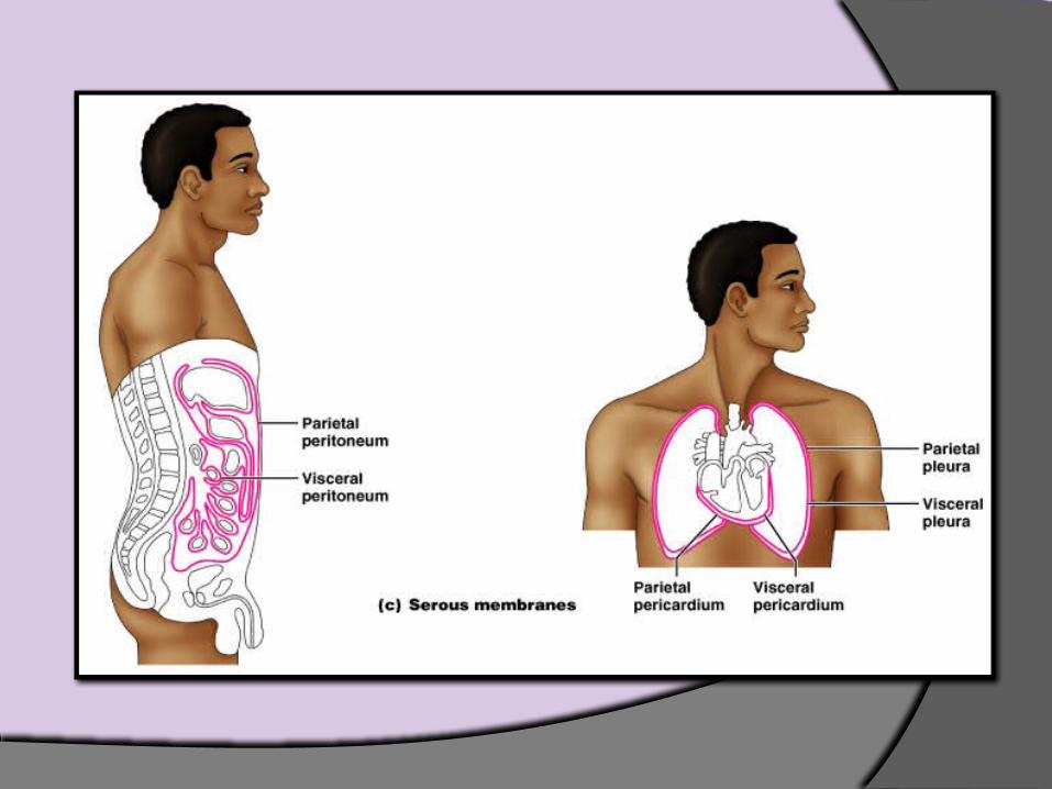
propria Serous membrane – slippery membranes
Simple squamous epithelium lying on areolar connective tissue
Line closed cavities○ Pleural, peritoneal, and pericardial cavities

Related Documents